Nuclear medicine in oncology. 1. Diagnosis 2. Therapy
|
|
|
- Holly Bruce
- 5 years ago
- Views:
Transcription
1 Nuclear medicine in oncology 1. Diagnosis 2. Therapy
2 Diagnosis - Conventional methods - Nonspecific radiopharmaceuticals cumulating in tumours - Specific radiopharmaceuticals (receptor- and immunoscintigraphy)

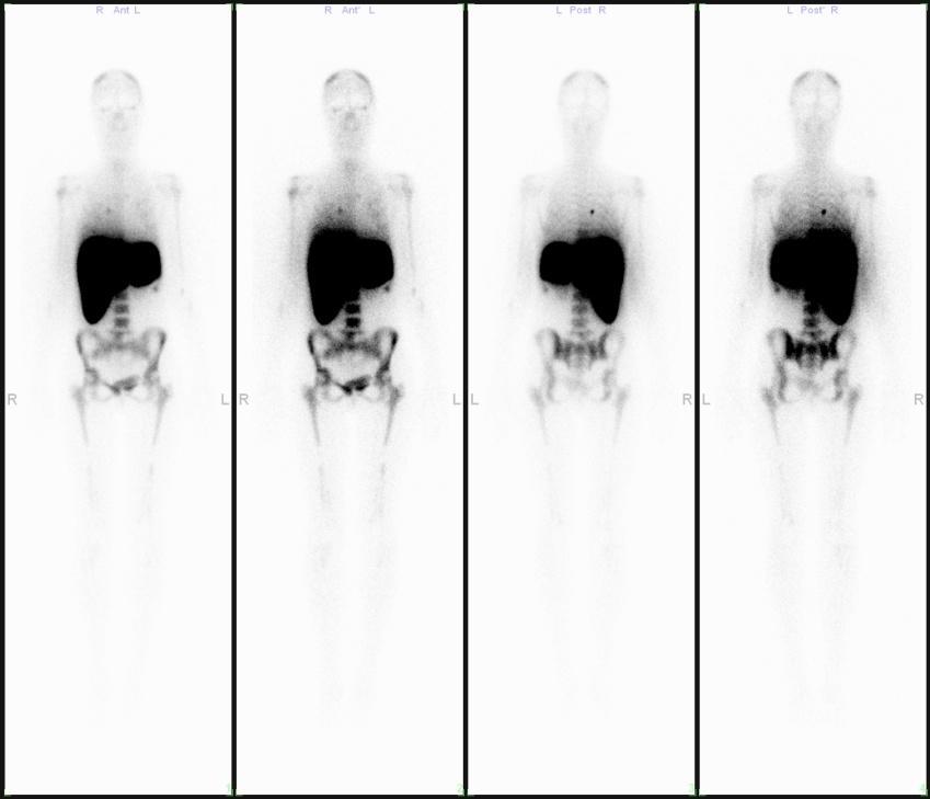
3 Bone scan multifocal metastases

4 Bone scan primary bone tumour Osteosarcoma tibiae l.s.

5 Osteosarcoma tibiae l.s. three-phase bone scan


6 Vertebra metastases Bone scintigraphy Bone marrow scintigraphy


7 Hodgkin disease Bone scintigraphy Bone marrow scintigraphy Bone marrow scintigraphy (SPECT)

8 Thyroid scintigraphy cold nodule

9 Colloidal and blood pool liver scan
 liver scan")
10 Colloidal, blood pool and hepatobiliary (HIDA) liver scan (FNH)
11 Perfusion lung scan - Perfusion lung scan shows the accurate regional perfusion of the lung - Injected subject: 99mTc-macroaggregate albumin with mean particles size of about 30 micrometer - It blocks only less than 0,1 % of precapillary arterioles

12 Normal perfusion lung scan
13 Indication of the perfusion scintigraphy - The search for pulmonary embolism (couple of the chest X-ray!) - The evaluation of regional lung function in patients with lung tumour before the operation - The evaluation of regional lung function in asthmatic and obstructive lung diseases - The assessment of regional lung function after the therapy

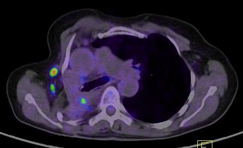
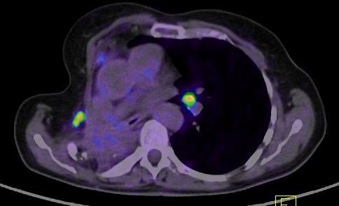
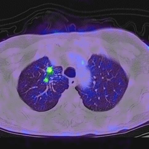
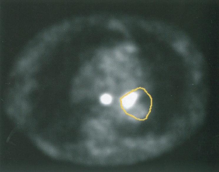
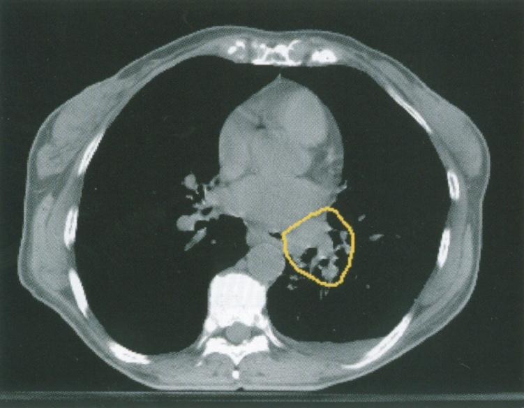
14 Lung cancer in the right upper lobe and in the left lung

15 Lung cancer in the right upper lobe and in the left lung CT SPECT SPECT/CT


16 Evaluation of regional lung function Periferial tumour in the right lung The activity of the lung: Both lung: cps Left lung: 48.3 % Right lung: 51.7 % Left upper lobe: 2597 cps Lingula: 2307 cps Left lower lobe: 3766 cps Right upper lobe: 3594 cps Right middle lobe: 2423 cps Right lower lobe: 3211 cps
17 Diagnosis - Conventional methods - Nonspecific radiopharmaceuticals cumulating in tumours - Specific radiopharmaceuticals (receptor- and immunoscintigraphy)
18 Radiopharmaceuticals cumulating nonspecifically in tumours - 99mTc-MIBI, (retentioned by mitochondrial membrane, primary role in the assestment of myocardial perfusion) - 99mTc-MDP (primary role in bone scintigraphy, tumor calcifications) TlCl (biologic properties similar to potassium, primary role in the assestment of myocardial perfusion) (e.g. brain, bone, lung, thyroid, parathyroid and breast tumours) - 67-Ga-citrate (similar to iron, binds to transferrin, e.g. lymphomas, lung and bone tumours) -FDG, C-11 -methionine (PET) (the greatest importance in clinical practice) PROBLEM: Not only tumour accumulation, positiv in inflammatory diseases too!)


")
19 Thyroid cc. - MIBI cumulation 99mTc 99mTc-MIBI (early) 99mTc-MIBI (delayed)

20 Thyroid cc. 99mTc-MIBI cumulation (SPECT/CT)

21 Parathyreoid adenoma subtraction protocoll
 CT SPECT")
22 Parathyreoid adenoma (SPECT/CT) CT SPECT SPECT/CT
 CT SPECT")
23 Parathyreoid adenoma (SPECT/CT) (retrosternal) CT SPECT SPECT/CT
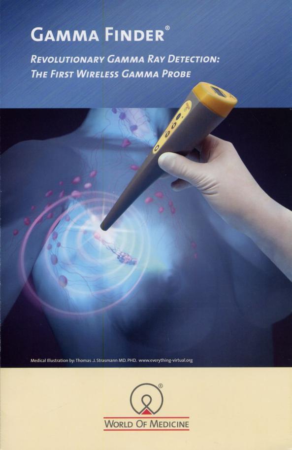
24 Intraoperatíve Gamma Probe

25 Extraosseal phosphate cumulation
 SPECT-image of the")
26 Extraosseal phosphate cumulation (meningeom) SPECT-image of the skull

27 Extraosseal phosphate cumulation

28 Extraosseal phosphate cumulation in m. gluteus maximus

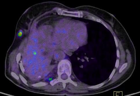
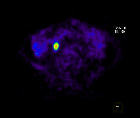
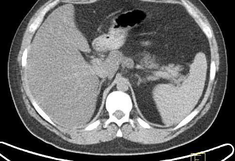
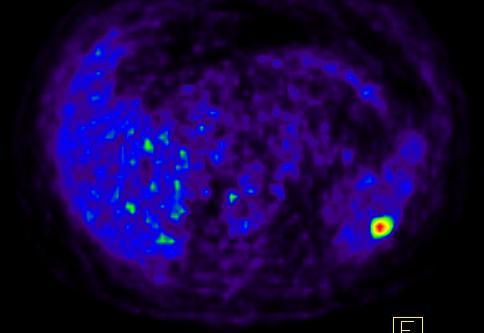
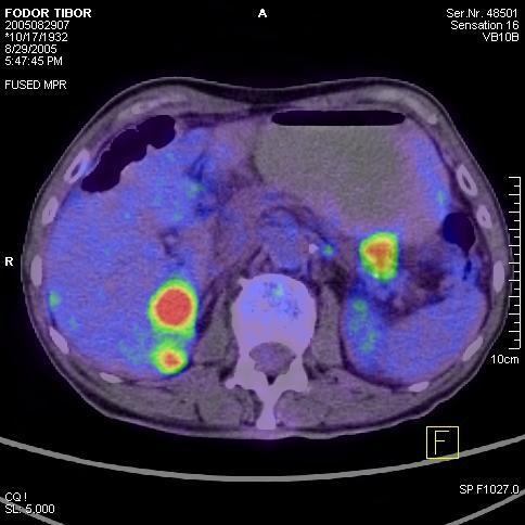
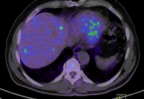
29 Extraosseal 99mTc-MDP cumulation in liver metastases
30 Scintimammography Radiopharmacon: - 99mTc-MIBI - 99mTc-MDP(together with bone scan!) - Taking early and delayed scans and SPECT image.
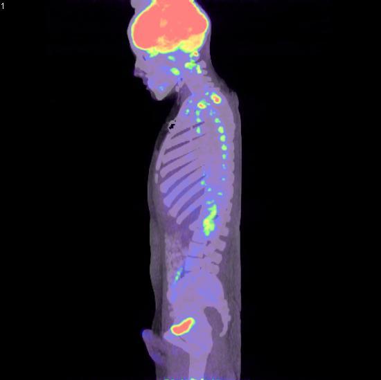
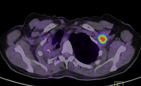
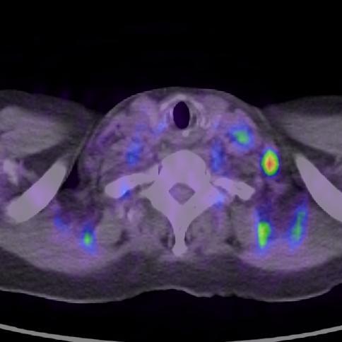
31 Pathological 99-mTc-MIBI cumulation in the right breast (cc ductale l.d.)
32 Pathological 99-mTc-MDP cumulation in the right breast (cc ductale l.d.)
")
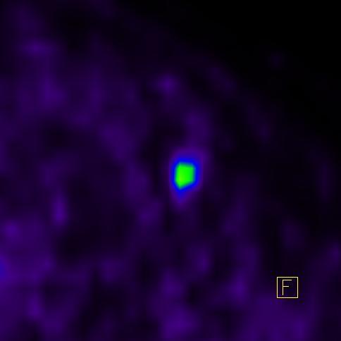
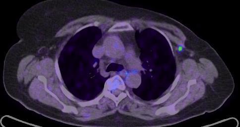
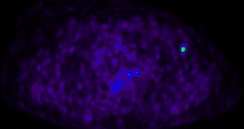
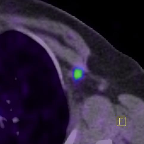
33 Pathological 99-mTc-MIBI cumulation in the right axilla (cc ductale l.d.)
34 Pathological 99-mTc-MDP cumulation in the right axilla (cc ductale l.d.)
35 The diagnostic value of scintimammogrphy according to histology 99mTc-MIBI 99mTc-MDP Sensitivity: 87% 81% Specificity: 88% 77% Validity: 88% 79% Today importance in dens breast
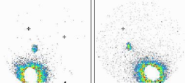
36 Sentinel lymph node (SLN) scintigraphy - The technical conception of sentinel lymph node biopsy is based on the process of lymphatic dissemination of tumour cells. - The peritumoral injected radiopharmaceutical passes the same way as the tumour cells. - The peritumoral injected tracer cumulates in the SLN, which is hereby detectable (gamma probe).
37 Why is SLN scintigraphy useful? - Unnecessary total axillary dissection may be avoided. (According to literature, if the SLN is negative, the other LN-s are in 97% negative too.) - The first LN, belonging to the tumour will be removed by all means. (According to literature the total axillary dissection gives in 10-30% false negative results!)
38 - The indication of SLN scintigraphy: early tumour stage (breast cancer, malignant melanoma, vulva cc.) - Method - peritumoral injection in 4-8 portions, using 99mTc-HSA colloid (99mTc-Senti-scint) - scans will be performed right after the injections, and after 1 and 3 hours again - the projection of SLN-s will be marked on the skin - operation on next day

39 SLN scintigraphy in breast cancer Antero-posterior Lateral-right
40 SLN scintigraphy in malignant melanoma Antero-posterior Lateral-right early 1. hour 1. hour


41 SLN scintigraphy in vulva cc. Sentinel lymph node examination FJ: squamous cell cancer metastasis in the left side recidiva 6 months later Anterior 1 hour Anterior 3 hours Anterior 24 hours

42 Rhabdomyosarcoma of the soft tissues of the right ankle 201-TlCl 67-Ga-citrate FDG

43 Ga-67 accumulation in NHL
44 PET radiopharmaceuticals in oncology Bloodflow Glucose-transzport Tumour-hypoxia Amino acid synthesis DNA-synthesis DNA-synthesis analog Tumour-receptor Chemotherapeutical agent Thyroid function 15O-water 18F-FDG 18F-misonidazole 11C-methionine 11C-thymidine 18F-FLT 68Ga-SMS 18F-fluorouracil 124I
45 SUV FDG accumulation in tumours Melanoma HG NHL HD Colorectal CA NSCLC Oesophageal CA Head/Neck CA HG sarcoma Ductal inv. breast CA Thyroid CA TesticularCA Pancreatic CA Recurrent ovarial CA LG NHL Bronchoalveolar CA Cervical CA Renal cell CA Lobular breast CA Mucinosus breast CA Prostate CA Primer ovarial CA Diff. Thyr. CA LG sarcoma HCC

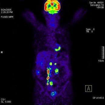
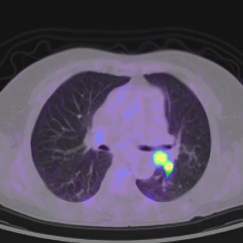
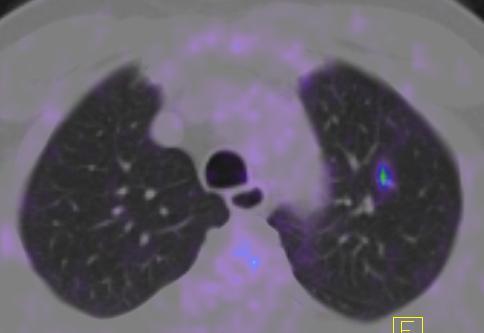
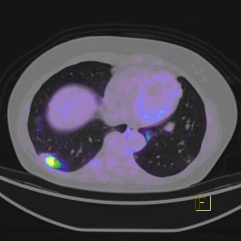
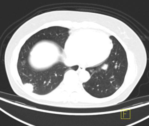
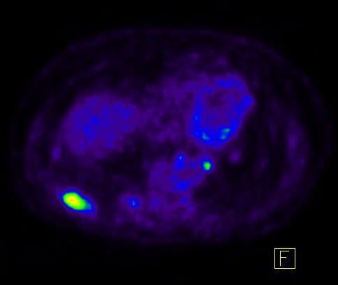
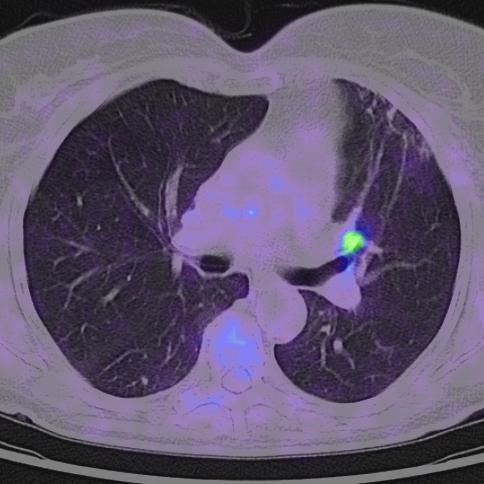
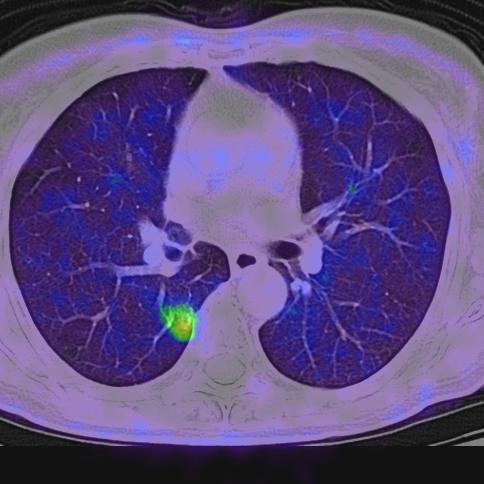
46 Adenocarcinoma in the right lung PET (FDG)

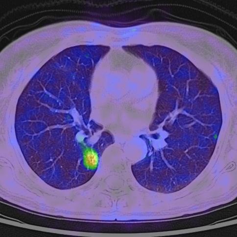
47 Adenocarcinoma in the left upper lobe PET (FDG)

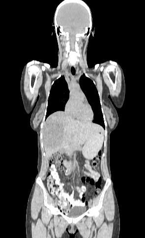
48 PET/CT CT PET
49 The indications of PET/CT Differentiation between malignant and benign lesions Residual tumor after radio- or chemotherapy Verification of tumour recurrence Staging (TNM) Optimization of biopsia Search of an unknown primary tumour Measuring of malignity Monitorization of the effectiveness of treatment Radiotherapy planning
50 Problems relating to differentiation between malignant and benign lesions FDG negative tumours The FDG uptake relates to the tumour grade and proliferation status FDG nonspecific radiopharmaceuticals, not only tumour accumulation, positiv in inflammatory diseases too!) Chemotherapy: 3 weeks Radiotherapy: 3 mounths


51 SUV FDG accumulation in tumours Melanoma HG NHL HD Colorectal CA NSCLC Oesophageal CA Head/Neck CA HG sarcoma Ductal inv. breast CA Thyroid CA TesticularCA Pancreatic CA Recurrent ovarial CA LG NHL Bronchoalveolar CA Cervical CA Renal cell CA Lobular breast CA Mucinosus breast CA Prostate CA Primer ovarial CA Diff. Thyr. CA LG sarcoma HCC
52 Problems relating to differentiation between malignant and benign lesions FDG negative tumours FDG nonspecific radiopharmaceuticals, not only tumour accumulation, positiv in inflammatory diseases too!) The FDG uptake relates to the tumour grade and proliferation status Chemotherapy: 3 weeks Radiotherapy: 3 mounths


53 FDG uptake in brown adipose tissue (hibernating fat)

54 FDG negative tumours - Prostate CA 18F-FDG 11C-Acetate


55 FDG negative, no residue NHL, 2,5 mounths after chemo- and radiotherapy


56 FDG pozitive, residual tumour NHL, 6 mounths after chemotherapy and embryonic stem cell transplantation
57 Tumour recurrence NSCLC St. post pulmonectomiam
58 Tumour recurrence ovarial CA


59 Tumour recurrence - sigma cc. presacralis rec.
60 Tumour recurrence Breast CA - Lgl. Metast.


61 Staging - NHL, extranodál manifestation Thill R, Nuklearmedizin, 1997:36: , Rini J et al Clin Nucl Med, 2002:27, 572

62 Staging - Adenocarcinoma pulm. l.s

63 Pancreas cc. - staging

64 Staging - adenocarcinoma sigmae - liver and lung mtastases
65 Biopsy

66 Biopsy


67 Search of an unknown primary tumour


68 Search of an unknown primary tumour SCLC

69 Measuring of malignity


70 Monitorization of the effectiveness of treatment before therapy after therapy Sigma tumour, liver matastases, after therapy (2. cycle)

71 Monitorization of the effectiveness of treatment Before th. After th. NSCLC


72 Monitorization of the effectiveness of treatment - NHL NHL -recurrence after chemotherapy and embryonic stem cell transplantation
73 Radiotherapy planning (correct focusing!)
74 THANKS!

75 Thanks for your attention!
Nuclear medicine methods in the urogenital system
 Nuclear medicine methods in the urogenital system Anatomy of the kidneys I. Anatomy of the kidneys II. The types of examinations Static examinations (scintigraphy): 1) the radiopharmaceutical is administered
Nuclear Medicine in Oncology
 Radiopharmaceuticals Nuclear Medicine in Oncology Practice Pharmaceutical Radionuc lide Function Tumor type Diphosphonates Tc-99m Osteoblast Bone tumor & metast. Ga-citrate Ga-67 Fe-analogue Bronchogenous
Clinical indications for positron emission tomography
 Clinical indications for positron emission tomography Oncology applications Brain and spinal cord Parotid Suspected tumour recurrence when anatomical imaging is difficult or equivocal and management will
Nuclear pulmonology. Katalin Zámbó Department of Nuclear Medicine
 Nuclear pulmonology Katalin Zámbó Department of Nuclear Medicine Imaging techniques Morphology Physiology Metabolism Molecules X-ray / CT MRI NM - SPECT/ PET MR spectroscopy fmri Ultrasound Hybrid imaging:
The Role of Nuclear Medicine in the Preoparative Diagnostics of Malignant Breast Tumours
 PhD theses The Role of Nuclear Medicine in the Preoparative Diagnostics of Malignant Breast Tumours Erzsébet Schmidt M.D. Doctorial School leader: Program leader: Tutor: Prof. József Bódis M.D. Prof. István
Molecular Imaging in the Development of Cancer Therapeutics. Johannes Czernin
 Molecular Imaging in the Development of Cancer Therapeutics Johannes Czernin Ahmanson Biological Imaging Division University of California Los Angeles Cancer Statistics Cancer Type 5-year Survival Rate
PET/CT Frequently Asked Questions
 PET/CT Frequently Asked Questions General Q: Is FDG PET specific for cancer? A: No, it is a marker of metabolism. In general, any disease that causes increased metabolism can result in increased FDG uptake
Index. Surg Oncol Clin N Am 16 (2007) Note: Page numbers of article titles are in boldface type.
 Note: Page numbers of article titles are in boldface type.") Surg Oncol Clin N Am 16 (2007) 465 469 Index Note: Page numbers of article titles are in boldface type. A Adjuvant therapy, preoperative for gastric cancer, staging and, 339 B Breast cancer, metabolic
Surg Oncol Clin N Am 16 (2007) 465 469 Index Note: Page numbers of article titles are in boldface type. A Adjuvant therapy, preoperative for gastric cancer, staging and, 339 B Breast cancer, metabolic
Nuclear medicine studies of the digestiv system. Zámbó Katalin Department of Nuclear Medicine
 Nuclear medicine studies of the digestiv system Zámbó Katalin Department of Nuclear Medicine Anatomy of the liver Liver scintigraphy The labelled colloid (200 MBq 99mTc-Fyton) is phagocyted by the Kuppfer-cells
Nuclear Medicine: Manuals. Nuclear Medicine. Nuclear imaging. Emission imaging: study types. Bone scintigraphy - technique
 Nuclear Medicine - Unsealed radioactive preparations the tracer mixes with the patients body fluids on a molecular level (e.g. after intravenous injection) - 3 main fields: - In vitro : measuring concentrations
F NaF PET/CT in the Evaluation of Skeletal Malignancy
 F NaF PET/CT in the Evaluation of Skeletal Malignancy Andrei Iagaru, MD September 26, 2013 School of of Medicine Ø Introduction Ø F NaF PET/CT in Primary Bone Cancers Ø F NaF PET/CT in Bone Metastases
objectives Pitfalls and Pearls in PET/CT imaging Kevin Robinson, DO Assistant Professor Department of Radiology Michigan State University
 objectives Pitfalls and Pearls in PET/CT imaging Kevin Robinson, DO Assistant Professor Department of Radiology Michigan State University To determine the regions of physiologic activity To understand
Contents. 3 Pneumology Introduction Positron Emission Tomography: Past and Present 1. 2 Fundamentals. xxx
 xxx IX Contents 1 Introduction Positron Emission Tomography: Past and Present 1 1.1 Survey.......................... 1 Physical and Biochemical Fundamentals.... 2 PET in National and International Medical
Radionuclide detection of sentinel lymph node
 Radionuclide detection of sentinel lymph node Sophia I. Koukouraki Assoc. Professor Department of Nuclear Medicine Medicine School, University of Crete 1 BACKGROUND The prognosis of malignant disease is
Los Angeles Radiological Society 62 nd Annual Midwinter Radiology Conference January 31, 2010
 Los Angeles Radiological Society 62 nd Annual Midwinter Radiology Conference January 31, 2010 Self Assessment Module on Nuclear Medicine and PET/CT Case Review FDG PET/CT IN LYMPHOMA AND MELANOMA Submitted
Page: 1 of 29. For this policy, PET scanning is discussed for the following 4 applications in oncology:
 Emission Tomography Scanning Page: 1 of 29 Last Review Status/Date: June 2015 Description Positron emission tomography (PET) scans are based on the use of positron-emitting radionuclide tracers coupled
Medical Policy An independent licensee of the Blue Cross Blue Shield Association
 PET Scanning: Oncologic Applications Page 1 of 42 Medical Policy An independent licensee of the Blue Cross Blue Shield Association Title: See also: Positron Emission Tomography (PET) Scanning: Oncologic
Oncologic Applications of PET Scanning
 6.01.26 Oncologic Applications of PET Scanning Section 6.0 Radiology Subsection Effective Date February 15, 2015 Original Policy Date January 26, 2009 Next Review Date December 2015 Description Positron
Radiology Pathology Conference
 Radiology Pathology Conference Sharlin Johnykutty,, MD, Cytopathology Fellow Sara Majewski, MD, Radiology Resident Friday, August 28, 2009 Presentation material is for education purposes only. All rights
Prof. Dr. NAGUI M. ABDELWAHAB,M.D.; MARYSE Y. AWADALLAH, M.D. AYA M. BASSAM, Ms.C.
 Role of Whole-body Diffusion MR in Detection of Metastatic lesions Prof. Dr. NAGUI M. ABDELWAHAB,M.D.; MARYSE Y. AWADALLAH, M.D. AYA M. BASSAM, Ms.C. Cancer is a potentially life-threatening disease,
PET IMAGING (POSITRON EMISSION TOMOGRAPY) FACT SHEET
 FACT SHEET") Positron Emission Tomography (PET) When calling Anthem (1-800-533-1120) or using the Point of Care authorization system for a Health Service Review, the following clinical information may be needed to
Positron Emission Tomography (PET) When calling Anthem (1-800-533-1120) or using the Point of Care authorization system for a Health Service Review, the following clinical information may be needed to
Hybrid systems in Medical Imaging
 Hybrid systems in Medical Imaging from PET/CT to PET/MR Osman Ratib, MD, PhD, FAHA Professor and chair Department of Medical Imaging and Information Sciences Head of division of Nuclear Medicine University
An Introduction to PET Imaging in Oncology
 January 2002 An Introduction to PET Imaging in Oncology Janet McLaren, Harvard Medical School Year III Basics of PET Principle of Physiologic Imaging: Allows in vivo visualization of structures by their
Ryan Niederkohr, M.D. Slides are not to be reproduced without permission of author
 Ryan Niederkohr, M.D. CMS: PET/CT CPT CODES 78814 Limited Area (e.g., head/neck only; chest only) 78815 78816 Regional (skull base to mid-thighs) True Whole Body (skull vertex to feet) SELECTING FIELD
Bone PET/MRI : Diagnostic yield in bone metastases and malignant primitive bone tumors
 Bone PET/MRI : Diagnostic yield in bone metastases and malignant primitive bone tumors Lars Stegger, Benjamin Noto Department of Nuclear Medicine University Hospital Münster, Germany Content From PET to
POSITRON EMISSION TOMOGRAPHY (PET)
") Status Active Medical and Behavioral Health Policy Section: Radiology Policy Number: V-27 Effective Date: 08/27/2014 Blue Cross and Blue Shield of Minnesota medical policies do not imply that members should
Status Active Medical and Behavioral Health Policy Section: Radiology Policy Number: V-27 Effective Date: 08/27/2014 Blue Cross and Blue Shield of Minnesota medical policies do not imply that members should
Cancers of unknown primary : Knowing the unknown. Prof. Ahmed Hossain Professor of Medicine SSMC
 Cancers of unknown primary : Knowing the unknown Prof. Ahmed Hossain Professor of Medicine SSMC Definition Cancers of unknown primary site (CUPs) Represent a heterogeneous group of metastatic tumours,
PET-MRI in malignant bone tumours. Lars Stegger Department of Nuclear Medicine University Hospital Münster, Germany
 PET-MRI in malignant bone tumours Lars Stegger Department of Nuclear Medicine University Hospital Münster, Germany Content From PET to PET/MRI General considerations Bone metastases Primary bone tumours
Nuclear Medicine Head and Neck Region. Bán Zsuzsanna, MD University of Pécs, Department of Nuclear Medicine
 Nuclear Medicine Head and Neck Region Bán Zsuzsanna, MD University of Pécs, Department of Nuclear Medicine Thyroid scintigraphy Parathyroid scintigraphy F18-FDG PET examinations in head and neck cancer
ROLE OF PET-CT IN BREAST CANCER, GUIDELINES AND BEYOND. Prof Jamshed B. Bomanji Institute of Nuclear Medicine UCL Hospitals London
 ROLE OF PET-CT IN BREAST CANCER, GUIDELINES AND BEYOND Prof Jamshed B. Bomanji Institute of Nuclear Medicine UCL Hospitals London CANCER Key facts Estimated 15.2 million new cases per year in 2015 worldwide
Positron Emission Tomography in Lung Cancer
 May 19, 2003 Positron Emission Tomography in Lung Cancer Andrew Wang, HMS III Patient DD 53 y/o gentleman presented with worsening dyspnea on exertion for the past two months 30 pack-year smoking Hx and
Molecular Imaging and Cancer
 Molecular Imaging and Cancer Cancer causes one in every four deaths in the United States, second only to heart disease. According to the U.S. Department of Health and Human Services, more than 512,000
Hybrid Imaging SPECT/CT PET/CT PET/MRI. SNMMI Southwest Chapter Aaron C. Jessop, MD
 Hybrid Imaging SPECT/CT PET/CT PET/MRI SNMMI Southwest Chapter 2014 Aaron C. Jessop, MD Assistant Professor, Department of Nuclear Medicine UT MD Anderson Cancer Center, Houston, Texas Complimentary role
List of Available TMAs in the PRN
 TMA RPCI_BrainCa01 RPCI_BrCa03 RPCI_BrCa04 RPCI_BrCa05 RPCI_BrCa0 RPCI_BrCa07 RPCI_BrCa08 RPCI_BrCa15 RPCI_BrCa1 RPCI_BrCa17 RPCI_BrCa18 RPCI_BrCa19 RPCI_BrCa20 RPCI_BrCa21 RPCI_BrCa24 RPCI_BrCa25 RPCI_BrCa2
PET/CT in breast cancer staging
 PET/CT in breast cancer staging Anni Morsing Consultant, PhD, DMSc Rigshospitalet 1 18F- FDG PET/CT for breastcancer staging Where is the clinical impact? To which women should 18F- FDG PET/CT be offered?
Using PET/CT in Prostate Cancer
 Using PET/CT in Prostate Cancer Legal Disclaimer These materials were prepared in good faith by MITA as a service to the profession and are believed to be reliable based on current scientific literature.
FDG PET/CT STAGING OF LUNG CANCER. Dr Shakher Ramdave
 FDG PET/CT STAGING OF LUNG CANCER Dr Shakher Ramdave FDG PET/CT STAGING OF LUNG CANCER FDG PET/CT is used in all patients with lung cancer who are considered for curative treatment to exclude occult disease.
Indications of PET/CT in oncology
 Monday, August 27, 2012 Session 1, 10:00-10:40 Indications of PET/CT in oncology Helle Westergren Hendel MD, PhD, assistant professor Bacelor in Leadership & Health Ecomomics Head of Clinical PET, Herlev
Medical Policy An independent licensee of the Blue Cross Blue Shield Association
 PET Scanning: Oncologic Applications Page 1 of 88 Medical Policy An independent licensee of the Blue Cross Blue Shield Association Title: Positron Emission Tomography (PET) Scanning: Oncologic Applications
PET/CT F-18 FDG. Objectives. Basics of PET/CT Imaging. Objectives. Basic PET imaging
 Basics of PET/CT Imaging Kevin Robinson, DO Department of Radiology Michigan State University Objectives Basic PET imaging Evaluating the therapeutic response Evaluating the big 5 Lymphoma Breast Lung
Recent initiatives of the FANC. Michel Biernaux Health Protection Service Health and Environment Department
 Recent initiatives of the FANC Michel Biernaux Michel.biernaux@fanc.fgov.be Health Protection Service Health and Environment Department Reminder objectives of the national survey : 1.Review the average
Cancer Program Report 2014
 Cancer Program Report 2014 Queen of the Valley Hospital St Joseph Health Queen of the Valley Hospital - 2014 Site Table Site Total Class Sex Group Cases Analytic NonAn M F 0 I II ALL SITES 661 494 167
Lung Cancer Imaging. Terence Z. Wong, MD,PhD. Department of Radiology Duke University Medical Center Durham, NC 9/9/09
 Lung Cancer Imaging Terence Z. Wong, MD,PhD Department of Radiology Duke University Medical Center Durham, NC 9/9/09 Acknowledgements Edward F. Patz, Jr., MD Jenny Hoang, MD Ellen L. Jones, MD, PhD Lung
Itroduction to the Nuclear Medicine: biophysics and basic principles. Zámbó Katalin Department of Nuclear Medicine
 Itroduction to the Nuclear Medicine: biophysics and basic principles Zámbó Katalin Department of Nuclear Medicine NUCLEAR MEDICINE Application of the radioactive isotopes in the diagnostics and in the
Quantitative Nuclear Medicine Imaging in Oncology. Susan E. Sharp, MD
 Quantitative Nuclear Medicine Imaging in Oncology Susan E. Sharp, MD Disclosures None Objectives Describe scoring systems used in oncologic nuclear medicine imaging Deauville scoring in lymphoma Curie
42 yr old male with h/o Graves disease and prior I 131 treatment presents with hyperthyroidism and undetectable TSH. 2 hr uptake 20%, 24 hr uptake 50%
 Pinhole images of the neck are acquired in multiple projections, 24hrs after the oral administration of approximately 200 µci of I123. Usually, 24hr uptake value if also calculated (normal 24 hr uptake
PET/MR:Techniques, Indications and Applications
 PET/MR:Techniques, Indications and Applications Franz Wolfgang Hirsch Professor and Head of the Department of Pediatric Radiology University Hospital Leipzig / Germany Children s Hospital University Leipzig
Department of Nuclear Medicine with Positron Emission Tomography
 (PET) Unit [1] Contact information: Registration: +48 41 367 4850 Main office: +48 41 367 4860 Fax: +48 41 367 4887 e-mail: zmnsco@onkol.kielce.pl [2] Head of the Department: Professor Janusz Braziewicz
Zurich, January 19, 2018
 Brain metastases as first presentation of malignancy: Immediate management, differential diagnosis; prevalence of primaries and suggested work-up Symposium on Brain Metastasis Cancer Center Zurich Zurich,
Macmillan Publications
 S1 S2 S3 S3 S3 S4 S5 S6 S7 S8 S8 S9 S10 S11 S11 S12 S13 S14 S15 S17 S18 S19 Bladder Cancer: Non-Invasive, Invasive and Advanced Bone Cancer: Primary, Secondary Colon Cancer, Anal Cancer, Rectal Cancer
Dr Sneha Shah Tata Memorial Hospital, Mumbai.
 Dr Sneha Shah Tata Memorial Hospital, Mumbai. Topics covered Lymphomas including Burkitts Pediatric solid tumors (non CNS) Musculoskeletal Ewings & osteosarcoma. Neuroblastomas Nasopharyngeal carcinomas
Case 1: 79 yr-old woman with a lump in upper outer quadrant of left breast.
 Case 1: 79 yr-old woman with a lump in upper outer quadrant of left breast. Giuliano Mariani Regional Center of Nuclear Medicine, University of Pisa Medical School, Pisa (Italy) Relevant history 79-yr
SPECT/CT Imaging of the Sentinel Lymph Node
 IAEA Regional Training Course on Hybrid Imaging SPECT/CT Imaging of the Sentinel Lymph Node Giuliano Mariani Regional Center of Nuclear Medicine, University of Pisa Medical School, Pisa, Italy Vilnius,
Nuclear Sciences and Medicine
 Nuclear Sciences and Medicine Rethy Chhem, MD, PhD (Edu), PhD (His), FRCPC Division of Human Health Guest Professor, Medical University of Vienna International Atomic Energy Agency Medical Imaging X-rays
performed to help sway the clinician in what the appropriate diagnosis is, which can substantially alter the treatment of management.
 Hello, I am Maura Polansky at the University of Texas MD Anderson Cancer Center. I am a Physician Assistant in the Department of Gastrointestinal Medical Oncology and the Program Director for Physician
Nonvisualization of sentinel node by lymphoscintigraphy in advanced breast cancer
 Radiology Case Reports Nonvisualization of sentinel node by lymphoscintigraphy in advanced breast cancer Volume 5, Issue 3, 2010 Brian Wosnitzer, MD; Rosna Mirtcheva, MD; and Munir Ghesani, MD Previous
CT PET SCANNING for GIT Malignancies A clinician s perspective
 CT PET SCANNING for GIT Malignancies A clinician s perspective Damon Bizos Head, Surgical Gastroenterology Charlotte Maxeke Johannesburg Academic Hospital Case presentation 54 year old with recent onset
What is Cancer? Petra Ketterl, MD Medical Oncology and Functional Medicine
 What is Cancer? Petra Ketterl, MD Medical Oncology and Functional Medicine What is Cancer? Layman s terms: cancer starts when cells grow out of control (in any place in the body) and crowd out normal cells
New Visions in PET: Surgical Decision Making and PET/CT
 New Visions in PET: Surgical Decision Making and PET/CT Stanley J. Goldsmith, MD Director, Nuclear Medicine Professor, Radiology & Medicine New York Presbyterian Hospital- Weill Cornell Medical Center
Breast Cancer PET/CT Imaging Protocol
 Breast Cancer PET/CT Imaging Protocol Scanning Protocol: Patients are scanned from the top of the neck through the pelvis. Arms-up position is used to avoid beam-hardening artifact in the chest and abdomen.
PET/CT in oncology. Positron emission tomography
 Clinical Medicine 2012, Vol 12, No 4: 368 72 PET/CT in oncology Fahim-Ul-Hassan, SpR Nuclear Medicine, Guy s Hospital, London; Gary J Cook, professor of Clinical PET, KCL Division of Imaging Sciences &
Subject: PET Scan With or Without CT Attenuation. Original Effective Date: 11/7/2017. Policy Number: MCR: 610. Revision Date(s): Review Date:
: Review Date:") Subject: PET Scan With or Without CT Attenuation Policy Number: MCR: 610 Revision Date(s): MHW Original Effective Date: 11/7/2017 Review Date: DISCLAIMER This Molina Clinical Review (MCR) is intended to
Subject: PET Scan With or Without CT Attenuation Policy Number: MCR: 610 Revision Date(s): MHW Original Effective Date: 11/7/2017 Review Date: DISCLAIMER This Molina Clinical Review (MCR) is intended to
FDG PET/CT in Lung Cancer Read with the experts. Homer A. Macapinlac, M.D.
 FDG PET/CT in Lung Cancer Read with the experts Homer A. Macapinlac, M.D. Patient with suspected lung cancer presents with left sided chest pain T3 What is the T stage of this patient? A) T2a B) T2b C)
PARATHYROID NUCLEAR MEDICINE IMAGING REVIEW DISCLOSURES
 PARATHYROID NUCLEAR MEDICINE IMAGING REVIEW Miguel Hernandez Pampaloni, M.D., Ph.D. Chief, Nuclear Medicine Assistant Professor of Radiology UCSF Department of Radiology and Biomedical Imaging DISCLOSURES
Take Home Quiz 1 Please complete the quiz below prior to the session. Use the Multiple Primary and Histology Rules
 Take Home Quiz 1 Please complete the quiz below prior to the session. Use the Multiple Primary and Histology Rules Case 1 72 year old white female presents with a nodular thyroid. This was biopsied in
2004 SNM Mid-Winter Educational Symposium
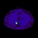
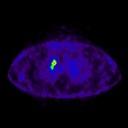
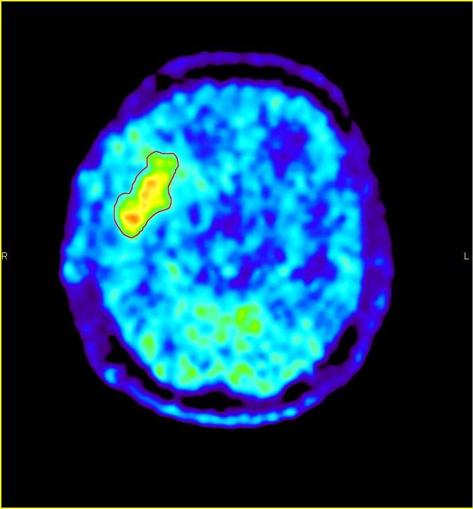
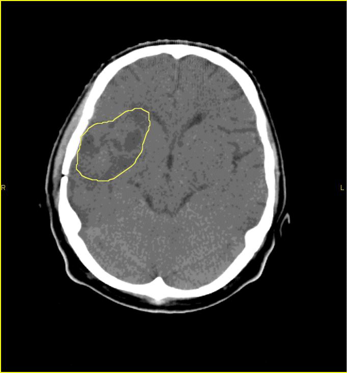
 1 2 20 Numeric values 15 10 5 0 SUV corr hs 18FDG Slope corr hs SUV corr hs Slope corr hs 11C-methionin 3 11C-methionine Grading and delineation of brain tumors Differentiation of malignant from benign
New imaging techniques: let there be light. Felix M. Mottaghy Department of Nuclear Medicine University Hospital KU Leuven
 New imaging techniques: let there be light Felix M. Mottaghy Department of Nuclear Medicine University Hospital KU Leuven Medical imaging and the pathology cascade Molecular/Cellular disturbance Alterations
THE PARATHYROID GLAND THEORY AND NUCLEAR MEDICINE PRACTICE
 THE PARATHYROID GLAND THEORY AND NUCLEAR MEDICINE PRACTICE George N. Sfakianakis MD Professor of Radiology and Pediatrics Director, Division of Nuclear Medicine UM/JMMC Miami FL October 2009 ENDONCRINE
Lugano classification: Role of PET-CT in lymphoma follow-up
 CAR Educational Exhibit: ID 084 Lugano classification: Role of PET-CT in lymphoma follow-up Charles Nhan 4 Kevin Lian MD Charlotte J. Yong-Hing MD FRCPC Pete Tonseth 3 MD FRCPC Department of Diagnostic
Original Policy Date
 MP 6.01.17 Oncologic Applications of PET Scanning Medical Policy Section Radiology Issue 12:2013 Original Policy Date 12:2013 Last Review Status/Date Reviewed with literature/12:2013 Return to Medical
Nuclear Medicine Diagnosis
 Anatomy : MRI Fonction : Nucl Med Nuclear Medicine Diagnosis Functional imaging = distribution of a (radio)- tracer in organs Each tracer is a SPY of a function, usually through a metabolic pathway flow
Case Scenario 1 Worksheet. Primary Site C44.4 Morphology 8743/3 Laterality 0 Stage/ Prognostic Factors
 CASE SCENARIO 1 9/10/13 HISTORY: Patient is a 67-year-old white male and presents with lesion located 4-5cm above his right ear. The lesion has been present for years. No lymphadenopathy. 9/10/13 anterior
Radiological staging of lung cancer. Shukri Loutfi,MD,FRCR Consultant Thoracic Radiologist KAMC-Riyadh
 Radiological staging of lung cancer Shukri Loutfi,MD,FRCR Consultant Thoracic Radiologist KAMC-Riyadh Bronchogenic Carcinoma Accounts for 14% of new cancer diagnoses in 2012. Estimated to kill ~150,000
Colorectal Cancer and FDG PET/CT
 Hybrid imaging in colorectal & esophageal cancer Emmanuel Deshayes IAEA WorkShop, November 2017 Colorectal Cancer and FDG PET/CT 1 Clinical background Cancer of the colon and rectum is one of the most
Lung. 10/24/13 Chest X-ray: 2.9 cm mass like density in the inferior lingular segment worrisome for neoplasm. Malignancy cannot be excluded.
 Lung Case Scenario 1 A 54 year white male presents with a recent abnormal CT of the chest. The patient has a history of melanoma, kidney, and prostate cancers. 10/24/13 Chest X-ray: 2.9 cm mass like density
L hyperfixation dans le suivi des lymphomes représente-t-elle toujours une maladie active?
 L hyperfixation dans le suivi des lymphomes représente-t-elle toujours une maladie active? Thierry Vander Borght UCL Mont-Godinne, Belgique FDG-PET in Lymphoma: Mont-Godinne Experience 03/2000 10/2002:
Icd 10 code for lung cancer with mets to bone
 Icd 10 code for lung cancer with mets to bone 1-10-2017 ICD-10 -CM Diagnosis Code. Cancer metastatic to bone ;. The majority of metastatic neoplasms to the bone are carcinomas. ICD - 10 -CM C79.51 is grouped.
Nuclear Medicine in HIV Update with FDG PET
 Nuclear Medicine in HIV Update with FDG PET Mike Sathekge, MB ChB, M Med (Nucl Med), PhD HOD: University of Pretoria Introduction Outline Brief about general nuclear medicine FDG in HIV: Update & Potential
THE ROLE OF CONTEMPORARY IMAGING AND HYBRID METHODS IN THE DIAGNOSIS OF CUTANEOUS MALIGNANT MELANOMA(CMM) AND MERKEL CELL CARCINOMA (MCC)
 AND MERKEL CELL CARCINOMA (MCC)") THE ROLE OF CONTEMPORARY IMAGING AND HYBRID METHODS IN THE DIAGNOSIS OF CUTANEOUS MALIGNANT MELANOMA(CMM) AND MERKEL CELL CARCINOMA (MCC) I.Kostadinova, Sofia, Bulgaria CMM some clinical facts The incidence
THE ROLE OF CONTEMPORARY IMAGING AND HYBRID METHODS IN THE DIAGNOSIS OF CUTANEOUS MALIGNANT MELANOMA(CMM) AND MERKEL CELL CARCINOMA (MCC) I.Kostadinova, Sofia, Bulgaria CMM some clinical facts The incidence
Chapter 10. Summary, conclusions and future perspectives
 Chapter 10 Summary, conclusions and future perspectives 10.1 SUMMARY In this thesis, a new tumor imaging tracer in nuclear medicine is studied. This 123 tracer, L-3-[ I]Iodo-alpha-methyl-tyrosine (IMT),
A 64 y.o. man presents to the hospital with persistent cough and hemoptysis. Fernando Mut Montevideo - Uruguay
 A 64 y.o. man presents to the hospital with persistent cough and hemoptysis Fernando Mut Montevideo - Uruguay Teaching case Bone # 1 A 64 y.o. man presents to the hospital with persistent cough and hemoptysis.
UK Musculoskeletal Oncology: Something for All Ages. Lars Wagner, MD Pediatric Hematology/Oncology University of Kentucky
 UK Musculoskeletal Oncology: Something for All Ages Lars Wagner, MD Pediatric Hematology/Oncology University of Kentucky Pediatric-Type Sarcomas of Bone and Soft Tissue The incidence of sarcoma continues
Principles of nuclear metabolic imaging. Prof. Dr. Alex Maes AZ Groeninge Kortrijk and KULeuven Belgium
 Principles of nuclear metabolic imaging Prof. Dr. Alex Maes AZ Groeninge Kortrijk and KULeuven Belgium I. Molecular imaging probes A. Introduction - Chemical disturbances will precede anatomical abnormalities
Tumor Imaging in Nuclear Medicine: Current Status and
 Tumor Imaging in Nuclear Medicine: Current Status and Future Prospects Bennett S. Greenspan, MD SNM MWM Albuquerque, NM January 30, 2010 Tumor Imaging Part I Current Status Current status agents that are
Thyroid nodules - medical and surgical management. Endocrinology and Endocrine Surgery Manchester Royal Infirmary
 Thyroid nodules - medical and surgical management JRE Davis NR Parrott Endocrinology and Endocrine Surgery Manchester Royal Infirmary Thyroid nodules - prevalence Thyroid nodules common, increase with
Theranostics in Nuclear Medicine
 Theranostics in Nuclear Medicine Patrick FLAMEN, MD, PhD Head Nuclear Medicine Institut Jules Bordet Université Libre de Bruxelles (U.L.B.) n Theranostics in Nuclear Medicine n A form of (nuclear) diagnostic
Lymphoma Read with the experts
 Lymphoma Read with the experts Marc Seltzer, MD Associate Professor of Radiology Geisel School of Medicine at Dartmouth Director, PET-CT Course American College of Radiology Learning Objectives Recognize
Medical imaging X-ray, CT, MRI, scintigraphy, SPECT, PET Györgyi Műzes
 Medical imaging X-ray, CT, MRI, scintigraphy, SPECT, PET Györgyi Műzes Semmelweis University, 2nd Dept. of Medicine Medical imaging: definition technical process of creating visual representations about
Value of true whole-body FDG- PET/CT scanning protocol in oncology and optimization of its use based on primary malignancy
 Value of true whole-body FDG- PET/CT scanning protocol in oncology and optimization of its use based on primary malignancy Ronnie Sebro MD, Ph.D Carina Mari Aparici MD, Miguel Hernandez Pampaloni MD, PhD
FieldStrength. Leuven research is finetuning. whole body staging
 FieldStrength Publication for the Philips MRI Community Issue 40 May 2010 Leuven research is finetuning 3.0T DWIBS for whole body staging The University Hospital of Leuven is researching 3.0T whole body
Radiopharmaceutical Activities Administered for Diagnostic and Therapeutic Procedures in Nuclear Medicine in Argentine: Results of a National Survey
 Radiopharmaceutical Activities Administered for Diagnostic and Therapeutic Procedures in Nuclear Medicine in Argentine: Results of a National Survey A. M. Bomben, C. A. Chiliutti Autoridad Regulatoria
Dr Alfred O Ankrah FCNP
 Dr Alfred O Ankrah FCNP Outline Introduction Brief history of Nuclear Medicine in Ghana Current situation of Nuclear Medicine in Ghana Use of Nuclear medicine in various disciplines Future of Nuclear Medicine
ACR TXIT TM EXAM OUTLINE
 ACR TXIT TM EXAM OUTLINE Major Domain Sub-Domain 1 Statistics 1.1 Study design 1.2 Definitions of statistical terms 1.3 General interpretation & analysis 1.4 Survival curves 1.5 Specificity/sensitivity
HEALTHFIRST 2011 RADIOLOGY PROGRAM CODE LIST
 HEALTHFIRST 2011 RADIOLOGY PROGRAM CODE LIST Outpatient Radiology utilization call Carecore at 1-877-773-6964 Modality CPT CODE Description CT SCANS 70450 CT HEAD/BRAIN W/O CONTRAST CT SCANS 70460 CT HEAD/BRAIN
The Concept of GOSTT
 IAEA Regional Training Course on Sentinel Lymph Node Mapping and Radioguided Surgery The Concept of GOSTT Giuliano Mariani Regional Center of Nuclear Medicine, University of Pisa Medical School, Pisa,
Methods of nuclear medicine
 Methods of nuclear medicine Per Wollmer Dept. of Translational Medicine Lund University Gamma camera Positron camera Both frequently combined with CT Ventilation/perfusion scanning Perfusion: Albumin macroaggregates
Assessing the lung and mediastinum in cancer-is tissue the issue? George Santis
 1 Assessing the lung and mediastinum in cancer-is tissue the issue? George Santis Optimal management of Cancer Histological diagnosis & accurate staging at presentation Molecular analysis of primary tumour
PET/CT in lung cancer
 PET/CT in lung cancer Andrei Šamarin North Estonia Medical Centre 3 rd Baltic Congress of Radiology 08.10.2010 Imaging in lung cancer Why do we need PET/CT? CT is routine imaging modality for staging of
DEPARTMENT OF ONCOLOGY ELECTIVE
 DEPARTMENT OF ONCOLOGY ELECTIVE 2015-2016 www.uwo.ca/oncology Oncology Elective Program Administrator: Ms. Kimberly Trudgeon Room A4-901C (Admin) LHSC London Regional Cancer Centre (Victoria Campus) Phone:
PET imaging of cancer metabolism is commonly performed with F18
 PCRI Insights, August 2012, Vol. 15: No. 3 Carbon-11-Acetate PET/CT Imaging in Prostate Cancer Fabio Almeida, M.D. Medical Director, Arizona Molecular Imaging Center - Phoenix PET imaging of cancer metabolism