Apoptosis and cancer. Cristina Muñoz Pinedo Bellvitge (IDIBELL)
|
|
|
- Mark Walton
- 6 years ago
- Views:
Transcription
1 Apoptosis and cancer Cristina Muñoz Pinedo Institut d Investigació Biomèdica de Bellvitge (IDIBELL)
2
3 Hanahan and Weinberg, Cell 2000
4 Apoptosis in cancer Tumors overexpress antiapoptotic proteins and display reduced levels or mutations of proapoptotic p p proteins. Damaged cells need to be eliminated to maintain genomic integrity. Growth factor deprivation should induce apoptosis. Matrix detachment should induce apoptosis. Immune response against tumors. Oncogene activation triggers apoptosis p
5 Apoptosis as a consequence of therapy Chemotherapy (antimetabolites, inhibitors of topoisomerases, alkylating agents ) Radioterapy Hormonal therapy (tamoxifen, aromatase inhibitors) antibodies (Trastuzumab- anti ErbB2)
6 Mutations ti involved in tumorogenesis, metastasis or resistance to therapy p53 frequently mutated. Bcl-2, Bcl-xL, Mcl-1 frequently overexpressed Mutations or downregulation of Bax, Bak etc. Reduced levels of caspases Amplifications in IAPs.
7 Apoptosis Programmed cell death? Current definition: caspase-dependent death. Morphological and biochemical criteria: - membrane blebbing; cell detachment and shrinkage; chromatin condensation. - other caspase-dependent features: - DNA ladder - subg1 DNA content - phosphatidyl-serine p exposure: Annexin-V binding
8 Pathways of apoptosis in vertebrates Stimulus extrinsic stimuli intrinsic stimuli Death receptor stress signal Adapter molecule DISC: FADD apoptosome: Apaf-1 Mitochondria Initiator caspase Caspase- 8/10 Caspase-9 cytochrome c Effector caspase caspase-3/7 = Apoptosis
 Nat Cell Bio.")
9 Apoptosis phases Phagocytosis Execution Decision Apoptosome / caspase activation Cell disintegration / DNA degradation Stimulus Regulation of Bcl-2 family members Mitochondrial permeabilization Activation of death receptors De-activation of survival pathways Cell stress / damage *Goldstein et al. (2000) Nat Cell Bio. 2:
10 green = cytochrome c-gfp blue = Annexin-Alexa647 purple = Propidium Iodide 4 min/frame. 15h Hela ActD
11 Red = TMRE (mitochondrial potential) Blue = annexin Green = yopro 1 (DNA) Muñoz-Pinedo, Lab on a Chip 2005


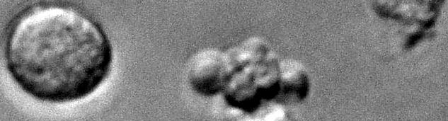


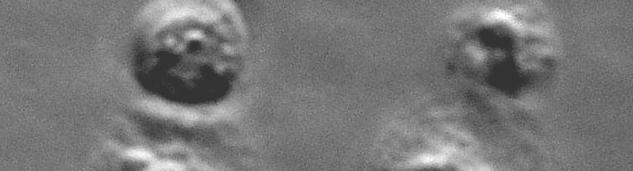

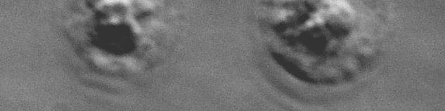
12 Apoptosis versus necrosis Glucose removal thapsigargin
13 Other forms of death truncated apoptosis: caspase-independent cell death (CICD) lack of key molecules; caspase inhibition autophagy? unclassified: Toll-like receptors cornification some forms of neuronal cell death perforin necrosis ischemia, heat shock, irradiation etc.
14
15 Caspases
16 Caspases are activated in cascades Initiator caspases (caspase-8, caspase-9) Cleave executioner caspases (caspase-3,-7) caspase-3 3heterotetramer t t
17 Caspases caspase-3 3heterotetramer t t Lavrik et al, J. Clin. Invest. 115 (2005)


18 Caspase-8 is activated by homo- dimerization
19 Riedl and Salvesen, Nat Rev MCB 2007


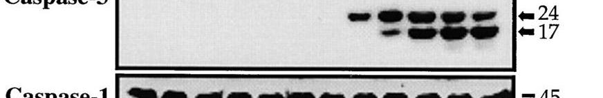
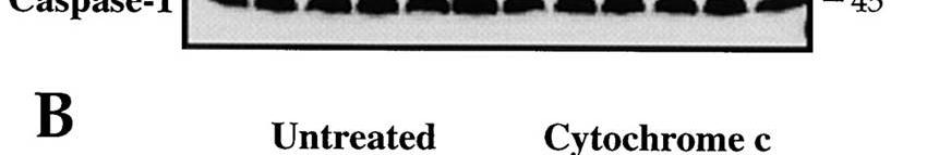
20 Slee et al, JCB 1999


21 Evolution of caspases: para- and metacaspases Human caspase-8 metacaspase 9 of A. thaliana Vercammen JCB 2007
22 Caspase Knockout phenotype Role in apoptosis Caspase-1 Mice develop normally Role in 'pyroptosis Caspase-2 I i i / i Mice have excess oocytes Premature aging Caspase-3 Perinatal lethality Executioner caspase Brain hyperplasia Caspase-6 Mice develop normally Executioner caspase Caspase-7 Perinatal lethality Executioner caspase Caspase-8 Caspase-9 Embryonic lethality. Impaired heart development and decreased pool of hematopoietic precursors. In humans, familial mutation leads to immunodeficiency Perinatal lethality Excess brain tissue Initiator/executioner caspase Initiator of apoptosis after DNA damage and during mitotic catastrophe Initiator caspase of the death receptor (extrinsic) apoptotic pathway Proteolytically activates downstream caspases and Bid Initiator caspase of the mitochondrial (intrinsic) apoptotic pathway Caspase-10 In humans, a familial mutation is linked with Putative initiator caspase of the extrinsic Caspase-11 type II autoimmune lymphoproliferative syndrome Mice develop normally Lymphocytes have a defect in actin depolymerization pathway Proteolytically activates downstream caspases and Bid Involved in neuronal cell death induced by MPTP and other pathological stimuli Caspase-12 Mice develop normally Initiator caspase in ER stress-dependent apoptosis Caspase-14 Mice skin exhibit reduced hydration levels and enhanced sensitivity to UVB Unknown Adapted from Galuzzi et al 2008
23 Caspases are frequently mutated or downregulated in tumors - caspase-1 downregulated - caspase-9 mutations (truncation) in colon cancer - Polymorphism in caspase-8 8(CASP8) gene, D302H, associated with high risk of breast cancer - mutations or inactivation of caspase-8 and caspase-10 in tumors - Mutations in caspase-3 - dominant mutations in caspase-7 in several carcinomas
24
25 Pathways of apoptosis
26 Extrinsic pathway Intrinsic or mitochondrial pathway Induced by extracellular ligands: FasL, TNFα, TRAIL Induced by signals that activate Bcl-2-like proteins: Relevance: - hormones - elimination of infected cells - lack of growth factors - homeostasis of immune - hipoxia, hypoglicemia, lack system, immune privilege of nutrients in general - role of TRAIL in innate - chemotherapeutic drugs immunity - antitumor drugs - inflammatory signals (non - moderate heat-shock apoptotic ti function) - DNA damage - etc.
27 Pathways of apoptosis in vertebrates Stimulus extrinsic stimuli intrinsic stimuli Death receptor stress signal Adapter molecule DISC: FADD apoptosome: Apaf-1 Mitochondria Initiator caspase Caspase- 8/10 Caspase-9 cytochrome c Effector caspase caspase-3/7 = Apoptosis
28 Logue and Martin, Biochem Soc Trans 2008
29 The extrinsic or death receptor pathway
30 Ligands and death receptors trigger the extrinsic pathway Wallach et al. Arthritis Res 2002
31 Role in cancer Fas, TNF, granzymes involved in CTL-mediated killing TNF homolog required for keeping epithelial tumors in check in Drosophila Caspase-8 behaves as a tumor suppressor Role in metastasis of caspase-8 and caspase-10 TRAIL
32 The extrinsic or death receptor pathway Stimulus extrinsic stimuli Death receptor Adapter molecule Initiator caspase DISC: FADD Caspase- 8/10 Effector caspase caspase-3/7 = Apoptosis
33 Inactivating mutations or downregulation of TRAIL receptors, caspase-8 and caspase-10 in many tumors Methylation of Fas and TRAIL-R1 FADD downregulated in thyroid and tongue carcinoma, mantle cell lymphoma FLIP expression increased in many tumors: colon and hepatocellular carcinoma, pancreatic cancer, melanoma Mutations in FADD, caspase-8 and caspase-10 higher in metastasis
34 Fas (CD95) DISC DISC (death-inducing signaling g complex)
35 TNF signalling
36 TRAIL signaling TRAIL-R3 TRAIL-R1 TRAIL-R2 TRAIL-R4 Osteoprotegerin DD DDDD DDDD DD FADD Caspase-8 APOPTOSIS
37 Wang, Oncogene 2008
38 TRAIL prevents tumorogenesis TRAIL mediates NK-induced cytotoxicity against tumor cells. Activated T cells exert e TRAIL-mediated apoptosis More tumors in TRAIL-deficient mice, spontaneusly and after carcinogen treatment.
39 TRAIL for therapy Tumor cells are more sensitive than nontransformed cells to TRAIL. rtrail, anti-trail receptor Abs efficient in xenografts In clinical trials: anti-trailr1 TRAILR1, anti-trailr2 O i ff t iti i t ll t Ongoing efforts on sensitizing tumor cells to TRAIL
40
41 Execution phase Stimulus extrinsic stimuli Death receptor Adapter molecule Initiator caspase DISC: FADD Caspase- 8/10 Effector caspase caspase-3/7 = Apoptosis
42 Caspases dismantle the cell by cleaving hundreds of proteins Taylor et al, Nature Reviews Molecular Cell Biology 9, (March 2008)
43 Mitochondria are inactivated during execution phase: ATP levels fall - Caspases are responsible for membrane potential drop - Cells keep respiring if caspases are inhibited. - Caspases are responsible for generation of ROS
44 Caspases inactivate t respiratory chain Caspases OM IMS IM I Q e - II Q e - e - III C e - IV 2H + + ½ O 2 H 2 O ADP V H + + Ψm - ATP V D A C A N T
45 Caspases inactivate Complex I to inhibit mitochondrial membrane potential WT Uncleavable mutant t red=mitochondrial potential green= cytochrome c-gfp Hela ActD 4 min/frame
46 Prevention of caspase-dependent p mitochondrial dysfunction delays cell death WT uncleavable mutant green = cytochrome c-gfp blue = Annexin-Alexa647 purple = Propidium Iodide 4 min/frame
47 Apoptotic cells are removed by macrophages Taylor et al, Nature Reviews Molecular Cell Biology 9, (March 2008)
48
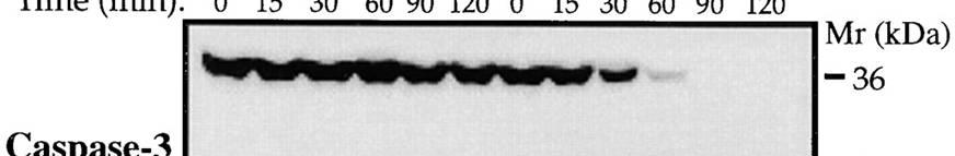
49 Measuring apoptosis Caspase activity and cleavage (by western-blot or enzymatic assays) Chromatin condensation (by microscopy) In tissues: TUNNEL, caspase cleavage. Annexin-V (some unspecificity)
50 Measuring apoptosis Caspase activity and cleavage (by western-blot or enzymatic assays) Chromatin condensation (by microscopy) In tissues: TUNNEL, caspase cleavage. Annexin-V (some unspecificity) Krysko et al, Methods 2008

 DNA")
51 Measuring apoptosis % cells with sub-g1 DNA content (flow citometer) DNA ladder
52
53 The mitochondrial pathway
54 Stimulus extrinsic stimuli intrinsic stimuli Death receptor stress signal Adapter molecule DISC: FADD apoptosome: Apaf-1 Mitochondria Initiator caspase Caspase- 8/10 Caspase-9 cytochrome c Effector caspase caspase-3/7 = Apoptosis
55 Relevance for tumorogenesis and tumor maintenance - Oncogene activation - DNA damage - growth factor removal - lack of oxygen - lack of nutrients - lack of substrate adhesion - chemotherapy - radiotherapy


56 Deprivation of survival factors induces apoptosis through the mitochondrial pathway IL-3 BH3 Adapter molecule Initiator caspase Effector caspase apoptosome: Apaf-1 Caspase-9 Apoptosis Mitochondria cytochrome c

57 Stimulus Chemotherapy induces apoptosis through the mitochondrial pathway stress signal Adapter molecule Initiator caspase Effector caspase apoptosome: Apaf-1 Caspase-9 Apoptosis Mitochondria cytochrome c
58
59 Bcl-2 family proteins
60
61 Phenotypes of BCL-null mice Pro-survival family members Adapted from Youle and Strasser, NRMCB 2008 BCL-2 Abnormal death of renal epithelial progenitors, melanocyte progenitors and mature B and T cells. Fatal polycystic kidney disease (100% mortality by 6 weeks), premature greying and lymphopoenia. BCL-XL BCL-W A1A Abnormal death of fetal erythroid progenitors and neuronal cells. 100% die around embryonic day 14 Abnormal death of developing sperm cells. Causes male sterility. Abnormally accelerated death of granulocytes and mast cells in culture. MCL1 Failure in implantation. Conditional knockout causes premature death of immature and mature B and T lymphoid cells, as well as haemopoietic stem cells. Pro-apoptotic BAX/BAK family members BAX BAK Mild lymphoid hyperplasia, male sterility due to sperm-cell differentiation defect. No obvious defects detected so far.
62 Bcl-2 proteins regulate mitochondrial permeabilization and cytochrome c release Hela staurosporine + ZVAD
63 Bax translocates during apoptosis GFP-Bax Cyt. c - ReAsH merge HeLa Staurosporine + ZVAD 2 min / frame
64 Multidomain pro- and anti-apoptotic Bcl-2 homologs are structurally similar Bax Bcl-W Bcl-xL
65
66 Bax and Bak are required for permeabilization of the outer mitochondrial i membrane Bax/Bak Bcl-2 tbid mitochondria matrix Cyt c BH3-only tbid Bax Bak Cytochrome c Caspase-9 Apoptosis
67 Basáñez and Hardwick, 2008
68 BH3-only proteins are selective for anti- apoptotic Bcl-2 proteins Youle and Strasser, NRMCB 2008
69 Bcl-2 proteins in cancer -Bcl-2 discovered as an oncogene - Antiapoptotic Bcl-2 family members behave as oncogenes - Proapoptotic Bcl-2 family members behave as - Proapoptotic Bcl-2 family members behave as tumor suppressors
70 Antiapoptotic Bcl-2 allow tumorogenesis by c-myc Hematopoietic progenitor cells are transduced with retroviral vectors, ex vivo, followed by reintroduction into lethally irradiated syngeneic recipients Beverly et al, Oncogene, 2009


71 Antiapoptotic Bcl-2 proteins are overexpressed in a range of human tumors Beverly et al, Oncogene, 2009
72 Antiapoptotic Bcl-2 proteins are overexpressed in tumors Bcl-2 Bcl-xL follicular lymphoma lung adenocarcinoma, ovarian tumors, murine hematopoietic malignancies Mcl-1 multiple myeloma and other hematopoietic malignancies Bfl-1 diffuse large-cell lymphoma, breast tumors Bcl-w lung tumors
73 Tumor cells frequently inactivate BH3-onlys or downregulate Bax and/or Bak Anthony G. Letai Nature Reviews Cancer 8, (February 2008)
74 Bcl2-like proapoptotic proteins are mutated or downregulated in human tumors Proapoptotic multidomain Bax oligodendroglioma, breast cancer, colon and gastric adenocarcinomas, hematopoietic ti malignancies, i Bak nonsmall cell lung carcinoma BH3-only Puma melanoma Bad squamous cell carcinomas, colon cancers Bim Bid renal cell carcinoma nonsmall cell lung carcinoma
75 Pro-apoptotic Puma and Noxa contribute to mycinduced lymphomagenesis Some BH3-only are upregulated in tumors: Noxa, BNIP3 Burton et al, Oncogene 2009
76 Bcl-2 family members modulate response to chemotherapy - In vitro, most BH3 proteins have been shown to modulate responses to drugs. -increased expression of antiapoptotic BCL2 in cell lines resistant to chemotherapy (increasing copy numbers or upregulation of mrna or protein). Frenzel et al, Apoptosis 2008
77 Addiction to anti-apoptotic Bcl-2 proteins Brunelle and Letai, JCS 2008
78

79 Mitochondrial permeabilization during apoptosis Stimulus intrinsic i i stimuli stress signal Adapter molecule Initiator caspase apoptosome: Apaf-1 Mitochondria cytochrome c Effector caspase Apoptosis Caspase-9
80 MOMP (mitochondrial outer membrane permeabilization) When mitos are permeabilized, many proteins from the intermembrane space are released: - Cytochrome c - Smac / DIABLO - Adenylate kinase 2 - Omi / HtrA2 - Endonuclease G - AIF Mitos are depolarized

81 Bax and Bak are required for permeabilization of the outer mitochondrial i membrane Bax/Bak Bcl-2 tbid mitochondria matrix Cyt c BH3-only tbid Bax Bak Cytochrome c Caspase-9 Apoptosis
82 The MOMP pore Bax and/or Bak are required for formation of the pore. It only targets the outer membrane. Different proteins up to at least 120 kda are released simultaneously from the pore.
83 Cytochrome c release takes 5 min within a Cytochrome c release takes 5 min within a single cell
84 cytochrome c-gfp and cyt.c-tetracysteinetetracysteine are released in 5 min GFP-cytocrome c cyt. c - tetracysteine REASH Hela - ActD+ZVAD 30ºC 1 min 30 /frame 2 h total
85 Smac/DIABLO, a second mitocondrial pro- apoptotic protein Stimulus intrinsic stimuli stress signal Adapter molecule Initiator caspase Effector caspase apoptosome: Apaf-1 Caspase-9 Apoptosis Mitochondria cytochrome c
86 Smac is released prior to mitochondrial depolarization, and simultaneously to cyt.c Smac-TC Smac- TC+TMRE Duration of release= 5.4 min Lag= 3.1 min Hela, Staurosporine + ZVAD
87 Smac is mutated t in tumors Renal cell carcinomas express less Smac than normal tissue, The expression is lower in advanced stages, Expression correlates with survival, Retransfection sensitizes RCC to TNF, TRAIL and cisplatin. Can cells survive after mitochondrial Can cells survive after mitochondrial permeabilization?
88 Omi/HtrA2, a serine-protease released during apoptosis. Omi and cyt.c are released simultaneously Cyt. C-GFP Omi-4cys Hela stably transfected with cyt.c- GFP; transiently with Omi-4CYS. Staurosporine + ZVAD, 3 min/picture Muñoz-Pinedo et al, PNAS 2006
89 AIF, apoptosis-inducing inducing factor? AIF is released slowly after cyt.c Time (min) cyt.c- GFP merge AIF-TC
90
91 Riedl and Salvesen, Nat Rev MCB 2007
92 Apaf-1 promoter methylated in several leukemias Downregulated in malignant melanoma, ovarian cancer Can cells survive after cyt.c release?
93
94 IAPs ( Inhibitor of Apoptosis Protein ) IAPs ( Inhibitor of Apoptosis Protein ) part of the BIR-containing protein family
95 IAPs may act at different levels Stimulus extrinsic stimuli intrinsic stimuli Death receptor stress signal Adapter molecule DISC: FADD apoptosome: Apaf-1 Mitochondria Initiator caspase Caspase- 8/10 Caspase-9 cytochrome c Effector caspase caspase-3/7 = Apoptosis
96 Gene Disease Comments Birc2 (ciap1) Birc3 (ciap2) Birc4 (XIAP) Multiple myeloma (MM) Various carcinomas MALT lymphoma Multiple myeloma Various carcinomas X-linked lymphoproliferativ e disorder (XLP) ciap1 and ciap2 are deleted in some MM cancers, as part of a larger trend of activating mutations ti for the alternative ti NF-B pathway (Annunziata et al., 2007; Keats et al., 2007). Hence, the negative regulatory role of ciap1 or ciap2 as E3 ubiquitin ligases for NIK is removed, allowing for NIK stabilization and activation of the alternative NF-B pathway. ciap1 and ciap2 genes, at 11q22, are commonly amplified in many cancers, with visibly increased expression of ciap1. This phenomenon is also conserved in some murine tumors that also lead to co-amplification of YAP. Both ciap1 and YAP have been shown to cooperate in transformation (Zender et al., 2006). ciap1 can also cooperate with Myc to transform. t f This results from ciap1's ability to induce the proteosomal degradation of the Myc antagonist Mad1 (Xu et al., 2007). ciap2 is translocated in t(11;18)(q21;q21)-bearing lymphomas (Dierlamm et al., 1999). The chromosomal translocation fuses the three BIR domains of ciap2 with the paracaspase domain of MALT1, while preserving the ciap2, NF-B-responsive, promoter. See above ciap1 and ciap2 genes, at 11q22, are commonly amplified in many cancers, with visibly increased expression of ciap1. While ciap2 expression is often overlooked. This DNA amplification is also conserved in some murine tumors, which also leads to co-amplification of YAP (Zender et al., 2006). ciap2 may also cooperate in transformation in these cases. Apoptosis of lymphocytes from XIAP-deficient patients is enhanced in response to various stimuli including CD3, Fas and TRAIL. In addition, XIAP-deficient XLP patients (XLP-2), like SAP-deficient XLP patients (XLP-1), have low numbers of NK T cells. XLP patients can exhibit a fatal response to EBV, the viral cause of infectious mononucleosis (presumably due to a failure(s) in NK and lymphocyte homeostasis and signaling) (Rigaud et al., 2006). Adapted from LaCasse et al, Oncogene 2008
97 IAPs and their inhibitors are altered in cancer - XAF1 is an IAP inhibitor underexpressed in many cancer cell lines (LOH), and a prognostic marker in bladder cancers. - Smac/DIABLO inhibits XIAP, ciap1, ciap2, survivin, livin and BRUCE.
98 IAP small molecule antagonists in clinical development Target Compound Class Stage Company XIAP AEG35156/GEM640 Antisense Phase 2 Aegera; Idera Xantags ( , ), TWX006, TWX024 Caspase-3 de-repressor Preclinical Embelin and derivatives Natural product Preclinical ciap1, ciap2, AEG40826/HGS1029, LBW242, compound 3, XIAP, livin compound 11 Joyant, Pfizer, Abbott Smac mimetic Phase 1 Aegera (Human Genome Sciences, USA), Novartis, Joyant Pfizer Abbott Compound C, compound 8, Smac mimetic Phase 1 Genentech, SF, CA BV6 GT-T, compound A Smac mimetic Preclinical Tetralogic, Malvern, PA, USA (Amgen, CA) SM-164, SM-122 Smac mimetic Preclinical Ascenta, Malvern, PA, USA ciap1 Bestatin methyl ester ciap1 degrad. promoter Preclinical Nippon Kayaku Co., Japan Bestatin actinonin hybrid, HAB-5(30b) ciap1 degrad. promoter Preclinical Nippon Kayaku Co. ciap2 Ro IKK ubiquitination Preclinical Roche, Palo Alto, CA, USA inhibitor Survivin LY /ISIS23722 Antisense Phase 2 Eli Lilly, Indianapolis, IN, USA (Isis, CA, USA) YM155 Transcriptional repressor Phase 2 Astellas, Tokyo, Japan Terameprocol (EM-1421) Transcriptional repressor Phase 2 Erimos, Raleigh, NC, USA Adapted from LaCasse et al, Oncogene 2008
99
100 Pathways of apoptosis in vertebrates Stimulus extrinsic stimuli intrinsic stimuli Death receptor stress signal Adapter molecule DISC: FADD apoptosome: Apaf-1 Mitochondria Initiator caspase Caspase- 8/10 Caspase-9 cytochrome c Effector caspase caspase-3/7 = Apoptosis
Mechanisms of Cell Death
 Mechanisms of Cell Death CELL DEATH AND FORMATION OF THE SEMICIRCULAR CANALS Carol M. Troy August 25, 2008 FROM: Fekete et al., Development 124: 2451 (1997) PHENOMENOLOGY OF CELL DEATH I. DEVELOPMENT A.
Apoptosis Chapter 9. Neelu Yadav PhD
 Apoptosis Chapter 9 Neelu Yadav PhD Neelu.Yadav@Roswellpark.org 1 Apoptosis: Lecture outline Apoptosis a programmed cell death pathway in normal homeostasis Core Apoptosis cascade is conserved Compare
Signaling Apoptosis. Scott André Oakes, M.D. Dept. of Pathology Univ. of Calif-San Francisco. Cyt c Release BAX/BAK. Apoptosome Formation
 Signaling Apoptosis Cyt c Release BAX/BAK Apoptosome Formation Caspase Activation Scott André Oakes, M.D. Dept. of Pathology Univ. of Calif-San Francisco Why do we need cell death? Sculpt Organs Control
#19 Apoptosis Chapter 9. Neelu Yadav PhD
 #19 Apoptosis Chapter 9 Neelu Yadav PhD Neelu.Yadav@Roswellpark.org Why cells decide to die? - Stress, harmful, not needed - Completed its life span Death stimulation or Stress Cell Survival Death Functions
Apoptotic Pathways in Mammals Dr. Douglas R. Green
 Apoptotic Pathways in Mammals Douglas R. Green 1 Apoptosis A form of cell death that is defined morphologically, and features a number of biochemical events Programmed cell death Cell death that occurs
Introduction to pathology lecture 5/ Cell injury apoptosis. Dr H Awad 2017/18
 Introduction to pathology lecture 5/ Cell injury apoptosis Dr H Awad 2017/18 Apoptosis = programmed cell death = cell suicide= individual cell death Apoptosis cell death induced by a tightly regulated
#19 Apoptosis Chapter 9. Neelu Yadav PhD
 #19 Apoptosis Chapter 9 Neelu Yadav PhD Neelu.Yadav@Roswellpark.org Why cells decide to die? - Stress, harmful, not needed - Completed its life span Death stimulation or Stress Cell Survival Death Functions
Apoptotic cell signaling in cancer progression and therapyw
 Integrative Biology Dynamic Article Links Cite this: Integr. Biol., 2011, 3, 279 296 www.rsc.org/ibiology REVIEW ARTICLE Apoptotic cell signaling in cancer progression and therapyw Jessica Plati, a Octavian
Apoptosome dysfunction in human cancer
 Apoptosis 2004; 9: 691 704 C 2004 Kluwer Academic Publishers Apoptosome dysfunction in human cancer K. M. Hajra and J. R. Liu Department of Obstetrics and Gynecology, University of Michigan Medical School,
GMS 6644: Apoptosis. Introduction
 GMS 6644: Apoptosis Introduction (Feb. 15, 2006) Lei Xiao, Ph.D. Department of Anatomy & Cell Biology UF Shands Cancer Center ARB Rm R4-250, 846-1199, lxiao@ufl.edu Outline of the Lecture Different types
Molecular biology :- Cancer genetics lecture 11
 Molecular biology :- Cancer genetics lecture 11 -We have talked about 2 group of genes that is involved in cellular transformation : proto-oncogenes and tumour suppressor genes, and it isn t enough to
Programmed Cell Death (apoptosis)
") Programmed Cell Death (apoptosis) Stereotypic death process includes: membrane blebbing nuclear fragmentation chromatin condensation and DNA framentation loss of mitochondrial integrity and release of
Programmed Cell Death (apoptosis) Stereotypic death process includes: membrane blebbing nuclear fragmentation chromatin condensation and DNA framentation loss of mitochondrial integrity and release of
p53 and Apoptosis: Master Guardian and Executioner Part 2
 p53 and Apoptosis: Master Guardian and Executioner Part 2 p14arf in human cells is a antagonist of Mdm2. The expression of ARF causes a rapid increase in p53 levels, so what would you suggest?.. The enemy
Supplementary Figures
 Supplementary Figures Figure S1. Validation of kinase regulators of ONC201 sensitivity. Validation and screen results for changes in cell viability associated with the combination of ONC201 treatment (1
Apoptosis Oncogenes. Srbová Martina
 Apoptosis Oncogenes Srbová Martina Cell Cycle Control point Cyclin B Cdk1 Cyclin D Cdk4 Cdk6 Cyclin A Cdk2 Cyclin E Cdk2 Cyclin-dependent kinase (Cdk) have to bind a cyclin to become active Regulation
The discovery of Bcl-2
 The discovery of Bcl-2 Bcl-2: B-cell lymphoma 2 The pro-survival subfamily of Bcl-2 protein family Cloning of Bcl-2 as the oncogene which is deregulated at t(14;18) lymphomas Pioneer works by Tsujimoto
Part I Molecular Cell Biology
 1 Part I Molecular Cell Biology RNA Regulation: Advances in Molecular Biology and Medicine, First Edition. Edited by Robert A. Meyers. 2014 Wiley-VCH Verlag GmbH & Co. KGaA. Published 2014 by Wiley-VCH
Problem Set 8 Key 1 of 8
 7.06 2003 Problem Set 8 Key 1 of 8 7.06 2003 Problem Set 8 Key 1. As a bright MD/PhD, you are interested in questions about the control of cell number in the body. Recently, you've seen three patients
Epigonal Conditioned Media from Bonnethead Shark, Sphyrna tiburo, Induces Apoptosis in a T-Cell Leukemia Cell Line, Jurkat E6-1
 Mar. Drugs 2013, 11, 3224-3257; doi:10.3390/md11093224 Article OPEN ACCESS marine drugs ISSN 1660-3397 www.mdpi.com/journal/marinedrugs Epigonal Conditioned Media from Bonnethead Shark, Sphyrna tiburo,
The Biochemistry of apoptosis
 The Biochemistry of apoptosis 1 1 The apoptosis is composed of multiple biochemical events 2 2 Biochemical, cellular, and molecular events in Apoptosis 1. Membrane blebbing; phosphatidyl serine exposure
Major apoptotic mechanisms and genes involved in apoptosis
 Tumor Biol. (2016) 37:8471 8486 DOI 10.1007/s13277-016-5035-9 REVIEW Major apoptotic mechanisms and genes involved in apoptosis Yağmur Kiraz 1,2 & Aysun Adan 1 & Melis Kartal Yandim 2 & Yusuf Baran 1,2
shehab Moh Tarek ... ManarHajeer
 3 shehab Moh Tarek... ManarHajeer In the previous lecture we discussed the accumulation of oxygen- derived free radicals as a mechanism of cell injury, we covered their production and their pathologic
ASCO Annual Meeting 2013, May 31 June , Chicago, IL
 Phase I Study of Safety and Pharmacokinetics of GDC-0917, an Antagonist of Inhibitor of Apoptosis Proteins in Patients with Refractory Solid Tumors or Lymphoma 1 A Tolcher, 1 K Papadopoulos, 1 A Patnaik,
Types of cell death and apoptosis resistance mechanisms. Institut for Experimental Cancer Research
 Types of cell death and apoptosis resistance mechanisms Prof.Dr.rer. rer. nat.anna Trauzold Institut for Experimental Cancer Research Physiological cell death Embryogenesis Control of the tissue size Renewal
APPLICATION NOTE 1850 Millrace Drive, Suite 3A Eugene, Oregon
 APPLICATION NOTE 185 Millrace Drive, Suite 3A Eugene, Oregon 973 In-Cell ELISA (ICE) Assay Platform Monitoring apoptosis in cells: a high-throughput, quantitative cellbased assay. Rev. Introduction: Apoptosis:
Prepared by Cyrus H. Nozad, MD, University of Tennessee and John Seyerle, MD, Ohio State University
 Allergy and Immunology Review Corner: Chapter 21 of Middleton s Allergy Principles and Practice, Seventh Edition, edited by N. Franklin Adkinson, et al. Chapter 21: Antigen-Presenting Dendritic Cells (Pages
Supplementary Information
 Supplementary Information Significance of p53 dynamics in regulating apoptosis in response to ionizing radiation, and polypharmacological strategies Bing Liu 1,, Divesh Bhatt 1,, Zoltan N. Oltvai 2, Joel
mirna Dr. S Hosseini-Asl
 mirna Dr. S Hosseini-Asl 1 2 MicroRNAs (mirnas) are small noncoding RNAs which enhance the cleavage or translational repression of specific mrna with recognition site(s) in the 3 - untranslated region
Follicular Lymphoma. ced3 APOPTOSIS. *In the nematode Caenorhabditis elegans 131 of the organism's 1031 cells die during development.
 Harvard-MIT Division of Health Sciences and Technology HST.176: Cellular and Molecular Immunology Course Director: Dr. Shiv Pillai Follicular Lymphoma 1. Characterized by t(14:18) translocation 2. Ig heavy
Apoptosis-based Therapies: Mechanisms and Applications
 Apoptosis-based Therapies: Mechanisms and Applications Perspective Bcl-2 family members, potential usage of BH3 domains as drug targets Bcl-2/xL inhibitors - Antisense, inhibitors of protein-protein interactions,
Table S1. New colony formation 7 days after stimulation with doxo and VCR in JURKAT cells
 Table S1. New colony formation 7 days after stimulation with and in JURKAT cells drug co + number of colonies 7±14 4±7 48±11 JURKAT cells were stimulated and analyzed as in Table 1. Drug concentrations
Apoptosis in chronic hepatitis C
 Apoptosis in chronic hepatitis C Dr med. Anna Parfieniuk-Kowerda Department of Infectious Diseases and Hepatology Medical University of Bialystok Poland APOPTOSIS Apoptosis - type I programmed cell death
Getting TRAIL back on track for cancer therapy
 (2014) 21, 1350 1364 & 2014 Macmillan Publishers Limited All rights reserved 1350-9047/14 www.nature.com/cdd OPEN Review Getting TRAIL back on track for cancer therapy J Lemke 1,2, S von Karstedt 1, J
Robbins and Cotran Pathologic Basis of Disease 8th Edition Odabrana poglavlja
 Robbins and Cotran Pathologic Basis of Disease 8th Edition Odabrana poglavlja Apoptosis Apoptosis is a pathway of cell death that is induced by a tightly regulated suicide program in which cells destined
C-Phycocyanin (C-PC) is a n«sjfc&c- waefc-jduble phycobiliprotein. pigment isolated from Spirulina platensis. This water- soluble protein pigment is
 is a n«sjfc&c- waefc-jduble phycobiliprotein. pigment isolated from Spirulina platensis. This water- soluble protein pigment is") ' ^Summary C-Phycocyanin (C-PC) is a n«sjfc&c- waefc-jduble phycobiliprotein pigment isolated from Spirulina platensis. This water- soluble protein pigment is of greater importance because of its various
' ^Summary C-Phycocyanin (C-PC) is a n«sjfc&c- waefc-jduble phycobiliprotein pigment isolated from Spirulina platensis. This water- soluble protein pigment is of greater importance because of its various
34 Apoptosis Programmed cell death is vital to the health and development of multicellular organisms.
 Principles of Biology contents 34 Apoptosis Programmed cell death is vital to the health and development of multicellular organisms. Apoptosis is the reason we have separate fingers and toes. During embryonic
Silibinin i activates p53-caspase-2 pathway and causes caspase-mediated cleavage of Cip1/p21 in apoptosis
 Silibinin i activates p53-caspase-2 pathway and causes caspase-mediated cleavage of Cip1/p21 in apoptosis induction in bladder transitional-cell papilloma RT4 cells: Evidence for a regulatory loop between
Mitochondria in apoptosis. Jean-Claude Martinou, MD, Ph.D Department of cell biology University of Geneva Geneva, Switzerland
 Mitochondria in apoptosis Jean-Claude Martinou, MD, Ph.D Department of cell biology University of Geneva Geneva, Switzerland The dual role of mitochondria in life and death QuickTime and a TIFF (Uncompressed)
Cancer. The fundamental defect is. unregulated cell division. Properties of Cancerous Cells. Causes of Cancer. Altered growth and proliferation
 Cancer The fundamental defect is unregulated cell division. Properties of Cancerous Cells Altered growth and proliferation Loss of growth factor dependence Loss of contact inhibition Immortalization Alterated
Cancer. Throughout the life of an individual, but particularly during development, every cell constantly faces decisions.
 Cancer Throughout the life of an individual, but particularly during development, every cell constantly faces decisions. Should it divide? Yes No--> Should it differentiate? Yes No-->Should it die? Yes-->Apoptosis
2 The Extrinsic Pathway of Apoptosis
 2 The Extrinsic Pathway of Apoptosis M. Stacey Ricci, ScD, and Wafik S. El-Deiry, MD, PhD Summary Defects in the extrinsic pathway are linked to several disease states, including cancer. Pharmacologic
Introduction. Cancer Biology. Tumor-suppressor genes. Proto-oncogenes. DNA stability genes. Mechanisms of carcinogenesis.
 Cancer Biology Chapter 18 Eric J. Hall., Amato Giaccia, Radiobiology for the Radiologist Introduction Tissue homeostasis depends on the regulated cell division and self-elimination (programmed cell death)
Cancer genetics
 Cancer genetics General information about tumorogenesis. Cancer induced by viruses. The role of somatic mutations in cancer production. Oncogenes and Tumor Suppressor Genes (TSG). Hereditary cancer. 1
The death receptors: signaling and modulation
 The death receptors: signaling and modulation 1 1 The extrinsic cell death pathway 2 Nat Rev Drug Discov. 2008 Dec;7(12):1001-12. 2 Death receptors Belong to the tumor necrosis factor (TNF) receptor gene
Chapter 1 CELL INJURY CELL DEATH CELL ADAPTATIONS. M.G.Rajanandh, Dept. of Pharmacy Practice, SRM College of Pharmacy, SRM University.
 Chapter 1 CELL INJURY CELL DEATH CELL ADAPTATIONS M.G.Rajanandh, Dept. of Pharmacy Practice, SRM College of Pharmacy, SRM University. CONCEPTS IN CELL INJURY The clinical signs and symptoms are several
Deregulation of signal transduction and cell cycle in Cancer
 Deregulation of signal transduction and cell cycle in Cancer Tuangporn Suthiphongchai, Ph.D. Department of Biochemistry Faculty of Science, Mahidol University Email: tuangporn.sut@mahidol.ac.th Room Pr324
Think Tank on Molecular Targets: Survival and Death Pathways in Cancer
 Think Tank on Molecular Targets: Survival and Death Pathways in Cancer Oncogenes Induce Cell Proliferation & Cell Death Proliferation Mitogens ONCOGENES Apoptosis Adapted from G Evan Survival Signals Block
RAS Genes. The ras superfamily of genes encodes small GTP binding proteins that are responsible for the regulation of many cellular processes.
 ۱ RAS Genes The ras superfamily of genes encodes small GTP binding proteins that are responsible for the regulation of many cellular processes. Oncogenic ras genes in human cells include H ras, N ras,
Bihong Zhao, M.D, Ph.D Department of Pathology
 Bihong Zhao, M.D, Ph.D Department of Pathology 04-28-2009 Is tumor self or non-self? How are tumor antigens generated? What are they? How does immune system respond? Introduction Tumor Antigens/Categories
Apoptosis and cutaneous melanoma
 @[;:(~1iiJ @@IiiJ@@IJ' 2007;22:21-31. Apoptosis and cutaneous melanoma Ricardo Vieira, Oscar Tellechea and America Figueiredo Department ofdermatology, Faculty ofmedicine, Coimbra University, Coimbra,
Pro-apoptotic signalling through Toll-like receptor 3 involves TRIF-dependent
 Pro-apoptotic signalling through Toll-like receptor 3 involves TRIF-dependent activation of caspase-8 and is under the control of inhibitor of apoptosis proteins in melanoma cells Arnim Weber, Zofia Kirejczyk,
Thesis for doctoral degree (Ph.D.) 2008 Regulation of mast cell survival and apoptosis. Mats Karlberg. Mats Karlberg
 2008 Regulation of mast cell survival and apoptosis. Mats Karlberg. Mats Karlberg") Thesis for doctoral degree (Ph.D.) 2008 Thesis for doctoral degree (Ph.D.) 2008 Regulation of mast cell survival and apoptosis Mats Karlberg Regulation of mast cell survival and apoptosis Mats Karlberg
Thesis for doctoral degree (Ph.D.) 2008 Thesis for doctoral degree (Ph.D.) 2008 Regulation of mast cell survival and apoptosis Mats Karlberg Regulation of mast cell survival and apoptosis Mats Karlberg
Molecular mechanisms of apoptosis Caspase-8-activation: death receptors and TRIF
 Molecular mechanisms of apoptosis Cell death by apoptosis occurs when a specialised intracellular signalling pathway is activated and kills the cell. Apoptosis is the most common way of cells to die in
Overview of cell death signaling pathways
 Cancer Biology & Therapy ISSN: 1538-4047 (Print) 1555-8576 (Online) Journal homepage: http://www.tandfonline.com/loi/kcbt20 Overview of cell death signaling pathways Zhaoyu Jin & Wafik S. El-Deiry To cite
The inhibitors of apoptosis (IAPs) as cancer targets
 as cancer targets") Apoptosis (2007) 12:1543 1568 DOI 10.1007/s10495-007-0087-3 ORIGINAL PAPER The inhibitors of apoptosis (IAPs) as cancer targets Allison M. Hunter Æ Eric C. LaCasse Æ Robert G. Korneluk Published online:
Apoptosis (2007) 12:1543 1568 DOI 10.1007/s10495-007-0087-3 ORIGINAL PAPER The inhibitors of apoptosis (IAPs) as cancer targets Allison M. Hunter Æ Eric C. LaCasse Æ Robert G. Korneluk Published online:
NFκB What is it and What s the deal with radicals?
 The Virtual Free Radical School NFκB What is it and What s the deal with radicals? Emily Ho, Ph.D Linus Pauling Institute Scientist Department of Nutrition and Food Management Oregon State University 117
Introduction: 年 Fas signal-mediated apoptosis. PI3K/Akt
 Fas-ligand (CD95-L; Fas-L) Fas (CD95) Fas (apoptosis) 年 了 不 度 Fas Fas-L 力 不 Fas/Fas-L T IL-10Fas/Fas-L 不 年 Fas signal-mediated apoptosis 度降 不 不 力 U-118, HeLa, A549, Huh-7 MCF-7, HepG2. PI3K/Akt FasPI3K/Akt
Cell death at the intestinal epithelial front line
 REVIEW ARTICLE Cell death at the intestinal epithelial front line Maria Eugenia Delgado, Thomas Grabinger and Thomas Brunner Chair of Biochemical Pharmacology, Department of Biology, University of Konstanz,
Analysis of nitric oxide-induced apoptotic signaling in PC12 rat phaeochromocytoma cells
 Analysis of nitric oxide-induced apoptotic signaling in PC12 rat phaeochromocytoma cells PhD thesis Judit Varga Doctoral school leader: Balázs Sümegi PhD, DSc Supervisor and program leader: József Szeberényi
Lecture 14 - The cell cycle and cell death
 02.17.10 Lecture 14 - The cell cycle and cell death The cell cycle: cells duplicate their contents and divide The cell cycle may be divided into 4 phases The cell cycle triggers essential processes (DNA
Warner-Lambert/Parke-Davis Award Lecture
 American Journal of Pathology, Vol. 157, No. 5, November 2000 Copyright American Society for Investigative Pathology Warner-Lambert/Parke-Davis Award Lecture Mechanisms of Apoptosis John C. Reed From The
Recent insights into the mechanism of glucocorticosteroidinduced
 Review (2002) 9, 6±19 ã 2002 Nature Publishing Group All rights reserved 1350-9047/02 $25.00 www.nature.com/cdd Recent insights into the mechanism of glucocorticosteroidinduced apoptosis *,1 1 Division
Cancer. The fundamental defect is. unregulated cell division. Properties of Cancerous Cells. Causes of Cancer. Altered growth and proliferation
 Cancer The fundamental defect is unregulated cell division. Properties of Cancerous Cells Altered growth and proliferation Loss of growth factor dependence Loss of contact inhibition Immortalization Alterated
Apoptosis as a Therapeutic Target in Cancer and Cancer Stem Cells: Novel Strategies and Futures Perspectives
 Chapter 5 Apoptosis as a Therapeutic Target in Cancer and Cancer Stem Cells: ovel Strategies and Futures Perspectives María A. García, Esther Carrasco, Alberto Ramírez, Gema Jiménez, Elena López-Ruiz,
ACTIVATION OF T LYMPHOCYTES AND CELL MEDIATED IMMUNITY
 ACTIVATION OF T LYMPHOCYTES AND CELL MEDIATED IMMUNITY The recognition of specific antigen by naïve T cell induces its own activation and effector phases. T helper cells recognize peptide antigens through
number Done by Corrected by Doctor Maha Shomaf
 number 19 Done by Waseem Abo-Obeida Corrected by Abdullah Zreiqat Doctor Maha Shomaf Carcinogenesis: the molecular basis of cancer. Non-lethal genetic damage lies at the heart of carcinogenesis and leads
Sensitization to death receptor stimuli and anchoragedependent cell death through induction of endoplasmic reticulum stress
 Sensitization to death receptor stimuli and anchoragedependent cell death through induction of endoplasmic reticulum stress by Kikanwa Brenda Lydia Hope Anyiwe A thesis submitted in conformity with the
APOPTOSIS, NECROSIS AND CANCER. Dr. S. P. Pattanayak
 APOPTOSIS, NECROSIS AND CANCER Dr. S. P. Pattanayak LEARNING OBJECTIVES At the end of the lecture, students should be able to: Know the importance of cell death. Define various modes of cell death. Identify
EXPLOITING THE CANCER GENOME: Strategies for the Discovery and Clinical Development of Targeted Molecular Therapeutics
 EXPLOITING THE CANCER GENOME: Strategies for the Discovery and Clinical Development of Targeted Molecular Therapeutics Timothy A. Yap and Paul Workman Supplemental Material Section 9 of this review discusses
DEATH AND ANTI-DEATH: TUMOUR RESISTANCE TO APOPTOSIS
 DEATH AND ANTI-DEATH: TUMOUR RESISTANCE TO APOPTOSIS Frederik H. Igney and Peter H. Krammer Every cell in a multicellular organism has the potential to die by apoptosis, but tumour cells often have faulty
Objectives. Abbas Chapter 11: Immunological Tolerance. Question 1. Question 2. Question 3. Definitions
 Objectives Abbas Chapter 11: Immunological Tolerance Christina Ciaccio, MD Children s Mercy Hospitals and Clinics February 1, 2010 To introduce the concept of immunologic tolerance To understand what factors
What would you observe if you fused a G1 cell with a S cell? A. Mitotic and pulverized chromosomes. B. Mitotic and compact G1 chromosomes.
 What would you observe if you fused a G1 cell with a S cell? A. Mitotic and pulverized chromosomes. B. Mitotic and compact G1 chromosomes. C. Mostly non-compact G1 chromosomes. D. Compact G1 and G2 chromosomes.
Cancer. Questions about cancer. What is cancer? What causes unregulated cell growth? What regulates cell growth? What causes DNA damage?
 Questions about cancer What is cancer? Cancer Gil McVean, Department of Statistics, Oxford What causes unregulated cell growth? What regulates cell growth? What causes DNA damage? What are the steps in
Virchow s Hypothesis lymphorecticular infiltration of cancer reflected the origin of cancer at sites of inflammation
 Virchow s Hypothesis 1863 lymphorecticular infiltration of cancer reflected the origin of cancer at sites of inflammation Barrett s esophagus/ Esophageal adenocarcinoma PSC / Cholangiocarcinoma Viral hepatitis
An Epstein-Barr virus-encoded microrna targets PUMA to promote host cell survival
 An Epstein-Barr virus-encoded microrna targets to promote host cell survival The Journal of Experimental Medicine 205(11): 2551-2560, 2008. 1 Elizabeth Yee-Wai Choy, Kam-Leung Siu, Kin-Hang Kok, Raymond
COURSE: Medical Microbiology, PAMB 650/720 - Fall 2008 Lecture 16
 COURSE: Medical Microbiology, PAMB 650/720 - Fall 2008 Lecture 16 Tumor Immunology M. Nagarkatti Teaching Objectives: Introduction to Cancer Immunology Know the antigens expressed by cancer cells Understand
609G: Concepts of Cancer Genetics and Treatments (3 credits)
") Master of Chemical and Life Sciences Program College of Computer, Mathematical, and Natural Sciences 609G: Concepts of Cancer Genetics and Treatments (3 credits) Text books: Principles of Cancer Genetics,
Master of Chemical and Life Sciences Program College of Computer, Mathematical, and Natural Sciences 609G: Concepts of Cancer Genetics and Treatments (3 credits) Text books: Principles of Cancer Genetics,
The Hallmarks of Cancer
 The Hallmarks of Cancer Theresa L. Hodin, Ph.D. Clinical Research Services Theresa.Hodin@RoswellPark.org Hippocrates Cancer surgery, circa 1689 Cancer Surgery Today 1971: Nixon declares War on Cancer
ER Stress in Retinal Degeneration in S334ter Rho Rats
 ER Stress in Retinal Degeneration in S334ter Rho Rats Vishal M. Shinde 1., Olga S. Sizova 1., Jonathan H. Lin 2, Matthew M. LaVail 3, Marina S. Gorbatyuk 1 * 1 Department of Cell Biology and Anatomy, University
Lecture 1: Carcinogenesis
 Lecture 1: Carcinogenesis Anti-cancer (oncology agents): These are perhaps the most dangerous of drugs, other than the narcotic analgesics. This is due to their toxicities. Killing or inhibiting cancer
BIO360 Fall 2013 Quiz 1
 BIO360 Fall 2013 Quiz 1 1. Examine the diagram below. There are two homologous copies of chromosome one and the allele of YFG carried on the light gray chromosome has undergone a loss-of-function mutation.
OVARY The surface of the ovary is covered with surface epithelium
 OVARY Cow The ovary, or female gonad, is: 1. an exocrine gland, producing oocytes 2. an endocrine gland, secreting hormones, i.e., estrogen and progesterone OVARY OVARY The surface of the ovary is covered
ciap using fragment based drug discovery
 Discovery of a potent dual antagonist of both XIAP and ciap using fragment based drug discovery Gianni Chessari, PhD TAT Congress 2012 Disclosures I am an employee of Astex Pharmaceuticals I will present
Test Bank for Robbins and Cotran Pathologic Basis of Disease 9th Edition by Kumar
 Link full download:https://getbooksolutions.com/download/test-bank-for-robbinsand-cotran-pathologic-basis-of-disease-9th-edition-by-kumar Test Bank for Robbins and Cotran Pathologic Basis of Disease 9th
Genome of Hepatitis B Virus. VIRAL ONCOGENE Dr. Yahwardiah Siregar, PhD Dr. Sry Suryani Widjaja, Mkes Biochemistry Department
 Genome of Hepatitis B Virus VIRAL ONCOGENE Dr. Yahwardiah Siregar, PhD Dr. Sry Suryani Widjaja, Mkes Biochemistry Department Proto Oncogen and Oncogen Oncogen Proteins that possess the ability to cause
Under the Radar Screen: How Bugs Trick Our Immune Defenses
 Under the Radar Screen: How Bugs Trick Our Immune Defenses Session 8: Apoptosis Marie-Eve Paquet and Gijsbert Grotenbreg Whitehead Institute for Biomedical Research Myxoma virus Poxvirus Infects rabbits
Acute lung injury in children : from viral infection and mechanical ventilation to inflammation and apoptosis Bern, R.A.
 UvA-DARE (Digital Academic Repository) Acute lung injury in children : from viral infection and mechanical ventilation to inflammation and apoptosis Bern, R.A. Link to publication Citation for published
BCL-W has a fundamental role in B cell survival and lymphomagenesis.
 Thomas Jefferson University Jefferson Digital Commons Department of Cancer Biology Faculty Papers Department of Cancer Biology 2-1-2017 BCL-W has a fundamental role in B cell survival and lymphomagenesis.
7/9/2008. Hypoxic cell injury. Consequences of hypoxia depend on cell type. Significance of hypoxia depends on:
 Cellular Adaptation and Cell Injury CLDavis Foundation On the Beach Lecture 2 Hypoxia, reperfusion, free radicals, and apoptosis R K Myers 2008 Hypoxic cell injury Hypoxia: any state of reduction of O
Cytokines, adhesion molecules and apoptosis markers. A comprehensive product line for human and veterinary ELISAs
 Cytokines, adhesion molecules and apoptosis markers A comprehensive product line for human and veterinary ELISAs IBL International s cytokine product line... is extremely comprehensive. The assays are
CATALOG OF ANTIBODIES FOR APOPTOSIS
 CATALOG OF ANTIBODIES FOR APOPTOSIS Table of Contents Mitochondrial Mediated Pathway...2 Inhibitors of Apoptosis Family...5 Death Receptor Signaling Pathway...8 Caspases... 12 Apoptosis Inducing Factor...
KEY CONCEPT QUESTIONS IN SIGNAL TRANSDUCTION
 Signal Transduction - Part 2 Key Concepts - Receptor tyrosine kinases control cell metabolism and proliferation Growth factor signaling through Ras Mutated cell signaling genes in cancer cells are called
BL-8040: BEST-IN-CLASS CXCR4 ANTAGONIST FOR TREATMENT OF ONCOLOGICAL MALIGNANCIES. Overview and Mechanism of Action Dr.
 BL-8040: BEST-IN-CLASS CXCR4 ANTAGONIST FOR TREATMENT OF ONCOLOGICAL MALIGNANCIES Overview and Mechanism of Action Dr. Leah Klapper, CSO 88 BL-8040: Novel CXCR4 Antagonist For Hematological Cancers Indications:
STUDY OF THE REGULATION AND SIGNALLING OF CDK2-CYCLIN O COMPLEXES DURING APOPTOSIS
 STUDY OF THE REGULATION AND SIGNALLING OF CDK2-CYCLIN O COMPLEXES DURING APOPTOSIS Doctoral thesis This thesis is inscribed in the Departament de Ciències Experimentals i de la Salut of the Universitat
Over the past decade, interest in cell death has intensified. Review. The Mitochondrial Death Pathway and Cardiac Myocyte Apoptosis
 Review This Review is part of a thematic series on Mitochondrial Dysfunction in Ischemia, which includes the following articles: Role of the Mitochondrial Permeability Transition in Myocardial Disease
Cell Quality Control. Peter Takizawa Department of Cell Biology
 Cell Quality Control Peter Takizawa Department of Cell Biology Cellular quality control reduces production of defective proteins. Cells have many quality control systems to ensure that cell does not build
SUPPLEMENTARY INFORMATION
 Figure S1 Induction of non-apoptotic death of SV40-transformed and primary DKO MEFs, and DKO thymocytes. (A-F) STS-induced non-apoptotic death of DKO MEF. (A, B) Reduced viability of DKO MEFs after exposure
Cell cycle and apoptosis
 Cell cycle and apoptosis Cell cycle Definition Stages and steps Cell cycle Interphase (G1/G0, S, and G2) Mitosis (prophase, metaphase, anaphase, telophase, karyokinesis, cytokinesis) Control checkpoints
Understanding sensitivity to BH3 mimetics: ABT-737 as a case study to foresee the complexities of personalized medicine
 Stamelos et al. Journal of Molecular Signaling 2012, 7:12 REVIEW Understanding sensitivity to BH3 mimetics: ABT-737 as a case study to foresee the complexities of personalized medicine Vasileios A Stamelos
Biochemistry of Carcinogenesis. Lecture # 35 Alexander N. Koval
 Biochemistry of Carcinogenesis Lecture # 35 Alexander N. Koval What is Cancer? The term "cancer" refers to a group of diseases in which cells grow and spread unrestrained throughout the body. It is difficult