Original article. A novel gene-protein assay for evaluating HER2 status in gastric cancer: simultaneous
|
|
|
- Bartholomew Haynes
- 6 years ago
- Views:
Transcription
1 Original article A novel gene-protein assay for evaluating HER2 status in gastric cancer: simultaneous analyses of HER2 protein over-expression and gene-amplification reveal intratumoral heterogeneity Yasunori Nishida Takeshi Kuwata Hiroaki Nitta Eslie Dennis Masaki Aizawa Takahiro Kinoshita Atsushi Ohtsu Atsushi Ochiai Y. Nishida T. Kuwata A. Ochiai: Pathology Division, National Cancer Center Hospital East, Kashiwa, Chiba, Japan Y. Nishida T. Kinoshita: Department of Surgical Oncology, National Cancer Center Hospital East, Kashiwa, Chiba, Japan Y. Nishida A. Ohtsu: Juntendo University Graduate School of Medicine, Bunkyo-ku, Tokyo, Japan H. Nitta E. Dennis: Scientific Affairs, Ventana Medical Systems, Inc., Tucson, AZ, USA M. Aizawa: Department of Surgery, Niigata Cancer Center Hospital, Niigata, Japan A. Ohtsu: Research Center for Innovative Oncology, National Cancer Center Hospital East, Kashiwa, Chiba, Japan 1
2 Correspondence Atsushi Ochiai Pathology Division, National Cancer Center Hospital East Kashiwanoha, Kashiwa, Chiba, , Japan Phone: Fax: Requests for reprints Atsushi Ochiai Pathology Division, National Cancer Center Hospital East Kashiwanoha, Kashiwa, Chiba, , Japan Short running head HER2 GPA in gastric cancer (26/40 characters limitation) Word count Manuscript excluding references 3893/4000 words limitation 2
3 Abstract Background Human epidermal growth factor receptor 2 (HER2) protein overexpression and gene amplification are important biomarkers for trastuzumab treatment in breast and gastric cancer patients. Gastric cancer demonstrates high rates of tumor heterogeneity which may influence the results of HER2 testing. A novel gene-protein assay (GPA) can allow simultaneous analysis of HER2 protein and gene status on a single slide. Methods Using the tissue microarray technique, the HER2 status of 875 gastric cancer cases was evaluated by immunohistochemistry (IHC), brightfield dual-color in situ hybridization (DISH) and GPA. Intratumoral phenotypic and genotypic heterogeneity were evaluated by comparing the HER2 status on two tissue cores from each case. Results There was excellent concordance between GPA and IHC (99.2%) as well as between GPA and DISH results (99.3%). HER2 positivity obtained by GPA was almost identical (99.8%) with the results obtained by IHC and DISH assays. Intratumoral phenotypic heterogeneity was more frequently observed in IHC 2+ cases (63.5%) compared with IHC 3+ cases (28.3%). Phenotypic heterogeneity (48.8%) was more frequently observed than genotypic heterogeneity (26.8%). Tumor heterogeneity was consistently observed from early to advanced stages. Conclusions HER2-positive gastric cancers demonstrated different HER2 protein expression 3
4 and gene amplification statuses within the same lesion in almost half the cases examined. The evaluation of both phenotypic and genotypic heterogeneity may contribute to a deeper understanding and improved prediction of clinical outcome in gastric cancer patients treated with trastuzumab. The newly established GPA technology may also be useful for developing biomarkers for other molecularly targeted therapies. (249/250 words limitation) Mini-abstract HER2-positive gastric cancers demonstrated different intratumoral HER2 protein expression and gene amplification statuses. A novel gene-protein assay may be useful for understanding and predicting the clinical outcome of gastric cancer. (30/30 words limitation) Key words Gastric cancer HER2 Tumor heterogeneity Gene-protein assay Immunohistochemistry 4
5 Introduction Human epidermal growth factor receptor 2 (HER2) is an oncogene overexpressed in approximately 10-30% of gastric cancers [1-3]. Trastuzumab (Herceptin ), a humanized monoclonal antibody against HER2, was originally developed for treating metastatic breast cancers [4]. The ToGA study [5] demonstrated that trastuzumab significantly improves overall survival compared with chemotherapy alone in advanced HER2-positive gastric and gastro-oesophageal junction cancers. In the ToGA study [5], HER2-positive gastric cancer was defined as overexpression of HER2 protein assessed by immunohistochemistry (IHC) and/or gene amplification by fluorescence in-situ hybridization (FISH). For evaluating overexpression of HER2 in gastric cancer, the immunohistochemical scoring system (IHC score 0, 1+ to 3+) used in breast cancer was employed. However, because of biological differences between breast and gastric cancer such as an increased frequency of tumor heterogeneity and a basolateral vs. circumferential membrane staining pattern, the ASCO/CAP HER2 IHC scoring criteria were modified specifically for gastric and esophagogastric junction cancers [1, 5]. More importantly, exploratory subgroup analyses of the ToGA study revealed that among HER2 FISH positive cases, high-level HER2 expression (IHC 3+ or 2+) was a favorable predictive marker for trastuzumab treatment. This data suggests that assessment of both HER2 protein overexpression and gene 5
6 amplification status may be useful in predicting the efficacy of trastuzumab therapy in gastric cancer. Several new molecularly targeted drugs against HER2 protein are currently being tested in vivo as well as in clinical studies [6-8], further highlighting the importance of accurate HER2 status assessment. Compared to breast cancer, gastric cancer shows higher rates of intratumoral heterogeneity of HER2 protein overexpression [1]. Although HER2 gene amplification status is also thought to be heterogeneous in gastric cancers [9], there are only a few studies of HER2 genotypic heterogeneity in gastric cancer [10, 11] and its clinical significance has not yet been determined. In the ToGA study, 22.4% of FISH positive gastric cancers showed only weak or no protein expression [5]. It is therefore important to establish the clinical significance of the correlation between HER2 protein overexpression and gene amplification. The gene-protein assay (GPA) is a newly established technique which allows both IHC and brightfield dual-color in situ hybridization (DISH) to be performed on a single slide, thereby enabling pathologists to examine both protein overexpression and gene amplification simultaneously at the single cell level. The utility of GPA technology has been demonstrated in breast cancer, especially in equivocal cases or cases showing intratumoral heterogeneity [12]. This study examined the diagnostic accuracy of GPA technology for evaluating HER2 6
7 status in gastric cancer, comparing GPA results with single IHC and DISH HER2 assays. In addition, we also analyzed intratumoral phenotypic and genotypic HER2 heterogeneity in over 800 gastric cancer cases examined by GPA. Materials and methods Cases and tissue microarray Tissue microarray (TMA) construction has been previously described by Aizawa et al [13]. Briefly, formalin-fixed paraffin embedded (FFPE) specimens from 1006 consecutive patients with gastric cancer who underwent surgical resection at the National Cancer Center Hospital East, Chiba, Japan between January 2003 and July 2007 were selected for constructing the TMAs. For each clinical case, a representative section was selected and two tissue cores (each 2.0 mm in diameter) were obtained from different tumor areas. Serial 4 μm sections were prepared and used for hematoxylin and eosin (H&E), IHC, DISH, and GPA staining. Clinicopathological parameters were obtained from the medical records. The study protocol was approved by the Institutional Review Board of the National Cancer Center, Japan. HER2 immunohistochemistry, dual-color in situ hybridization, and gene-protein assay HER2 IHC, HER2 and chromosome 17 centromere (CEN17) DISH and HER2 GPA 7
8 assays were performed as previously described by Nitta et al [12]. Briefly, for HER2 IHC, HER2 protein expression was detected using the PATHWAY HER-2/neu rabbit monoclonal antibody (clone 4B5; Ventana Medical Systems, Inc., Tucson, AZ, USA) and the iview DAB Detection Kit (Ventana) on a BenchMark XT automated slide staining system (Ventana). For HER2 DISH, HER2 gene and CEN17 targets were visualized with the ultraview SISH DNP Detection Kit (Ventana) and the ultraview Red ISH DIG Detection Kit (Ventana), respectively, after hybridizing with the INFORM HER2 Dual ISH DNA Probe Cocktail (Ventana). For HER2 GPA, the HER2 IHC protocol was followed by the HER2 and CEN17 DISH protocol in which HybReady (a hybridization buffer, Ventana) was replaced with HybClear (Ventana). HybClear contains naphthol phosphate as a blocker. All tissue sections were counterstained with Hematoxylin II (Ventana) and Bluing Reagent (Ventana). Air-dried glass slides were coverslipped using the Tissue-Tek Film Automated Coverslipper (Sakura Finetek Japan, Tokyo, Japan). Only one optimized protocol each for HER2 DISH and HER2 GPA was performed for all gastric cancer TMA slides. Evaluation of HER2 status To evaluate HER2 protein overexpression on the IHC and GPA slides, the ToGA study scoring system for surgically resected gastric cancer tissue was used [5]. For evaluating the HER2 8
9 gene amplification status of DISH slides, the manufacturer s instructions were followed. Briefly, the HER2/CEN17 ratio was determined by counting HER2 gene signals (black dots) and CEN17 signals (red dots) in 20 representative tumor cell nuclei. When this ratio was between 1.8 and 2.2, in situ hybridization (ISH) signals in an additional 20 nuclei were counted, and the HER2/CEN17 ratio in total of 40 nuclei was calculated. HER2 gene status was reported as non-amplified if HER2/CEN17 < 2.0 or amplified if HER2/CEN17 > 2.0. The GPA slides were evaluated using the same IHC and DISH scoring criteria described above. Cases with a HER2 IHC score of 3+ and/or HER2 gene amplification were defined as HER2 positive in accordance with the criteria used in the ToGA study [5]. In comparison, we also analyzed HER2 status based on European criteria [14], where HER2 IHC scores of 3+ or IHC scores of 2+ with HER2 gene amplification are defined as HER2 positive. All tissue cores stained for HER2 IHC, DISH and GPA were evaluated by YN and TK. All tissue cores stained with each staining method were evaluated independently from results by the other staining methods. Intratumoral heterogeneity of HER2 protein overexpression and HER2 gene amplification In this study, intratumoral heterogeneity of HER2 protein expression (phenotypic heterogeneity) was defined as different IHC scores on two separate tissue cores. It should be 9
10 noted that cases with an inter-core discrepancy of IHC 1+ and 0 were not considered phenotypically heterogeneous because both IHC 1+ and 0 are clinically considered negative. Intratumoral heterogeneity of HER2 gene amplification (genotypic heterogeneity) was defined as different gene amplification statuses (positive vs. negative) between two tissue cores. In addition, intratumoral heterogeneity of HER2 protein overexpression in a single TMA core (intra-core phenotypic heterogeneity) was defined as different IHC scores within a single core with < 50% of tumor cells representing the highest IHC score. Intra-core genotypic heterogeneity was not assessed. Statistical Analyses Kappa coefficients were calculated for assay agreements for each analysis. The clinical characteristics between the two groups were compared using the chi-square test for non-continuous variables and the t-test for continuous variables. All p values reported are two-sided, and p < 0.05 is considered statistically significant. All analyses were performed using IBM SPSS Statistics 21 package software (SPSS Inc., Tokyo, Japan). Results Among the 1006 clinical cases (2012 tissue cores), 1980 tissue cores were confirmed to 10
11 have sufficient tumor cells and were eligible for IHC and DISH analyses. HER2 gene amplification status could not be evaluated in 194 cases by DISH because of inadequate staining levels including weak/absent CEN17 and/or HER2 signals in internal positive control cells or tumor nuclei. No modifications of the HER2 DISH protocol were made to accommodate these cases and they were excluded. 875 cases (1750 cores) were confirmed to have evaluable tumor cell areas in both cores for both IHC and DISH and were eligible for GPA and further analysis. Characteristics of these 875 cases are listed in Table 1S (electronic supplementary material). Concordance of resulting HER2 status by IHC, DISH, and GPA methods Serial sections from each TMA block were prepared and stained for HER2 IHC, DISH, and GPA (Fig.1). The results of the comparison between HER2 IHC scores obtained by single IHC and the GPA scores are shown in Table 1. One thousand seven hundred thirty-six cores demonstrated the same IHC score between the single IHC and GPA assays. The remaining 14 cores showed only single score differences. The concordance rate between these two methods was 99.2% (1736/1750 cores). The Kappa value between the IHC score and the GPA IHC score was HER2 DISH and GPA concordance results for gene amplification are shown in Table 2. HER2 gene amplification was observed in 167 out of 1750 cores (9.5%) by DISH and in 163 out 11
12 of 1750 cores by GPA (9.3%). There were four cores in which gene amplification could be detected by GPA but not by DISH. The HER2/CEN17 ratio of all four discordant cores was between 1.8 and 2.2. In addition, there were eight cores in which the gene copy number could not be counted on the GPA stained slide because the CEN17 signals (red dots) were obscured by strong 3,3 -diaminobenzidene (DAB) staining for HER2 protein. Since these eight cores were all IHC score 3+, gene amplification status did not influence the final HER2 status. The concordance rate between DISH and GPA for HER2 gene amplification was 99.3% (1738/1750 cores). The Kappa value between DISH and GPA DISH results was Finally, HER2 status as defined by the ToGA study [5] was compared between IHC/DISH and GPA (Table 3). By examining single HER2 IHC and DISH assays, 96 cases were HER2 positive (51 IHC score 3+, 45 IHC 0, 1+ and 2+/gene amplified), while 98 cases were HER2 positive by GPA. Two cases were positive only by GPA. These cases were scored IHC 0 and non-amplified by single IHC/DISH. However, they were scored as IHC 0 and amplified by GPA. The concordance rate between the two methods was 99.8% (873/875 cases). The agreement between IHC/DISH and GPA was excellent (kappa value of 0.99). GPA detected all HER2 positive cases evaluated by single IHC/DISH. Moreover, two additional cases were identified as HER2 positive using GPA. In addition, according to the European scoring criteria described by 12
13 Albarello et al [14], 84 cases were HER2 positive using the single assays (51 IHC score 3+, 33 IHC 2+/gene amplified), while 83 cases were HER2 positive by GPA (Table 2S). Only one case was negative by GPA. This case was scored IHC 2+ and amplified by single IHC and DISH assays, and IHC 1+ and amplified by GPA. The concordance rate between these two methods was 99.9% (874/875 cases). The agreement between IHC/DISH and GPA was excellent (kappa value of 0.99). Correlation between HER2 IHC score and gene amplification status 875 cases were analyzed to compare IHC scores and gene amplification status obtained by single IHC and DISH assays. As shown in Table 3S, all 51 IHC 3+ cases had gene amplification, whereas only 33 out of 76 (43.4%) IHC 2+cases had gene amplification. There were 12 cases with IHC scores of 0 or 1+ and positive HER2 gene amplification by IHC/DISH. HER2 heterogeneity in protein expression and gene amplification The association between intratumoral phenotypic heterogeneity and genotypic heterogeneity is shown in Table 4S. There were 764 cases (87.3%) with the same IHC scores between two cores, while 111 cases (12.7%) demonstrated different IHC scores (Table 4). After excluding 49 cases with IHC scores 0 and 1+ (See Materials and Methods), 62 cases (7.1%) were assessed for phenotypic heterogeneity. In 875 cases, HER2 protein expression of > 2+ intensity in 13
14 at least one core was observed in 127 cases. Of these, 76 were IHC 2+ and 51 were IHC 3+. Phenotypic heterogeneity was more frequently observed in IHC 2+ cases (47/74; 63.5%) than IHC 3+ cases (15/53; 28.3%). HER2 gene amplification was observed in 93 out of 875 cases. Of these, 25 showed HER2 gene amplification in only one of two cores and were therefore assigned as cases with genotypic heterogeneity (Table 5). Among these 25 cases with genotypic heterogeneity, 15 showed phenotypic heterogeneity, 3 showed protein overexpression in both cores and 7 were IHC negative (0/1+) in both cores. Finally, 71 cases had either phenotypic or genotypic heterogeneity or both (Fig.2 a-d) after excluding one case in which amplification status by GPA could not be evaluated (Table 5S). Among 14 cases with IHC 3+ as the highest score, 7 cases showed IHC 2+ in the other core (IHC 3+/2+) and all possessed homogenous gene amplification, while the remaining 7 cases were IHC 3+/0 and showed gene amplification only in IHC 3+ cores. In contrast, among 50 cases with IHC 2+ as the highest score (IHC2+/1+ or 0), 4 (8.0%) and 11 cases (22.0%) showed homogenous and heterogeneous gene amplification, respectively. As a final HER2 status assessment, among 98 HER2 GPA positive cases based on ToGA study IHC and gene amplification criteria, there were 25 cases (26.9%) showing discrepant status 14
15 between two tissue cores (Table 6S). Intra-core heterogeneity 200 cores with a IHC score of 2+ or 3+ were evaluated for intra-core heterogeneity of HER2 protein expression (intra-core phenotypic heterogeneity) (Fig.2 e-g). Sixty-nine out of 109 cores with IHC 2+ (63.3%) showed intra-core phenotypic heterogeneity, compared to only 9 out of 91 cores (9.9%) with IHC3+. In 62 cases with phenotypic heterogeneity between two cores, intra-core heterogeneity was observed in 44 cases (71.0%). HER2 heterogeneity and other clinicopathological factors Clinicopathological characteristics of cases with or without phenotypic heterogeneity and genotypic heterogeneity are shown in Table 7S and 8S. In the cases of phenotypic heterogeneity, there were no significant differences in any clinicopathologic characteristics (age, gender, histology, tumor location, macroscopic type, TNM stage). However, phenotypic and genotypic heterogeneity were more frequently observed in early stage cancers (Table 7S and 8S), suggesting that gastric cancer possesses heterogeneous characteristics early in tumor development. This is consistent with data suggesting that HER2-positive tumors may not have a growth advantage over HER2-negative tumors. 15
16 Discussion This study demonstrated that: 1) HER2 testing results by GPA have good concordance with single IHC and DISH assays in gastric cancer, 2) there are high frequencies of phenotypic and genotypic HER2 intratumoral heterogeneity in gastric cancer, and 3) HER2 genetic and phenotypic heterogeneity is more frequently observed in early stages of gastric cancer development. Tubbs et al. [15] reported a dual HER2 protein and HER2 gene assay for breast cancer in Two following studies further demonstrated the feasibility of this assay in breast cancer [16, 17]. Hirschmann et al. [18] reported the simultaneous analysis of HER2 gene and HER2 protein on a single slide in a small study with 25 gastric cancers, in which the same antibody and DISH probes from the current study were used but without naphthol phosphate. Recently, Nitta et al. described the diagnostic utility of the HER2 GPA technology in breast cancer, especially in equivocal cases or cases showing intratumoral heterogeneity of HER2 [12]. The present study is the first to evaluate the concordance of HER2 status between conventional methods (single IHC and DISH assays) and GPA in a large number of gastric cancer cases. The agreement rates are similar to those seen in breast cancer, with Nitta et al. reporting an overall percent agreement of % for IHC and % for DISH [12]. We conclude that GPA is equivalent to single 16
17 IHC and DISH for evaluation of HER2 protein expression and gene amplification status in gastric cancer. In this study, there were 194 cases that could not be evaluated for HER2 gene amplification because of inadequate staining in tumor or internal control cell nuclei. While the exact reason for the ISH staining failure could not be identified, possible reasons may be pre-analytical variation such as fixation duration or time after the paraffin blocks prepared. It should also be noted that the CEN17 signals could not be assessed in tumor cell nuclei in 8 cores with high HER2 gene amplification because of strong DAB staining obscuring the CEN17 signals. Although Hirschmann et al. [18] also expressed concern about this problem, false-negative results are unlikely since the final HER2 status can be determined by the IHC score regardless of HER2 gene amplification status. Regarding the four cores in our study in which gene amplification could be detected by GPA but not by DISH, it is our assumption that under the guidance of IHC staining, tumor cells with HER2 amplification are more precisely selected for gene copy number evaluation. The College of American Pathologists (CAP) issued the supplemental guideline in 2009 to define breast cancer tumors that are genetically heterogeneous. They defined these as tumors with at least 5% but fewer than 50% of nuclei having a HER2/CEN17 ratio >2.2 [19]. The
18 ASCO/CAP HER2 guideline update [20] referred to ISH heterogeneity and recommended a standardized method for ISH interpretation that included scanning of the entire slide prior to counting and/or using an IHC HER2 test to define areas of potential amplification. In gastric cancer, there are no guidelines for tumor heterogeneity assessment and the clinical significance of this finding has not yet been determined. Compared to breast cancer, gastric cancer shows a higher frequency of heterogeneity in HER2 expression [1] and HER2 gene amplification [9]. Yang et al. [21] reported a 79.3% rate of heterogeneous HER2 protein expression by IHC in gastric cancer, while HER2 genetic heterogeneity was found in 44.0% of cases. Our results are consistent with this study with phenotypic heterogeneity observed more frequently than genotypic heterogeneity. Kim et al. [22] evaluated the proportion of positively stained tumor areas in relation to HER2 scores in gastric cancer. They found that heterogeneity was more prevalent in IHC 2+ cases, with 90.9% of IHC 3+ cases but only 40.9% of IHC 2+ cases staining more than 50% of the tumor area. In our study, phenotypic heterogeneity was observed in 63.5% of IHC 2+ cases, in contrast to 28.3% in IHC 3+ cases, consistent with the previous report. In the ToGA study [5], about 22% of HER2-positive cases showed gene amplification without protein overexpression (FISH+/IHC 0 or 1+). In this study, 12 cases of IHC 0 or 1+ with gene amplification were identified. The biological nature and clinical outcomes associated with 18
19 this patient population have yet to be determined. Simultaneous analysis of HER2 protein expression and gene amplification at the single cell level by GPA may be applicable to further investigation in this area. In the current study, all IHC 3+ cases had HER2 gene amplification, whereas 43.4% of IHC 2+ cases had no gene amplification. These results are consistent with the previous report by Kim et al. [22] and others [1, 2]. In addition, it should be noted that focal areas consisting of a few IHC positive tumor cells with HER2 gene amplification were observed. GPA may contribute to the accurate evaluation of HER2 status in such cases. Lee et al. [10] studied the clinical significance of tumor heterogeneity, finding that intratumoral HER2 heterogeneity in gastric cancer was significantly associated with longer disease-free survival. They reported that cases with diffuse or mixed Lauren histological subtype tended to have heterogeneous rather than homogeneous HER2 expression. In this study, we also showed poorly-differentiated tumors tend to have high rate of heterogeneity for HER2 expression or amplification. These observations may rise useful information for pathologists, since they could predict heterogeneous HER2 status (expression or amplification) based on their routine histological examination. Moreover, they reported that the frequency of tumor heterogeneity was comparable between early and advanced stages, suggesting that tumors acquire a certain degree of diversity early in their development. Since HER2 positive gastric cancers are 19
20 reported to have comparable clinical behavior [9, 23-25] to HER2 negative gastric cancers, HER2-positive tumor cells may not have a growth/survival advantage over those that are HER2-negative. Regarding gastric carcinogensis, the overexpression of the mutated p53 gene is a major genetic event [26]. Kataoka et al. [27] reported that a strong correlation between p53 overexpression and HER2 positivity. The possibility of an association of p53 overexpression with HER2 status and heterogeneity needs to be determined by further studies. Conclusions HER2-positive gastric cancers demonstrate different HER2 protein expression and gene amplification statuses within the same lesion. It may be important to evaluate both phenotypic and genotypic heterogeneity for a deeper understanding and improved prediction of clinical outcome in gastric cancer patients treated with trastuzumab and similar targeted therapies. The newly established GPA technology described here may be useful for establishing biomarkers for other molecularly targeted drugs. Disclosure/Conflict of Interest Hiroaki Nitta and Eslie Dennis are full-time employees of Ventana Medical Systems, Inc. Atsushi Ochiai is a consultant of Ventana Medical Systems, Inc. 20
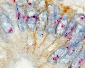
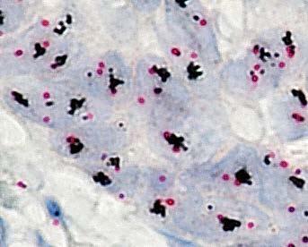
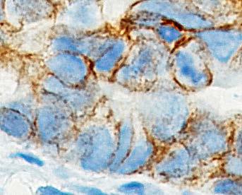
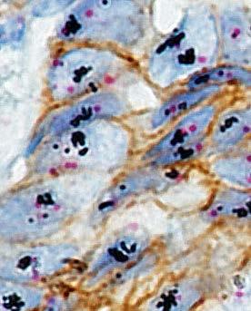
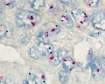
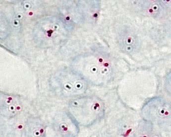
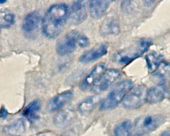
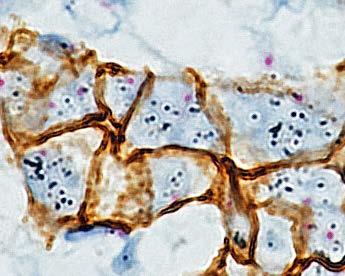
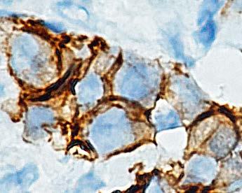
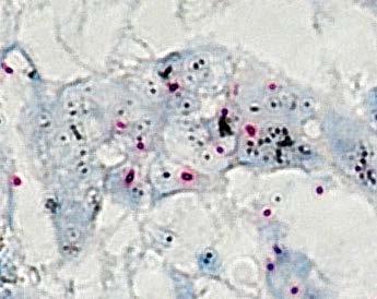
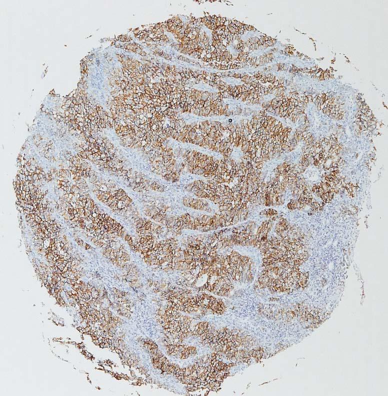
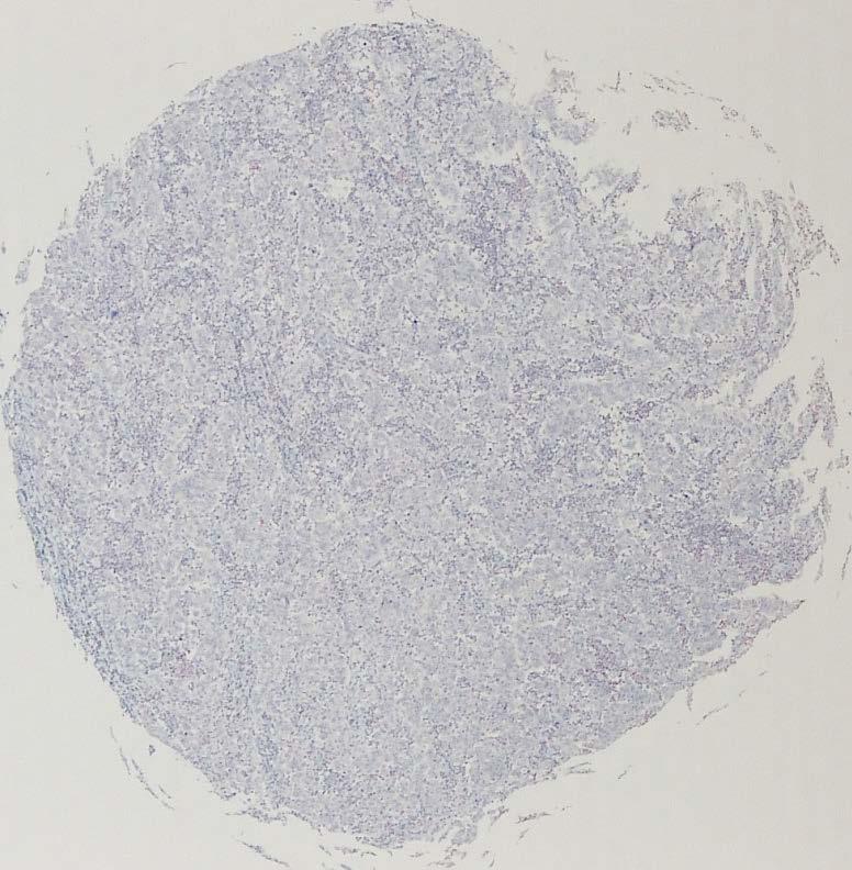
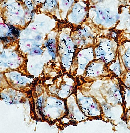
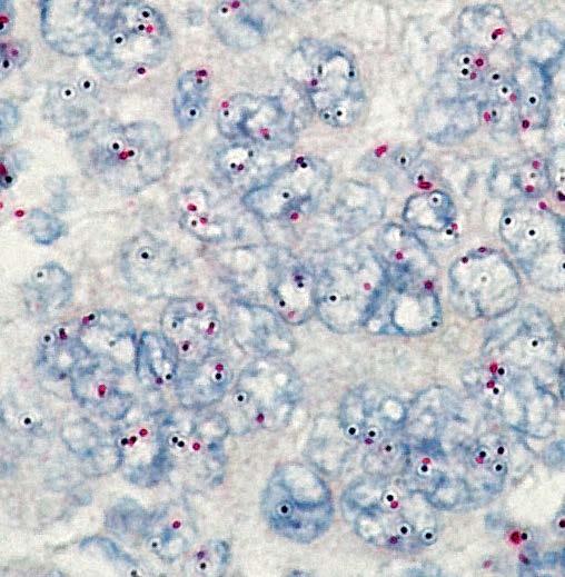
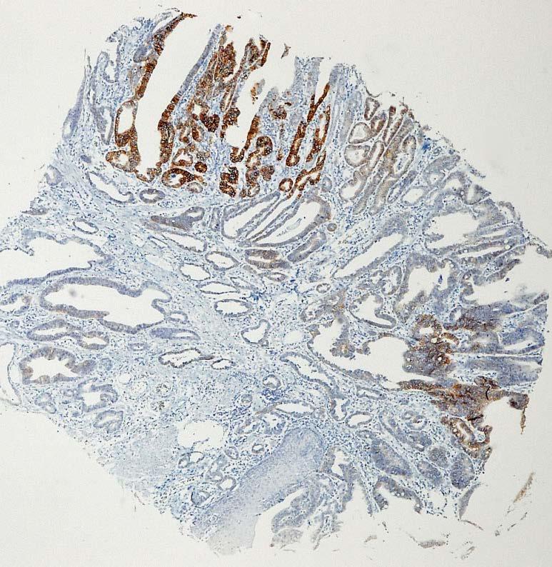
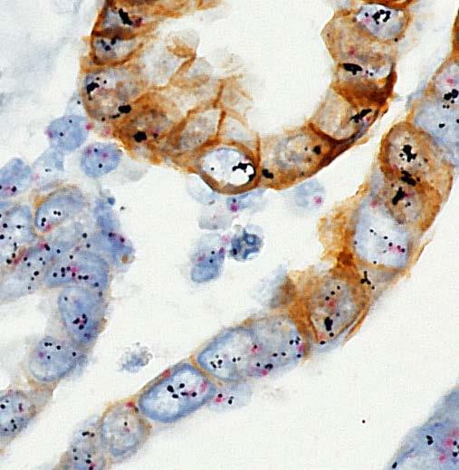
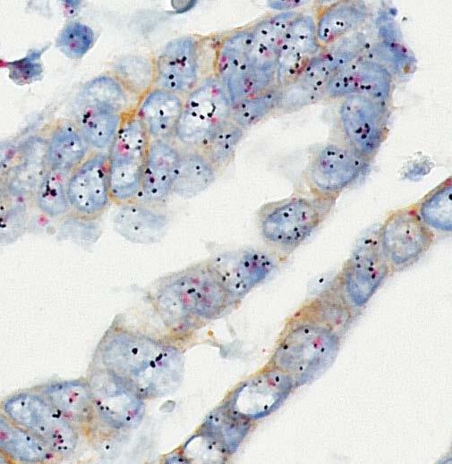
21 Acknowledgments We would like to thank Drs. Eric Walk and Ruediger Ridder, Medical and Scientific Affairs, Ventana Medical Systems, Inc., for their pro bono review of this manuscript. Figure legends Fig.1 (a-l) Immunohistochemical (a,d,g,j), dual-color in situ hybridization (b,e,h,k) and gene-protein assay (c,f,i,l) staining examples from tissue microarray samples. (a-c) HER2 immunohistochemistry (IHC) 0 case without gene amplification. (d-f) IHC 2+ case without gene amplification. (g-i) IHC 2+ case with gene amplification. (j-l) IHC 3+ case with gene amplification. Fig.2 (a-d) Two TMA cores (A and B) obtained from the same lesion demonstrate intratumoral phenotypic and genotypic heterogeneity. (a,c) Core A was immunohistochemistry (IHC) 3+ with gene amplification. (b,d) Core B was IHC 0 without gene amplification. (c,d 60x.) (e-g) Intra-core phenotypic heterogeneity. (e) Heterogeneity of HER2 protein overexpression within one TMA core. (f,g) Areas with 21
22 different immunohistochemistry (IHC) scores were observed at the cell-to-cell level. IHC 3+ and IHC 1+/0 area (f) and IHC negative area (g). Homogeneous gene amplification was observed in spite of heterogeneous protein overexpression. (f,g 60x.) References 1. Hofmann M, Stoss O, Shi D, Buttner R, van de Vijver M, Kim W, et al. Assessment of a HER2 scoring system for gastric cancer: results from a validation study. Histopathology. 2008;52(7): Yano T, Doi T, Ohtsu A, Boku N, Hashizume K, Nakanishi M, et al. Comparison of HER2 gene amplification assessed by fluorescence in situ hybridization and HER2 protein expression assessed by immunohistochemistry in gastric cancer. Oncol Rep. 2006;15(1): Tanner M, Hollmen M, Junttila TT, Kapanen AI, Tommola S, Soini Y, et al. Amplification of HER-2 in gastric carcinoma: association with Topoisomerase IIalpha gene amplification, intestinal type, poor prognosis and sensitivity to trastuzumab. Ann Oncol. 2005;16(2): Slamon DJ, Leyland-Jones B, Shak S, Fuchs H, Paton V, Bajamonde A, et al. Use of chemotherapy plus a monoclonal antibody against HER2 for metastatic breast cancer that overexpresses HER2. N Engl J Med. 2001;344(11):
23 5. Bang YJ, Van Cutsem E, Feyereislova A, Chung HC, Shen L, Sawaki A, et al. Trastuzumab in combination with chemotherapy versus chemotherapy alone for treatment of HER2-positive advanced gastric or gastro-oesophageal junction cancer (ToGA): a phase 3, open-label, randomised controlled trial. Lancet. 2010;376(9742): Wainberg ZA, Anghel A, Desai AJ, Ayala R, Luo T, Safran B, et al. Lapatinib, a dual EGFR and HER2 kinase inhibitor, selectively inhibits HER2-amplified human gastric cancer cells and is synergistic with trastuzumab in vitro and in vivo. Clin Cancer Res. 2010;16(5): Yamashita-Kashima Y, Iijima S, Yorozu K, Furugaki K, Kurasawa M, Ohta M, et al. Pertuzumab in combination with trastuzumab shows significantly enhanced antitumor activity in HER2-positive human gastric cancer xenograft models. Clin Cancer Res. 2011;17(15): Orphanos G, Kountourakis P. Targeting the HER2 receptor in metastatic breast cancer. Hematol Oncol Stem Cell Ther. 2012;5(3): Marx AH, Tharun L, Muth J, Dancau AM, Simon R, Yekebas E, et al. HER-2 amplification is highly homogenous in gastric cancer. Hum Pathol. 2009;40(6): Lee HE, Park KU, Yoo SB, Nam SK, Park do J, Kim HH, et al. Clinical significance of intratumoral HER2 heterogeneity in gastric cancer. Eur J Cancer. 2013;49(6): Cho EY, Park K, Do I, Cho J, Kim J, Lee J, et al. Heterogeneity of ERBB2 in gastric 23
24 carcinomas: a study of tissue microarray and matched primary and metastatic carcinomas. Mod Pathol. 2013;26(5): Nitta H, Kelly BD, Padilla M, Wick N, Brunhoeber P, Bai I, et al. A gene-protein assay for human epidermal growth factor receptor 2 (HER2): brightfield tricolor visualization of HER2 protein, the HER2 gene, and chromosome 17 centromere (CEN17) in formalin-fixed, paraffin-embedded breast cancer tissue sections. Diagn Pathol. 2012;7: Aizawa M, Nagatsuma AK, Kitada K, Kuwata T, Fujii S, Kinoshita T, et al. Evaluation of HER2-based biology in 1,006 cases of gastric cancer in a Japanese population. Gastric Cancer. 2014;17(1): Albarello L, Pecciarini L, Doglioni C. HER2 testing in gastric cancer. Adv Anat Pathol. 2011;18(1): Tubbs R, Pettay J, Hicks D, Skacel M, Powell R, Grogan T, et al. Novel bright field molecular morphology methods for detection of HER2 gene amplification. J Mol Histol. 2004;35(6): Downs-Kelly E, Pettay J, Hicks D, Skacel M, Yoder B, Rybicki L, et al. Analytical validation and interobserver reproducibility of EnzMet GenePro: a second-generation bright-field metallography assay for concomitant detection of HER2 gene status and protein expression in 24
25 invasive carcinoma of the breast. Am J Surg Pathol. 2005;29(11): Reisenbichler ES, Horton D, Rasco M, Andea A, Hameed O. Evaluation of dual immunohistochemistry and chromogenic in situ hybridization for HER2 on a single section. Am J Clin Pathol. 2012;137(1): Hirschmann A, Lamb TA, Marchal G, Padilla M, Diebold J. Simultaneous analysis of HER2 gene and protein on a single slide facilitates HER2 testing of breast and gastric carcinomas. Am J Clin Pathol. 2012;138(6): Vance GH, Barry TS, Bloom KJ, Fitzgibbons PL, Hicks DG, Jenkins RB, et al. Genetic heterogeneity in HER2 testing in breast cancer: panel summary and guidelines. Arch Pathol Lab Med. 2009;133(4): Wolff AC, Hammond ME, Hicks DG, Dowsett M, McShane LM, Allison KH, et al. Recommendations for human epidermal growth factor receptor 2 testing in breast cancer: American Society of Clinical Oncology/College of American Pathologists clinical practice guideline update. J Clin Oncol. 2013;31(31): Yang J, Luo H, Li Y, Li J, Cai Z, Su X, et al. Intratumoral heterogeneity determines discordant results of diagnostic tests for human epidermal growth factor receptor (HER) 2 in gastric cancer specimens. Cell Biochem Biophys. 2012;62(1):
26 22. Kim KC, Koh YW, Chang HM, Kim TH, Yook JH, Kim BS, et al. Evaluation of HER2 protein expression in gastric carcinomas: comparative analysis of 1,414 cases of whole-tissue sections and 595 cases of tissue microarrays. Ann Surg Oncol. 2011;18(10): Grabsch H, Sivakumar S, Gray S, Gabbert HE, Muller W. HER2 expression in gastric cancer: Rare, heterogeneous and of no prognostic value - conclusions from 924 cases of two independent series. Cell Oncol. 2010;32(1-2): Park DI, Yun JW, Park JH, Oh SJ, Kim HJ, Cho YK, et al. HER-2/neu amplification is an independent prognostic factor in gastric cancer. Dig Dis Sci. 2006;51(8): Kim MA, Jung EJ, Lee HS, Lee HE, Jeon YK, Yang HK, et al. Evaluation of HER-2 gene status in gastric carcinoma using immunohistochemistry, fluorescence in situ hybridization, and real-time quantitative polymerase chain reaction. Hum Pathol. 2007;38(9): Yokozaki H, Kuniyasu H, Kitadai Y, Nishimura K, Todo H, Ayhan A, et al. p53 point mutations in primary human gastric carcinomas. J Cancer Res Clin Oncol. 1992;119(2): Kataoka Y, Okabe H, Yoshizawa A, Minamiguchi S, Yoshimura K, Haga H, et al. HER2 expression and its clinicopathological features in resectable gastric cancer. Gastric Cancer. 2013;16(1):


27 IHC DISH GPA a b c d e f g h i j k l
28 a b c d
29 e f g
30 Table 1 Concordance between HER2 IHC score and GPA IHC score on 1750 cores of 875 cases IHC score GPA IHC score Total (%) (82.5) (6.1) (6.2) (5.2) Total (%) 1445 (82.6) 105 (6.0) 112 (6.4) 88 (5.0) 1750 cores HER2: human epidermal grows factor receptor 2; IHC: immunohistochemistry; GPA: gene-protein assay
31 Table 2 Concordance of HER2 status (amplified/non-amplified) between DISH and GPA DISH on 1750 cores of 875 cases DISH GPA DISH Non-amplified Amplified Total (%) Non-amplified (90.2) Amplified (9.3) Not determined (0.5) Total (%) 1583 (90.5) 167 (9.5) 1750 cores HER2: human epidermal grows factor receptor 2; DISH: dual-color in situ hybridization; GPA: gene-protein assay
32 Table 3 Concordance of HER2 final status (positive/negative)* between single IHC/DISH and GPA on 875 cases IHC/DISH GPA IHC/DISH Negative Positive Total (%) Negative (88.8) Positive (11.2) Total (%) 779 (89.0) 96 (11.0) 875 cases * HER2 IHC score of 3+ and/or HER2 gene amplification were defined as HER2 positive [5] HER2: human epidermal grows factor receptor 2; IHC: immunohistochemistry; DISH: dual-color in situ hybridization; GPA: gene-protein assay
33 Table 4 Concordance of GPA IHC score between two cores on 875 cases Core B Core A Total Total cases Two cores (Core A and Core B) were obtained form same lesion GPA: gene-protein assay; IHC: immunohistochemistry
34 Table 5 Concordance of HER2 gene amplification by GPA between two cores on 875 cases core B core A Non-amplified Amplified Not determined Total Non-amplified Amplified Not determined Total cases Two cores (Core A and Core B) were obtained form same lesion GPA: gene-protein assay; DISH: dual-color in situ hybridization
35 Table 1S Clnicopathological and treatment-related characteristics of the 875 cases Characteristics Age (years, mean ± SD) 62.9 ± 10.9 Gender, n (%) male 591 (67.5) female 284 (32.5) Histological feature, n (%) Papillary 22 (2.5) Tubular 402 (45.9) Poorly differentiated / Signet ring cell 432 (49.4) Mucinous 19 (2.2) Tumor location, n (%) Esophageal junction 29 (3.3) Proximal third of stomach 184 (21.0) Middle third of stomach 406 (46.4) Distal third of stomach 256 (29.3) Macroscopic type, n (%) Type (50.3) Type 1 21 (2.4)
36 Type (12.0) Type (25.5) Type 4 70 (8.0) Type 5 16 (1.8) pt Stage, n (%) T1 426 (48.7) T2 106 (12.1) T3 206 (23.5) T4 137 (15.7) pn Stage, n (%) N0 520 (59.4) N (40.5) Nx 1 (0.1) ptnm stage, n (%) Stage I 457 (52.2) Stage II 188 (21.5) Stage III 159 (18.2) Stage IV 71 (8.1) Resection margin, n (%) R0 821 (93.8) R (6.2)
37 Neo-adjuvant chemotherapy, n (%) Present 46 (5.3) Absent 829 (94.7) Adjuvant chemotherapy, n (%) Present 64 (7.3) Absent 811 (92.7)
38 Table 2S Concordance of HER2 final status (positive/negative)* between single IHC/DISH and GPA on 875 cases IHC/DISH GPA IHC/DISH Negative Positive Total (%) Negative (90.5) Positive (9.5) Total (%) 791 (90.4) 84 (9.6) 875 cases * HER2 IHC score of 3+ or IHC score of 2+ with HER2 gene amplification were defined as HER2 positive [14] HER2: human epidermal grows factor receptor 2; IHC: immunohistochemistry; DISH: dual-color in situ hybridization; GPA: gene-protein assay
39 Table 3S IHC score and HER2 gene amplification by IHC and DISH on 875 cases IHC DISH Total (%) Non-amplified (89.0) Amplified (11.0) Total (%) 681 (77.8) 67 (7.7) 76 (8.7) 51 (5.8) 875 cases HER2: human epidermal grows factor receptor 2; IHC: immunohistochemistry; DISH: dual-color in situ hybridization
40 Table 4S Association between intratumoral heterogeneity of HER2 protein overexpression and gene amplification by comparing two cores GPA IHC GPA DISH Negative score Heterogeneous Homogeneous total (0/1+) overexpression overexpression No amplification Heterogeneous amplification Homogeneous amplification Not determined total cases HER2: human epidermal grows factor receptor 2; GPA: gene-protein assay; IHC: immunohistochemistry; DISH: dual-color in situ hybridization
41 Table 5S Cases with intratumoral heterogeneity of HER2 protein overexpression or gene amplification core A core B Case No. GPA IHC GPA DISH GPA IHC GPA DISH 1 3+ A 2+ A 2 3+ A 2+ A 3 3+ A 2+ A 4 3+ A 2+ A 5 3+ A 2+ A 6 3+ A 2+ A 7 3+ A 2+ A 8 3+ A A A A A A A A A 2+ -
42 17 2+ A A 1+ A A 1+ A A 1+ A A 1+ A
43 A A A A A A A A
44 A A A A A A A 0 - Two cores (Core A and Core B) were obtained form same lesion HER2: human epidermal grows factor receptor 2; IHC: immunohistochemistry; DISH: dual-color in situ hybridization; GPA: gene-protein assay; A: amplified; -: non-amplified
45 Table 6S Concordance of HER2 final status (positive/negative) by GPA between two cores on 875 cases core B core A Negative Positive Total Negative Positive Total cases Two cores (Core A and Core B) were obtained form same lesion HER2: human epidermal grows factor receptor 2; GPA: gene-protein assay; IHC: immunohistochemistry; DISH: dual-color in situ hybridization
46 Table 7S Patient characteristics of the 875 cases according to intratumoral heterogeneity of HER2 protein overexpression by gene-protein assay Characteristics HER2 protein expression p a negative positive positive heterogeneous homogeneous Age (years, mean ± SD) 62.3 ± ± ± Gender, n male female Histological feature, n 0.63 b Well (papillary, tubular) Poorly (poorly, signet cell) Others Tumor location, n 1.00 Esophageal junction Stomach Macroscopic type, n 0.72 c Type
47 Type 1, Type 3, Type pt Stage, n 0.12 d T T T T pn Stage, n 0.12 e N N Nx ptnm stage, n 0.15 f Stage I Stage II Stage III Stage IV a comparison between cases of heterogeneous and homogeneous protein overexpression b well versus poorly c Type 0 versus Type1-4
48 d T1-2 versus T3-4 e N0 versus N1-3 f Stage I-II versus Stage III-IV HER2: human epidermal grows factor receptor 2
49 Table 8S Patient characteristics of the evaluable 870 cases according to intratumoral heterogeneity of HER2 gene amplification by gene-protein assay Characteristics HER2 gene amplification p a negative positive positive heterogeneous homogeneous Age (years, mean ± SD) 62.6 ± ± ± Gender, n 0.76 male female Histological feature, n b Well (papillary, tubular) Poorly (poorly, signet cell) Others Tumor location, n 1.00 Esophageal junction Stomach Macroscopic type, n 0.48 c Type
50 Type 1, Type 3, Type pt Stage, n 0.62 d T T T T pn Stage, n 0.57 e N N Nx ptnm stage, n 0.45 f Stage I Stage II Stage III Stage IV a comparison between cases of heterogeneous and homogeneous protein overexpression b well versus poorly, X2 test c Type 0 versus Type1-4
51 d T1-2 versus T3-4 e N0 versus N1-3 f Stage I-II versus Stage III-IV HER2: human epidermal grows factor receptor 2
HER2 Gene Protein Assay Is Useful to Determine HER2 Status and Evaluate HER2 Heterogeneity in HER2 Equivocal Breast Cancer
 HER2 Gene Protein Assay Is Useful to Determine HER2 Status and Evaluate HER2 Heterogeneity in HER2 Equivocal Breast Cancer Yanjun Hou, MD, PhD, 1 Hiroaki Nitta, PhD, 2 and Zaibo Li, MD, PhD 1 From the
Editorial New guidelines for HER2 pathological diagnostics in gastric cancer
 bs_bs_banner Pathology International 2016; 66: 57 62 doi:10.1111/pin.12390 Editorial New guidelines for HER2 pathological diagnostics in gastric cancer Ryo Wada, 1 Kenichi Hirabayashi, 2 Nobuyuki Ohike
Nitta et al. Diagnostic Pathology 2012, 7:60
 Nitta et al. Diagnostic Pathology 2012, 7:60 METHODOLOGY Open Access A gene-protein assay for human epidermal growth factor receptor 2 (HER2): brightfield tricolor visualization of HER2 protein, the HER2
Dr. dr. Primariadewi R, SpPA(K)
") Curriculum Vitae Dr. dr. Primariadewi R, SpPA(K) Education : Medical Doctor from UKRIDA Doctoral Degree from Faculty of Medicine University of Indonesia Pathologist Specialist and Consultant from Faculty
Curriculum Vitae Dr. dr. Primariadewi R, SpPA(K) Education : Medical Doctor from UKRIDA Doctoral Degree from Faculty of Medicine University of Indonesia Pathologist Specialist and Consultant from Faculty
HER2 Status and Its Heterogeneity in Gastric Carcinoma of Vietnamese Patient
 Journal of Pathology and Translational Medicine 2017; 51: 396-402 ORIGINAL ARTICLE HER2 Status and Its Heterogeneity in Gastric Carcinoma of Vietnamese Patient Dang Anh Thu Phan Vu Thien Nguyen Thi Ngoc
HER-2/neu Analysis in Gastroesophageal and Gastric Adenocarcinoma Keith Daniel Bohman, MD
 HER-2/neu Analysis in Gastroesophageal and Gastric Adenocarcinoma Keith Daniel Bohman, MD Carcinoma of the stomach is the fourth most common cancer and second most frequent cause of cancer-related mortality
Expression of human epidermal growth factor receptor 2 in primary and paired parenchymal recurrent and/or metastatic sites of gastric cancer
 MOLECULAR AND CLINICAL ONCOLOGY 2: 751-755 Expression of human epidermal growth factor receptor 2 in primary and paired parenchymal recurrent and/or metastatic sites of gastric cancer RYOSUKE SHIBATA 1,
Kristen E. Muller, DO, Jonathan D. Marotti, MD, Vincent A. Memoli, MD, Wendy A. Wells, MD, and Laura J. Tafe, MD
 AJCP / Original Article Impact of the 2013 ASCO/CAP HER2 Guideline Updates at an Academic Medical Center That Performs Primary HER2 FISH Testing Increase in Equivocal Results and Utility of Reflex Immunohistochemistry
MEDICAL POLICY. Proprietary Information of YourCare Health Plan
 MEDICAL POLICY SUBJECT: HER-2 TESTING IN INVASIVE BREAST OR PAGE: 1 OF: 7 If the member's subscriber contract excludes coverage for a specific service it is not covered under that contract. In such cases,
Journal of Breast Cancer
 ORIGINAL ARTICLE Journal of Breast Cancer J Breast Cancer 2009 December; 12(4): 235-40 DOI: 10.4048/jbc.2009.12.4.235 Comparison of Silver-Enhanced in situ Hybridization and Fluorescence in situ Hybridization
Molecular Probes Introducing 14 new probes
 Molecular Probes Introducing 14 new probes Gene and Chromosome Probes Dual Colour ISH INFORM HER2 Dual ISH DNA Probe Cocktail Assay Product Part Number INFORM HER2 Dual ISH DNA Probe Cocktail 800-4422
HER-2/neu Marker Examination using Immunohistochemical Method in Patients Suffering from Gastric Adenocarcinoma
 IJMCM Autumn 203, Vol 2, No 4 Original Article HER-2/neu Marker Examination using Immunohistochemical Method in Patients Suffering from Gastric Adenocarcinoma Kourosh Movagharnejad, Majid Sharbatdaran,
RESEARCH ARTICLE. Fatih Selcukbiricik 1 *, Sibel Erdamar 2, Evin Buyukunal 3, Suheyla Serrdengecti 3, Fuat Demirelli 3. Abstract.
 DOI:http://dx.doi.org/10.731/APJCP.201.15.2.10607 RESEARCH ARTICLE Is Her-2 Status in the Primary Tumor Correlated with Matched Lymph Node Metastases in Patients with Gastric Cancer Undergoing Curative
Reviewer's report. Version: 1 Date: 24 May Reviewer: Cathy Moelans. Reviewer's report:
 Reviewer's report Title: Validation of HER2 testing with core needle biopsy specimens from primary breast cancers in terms of interobserver reproducibility and concordance with surgically resected specimens
Version 2 of these Guidelines were drafted in response to published updated ASCO/CAP HER2 test Guideline Recommendations-
 Introduction: These guidelines represent systematically developed statements to assist in the provision of quality assured HER2 testing in breast and gastric/ gastro-oesophageal carcinoma. They are based
ONCOLOGY LETTERS 5: , 2013
 ONCOLOGY LETTERS 5: 559-563, 2013 The incidence and prognostic value of HER2 overexpression and cyclin D1 expression in patients with gastric or gastroesophageal junction adenocarcinoma in Israel GIL BAR-SELA
MEDICAL POLICY. Proprietary Information of Excellus Health Plan, Inc. A nonprofit independent licensee of the BlueCross BlueShield Association
 MEDICAL POLICY SUBJECT: HER-2 TESTING IN INVASIVE BREAST OR PAGE: 1 OF: 7 If a product excludes coverage for a service, it is not covered, and medical policy criteria do not apply. If a commercial product,
HER2 status assessment in breast cancer. Marc van de Vijver Academic Medical Centre (AMC), Amsterdam
, Amsterdam") HER2 status assessment in breast cancer Marc van de Vijver Academic Medical Centre (AMC), Amsterdam 13e Bossche Mamma Congres 17 th June 2015 Modern cancer therapies are based on sophisticated molecular
HER2 status assessment in breast cancer Marc van de Vijver Academic Medical Centre (AMC), Amsterdam 13e Bossche Mamma Congres 17 th June 2015 Modern cancer therapies are based on sophisticated molecular
Review Article The assessment of HER2 status in breast cancer: the past, the present, and the future
 Pathology International 2016; 66: 313 324 doi:10.1111/pin.12407 Review Article The assessment of HER2 status in breast cancer: the past, the present, and the future Hiroaki Nitta, 1 Brian D. Kelly, 2 Craig
Priti Lal, MD, 1 Paulo A. Salazar, 1 Clifford A. Hudis, MD, 2 Marc Ladanyi, MD, 1 and Beiyun Chen, MD, PhD 1. Abstract
 Anatomic Pathology / DUAL- VS SINGLE-COLOR SCORING IN IMMUNOHISTOCHEMICAL AND FISH HER-2 TESTING HER-2 Testing in Breast Cancer Using Immunohistochemical Analysis and Fluorescence In Situ Hybridization
Quantitative Image Analysis of HER2 Immunohistochemistry for Breast Cancer
 Quantitative Image Analysis of HER2 Immunohistochemistry for Breast Cancer Guideline from the College of American Pathologists Early Online Release Publication: Archives of Pathology & Laboratory Medicine
SATOSHI IKEDA. Department of Pathology, Tsuchiura Kyodo General Hospital, Tsuchiura, Ibaraki , Japan
 1084 Novel and simple method of double detection using fluorescence in situ hybridization and fluorescence immunostaining of formalin fixed paraffin embedded tissue sections SATOSHI IKEDA Department of
Incidence of brain metastases in HER2+ gastric or gastroesophageal junction adenocarcinoma.
 Incidence of brain metastases in HER2+ gastric or gastroesophageal junction adenocarcinoma. Christophe Blay, Dan Cristian Chiforeanu, Eveline Boucher, Florian Cabillic, Romain Desgrippes, Bérengère Leconte,
Targeting the Oncogenic Pathway as Opposed to the Primary Tumor Site: HER2 as an Example
 Targeting the Oncogenic Pathway as Opposed to the Primary Tumor Site: HER2 as an Example Dennis J Slamon, MD, PhD Professor of Medicine Chief, Division of Hematology/Oncology; Director of Clinical/Translational
Resident Short Review. HER2/neu Gene Amplification and Protein Overexpression in Gastric and Gastroesophageal Junction Adenocarcinoma
 Resident Short Review HER2/neu Gene Amplification and Protein Overexpression in Gastric and Gastroesophageal Junction Adenocarcinoma A Review of Histopathology, Diagnostic Testing, and Clinical Implications
Correlation of Lauren s histological type and expression of E-cadherin and HER-2/ neu in gastric adenocarcinoma
 IJPLM. 2016;2(1):OA2 ORIGINAL ARTICLES Correlation of Lauren s histological type and expression of E-cadherin and HER-2/ neu in gastric adenocarcinoma Khushboo Dewan 1*, Renu Madan 2, P Sengupta 3 1, 2,
HER2 CISH pharmdx TM Kit Interpretation Guide Breast Cancer
 P A T H O L O G Y HER2 CISH pharmdx TM Kit Interpretation Guide Breast Cancer FROM CERTAINTY COMES TRUST For in vitro diagnostic use HER2 CISH pharmdx Kit HER2 CISH pharmdx Kit is intended for dual-color
Three Hours Thirty Minutes
 INTERPRETATION HER2 IQFISH pharmdx TM Interpretation Guide Three Hours Thirty Minutes it s about time Breast carcinoma (FFPE) stained with HER2 IQFISH pharmdx Gastric cancer (FFPE) stained with HER2 IQFISH
CANCER. Clinical Validation of Breast Cancer Predictive Markers
 Clinical Validation of Breast Cancer Predictive Markers David Hicks, MD Loralee McMahon, MS, HTL(ASCP) CANCER The human body is composed of billions of highly regulated cells Cancer cells no longer respond
HER2 ISH (BRISH or FISH)
") Assessment Run H14 2018 HER2 ISH (BRISH or FISH) Material Table 1. Content of the multi-block used for the NordiQC HER2 ISH assessment, run H14 HER2 IHC* IHC score Dual - SISH** FISH*** FISH*** HER2/chr17
Assessment Run H14 2018 HER2 ISH (BRISH or FISH) Material Table 1. Content of the multi-block used for the NordiQC HER2 ISH assessment, run H14 HER2 IHC* IHC score Dual - SISH** FISH*** FISH*** HER2/chr17
IT S ABOUT TIME. IQFISH pharmdx Interpretation Guide THREEHOURSTHIRTYMINUTES. HER2 IQFISH pharmdxtm. TOP2A IQFISH pharmdxtm
 I N T E R P R E TAT I O N IQFISH pharmdx Interpretation Guide TM HER2 IQFISH pharmdxtm TOP2A IQFISH pharmdxtm Breast carcinoma (FFPE) stained with HER2 IQFISH pharmdx Breast carcinoma (FFPE) stained with
HER2 FISH pharmdx TM Interpretation Guide - Breast Cancer
 P A T H O L O G Y HER2 FISH pharmdx TM Interpretation Guide - Breast Cancer For In Vitro Diagnostic Use FDA approved as an aid in the assessment of patients for whom Herceptin TM (trastuzumab) treatment
Original Article. Jennifer Jeung, MD; Roshan Patel, MD; Lizette Vila, MD; Dara Wakefield, MD; Chen Liu, MD, PhD
 Original Article Quantitation of HER2/neu Expression in Primary Gastroesophageal Adenocarcinomas Using Conventional Light Microscopy and Quantitative Image Analysis Jennifer Jeung, MD; Roshan Patel, MD;
Genetic heterogeneity in HER2/neu testing by fluorescence in situ hybridization: a study of 2522 cases
 Modern Pathology () 5, 683 688 & USCAP, Inc. All rights reserved 893-395/ $3. 683 Genetic heterogeneity in HER/neu testing by fluorescence in situ hybridization: a study of 5 cases Martin C Chang,,3, Janet
2017 OPTIONS FOR INDIVIDUAL MEASURES: REGISTRY ONLY. MEASURE TYPE: Process
 Measure #449 (NQF 1857): HER2 Negative or Undocumented Breast Cancer Patients Spared Treatment with HER2-Targeted Therapies National Quality Strategy Domain: Efficiency and Cost Reduction 2017 OPTIONS
Optimal algorithm for HER2 testing
 Optimal algorithm for HER2 testing The revised definition of IHC 2+ (equivocal) is invasive breast cancer with Weak to moderate complete membrane staining observed in >10% of tumor cells. (see Figure 1
RESEARCH ARTICLE. Immuno-Histochemical Assessment of HER2NEU Expression in Gastric Adenocarcinoma in North Karnataka, India
 DOI:10.22034/APJCP.2018.19.5.1381 RESEARCH ARTICLE Editorial Process: Submission:01/27/2018 Acceptance:03/29/2018 Immuno-Histochemical Assessment of HER2NEU Expression in Gastric Adenocarcinoma in North
CME/SAM ABSTRACT. AJCP / Original Article
 Clinicopathologic Significance of the Intratumoral Heterogeneity of HER2 Gene Amplification in HER2- Positive Breast Cancer Patients Treated With Adjuvant Trastuzumab Hee Jin Lee, MD, PhD, 1 Joo Young
Association (2013), 16(1): の論文は出版社版でありません 引用の際には出版社版をご確認ご利用ください This is not the publish
, 16(1): の論文は出版社版でありません 引用の際には出版社版をご確認ご利用ください This is not the publish") Title HER2 expression and its clinicopath resectable gastric cancer. Kataoka, Yoshiki; Okabe, Hiroshi; Y Author(s) Minamiguchi, Sachiko; Yoshimura, Ke Sakai, Yoshiharu Gastric cancer : official journal
Title HER2 expression and its clinicopath resectable gastric cancer. Kataoka, Yoshiki; Okabe, Hiroshi; Y Author(s) Minamiguchi, Sachiko; Yoshimura, Ke Sakai, Yoshiharu Gastric cancer : official journal
HER2/neu Amplification in Breast Cancer Stratification by Tumor Type and Grade
 Anatomic Pathology / HER2/NEU AMPLIFICATION IN BREAST CANCER HER2/neu Amplification in Breast Cancer Stratification by Tumor Type and Grade Elise R. Hoff, MD, Raymond R. Tubbs, DO, Jonathan L. Myles, MD,
Importance of confirming HER2 overexpression of recurrence lesion in breast cancer patients
 Breast Cancer (2013) 20:336 341 DOI 10.1007/s12282-012-0341-6 ORIGINAL ARTICLE Importance of confirming HER2 overexpression of recurrence lesion in breast cancer patients Rikiya Nakamura Naohito Yamamoto
It s not a four legged animal anymore. Disclosure
 It s not a four legged animal anymore Parminder Singh, MD Assistant Professor of Medicine Division hematology and oncology No disclosures Disclosure 1 Four legged animal which use the tips of their toes,
Welcome! HER2 TESTING DIAGNOSTIC ACCURACY 4/11/2016
 HER2 TESTING DIAGNOSTIC ACCURACY Can t We Finally Get It Right? Allen M. Gown, M.D. Medical Director and Chief Pathologist PhenoPath Laboratories Seattle, Washington Clinical Professor of Pathology University
Journal of Breast Cancer
 Journal of Breast Cancer ORIGINAL ARTICLE J Breast Cancer 2011 December; 14(4): 276-282 Silver-Enhanced In Situ Hybridization as an Alternative to Fluorescence In Situ Hybridization for Assaying HER2 Amplification
What is HER2 positive breast cancer in 2018? Updated ASCO-CAP guidelines. Giuseppe Viale University of Milan European Institute of Oncology
 What is HER2 positive breast cancer in 2018? Updated ASCO-CAP guidelines Giuseppe Viale University of Milan European Institute of Oncology Mission accomplished! First alarming results Breast Intergroup
Immunohistochemical Expression of Her2/neu in Gastric Carcinomas in Egyptian Patients
 Research Article imedpub Journals www.imedpub.com Journal of Clinical Pathology and Diagnosis Abstract Immunohistochemical Expression of Her2/neu in Gastric Carcinomas in Egyptian Patients Background:
Breast Cancer Interpretation Guide
 Breast Cancer Interpretation Guide UCT D O R P NEW ERBB2/ C E P S ht e ZytoLig lor Prob o C l a u 2D D17S12 ng to the i d r o c c a ting for re-tes idelines 2013 ASCO Gu Breast Cancer Interpretation Guide
Assessment Run B HER2 IHC
 Assessment Run B24 2017 HER2 IHC Material The slide to be stained for HER2 comprised the following 5 materials: IHC: HER2 Score* (0, 1+, 2+, 3+) FISH: HER2 gene/chr 17 ratio** 1. Breast carcinoma, no.
HER2/neu Evaluation of Breast Cancer in 2019
 HER2/neu Evaluation of Breast Cancer in 2019 A.A. Sahin, M.D. Professor of Pathology and Translation Molecular Pathology Section Chief of Breast Pathology ERBB2 (HER2) Background 185-kDa membrane protein
# Best Practices for IHC Detection and Interpretation of ER, PR, and HER2 Protein Overexpression in Breast Cancer
 #1034 - Best Practices for IHC Detection and Interpretation of ER, PR, and HER2 Protein Overexpression in Breast Cancer Richard W. Cartun, MS, PhD Andrew Ricci, Jr, MD Department of Pathology Hartford
FAQs for UK Pathology Departments
 FAQs for UK Pathology Departments This is an educational piece written for Healthcare Professionals FAQs for UK Pathology Departments If you would like to discuss any of the listed FAQs further, or have
HER2 expression in gastric cancer: Rare, heterogeneous and of no prognostic value conclusions from 924 cases of two independent series
 Cellular Oncology 32 (2010) 57 65 57 DOI 10.3233/CLO-2009-0497 IOS Press HER2 expression in gastric cancer: Rare, heterogeneous and of no prognostic value conclusions from 924 cases of two independent
NIH Public Access Author Manuscript Cancer Epidemiol Biomarkers Prev. Author manuscript; available in PMC 2011 January 1.
 NIH Public Access Author Manuscript Published in final edited form as: Cancer Epidemiol Biomarkers Prev. 2010 January ; 19(1): 144 147. doi:10.1158/1055-9965.epi-09-0807. Feasibility Study for Collection
Product Introduction
 Product Introduction Product Codes: HCL026, HCL027 and HCL028 Contents Introduction to HER2 2 HER2 immunohistochemistry 3 Cell lines as controls 5 HER2 Analyte Control DR IHC 7 HER2 Analyte Control DR
T he HER2/neu type 1 tyrosine kinase growth factor
 710 ORIGINAL ARTICLE HER2 amplification status in breast cancer: a comparison between immunohistochemical staining and fluorescence in situ hybridisation using manual and automated quantitative image analysis
2019 COLLECTION TYPE: MIPS CLINICAL QUALITY MEASURES (CQMS) MEASURE TYPE: Process High Priority
 MEASURE TYPE: Process High Priority") Quality ID #449 (NQF 1857): HER2 Negative or Undocumented Breast Cancer Patients Spared Treatment with HER2-Targeted Therapies National Quality Strategy Domain: Efficiency and Cost Reduction Meaningful
Quality ID #449 (NQF 1857): HER2 Negative or Undocumented Breast Cancer Patients Spared Treatment with HER2-Targeted Therapies National Quality Strategy Domain: Efficiency and Cost Reduction Meaningful
Androgen Receptor Expression in Renal Cell Carcinoma: A New Actionable Target?
 Androgen Receptor Expression in Renal Cell Carcinoma: A New Actionable Target? New Frontiers in Urologic Oncology Juan Chipollini, MD Clinical Fellow Department of Genitourinary Oncology Moffitt Cancer
Guideline. Associated Documents ASCO CAP 2018 GUIDELINES and SUPPLEMENTS -
 Guideline Subject: ASCO CAP 2018 HER2 Testing for Breast Cancer Guidelines - Recommendations for Practice in Australasia Approval Date: December 2018 Review Date: December 2022 Review By: HER2 testing
Surgical Pathology Lab of the Future. Thomas M. Grogan, M.D. Professor of Pathology, University of Arizona Founder, Ventana Medical Systems, Inc.
 Surgical Pathology Lab of the Future Thomas M. Grogan, M.D. Professor of Pathology, University of Arizona Founder, Ventana Medical Systems, Inc. 28 April 2010 Objective Demonstrate how the next generation
Detection of Anaplastic Lymphoma Kinase (ALK) gene in Non-Small Cell lung Cancer (NSCLC) By CISH Technique
 gene in Non-Small Cell lung Cancer (NSCLC) By CISH Technique") Cancer and Clinical Oncology; Vol. 7, No. 1; 2018 ISSN 1927-4858 E-ISSN 1927-4866 Published by Canadian Center of Science and Education Detection of Anaplastic Lymphoma Kinase (ALK) gene in Non-Small Cell
Cancer and Clinical Oncology; Vol. 7, No. 1; 2018 ISSN 1927-4858 E-ISSN 1927-4866 Published by Canadian Center of Science and Education Detection of Anaplastic Lymphoma Kinase (ALK) gene in Non-Small Cell
Minimum biopsy set for HER2 evaluation in gastric and gastro-esophageal junction cancer
 THIEME Original article Minimum biopsy set for HER2 evaluation in gastric and gastro-esophageal junction cancer Authors Institutions Irene Gullo 1, *, Federica Grillo 1, *, Luca Molinaro 2, Matteo Fassan
Data Supplement 1: 2013 Update Rationale and Background Information
 Recommendations for Human Epidermal Growth Factor Receptor 2 Testing in Breast Cancer: American Society of Clinical Oncology /College of American Pathologists Clinical Practice Guideline Update (edited
Supplementary Online Content
 Supplementary Online Content Fumagalli D, Venet D, Ignatiadis M, et al. RNA Sequencing to predict response to neoadjuvant anti-her2 therapy: a secondary analysis of the NeoALTTO randomized clinical trial.
EARLY ONLINE RELEASE
 EARLY ONLINE RELEASE Note: This article was posted on the Archives Web site as an Early Online Release. Early Online Release articles have been peer reviewed, copyedited, and reviewed by the authors. Additional
Assessment Run B HER2 IHC
 Assessment Run B26 208 HER2 IHC Material The slide to be stained for HER2 comprised the following 5 materials: IHC: HER2 Score* (0, +, 2+, 3+) FISH: HER2 gene/chr 7 ratio**. Breast carcinoma, no. 2+..3
Comparison of Immunohistochemical and Fluorescence In Situ Hybridization Assessment of HER-2 Status in Routine Practice
 Anatomic Pathology / ASSESSMENT OF HER-2 STATUS Comparison of Immunohistochemical and Fluorescence In Situ Hybridization Assessment of HER-2 Status in Routine Practice Michelle Dolan, MD, 1 and Dale Snover,
Droplet digital PCR using HER2/EIF2C1 ratio for detection of HER2 amplification in breast cancer tissues
 https://doi.org/10.1007/s12032-018-1210-8 ORIGINAL PAPER Droplet digital PCR using HER2/EIF2C1 ratio for detection of HER2 amplification in breast cancer tissues Anchalee Tantiwetrueangdet 1 Ravat Panvichian
Considerable advances in the therapy of breast cancer
 HER-2/neu Status in Breast Cancer Metastases to the Central Nervous System Kelly C. Lear-Kaul, MD; Hye-Ryoung Yoon, MD; Bette K. Kleinschmidt-DeMasters, MD; Loris McGavran, PhD; Meenakshi Singh, MD Context.
Received 04 November 2008; Accepted in revision 09 January 2009; Available online 20 January 2009
 Int J Clin Exp Pathol (2009) 2, 476-480 www.ijcep.com/ijcep811001 Original Article Immunohistochemical Detection of Estrogen and Progesterone Receptor and HER2 Expression in Breast Carcinomas: Comparison
CME/SAM. Abstract. Anatomic Pathology / HER2/neu Results in Breast Cancer
 Anatomic Pathology / HER2/neu Results in Breast Cancer Effect of Ischemic Time, Fixation Time, and Fixative Type on HER2/neu Immunohistochemical and Fluorescence In Situ Hybridization Results in Breast
Prediction of HER2 gene status in Her2 2 þ invasive breast cancer: a study of 108 cases comparing ASCO/CAP and FDA recommendations
 & 2009 USCAP, Inc All rights reserved 0893-3952/09 $32.00 www.modernpathology.org Prediction of HER2 gene status in Her2 2 þ invasive breast cancer: a study of 108 cases comparing ASCO/CAP and FDA recommendations
Template for Reporting Results of Biomarker Testing of Specimens From Patients With Carcinoma of the Breast
 Template for Reporting Results of Biomarker Testing of Specimens From Patients With Carcinoma of the Breast Version: Template Posting Date: January 2018 Includes requirements from the 2017 CAP Accreditation
A Study Comparing Conventional Brightfield Microscopy, Image Analysis-Assisted Microscopy, and Interobserver Variation
 Effects of the Change in Cutoff Values for Human Epidermal Growth Factor Receptor 2 Status by Immunohistochemistry and Fluorescence In Situ Hybridization A Study Comparing Conventional Brightfield Microscopy,
Assessment Run B HER-2 IHC. HER-2/chr17 ratio**
 Assessment Run B2 20 HER-2 IHC Material The slide to be stained for HER-2 comprised the following 5 tissues: IHC HER-2 Score* (0, +, 2+,3+) FISH HER-2/chr7 ratio**. Breast ductal carcinoma 0..3 2. Breast
Immunohistochemical classification of breast tumours
 Immunohistochemical classification of breast tumours Workshop in Diagnostic Immunohistochemistry September 19 th - 21 th 2018 Anne-Vibeke Lænkholm Department of Surgical Pathology, Zealand University Hospital,
Intratumoral Heterogeneity in Breast Cancer: A Case Report and Molecular Discussion
 Original Case Report Article Middle East Journal of Cancer; July October 20152018; 6(3): 9(4): 339-343 Intratumoral Heterogeneity in Breast Cancer: A Case Report and Molecular Discussion Akbar Safaei,
Introduction. The HER2 Testing Expert Panel has identified five Clinical Questions that form the core of this Focused Update.
 Human Epidermal Growth Factor Receptor 2 Testing in Breast Cancer: American Society of Clinical Oncology/ College of American Pathologists Clinical Practice Guideline Focused Update Wolff, et al. Introduction
Assessment of Breast Cancer with Borderline HER2 Status Using MIP Microarray
 Assessment of Breast Cancer with Borderline HER2 Status Using MIP Microarray Hui Chen, Aysegul A Sahin, Xinyan Lu, Lei Huo, Rajesh R Singh, Ronald Abraham, Shumaila Virani, Bal Mukund Mishra, Russell Broaddus,
First released in 2007 and updated in 2013, the recommendations
 Special Article Human Epidermal Growth Factor Receptor 2 Testing in Breast Cancer American Society of Clinical Oncology/College of American Pathologists Clinical Practice Guideline Focused Update Antonio
On May 4 and 5, 2002, the College of American Pathologists
 College of American Pathologists Conference Conference Summary, Strategic Science Symposium Her-2/neu Testing of Breast Cancer Patients in Clinical Practice Richard J. Zarbo, MD, DMD; M. Elizabeth H. Hammond,
A Retrospective Analysis of Clinical Utility of AJCC 8th Edition Cancer Staging System for Breast Cancer
 Elmer ress Original Article World J Oncol. 2017;8(3):71-75 A Retrospective Analysis of Clinical Utility of AJCC 8th Edition Cancer Staging System for Breast Cancer Hui Hu a, Wei Wei a, Xin Yi a, Ling Xin
COMPUTER-AIDED HER-2/neu EVALUATION IN EXTERNAL QUALITY ASSURANCE (EQA) OF BREAST CANCER SCREENING PROGRAMME
 OF BREAST CANCER SCREENING PROGRAMME") COMPUTER-AIDED HER-2/neu EVALUATION IN EXTERNAL QUALITY ASSURANCE (EQA) OF BREAST CANCER SCREENING PROGRAMME Maria Lunardi MD Anatomic Pathology Fracastoro Hospital San Bonifacio, Verona -Italy HER2-neu
COMPUTER-AIDED HER-2/neu EVALUATION IN EXTERNAL QUALITY ASSURANCE (EQA) OF BREAST CANCER SCREENING PROGRAMME Maria Lunardi MD Anatomic Pathology Fracastoro Hospital San Bonifacio, Verona -Italy HER2-neu
위장관종양의표적치료를위한 면역조직화학적및분자병리학적 진단기법 이혜승 분당서울대학교병원병리과
 위장관종양의표적치료를위한 면역조직화학적및분자병리학적 진단기법 이혜승 분당서울대학교병원병리과 Contents HER2 testing in gastric cancer - Clinicopathologic significance of HER2 - Practical approach of HER2 testing Resistance to anti-egfr therapy
CME/SAM. Abstract. Anatomic Pathology / Image Analysis of HER2 Immunostaining
 Anatomic Pathology / Image Analysis of HER2 Immunostaining Image Analysis of HER2 Immunohistochemical Staining Reproducibility and Concordance With Fluorescence In Situ Hybridization of a Laboratory-Validated
HER2+ Breast Cancer Review of Biologic Relevance and Optimal Use of Diagnostic Tools
 Anatomic Pathology / HER2: BIOLOGIC RELEVANCE AND DIAGNOSIS HER2+ Breast Cancer Review of Biologic Relevance and Optimal Use of Diagnostic Tools David G. Hicks, MD, 1 and Swati Kulkarni, MD 2 Key Words:
Evolution of Pathology
 1 Traditional pathology Molecular pathology 2 Evolution of Pathology Gross Pathology Cellular Pathology Morphologic Pathology Molecular/Predictive Pathology Antonio Benivieni (1443-1502): First autopsy
Milestones in Her 2 Testing
 Human Epidermal Growth Factor Receptor 2 (HER2) Testing - Validation, Application and Correlation Her2 is encoded by the C-erbB2 gene and is one of four oncoproteins belonging to the Human Epidermal Growth
2018 OPTIONS FOR INDIVIDUAL MEASURES: REGISTRY ONLY. MEASURE TYPE: Process
 Quality ID #450 (NQF 1858): Trastuzumab Received By Patients With AJCC Stage I (T1c) III And HER2 Positive Breast Cancer Receiving Adjuvant Chemotherapy National Quality Strategy Domain: Effective Clinical
Breast cancer: Molecular STAGING classification and testing. Korourian A : AP,CP ; MD,PHD(Molecular medicine)
") Breast cancer: Molecular STAGING classification and testing Korourian A : AP,CP ; MD,PHD(Molecular medicine) Breast Cancer Theory: Halsted Operative breast cancer is a local-regional disease The positive
Breast cancer: Molecular STAGING classification and testing Korourian A : AP,CP ; MD,PHD(Molecular medicine) Breast Cancer Theory: Halsted Operative breast cancer is a local-regional disease The positive
Immunohistochemical (IHC) HER-2/neu and Fluorescent- In Situ Hybridization (FISH) Gene Amplification of Breast Cancer in Indian Women
 HER-2/neu and Fluorescent- In Situ Hybridization (FISH) Gene Amplification of Breast Cancer in Indian Women") Comparison of IHC and FISH for HER-2/neu Status of Breast Cancer in Indian Women RESEARCH COMMUNICATION Immunohistochemical (IHC) HER-2/neu and Fluorescent- In Situ Hybridization (FISH) Gene Amplification
Comparison of IHC and FISH for HER-2/neu Status of Breast Cancer in Indian Women RESEARCH COMMUNICATION Immunohistochemical (IHC) HER-2/neu and Fluorescent- In Situ Hybridization (FISH) Gene Amplification
HER2/neu testing for anti-her2-based therapies in patients with unresectable and/or metastatic gastric cancer
 JCP Online First, published on May 8, 2012 as 10.1136/jclinpath-2012-200774 Original article 1 Gastrointestinal Cancer Clinical Research Unit, Spanish National Cancer Research Centre, Madrid, Spain 2 Medical
Department of Pathology, Loyola University Medical Center, Maywood, IL 60153, USA 2
 Hindawi Publishing Corporation Pathology Research International Volume 2012, Article ID 947041, 7 pages doi:10.1155/2012/947041 Clinical Study The Effect of Cold Ischemia Time and/or Formalin Fixation
CME/SAM. HER2 Heterogeneity Affects Trastuzumab Responses and Survival in Patients With HER2-Positive Metastatic Breast Cancer
 AJCP / Original Article HER2 Heterogeneity Affects Trastuzumab Responses and Survival in Patients With HER2-Positive Metastatic Breast Cancer Hee Jin Lee, MD,,7 An Na Seo, MD, Eun Joo Kim, Min Hye Jang,
Applications of IHC. Determination of the primary site in metastatic tumors of unknown origin
 Applications of IHC Determination of the primary site in metastatic tumors of unknown origin Classification of tumors that appear 'undifferentiated' by standard light microscopy Precise classification
Author s response to reviews
 Author s response to reviews Title: Association between HER2 status in gastric cancer and clinicopathological features: a retrospective study using whole-tissue sections Authors: Renato Santos Laboissiere
System-wide Ownership Group: Allina Health Breast Program Committee. Hospital Division Quality Council: August 2018
 Oncology Clinical Service Line System-wide Consensus Guidelines: Evaluation and Management of Breast Lumpectomy and Mastectomy Specimens by Surgeons and Pathologists These guidelines apply to clinical
Research Article HER2 Protein Overexpression and Gene Amplification in Plasmacytoid Urothelial Carcinoma of the Urinary Bladder
 Disease Markers Volume 2016, Article ID 8463731, 6 pages http://dx.doi.org/10.1155/2016/8463731 Research Article HER2 Protein Overexpression and Gene Amplification in Plasmacytoid Urothelial Carcinoma
Original Article. Overexpression of Her2/Neu in Gastric Carcinoma: Association with Histological Type, Tumor Grade and H.
 Original Article Overexpression of Her2/Neu in Gastric Carcinoma: Association with Histological Type, Tumor Grade and H. Pylori Infection Piyali Ghosh 1, Indranil Chakrabarti 2, Sourav Bhowmick 3, Mimi
College of American Pathologists. Pathology Performance Measures included in CMS 2012 PQRS
 College of American Pathologists Pathology Performance Measures included in CMS 2012 PQRS Breast Cancer Resection Pathology Reporting Measure #99 pt category (primary tumor) and pn category (regional lymph