Contrast-enhanced Breast MRI RSSA 2013
|
|
|
- Louise Carroll
- 6 years ago
- Views:
Transcription


1 Contrast-enhanced Breast MRI RSSA 2013 Prof. dr. Maurice van den Bosch University Medical Center Utrecht, the Netherlands


2
3 Index 1) Breast cancer 2) Why MRI of the breast 3) Technique 4) Interpretation of breast MRI 5) Clinical indications 6) 7T High Resolution Breast MRI
4 Breast cancer - Most common type of cancer in women - Diagnosis: women / year - Early detection favors prognosis - Different diagnostic strategies - Mammography is 1 st modality: - Sensitivity: 69-81% - Specificity: 40-60% - Decreased accuracy in dense breasts
5 Why MRI of the breast - MRI is extremely sensitive to enhancement by contrast regardless of breast density or composition - Tumor angiogenesis leads to preferential enhancement of cancers with intravenous contrast - Sensitivity of MRI for invasive tumors is high > 95%

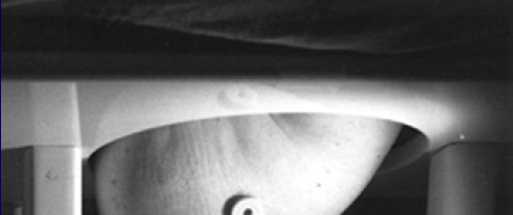
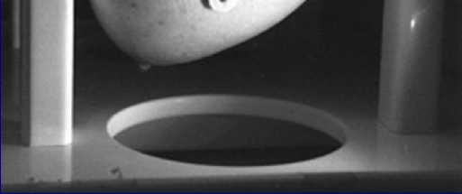
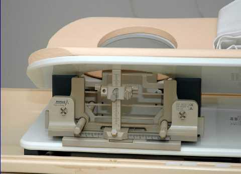
6 Technique - Patient is prone positioned - Dedicated 8 channel phased-array breast coil - 1.5T or 3.0T closed bore magnet
7 Technique - Standard MRI protocol bilateral breast imaging: m SURVEY (MST) SynBody (Refscan) T1 FFE (Tra) T2 SPAIR (Tra) T1 DYN THRIVE (Tra) T1 3Dhr postgd (Tra)
8 Technique 3.0 T MRI protocol UMC Utrecht: T1 3D FFE DYN (axial) (FOV 340 mm, matrix 352x384 matrix, 1.0 mm slice thickness, gap: 0, TR/TE shortest/shortest, flip angle: 30, scan duration each dynamic scan: 50 sec, started after intravenous bolus injection of 0.1 mmol/kg Magnevist), 8 dynamic series

9 Interpretation of breast MRI American College of Radiology BI-RADS MRI lexicon Description of a mass: - Margin: smooth, irregular, spiculated - Shape: round, oval, lobulated, stellate - Enhancement: homogeneous, heterogeneous, rim - Associated findings: edema or skin thickening Lesion diagnosis working group, JMRI 1999,10:982-90
: slow, medium or fast - Delayed phase: persistent, plateau, or")
10 Interpretation of breast MRI American College of Radiology BI-RADS MRI lexicon Enhancement kinetics: - Initial enhancement (within the first 2 minutes): slow, medium or fast - Delayed phase: persistent, plateau, or washout

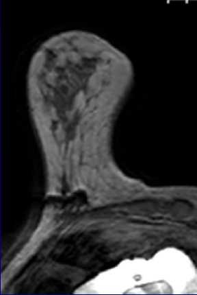
11 Interpretation of breast MRI Spiculated homogeneous enhancing mass, rapid initial enhancement followed by washout: BI-RADS MRI: V PA: invasive ductal carcinoma

12 Interpretation of breast MRI Homogeneous enhancing oval mass with smooth borders. Slow initial enhancement followed by persistent: BI-RADS MRI: II PA: fibroadenoma
13 Clinical indications 1) Screening of high risk women 2) Diagnosis of indeterminate breast lesions 3) Pre-operative staging of known breast cancer 4) Imaging of patients with positive axillary node 5) Evaluation of response to neo-adjuvant chemotherapy

14 Clinical indications Screening of high risk patient: PA: invasive adenoma carcinoma


15 Clinical indications Diagnosis of indeterminate mammographic lesion MRI-guided localization, PA: Fibroadenoma

16 Clinical indications Staging of known breast malignancy PA: skeletal metastasis: 2.3x1.5 cm


17 Clinical indications Positive axillary lymph node PA: invasive ductal carcinoma 6x5 mm


18 Clinical indications Response to neo-adjuvant chemotherapy Before 6 FEC After chemotherapy


19 Clinical indications Staging of known breast malignancy (MONET trial) PA: multicentric invasive ductal carcinoma
20 MONET Randomized Clinical Trial
21 MONET Randomized Clinical Trial Purpose: Does CE-MRI in addition to mammography/us in patients with non-palpable lesions improve breast cancer management
22 MONET Trial Methods women with nonpalpable 3-5 lesions were randomized - Group A: N=211 mammography, US, LCNB (routine care) - Group B: N=207 routine care + CE-MRI prior to biopsy - Patients with malignancy underwent surgery
23 MONET Trial Methods Primary endpoint: Rate of additional surgical procedures: a) Re-excisions b) Conversions to Mastectomy

24 MONET Results
25 MONET Results
26 MONET Results - The primary BCS rate was similar in both groups: 66% controls vs 68% MRI group - Number of re-excisions due to positive margins BCS 6/50 (12%) controls vs 18/53 (34%) MRI group (p=0.008) - The numbers of conversions to mastectomy did not differ
27 MONET Trial Conclusion MRI in addition to routine clinical care was paradoxically associated with an increased reexcision rate MRI should not be used routinely for preoperative work-up in these patients
28 MRI-guided biopsy - MRI can detect occult breast lesions % of patients with breast cancer have multifocal disease - 15% of high risk patients have IEL s - Always second look ultrasound - MRI guided VAB has been developed to allow selective tissue sampling of MRI only lesions:
29 MRI-guided biopsy - MRI can detect occult breast lesions % of patients with breast cancer have multifocal disease - 15% of high risk patients have IEL s - Always second look ultrasound - MRI guided VAB has been developed to allow selective tissue sampling of MRI only lesions:
30 MRI-guided biopsies
31 MRI-guided biopsies
32 New developments 7.0T High Field Breast Imaging: Promises: - Higher field strength results in linearly increased SNR - Higher SNR enables increased spatial resolution (DCIS) - Higher SNR can be exchanged for faster scanning techniques - More sensitive for phase changes (calcification detection)

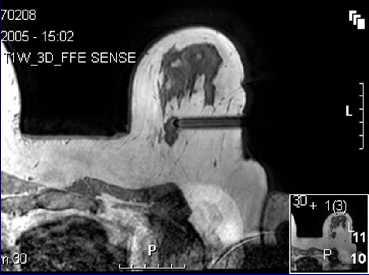
33 7.0T Feasibility Study - We started a feasibility study comparing 3T with 7T - Included were women >18 yrs with BIRADS 4 or 5 mass lesions
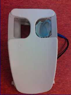
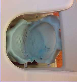
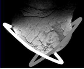
34 7.0T Feasibility Study - Unilateral two channel breast coil
35 7.0T Feasibility Study - Scan protocol T1w_GRE dynamic (7:25min) 7x T1w GRE Injection of Gd after 1 st series FA 10, TR/TE 5.0/2.0 ms, FOV mm 3 Voxel size 1.0x1.0x2.0 mm 3 Temporal resolution 63s T1w_GRE Highres (13:30min) FA 12, TR/TE 7.0/2.9 ms, FOV mm 3 Voxel size 0.45x0.57x0.45 mm 3


36 7.0T Feasibility Study: methods Two radiologists Dynamic series - Image quality excellent / good / sufficient / insufficient - Lesion identification + BI-RADS-MRI High-resolution imaging - Added value none / increased reader confidence / change in interpretation / other Invasive ductal carcinoma
37 7.0T Feasibility Study: Results 20 women with 23 lesions BI-RADS 4 n=7 BI-RADS 5 n=16 19 malignant invasive ductal carcinoma n=12 invasive lobular carcinoma n=2 invasive ductulolubular carcinoma n=5 4 non-malignant cyst n=2 slerosis n=1 radial scar n=1


38 7.0T Feasibility Study: Case T1w GRE dynamic pre-contrast T1w GRE dynamic post-contrast T1w GRE high resolution BI-RADS-MRI V Pathology: IDC Enhancement (%) Dynamic series (no.)
39 7.0T Feasibility Study: Results Image quality dynamic series excellent n=1 (R1) n=0 (R2) good n=10 (R1) n=12 (R2) sufficient n=8 (R1) n=8 (R2) insufficient n=1 (R1) n=0 (R2) Lesion identification + BI-RADS-MRI analysis - All malignant lesions + 1 non-malignant lesion identified - 3 non-malignant lesions were not identified
")
 59% (10/17) Change")
 Other 0% (0/17) 0%")
40 7.0T Feasibility Study: Results HR scans R1 R2 None 12% (2/17) 29% (5/17) Increased confidence 88% (15/17) 59% (10/17) Change in interpretation 18% (3/17) 41% (7/17) Other 0% (0/17) 0% (0/17)
41 7.0T Feasibility Study: Challenges - B1 inhomogeneity (Spin echo imaging) - B0 inhomogeneity (Uneven fat-suppression) - Field of view (Unilateral imaging)
42 Conclusion - Clear indications for anatomical breast imaging at 3T - Imaging and biopsy should be available - Imaging at 7.0T: - Is feasible - High spatial resolution


43 Thank you for your attention
Pitfalls and Limitations of Breast MRI. Susan Orel Roth, MD Professor of Radiology University of Pennsylvania
 Pitfalls and Limitations of Breast MRI Susan Orel Roth, MD Professor of Radiology University of Pennsylvania Objectives Review the etiologies of false negative breast MRI examinations Discuss the limitations
Armed Forces Institute of Pathology.
 Armed Forces Institute of Pathology www.radpath.com Armed Forces Institute of Pathology Breast Disease www.radpath.org Armed Forces Institute of Pathology Interpretation of Breast MRI Leonard M. Glassman
Leonard M. Glassman MD
 BI-RADS The New BI-RADS Leonard M. Glassman MD FACR Former Chief of Breast Imaging American Institute for Radiologic Pathology Washington Radiology Associates, PC Breast Imaging Reporting and Data System
Mammographic imaging of nonpalpable breast lesions. Malai Muttarak, MD Department of Radiology Chiang Mai University Chiang Mai, Thailand
 Mammographic imaging of nonpalpable breast lesions Malai Muttarak, MD Department of Radiology Chiang Mai University Chiang Mai, Thailand Introduction Contents Mammographic signs of nonpalpable breast cancer
Feasibility of MRI-guided large-core-needle biopsy of suspiscious breast lesions at 3T
 Eur Radiol (2009) 19: 1639 1644 DOI 10.1007/s00330-009-1310-0 BREAST Nicky H. G. M. Peters Carla Meeuwis Chris J. G. Bakker Willem P. Th. M. Mali Arancha M. Fernandez-Gallardo Richard van Hillegersberg
Successful Breast MRI Program : The ingredients
 Successful Breast MRI Program : The ingredients Dr. Smriti Hari Associate Professor Deptt. Of Radiology All India Institute of Medical Sciences New Delhi How to perform Breast MRI Breast MRI descriptors
Computer-aided detection (CAD) for breast MRI: evaluation of efficacy at 3.0 T
 for breast MRI: evaluation of efficacy at 3.0 T") Eur Radiol (2010) 20: 522 528 DOI 10.1007/s00330-009-1573-5 BREAST Carla Meeuwis Stephanie M. van de Ven Gerard Stapper Arancha M. Fernandez Gallardo Maurice A. A. J. van den Bosch Willem P. Th. M. Mali
Eur Radiol (2010) 20: 522 528 DOI 10.1007/s00330-009-1573-5 BREAST Carla Meeuwis Stephanie M. van de Ven Gerard Stapper Arancha M. Fernandez Gallardo Maurice A. A. J. van den Bosch Willem P. Th. M. Mali
Imaging in breast cancer. Mammography and Ultrasound Donya Farrokh.MD Radiologist Mashhad University of Medical Since
 Imaging in breast cancer Mammography and Ultrasound Donya Farrokh.MD Radiologist Mashhad University of Medical Since A mammogram report is a key component of the breast cancer diagnostic process. A mammogram
Categorical Classification of Spiculated Mass on Breast MRI
 Categorical Classification of Spiculated Mass on Breast MRI Poster No.: C-1974 Congress: ECR 2013 Type: Authors: Scientific Exhibit Y. Kanda 1, S. Kanao 2, M. Kataoka 2, K. Togashi 2 ; 1 Kyoto City/JP,
Breast MRI Update. Jeffrey C. Weinreb, MD, FACR Yale University School of Medicine
 Breast MRI Update Jeffrey C. Weinreb, MD, FACR jeffrey.weinreb@yale.edu Yale University School of Medicine I disclose the following financial relationships with relevant commercial interests: Bracco Bayer
BI-RADS and Breast MRI. Kathy Borovicka, M.D. Thursday February 15, 2018
 BI-RADS and Breast MRI Kathy Borovicka, M.D. Thursday February 15, 2018 Learning Objectives Be familiar with the Breast Imaging Reporting and Data System (BI-RADS) Understand the components of a breast
MRI in breast cancer: diagnosis and intervention. Dr Sue Barter Addenbrookes Hospital, Cambridge UK
 MRI in breast cancer: diagnosis and intervention Dr Sue Barter Addenbrookes Hospital, Cambridge UK Intervention will be discussed in High Risk Screening! Indications UK and Europe: Breast MRI is well established
Criteria of Malignancy. Evaluation Score
 30 5 Diagnostic Criteria Criteria of Malignancy Table 5.2 lists criteria in contrast-enhancing MR mammography that strongly indicate the presence of malignancy or are unspecific. Unifactorial evaluation
BREAST MRI. VASILIKI FILIPPI RADIOLOGIST CT MRI & PET/CT Departments Hygeia Hospital, Athens, Greece
 BREAST MRI VASILIKI FILIPPI RADIOLOGIST CT MRI & PET/CT Departments Hygeia Hospital, Athens, Greece Breast ΜR Imaging (MRM) Breast MR imaging is an extremely powerful diagnostic tool, that when used in
MRI features of Triple-negative breast cancer: our experience.
 MRI features of Triple-negative breast cancer: our experience. Poster No.: C-1852 Congress: ECR 2013 Type: Scientific Exhibit Authors: V. Bertani, A. Gualano, V. Londero, A. Dal Col, M. Marcon, P. 1 2
Standard Breast Imaging Modalities. Lilian Wang, M.D. Breast Imaging Section Department of Radiology Northwestern Medicine
 Standard Breast Imaging Modalities Lilian Wang, M.D. Breast Imaging Section Department of Radiology Northwestern Medicine Overview Standard breast imaging modalities Mammography Ultrasound MRI Imaging
Here are examples of bilateral analog mammograms from the same patient including CC and MLO projections.
 Good afternoon. It s my pleasure to be discussing Diagnostic Breast Imaging over the next half hour. I m Wei Yang, Professor of Diagnostic Radiology and Chief, the Section of Breast Imaging as well as
Value of the BI-RADS classification in MR-Mammography for diagnosis of benign and malignant breast tumors
 Eur Radiol (2011) 21:2475 2483 DOI 10.1007/s00330-011-2210-7 BREAST Value of the BI-RADS classification in MR-Mammography for diagnosis of benign and malignant breast tumors Christian Sohns & Martin Scherrer
Emerging Techniques in Breast Imaging: Contrast-Enhanced Mammography and Fast MRI
 Emerging Techniques in Breast Imaging: Contrast-Enhanced Mammography and Fast MRI Lilian Wang, M.D. Breast Imaging Section Department of Radiology Northwestern Medicine Overview Rationale for new imaging
ROLE OF MRI IN SCREENING, DIAGNOSIS AND MANAGEMENT OF BREAST CANCER. B.Zandi Professor of Radiology
 ROLE OF MRI IN SCREENING, DIAGNOSIS AND MANAGEMENT OF BREAST CANCER B.Zandi Professor of Radiology Introduction In the USA, Breast Cancer is : The Most Common Non-Skin Cancer The Second Leading cause of
MR sin plass i brystkreftdiagnostikk, dagens anbefalinger og fremtidsperspektiver
 MR sin plass i brystkreftdiagnostikk, dagens anbefalinger og fremtidsperspektiver Kathinka Kurz, MD, PhD, seksjonsoverlege SUS, kathinka.dehli.kurz@sus.no Technique - Subtraction Without contrast agent
Anyone can get breast cancer BREAST MRI BREAST CANCER. The incidence of getting breast cancer is 1:19 in Malaysia
 Anyone can get breast cancer BREAST MRI KATE Datin Dr Fatimah Moosa Sunway Medical Centre DATIN SERI ENDON KYLIE SIZE DOES NOT MAKE A DIFFERENCE BREAST CANCER The incidence of getting breast cancer is
EARLY DETECTION: MAMMOGRAPHY AND SONOGRAPHY
 EARLY DETECTION: MAMMOGRAPHY AND SONOGRAPHY Elizabeth A. Rafferty, M.D. Avon Comprehensive Breast Center Massachusetts General Hospital Harvard Medical School Breast Cancer Screening Early detection of
Triple-negative breast cancer: which typical features can we identify on conventional and MRI imaging?
 Triple-negative breast cancer: which typical features can we identify on conventional and MRI imaging? Poster No.: C-1862 Congress: ECR 2013 Type: Educational Exhibit Authors: V. Bertani 1, A. Gualano
National Diagnostic Imaging Symposium 2013 SAM - Breast MRI 1
 National Diagnostic Imaging Symposium 2013 December 8-12, 2013 Disney s Yacht Club Resort Lake Buena Vista, Florida Self Assessment Module Questions, Answers and References Day SAM Title - Each SAM title
S. Murgo, MD. Chr St-Joseph, Mons Erasme Hospital, Brussels
 S. Murgo, MD Chr St-Joseph, Mons Erasme Hospital, Brussels? Introduction Mammography reports are sometimes ambiguous and indecisive. ACR has developped the BIRADS. BIRADS consists of a lexicon in order
BI-RADS Update. Martha B. Mainiero, MD, FACR, FSBI Brown University Rhode Island Hospital
 BI-RADS Update Martha B. Mainiero, MD, FACR, FSBI Brown University Rhode Island Hospital No Disclosures BI-RADS History 1980s Quality Issues ACR Accreditation BI-RADS 1994 2003 4 th Edition MRI, US January
BREAST IMAGING and NEW IMAGING MODALITIES- A Surgeons view
 BREAST IMAGING and NEW IMAGING MODALITIES- A Surgeons view DR CHANTEL THORNTON SPECIALIST BREAST CANCER SURGEON BMSc (hons) MBBS (hons) FRACS Epworth Hospital, Richmond- Agora Centre for Women s Health
WHICH INDICATION FOR BREAST MRI?
 WHICH INDICATION FOR BREAST MRI? Dr. P. De Visschere, Prof. Dr. G. Villeirs Genitourinary Radiology and Mammography University Hospital Gent Symposium Belgian Menopause Society 13/03/2010 Which Indication
Lesion Imaging Characteristics Mass, Favoring Benign Circumscribed Margins Intramammary Lymph Node
 Lesion Imaging Characteristics Mass, Favoring Benign Circumscribed Margins Intramammary Lymph Node Oil Cyst Mass, Intermediate Concern Microlobulated Margins Obscured Margins Mass, Favoring Malignant Indistinct
Ultrasonography. Methods. Brief Description. Indications. Device-related Prerequisites. Technical Requirements. Evaluation Criteria
 1 Ultrasonography Brief Description Imaging modality using sound waves Tissue-specific wave reflection. Indications Evaluation of palpable breast nodules Evaluation of clinically occult mammographic findings
EARLY DETECTION: MAMMOGRAPHY AND SONOGRAPHY
 EARLY DETECTION: MAMMOGRAPHY AND SONOGRAPHY Elizabeth A. Rafferty, M.D. Avon Comprehensive Breast Center Massachusetts General Hospital Harvard Medical School Breast Cancer Screening Early detection of
ORIGINAL ARTICLE EVALUATION OF BREAST LESIONS USING X-RAY MAMMOGRAM WITH HISTOPATHOLOGICAL CORRELATION
 Available online at www.journalijmrr.com INTERNATIONAL JOURNAL OF MODERN RESEARCH AND REVIEWS IJMRR ISSN: 2347-8314 Int. J. Modn. Res. Revs. Volume 3, Issue 10, pp 807-814, October, 2015 ORIGINAL ARTICLE
MRI BI-RADS: How to make it out?
 MRI BI-RADS: How to make it out? Poster No.: C-1850 Congress: ECR 2016 Type: Educational Exhibit Authors: M. Ben Ammar, A. Ben Miled, O. Ghdes, S. Harguem, A. Gaja, N. Mnif; Tunis/TN Keywords: Breast,
Amammography report is a key component of the breast
 Review Article Writing a Mammography Report Amammography report is a key component of the breast cancer diagnostic process. Although mammographic findings were not clearly differentiated between benign
Rate of Malignancy in MRI-Detected Probably Benign (BI-RADS 3) Lesions
 Lesions") Women s Imaging Original Research Spick et al. Malignancy in MRI BI-RADS 3 Lesions Women s Imaging Original Research Claudio Spick 1,2 Dieter H. M. Szolar 1 Pascal A. Baltzer 2 Manfred Tillich 1 Pia Reittner
Women s Imaging Original Research Spick et al. Malignancy in MRI BI-RADS 3 Lesions Women s Imaging Original Research Claudio Spick 1,2 Dieter H. M. Szolar 1 Pascal A. Baltzer 2 Manfred Tillich 1 Pia Reittner
MEDICAL IMAGING AND BREAST DISEASE HOW CAN WE HELP YOU?
 MEDICAL IMAGING AND BREAST DISEASE HOW CAN WE HELP YOU? Barbara M. Preston, M.D. SCREENING MAMMOGRAPHY AVERAGE RISK PATIENTS KAISER RECOMMENDATION: ALL WOMEN (INCLUDING TRANSGENDER FEMALES) Every 1-21
Triple Negative Breast Cancer: Clinical Presentation and Multimodality Imaging Characteristics
 Triple Negative Breast Cancer: Clinical Presentation and Multimodality Imaging Characteristics Poster No.: R-0141 Congress: RANZCR-AOCR 2012 Type: Scientific Exhibit Authors: O. H. Woo, S. Jang, K. R.
Breast Ultrasound: Improving Your Skills & Patient Care
 Breast Ultrasound: Improving Your Skills & Patient Care Objectives Discuss US techniques available for image optimization. Review & compare the US appearances of benign & malignant masses. Cherie M. Kuzmiak,
Vacuum-assisted breast biopsy using computer-aided 3.0 T- MRI guidance: diagnostic performance in 173 lesions
 Vacuum-assisted breast biopsy using computer-aided 3.0 T- MRI guidance: diagnostic performance in 173 lesions Poster No.: C-2870 Congress: ECR 2017 Type: Scientific Exhibit Authors: A. Pozzetto, L. Camera,
Detailed Program of the second BREAST IMAGING AND INTERVENTIONS PROGRAM am am : Clinician s requirements from breast imaging
 Detailed Program of the second BREAST IMAGING AND INTERVENTIONS PROGRAM 2012 Day one, 2 nd November BREAST IMAGING AND INTERVENTIONS PROGRAM 2012 9.00 AM 9.10 am Introduction 9.10 am - 9.30 am : Clinician
BREAST MRI. Elizabeth A. Rafferty, M.D. Avon Comprehensive Breast Center Massachusetts General Hospital Harvard Medical School
 BREAST MRI Elizabeth A. Rafferty, M.D. Avon Comprehensive Breast Center Massachusetts General Hospital Harvard Medical School BREAST MRI Any assessment of the breast parenchyma requires the administration
Large health system benefits from multiple scanners for breast MRI
 Publication for the Philips MRI Community Issue 42 DECEMBER 2010 Large health system benefits from multiple scanners for breast MRI Breast MR and MR-guided biopsy are daily practice at WellStar Health
NEW SUBTRACTION ALGORITHMS FOR EVALUATION OF BREAST LESIONS ON DYNAMIC CONTRAST ENHANCED MR MAMMOGRAPHY
 A-056 NEW SUBTRACTION ALGORITHMS FOR EVALUATION OF BREAST LESIONS ON DYNAMIC CONTRAST ENHANCED MR MAMMOGRAPHY So Hee Cho, M.D., Byung Gil Choi, M.D., Hak Hee Kim, M.D., Euy Neyng Kim, M.D., Bum-soo Kim,
Imaging the Symptomatic Patient. Avice M.O Connell MD,FACR,FSBI Professor of Imaging Sciences Director, Women s Imaging University of Rochester
 Imaging the Symptomatic Patient Avice M.O Connell MD,FACR,FSBI Professor of Imaging Sciences Director, Women s Imaging University of Rochester The four most common symptoms Mass Pain Discharge Infection
MP Magnetic Resonance Imaging for Detection and Diagnosis of Breast Cancer
 Medical Policy MP 6.01.29 BCBSA Ref. Policy: 6.01.29 Last Review: 09/19/2018 Effective Date: 09/19/2018 Section: Radiology Related Policies 6.01.45 Computer-Aided Evaluation of Malignancy With Magnetic
Index. C Calcifications fat necrosis 1, 61 fat necrosis 4, 69 nipple/peri-areolar involvement 1, 165
 A ADH. See Atypical ductal hyperplasia (ADH) American College of Radiology (ACR), BI-RADS background parenchymal enhancement, 8, 9, 81, 82 fibroglandular tissue guidelines, 6 American Joint Committee on
Dynamic contrast enhanced breast MRI at 7T and 3T: an intra individual comparison study
 DOI 10.1186/s40064-015-1654-7 RESEARCH Open Access Dynamic contrast enhanced breast MRI at 7T and 3T: an intra individual comparison study Gisela L. G. Menezes *, Bertine L. Stehouwer, Dennis W. J. Klomp,
Breast MRI: Friend or Foe?
 Breast MRI: Friend or Foe? UCSF Postgraduate Course May 18, 2013 Cheryl Ewing, MD Clinical Professor of Surgery UCSF Department of Surgery APPLEGATE HAS DOUBLE MASTECTOMY IN CANCER SCARE DIAGNOSED WITH
ORIGINAL PAPER. Background parenchymal enhancement in preoperative breast MRI
 Nagoya J. Med. Sci. 77. 373 ~ 382, 2015 ORIGINAL PAPER Background parenchymal enhancement in preoperative breast MRI Satoko Kohara 1, Satoko Ishigaki 1, Hiroko Satake 1, Akiko Kawamura 1, Hisashi Kawai
Using lesion washout volume fraction as a biomarker to improve suspicious breast lesion characterization
 JOURNAL OF APPLIED CLINICAL MEDICAL PHYSICS, VOLUME 16, NUMBER 5, 2015 Using lesion washout volume fraction as a biomarker to improve suspicious breast lesion characterization Jie Huang, a Sarah M. Schafer,
The role of apparent diffusion coefficient (ADC) and relative ADC in the evaluation of breast masses
 and relative ADC in the evaluation of breast masses") The role of apparent diffusion coefficient (ADC) and relative ADC in the evaluation of breast masses Poster No.: C-1749 Congress: ECR 2014 Type: Scientific Exhibit Authors: U. Aksoy Ozcan 1, A. Öz 2, S.
The role of apparent diffusion coefficient (ADC) and relative ADC in the evaluation of breast masses Poster No.: C-1749 Congress: ECR 2014 Type: Scientific Exhibit Authors: U. Aksoy Ozcan 1, A. Öz 2, S.
Breast Cancer. Most common cancer among women in the US. 2nd leading cause of death in women. Mortality rates though have declined
 Breast Cancer Most common cancer among women in the US 2nd leading cause of death in women Mortality rates though have declined 1 in 8 women will develop breast cancer Breast Cancer Breast cancer increases
Pitfalls of Dynamic Contrast Enhanced MR Mammography (DCE-MRM) in Evaluation of Post-Biopsy Suspicious Breast Lesions
 in Evaluation of Post-Biopsy Suspicious Breast Lesions") Med. J. Cairo Univ., Vol. 86, No. 3, June: 1513-1522, 2018 www.medicaljournalofcairouniversity.net Pitfalls of Dynamic Contrast Enhanced MR Mammography (DCE-MRM) in Evaluation of Post-Biopsy Suspicious
Med. J. Cairo Univ., Vol. 86, No. 3, June: 1513-1522, 2018 www.medicaljournalofcairouniversity.net Pitfalls of Dynamic Contrast Enhanced MR Mammography (DCE-MRM) in Evaluation of Post-Biopsy Suspicious
Breast Cancer. Saima Saeed MD
 Breast Cancer Saima Saeed MD Breast Cancer Most common cancer among women in the US 2nd leading cause of death in women 1 in 8 women will develop breast cancer Incidence/mortality rates have declined Breast
BREAST MRI. Elizabeth A. Rafferty, M.D. Avon Comprehensive Breast Center Massachusetts General Hospital Harvard Medical School
 BREAST MRI Elizabeth A. Rafferty, M.D. Avon Comprehensive Breast Center Massachusetts General Hospital Harvard Medical School BREAST MRI Any assessment of the breast parenchyma requires the administration
Breast Imaging: Multidisciplinary Approach. Madelene Lewis, MD Assistant Professor Associate Program Director Medical University of South Carolina
 Breast Imaging: Multidisciplinary Approach Madelene Lewis, MD Assistant Professor Associate Program Director Medical University of South Carolina No Disclosures Objectives Discuss a multidisciplinary breast
Ductal carcinoma in situ: ultrasound, mammography and MRI features with pathologic correlation
 Ductal carcinoma in situ: ultrasound, mammography and MRI features with pathologic correlation Poster No.: C-2252 Congress: ECR 2013 Type: Educational Exhibit Authors: L. Fernandes, H. A. M. R. Tinto,
BI-RADS MRI: A Primer
 Erguvan- ogan et al. I- RS MRI Women s Imaging Pictorial Essay WOMEN S IMGING asak Erguvan-ogan 1 Gary J. Whitman 1 nne. Kushwaha 1,2 Michael J. Phelps 1,3 Peter J. empsey 1 Erguvan-ogan, Whitman GJ, Kushwaha,
Essentials of Clinical MR, 2 nd edition. 73. Urinary Bladder and Male Pelvis
 73. Urinary Bladder and Male Pelvis Urinary bladder carcinoma is best locally staged with MRI. It is important however to note that a thickened wall (> 5 mm) is a non-specific finding seen in an underfilled
Magnetic Resonance Imaging of the Female Breast
 January 2002 Magnetic Resonance Imaging of the Female Breast Its Role, Indication, Technique & Future. Christian Zechmann, Heidelberg Medical School Year, IV Dr. Introduction Breast cancer is the most
Can magnetic resonance imaging obviate the need for biopsy for microcalcifications?
 Original Article Can magnetic resonance imaging obviate the need for biopsy for microcalcifications? Shinya Yamamoto, Takashi Chishima Department of Breast Surgery, Yokohama Rosai Hospital, Yokohama 222-0036,
Case Scenario 1 History and Physical 3/15/13 Imaging Pathology
 Case Scenario 1 History and Physical 3/15/13 The patient is an 84 year old white female who presented with an abnormal mammogram. The patient has a five year history of refractory anemia with ringed sideroblasts
RADIOLOGIC EVALUATION OF BREAST CANCER
 RADIOLOGIC EVALUATION OF BREAST CANCER Orsolya Farkas, Gabriella Bodrogi and Gábor Szalai Department of Radiology, Pécs University Orsifarkas@yahoo.com Complex evaluation of the breast Patient history
Mammographic evaluation of palpable breast masses with pathological correlation: a tertiary care centre study in Nepal
 Original article 21 Mammographic evaluation of palpable breast masses with pathological correlation: a tertiary care centre study in Nepal G. Gurung, R. K. Ghimire, B. Lohani Department of Radiology and
RSNA, /radiol Appendix E1. Methods
 RSNA, 2016 10.1148/radiol.2016151097 Appendix E1 Methods US and Near-infrared Data Acquisition Four optical wavelengths (740 nm, 780 nm, 808 nm, and 830 nm) were used to sequentially deliver the light
ACR Breast MRI Accreditation Program - DRAFT
 ACR Breast MRI Accreditation Program - DRAFT Donna M. Reeve, MS, DABR, DABMP Department of Imaging Physics Educational Objectives Provide an overview of the ACR Breast MRI Accreditation Program (BMRAP)
Case study 1. Rie Horii, M.D., Ph.D. Division of Pathology Cancer Institute Hospital, Japanese Foundation for Cancer Research
 NCCN/JCCNB Seminar in Japan April 15, 2012 Case study 1 Rie Horii, M.D., Ph.D. Division of Pathology Cancer Institute Hospital, Japanese Foundation for Cancer Research Present illness: A 50y.o.premenopausal
8/31/2016 HIDING IN PLAIN SITE, ARCHITECTURAL DISTORTIONS AND BREAST ASYMMETRIES ARCHITECTURAL DISTORTIONS ARCHITECTURAL DISTORTIONS
 HIDING IN PLAIN SITE, ARCHITECTURAL DISTORTIONS AND BREAST ASYMMETRIES DEBORAH THAMES R.T. (R)(M)(QM) ARCHITECTURAL DISTORTIONS Definition is disruption of the natural flow of breast pattern towards the
Ana Sofia Preto 19/06/2013
 Ana Sofia Preto 19/06/2013 Understanding the underlying pathophysiologic processes leading to the various types of calcifications Description and illustration of the several types of calcifications, according
Case Scenario 1: This case has been slightly modified from the case presented during the live session to add clarity.
 Case Scenario 1: This case has been slightly modified from the case presented during the live session to add clarity. Background: 46 year old married premenopausal female with dense breasts has noticed
Breast Cancer Imaging
 Breast Cancer Imaging I. Policy University Health Alliance (UHA) will cover breast imaging when such services meet the medical criteria guidelines (subject to limitations and exclusions) indicated below.
Breast Imaging Lexicon
 9//201 200 BI RADS th Edition 201 BI RADS th Edition Breast Imaging Lexicon Mammographic Pathology and Assessment Categories Deborah Thames, R.T.(R)(M)(QM) The Advanced Health Education Center Nonmember:
CDIS: what's beyond microcalcifications? - Pictorial essay
 CDIS: what's beyond microcalcifications? - Pictorial essay Poster No.: C-1096 Congress: ECR 2014 Type: Educational Exhibit Authors: R. N. Lucas, C. A. S. Ruano, I. Oliveira, J. M. G. Lourenco, Z. 1 1 1
Breast Imaging Update: Old Dog New Tricks
 Breast Imaging Update: Old Dog New Tricks Claire McKay, DO M&S Imaging Assoc. San Antonio, TX cmckayhart@juno.com Goals Describe modalities available, old and new Provide understanding of pros and cons
Original Research JOURNAL OF MAGNETIC RESONANCE IMAGING 32: (2010)
") JOURNAL OF MAGNETIC RESONANCE IMAGING 32:1117 1123 (2010) Original Research Peripheral Hyperintense Pattern on T2-Weighted Magnetic Resonance Imaging (MRI) in Breast Carcinoma: Correlation With Early Peripheral
JOURNAL OF MAGNETIC RESONANCE IMAGING 32:1117 1123 (2010) Original Research Peripheral Hyperintense Pattern on T2-Weighted Magnetic Resonance Imaging (MRI) in Breast Carcinoma: Correlation With Early Peripheral
When do you need PET/CT or MRI in early breast cancer?
 When do you need PET/CT or MRI in early breast cancer? Elizabeth A. Morris MD FACR Chief, Breast Imaging Service Memorial Sloan-Kettering Cancer Center NY, NY Objectives What is the role of MRI in initial
Breast MRI, digital mammography and breast tomosynthesis: Comparison of three methods for early detection of breast cancer
 BOSNIAN JOURNAL OF BASIC MEDICAL SCIENCES RESEARCH ARTICLE WWW.BJBMS.ORG Breast MRI, digital mammography and breast tomosynthesis: Comparison of three methods for early detection of breast cancer Dragana
Benign, Reactive and Inflammatory Lesions of the Breast
 Benign, Reactive and Inflammatory Lesions of the Breast Marilin Rosa, MD Associate Member Section Head of Breast Pathology Department of Anatomic Pathology Program Director, Breast Pathology Fellowship
AMSER Case of the Month: November 2018
 AMSER Case of the Month: November 2018 52 year old female with an abnormal screening mammogram Areeg Rehman, MS 4 Nova Southeastern University Rebecca T. Sivarajah, MD Penn State University College of
Patient Outcomes in Canceled MRI-Guided Breast Biopsies
 Women s Imaging Original Research Outcomes After Canceled MRI-Guided Breast Biopsies Women s Imaging Original Research Bethany L. Niell 1 Janie M. Lee 1, 2 Christopher Johansen 3 Elkan F. Halpern 4 Elizabeth
Table 1. Classification of US Features Based on BI-RADS for US in Benign and Malignant Breast Lesions US Features Benign n(%) Malignant n(%) Odds
 Malignant n(%) Odds") 215 Table 1. Classification of US Features Based on BI-RADS for US in Benign and Malignant Breast Lesions US Features Benign n(%) Malignant n(%) Odds ratio 719 (100) 305(100) Shape Oval 445 (61.9) 019
215 Table 1. Classification of US Features Based on BI-RADS for US in Benign and Malignant Breast Lesions US Features Benign n(%) Malignant n(%) Odds ratio 719 (100) 305(100) Shape Oval 445 (61.9) 019
Case Scenario 1: This case has been slightly modified from the case presented during the live session to add clarity.
 Case Scenario 1: This case has been slightly modified from the case presented during the live session to add clarity. Background: 46 year old married premenopausal female with dense breasts has noticed
University of Washington Radiology Review Course: Strange and Specific Diagnoses. Case #1
 University of Washington Radiology Review Course: Strange and Specific Diagnoses Katherine E. Dee, MD Seattle Breast Center Via Radiology 2014 Case #1 37 year old presents with bilateral palpable lumps.
Correlation between lesion type and the additional value of digital breast tomosynthesis
 Correlation between lesion type and the additional value of digital breast tomosynthesis Poster No.: C-1604 Congress: ECR 2011 Type: Scientific Exhibit Authors: C. Van Ongeval, L. Cockmartin, A. Van Steen,
Compressive Re-Sampling for Speckle Reduction in Medical Ultrasound
 Compressive Re-Sampling for Speckle Reduction in Medical Ultrasound Professor Richard Mammone Rutgers University Email Phone Number Christine Podilchuk, Lev Barinov, Ajit Jairaj and William Hulbert ClearView
Financial Disclosures
 Financial Disclosures 3D Mammography: The Latest Developments in the Breast Imaging Arena I have no financial disclosures Dr. Katharine Lampen-Sachar Breast and Body Radiologist Radiology Associates of
Throughout this policy, bracketed numbers link topics across multiple sections according to the indication numbers in the following list.
 Subject: Magnetic Resonance Imaging of the Breast Page: 1 of 33 Last Review Status/Date: September 2015 Magnetic Resonance Imaging of the Breast Description Magnetic resonance imaging (MRI) of the breast
The role of MRI in assessment of asymmetrical breast densities
 The Egyptian Journal of Radiology and Nuclear Medicine (2010) 41, 501 508 Egyptian Society of Radiology and Nuclear Medicine The Egyptian Journal of Radiology and Nuclear Medicine www.elsevier.com/locate/ejrnm
Case Report Tubular Carcinoma of the Breast: Advantages and Limitations of Breast Tomosynthesis
 Case Reports in Radiology Volume 2016, Article ID 3906195, 4 pages http://dx.doi.org/10.1155/2016/3906195 Case Report Tubular Carcinoma of the Breast: Advantages and Limitations of Breast Tomosynthesis
Ultrasound of the Breast BASICS FOR THE ORDERING CLINICIAN
 Ultrasound of the Breast BASICS FOR THE ORDERING CLINICIAN Breast Ultrasound Anatomy Skin Breast Parenchyma Pectoralis Fascia Pectoralis Breast Ultrasound Anatomy Indications for Breast Ultrasound Palpable
New Imaging Modalities for better Screening and Diagnosis
 New Imaging Modalities for better Screening and Diagnosis Miri Sklair-Levy, MD Department of Diagnostic Imaging Sheba Medical Center, Sackler School of Medicine, Tel Aviv University Department of Diagnostic
BI-RADS Categorization As a Predictor of Malignancy 1
 Susan G. Orel, MD Nicole Kay, BA Carol Reynolds, MD Daniel C. Sullivan, MD BI-RADS Categorization As a Predictor of Malignancy 1 Index terms: Breast, biopsy, 00.1261 Breast neoplasms, localization, 00.125,
ACR MRI Accreditation Program. ACR MRI Accreditation Program Update. Educational Objectives. ACR accreditation. History. New Modular Program
 ACR MRI Accreditation Program Update Donna M. Reeve, MS, DABR, DABMP Department of Imaging Physics University of Texas M.D. Anderson Cancer Center Educational Objectives Present requirements of the new
Non-mass Enhancement on Breast MRI. Aditi A. Desai, MD Margaret Ann Mays, MD
 Non-mass Enhancement on Breast MRI Aditi A. Desai, MD Margaret Ann Mays, MD Breast MRI Important screening and diagnostic tool, given its high sensitivity for breast cancer detection Breast MRI - Indications
Contrast-Enhanced Spectral Mammography
 Contrast-Enhanced Spectral Mammography Illuminating Breast Cancer Detection SenoBright HD TM gehealthcare.com/senobright Mammography is the most reliable imaging technique for breasts, but limitations
Tomosynthesis and breast imaging update. Dr Michael J Michell Consultant Radiologist King's College Hospital NHS Foundation Trust
 Tomosynthesis and breast imaging update Dr Michael J Michell Consultant Radiologist King's College Hospital NHS Foundation Trust Breast imaging new technology BREAST CANCER FLT PET shows different grades
Medical Policy An independent licensee of the Blue Cross Blue Shield Association
 Magnetic Resonance Imaging (MRI) of the Breast Page 1 of 51 Medical Policy An independent licensee of the Blue Cross Blue Shield Association Title: Magnetic Resonance Imaging (MRI) of the Breast Professional
We are IntechOpen, the world s leading publisher of Open Access books Built by scientists, for scientists. International authors and editors
 We are IntechOpen, the world s leading publisher of Open Access books Built by scientists, for scientists 3,800 116,000 120M Open access books available International authors and editors Downloads Our
The Lactating Breast: Contrast-enhanced MR Imaging of Normal Tissue and Cancer 1
 Breast Imaging Leandro A. Espinosa, MS Bruce L. Daniel, MD Logi Vidarsson, MSEE Marowan Zakhour, MD Debra M. Ikeda, MD Robert J. Herfkens, MD Published online 10.1148/radiol.2372040837 2005; 237:429 436