UNDERSTANDING SPECIAL STAINS
|
|
|
- Ezra Holmes
- 6 years ago
- Views:
Transcription
1 Vet Times The website for the veterinary profession UNDERSTANDING SPECIAL STAINS Author : MELANIE DOBROMYLSKYJ Categories : Vets Date : April 14, 2014 MELANIE DOBROMYLSKYJ explains some of the most common special stains used in diagnostic laboratories, their main indications and clinical situations where they can aid diagnosis Summary Do you want to know why your pathologist has delayed the final report on your case, for the sake of some oddsounding special stain? This article aims to summarise the most common special stains used in a diagnostic laboratory setting, their main indications and the corresponding clinical situations where they might be used to aid diagnosis. These include the metachromatic stains, for example Giemsa, used for blood smears, mast cells and some fungal and protozoal infections, stains for infectious agents, such as Gram (bacteria), Warthin-Starry (spirochetes), Ziehl-Neelsen (acid-fast bacteria), periodic acid-schiff (fungi, glycogen), and stains for materials, such as pigments, fat, amyloid and calcium. Key words histology, special stains, infectious agents, neoplasia THE haematoxylin and eosin (H and E) stain is the routine, everyday stain used in histological sections and will probably be the first slide a pathologist looks at for any given case. Haematoxylin colours nuclei and a few other objects blue (referred to as basophilic) while the eosin counterstains the cytoplasm in various shades of pink (termed pale to brightly to deeply 1 / 17
2 eosinophilic depending on the precise shade). Structures do not have to be acidic or basic to be called basophilic or eosinophilic, the terminology is simply based on the affinity to these dyes. Other, endogenous colours may also be present in a section, for example erythrocytes are bright red, melanin is brown-black and other pigments may be yellow, golden-brown or green. Based on the appearance of this initial section, a pathologist may request further special stains for a number of reasons. Some specific tissue structures simply do not stain well with routine H and E stains. For example, the reticular fibres in the liver require a silver stain, such as Gordon-Sweets, to allow better assessment of liver architecture, while Masson s trichrome stains connective tissues, highlighting the presence of fibrosis in some cases of liver disease (Figure 1). Hydrophobic structures also tend to remain clear since these are usually rich in fats; this includes adipocytes and the myelin around neuron axons. Some special stains aid better identification of poorly differentiated neoplastic cells (such as mast cells or a mucin-secreting neoplasia), or may highlight the presence of infectious agents. Other stains allow more precise identification of deposits such as amyloid, calcium and a variety of pigments. This article is not intended as an exhaustive list of special stains because there are very many of them even Table 1 does not list every stain available to the histopathologist but it will concentrate on the ones most often used in routine histopathology cases. Metachromatic stains Metachromatic stains are those that have the ability to produce different colours with various histologic or cytologic structures. Examples include Giemsa, Wright and Diff-Quik stains. The Giemsa stain has multiple purposes within the diagnostic laboratory. It is one of the classic stains for peripheral blood smears and bone marrow specimens, and is also useful for staining some fungal species such as Histoplasma and some intracellular protozoa, such as Leishmania. Another major use of Giemsa, as well as other metachromatic stains, such as toluidine blue and astra blue, is the identification of mast cells because it stains the cytoplasmic granules present in mast cells a magenta to purple colour. This may aid further differentiation of a round cell tumour as being of mast cell origin, especially in some of the more poorly differentiated tumours where the granules may not be obvious in routine H and E sections. In other cases, where increased numbers of mast cells are present in a lesion that is otherwise inflammatory in appearance, a Giemsa stain can help reveal how many mast cells are present, whether they form clusters, how pleomorphic they are, whether they contain mitotic figures and whether the pathologist should be suspicious of an underlying neoplasm. The Giemsa stain can also help when assessing mast cell tumour margins and scar line resections; although it is 2 / 17
3 important to remember it cannot distinguish between neoplastic mast cells and the non-neoplastic mast cells that are migrating to the site of the tumour, attracted by the release of histamine and other bioactive substances. Stains for infectious agents Gram and Warthin- Starry stains Gram staining is a method for highlighting the presence and morphology of bacterial populations within a section; it also differentiates bacterial species into two large groups Gram-positive and Gram-negative based on the chemical and physical properties of their cell walls. It detects peptidoglycan, which is present in a thick layer in Gram-positive bacteria and results in a blue/ purple colour. Gram-negative bacteria generally have a thin layer of peptidoglycan between two membranes, which results in a pink/red colour. Not all bacteria can be definitively classified by this technique, thus forming Gram-variable and Gramindeterminate groups as well. Gram-positive bacteria include many well-known genera, such as Bacillus, Listeria, Staphylococcus, Streptococcus, Enterococcus and Clostridium, while Gram-negative bacteria include, among others, Bordetella, Campylobacter, Enterobacter, Escherichia coli, Helicobacter, Pasteurella, Pseudomonas and Salmonella. However, Gram staining is not always reliable in histological sections and follow-up culture and full identification of potentially significant bacterial populations seen on histology is usually recommended. Warthin-Starry is a silver nitrate-based staining technique used to detect spirochetes, such as Leptospira and Borrelia, as well as Helicobacter species present in gastric biopsies. Ziehl-Neelsen stain The Ziehl-Neelsen (ZN) stain, also known as the acid-fast stain, is a special stain used to identify acid-fast organisms, mainly mycobacterial species such as Mycobacterium bovis, M paratuberculosis and M avium. These may be present within suspicious lesions in very low numbers, which means identification on routine H and E sections is often nearly impossible. Specialised cultures or molecular techniques, such as PCR, are required to confirm the identification of Mycobacterium species and to rule out other acid-fast bacilli (although other types of bacilli are extremely unlikely) and also to precisely identify the species of Mycobacterium involved. There is some zoonotic potential from Mycobacterium species, adding importance to accurate identification and reporting of such infections. The ZN stain can also be used to identify intranuclear lead inclusion bodies. Wright s stain 3 / 17
4 Leishmaniasis generally causes a chronic inflammatory response of granulomatous nature, due to the parasite targeting phagocytic cells, such as macrophages. It can also provoke a type IV hypersensitivity response. Several forms of leishmaniasis occur, including cutaneous, mucocutaneous and visceral forms. Within the dog, the intracellular parasite is detected by Wright s stain on blood smears, lymph node aspirates or in histological sections, where it assumes a non-flagellated form. Periodic acid-schiff Periodic acid-schiff (PAS) is a staining method used to detect glycogen and other polysaccharides in tissues. Probably the most common use of PAS in diagnostic pathology is for fungal infections, such as Malassezia, fungal hyphae and Candida (living fungi). Other stains, especially silver stains, are also used to detect fungal organisms, for example Grocott s methenamine silver stain (fungi both dead and alive). The presence of glycogen can be confirmed in tissue sections by using diastase to digest the glycogen from a section, then comparing a diastase-digested PAS section with a normal PAS section. If the positive-staining material present in the PAS slide is glycogen, then it will be absent from the corresponding location on the diastase-digested slide. This technique can be used on liver sections to confirm intracytoplasmic material is glycogen, as opposed to fat, and can be used on neurological tissues to distinguish glycogen storage diseases from other types of storage disease (Figure 2). Stains for amyloid Amyloid is a fairly homogeneous, nondescript eosinophilic material on routine H and E stained sections, and its presence needs to be confirmed by special stains such as Congo red. This stain colours amyloid an orangered colour, and when viewed under a polarised light the material demonstrates apple-green birefringence. Amyloid may be present in a range of diseases. Primary amyloidosis is the most common systemic or generalised form, and is most often due to plasma cell or B-cell dyscrasias, such as multiple myeloma and other monoclonal B-cell proliferations. These can result in increased amounts of immunoglobulin light chain (AL amyloid), the basis for the type of amyloid present in these cases. Secondary or reactive amyloidosis is the form that is generated from increased amounts of the acute phase protein serum amyloid A (AA amyloid) and is generally associated with chronic inflammatory conditions or neoplasia, although it can also be idiopathic. Familial amyloidosis is a hereditary and systemic condition that can affect various organ systems. It is seen in Abyssinian cats and Shar Pei dogs (Figure 3), where deposition occurs in the kidney, and in Siamese cats, where it occurs in the liver. 4 / 17
5 Stains for calcium The presence of calcium in tissues is confirmed by the von Kossa stain, which colours calcium black. Pathological calcification of tissues falls into two broad categories, dystrophic (serum calcium levels are normal, but the tissue is not) and metastatic (serum calcium levels are increased, but the tissues are normal). Dystrophic calcification can occur in necrotic tissues, such as in the centre of granulomas in tuberculosis and Johne s disease. It can also be seen in the skin of dogs, for example calcinosis cutis associated with hyperadrenocorticism (Cushing s disease) and calcinosis circumscripta (occurring in the skin and other soft tissues) associated with sites of repeated trauma. Metastatic calcification can be seen associated with renal failure (secondary hyperparathyroidism), vitamin D toxicosis (ingestion of calcinogenic plants, rodenticides or even the owner s tube of psoriasis cream Figure 4), primary hyperparathyroidism, pseudohyperparathyroidism (release of parathyroid-related protein from certain tumours) and destructive bone lesions. Pigment stains Masson- Fontana Melanocytic tumours can represent a diagnostic challenge. Poorly melanised or amelanotic tumours can vary markedly in their cellular morphology, and in the absence of melanin as a clue, they can resemble a large number of other tumours. Masson-Fontana, a stain that detects melanin pigment, can aid diagnosis in poorly melanised tumours by highlighting the presence of any small amounts of melanin present in the sections. Permanganate bleach Large amounts of melanin pigment in the tumour cells can obscure both nuclear and cellular morphology, making histological assessment of malignancy difficult. Bleaching sections with permanganate bleach removes the colour of the pigment and allows clearer assessment of nuclear morphology and the mitotic index, important features for distinguishing between malignant or benign tumours when taken together with anatomical location. Other pigment stains Various stains are available to help the pathologist distinguish between pigments present in histological sections. 5 / 17
6 For example, the Dunn-Thompson stain for haemoglobin may be of use in cases of intravascular haemolysis leading to deposition of haemoglobin within renal tubules. The Fouchet stain for bile pigments may be used in cases of cholestasis (Figure 5). Perl s Prussian blue is used to highlight the presence of haemosiderin and to distinguish it from other pigments. Examples include the presence of haemosiderin in alveolar macrophages seen with passive chronic congestion of the lungs (so-called heart-failure cells ) and when excess iron is released during red blood cell breakdown, for example as seen with auto-immune haemolytic anaemia. Fat stains Various stains are used to detect the presence of fat in a section, although these require the use of frozen tissue sections. This is because the normal routine processing of tissues removes the vast majority of fat from sections, simply leaving an empty space in cells and tissues. Pathologists may be suspicious that these spaces previously contained fats, either in the form of lipid, lipoproteins or triglycerides, but the need to perform special stains, such as oil red O and Sudan III to confirm its presence. For example, the presence of lipid in a poorly differentiated sarcoma may aid the further diagnosis of the tumour as a liposarcoma. Presence of lipid in vacuolated hepatocytes may aid diagnosis of a hepatic lipidosis, as opposed to a disease involving hepatic storage of other substances, such as glycogen. Figure 6 demonstrates the use of oil red O staining to highlight fat in vacuoles of neoplastic cells; this section is from one of multiple abdominal tumours in a horse that presented with marked and recurrent ascites. Oil red O stain on frozen sections together with immunohistochemical stains and electron microscopy confirmed the diagnosis of a lipid-rich mesothelioma in this case, the first report of this type of tumour in a horse. Reference 1. Dobromylskyj M J, Copas V, Durham A, Hughes T K and Patterson-Kane J C (2011). Disseminated lipid-rich peritoneal mesothelioma in a horse, Journal of Veterinary Diagnostic Investigation 23(3): / 17
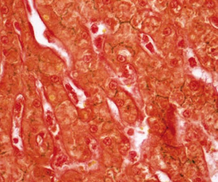
 Section from the liver of a bongo antelope in a zoological collection, with chronic diffuse hepatic fibrosis.")
7 Figure 1. Masson s trichrome. a) Section from the liver of a bongo antelope in a zoological collection, with chronic diffuse hepatic fibrosis. Liver, Masson s trichrome, 4x. b) section from a normal liver for comparison 4x. 7 / 17
 Section from the liver of a bongo antelope in a zoological collection, with chronic diffuse hepatic fibrosis.")
8 Figure 1. Masson s trichrome. a) Section from the liver of a bongo antelope in a zoological collection, with chronic diffuse hepatic fibrosis. Liver, Masson s trichrome, 4x. b) section from a normal liver for comparison 4x. 8 / 17

9 Figure 2. Periodic acid-schiff stain of a storage disease. Section from the brain of a dog with a glycogen storage disease. PAS-positive material is present within multiple neurons. This case was submitted to the Veterinary Diagnostic Services, University of Glasgow. Brain, PAS, 10x. 9 / 17
 is amyloid, colouring it red.")
10 Figure 3. Congo red stain. Section from the kidney of a four-year old Shar Pei dog with renal amyloidosis. Congo red staining confirms the material present within the glomeruli (which is eosinophilic on H and E) is amyloid, colouring it red. Kidney, Congo red, 40x. 10 / 17

11 Figure 4. Von Kossa stain. Section from the kidney of a puppy that had ingested its owner s tube of psoriasis cream, resulting in vitamin D toxicosis and widespread, severe calcification of multiple tissues. A von Kossa stain of the mesenteric adipose tissues colours the extensive calcium deposits black. von Kossa, 20x. 11 / 17
 kidney: many renal tubules are distended by bright orange-red, amorphous to crystalline material, which stains yellow with a Fouchet stain (20x), allowing differentiation between haemoglobin and")
12 Figure 5. Fouchet stain. Sections from the kidney and liver of a tapir. a) kidney: many renal tubules are distended by bright orange-red, amorphous to crystalline material, which stains yellow with a Fouchet stain (20x), allowing differentiation between haemoglobin and bile, which stains emerald green with Fouchet, as in b). section from the liver, 40x. Final diagnosis in this case was of a haemoglobinuric nephrosis. 12 / 17
13 13 / 17
 kidney: many renal tubules are distended by bright orange-red, amorphous to crystalline material, which stains yellow with a Fouchet stain (20x), allowing differentiation between haemoglobin and")
14 Figure 5. Fouchet stain. Sections from the kidney and liver of a tapir. a) kidney: many renal tubules are distended by bright orange-red, amorphous to crystalline material, which stains yellow with a Fouchet stain (20x), allowing differentiation between haemoglobin and bile, which stains emerald green with Fouchet, as in b). section from the liver, 40x. Final diagnosis in this case was of a haemoglobinuric nephrosis. 14 / 17
15 Figure 6. Oil red O. Section from one of multiple abdominal tumours in a horse that presented with marked and recurrent ascites. Oil red O stain on frozen sections reveals positive-staining (red) vacuoles within many tumour cells, which together with immunohistochemical stains and electron microscopy confirmed the diagnosis of a lipid-rich mesothelioma, the first reported case in a horse (submitted to the Veterinary Diagnostic Services, University of Glasgow) 1. Abdominal mass; Oil red O, 20x. 15 / 17
16 16 / 17
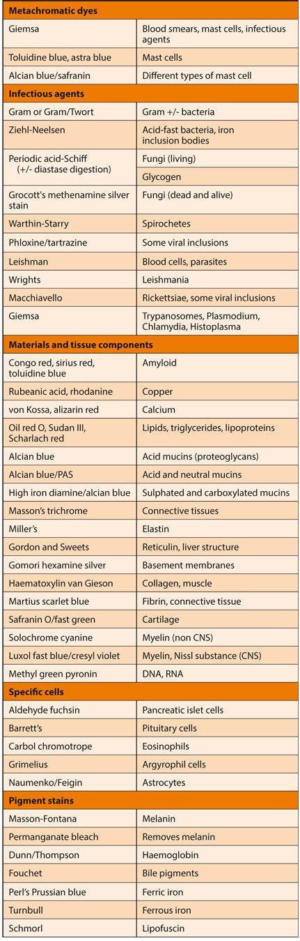
17 Table 1. A selection of special stains available to the histopathologist 17 / 17 Powered by TCPDF (
The Oral Histology Series Series 5 Special Stains
 The Oral Histology Series Series 5 Special Stains DAVID E. KLINGMAN, Lt Col, USAF, DC Views and opinions expressed are those of the author(s) and do not reflect official policy or position of the United
Atlas of Stains. Special Stains on Artisan Link Pro
 Atlas of Stains Special Stains on Artisan Link Pro Intended use Routinely processed samples (paraffin-embedded) may be used. The preferred fixative is neutral buffered formalin. The clinical interpretation
Schedule of Accreditation issued by United Kingdom Accreditation Service 2 Pine Trees, Chertsey Lane, Staines-upon-Thames, TW18 3HR, UK
 2 Pine Trees, Chertsey Lane, Staines-upon-Thames, TW18 3HR, UK Cellular Pathology Department University College London Hospital Rockefeller Building University Street London WC1E 6JJ Contact: Gavyn Barrett
BLIZARD INSTITUTE CORE PATHOLOGY ATLAS OF TINCTORIAL STAINS
 BLIZARD INSTITUTE CORE PATHOLOGY ATLAS OF TINCTORIAL STAINS Contents Introduction... 3 Background to Tinctorial Stains... 3 Haematoxylin and Eosin Stain (H&E)... 3 Connective Tissue Stains... 4 Nucleic
This is Learning Component 6 in Learning Module 1. We will show examples of features ( things ) including mineral deposits, urates, pigments, dust,
 including mineral deposits, urates, pigments, dust,") This is Learning Component 6 in Learning Module 1. We will show examples of features ( things ) including mineral deposits, urates, pigments, dust, plant material, and amyloid. 1 Calcium salts are the
This is Learning Component 6 in Learning Module 1. We will show examples of features ( things ) including mineral deposits, urates, pigments, dust, plant material, and amyloid. 1 Calcium salts are the
Presented by: Dr. Giuseppe Molinaro Dr. Davide De Biase
 Presented by: Dr. Giuseppe Molinaro Dr. Davide De Biase Dog Spayed Female LABRADOR RETRIEVER 3 Years old VACCINATIONS ANTIPARASITIC COMMERCIAL DIET VOMITING FOR A MONTH DULLNESS WEIGHT LOSS INAPPETANCE
PATHOLOGY Intracellular Degeneration LAB 1
 PATHOLOGY Intracellular Degeneration LAB 1 Cellular swelling Liver Organ :- Liver Lesion :- 1. Narrowing of hepatic sinusoids due to the swelling of hepatocyte. 2. The cytoplasm of affected hepatocyte
Histopathology: Cell necrosis and cytoplasmic accumulations
 Histopathology: Cell necrosis and cytoplasmic accumulations These presentations are to help you identify basic histopathological features. They do not contain the additional factual information that you
Fixation... Questions 1 Answers 16. Processing... Questions 25 Answers 36. Safety... Questions 67 Answers 73
 Table of Contents Fixation... Questions 1 Answers 16 Processing... Questions 25 Answers 36 Instrumentation... Questions 43 Answers 58 Safety... Questions 67 Answers 73 Laboratory Mathematics & Solution
Cell injury, adaptation and death. Unite one Second Lab.
 Cell injury, adaptation and death Unite one Second Lab. The two lung abscesses seen here are examples of liquefactive necrosis in which there is a liquid center in an area of tissue injury. One abscess
Preface 1. Fixation and Processing 1
 Contents Preface xi 1. Fixation and Processing 1 Fixation 1 Processing 2 What Should Be Seen in a Well-Fixed, Well-Processed Specimen Stained with Hematoxylin and Eosin 3 Problems Encountered With Fixation
Avian Pathology. Bacterial diseases: histo slides. ECVP-ESVP Summer School 2012 Frédérique NGUYEN
 Avian Pathology Bacterial diseases: histo slides ECVP-ESVP Summer School 2012 Frédérique NGUYEN Bacterial diseases: histo slides B1. Turkey. Organs? Morphologic diagnosis? Special procedure? B2. Hen. Organ?
Cytology of Inflammatory Cutaneous lesions in the Dog and Cat
 Cytology of Inflammatory Cutaneous lesions in the Dog and Cat Rick L. Cowell, DVM, MS, MRCVS, Diplomate ACVP Clinical Pathologist IDEXX Laboratories Inc A. Cytologic Patterns of Inflammation: 1. Neutrophilic
HISTOPATHOLOGY. Shannon Martinson
 HISTOPATHOLOGY Shannon Martinson March 2013 Case #1 History: 8 year old beagle Neck pain for the past couple of weeks Paresis, followed by paralysis developed over the past few days Gross Description courtesy
VPM Pigment and other tissue deposits. Shannon Martinson
 VPM 152 - Pigment and other tissue deposits Shannon Martinson http://people.upei.ca/smartinson/ Case 1 Signalment: 2 month old heifer beef calf Clinical History: Lateral recumbency for 4 days Tachycardia,
Characteristics of Mycobacterium
 Mycobacterium Characteristics of Mycobacterium Very thin, rod shape. Culture: Aerobic, need high levels of oxygen to grow. Very slow in grow compared to other bacteria (colonies may be visible in up to
Special Stains in Dermatopathology
 Application Special Stains in Dermatopathology Jameel Ahmad Brown, MD Bruce R. Smoller, MD Fellow: Division of Dermatopathology University of Arkansas for Medical Sciences 4301 W. Markham St. Little Rock,
Extracellular degeneration
 Extracellular degeneration By Dr. Hemn Hassan Othman PhD, Pathology Fall 2016 1/17/2017 1 Extracellular Degenerations I / Hyaline Degeneration (Hyalinization): The ward hyaline is derived from the Latin
Necrosis is death of cells and tissues in the living animal. Focal/ Multifocal necrosis- terms used for one
 Necrosis Necrosis Necrosis is death of cells and tissues in the living animal. Focal/ Multifocal necrosis- terms used for one or more, small, clearly defined areas of necrosis. Diffuse necrosis- term used
number Done by Corrected by Doctor Mousa Al-Abbadi
 number 11 Done by Husam Abu-Awad Corrected by Muhammad Tarabieh Doctor Mousa Al-Abbadi The possible outcomes of an acute inflammation are the following: 1- A complete resolution in which the tissue returns
PROGNOSTIC TESTING INTO FELINE CUTANEOUS MAST CELL TUMOURS
 Vet Times The website for the veterinary profession https://www.vettimes.co.uk PROGNOSTIC TESTING INTO FELINE CUTANEOUS MAST CELL TUMOURS Author : Melanie Dobromylskyj Categories : Vets Date : October
Non-hematogenous endogenous pigments
 Non-hematogenous endogenous pigments 0 This group contains the following : 1. Melanins. 2. Lipofuscins. 3. Chromaffin. 4. Pseudomelanosis. 5. Dubin-Johnson pigments. 6. Ceroid-type lipofuscins. 7. Hamazaki-Weisenberg
PATHOPHYSIOLOGY. DEFINED Involves the study of function that results from disease processes.
 DEFINED Involves the study of function that results from disease processes. What is pathology? Pathology is the branch of medical sciences that treats the essential nature of disease, especially the changes
CYTOLOGY OF THE LIVER
 CYTOLOGY OF THE LIVER Maxey L. Wellman, DVM, PhD, DACVP (Clinical Pathology) Professor, Department of Veterinary Biosciences, College of Veterinary Medicine, The Ohio State University, Columbus, OH, USA
Bone Marrow Pathology. Part 1. R.S. Riley, M.D., Ph.D.
 Bone Marrow Pathology Part 1 R.S. Riley, M.D., Ph.D. Bone Marrow Pathology Bone marrow basics Red cell diseases White cell diseases Other diseases Bone Marrow Pathology Bone marrow basics Hematopoiesis
Diagnostic Cytology of Cancer Cases
 Diagnostic Cytology of Cancer Cases Somporn Techangamsuwan Companion Animal Cancer Research Unit (CAC-RU) Department of Pathology, Faculty of Veterinary Science, Chulalongkorn University 1 Tumor or Non-tumor
Histopathology: chronic inflammation
 Histopathology: chronic inflammation These presentations are to help you identify, and to test yourself on identifying, basic histopathological features. They do not contain the additional factual information
Schedule of Accreditation issued by United Kingdom Accreditation Service 2 Pine Trees, Chertsey Lane, Staines-upon-Thames, TW18 3HR, UK
 Schedule of Accreditation 2 Pine Trees, Chertsey Lane, Staines-upon-Thames, TW18 3HR, UK Department of Eye Pathology 1 st Floor, Cayton Street Building UCL Institute of Ophthalmology 11-43 Bath Street
SECTION 2 CELL INJURY
 Adapted myocyte Normal myocyte Reversibly-injured myocyte SECTION 2 CELL INJURY Cell death 5/4/2014 1 5/4/2014 2 Reversible Degeneration Irreversible Cellular Swelling Fatty Change Hyaline Change Amyloid
Proceeding of the SEVC Southern European Veterinary Conference
 www.ivis.org Proceeding of the SEVC Southern European Veterinary Conference Oct. 17-19, 2008 Barcelona, Spain http://www.sevc.info Reprinted in the IVIS website with the permission of the SEVC www.ivis.org
COLONIC ADENOCARCINOMA WITH MALAKOPLAKIA OF COLON: A CASE REPORT Chitrawati Bal Gargade 1, Abhay Yashwant Desai 2
 COLONIC ADENOCARCINOMA WITH MALAKOPLAKIA OF COLON: A CASE REPORT Chitrawati Bal Gargade 1, Abhay Yashwant Desai 2 HOW TO CITE THIS ARTICLE: Chitrawati Bal Gargade, Abhay Yashwant Desai. Colonic Adenocarcinoma
Pathology of the Hematopoietic System. Case studies
 Pathology of the Hematopoietic System Case studies Shannon Martinson, September 2015 Signalment: 9 yr-old MC cat Case Study 1 History: Cat had been anorexic and developed bleeding in the eyes Physical
The basis of Disease
 General Curriculum The basis of Disease ZHOU REN 周韧 Prof., M.D., Ph.D. Institute of Pathology & Forensic Medicine Department of Pathology & Patho-physiology Zhenjiang University Judicial Evidence & Evaluation
RED BLOOD CELLS IMPORTANCE OF FILM EXAMINATION: PART ONE
 Vet Times The website for the veterinary profession https://www.vettimes.co.uk RED BLOOD CELLS IMPORTANCE OF FILM EXAMINATION: PART ONE Author : Mark Richer Categories : Vets Date : March 25, 2013 Mark
Case: The patient is a 73 year old woman with vague complaints of dyspepsia and abdominal pain. Upper endoscopy showed features of gastritis and a
 Case: The patient is a 73 year old woman with vague complaints of dyspepsia and abdominal pain. Upper endoscopy showed features of gastritis and a nodular lesion in the body of the stomach. The patient
Histopathology: skin pathology
 Histopathology: skin pathology These presentations are to help you identify, and to test yourself on identifying, basic histopathological features. They do not contain the additional factual information
The Differentiation of Yeast and Yeast-Like Forms in Human Tissues. Introduction. Histochemical Stains Used to Detect Fungi. Histopathologic Diagnoses
 The Differentiation of Yeast and Yeast-Like Forms in Human Tissues Gary W. Procop, MD Chair, Clinical Pathology Staff, Anatomic Pathology Director, Molecular Microbiology, Mycology, and Parasitology Cleveland
2014 CURRENT ISSUES IN PATHOLOGY
 2014 CURRENT ISSUES IN PATHOLOGY SPECIAL STAINS IN LIVER BIOPSY PATHOLOGY Sanjay Kakar, MD University of California, San Francisco Trichrome stain : (1) Assess degree of fibrosis. H&E stain is not reliable
Dermatology Diagnostics: Cutaneous Cytology
 DERMATOLOGY Dermatology Diagnostics: Cutaneous Cytology Chris Reeder, DVM, DACVD BluePearl Veterinary Partners, Franklin, Tennessee shutterstock.com/komsan Loonprom Cytology is one of the most important
Almost any suspected tumor can be aspirated easily and safely. Some masses are more risky to aspirate including:
 DOES THIS PATIENT HAVE CANCER? USING IN-HOUSE CYTOLOGY TO HELP YOU MAKE THIS DIAGNOSIS. Joyce Obradovich, DVM, Diplomate, ACVIM (Oncology) Animal Cancer & Imaging Center, Canton, Michigan Almost every
Neuropathology Inflammation, Infection, Demyelination in the CNS
 Neuropathology Inflammation, Infection, Demyelination in the CNS PathoBasic 2016-09-20 Jürgen Hench Inflammation in the CNS inflammation generally as a reaction against pathogen, substance, necrotic, or
3. Staining solutions for histology and cytology Mounting and embedding media... 60
 INDEX 1. Hematoxylin stains... 4 2. Eosin... 6 3. Staining solutions for histology and cytology... 8 4. Reagents I... 48 5. Reagents II... 55 6. Decalcifying solutions... 58 7. Fixatives... 59 8. Formalins...
Dr. Issraa Ali Hussein
 CLINICAL 09888888;rCYTOLOGY Dr. Issraa Ali Hussein objectives Define diagnostic cytology (clinical cytology). Explain the differences between histopathology and cytopathology. Recognize the methods for
Schedule of Accreditation issued by United Kingdom Accreditation Service 2 Pine Trees, Chertsey Lane, Staines-upon-Thames, TW18 3HR, UK
 2 Pine Trees, Chertsey Lane, Staines-upon-Thames, TW18 3HR, UK Issue No: 002 Issue date: 05 March 2018 Berkshire and Surrey Pathology Services Department of Histopathology Wexham Park Hospital Slough Berkshire
Blood: Functions. Liquid connective tissue 3 general functions 1. Transportation. 2. Regulation. 3. Protection
 Blood Elements Lecture Objectives List blood components. Classify formed elements of blood. Discuss the scientific basis of the above classification. Describe the basic structure of erythrocytes and criteria
VPM Pigment and other tissue deposits. Shannon Martinson
 VPM 152 - Pigment and other tissue deposits Shannon Martinson http://people.upei.ca/smartinson/ Case 1: Signalment: 2 month old heifer beef calf Clinical History: Lateral recumbency for 4 days. Tachycardia,
Schedule of Accreditation issued by United Kingdom Accreditation Service 2 Pine Trees, Chertsey Lane, Staines-upon-Thames, TW18 3HR, UK
 2 Pine Trees, Chertsey Lane, Staines-upon-Thames, TW18 3HR, UK 05/09/2018 Cellular Pathology Level 2 Esher Wing Galsworthy Road Kingston upon Thames KT2 7QB Contact: Dr Sussan Gharaie Tel: +44 (0)208 934
Histopathology: granulomatous inflammation, including tuberculosis
 Histopathology: granulomatous inflammation, including tuberculosis These presentations are to help you identify basic histopathological features. They do not contain the additional factual information
Cytology of NeoPlasia
 PEEr reviewed Cytology of NeoPlasia An Essential Component of Diagnosis Anne Barger, DVM, MS, Diplomate ACVP Cytology is a quick, easy, and inexpensive diagnostic tool. It is commonly used for the diagnosis
العصوي الوعاي ي الورام = angiomatosis Bacillary
 1 / 7 BACILLARY ANGIOMATOSIS Epidemiology BA is most commonly seen in patients with acquired immunodeficiency syndrome (AIDS) and a CD4 count less than 50 cells/mm 3, with an incidence of 1.2 cases per
Pitfalls in thyroid tumor pathology. Prof.Valdi Pešutić-Pisac MD, PhD
 Pitfalls in thyroid tumor pathology Prof.Valdi Pešutić-Pisac MD, PhD Too many or... Tumour herniation through a torn capsule simulating capsular invasion fibrous capsule with a sharp discontinuity, suggestive
Burkitt lymphoma. Sporadic Endemic in Africa associated with EBV Translocations involving MYC gene on chromosome 8
 Heme 8 Burkitt lymphoma Sporadic Endemic in Africa associated with EBV Translocations involving MYC gene on chromosome 8 Most common is t(8;14) Believed to be the fastest growing tumor in humans!!!! Morphology
Objectives. Salivary Gland FNA: The Milan System. Role of Salivary Gland FNA 04/26/2018
 Salivary Gland FNA: The Milan System Dr. Jennifer Brainard Section Head Cytopathology Cleveland Clinic Objectives Introduce the Milan System for reporting salivary gland cytopathology Define cytologic
Pigments and accumulations
 Pigments and accumulations Intracellular Accumulations Normal cellular constituent vs. abnormal substance Transient vs. permanent Harmless vs. toxic Cytoplasm vs. nucleus Cell produced vs. produced other
Schedule of Accreditation issued by United Kingdom Accreditation Service 2 Pine Trees, Chertsey Lane, Staines-upon-Thames, TW18 3HR, UK
 2 Pine Trees, Chertsey Lane, Staines-upon-Thames, TW18 3HR, UK 142-144 New Cavendish Street W1W 6YF Contact: Mr Tel: +44 (0) 20 7299 4490 Fax: +44 (0)20 7299 4491 E-Mail: Neil.Barrett@unilabs.com Website:
Cellular Pathology. Histopathology Lab #2 (web) Paul Hanna Jan 2018
 Paul Hanna Jan 2018") Cellular Pathology Histopathology Lab #2 (web) Paul Hanna Jan 2018 Slide #91 Clinical History: a necropsy was performed on an aged cat the gross pathological changes included: widespread subcutaneous edema
Cellular Pathology Histopathology Lab #2 (web) Paul Hanna Jan 2018 Slide #91 Clinical History: a necropsy was performed on an aged cat the gross pathological changes included: widespread subcutaneous edema
Plasma cell myeloma (multiple myeloma)
") Plasma cell myeloma (multiple myeloma) Common lymphoid neoplasm, present at old age (70 years average) Remember: plasma cells are terminally differentiated B-lymphocytes that produces antibodies. B-cells
Plasma cell myeloma (multiple myeloma) Common lymphoid neoplasm, present at old age (70 years average) Remember: plasma cells are terminally differentiated B-lymphocytes that produces antibodies. B-cells
Contributions to Anatomic Pathology, over the years
 Contributions to Anatomic Pathology, over the years Anatomic Pathology, part 1 G.B. Morgagni Xavier Bichat Rudolf Wirchow Anatomic Pathology, part 1 Anatomic pathology materials: morphological samples
HISTOPATHOLOGY. Introduction
 HISTOPATHOLOGY Introduction Contacts Services offered Pathology tissue request Laboratory hours Special instructions Histopathology reports List of specimens Introduction The Histopathology section of
Disclosures. Parathyroid Pathology. Objectives. The normal parathyroid 11/10/2012
 Disclosures Parathyroid Pathology I have nothing to disclose Annemieke van Zante MD/PhD Assistant Professor of Clinical Pathology Associate Chief of Cytopathology Objectives 1. Review the pathologic features
Schedule of Accreditation issued by United Kingdom Accreditation Service 2 Pine Trees, Chertsey Lane, Staines-upon-Thames, TW18 3HR, UK
 2 Pine Trees, Chertsey Lane, Staines-upon-Thames, TW18 3HR, UK Blizard Institute Core Pathology Pathology and Pharmacy Building Second Floor 80 Newark Street London E1 2ES Contact: Pauline Levey Tel: +44
VETERINARY HEMATOLOGY ATLAS OF COMMON DOMESTIC AND NON-DOMESTIC SPECIES COPYRIGHTED MATERIAL SECOND EDITION
 VETERINARY HEMATOLOGY ATLAS OF COMMON DOMESTIC AND NON-DOMESTIC SPECIES SECOND EDITION COPYRIGHTED MATERIAL CHAPTER ONE HEMATOPOIESIS GENERAL FEATURES All blood cells have a finite life span, but in normal
number Done by Corrected by Doctor Heyam Awad
 number 4 Done by Waseem Abu Obeida Corrected by Saad Al-Hayek Doctor Heyam Awad Cell injury -in the previous lectures we talked about the causes (etiology) and the mechanism (pathogenesis) of cell injury.
number Done by Corrected by Doctor موسى العبادي
 number 12 Done by Corrected by Doctor موسى العبادي Morphology of Granulomatous Inflammations The first image (left) shows a lung alveolus in which necrosis is taking place. The image below it shows the
Schedule of Accreditation issued by United Kingdom Accreditation Service 2 Pine Trees, Chertsey Lane, Staines-upon-Thames, TW18 3HR, UK
 United Kingdom Accreditation Service 2 Pine Trees, Chertsey Lane, Staines-upon-Thames, TW18 3HR, UK North Tyneside General Hospital Rake Lane North Shields Tyne & Wear NE29 8NH Contact: Ian Taylor Tel:
Staining Technology and Bright- Field Microscope Use
 Staining Technology and Bright- Field Microscope Use 2 Abstract We will introduce bright-field microscope use, practice Gram staining with foodborne pathogens, and practice endospore staining with Bacillus
BenchMark Special Stains. Product guide
 BenchMark Special Stains Product guide Quick reference table Product name Ordering code Catalog number Tissue thickness Tests per kit Total vials in kit package *Not all vials in the kit are used at one
Pathology of the Hematopoietic System
 Pathology of the Hematopoietic System Lecture 1: Introduction, Bone Marrow, and Blood Cells http://people.upei.ca/smartinson/ Shannon Martinson, April 2018 Hematopoietic system - Introduction Myeloid Tissue
Autopsy findings in 51 year-old man with mantle cell lymphoma
 Autopsy findings in 51 year-old man with mantle cell lymphoma Bobbi S. Pritt, MD, MSc Professor of Laboratory Medicine and Pathology Mayo Clinic Disclosure of Relevant Financial Relationships USCAP requires
Carbohydrates and mucins. Dr Phil Bryant, Wales, UK
 Carbohydrates and mucins Dr Phil Bryant, Wales, UK Carbohydrates Provide cells with energy by converting to glucose Excess glucose stored in liver and muscle as glycogen Residual unstored glycogen is turned
From Morphology to Molecular Pathology: A Practical Approach for Cytopathologists Part 1-Cytomorphology. Songlin Zhang, MD, PhD LSUHSC-Shreveport
 From Morphology to Molecular Pathology: A Practical Approach for Cytopathologists Part 1-Cytomorphology Songlin Zhang, MD, PhD LSUHSC-Shreveport I have no Conflict of Interest. FNA on Lymphoproliferative
LYMPH GLAND. By : Group 1
 LYMPH GLAND By : Group 1 ANATOMY LYMPH NODE Lymphatic Organs Red bone marrow Thymus gland Lymph nodes Lymph nodules Spleen Primary organs Secondary organs Lymph Nodes Firm, smooth-surfaced, bean-shaped
G3.02 The malignant potential of the neoplasm should be recorded. CG3.02a
 G3.02 The malignant potential of the neoplasm should be recorded. CG3.02a Conventional adrenocortical neoplasm. Each of the below parameters is scored 0 when absent and 1 when present. 3 or more of these
Diagnostic problems in uterine smooth muscle tumors
 Diagnostic problems in uterine smooth muscle tumors Marina Kos Ljudevit Jurak Clinical Department of Pathology, Clinical Hospital Center Sestre milosrdnice, Zagreb Institute of Pathology, University of
Schedule of Accreditation issued by United Kingdom Accreditation Service 2 Pine Trees, Chertsey Lane, Staines-upon-Thames, TW18 3HR, UK
 2 Pine Trees, Chertsey Lane, Staines-upon-Thames, TW18 3HR, UK Lister Hospital Contact: Heather Taylor Coreys Mill Lane Tel: +44 (0) 01438 285086 Stevenage E-Mail: Heathertaylor3@nhs.net SG1 4AB Website:
Cutaneous mast cell tumours staging and histological grading
 Vet Times The website for the veterinary profession https://www.vettimes.co.uk Cutaneous mast cell tumours staging and histological grading Author : MELANIE DOBROMYLSKYJ Categories : Vets Date : September
Prokaryotic Cell Structure
 Prokaryotic Cell Structure Chapter 3 Prokaryotes vs Eukaryotes DNA Prokaryotes Eukaryotes Organelles Size & Organization Kingdoms 1 Where do viruses fit in? Acellular microorganisms Cannot reproduce outside
Prokaryotic Cell Structure
 Prokaryotic Cell Structure Chapter 3 Prokaryotes vs Eukaryotes DNA Prokaryotes Eukaryotes Organelles Size & Organization Kingdoms Where do viruses fit in? Acellular microorganisms Cannot reproduce outside
ASSESSMENT OF AVIAN ANAEMIA
 Vet Times The website for the veterinary profession https://www.vettimes.co.uk ASSESSMENT OF AVIAN ANAEMIA Author : KATE ENGLISH Categories : Vets Date : July 21, 2008 KATE ENGLISH discusses how anaemia
Mucoprotein-containing histiocytes (muciphages)
") J. clin. Path. (1966), 19, 368 Mucoprotein-containing histiocytes (muciphages) in the rectum J. G. AZZOPARDI AND D. J. EVANS' From the Department of Morbid Anatomy, Postgraduate Medical School, London
J. clin. Path. (1966), 19, 368 Mucoprotein-containing histiocytes (muciphages) in the rectum J. G. AZZOPARDI AND D. J. EVANS' From the Department of Morbid Anatomy, Postgraduate Medical School, London
ataxia, head tremors and mild inappentence, was given palliative care
 2013-5-2 Cerebellum, Spleen-Raccoon Ahmed M. Abubakar BOVINE PATHOLOGY CONTRIBUTING INSTITUTION :College of Veterinary Medicine UC Davies Signalment: Wild-caught juvenile male raccoon, ( Procyon lotor)
Pathology of the Hematopoietic System GROSS/HISTO LAB
 Pathology of the Hematopoietic System GROSS/HISTO LAB Paul Hanna (thanks to Dr s Aburto, Martinson & Fenton) Fall 2014 Slide 1 Spleen from a Beaver Give a morphologic diagnosis and possible etiology &
Mast Cell Tumors in Dogs
 Mast Cell Tumors in Dogs 803-808-7387 www.gracepets.com These notes are provided to help you understand the diagnosis or possible diagnosis of cancer in your pet. For general information on cancer in pets
Lipid content of malignant lymphomas
 J. clin. Path. (1968), 1, 6-69 Lipid content of malignant lymphomas D. H. WRIGHT1 From Makerere University College Medical School, Kampala, Uganda SYNOPSIS Lipid staining, using oil red 0 and Sudan black
WHAT IS YOUR DIAGNOSIS?
 WHAT IS YOUR DIAGNOSIS? A 21 month old, female neutered Cockapoo presented with a 5 day history of trembling. The dog had been in the owners possession since a 7 week old puppy, and was up-to-date with
Microscopic Examination of Urine
 Download http://www.vetlab.com/kova.htm Definition of urine sediment: all solid materials suspended in the urine - a semiquantative evaluation of the urine sediment Significance of formed elements in the
AFIP MINIBOARD EXAMINATION MAY 2008 LAB ANIMAL PATHOLOGY
 AFIP MINIBOARD EXAMINATION MAY 2008 LAB ANIMAL PATHOLOGY 1. Hallmark lesions of infection with polytropic strains of mouse hepatitis virus include: A. Lymphocytic syncytia in mesenteric lymph nodes B.
PRICE LIST August 2017-July 2018
 PRICE LIST August 2017-July 2018 Necropsy... 1 Cytology... 2 Biopsy... 2 Histology special stains... 3 Small animal profiles... 4 Equine profiles... 5 Large animal profiles... 5 Standard biochemistry s...
LECTURE OUTLINE: CTP (Connective Tissues Proper) (Ordinary Connective Tissues)
 (Ordinary Connective Tissues)") LECTURE OUTLINE: CTP (Connective Tissues Proper) (Ordinary Connective Tissues) General Definition: Tissues composed of cells embedded in an extracellular (intercellular) matrix, consisting of ground substance
LECTURE OUTLINE: CTP (Connective Tissues Proper) (Ordinary Connective Tissues) General Definition: Tissues composed of cells embedded in an extracellular (intercellular) matrix, consisting of ground substance
WSC , Conference 9, Case 1. Tissue from a nyala.
 WSC 2009-2010, Conference 9, Case 1. Tissue from a nyala. MICROSCOPIC DESCRIPTION: Heart, atrium (1 pt.): Approximately 40% of the atrial myocardium is replaced by areas of fibrous connective tissue (1
Lymphoma Tumor Board Quiz! Laboratory Hematology: Basic Cell Morphology
 Lymphoma Tumor Board Quiz! Laboratory Hematology: Basic Cell Morphology CABOT RINGS Cabot rings in a patient with hemolytic anemia. Cabot ring (red arrow) and Howell-Jolly body (blue arrow). Observed in
POSTGRADUATE CERTIFICATE DERMATOLOGY. PgC & GPCert (DERM) SYLLABUS
 SYLLABUS") POSTGRADUATE CERTIFICATE DERMATOLOGY PgC & GPCert (DERM) 2018 SYLLABUS POSTGRADUATE CERTIFICATE IN DERMATOLOGY PgC & GPCert (Derm) 2018 SYLLABUS This syllabus is designed as a guideline to the key areas
POSTGRADUATE CERTIFICATE DERMATOLOGY PgC & GPCert (DERM) 2018 SYLLABUS POSTGRADUATE CERTIFICATE IN DERMATOLOGY PgC & GPCert (Derm) 2018 SYLLABUS This syllabus is designed as a guideline to the key areas
Schedule of Accreditation issued by United Kingdom Accreditation Service 2 Pine Trees, Chertsey Lane, Staines-upon-Thames, TW18 3HR, UK
 2 Pine Trees, Chertsey Lane, Staines-upon-Thames, TW18 3HR, UK Laboratory locations: Department of Histopathology Royal United Hospitals NHS Foundation Trust Combe Park Bath BA1 3NG Contact: Lesley Shipway
Participants Identification No. % Evaluation. Mitotic figure Educational Erythrocyte precursor, abnormal 1 0.
 Cell Identification Mitotic figure 212 99.5 Educational Erythrocyte precursor, abnormal BMD-02 The arrowed cell is a mitotic figure. It was correctly identified by 99.5% of the participants. A cell containing
FORELIMB SWEAT GLAND ADENOCARCINOMA IN A CAT
 I: 2047-2051 ISSN: 2277 4998 FORELIMB SWEAT GLAND ADENOCARCINOMA IN A CAT ABEDI G 1, HESARAKI S 2, ASGHARI A 1* 1: Department of Clinical Science, Science and Research branch, Islamic Azad University,
Histopathology: Glomerulonephritis and other renal pathology
 Histopathology: Glomerulonephritis and other renal pathology These presentations are to help you identify basic histopathological features. They do not contain the additional factual information that you
Introduction to Haematology. Prof Roger Pool Department of Haematology University of Pretoria
 Introduction to Haematology Prof Roger Pool Department of Haematology University of Pretoria Suggested reading Haematology at a Glance Atul Mehta & Victor Hoffbrand Second Edition Published by Blackwell
Proudly Presents: CLINICAL PATHOLOGY
 Chicago Veterinary Medical Association Shaping the Future of Veterinary Medicine - Promoting the Human-Animal Bond Proudly Presents: CLINICAL PATHOLOGY With: ANNE BARGER DVM, MS, DACVP April 13, 2016 Chicago
Presentation material is for education purposes only. All rights reserved URMC Radiology Page 1 of 98
 Presentation material is for education purposes only. All rights reserved. 2011 URMC Radiology Page 1 of 98 Radiology / Pathology Conference February 2011 Brooke Koltz, Cytopathology Resident Presentation
Prelab #4 BLOOD; BONE MARROW; RESPIRATORY; INTEGUEMENT Page 1
 Prelab #4 BLOOD; BONE MARROW; RESPIRATORY; INTEGUEMENT Page 1 Blood Slide 101 This a classic slide of blood cells using a Wright stain. Inspect red blood cells and their appearance. Note the approximate