Contrast-Enhanced Breast Tomosynthesis: Combining the Best of Both Worlds for Better Breast-Cancer Diagnosis
|
|
|
- Amy Tate
- 6 years ago
- Views:
Transcription
1 Contrast-Enhanced Breast Tomosynthesis: Combining the Best of Both Worlds for Better Breast-Cancer Diagnosis T Wu (twu2@partners.org), E Rafferty, R Moore, D Kopans, Massachusetts General Hospital, Boston, MA Lay-language description of paper TU-E Tuesday, July 27, 4PM 2004 AAPM Annual Meeting, Pittsburgh, PA Introduction Mammography is currently the most effective breast cancer screening technique. It has been shown by clinical studies that mammography has reduced the mortality by 30~50%. However, mammography is not a perfect technique in that about 30% of breast cancers are missed. A major limit to the sensitivity of conventional mammography is the superimposition of the breast tissue. A mammogram is a two-dimensional (2D) projection image from a three-dimensional breast volume. As a result, the various layers of the breast tissues are superimposed on each other in a single 2D image. For this reason, normal tissue may obscure more deeply buried tumors, making difficult to detect the tumor. Conversely, superimposed normal breast tissues have been known to sometimes produce "false lesions" that appear like a cancerous tumor on a mammogram. This is one reason for the unnecessary call-backs of the patient in 2D mammography. Digital breast tomosynthesis (DBT) is a technique that provides 3D structural information of the breast. By removing the superimposition of breast tissues, DBT potentially can improve the detection and diagnosis of breast cancers, as well as reducing unnecessary call-backs. The diagnostic capability of DBT may be further improved by the use of contrast agent. A contrast agent provides "functional" information of the breast lesion and its usage with 2D digital mammography is being investigated. Digital Breast Tomosynthesis (DBT) DBT is an exciting new development in breast imaging. It yields multiple high-resolution image slices that are located at different depth in a breast volume. As a result, it greatly reduces the problems of superimposed tissue that obscure tumors or create the illusion of cancer. Other researchers have investigated tomosynthesis for applications such as angiography, chest imaging, hand joint imaging and dental imaging. We have conducted the first clinical studies on using tomosynthesis for breast imaging. Our initial clinical studies showed compared with conventional mammography, DBT found more cancers (~16%) by removing the obscuring effects of superimposed normal tissue. It also reduced false-positive patient callbacks (85%) by eliminating fake lesions" that appeared in mammograms caused by the superimposed normal structures projected on a 2D image. Currently, DBT is at the research stage. A 3000-patient DBT screening study is being performed at the Massachusetts General Hospital (MGH). We believe the result will further prove the advantage of DBT. GE has obtained a license from MGH for building clinical DBT units. While


2 further FDA approval is still needed, there should not be any technological obstacles to the clinical use of DBT. Figure 1. Schematic of a tomosynthesis setup for breast imaging.

 The MLO-view mammogram of the left breast of a patient.")
 A tomosynthesis slice located 12 mm under the breast surface, which")
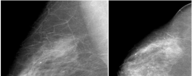
3 A B C Figure 2. Comparison of conventional mammography and breast tomosynthesis: potential of tomosynthesis to improve sensitivity. (A) The MLO-view mammogram of the left breast of a patient. A cancer (arrow) is obscured by superimposed breast tissues. (B) A tomosynthesis slice located 12 mm under the breast surface, which shows normal breast tissue. (C) A tomosynthesis slice located 28 mm under the breast surface that clearly shows the cancer (arrow).
4 3A

5 Z = 10 mm Z = 20 mm Z = 30 mm Z = 40 mm 3B
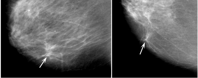
6 Figure 3. Comparison of conventional mammography and breast tomosynthesis: potential of tomosynthesis to improve specificity. (A) Two mammogram views show a finding with illdefined boundary (marked by arrows) in the left breast of a patient. (B) The finding is not shown in tomosynthesis slices from Z = 10 mm to 40 mm. The patient was called back and additional diagnostic mammograms proved that the finding in her original mammograms was caused by superimposed breast tissues. Contrast-Enhanced Imaging The "functional" information (i.e., the dynamics of tumor blood supply) of a lesion can be yielded by contrast agents. A contrast agent is usually required in breast MRI, which is approved by the FDA for use as a supplemental tool to mammography to help diagnose breast cancer. In the technique, patients get an injection of a gadolinium-based contrast agent that concentrates in abnormal breast tissue and lights up these regions. Although breast MR has very good sensitivity (detecting the abnormalities), its specificity (distinguishing between cancerous and noncancerous findings) is relatively poor, which may lead to unnecessary breast biopsies. In addition, MRI is an expensive exam and takes longer time (>30 minutes) than mammography. Contrast-enhanced digital mammography is being investigated, but it is still a 2D imaging technique compromised by overlapping structures. Combination of DBT and Contrast-Enhancement Our goal is to combine the advantages of DBT and contrast imaging. DBT is a 3D mammography technique while a contrast agent provides physiological information of the findings. Compared with 2D contrast-enhanced digital mammography, the superimposed enhanced breast tissue can be separated by DBT, so the morphology (shape) information of the enhanced lesion can be better characterized. Compared with breast MRI, contrast-enhanced DBT has significantly higher resolution (~0.1mm) than MRI (~1mm) because of the high spatial resolution of the digital detector used in DBT. The tumor can also be segmented from surrounding the tissue in order to measure the dynamic curve of the contrast as done in MR. The current experiments on contrast-enhanced DBT study the capability of DBT in separating superimposed tissues that are enhanced by contrast agent, as well as the change in contrast enhancement over time. The experiments on specimens are different than in vivo imaging because contrast agent is injected and flow in interstitial spaces in the specimen, and the change in contrast enhancement in a specimen is different than that in a patient s breast. Our result shows that DBT can separate superimposed tissues are enhanced by contrast agent and the borders of the enhanced features are much better characterized. The change in contrast enhancement over time (take-up and wash-out of the contrast agent) can also be qualitatively characterized. For in vivo imaging, some practical issues need to be studied such as breast compression, registration of pre- and post-injection images.
7 Figure 4A

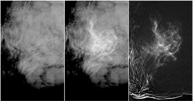
8 Figure 4B Figure 4. An experiment in contrast-enhanced tomosynthesis was performed using a 52 mm thick mastectomy specimen. The pre-contrast and post-contrast subtraction can be easily performed since the specimen did not move during the injection. (A) The pre-contrast (left) and post-contrast (middle) projections and the subtraction image (right). (B) Pre-contrast (left column) and post-contrast (middle column) tomosynthesis slices, and the subtraction images (right column). The depths of the three slices are 15 mm (1st row), 25 mm (2nd row) and 33 mm (3rd row) respectively. Structures and spaces between tissues in the specimen are enhanced in both projection and reconstruction images, while the reconstruction images separate the enhanced tissues and spaces that are overlapped in projection images.


9 2 mm 5A Z = 11 mm Z = 15 mm 5B 5C
10 Figure 5. Magnified field of views (FOVs) (A) A FOV on a projection image after subtraction. (B) A FOV at the same in-plane location on a tomosynthesis slice 11 mm under the specimen surface. (C) A FOV on another tomosynthesis slice 15 mm under the specimen surface. Two features that are enhanced by the contrast agent are marked by circles in (A). The conspicuity of the features is compromised by the overlapping structures that are also enhanced by the contrast agent. The features are better perceived in tomosynthesis slices (B) and (C), with their borders clearly characterized.
CURRENTLY FDA APPROVED ARE FULL FIELD DIGITAL MAMMOGRAPHY SYSTEMS AND FILM SCREEN STILL BEING USED AT SOME INSTITUTIONS
 ABBY DUROJAYE,M.D CURRENTLY FDA APPROVED ARE FULL FIELD DIGITAL MAMMOGRAPHY SYSTEMS AND FILM SCREEN STILL BEING USED AT SOME INSTITUTIONS BOTH HAVE BEEN SHOWN TO BE EFFECTIVE TOOLS EARLY DETECTION OF BREAST
Breast Tomosynthesis An additional screening tool in the fight against breast cancer
 What to Expect Breast Tomosynthesis An additional screening tool in the fight against breast cancer Every woman over 40 should be examined for breast cancer once a year. American Cancer Society What to
WHAT TO EXPECT. Breast Tomosynthesis An additional screening tool in the fight against breast cancer HOLOGIC. The Women's Health Company
 WHAT TO EXPECT Breast Tomosynthesis An additional screening tool in the fight against breast cancer HOLOGIC The Women's Health Company ...,. Screening for breast cancer Doctors and scientists agree that
Since its introduction in 2000, digital mammography has become
 Review Article Smith A, PhD email : Andrew.smith@hologic.com Since its introduction in 2000, digital mammography has become an accepted standard of care in breast cancer screening and has paved the way
Digital breast tomosynthesis
 GE Healthcare Digital breast tomosynthesis Daniel B. Kopans, M.D., F.A.C.R. Professor of Radiology Harvard Medical School Senior Radiologist - Breast Imaging Division Massachusetts General Hospital Since
A Breast Surgeon s Use of Three Dimensional Specimen Tomosynthesis
 A Breast Surgeon s Use of Three Dimensional Specimen Tomosynthesis Cary S. Kaufman MD, FACS Associate Clinical Professor of Surgery A Breast Surgeon s Use of Three Dimensional Specimen Tomosynthesis Cary
Update of Digital Breast Tomosynthesis. Susan Orel Roth, MD
 Update of Digital Breast Tomosynthesis Susan Orel Roth, MD NCI estimates that : Why DBT? Approximately 20% of breast cancers are missed at mammography screening Average recall rates approximately 10%
TOMOSYNTHESIS: WORTH ALL THE HYPE?
 X-Ray Associates of New Mexico, P.C. TOMOSYNTHESIS: WORTH ALL THE HYPE? MICHAEL N. LINVER, MD, FACR MAMMOGRAPHY: THE GOOD, THE PRETTY GOOD, & THE NOT SO GOOD MAMMOGRAPHY: THE GOOD, THE PRETTY GOOD, & THE
WHAT TO EXPECT. Genius 3D MAMMOGRAPHY Exam. The most exciting advancement in mammography in over 30 years
 WHAT TO EXPECT Genius 3D MAMMOGRAPHY Exam The most exciting advancement in mammography in over 30 years 91% of patients agree the quality of care provided by the facility was better with a Genius 3D MAMMOGRAPHY
3D Mammography. The most exciting advancement in mammography in over 30 years
 What to Expect 3D Mammography The most exciting advancement in mammography in over 30 years Screening for breast cancer Doctors and scientists agree that early detection is the best defense against breast
Breast tomosynthesis reduces radiologist performance variability compared to digital mammography
 Breast tomosynthesis reduces radiologist performance variability compared to digital mammography Andrew Smith 1, Elizabeth Rafferty 2, Loren Niklason 1 1 Hologic, Inc., Bedford MA, USA 2 Massachusetts
WHAT TO EXPECT. Genius 3D Mammography Exam. The most exciting advancement in mammography in over 30 years
 WHAT TO EXPECT Genius 3D Mammography Exam The most exciting advancement in mammography in over 30 years Screening for breast cancer Doctors and scientists agree that early detection is the best defense
Financial Disclosures
 Financial Disclosures 3D Mammography: The Latest Developments in the Breast Imaging Arena I have no financial disclosures Dr. Katharine Lampen-Sachar Breast and Body Radiologist Radiology Associates of
Digital Breast Tomosynthesis from a first idea to clinical routine
 International Master Programm Biomedical Engineering Digital Breast Tomosynthesis from a first idea to clinical routine Historical background 2D imaging of 3D objects has important limitations Jörg Barkhausen
Fundamentals of Breast Tomosynthesis
 Fundamentals of Breast Tomosynthesis Improving the Performance of Mammography Andrew Smith, Ph.D. This white paper is one in a series of research overviws on advanced technologies in women s healthcare.
the one name in cancer care.
 the one name in cancer care. Landmark study evaluating close to half a million mammography exams published in the Journal of the American Medical Association (JAMA) 1 Hologic 3D Mammography Significantly
Breast positioning system for full field digital mammography and digital breast tomosynthesis system
 Breast positioning system for full field digital mammography and digital breast tomosynthesis system Mari Varjonen* a, Martti Pamilo b, Pirjo Hokka b, Riina Hokkanen a, Pekka Strömmer a a Planmed Oy Asentajankatu
Breast Tomosynthesis. What is breast tomosynthesis?
 Scan for mobile link. Breast Tomosynthesis Breast tomosynthesis is an advanced form of mammography, a specific type of breast imaging that uses low-dose x-rays to detect cancer early when it is most treatable.
Mammography limitations. Clinical performance of digital breast tomosynthesis compared to digital mammography: blinded multi-reader study
 Clinical performance of digital breast tomosynthesis compared to digital mammography: blinded multi-reader study G. Gennaro (1), A. Toledano (2), E. Baldan (1), E. Bezzon (1), C. di Maggio (1), M. La Grassa
Corporate Medical Policy
 Corporate Medical Policy File Name: Origination: Last CAP Review: Next CAP Review: Last Review: digital_breast_tomosynthesis 3/2011 6/2016 6/2017 11/2016 Description of Procedure or Service Conventional
Volume 14 - Issue 3, Matrix
 Volume 14 - Issue 3, 2014 - Matrix Digital Breast Tomosynthesis for Screening and Diagnosis of Breast Cancer Author ECRI ECRI Institute 29 Broadwater Road Suite 104 Welwyn Garden City AL7 3BQ United Kingdom
WHAT TO EXPECT. Genius 3D MAMMOGRAPHY Exam. The most exciting advancement in mammography in over 30 years
 WHAT TO EXPECT Genius 3D MAMMOGRAPHY Exam The most exciting advancement in mammography in over 30 years 91% of patients agree the quality of care provided by the facility was better with a Genius 3D MAMMOGRAPHY
8/3/2016. DBT Physics Basic to Advanced: Primer On Tomosynthesis. Tomosynthesis Pedigree
 DBT Physics Basic to Advanced: Primer On Tomosynthesis Andrew D. A. Maidment, Ph.D. University of Pennsylvania Department of Radiology Acknowledgements of Support Research support from the Komen Foundation,
Emerging Techniques in Breast Imaging: Contrast-Enhanced Mammography and Fast MRI
 Emerging Techniques in Breast Imaging: Contrast-Enhanced Mammography and Fast MRI Lilian Wang, M.D. Breast Imaging Section Department of Radiology Northwestern Medicine Overview Rationale for new imaging
Standard Breast Imaging Modalities. Lilian Wang, M.D. Breast Imaging Section Department of Radiology Northwestern Medicine
 Standard Breast Imaging Modalities Lilian Wang, M.D. Breast Imaging Section Department of Radiology Northwestern Medicine Overview Standard breast imaging modalities Mammography Ultrasound MRI Imaging
Contrast-Enhanced Digital Mammography
 2015 ARRS Breast Symposium Contrast-Enhanced Digital Mammography John Lewin, M.D. Diversified Radiology of Colorado CEDM - Outline History Technique Literature Review / Cases Clinical Status Inexpensive,
EARLY DETECTION: MAMMOGRAPHY AND SONOGRAPHY
 EARLY DETECTION: MAMMOGRAPHY AND SONOGRAPHY Elizabeth A. Rafferty, M.D. Avon Comprehensive Breast Center Massachusetts General Hospital Harvard Medical School Breast Cancer Screening Early detection of
Epworth Healthcare Benign Breast Disease Symposium. Sat Nov 12 th 2016
 Epworth Healthcare Benign Breast Disease Symposium Breast cancer is common Sat Nov 12 th 2016 Benign breast disease is commoner, and anxiety about breast disease commoner still Breast Care Campaign UK
THE DIAGNOSTIC WORKUP: THE TEAM APPROACH
 X-Ray Associates of New Mexico, P.C. THE DIAGNOSTIC WORKUP: THE TEAM APPROACH MICHAEL N. LINVER, MD, FACR DAWN DERENBURGER, RTRM Disclosure There are no conflicts of interest or relevant financial interests
The Radiology Aspects
 REQUIREMENTS FOR INTERNATIONAL ACCREDITATION OF BREAST CENTERS/UNITS The Radiology Aspects Miri Sklair-Levy, Israel RADIOLOGY GUIDELINES FOR QUALITY ASSURANCE IN BREAST CANCER SCREENING AND DIAGNOSIS Radiologists
Breast Cancer Screening and High Risk
 Breast Cancer Screening and High Risk Mary Freyvogel, DO Breast Surgeon Clinical Assistant Professor of Surgery University Hospitals Case Medical Center St. John Medical Center / Elyria Medical Center
#46: DIGITAL TOMOSYNTHESIS: What is the Data Really Showing? TERMS (AKA) WHAT IS TOMOSYNTHESIS? 3/3/2014. Digital breast tomosynthesis =
 WHAT IS TOMOSYNTHESIS? 3/3/2014. Digital breast tomosynthesis =") #46: DIGITAL TOMOSYNTHESIS: What is the Data Really Showing? January K. Lopez, MD Hoag Breast Care Center Newport Beach, CA Disclosures: None TERMS (AKA) Digital breast tomosynthesis = DBT Tomo 3D Full
#46: DIGITAL TOMOSYNTHESIS: What is the Data Really Showing? January K. Lopez, MD Hoag Breast Care Center Newport Beach, CA Disclosures: None TERMS (AKA) Digital breast tomosynthesis = DBT Tomo 3D Full
Image processing mammography applications
 Image processing mammography applications Isabelle Bloch Isabelle.Bloch@telecom-paristech.fr http://perso.telecom-paristech.fr/bloch LTCI, Télécom ParisTech Mammography p.1/27 Image processing for mammography
Radiation Dosimetry in Digital Breast Tomosynthesis. March, 2015 William J. O Connel, Dr. Ph, Senior Medical Physicist
 Radiation Dosimetry in Digital Breast Tomosynthesis March, 2015 William J. O Connel, Dr. Ph, Senior Medical Physicist Imagination at work. Syllabus 1. Introduction 2. Dosimetry in Mammography 3. Dosimetry
Pitfalls and Limitations of Breast MRI. Susan Orel Roth, MD Professor of Radiology University of Pennsylvania
 Pitfalls and Limitations of Breast MRI Susan Orel Roth, MD Professor of Radiology University of Pennsylvania Objectives Review the etiologies of false negative breast MRI examinations Discuss the limitations
Women s Imaging Original Research
 Women s Imaging Original Research Brandt et al. DBT for Screening Recalls Without Calcifications Women s Imaging Original Research FOCUS ON: Kathleen R. Brandt 1 Daniel A. Craig 1 Tanya L. Hoskins 2 Tara
Updates in Mammography. Dr. Yang Faridah A. Aziz Department of Biomedical Imaging University Malaya Medical Centre
 Updates in Mammography Dr. Yang Faridah A. Aziz Department of Biomedical Imaging University Malaya Medical Centre Updates in Mammography Breast Imaging Dr. Yang Faridah A. Aziz Department of Biomedical
Mammographic imaging of nonpalpable breast lesions. Malai Muttarak, MD Department of Radiology Chiang Mai University Chiang Mai, Thailand
 Mammographic imaging of nonpalpable breast lesions Malai Muttarak, MD Department of Radiology Chiang Mai University Chiang Mai, Thailand Introduction Contents Mammographic signs of nonpalpable breast cancer
Mammography. What is Mammography?
 Scan for mobile link. Mammography Mammography is a specific type of breast imaging that uses low-dose x-rays to detect cancer early before women experience symptoms when it is most treatable. Tell your
Diagnostic Medical Physicist Via Christi Hospitals Wichita, Wichita, KS
 Digital Breast Tomosynthesis SWAAPM Meeting 30 Mar 2012 Jerry A. Thomas, MS, FAAPM, DABR, CHP, DABSNM Diagnostic Medical Physicist Via Christi Hospitals Wichita, Wichita, KS Talk Overview Breast Cancer
Advances in Breast Cancer Diagnosis and Treatment. Heidi Memmel, MD FACS Surgical Director of Caldwell Breast Center September 26, 2015
 Advances in Breast Cancer Diagnosis and Treatment Heidi Memmel, MD FACS Surgical Director of Caldwell Breast Center September 26, 2015 Advances in Breast Cancer Diagnosis and Treatment Recommendations
The Future of Breast MRI Improving Outcomes
 The Future of Breast MRI Improving Outcomes Connie Lehman MD PhD Professor of Radiology Harvard Medical School Director of Breast Imaging Massachusetts General Hospital Opportunities New technology provides
Anyone can get breast cancer BREAST MRI BREAST CANCER. The incidence of getting breast cancer is 1:19 in Malaysia
 Anyone can get breast cancer BREAST MRI KATE Datin Dr Fatimah Moosa Sunway Medical Centre DATIN SERI ENDON KYLIE SIZE DOES NOT MAKE A DIFFERENCE BREAST CANCER The incidence of getting breast cancer is
What s New in Breast Imaging. Jennifer A. Harvey, M.D., FACR Professor of Radiology University of Virginia
 What s New in Breast Imaging Jennifer A. Harvey, M.D., FACR Professor of Radiology University of Virginia Disclosure Hologic, Inc. Shareholder and research agreement. Volpara Solutions, Ltd. Shareholder
Digital breast tomosynthesis (DBT) occult breast cancers: clinical, radiological and histopathological features.
 occult breast cancers: clinical, radiological and histopathological features.") Digital breast tomosynthesis (DBT) occult breast cancers: clinical, radiological and histopathological features. Poster No.: C-1707 Congress: ECR 2015 Type: Scientific Exhibit Authors: V. Vinci 1, A. Iqbal
Digital breast tomosynthesis (DBT) occult breast cancers: clinical, radiological and histopathological features. Poster No.: C-1707 Congress: ECR 2015 Type: Scientific Exhibit Authors: V. Vinci 1, A. Iqbal
BREAST IMAGING and NEW IMAGING MODALITIES- A Surgeons view
 BREAST IMAGING and NEW IMAGING MODALITIES- A Surgeons view DR CHANTEL THORNTON SPECIALIST BREAST CANCER SURGEON BMSc (hons) MBBS (hons) FRACS Epworth Hospital, Richmond- Agora Centre for Women s Health
Digital Breast Tomosynthesis: A Technological Review
 Digital Breast Tomosynthesis: A Technological Review Matija Males 1, Danijel Mileta 2, Mislav Grgic 1 1 University of Zagreb, Faculty of EE and Comp, Unska 3/XII, HR-10000 Zagreb, Croatia 2 Ministry of
Current Status of Supplementary Screening With Breast Ultrasound
 Current Status of Supplementary Screening With Breast Ultrasound Stephen A. Feig, M.D., FACR Fong and Jean Tsai Professor of Women s Imaging Department of Radiologic Sciences University of California,
Here are examples of bilateral analog mammograms from the same patient including CC and MLO projections.
 Good afternoon. It s my pleasure to be discussing Diagnostic Breast Imaging over the next half hour. I m Wei Yang, Professor of Diagnostic Radiology and Chief, the Section of Breast Imaging as well as
Contrast Enhanced Spectral Mammography (CESM) Updates
 Updates") Contrast Enhanced Spectral Mammography (CESM) Updates Georgeta Mihai, PhD, DABR Medical Physicist, BIDMC, Boston Assistant Professor, Harvard Medical School, Boston Disclosures None Acknowledgments: Da
Contrast Enhanced Spectral Mammography (CESM) Updates Georgeta Mihai, PhD, DABR Medical Physicist, BIDMC, Boston Assistant Professor, Harvard Medical School, Boston Disclosures None Acknowledgments: Da
Breast Imaging & You
 Breast Imaging & You What s Inside: Breast Imaging... 2 Digital Breast Tomosynthesis (DBT) mammograms... 4 Breast cancer screening... 6 Dense breast tissue... 8 Automated Breast Ultrasound (ABUS)... 9
Tomosynthesis and breast imaging update. Dr Michael J Michell Consultant Radiologist King's College Hospital NHS Foundation Trust
 Tomosynthesis and breast imaging update Dr Michael J Michell Consultant Radiologist King's College Hospital NHS Foundation Trust Breast imaging new technology BREAST CANCER FLT PET shows different grades
FDA Executive Summary
 Meeting of the Radiological Devices Advisory Panel On October 24, 22, the panel will discuss, make recommendations, and vote on a premarket approval application supplement (P83/S) to expand the indications
Screening Mammography: Who, what, where, when, why and how?
 Screening Mammography: Who, what, where, when, why and how? Jillian Lloyd, MD, MPH Breast Surgical Oncologist University Surgical Oncology Department of Surgery University of Tennessee Medical Center Disclosures
Outline. Digital Breast Tomosynthesis: Update and Pearls for Implementation. Tomosynthesis Dataset: 2D/3D (Hologic Combo Acquisition)
") Outline Digital Breast Tomosynthesis (DBT) the new standard of care Digital Breast Tomosynthesis: Update and Pearls for Implementation Emily F. Conant, M.D. Professor, Chief of Breast Imaging Department
Outline Digital Breast Tomosynthesis (DBT) the new standard of care Digital Breast Tomosynthesis: Update and Pearls for Implementation Emily F. Conant, M.D. Professor, Chief of Breast Imaging Department
MANAGEMENT OF DENSE BREASTS. Nichole K Ingalls, MD, MPH NW Surgical Specialists September 25, 2015
 MANAGEMENT OF DENSE BREASTS Nichole K Ingalls, MD, MPH NW Surgical Specialists September 25, 2015 No financial disclosures National Cancer Institute National Cancer Institute Increased Cancer Risk... DENSITY
AN ALGORITHM FOR EARLY BREAST CANCER DETECTION IN MAMMOGRAMS
 AN ALGORITHM FOR EARLY BREAST CANCER DETECTION IN MAMMOGRAMS Isaac N. Bankman', William A. Christens-Barryl, Irving N. Weinberg2, Dong W. Kim3, Ralph D. Semmell, and William R. Brody2 The Johns Hopkins
Breast Tomosynthesis
 Breast Tomosynthesis The Use of Breast Tomosynthesis in a Clinical Setting 2 What s Inside Introduction... 1 Initial Hologic Clinical Trial Purpose and Methodology... 1 Clinical Trial Results... 2 Improved
arxiv: v2 [cs.cv] 8 Mar 2018
![arxiv: v2 [cs.cv] 8 Mar 2018](/thumbs/87/97094636.jpg "arxiv: v2 [cs.cv] 8 Mar 2018") Automated soft tissue lesion detection and segmentation in digital mammography using a u-net deep learning network Timothy de Moor a, Alejandro Rodriguez-Ruiz a, Albert Gubern Mérida a, Ritse Mann a, and
Automated soft tissue lesion detection and segmentation in digital mammography using a u-net deep learning network Timothy de Moor a, Alejandro Rodriguez-Ruiz a, Albert Gubern Mérida a, Ritse Mann a, and
Correlation between lesion type and the additional value of digital breast tomosynthesis
 Correlation between lesion type and the additional value of digital breast tomosynthesis Poster No.: C-1604 Congress: ECR 2011 Type: Scientific Exhibit Authors: C. Van Ongeval, L. Cockmartin, A. Van Steen,
CenSSIS Multimode Cancer Imaging via Digital Breast Tomosynthesis
 CenSSIS Multimode Cancer Imaging via Digital Breast Tomosynthesis RPI Electrical Impedance Laboratory MGH Diffuse Optical Tomography Laboratory MGH Breast Imaging Laboratory Richard Moore Gordon-CenSSIS
Breast Imaging & You
 Breast Imaging & You What s Inside: Breast Imaging... 2 Digital Breast Tomosynthesis (DBT) mammograms... 4 Breast cancer screening... 6 Dense breast tissue... 8 Automated breast ultrasound (ABUS)... 9
Supplemental Screening for Dense Breasts. Reagan Leverett, MD, MS
 Supplemental Screening for Dense Breasts Reagan Leverett, MD, MS Outline Anatomy and Density Risk of dense breasts Theory of Supplemental Screening Options for supplemental screening Tomosynthesis Ultrasound
Medical Policy An independent licensee of the Blue Cross Blue Shield Association
 Digital Breast Tomosynthesis Page 1 of 31 Medical Policy An independent licensee of the Blue Cross Blue Shield Association Title: Digital Breast Tomosynthesis Professional Institutional Original Effective
Armed Forces Institute of Pathology.
 Armed Forces Institute of Pathology www.radpath.com Armed Forces Institute of Pathology Breast Disease www.radpath.org Armed Forces Institute of Pathology Interpretation of Breast MRI Leonard M. Glassman
Henda s Law. Supplemental screening for women with dense breast tissue and increased risk
 . Henda s Law Supplemental screening for women with dense breast tissue and increased risk The 2011 Texas Legislature passed House Bill 2102 which is effective 1st September 2011. The law is informally
Benign Intraparenchymal Scarring in the DBT Era
 Benign Intraparenchymal Scarring in the DBT Era David Gruen, MD, MBA, FACR Director of Women s Imaging, Stamford Health, Stamford, CT With the advent of Digital Breast Tomosynthesis (DBT or 3D mammography),
Why Choose Breast Radiology?
 Why Choose Breast Radiology? Hannah Gay ST6, St George s Hospital BSBR exec committee trainee representative. Introduction Breast cancer is the most common cancer in the UK. Lifetime risk of 1 in 8 for
Case Report Tubular Carcinoma of the Breast: Advantages and Limitations of Breast Tomosynthesis
 Case Reports in Radiology Volume 2016, Article ID 3906195, 4 pages http://dx.doi.org/10.1155/2016/3906195 Case Report Tubular Carcinoma of the Breast: Advantages and Limitations of Breast Tomosynthesis
Molecular Breast Imaging: History and Recent Developments
 Molecular Breast Imaging: History and Recent Developments Associate Professor, Department of Imaging Physics The University of Texas MD Anderson Cancer Center, Houston, Texas Educational Objectives 1.
Breast Tomosynthesis
 Breast Tomosynthesis The Use of Breast Tomosynthesis in a Clinical Setting 2 What s Inside Introduction... 1 Initial Hologic Clinical Trial Purpose and Methodology... 1 Clinical Trial Results... 2 Improved
EARLY DETECTION: MAMMOGRAPHY AND SONOGRAPHY
 EARLY DETECTION: MAMMOGRAPHY AND SONOGRAPHY Elizabeth A. Rafferty, M.D. Avon Comprehensive Breast Center Massachusetts General Hospital Harvard Medical School Breast Cancer Screening Early detection of
Breast MRI, digital mammography and breast tomosynthesis: Comparison of three methods for early detection of breast cancer
 BOSNIAN JOURNAL OF BASIC MEDICAL SCIENCES RESEARCH ARTICLE WWW.BJBMS.ORG Breast MRI, digital mammography and breast tomosynthesis: Comparison of three methods for early detection of breast cancer Dragana
Innovations and Applications of Tomosynthesis. Andrew D. A. Maidment, Ph.D. University of Pennsylvania Department of Radiology
 Innovations and Applications of Tomosynthesis Andrew D. A. Maidment, Ph.D. University of Pennsylvania Department of Radiology Acknowledgements of Support Grant support from the Komen Foundation, DOD, NIH,
Policy Library Clinical Advantages of Digital Breast Tomosynthesis in Symptomatic Patients
 Policy Library Clinical Advantages of Digital Breast Tomosynthesis in Symptomatic Patients Version: 1 Approved by: Faculty of Clinical Radiology Council Date of approval: Click and type: day month and
Digital Breast Tomosynthesis in the Diagnostic Environment: A Subjective Side-by-Side Review
 Women s Imaging Original Research Hakim et al. Digital Breast Tomosynthesis Women s Imaging Original Research Christiane M. Hakim 1 Denise M. Chough 1 Marie A. Ganott 1 Jules H. Sumkin 1 Margarita L. Zuley
Breast asymmetries in mammography: Management
 Breast asymmetries in mammography: Management Poster No.: C-1026 Congress: ECR 2015 Type: Educational Exhibit Authors: V. de Lara Bendahan 1, F. J. Hidalgo Ramos 2, J. L. Ortega Garcia 3, Keywords: DOI:
Experience with Tomosynthesis (3-D) Mammography in a Mobile Setting and Current Controversies
 Mammography in a Mobile Setting and Current Controversies") Experience with Tomosynthesis (3-D) Mammography in a Mobile Setting and Current Controversies Jennifer Lance, MHSA, MS Jerry McLarty, PhD LSU Feist-Weiller Cancer Center Shreveport LA Sept 15, 2008 A brief
Experience with Tomosynthesis (3-D) Mammography in a Mobile Setting and Current Controversies Jennifer Lance, MHSA, MS Jerry McLarty, PhD LSU Feist-Weiller Cancer Center Shreveport LA Sept 15, 2008 A brief
DOUG LAKE, MD, MRMD (MRSC) RADIOLOGIST, MCFARLAND CLINIC, PC ADJUNCT CLINICAL ASSISTANT PROFESSOR, DEPARTMENT OF RADIOLOGY, STANFORD HEALTH CARE
 RADIOLOGIST, MCFARLAND CLINIC, PC ADJUNCT CLINICAL ASSISTANT PROFESSOR, DEPARTMENT OF RADIOLOGY, STANFORD HEALTH CARE") DOUG LAKE, MD, MRMD (MRSC) RADIOLOGIST, MCFARLAND CLINIC, PC ADJUNCT CLINICAL ASSISTANT PROFESSOR, DEPARTMENT OF RADIOLOGY, STANFORD HEALTH CARE ADVANCES IN DIAGNOSTIC IMAGING DISCLOSURES I own shares
DOUG LAKE, MD, MRMD (MRSC) RADIOLOGIST, MCFARLAND CLINIC, PC ADJUNCT CLINICAL ASSISTANT PROFESSOR, DEPARTMENT OF RADIOLOGY, STANFORD HEALTH CARE ADVANCES IN DIAGNOSTIC IMAGING DISCLOSURES I own shares
now a part of Electronic Mammography Exchange: Improving Patient Callback Rates
 now a part of Electronic Mammography Exchange: Improving Patient Callback Rates Overview This case study explores the impact of a mammography-specific electronic exchange network on patient callback rates
TOMOSYNTHESIS. Daniela Bernardi. U.O. Senologia Clinica e Screening mammografico APSS Trento, Italy
 TOMOSYNTHESIS Daniela Bernardi U.O. Senologia Clinica e Screening mammografico APSS Trento, Italy BACKGROUND early detection through screening MAMMOGRAPHY is associated with reduced breast cancer morbidity
1. Screening, Diagnosis and Surgical Management of Breast Cancer
 1. Screening, Diagnosis and Surgical Management of Breast Cancer Dr Melanie Walker, MBBS, FRACS (Breast Surgeon) Oncoplastic Breast Surgery Combination of optimal cancer surgery with plastic surgical techniques
Effect of non- homogeneous breast 3ssue on mean glandular dose assessment in digital breast tomosynthesis
 Effect of nonhomogeneous breast 3ssue on mean glandular dose assessment in digital breast tomosynthesis M. Bap3sta, S. Di Maria, C. Figueira, L. Orvalho, A. Silva, P. Vaz, M. Zankl o Mo3va3on Mammography
Breast Imaging Update: Old Dog New Tricks
 Breast Imaging Update: Old Dog New Tricks Claire McKay, DO M&S Imaging Assoc. San Antonio, TX cmckayhart@juno.com Goals Describe modalities available, old and new Provide understanding of pros and cons
Look differently. Invenia ABUS. Automated Breast Ultrasound
 Look differently. Invenia ABUS Automated Breast Ultrasound InveniaTM ABUS from GE Healthcare offers a view beyond mammography, with breast screening technology that looks differently. 40 % The unseen risk.
Objectives. Explanation of Radiation Dose Terminology 10/9/2018. What are these lines?
 Oh no, it s not!!! with Breast Tomosynthesis It s not what you may be thinking, so I ll tell you! IT IS A VIETNAMESE PUMPKIN! Oh yes, it is! Objectives Explain radiation dose terminology Recognize 2D and
What Does Mammography Follow Up Involve?
 IOWA RADIOLOGY 1 What Does Mammography Follow Up Involve? 515-226-9810 Ankeny Clive Downtown Des Moines Lakeview IOWA RADIOLOGY 2 Table of Contents Introduction... 1 Imaging... 2 Mammography and Ultrasound...
Melissa Hartman, DO Women s Health Orlando VA Medical Center
 Melissa Hartman, DO Women s Health Orlando VA Medical Center Most common non-skin cancer and Second deadliest cancer in women Majority are diagnosed by abnormal screening study An approach to breast cancer
RECENT ADVANCES IN CLINICAL MR OF ARTICULAR CARTILAGE
 In Practice RECENT ADVANCES IN CLINICAL MR OF ARTICULAR CARTILAGE By Atsuya Watanabe, MD, PhD, Director, Advanced Diagnostic Imaging Center and Associate Professor, Department of Orthopedic Surgery, Teikyo
BREAST HEALTH SOLUTIONS. See what others miss.
 HMH 3D Mammography BREAST HEALTH SOLUTIONS See what others miss. 2 BREAST HEALTH SOLUTIONS Genius 3D MAMMOGRAPHY exams are clinically proven to detect 41% More invasive breast cancer, while simultaneously
Can Digital Breast Tomosynthesis(DBT) Perform Better than Standard Digital Mammography Workup in a Breast Cancer Assessment Clinic?
 Perform Better than Standard Digital Mammography Workup in a Breast Cancer Assessment Clinic?") Can Digital Breast Tomosynthesis(DBT) Perform Better than Standard Digital Mammography Workup in a Breast Cancer Assessment Clinic? Accepted for publication in European Radiology Authors: S Mall, J Noakes,
Can Digital Breast Tomosynthesis(DBT) Perform Better than Standard Digital Mammography Workup in a Breast Cancer Assessment Clinic? Accepted for publication in European Radiology Authors: S Mall, J Noakes,
Digital Breast Tomosynthesis
 Digital Breast Tomosynthesis Policy Number: Original Effective Date: MM.05.012 06/28/2013 Line(s) of Business: Current Effective Date: HMO; PPO; QUEST 06/28/2013 Section: Radiology Place(s) of Service:
Opportunities and Innovations in Digital Mammography John M. Sandrik, Ph.D. GE Healthcare Milwaukee, WI
 Opportunities and Innovations in Digital Mammography John M. Sandrik, Ph.D. GE Healthcare Milwaukee, WI john.sandrik@med.ge.com with many thanks to Vince Polkus, Advanced Applications Product Mgr. 1 Content
8/31/2016 HIDING IN PLAIN SITE, ARCHITECTURAL DISTORTIONS AND BREAST ASYMMETRIES ARCHITECTURAL DISTORTIONS ARCHITECTURAL DISTORTIONS
 HIDING IN PLAIN SITE, ARCHITECTURAL DISTORTIONS AND BREAST ASYMMETRIES DEBORAH THAMES R.T. (R)(M)(QM) ARCHITECTURAL DISTORTIONS Definition is disruption of the natural flow of breast pattern towards the
Improving Reading Time of Digital Breast Tomosynthesis with Concurrent Computer Aided Detection
 White Paper Improving Reading Time of Digital Breast Tomosynthesis with Concurrent Computer Aided Detection WHITE PAPER 2 3 Abstract PowerLook Tomo Detection, a concurrent computer-aided detection (CAD)
Screening Mammograms: Questions and Answers
 CANCER FACTS N a t i o n a l C a n c e r I n s t i t u t e N a t i o n a l I n s t i t u t e s o f H e a l t h D e p a r t m e n t o f H e a l t h a n d H u m a n S e r v i c e s Screening Mammograms:
Detailed Program of the second BREAST IMAGING AND INTERVENTIONS PROGRAM am am : Clinician s requirements from breast imaging
 Detailed Program of the second BREAST IMAGING AND INTERVENTIONS PROGRAM 2012 Day one, 2 nd November BREAST IMAGING AND INTERVENTIONS PROGRAM 2012 9.00 AM 9.10 am Introduction 9.10 am - 9.30 am : Clinician
Ge elastography cpt codes
 Ge elastography cpt codes Aetna considers digital mammography a medically necessary acceptable alternative to film mammography. Currently, there are no guideline recommendations from leading medical professional
B R E A S T I M A G I N G S O L U T I O N S. Selenia Dimensions A Revolution in Breast Imaging
 B R E A S T I M A G I N G S O L U T I O N S Selenia Dimensions A Revolution in Breast Imaging The promise of breast tomosynthesis is here Hologic has been at the forefront of the industry s transformation
Full ultrasound breast volumes. Faster scans. Streamlined workflow. ACUSON S2000 Automated Breast Volume Scanner. Answers for life.
 Full ultrasound breast volumes. Faster scans. Streamlined workflow. ACUSON S2000 Automated Breast Volume Scanner Answers for life. 1 ACQUIRE An automated whole breast solution. Reduced acquisition time.