Mammography. Background and Perspective. Mammography Evolution. Background and Perspective. T.R. Nelson, Ph.D. x41433
|
|
|
- Ashlyn Elliott
- 6 years ago
- Views:
Transcription
1 Background and Perspective 2005 (in US) Women Men Mammography Invasive Breast Cancer Diagnosed 211,240 1,690 Noninvasive Breast Cancer Diagnosed 58,940 Deaths from Breast Cancer 40, T.R. Nelson, Ph.D. x41433 Background and Perspective Breast Cancer is the most common form of cancer among women other than skin cancer Breast Cancer is the second leading cause of cancer death in women after lung cancer A woman's chance of developing breast cancer increases significantly with age. A woman's chance of developing breast cancer sometime in her lifetime is approximately 1 in 7 The chance that breast cancer will be responsible for a woman's death is about 1 in 33 If detected in the earliest stages, the five-year survival rate for breast cancer is 98%. Mammography Evolution Introduced in the early 1950 s little medical benefit poor image quality Xerography introduced in 70 s and 80 s high dose good resolution and edge enhancement poor contrast sensitivity ACR mammography accreditation program started mid 80 s improved quality control improved dosimetry Mammography Quality Standards Act (MQSA) passed in 1992


2 Mammography Evolution Complementary Imaging Technology Ultrasound Cyst / solid differentiation Biopsy guidance Operator dependent MRI High contrast sensitivity Visualize silicone implants Breast cancer staging Generally requires contrast Thermography Infrared imaging State-of-the-Art - Full Field Digital Mammography X-ray mammography is current gold standard Cancer identification High contrast between normal and cancer tissues Detects calcifications
 x100 Gland")
3 Soft Tissue Contrast - Breast Mammography Equipment Contrast = (Gland - Cancer) x100 Gland C D B A
4 Mammography Equipment - X-ray Tube Design Anode Molybdenum Rhodium Tungsten Characteristic radiation Molybdenum , 19.6 kev Rhodium , 22.7 kev Anode Angle determines field-of-view Cathode over chest wall Anode over nipple Grounded to reduce space charge Mammography Equipment - X-ray Tube Design

5 Mammography Equipment - Anode - Cathode Axis Mammography Equipment - Focal Spots Small focal spots Reduce blurring High magnification Size depends on use mm for contact imaging mm for magnification imaging Size depends on SID 0.4 mm for SID > 66 cm 0.3 mm for SID < 65 cm Size varies with position in field Mammography Equipment - Focal Spot Size Variation Mammography Equipment - Focal Spot Size Variation Focal spot size estimated with slit camera or pinhole camera. Effective resolution measured with bar pattern (up to 20 lp/mm). Measurement incorporates contribution of all components (i.e. image receptor, focal spot, tube motion, etc.)
6 Mammography Equipment - Beam Quality
7 Mammography Equipment - Filtration X-ray Beam Filtration: Inherent: ~1 mm Be Added: Mo, Rh eliminates low (and high) energy x-rays Mammography Equipment - Filtration Mammography Equipment - Filtration
8 Mammography Equipment - Filtration Mammography Equipment - Filtration Tungsten (W) characteristic x-rays from L shell Mammography Equipment - Filtration


9 Mammography Equipment - HVL Half-value Layer (HVL) Reflects beam hardness How much soft radiation is present in beam Small HVL - too much soft radiation Dose without information Large HVL - more penetrating beam Aged or pitted anode Too much filtration Depends on Inherent filtration Added filtration Compression paddle composition Mammography Equipment - HVL Mammography Equipment - HVL
10 Mammography Equipment - Tube Output Mammography Equipment - Collimation Must have at least 7.0 mgy/sec at 28 kvp in Mo/Mo mode to meet MQSA Light - x-ray field congruence to <1% for any edge and < 2% overall Must extend to chest wall


11 Mammography Equipment - Collimation Mammography Equipment - Exposure Control Automatic Exposure Control Uses sensor to monitor exposure Shuts off at pre-determined amount of radiation Compensates for breast thickness and density May use short (< 100 ms) pre-exposure to set technique Designed to produce optimum and consistent density Adjustments to increase/decrease density Includes a backup timer Mammography Equipment - Exposure Control Mammography Equipment - Technique Chart


12 Mammography Equipment - Compression Mammography Equipment - Compression Improves image quality, reduces motion, decreases thickness, decreases blurring, lowers radiation dose Mammography Equipment - Compression Spreads tissue over localized area
13 Mammography Equipment - Scatter - Primary Ratio

14 Mammography Equipment - Anti-scatter Grids - Magnification Source - Object Distance $"!#,"!#+" M = 1; 0.3 mm Focal Spot; 200 um Pixel Size M = 2; 0.3 mm Focal Spot; 200 um Pixel Size M = 3; 0.3 mm Focal Spot; 200 um Pixel Size M = 1; 0.1 mm Focal Spot; 200 um Pixel Size M = 2; 0.1 mm Focal Spot; 200 um Pixel Size M = 3; 0.1 mm Focal Spot; 200 um Pixel Size!#*" Object - Image Detector Distance MTF of the focal spot decreases with increasing magnification MTF of the detector increases with magnification Optimum magnification depends on both focal spot MTF and detector MTF Geometric magnification derives no resolution improvement from large focal spot - focal spot blur dominates Geometric magnification shows resolution improvement when detector has large detector pixels sizes - smaller effective pixel size!"#$!#)"!#("!#'"!#&"!#%"!#$" The focal spot MTF degraded with magnification while the detector MTF improved with magnification. Representa- tive results shown in Fig. 1 demonstrate that the improvement of the MTF depended on the tradeoff between focal spot size and pixel size. A large focal spot (0.6 mm) resulted in little or no resolution improvement with the use of geometric magnification. Since the focal spot blur dominated the system sharpness, reducing the effective pixel size did not compensate for the loss of resolution. A focal spot of 0.3 mm and pixel sizes of 50, 100, and 150 um showed an improvement in resolution for lower frequencies but not for higher frequencies suggesting a task dependent tradeoff for this combination (i.e., depending on the characteristics of the features that need to be imaged, different parameters may be optimal). A 0.3 mm focal spot with a 200 um pixel size showed an improved MTF for all magnification values although there was an optimum magnification. Magnification with a 0.1 mm focal spot resulted in improved MTF out to very high frequencies regardless of pixel size; specifically for large pixel sizes as the resolution of systems with large pixel sizes and small focal spots were dominated by the pixel size. Magnification in such systems resulted in a smaller effective pixel size thus increasing the overall system resolution. Geometric magnification increased the cut-off frequency of the system. The MTF was seen to improve with magnification for lower frequencies but a crossover point occurred for most geometries, Object where the Plane focal spot blurring Resolution became more dominant. The improvement in the MTF!"!" (" $" $!" %" resolution of $(" the &" system. %!" '" %(" &!" )" %&'(')$#*+,-+./0$1)&2334$ was particularly noteworthy for systems with larger pixel sizes and smaller focal spot widths since the effective pixel size in the object plane was reduced by magnification, thus reducing the overall Boyce et.al. Imaging properties of digital magnification radiography, Medical Physics, 2006


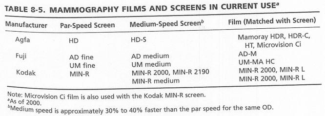
15 Mammography Equipment - Screen-Film Systems Mammography Equipment - Screen-Film Systems Mammography Equipment - Screen-Film Systems Mammography Equipment - Screen-Film Systems Limiting resolution approximately 20 lp/mm Requires approximately mr exposure for optimal OD Regular film requires approximately 2 mr


16 Mammography Equipment - Screen-Film Systems Mammography Equipment - Screen-Film Systems Processing quality control is essential to mammography success
17 Mammography Equipment - Screen-Film Systems Mammography Equipment - Screen-Film Systems Peak location shows where greatest contrast occurs. Provides sensitive means to monitor processor QC over time Mammography Equipment - Screen-Film Systems Extended processing used to improve performance in standard film processors Mammography Equipment - Viewing Conditions Optimal viewing requires: High view box / monitor luminance (cd/m 2 ) Mammography view box 3000 cd/m 2 Standard view box 1500 cd/m 2 Masking of non-image areas on view box Low ambient light levels (< 50 lux or lumens/m 2 ) Moon ~ 1 lux Normal room lighting ~ lux Bright light should be available

 Digital image -> input to")


18 Mammography Equipment - FFDM Mammography Equipment - FFDM Digital Imaging Detector Large dynamic range Reasonable spatial resolution (300 µm) Digital image -> input to CAD system Expensive ~ $300k Digital Detector Film-Screen Mammography Equipment - Stereotatic Biopsy Mammography Equipment - Stereotatic Biopsy Small field of view: 25 x 25 mm 1k x 1k CCD detector - 25 µm


 depicts a")

19 Mammography Equipment - Stereotatic Biopsy Mammography Equipment - Tomosynthesis Tomosynthesis comparable dose to Projetion Mammography Equipment - Tomosynthesis Mammography Equipment - Tomosynthesis Cranial-caudal conventional mammography view (A) of a middle-aged woman presenting with a palpable mass indicated by a metallic BB marker. Tomosynthesis 1 mm thick image (B) depicts a circumscribed mass (arrow) Mark A. Helvie,, Digital Mammography Imaging: Breast Tomosynthesis and Advanced Applications, Radiol Clin North Am. Sep 2010; 48(5):
 of imaging system Preferred image density Breast thickness Breast composition Fat")

20 Mammography Equipment - Tomosynthesis Vertical Section Projection Tomo Mammography Equipment - Dose Factors affecting dose Speed (efficiency) of imaging system Preferred image density Breast thickness Breast composition Fat less dense than gland kvp selected High kvp better penetration High kvp lower contrast High kvp lower dose Filtration used Mo or Rh Presence and type of grid Typical Bucky factor ~2x Mammography Equipment - Dose Mammography Equipment - Dose

21 Mammography Equipment - Dose Glandular Dose (in mrad) for 1 Roentgen Entrance Exposure 4.2-cm Breast Thickness Glandular Dose (in mrad) for 1 Roentgen Entrance Exposure 4.2-cm Breast Thickness Assumptions: 50% Adipose/50% Glandular Breast Tissue using a Mo/Mo Target-Filter Assumptions: 50% Adipose/50% Glandular Breast Tissue using a Mo/Rh Target-Filter kvp HVL Entrance Dose (mgy - Console) Entrance Dose (mgy - Measured) Glandular Dose (mgy - Console) Glandular Dose (mgy) mgy (meter/ese) HVL kvp Mammography Equipment - MQSA QA Mammography facilities must be: Accredited by ACR (or agreement states) Meet standards Initial qualifications of team Physicians, physicists, technologists Continuing education of team Continuing experience of team Equipment Quality control program Image quality Certified by FDA Granted when facility is accredited
22 Mammography Equipment - MQSA QA Mammography Equipment - MQSA QA Mammography Equipment - MQSA QA Mammography Equipment - MQSA QA


")
23 Mammography Equipment - MQSA QA Mammography Equipment - MQSA QA Mammography Equipment - MQSA QA Mammography T.R. Nelson, Ph.D. x41433 tnelson@ucsd.edu ACR Phantom ACR insert (no scatter)
RADIATION PROTECTION IN DIAGNOSTIC AND INTERVENTIONAL RADIOLOGY. L19: Optimization of Protection in Mammography
 IAEA Training Material on Radiation Protection in Diagnostic and Interventional Radiology RADIATION PROTECTION IN DIAGNOSTIC AND INTERVENTIONAL RADIOLOGY L19: Optimization of Protection in Mammography
Radiation Dosimetry in Digital Breast Tomosynthesis. March, 2015 William J. O Connel, Dr. Ph, Senior Medical Physicist
 Radiation Dosimetry in Digital Breast Tomosynthesis March, 2015 William J. O Connel, Dr. Ph, Senior Medical Physicist Imagination at work. Syllabus 1. Introduction 2. Dosimetry in Mammography 3. Dosimetry
BICOE Breast Imaging Center of Excellence. What is it? - Requirements. National Mammography Database. What do you get? ACR Accreditation in:
 BICOE Breast Imaging Center of Excellence What is it? - Requirements William Geiser, MS DABR Senior Medical Physicist MD Anderson Cancer Center Houston, Texas wgeiser@mdanderson.org ACR Accreditation in:
BICOE Stereotactic Breast Biopsy and Breast Ultrasound Accreditation. Introduction. Educational Objectives
 BICOE Stereotactic Breast Biopsy and Breast Ultrasound Accreditation William Geiser, MS DABR Senior Medical Physicist MD Anderson Cancer Center Houston, Texas wgeiser@mdanderson.org 1 Introduction Objectives
Implementation of a New Tomosynthesis Program: A Physicists Perspective
 Implementation of a New Tomosynthesis Program: A Physicists Perspective Bill Geiser, MS DABR Senior Medical Physicist wgeiser@mdanderson.org 1 Conflict of Interest None of the authors nor their immediate
Disclosures. Outline. Learning Objectives. Introduction. Introduction. Stereotactic Breast Biopsy vs Mammography: Image Quality and Dose.
 Disclosures Stereotactic Biopsy vs Mammography: and Dose None Vikas Patel, PhD, DABR Upstate Medical Physics 2014 Annual Meeting The American Association of Physicists in Medicine Austin, TX Learning Objectives
Patient Dosimetry in Mammography and Tomosynthesis:
 2013 ICTP/IAEA Training Course on Radiation Protection of Patients Trieste Patient Dosimetry in Mammography and Tomosynthesis: What to measure, why and how John M. Boone, Ph.D., FAAPM, FSBI, FACR Professor
Mammography. What is Mammography? What are some common uses of the procedure?
 Mammography What is Mammography? Mammography is a specific type of imaging that uses a low-dose x-ray system to examine breasts. A mammography exam, called a mammogram, is used to aid in the early detection
Mammograms Obtained with Rhodium vs Molybdenum Anodes: Contrast and Dose Differences
 1313 C. Kimme-Smith1 J. Wang2 N. DeBruhl1 M. Basic2 L. W. Bassett1 Received October 27, 1 993; accepted after revision January 25, 1994. Presented at the annual meeting of the American Roentgen Ray Society,
Hong Kong College of Radiologists Mammography Statement
 Hong Kong College of Radiologists Mammography Statement The Hong Kong College of Radiologists would like to give the following comments concerning mammography. Mammography screening: Breast cancer is the
The American College of Radiology Digital Mammography QC Manual: Frequently Asked Questions (Revised 12/12/2018; new and updated items in red)
") The American College of Radiology Digital Mammography QC Manual: Frequently Asked Questions (Revised 12/12/2018; new and updated items in red) Table of Contents General... 1 Applicability... 3 Transitioning
The American College of Radiology Digital Mammography QC Manual: Frequently Asked Questions (Revised 12/12/2018; new and updated items in red) Table of Contents General... 1 Applicability... 3 Transitioning
Breast Imaging Essentials
 Breast Imaging Essentials Module 1 Transcript 2016 ASRT. All rights reserved. Breast Imaging Essentials Module 1 - Fundamentals of Breast Imaging 1. ASRT Animation 2. Welcome Welcome to Module 1 of Breast
Overview. ACR Accreditation Update in Mammography. New ACR Activities. Requirements Today. What s New For Tomorrow. ACR: Recognized by FDA and CMS
 ACR Accreditation Update in Mammography Eric Berns, PhD University of Colorado Hospital Denver Health Medical Center Denver, CO *No financial disclosures to report Overview New ACR Activities Requirements
Mammography. What is Mammography?
 Scan for mobile link. Mammography Mammography is a specific type of breast imaging that uses low-dose x-rays to detect cancer early before women experience symptoms when it is most treatable. Tell your
Kish chakrabarti, Ph.D. Senior Physicist CDRH/FDA
 Facility Certification Extension Requirements, Quality Assurance and Medical Physicists role for Hologic Selenia Dimensions Digital Breast Tomosynthesis (DBT) System Kish chakrabarti, Ph.D. Senior Physicist
Mdiagnosing breast by using x-ray to both breasts. It
 Asian Journal of Medical and Clinical Sciences Estimation radiation risk during mammography in Sudan 1 1 1, Hifaa.Mohamed Khair, Hiba.Osman, Abdelmoneim Sulieman* 1 Al Neelain University, Faculty of Science
Using Monte Carlo Method for Evaluation of kvp & mas variation effect on Absorbed Dose in Mammography
 Using Monte Carlo Method for Evaluation of kvp & mas variation effect on Absorbed Dose in Mammography Poster No.: C-2078 Congress: ECR 2011 Type: Authors: Keywords: DOI: Scientific Exhibit F. Salmani Rezaei,
Opportunities and Innovations in Digital Mammography John M. Sandrik, Ph.D. GE Healthcare Milwaukee, WI
 Opportunities and Innovations in Digital Mammography John M. Sandrik, Ph.D. GE Healthcare Milwaukee, WI john.sandrik@med.ge.com with many thanks to Vince Polkus, Advanced Applications Product Mgr. 1 Content
Q. Where can I find out if my state currently requires stereotactic breast biopsy accreditation?
 The American College of Radiology Stereotactic Breast Biopsy Accreditation Program: Frequently Asked Questions (Revised: September 7, 2017; updated questions in red) Table of Contents Application - General...
Direct half value layer measurements in mammography - is near enough good enough?
 Direct half value layer measurements in mammography - is near enough good enough? Poster No.: R-0127 Congress: Type: Authors: 2014 CSM Scientific Exhibit J. Diffey 1, L. Cartwright 2, J. Crocker 1, J.
Update on MQSA and Mammography Accreditation
 MQSA - Who s Who Update on MQSA and Mammography Accreditation The Law: Mammography Quality Standards Act (MQSA) The Regulator: US Food and Drug Administration (FDA) Priscilla F. Butler, M.S. Senior Director,
Radiographic techniques in screen-film mammography
 JOURNAL OF APPLIED CLINICAL MEDICAL PHYSICS, VOLUME 3, NUMBER 3, SUMMER 2002 Radiographic techniques in screen-film mammography Thomas R. LaVoy* 4314 Kelsey Drive, Syracuse, New York 13215 Walter Huda
Update on the ACR FFDM QC Manual
 Update on the ACR FFDM QC Manual Eric Berns, PhD University of Colorado Hospital Denver Health Medical Center Denver, CO AAPM 2011 Vancouver, Canada 9000 8500 8000 7500 7000 6500 6000 5500 5000 4500 4000
Breast Tomosynthesis. What is breast tomosynthesis?
 Scan for mobile link. Breast Tomosynthesis Breast tomosynthesis is an advanced form of mammography, a specific type of breast imaging that uses low-dose x-rays to detect cancer early when it is most treatable.
Implementation of the 2012 ACR CT QC Manual in a Community Hospital Setting BRUCE E. HASSELQUIST, PH.D., DABR, DABSNM ASPIRUS WAUSAU HOSPITAL
 Implementation of the 2012 ACR CT QC Manual in a Community Hospital Setting BRUCE E. HASSELQUIST, PH.D., DABR, DABSNM ASPIRUS WAUSAU HOSPITAL Conflict of Interest Disclaimer Employee of Aspirus Wausau
Dosimetry in digital mammography
 Dosimetry in digital mammography Professor David Dance NCCPM, Royal Surrey County Hospital, Guildford, United kingdom Outline Why do dosimetry? History Essentials of European breast dosimetry protocol
Overview. ACR Update on FFDM Accreditation. New ACR Activities. QC Today. QC Tomorrow. ACR Breast Imaging Centers of Excellence BICOE
 ACR Update on FFDM Accreditation Overview New ACR Activities Eric Berns, PhD University of Colorado Hospital Denver Health Medical Center Denver, CO QC Today QC Tomorrow *No financial disclosures to report
POSITIONING ACR REQUIREMENTS IMAGE REVIEW CATEGORIES DEFICIENCIES IN POSITIONING ACR REQUIREMENTS 3/28/2016 NUMBER 1 REASON FOR ACR FAILURE
 CERTIFYING AGENCIES MAMMOGRAPHY CLINICAL IMAGE EVALUATION Pam Fulmer, BA RT (R)(M)(QM) FDA approved certifying states States can only certify facilities within their state borders Illinois Iowa South Carolina
Updates in Mammography. Dr. Yang Faridah A. Aziz Department of Biomedical Imaging University Malaya Medical Centre
 Updates in Mammography Dr. Yang Faridah A. Aziz Department of Biomedical Imaging University Malaya Medical Centre Updates in Mammography Breast Imaging Dr. Yang Faridah A. Aziz Department of Biomedical
Mammography Quality Control: A Refresher
 Mammography Quality Control: A Refresher MATT WAIT, MS, DABR Objectives Attendees will re-familiarize themselves with the purpose of quality assurance and quality control Attendees will re-familiarize
Introduction 1. Executive Summary 5
 Roman_pages 20-09-2005 21:01 Pagina IX Table of contents Introduction 1 Executive Summary 5 1. Epidemiological guidelines for quality assurance in breast cancer screening 15 1.10 Introduction 17 1.20 Local
Breast Tomosynthesis An additional screening tool in the fight against breast cancer
 What to Expect Breast Tomosynthesis An additional screening tool in the fight against breast cancer Every woman over 40 should be examined for breast cancer once a year. American Cancer Society What to
Maintaining a quality mammography facility: Overview of ACR and MQSA Requirements and Update on BI-RADS 5 TH Edition
 Maintaining a quality mammography facility: Overview of ACR and MQSA Requirements and Update on BI-RADS 5 TH Edition Sandra S. Rao, M.D. Northwestern Medicine I have nothing to disclose. Objectives: Review
WHAT TO EXPECT. Breast Tomosynthesis An additional screening tool in the fight against breast cancer HOLOGIC. The Women's Health Company
 WHAT TO EXPECT Breast Tomosynthesis An additional screening tool in the fight against breast cancer HOLOGIC The Women's Health Company ...,. Screening for breast cancer Doctors and scientists agree that
EXAMINATION CONTENT SPECIFICATIONS ARRT BOARD APPROVED: JANUARY 2017 IMPLEMENTATION DATE: JULY 1, 2017
 EXAMINATION CONTENT SPECIFICATIONS Mammography The purpose of the mammography examination is to assess the knowledge and cognitive skills underlying the intelligent performance of the tasks typically required
SenoBright Contrast Enhanced Spectral Mammography Technology. Ann-Katherine Carton Sylvie Saab-Puong Matt Suminski
 SenoBright Contrast Enhanced Spectral Mammography Technology Ann-Katherine Carton Sylvie Saab-Puong Matt Suminski White Paper October 2012 SenoBright Contrast Enhanced Spectral Mammography Technology Ann-Katherine
The Mammography Examination
 JANUARY 2015 5 The Mammography Examination The purpose of The American Registry of Radiologic Technologists (ARRT ) Mammography Examination is to assess the knowledge and cognitive skills underlying the
Breast Imaging & You
 Breast Imaging & You What s Inside: Breast Imaging... 2 Digital Breast Tomosynthesis (DBT) mammograms... 4 Breast cancer screening... 6 Dense breast tissue... 8 Automated Breast Ultrasound (ABUS)... 9
Digital Breast Tomosynthesis from a first idea to clinical routine
 International Master Programm Biomedical Engineering Digital Breast Tomosynthesis from a first idea to clinical routine Historical background 2D imaging of 3D objects has important limitations Jörg Barkhausen
8/3/2016. DBT Physics Basic to Advanced: Primer On Tomosynthesis. Tomosynthesis Pedigree
 DBT Physics Basic to Advanced: Primer On Tomosynthesis Andrew D. A. Maidment, Ph.D. University of Pennsylvania Department of Radiology Acknowledgements of Support Research support from the Komen Foundation,
Andrew Karellas, PhD
 Advanced Imaging for Breast Cancer: Screening, Diagnosis, and Assessing Response to Therapy The Role of Tomosynthesis Andrew Karellas, PhD Department of Radiology University of Massachusetts Medical School
Clinical evaluation of breast dose and the factors affecting breast dose in screen-film mammography
 Diagn Interv Radiol 2007; 13:134 139 Turkish Society of Radiology 2007 BREAST IMAGING ORIGINAL ARTICLE Clinical evaluation of breast dose and the factors affecting breast dose in screen-film mammography
Quality Control in Mammography: Results from a study in Recife, Brazil. H. J. Khoury, V. S. de Barros, M. Sampaio
 Quality Control in Mammography: Results from a study in Recife, Brazil H. J. Khoury, V. S. de Barros, M. Sampaio Grupo de Dosimetria e Instrumentação, Universidade Federal de Pernambuco, Rua Prof. Luiz
PNRA-RG (Rev. 0) December, 2017 PROTECTION OF PATIENTS IN DIAGNOSTIC RADIOLOGY REGULATORY GUIDE PAKISTAN NUCLEAR REGULATORY AUTHORITY
 December, 2017 PROTECTION OF PATIENTS IN DIAGNOSTIC RADIOLOGY REGULATORY GUIDE PAKISTAN NUCLEAR REGULATORY AUTHORITY") PNRA-RG-904.05 (Rev. 0) December, 2017 PROTECTION OF PATIENTS IN DIAGNOSTIC RADIOLOGY REGULATORY GUIDE PAKISTAN NUCLEAR REGULATORY AUTHORITY For Further Details Directorate of Policies & Procedures PAKISTAN
PNRA-RG-904.05 (Rev. 0) December, 2017 PROTECTION OF PATIENTS IN DIAGNOSTIC RADIOLOGY REGULATORY GUIDE PAKISTAN NUCLEAR REGULATORY AUTHORITY For Further Details Directorate of Policies & Procedures PAKISTAN
Breast CT and Dosimetry
 2013 ICTP/IAEA Training Course on Radiation Protection of Patients Trieste Breast CT and Dosimetry John M. Boone, Ph.D., FAAPM, FSBI, FACR Professor and Vice Chair (Research) of Radiology Professor of
Contrast-Enhanced Digital Mammography
 2015 ARRS Breast Symposium Contrast-Enhanced Digital Mammography John Lewin, M.D. Diversified Radiology of Colorado CEDM - Outline History Technique Literature Review / Cases Clinical Status Inexpensive,
Preparing for Medical Physics Components of the ABR Core Examination
 Preparing for Medical Physics Components of the ABR Core Examination The ABR core examination for radiologists contains material on medical physics. This content is based on the medical physics that is
Computed tomography Acceptance testing and dose measurements
 Computed tomography Acceptance testing and dose measurements Jonas Andersson Medical Physicist, Ph.D. Department of Radiation Sciences University Hospital of Norrland, Umeå Sweden Contents The Computed
Comparison Between Film-Screen and Digital Mammography for Woman Breast Cancer Screening: Mean Glandular Dose
 Academic Journal of Cancer Research 7 (2): 162-167, 2014 ISSN 1995-8943 IDOSI Publications, 2014 DOI: 10.5829/idosi.ajcr.2014.7.2.83313 Comparison Between Film-Screen and Digital Mammography for Woman
IAEA TECDOC 1517 Quality Control in Mammography Software
 IAEA TECDOC 1517 Quality Control in Mammography Software Fredy Somarriba a, Patricia Mora b*, Margarita Chevalier c and Raul Ramirez d a Universidad Nacional Autónoma de Nicaragua, Managua, Nicaragua.
Method for determination of the mean fraction of glandular tissue in individual female breasts using mammography
 INSTITUTE OF PHYSICS PUBLISHING Phys. Med. Biol. 50 (2005) 5953 5967 PHYSICS IN MEDICINE AND BIOLOGY doi:10.1088/0031-9155/50/24/013 Method for determination of the mean fraction of glandular tissue in
FACT: FACT: Breast Cancer Staging. For cancer to occur, something must damage nucleus of the cell. Stage I. Stage II 10/9/2018
 Digital Breast Tomosynthesis (DBT) Pathology Findings: Case Studies and Beyond For cancer to occur, something must damage nucleus of the cell. Advanced Health Education Center www.aheconline.com Copyright
PATIENT DOSES FROM SCREEN-FILM AND FULL-FIELD DIGITAL MAMMOGRAPHY IN A POPULATION BASED SCREENING PROGRAMME
 Number of mammography X-ray sets Radiation Protection Dosimetry (year), Vol. 0, No. 0, pp. 0 0 DOI: 10.1093/rpd/nc0000 SECTION TITLE HERE PATIENT DOSES FROM SCREEN-FILM AND FULL-FIELD DIGITAL MAMMOGRAPHY
Breast positioning system for full field digital mammography and digital breast tomosynthesis system
 Breast positioning system for full field digital mammography and digital breast tomosynthesis system Mari Varjonen* a, Martti Pamilo b, Pirjo Hokka b, Riina Hokkanen a, Pekka Strömmer a a Planmed Oy Asentajankatu
Screening Mammograms: Questions and Answers
 CANCER FACTS N a t i o n a l C a n c e r I n s t i t u t e N a t i o n a l I n s t i t u t e s o f H e a l t h D e p a r t m e n t o f H e a l t h a n d H u m a n S e r v i c e s Screening Mammograms:
BREAST DOSE SURVEYS IN THE NHSBSP: SOFTWARE AND INSTRUCTION MANUAL Version 2.0
 BREAST DOSE SURVEYS IN THE NHSBSP: SOFTWARE AND INSTRUCTION MANUAL Version 2.0 Report 04/ July 2004 K C Young National Co-ordinating Centre for the Physics of Mammography Guildford CONTENTS 1. INTRODUCTION...2
Examination of dose evaluation method for mammography: comparison of ACR, Euref and IAEA
 Examination of dose evaluation method for mammography: comparison of ACR, Euref and IAEA Poster No.: C-0618 Congress: ECR 2011 Type: Authors: Scientific Paper T. Otsuka 1, S. Suzuki 1, M. Ogura 1, T. Igarashi
Breast dosimetry system in screen/film mammography
 Bull Fae Health Sei, Okayama Univ Med Seh, 10: 99-106, 2000 (Original article) Breast dosimetry system in screen/film mammography Sachiko GOTO, Yoshiharu AZUMA, Toshinori MARUYAMA, Yoshitada NAKAGIRI,
CT Quality Control Manual FAQs
 CT Quality Control Manual FAQs General Question: How often will the QC Manual be updated and how will those updates be communicated? Answer: The ACR CT Physics Subcommittee will review any comments, issues
Breast Imaging & You
 Breast Imaging & You What s Inside: Breast Imaging... 2 Digital Breast Tomosynthesis (DBT) mammograms... 4 Breast cancer screening... 6 Dense breast tissue... 8 Automated breast ultrasound (ABUS)... 9
PhD Title: Improving QA/QC in mammography screening and breast in Montenegro
 3rd Doctoral Coordinated Research Project (CRP) on Advances in medical imaging techniques (E2.40.19) MNE BEL PhD Title: Improving QA/QC in mammography screening and breast in Montenegro MNE BEL PhD Candidate:
Course Syllabus + Study Guide for Lecture and Laboratory
 1811 0916BERGEN COMMUNITY COLLEGE Division of Health Professions/Radiography Program Fall 2016 A. General Course Information Title: Radiography I Credits: 5 Semester: Fall (6 hrs. laboratory and 3 hrs.
2001 AAPM Summer School Seattle, Washington ACR R/F Phantom
 2001 AAPM Summer School Seattle, Washington ACR R/F Phantom Charles R. Wilson, Ph.D., FACR Medical College of Wisconsin Milwaukee, Wisconsin MCMC Circa 1975 ACR R/F Phantom This lecture has been approved
Contrast Enhanced Spectral Mammography (CESM) Updates
 Updates") Contrast Enhanced Spectral Mammography (CESM) Updates Georgeta Mihai, PhD, DABR Medical Physicist, BIDMC, Boston Assistant Professor, Harvard Medical School, Boston Disclosures None Acknowledgments: Da
Contrast Enhanced Spectral Mammography (CESM) Updates Georgeta Mihai, PhD, DABR Medical Physicist, BIDMC, Boston Assistant Professor, Harvard Medical School, Boston Disclosures None Acknowledgments: Da
Breast Cancer Screening
 Scan for mobile link. Breast Cancer Screening What is breast cancer screening? Screening examinations are tests performed to find disease before symptoms begin. The goal of screening is to detect disease
9/3/2015. New Mammographic Modality Training. Mammographer. Qualified Radiologic Technologist. Tomosynthesis Training
 FDA Required Breast Tomosynthesis Training New Mammographic Modality Training On any new mammography technology, such as breast tomosynthesis, the Mammography Quality Standards Act (MQSA) (http:fda.gov/radiation-emitting
Accreditation Case Review: Mammography and Stereotactic Biopsy
 Accreditation Case Review: Mammography and Stereotactic Biopsy Brett T. Parkinson MD Breast Imaging Director Breast Care Services Intermountain Healthcare Chair, ACR Committee on Mammography Accreditation
What s New in Breast Imaging. Jennifer A. Harvey, M.D., FACR Professor of Radiology University of Virginia
 What s New in Breast Imaging Jennifer A. Harvey, M.D., FACR Professor of Radiology University of Virginia Disclosure Hologic, Inc. Shareholder and research agreement. Volpara Solutions, Ltd. Shareholder
1 Course Syllabus + Study Guide for Lecture and Laboratory
 1 Course Syllabus + Study Guide for Lecture and Laboratory /BERGEN COMMUNITY COLLEGE Division of Health Professions/Radiography Program Fall 2014 A. General Course Information Title: Radiography I Credits:
Application of international standards to diagnostic radiology dosimetry
 Application of international standards to diagnostic radiology dosimetry Poster No.: C-780 Congress: ECR 2009 Type: Scientific Exhibit Topic: Physics in Radiology Authors: I. D. McLean, A. Meghzifene,
Investigation of the clinical performance of a novel solid-state diagnostic dosimeter
 JOURNAL OF APPLIED CLINICAL MEDICAL PHYSICS, VOLUME 16, NUMBER 4, 2015 Investigation of the clinical performance of a novel solid-state diagnostic dosimeter Jason Tse, a Donald McLean Medical Physics and
CURRENTLY FDA APPROVED ARE FULL FIELD DIGITAL MAMMOGRAPHY SYSTEMS AND FILM SCREEN STILL BEING USED AT SOME INSTITUTIONS
 ABBY DUROJAYE,M.D CURRENTLY FDA APPROVED ARE FULL FIELD DIGITAL MAMMOGRAPHY SYSTEMS AND FILM SCREEN STILL BEING USED AT SOME INSTITUTIONS BOTH HAVE BEEN SHOWN TO BE EFFECTIVE TOOLS EARLY DETECTION OF BREAST
As Introduced. 132nd General Assembly Regular Session S. B. No
 132nd General Assembly Regular Session S. B. No. 121 2017-2018 Senator Eklund Cosponsors: Senators Yuko, Schiavoni A B I L L To amend sections 1751.62, 3923.52, and 3923.54 of the Revised Code to include
Pilot study of patient and phantom breast dose measurements in Bulgaria
 Pol J Med Phys Eng. 2008;14(1):21-32. PL ISSN 1425-4689 doi: 10.2478/v10013-008-0003-3 website: http://www.pjmpe.waw.pl Simona Avramova-Cholakova, Jenia Vassileva Pilot study of patient and phantom breast
Skin Model and its impact on Digital Mammography
 Skin Model and its impact on Digital Mammography Rodrigo T. Massera; Alessandra Tomal Institute of Physics "Gleb Wataghin University of Campinas Campinas, Brazil 1 Outline Motivation Methodology Results
Routine Quality Assurance Cookbook
 This Cookbook is a companion guide to the AIUM Routine Quality Assurance (QA) for Diagnostic Ultrasound Equipment document, which outlines the basic QA requirements for AIUM-accredited practices. The Guide
Radiation Dose Reduction: Should You Use a Bismuth Breast Shield?
 Radiation Dose Reduction: Should You Use a Bismuth Breast Shield? Lincoln L. Berland, M.D., F.A.C.R. Michael V. Yester, Ph.D. University of Alabama at Birmingham Breast Radiation on CT Use of chest CT
Establishment of the new IEC mammography qualities in a clinical system used for instruments calibration
 Establishment of the new IEC 61267 mammography qualities in a clinical system used for instruments calibration 1 Corrêa, E.L.; 2 Vivolo, V.; 3 Potiens, M.P.A. Abstract Instituto de Pesquisas Energéticas
Debbie Childs RDMS, RVT Sonographer Murphy Medical Center Murphy, NC
 Debbie Childs RDMS, RVT Sonographer Murphy Medical Center Murphy, NC Worked at Murphy Medical Center as a sonographer for 18 years Registered in Abdomen, OB/GYN, Breast, & Vascular Ultrasound ACR Accredited
REGULATION: QUALITY ASSURANCE PROGRAMS FOR MEDICAL DIAGNOSTIC X-RAY INSTALLATIONS N.J.A.C. 7:28-22
 REGULATION: QUALITY ASSURANCE PROGRAMS FOR MEDICAL DIAGNOSTIC X-RAY INSTALLATIONS N.J.A.C. 7:28-22 New Jersey Department of Environmental Protection Bureau of X-ray Compliance PO Box 420, Mail Code 25-01
CASE STUDIES. Population. Compression
 CASE STUDIES > Critical Insight for Breast Imaging Quality and Workflow VolparaAnalytics provides key mammography metrics to support breast imaging centers in delivering high quality breast screening services
The Radiology Aspects
 REQUIREMENTS FOR INTERNATIONAL ACCREDITATION OF BREAST CENTERS/UNITS The Radiology Aspects Miri Sklair-Levy, Israel RADIOLOGY GUIDELINES FOR QUALITY ASSURANCE IN BREAST CANCER SCREENING AND DIAGNOSIS Radiologists
Dental Extraoral X-ray Systems
 Dental Extraoral X-ray Systems PROPOSED REVISIONS TO 4732.XXXX, 1.0 4732.#### DENTAL EXTRAORAL X-RAY SYSTEMS; STATIONARY AND MOBILE. Subpart 1. Applicability. A registrant s x-ray system used for dental
Standard Breast Imaging Modalities. Lilian Wang, M.D. Breast Imaging Section Department of Radiology Northwestern Medicine
 Standard Breast Imaging Modalities Lilian Wang, M.D. Breast Imaging Section Department of Radiology Northwestern Medicine Overview Standard breast imaging modalities Mammography Ultrasound MRI Imaging
HISTORY OF MQSA AND ACR
 HISTORY OF MQSA AND ACR DEBORAH THAMES R.T. (R)(M)(QM) WHY MQSA? In the United States, there was a lack of standards in mammography imaging. Reporting Imaging Type of imaging screening/diagnostic Equipment
Effects of Quality Assurance Regulatory Enforcement on Performance of Mammography Systems: Evidence From Large- Scale Surveys in Taiwan
 Quality Assurance in Mammography Medical Physics and Informatics Original Research Medical Physics and Informatics Original Research Yi-Shuan Hwang 1 Hui-Yu Tsai 2 Chien-Chuan Chen 1 Pei-Kwei Tsay 3 Huay-Ben
ACR MRI Accreditation Program. ACR MRI Accreditation Program Update. Educational Objectives. ACR accreditation. History. New Modular Program
 ACR MRI Accreditation Program Update Donna M. Reeve, MS, DABR, DABMP Department of Imaging Physics University of Texas M.D. Anderson Cancer Center Educational Objectives Present requirements of the new
History of MQSA ACR creates testing procedures Oct Mammography Quality Standards Act Dec 1993 Interim Regulations Oct All mammog
 15 Years of MQSA, What works, what doesn t Jerry Soen, MS DABR History of MQSA 1990-9191 ACR creates testing procedures Oct. 1992 Mammography Quality Standards Act Dec 1993 Interim Regulations Oct. 1994
Prof. Dr. Doğan BOR Ankara University Institute of Nuclear Science
 PATIENT DOSIMETRY IN DIAGNOSTIC RADIOLOGY MODALITIES Prof. Dr. Doğan BOR Ankara University Institute of Nuclear Science Ankara University Institute of Nuclear Science USE OF RADIATION! INCREASING? Natural
Challenges to Delivery of High Quality Mammography
 Challenges to Delivery of High Quality Mammography Overview of Current Challenges Barbara Monsees, Washington University Geographic Access, Equity and Impact on Quality Tracy Onega, Dartmouth Medical School
Financial Disclosures
 Financial Disclosures 3D Mammography: The Latest Developments in the Breast Imaging Arena I have no financial disclosures Dr. Katharine Lampen-Sachar Breast and Body Radiologist Radiology Associates of
GATE MONTE CARLO DOSIMETRY SIMULATION OF MARS SPECTRAL CT
 GATE MONTE CARLO DOSIMETRY SIMULATION OF MARS SPECTRAL CT R Aamir, C Lowe, J Damet, P Carbonez, A P H Butler, N Schleich, N G Anderson MARFO, Emmanuel Geant4 User Workshop 2017, Wollongong Overview MARS
-Ray Producing Equipment
 Temple University R adiation Safety Guide For Diagnostic Imaging X -Ray Producing Equipment Environmental Health And Safety Radiation Safety Department Table of Content Page 1. Radiation 2 A. Radiation
Excellence by experience Analog Mammography. MAMMOMAT 1000 and MAMMOMAT 3000 Nova. Answers for life.
 Excellence by experience Analog Mammography MAMMOMAT 1000 and MAMMOMAT 3000 Nova Answers for life. Excellence by experience 5000 sold systems. More than 30 years experience. Clear results. Breast cancer
Survey of Mammography Practice: Initial Results from Serbia
 Survey of Mammography Practice: Initial Results from Serbia Olivera Ciraj Bjelac *, Dusko Kosutic, Danijela Arandjic, Milojko Kovacevic Vinca Institute of Nuclear Sciences, Radiation and Environmental
X-Ray & CT Physics / Clinical CT
 Computed Tomography-Basic Principles and Good Practice X-Ray & CT Physics / Clinical CT INSTRUCTORS: Dane Franklin, MBA, RT (R) (CT) Office hours will be Tuesdays from 5pm to 6pm CLASSROOM: TIME: REQUIRED
AAPM Annual Meeting. ACR Accreditation Update in CT
 AAPM Annual Meeting ACR Accreditation Updates in CT, Ultrasound, Mammography and MRI: ACR Accreditation Update in CT Michael McNitt-Gray, PhD, DABR, FAAPM Professor, Department of Radiological Sciences
Breast Imaging Update: Old Dog New Tricks
 Breast Imaging Update: Old Dog New Tricks Claire McKay, DO M&S Imaging Assoc. San Antonio, TX cmckayhart@juno.com Goals Describe modalities available, old and new Provide understanding of pros and cons
Chapter 10: Special Topics
 Chapter 10: Special Topics Slide set of 71 slides based on the chapter authored by D. McLean and J. Shepherd of the publication (ISBN 978-92-0-131010-1): Diagnostic Radiology Physics: A Handbook for Teachers