Histopathology: gastritis and peptic ulceration
|
|
|
- Johnathan Basil Maxwell
- 6 years ago
- Views:
Transcription
1 Histopathology: gastritis and peptic ulceration These presentations are to help you identify, and to test yourself on identifying, basic histopathological features. They do not contain the additional factual information that you need to learn about these topics, or necessarily all the images from resource sessions. This presentation contains images of basic histopathological features of Helicobacter gastritis, peptic ulceration and gastric adenocarcinoma. Before viewing this presentation you are advised to review relevant histology, relevant sections in a pathology textbook, relevant lecture notes, relevant sections of a histopathology atlas and the histopathology power point presentations on inflammation. Copyright University of Adelaide 2011 Med 1 students should have an understanding of Helicobacter gastritis and peptic ulceration (semester 2)
. It is difficult to appreciate that these are neutrophils at this magnification.")
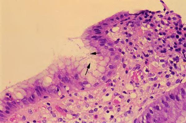
2 Helicobacter pylori causes an active chronic gastritis (image). The active or activity refers to the presence of acute inflammation in addition to chronic inflammation. Neutrophils are present in the lamina propria of the mucosa and infiltrate the epithelium (black arrows). It is difficult to appreciate that these are neutrophils at this magnification. The image shows excessive numbers of chronic inflammatory cells in the lamina propria (black stars) indicating chronic inflammation.

3 Active chronic gastritis with intestinal metaplasia as seen with Helicobacter pylori. Goblet cells (black arrows) are not normally seen in the gastric epithelium. Their presence here represents intestinal metaplasia. This change occurs as a result of the effects of H. pylori. It can also occur in autoimmune gastritis. There are too many chronic inflammatory cells in the lamina propria (black stars) indicating chronic inflammation.
4 Helicobacter
 necrotic slough and acute inflammatory exudate (yellow star) b) granulation tissue (red star) c) scar tissue (blue")
5 M Very low power view of a chronic peptic ulcer of the stomach. This one only penetrates into submucosa. There are classically several layers in the base of a chronic peptic ulcer: a) necrotic slough and acute inflammatory exudate (yellow star) b) granulation tissue (red star) c) scar tissue (blue stars) Patchy aggregates of lymphocytes (seen as dark areas/tiny dark dots due to their high N:C ratio on this low power) are also noted around the ulcer (black arrows). Make sure you can identify the layers of the wall. M: mucosa SM: submucosa MP: muscularis propria S: serosa Ulcer crater MP SM S SM MP

6 M SM MP Very low power view of the edge of a chronic peptic ulcer of the stomach. This one penetrates through muscularis propria into serosa. Black star: ulcer floor. M: mucosa SM: submucosa MP: muscularis propria

7 Chronic gastric ulcer. High power view of floor of ulcer. Black star: necrotic tissue Yellow star: oedematous vascular granulation tissue.

8 Chronic gastric ulcer. High power view of vascular granulation tissue in floor of ulcer. Black arrows: capillaries Yellow stars: macrophages, lymphocytes and plasma cells.
 is a very common investigation.")

9 Gastric biopsies. Endoscopy (upper and lower GI) is a very common investigation. Biopsies are frequently taken from lesions or abnormal areas of mucosa. Such endoscopic biopsies are from 1-3mm in diameter and comprise mucosa +/- a tiny bit of submucosa.

 which typically more diffusely infiltrates the stomach wall.")
10 A B Helicobacter pylori predisposes to the intestinal type of adenocarcinoma of the stomach, the cells of which form glands (black arrows, image A) and is thought to develop as result of intestinal metaplasia and dysplasia in chronic gastritis. Macroscopically these tumours form localised ulcerated lesions. The other main type of adenocarcinoma of the stomach is the signet ring type (image B) which typically more diffusely infiltrates the stomach wall. The tumour cells contain mucin which pushes the nucleus to one side, the overall effect resembling a signet ring (yellow arrows, image B).
Histopathology: chronic inflammation
 Histopathology: chronic inflammation These presentations are to help you identify, and to test yourself on identifying, basic histopathological features. They do not contain the additional factual information
Histopathology: healing
 Histopathology: healing These presentations are to help you identify, and to test yourself on identifying, basic histopathological features. They do not contain the additional factual information that
Histopathology: pulmonary pathology
 Histopathology: pulmonary pathology These presentations are to help you identify basic histopathological features. They do not contain the additional factual information that you need to learn about these
The surface mucous cells and the cardiac and pyloric glands secrete mucus which protects the stomach from self-digestion.
 PATHOLOGY OF THE STOMACH Stomach mucosa Gastric mucosa is covered by a layer of mucus. The mucosal glands comprise the cardiac glands, the fundic glands in the fundus and body of the stomach, and the pyloric
Gastric Cancer Histopathology Reporting Proforma
 Gastric Cancer Histopathology Reporting Proforma Mandatory questions (i.e. protocol standards) are in bold (e.g. S1.01). S1.01 Identification Family name Given name(s) Date of birth Sex Male Female Intersex/indeterminate
Histopathology: skin pathology
 Histopathology: skin pathology These presentations are to help you identify, and to test yourself on identifying, basic histopathological features. They do not contain the additional factual information
Gastrointestinal pathology 1. Upper GI tract
 Gastrointestinal pathology 1. Upper GI tract Tumors of The Salivary Glands Benign Malignant Pleomorphic adenoma (50%) Mucoepidermoid cc (15%) Warthin tumor (5%) Adenocarcinoma NOS (6%) Oncocytoma (2%)
General Structure of Digestive Tract
 Dr. Nabil Khouri General Structure of Digestive Tract Common Characteristics: Hollow tube composed of a lumen whose diameter varies. Surrounded by a wall made up of 4 principal layers: Mucosa Epithelial
Histopathology: Cervical HPV and neoplasia
 Histopathology: Cervical HPV and neoplasia These presentations are to help you identify basic histopathological features. They do not contain the additional factual information that you need to learn about
Histopathology: granulomatous inflammation, including tuberculosis
 Histopathology: granulomatous inflammation, including tuberculosis These presentations are to help you identify basic histopathological features. They do not contain the additional factual information
Histopathology: Hypertension and diabetes in the kidney These presentations are to help you identify basic histopathological features.
 Histopathology: Hypertension and diabetes in the kidney These presentations are to help you identify basic histopathological features. They do not contain the additional factual information that you need
Histopathology: Vascular pathology
 Histopathology: Vascular pathology These presentations are to help you identify basic histopathological features. They do not contain the additional factual information that you need to learn about these
Gastrointestinal pathology 2018 lecture 4. Dr Heyam Awad FRCPath
 Gastrointestinal pathology 2018 lecture 4 Dr Heyam Awad FRCPath Topics to be covered Peptic ulcer disease Hiatal hernia Gastric neoplasms Peptic ulcer disease (PUD)= chronic gastric ulcer Causes H pylori
PATHOLOGY OF NON NEOPLASTIC LESIONS OF THE UPPER GASTROINTESTINAL TRACT.
 PATHOLOGY OF NON NEOPLASTIC LESIONS OF THE UPPER GASTROINTESTINAL TRACT. OESOPHAGEAL LESIONS OESOPHAGITIS AND OTHER NON NEOPLASTIC DISORDERS Corrosive Gastroesophageal reflux (GERD), Pills, Acid intake,
Oesophagus and Stomach update dysplasia and early cancer
 Oesophagus and Stomach update dysplasia and early cancer Dr Tim Bracey STR teaching 13/4/16 Please check pathkids.com for previous talks One of the biggest units in the country (100 major resections per
Gastroenterology Tutorial
 Gastroenterology Tutorial Gastritis Poorly defined term that refers to inflammation of the stomach. Infection with H. pylori is the most common cause of gastritis. Most patients remain asymptomatic Some
Gastrointestinal pathology 2018 lecture 2. Dr Heyam Awad FRCPath
 Gastrointestinal pathology 2018 lecture 2 Dr Heyam Awad FRCPath Eosinophilic esophagitis Incidence of eosinophilic gastritis is increasing. Symptoms: food impaction and dysphagia. Histology: infiltration
Histopathology: Cell necrosis and cytoplasmic accumulations
 Histopathology: Cell necrosis and cytoplasmic accumulations These presentations are to help you identify basic histopathological features. They do not contain the additional factual information that you
Correlation between Gastric Mucosal Morphologic Patterns and Histopathological Severity of
 Hindawi Publishing Corporation BioMed Research International Volume 2015, Article ID 808505, 7 pages http://dx.doi.org/10.1155/2015/808505 Research Article Correlation between Gastric Mucosal Morphologic
Greater Manchester & Cheshire Guidelines for Pathology Reporting for Oesophageal and Gastric Malignancy
 Greater Manchester & Cheshire Guidelines for Pathology Reporting for Oesophageal and Gastric Malignancy Authors: Dr Gordon Armstrong, Dr Sue Pritchard 1. General Comments 1.1 Cancer reporting: Biopsies
Helicobacter and gastritis
 1 Helicobacter and gastritis Dr. Hala Al Daghistani Helicobacter pylori is a spiral-shaped gram-negative rod. H. pylori is associated with antral gastritis, duodenal (peptic) ulcer disease, gastric ulcers,
MECHANISMS OF HUMAN DISEASE: LABORATORY SESSIONS GASTROINTESTINAL (GI) PATHOLOGY LAB #1. January 06, 2012
 PATHOLOGY LAB #1. January 06, 2012") MECHANISMS OF HUMAN DISEASE: LABORATORY SESSIONS GASTROINTESTINAL (GI) PATHOLOGY LAB #1 GOAL: January 06, 2012 Faculty Copy 1. Describe the basis morphologic and pathophysiologic changes which occur in
MECHANISMS OF HUMAN DISEASE: LABORATORY SESSIONS GASTROINTESTINAL (GI) PATHOLOGY LAB #1 GOAL: January 06, 2012 Faculty Copy 1. Describe the basis morphologic and pathophysiologic changes which occur in
Common Inflammatory Gastrointestinal Disorders: Endoscopic and Pathologic Correlations
 Common Inflammatory Gastrointestinal Disorders: Endoscopic and Pathologic Correlations Nicole C. Panarelli, M.D. Attending Pathologist Montefiore Medical Center Associate Professor of Pathology - Albert
Histopathology of Endoscopic Resection Specimens from Barrett's Esophagus
 Histopathology of Endoscopic Resection Specimens from Barrett's Esophagus Br J Surg 38 oct. 1950 Definition of Barrett's esophagus A change in the esophageal epithelium of any length that can be recognized
The Pathologist s Role in the Diagnosis and Management of Neoplasia in Barrett s Oesophagus Cian Muldoon, St. James s Hospital, Dublin
 The Pathologist s Role in the Diagnosis and Management of Neoplasia in Barrett s Oesophagus Cian Muldoon, St. James s Hospital, Dublin 24.06.15 Norman Barrett Smiles [A brief digression - Chair becoming
SAM PROVIDER TOOLKIT
 THE AMERICAN BOARD OF PATHOLOGY Maintenance of Certification (MOC) Program SAM PROVIDER TOOLKIT Developing Self-Assessment Modules (SAMs) www.abpath.org The American Board of Pathology (ABP) approves educational
Histopathological study of Helicobacter Pylori bearing Gastric Biopsies for Gastric Lesions
 ORIGINAL ARTICLE Histopathological study of Helicobacter Pylori bearing Gastric Biopsies for Gastric Lesions MULAZIM HUSSAIN BUKHARI, SAMINA QAMAR*, SHAHBAZ AHMAD QURESHI*, MALIK MAQSOOD ANWAR** ABSTRACT
Earlyoesophagealcancer. dr. Nina Zidar Institute of Pathology Faculty ofmedicine University of Ljubljana Slovenia
 Earlyoesophagealcancer dr. Nina Zidar Institute of Pathology Faculty ofmedicine University of Ljubljana Slovenia Early carcinoma of oesophagus = tumor limited to mucosa or submucosa, not extending into
Key words : low-grade MALT lymphoma, epithelial change, empty lamina propria, B-cell clonality, H. pylori, eradication, long-term follow-up
 Department of Pathology, The University of Tokushima School of Medicine, Tokushima, Japan ; Department of Oral and Maxillofacial Surgery, The University of Tokushima School of Dentistry, Tokushima, Japan
Histopathogenesis of intestinal metaplasia: minute
 J Clin Pathol 1987;40:13-18 Histopathogenesis of intestinal metaplasia: minute lesions of intestinal metaplasia in ulcerated stomachs K MUKAWA, T NAKAMURA, G NAKANO, Y NAGAMACHI From the First Department
A 35 yr old man presents with a month history of burning epigastric pain that occurs between meals------
 A 35 yr old man presents with a month history of burning epigastric pain that occurs between meals------ The pain can be relieved by food or antiacids------ He denies taking aspirin or NSAID------ Lab
HDF Case CRYPTOSPORIDIOSE
 HDF Case 986949 CRYPTOSPORIDIOSE 45 yo male with severe diarrhea. Known HIV positive. Endoscopic biopsy of duodenum, the colon and ileum. EXUDATIVE CHANGES GRANULAR BASOPHILIC BODIES Colonic biopsy shows
Pathogenesis Most individuals with the infection also have the associated gastritis but are asymptomatic
 STOMACH Chronic Gastritis The presence of chronic inflammatory changes in the mucosa leading eventually to mucosal atrophy and epithelial metaplasia. In the Western world the prevalence of chronic gastritis
Gastric and Oesophageal Neuroendocrine tumours. Dr Tim Bracey, Consultant Pathologist MBChB PhD MRCS FRCPath
 Gastric and Oesophageal Neuroendocrine tumours Dr Tim Bracey, Consultant Pathologist MBChB PhD MRCS FRCPath Intestinal (and BO) endocrine cells in crypt bases NE cell (granules towards vessels) Paneth
Quiz Adenocarcinoma of the distal stomach has been increasing in the last 20 years. a. True b. False
 Quiz 1 1. Which of the following are risk factors for esophagus cancer. a. Obesity b. Gastroesophageal reflux c. Smoking and Alcohol d. All of the above 2. Adenocarcinoma of the distal stomach has been
HDF Case Whipple s disease
 HDF Case 952556 Whipple s disease 63 yo female complaining of a diarrhea for 2 months, weigth loss (12 Kg in 3 months), and joint pains. Duodenal biopsy performed. Scanning view, enlarged intestinal villi,
Histopathological Characteristics of Atrophic Gastritis in Adult Population
 Journal of Pharmacy and Pharmacology 3 (2015) 133-138 doi: 10.17265/2328-2150/2015.03.004 D DAVID PUBLISHING Histopathological Characteristics of Atrophic Gastritis in Adult Population Marija Milićević,
Update on the pathological classification of gastritis. Hala El-Zimaity, M.D. M.S. Epidemiology McMaster University Hamilton, Ontario Canada
 Update on the pathological classification of gastritis Hala El-Zimaity, M.D. M.S. Epidemiology McMaster University Hamilton, Ontario Canada CLASSIFICATION GASTRITIS GASTROPATHY 1. Acute 2. Chronic 3. Uncommon
Research Article Correlation between the Intensity of Helicobacter pylori Colonization and Severity of Gastritis
 Hindawi Gastroenterology Research and Practice Volume 2017, Article ID 8320496, 5 pages https://doi.org/10.1155/2017/8320496 Research Article Correlation between the Intensity of Helicobacter pylori Colonization
HASPI Medical Anatomy & Physiology 15a Lab Activity
 HASPI Medical Anatomy & Physiology 15a Lab Activity Name(s): Period: Date: The Digestive System Digestion is an important process that involves breaking down food and drink into small molecules that can
(A) Anemia (B) Malaria (C) Dehydration due to excess fluid loss. A. Incorrect! Anemia is symptomatic of sickle cell anemia, but is not the root cause.
 Anemia (B) Malaria (C) Dehydration due to excess fluid loss. A. Incorrect! Anemia is symptomatic of sickle cell anemia, but is not the root cause.") USMLE Step 1- Problem Drill 13: Histology Question No. 1 of 10 1. An 8 year old male complains of fatigue and dactylitis, and has a history of recurrent bacterial infections. Examination of a blood smear
USMLE Step 1- Problem Drill 13: Histology Question No. 1 of 10 1. An 8 year old male complains of fatigue and dactylitis, and has a history of recurrent bacterial infections. Examination of a blood smear
Upper GIT IV Gastric cancer
 Upper GIT IV Gastric cancer Luigi Tornillo PathoBasic 23.10.2014 Pathology Introduction Classification Morphogenesis Problems Intraepithelial neoplasia Surveillance EGJ Predictive factors Gastric cancer
Case: The patient is a 73 year old woman with vague complaints of dyspepsia and abdominal pain. Upper endoscopy showed features of gastritis and a
 Case: The patient is a 73 year old woman with vague complaints of dyspepsia and abdominal pain. Upper endoscopy showed features of gastritis and a nodular lesion in the body of the stomach. The patient
Histopathology: Glomerulonephritis and other renal pathology
 Histopathology: Glomerulonephritis and other renal pathology These presentations are to help you identify basic histopathological features. They do not contain the additional factual information that you
Gastritis (and gastropathy) Dr Ian Brown Envoi Pathology Brisbane, Australia
 Dr Ian Brown Envoi Pathology Brisbane, Australia") Gastritis (and gastropathy) Dr Ian Brown Envoi Pathology Brisbane, Australia ianbrown@envoi.com.au Topics for discussion Classification of gastritis Minimal diagnostic criteria for gastritis H.pylori negative
Gastritis (and gastropathy) Dr Ian Brown Envoi Pathology Brisbane, Australia ianbrown@envoi.com.au Topics for discussion Classification of gastritis Minimal diagnostic criteria for gastritis H.pylori negative
IBD. Crohn s. Outline. Ulcerative colitis versus Crohn s disease: is biopsy useful? UC vs. Crohn s? Is it easy? Biopsy settings 21/07/2017 IBD
 Outline Ulcerative colitis versus Crohn s disease: is biopsy useful? Roger Feakins Colorectal biopsies Ileal and upper GI biopsies Special situations New techniques Summary Inflammatory bowel disease (IBD)
Alimentary Canal (I)
") Alimentary Canal (I) Esophagus and Stomach (Objectives) By the end of this lecture, the student should be able to discuss the microscopic structure in correlation with the function of the following organs:
Alimentary Canal (I) Esophagus and Stomach (Objectives) By the end of this lecture, the student should be able to discuss the microscopic structure in correlation with the function of the following organs:
Anatomic and pathological aspects in the pathology of malignant gastric tumors
 Romanian Journal of Morphology and Embryology 2006, 47(2):163 168 ORIGINAL PAPER Anatomic and pathological aspects in the pathology of malignant gastric tumors IZABELLA SARGAN 1), A. MOTOC 1), MONICA-ADRIANA
- Helicobacter - THE EASE AND DIFFICULTY OF A NEW DISCOVERY. Robin Warren
 - Helicobacter - THE EASE AND DIFFICULTY OF A NEW DISCOVERY Robin Warren EARLY DAYS First reports 100 years ago considered spirochaetes 1940 Freedburg saw curved organisms in the stomach 1954 Palmer: Freedburg
Small Bowel Cases. Introduction. Introduction, Continued 12/7/2011. Lesions Found on endoscopic biopsies Just Like Signing Out
 Small Bowel Cases Lesions Found on endoscopic biopsies Just Like Signing Out Introduction Small intestinal biopsies have a few special pitfalls, for example: Neuroendocrine tumors are readily mistaken
Polyps in general: is a descriptive term of forming a mass that is exophytic & polypoid.
 ميحرلا نمحرلا هللا مسب Gastric Tumors: Benign tumours & tumor-like conditions: -Mucosal: Gastric polyps (they are uncommon) -Mesenchymal tumours: Leiomyoma & Lipoma (can occur anywhere in the body) Malignant:
Epithelia will be discussed according to the following scheme: Type Number of layers Shape Line drawing. Squamous Cuboidal Columnar
 Epithelia Epithelia will be discussed according to the following scheme: Type Number of layers Shape Line drawing Simple Squamous Cuboidal Columnar Covering and Lining epithelium Pseudostratified Stratified
A218 : Esophagus cancer tissues. (formalin fixed)
") (formalin fixed) For research use only Specifications: No. of cases: 40 Tissue type: Esophagus cancer tissues No. of spots: 2 spots from each cancer case (80 spots) 4 non-neoplastic spots (4 spots) Total
(formalin fixed) For research use only Specifications: No. of cases: 40 Tissue type: Esophagus cancer tissues No. of spots: 2 spots from each cancer case (80 spots) 4 non-neoplastic spots (4 spots) Total
Imaging in gastric cancer
 Imaging in gastric cancer Gastric cancer remains a deadly disease because of late diagnosis. Adenocarcinoma represents 90% of malignant tumors. Diagnosis is based on endoscopic examination with biopsies.
Small intestine. Small intestine
 General features Tubular organ longest part; 5-6 m most of chemical digestion absorption of nutrients reabsorption of H2O occurs. Two structural features; maximize the lumenal surface area villi microvilli
1. Esophageal diverticulum located above the upper esophageal sphincter is called
 Test Bank for Robbins Basic Pathology 9th Edition by Kumar Link full download: http://testbankair.com/download/test-bank-for-robbins-basic-pathology-9thedition-by-kumar/ Chapter 14: Oral Cavity and Gastrointestinal
WSC , Conference 9. Case 1. Tissue from a rhesus macaque.
 Case 1. Tissue from a rhesus macaque. MICROSCOPIC DESCRIPTION: Esophagus: There is multifocal loss of the mucosal lining (1 pt). In these areas, the denuded subepithelial fibrous connective tissue is infiltrated
32 Adenocarcinoma of the oesophagogastric junction
 Adenocarcinoma of the oesophagogastric junction S.J. Spechler P. Hainaut M.F. Dixon R. Lambert R. Genta R. Siewert Definition Adenocarcinomas that straddle the junction of the oesophagus and stomach are
Alimentary Tract. Wang Lin 王琳 Department of pathology School of basic medical sciences
 Alimentary Tract Wang Lin 王琳 Department of pathology School of basic medical sciences Alimentary Tract Carcinoma Peptic ulcer Carcinoma Gastritis Peptic ulcer Carcinoma Appendicitis Anatomy of the stomach
Gastrooesophageal reflux disease. Jera Jeruc Institute of pathology, Faculty of Medicine, Ljubljana, Slovenia
 Gastrooesophageal reflux disease Jera Jeruc Institute of pathology, Faculty of Medicine, Ljubljana, Slovenia Reflux esophagitis (RE) GERD: a spectrum of clinical conditions and histologic alterations resulting
3/30/2017. Disclosure of Relevant Financial Relationships. Case 5: Polypoid mass in ulcerative colitis. Case 5. TC Smyrk
 Case 5: Polypoid mass in ulcerative colitis TC Smyrk Disclosure of Relevant Financial Relationships USCAP requires that all faculty in a position to influence or control the content of CME disclose any
American Journal of Gastroenterology. Volumetric Laser Endomicroscopy Detects Subsquamous Barrett s Adenocarcinoma
 Volumetric Laser Endomicroscopy Detects Subsquamous Barrett s Adenocarcinoma Journal: Manuscript ID: AJG-13-1412.R1 Manuscript Type: Letter to the Editor Keywords: Barrett-s esophagus, Esophagus, Endoscopy
WSC , Conference 9, Case 1. Tissue from a nyala.
 WSC 2009-2010, Conference 9, Case 1. Tissue from a nyala. MICROSCOPIC DESCRIPTION: Heart, atrium (1 pt.): Approximately 40% of the atrial myocardium is replaced by areas of fibrous connective tissue (1
Clinico-Pathological Study of Endoscopic Gastric Biopsies
 Original Article Clinico-Pathological Study of Endoscopic Gastric Biopsies SumalathaKasturi, Raju Mulaka 2 Associate Professor Department of Pathology Chalmeda Anand Rao Institute of Medical Sciences Karimnagar,
Regression of Advanced Gastric MALT Lymphoma after the Eradication of Helicobacter pylori
 Gut and Liver, Vol. 6, No. 2, April 2012, pp. 270-274 CASE REPORT Regression of Advanced Gastric MALT Lymphoma after the Eradication of Helicobacter pylori Soo-Kyung Park, Hwoon-Yong Jung, Do Hoon Kim,
This is the second learning component (Learning Component 2) in our first learning module (Learning Module 1). In this component we review a very
 in our first learning module (Learning Module 1). In this component we review a very") This is the second learning component (Learning Component 2) in our first learning module (Learning Module 1). In this component we review a very basic response to injury inflammation. We ll look at examples
This is the second learning component (Learning Component 2) in our first learning module (Learning Module 1). In this component we review a very basic response to injury inflammation. We ll look at examples
University Mainz. Early Gastric Cancer. Ralf Kiesslich. Johannes Gutenberg University Mainz, Germany. Early Gastric Cancer 15.6.
 Ralf Kiesslich Johannes Gutenberg University Mainz, Germany DIAGNOSIS Unmask lesions - Chromoendoscopy -NBI Red flag technology - Autofluorescence Surface and detail analysis - Magnifying endoscopy - High
Digestive system L 2. Lecturer Dr. Firdous M. Jaafar Department of Anatomy/Histology section
 Digestive system L 2 Lecturer Dr. Firdous M. Jaafar Department of Anatomy/Histology section objectives 1-Describe the general structure of digestive tract: a-mucosa. b-submucosa. c-muscularis externa d-adventitia
LARYNGEAL DYSPLASIA. Tomas Fernandez M; 3 rd year ENT resident, Son Espases University Hospital
 LARYNGEAL DYSPLASIA Tomas Fernandez M; 3 rd year ENT resident, Son Espases University Hospital INTRODUCTION Laryngeal cancer constitutes 1-2% of all malignancies diagnosed worldwide Survival is related
Unexpected Findings at Endoscopy
 The Endoscopic Incidentaloma: What to Tell Your Patient t with Unexpected Endoscopic Findings: Gastric Intestinal Metaplasia, Silent Ileitis, Carcinoid David Greenwald, MD Montefiore Medical Center Albert
The effect of proton pump inhibitors on the gastric mucosal microenvironment
 Original papers The effect of proton pump inhibitors on the gastric mucosal microenvironment Yen-Chun Peng,,, A F, Lan-Ru Huang, A, C, E, Hui-Ching Ho, C, Chi-Sen Chang, C, E, Shou-Wu Lee, E, Ching-Chang
Abstracting Upper GI Cancer Incidence and Treatment Data Quiz 1 Multiple Primary and Histologies Case 1 Final Pathology:
 Abstracting Upper GI Cancer Incidence and Treatment Data Quiz 1 Multiple Primary and Histologies Case 1 A 74 year old male with a history of GERD presents complaining of dysphagia. An esophagogastroduodenoscopy
الله الر ح م ن الر ح يم مسب
 بسم رلا هللارلا هللا This is the second histology lecture in the GI system. In this lecture, we will discuss the histology of the esophagus, stomach, and small intestine so prepare yourself.this sheet
Correlation Between Endoscopic and Histological Findings in Different Gastroduodenal Lesion and its Association with Helicobacter Pylori
 ORIGINAL ARTICLE Correlation Between Endoscopic and Histological Findings in Different Gastroduodenal Lesion and its Association with Helicobacter Pylori *A. Sultana 1, SM Badruddoza 2, F Rahman 3 1 Dr.
African Trypanosomes
 African Trypanosomes Giemsa-stained blood smear of African trypanosomes viewed under the 100X objective lens. The block arrows denote trypomastigote forms of the African trypanosomes found within the blood
5/2/2018. Low Grade Dysplasia of GI Tract. High Grade Dysplasia of GI Tract. Dysplasia in Gastrointestinal Tract: Practical Pearls and Issues
 Dysplasia in Gastrointestinal Tract: Practical Pearls and Issues Arief Suriawinata, M.D. Professor of Pathology and Laboratory Medicine Geisel School of Medicine at Dartmouth Department of Pathology and
Case Report Features of the Atrophic Corpus Mucosa in Three Cases of Autoimmune Gastritis Revealed by Magnifying Endoscopy
 Volume 2012, Article ID 368160, 4 pages doi:10.1155/2012/368160 Case Report Features of the Atrophic Corpus Mucosa in Three Cases of Autoimmune Gastritis Revealed by Magnifying Endoscopy Kazuyoshi Yagi,
MALT LYMPHOMA. Silvia Montoto, St Bartholomew s Hospital, London, UK ESMO Preceptorship on Lymphoma
 MALT LYMPHOMA Silvia Montoto, St Bartholomew s Hospital, London, UK ESMO Preceptorship on Lymphoma Lugano, 3-4 November 2017 Disclosures: Roche: honoraria Gilead: travel grant ESMO Preceptorship on Lymphoma
GI Histology Lab 1. Prepared by: Zeina Kalaji
 GI Histology Lab 1 Prepared by: Zeina Kalaji Lip ORAL MUCOSA -Arrow shows labial salivary glands in the submucosa. VERMILLION transitional zone. SKIN Stratified Squamous epithelium, keratinized -Arrow
A Chronic or Recurring Pattern of Esophagitis Resembling Allergic Contact Dermatitis
 Anatomic Pathology / Lymphocytic Esophagitis Lymphocytic Esophagitis A Chronic or Recurring Pattern of Esophagitis Resembling Allergic Contact Dermatitis Julianne K. Purdy, MD, Henry D. Appelman, MD, Christopher
Dr Nadine Gravett School of Anatomical Sciences Room 2B10B
 Dr Nadine Gravett School of Anatomical Sciences Room 2B10B Nadine.Gravett@wits.ac.za Oral cavity Mechanical breakdown Formation of bolus Oesophagus Conduit from mouth to stomach Stomach Digestion Temporary
Lymphocytic Gastritis, Isolated Type Occurring in Family Members. A Case Report.
 Lymphocytic Gastritis, Isolated Type Occurring in Family Members. A Case Report. Alan Shienbaum, DO; AndriyPavlenko, MD; Jun Liu, MD, PhD; Janusz J Godyn, MD. Pathology Department, Kennedy University Hospitals,
Small Intestine. Protocol revision date: January 2005 Based on AJCC/UICC TNM, 6 th edition
 Small Intestine Protocol applies to all invasive carcinomas of the small intestine, including those with focal endocrine differentiation. Excludes carcinoid tumors, lymphomas, and stromal tumors (sarcomas).
RAPID DIAGNOSIS OF HELICOBACTER PYLORI INFECTION IN GASTRIC IMPRINT SMEARS
 RAPID DIAGNOSIS OF HELICOBACTER PYLORI INFECTION IN GASTRIC IMPRINT SMEARS Gurjeet Kaur 1, Manoharan Madhavan 1, Amir Hakim Basri 2, Abdul Hamid Mat Sain 3, Mohd Shaiful Bahrun Hussain 3, Mohd Kamal Yatiban
CT Evaluation of Bowel Wall Thickening. Dr: Adel El Badrawy; M.D. Lecturer of Radio Diagnosis Faculty of Medicine Mansoura University.
 CT Evaluation of Bowel Wall Thickening By Dr: Adel El Badrawy; M.D. Lecturer of Radio Diagnosis Faculty of Medicine Mansoura University. The CT findings of bowel wall thickening includes 1 Degree of thickening.
The Nobel Prize in Physiology or Medicine for 2005
 The Nobel Prize in Physiology or Medicine for 2005 jointly to Barry J. Marshall and J. Robin Warren for their discovery of "the bacterium Helicobacter pylori and its role in gastritis and peptic ulcer
SAMs Guidelines DEVELOPING SELF-ASSESSMENT MODULES TEST QUESTIONS. Ver. #
 SAMs Guidelines DEVELOPING SELF-ASSESSMENT MODULES TEST Ver. #5-02.12.17 GUIDELINES FOR DEVELOPING SELF-ASSESSMENT MODULES TEST The USCAP is accredited by the American Board of Pathology (ABP) to offer
Helicobacter pylori Improved Detection of Helicobacter pylori
 DOI:http://dx.doi.org/10.7314/APJCP.2016.17.4.2099 RESEARCH ARTICLE Improved Detection of Helicobacter pylori Infection and Premalignant Gastric Mucosa Using Conventional White Light Source Gastroscopy
Barrett s Esophagus: Old Dog, New Tricks
 Barrett s Esophagus: Old Dog, New Tricks Stuart Jon Spechler, M.D. Chief, Division of Gastroenterology, VA North Texas Healthcare System; Co-Director, Esophageal Diseases Center, Professor of Medicine,
Dana Alrafaiah. Dareen Abu Shalbak. Mohammad Almuhtaseb. 1 P a g e
 2 Dana Alrafaiah Dareen Abu Shalbak Mohammad Almuhtaseb 1 P a g e Esophagus: A muscular tube that is 25 cm long, but if measured from the incisors it would be 45cm long. Extends from C6 of cervical vertebra,
SESSION 1: GENERAL (BASIC) PATHOLOGY CONCEPTS Thursday, October 16, :30am - 11:30am FACULTY COPY
 PATHOLOGY CONCEPTS Thursday, October 16, :30am - 11:30am FACULTY COPY") SESSION 1: GENERAL (BASIC) PATHOLOGY CONCEPTS Thursday, October 16, 2008 9:30am - 11:30am FACULTY COPY GOAL: Describe the basic morphologic (structural) changes which occur in various pathologic conditions.
SESSION 1: GENERAL (BASIC) PATHOLOGY CONCEPTS Thursday, October 16, 2008 9:30am - 11:30am FACULTY COPY GOAL: Describe the basic morphologic (structural) changes which occur in various pathologic conditions.
Histology Lab. looking at microscopic pictures of tissues, for more information use Junqueira book and you can use BlueHistolgy website
 Done By: Aseel Twaijer & Laith Sorour Histology Lab *These notes help in differentiating tissues and you must read them while looking at microscopic pictures of tissues, for more information use Junqueira
Greater Manchester & Cheshire Guidelines for Pathology Reporting of Oesophageal and Gastric Malignancy
 Greater Manchester & Cheshire Guidelines for Pathology Reporting of Oesophageal and Gastric Malignancy Authors: Dr Stephen Hayes, Dr David Bisset, Dr Gordon Armstrong, Dr Sue Pritchard 1. General Comments
HISTOLOGY. GIT Block 432 Histology Team. Lecture 1: Alimentary Canal (1) (Esophagus & Stomach) Done by: Ethar Alqarni Reviewed by: Ibrahim Alfuraih
 (Esophagus & Stomach) Done by: Ethar Alqarni Reviewed by: Ibrahim Alfuraih") HISTOLOGY Lecture 1: Alimentary Canal (1) (Esophagus & Stomach) Done by: Ethar Alqarni Reviewed by: Ibrahim Alfuraih Color Guide: Black: Slides. Red: Important. Green: Doctor s notes. Blue: Explanation.
HISTOLOGY Lecture 1: Alimentary Canal (1) (Esophagus & Stomach) Done by: Ethar Alqarni Reviewed by: Ibrahim Alfuraih Color Guide: Black: Slides. Red: Important. Green: Doctor s notes. Blue: Explanation.
Cyclooxygenase-2 Expression in Gastric Antral Mucosa Before and After Eradication of Helicobacter pylori Infection
 THE AMERICAN JOURNAL OF GASTROENTEROLOGY Vol. 94, No. 5, 1999 1999 by Am. Coll. of Gastroenterology ISSN 0002-9270/99/$20.00 Published by Elsevier Science Inc. PII S0002-9270(99)00126-4 Cyclooxygenase-2
DIGESTIVE TRACT ESOPHAGUS
 DIGESTIVE TRACT From the lower esophagus to the lower rectum four fundamental layers comprise the wall of the digestive tube: mucosa, submucosa, muscularis propria (externa), and adventitia or serosa (see
Part 1. A pragmatic approach to common problems in esophageal biopsy pathology
 Part 1 A pragmatic approach to common problems in esophageal biopsy pathology How I can help more patients than I hurt Barbara J McKenna University of Michigan barbmcke@med.umich.edu Case 1 The following
The doctor mentioned a few things about the esophagus from the previous lecture:
 السالم عليكم [HISOLOGY 2] April 27, 2014 The doctor mentioned a few things about the esophagus from the previous lecture: Esophagus - It is about 25 cm in length (from the incisor it is 45 cm) Histological
A COMPARATIVE STUDY BETWEEN IMMUNOHISTOCHEMISTRY, HEMATOXYLIN & EOSIN AND GEIMSA STAIN FOR HELICOBACTER PYLORI DETECTION IN CHRONIC GASTRITIS
 Original Research Article Pathology International Journal of Pharma and Bio Sciences ISSN 0975-6299 A COMPARATIVE STUDY BETWEEN IMMUNOHISTOCHEMISTRY, HEMATOXYLIN & EOSIN AND GEIMSA STAIN FOR HELICOBACTER
Gastrointestinal Disorders. Disorders of the Esophagus 3/7/2013. Congenital Abnormalities. Achalasia. Not an easy repair. Types
 Gastrointestinal Disorders Congenital Abnormalities Disorders of the Esophagus Types Stenosis Atresia Fistula Newborn aspirates while feeding. Pneumonia Not an easy repair Achalasia Lack of relaxation