Does digital IHC need digital tissue controls?
|
|
|
- Brent Thompson
- 6 years ago
- Views:
Transcription
1 VILNIUS UNIVERSITY Digital immunohistochemistry platform for the staining variation monitoring based on integration of image and statistical analyses with laboratory information system Arvydas Laurinavicius 12th European Congress on Digital Pathology Paris, France, June 2014
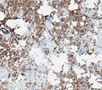
2 Does digital IHC need digital tissue controls? Conventional IHC staining quality is monitored by semiquantitative visual evaluation of tissue (multi)controls Image analysis may require more sensitive quality control Intra- and interlaboratory IHC staining variation may be a major obstacle for DIA adoption We have previously shown intra-laboratory HER2 IHC variation by digital analysis means


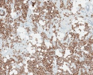
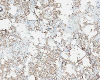
3 HER2 average membrane staining intensity variation Batch/Serial section # Laurinavicius, Besusparis, et al Diagnostic Pathology 2013.
4 Integrated IHC staining monitor by and for digital analysis Explore Ki67 IHC staining variation in our laboratory routine, set up LIS integration of the IHC tissue multi-controls, monitored by image analysis, and supported by automated statistical analysis feedback
5 Consecutive sections (n=69) of one TMA block containing 10 cores of breast cancer tissue used as tissue controls in routine Ki67 IHC testing Ventana slide label BarcodeID was sent to the LIS to register the serial section sequence, then used for statistical analyses Slides stained and scanned (Aperio ScanScope XT), IA performed by the Aperio/Leica Colocalization and Genie Classifier/Nuclear algorithms SQL-based SpectrumPlus/LIS integration ensured automated statistical analysis of the IA data by the SAS Enterprise Guide project Factor analysis and plot visualizations to explore slide-to-slide variation of the Ki67 IHC staining results in the control tissue


6 Workflow TMA block with 10 control tissue cores Serial sections stored at +4C Used on demand for routine Ki67 TMALAB design applied for the tissue controls Slides stained and scanned Ventana BarcodeID stored in LIS when slide label printed Colocalization Genie/Nuclear performed on the TMA and WSI SAS project rerun from the Spectrum/LIS SQL query Statistical feedback generated


7 Intraslide Intertissue Intraslide Intertissue
8 Variation of individual 10 spots


9 Slide-to-slide variation of the TMA multicontrols Total Nuclei Total Stained Area (mm2) Positive Nuclei Brown Intensity The sequence of Ventana slide Barcode on the x axis to represent consecutive serial sections of TMA blocks of the 10 multi-tissue control cores (labelled as SampleID),

 Brown/Blue Intensity")


10 Slide-to-slide variation of the TMA multicontrols Percent Positive Nuclei Blue Intensity Positive Density (Pos Nuclei/mm 2 ) Brown/Blue Intensity ratio The sequence of Ventana slide Barcode on the x axis to represent consecutive serial sections of TMA blocks of the 10 multi-tissue control cores (labelled as SampleID),

11 Factor pattern of Brown and Blue intensity indicators from Colocalization Analysis in 10 individual TMA cores Total Intensity Both Brown and Blue lighter Relatively deeper Blue Brown/Blue balance

12 e.g., Sum (Area) Sum (Total Nuclei) Median (Intensity) etc Variation of aggregated data from the 10 TMA spot images super-multi-tissue control

13 Factor pattern of Colocalization and Genie/Nuclear analysis outputs aggregated from 10 TMA cores IHC positivity High Ki67 LI, Dark Brown More tissue, Dark Blue Amount of Tissue Detected with Darker Blue
 Aggregated data from 10 TMA cores plotted against the sequence of Ventana Barcode on the x axis")
14 Relevant inverse covariation of selected image analysis variables Median Blue Intensity (Colocalization) Area of Analysis (Genie) Aggregated data from 10 TMA cores plotted against the sequence of Ventana Barcode on the x axis
15 The solution enabled continuous monitor of IHC multi-tissue controls by the means of IA, followed by automated statistical analysis, integrated into the laboratory workflow. Even in consecutive serial tissue sections, tissue-related factors affected the IHC IA results; meanwhile, less intense blue counterstain was associated with less amount of tissue, detected by the IA tools.
16 Remarks Scanning variation was not addressed in our experiment, same scanner used. The IA issues with hematoxylin counterstain in IHC, have been highlighted, alternative counterstaining and IA techniques have been proposed Brey at al J Histochem Cytochem 2003, Pham et al Diagn Pathol 2007, Stefanovic et albiotechnic & histochemistry 2013, Zehntner et al J Histochem Cytochem 2008, Bernardo et al Microsc Microanal 2009] Another approach - measuring signal-to-noise ratio of the images to evaluate quality before IA (Ali et al Br J Cancer 2013), however, adjustment of the images and/or analyses may require effort. Our data simulate reproducibility of the Ki67 index in the consecutive sections of one 1 mm diameter core: variation of Ki67% (the IA result) was satisfactory (standard deviation in all 10 cores ranged from 3 to 8, relative error within the range of 0.07 to 0.39) the variation of cell numbers detected (the process) was higher. Inter-laboratory IHC staining variation is likely to be more significant: international Ki67 reproducibility study (Polley et al J Natl Cancer Inst 2013) revealed unsatisfactory results of visual estimation which was even worse when the slides were stained locally. Further optimization and standardization of IHC procedures, especially, when applying IA tools with unknown sensitivity to the staining/scanning variation. Ideally, IA tools should be robust and resistant to the IHC staining and scanning variations.


17 Aida Laurinavičienė and Paulette Herlin Daiva Lesciute- Krilaviciene Yasir Iqbal Benoit Plancoulaine Darius Raudeliunas Raimundas Meškauskas Indra Baltrušaitytė, Justinas Besusparis A COMPREHENSIVE BIOMARKER INTRA- TUMOUR HETEROGENEITY EVALUATION BY DIGITAL IMMUNOHISTOCHEMISTRY IMAGE ANALYSIS This research is funded by European Social Fund under the Global Grant measure.

18
Education 2012 m. Vilnius University, National Cancer Institute, PhD studies 1995 m. Vilnius University, Faculty of Natural Sciences, Master s degree
 CURRICULUM VITAE AIDA LAURINAVIČIENĖ 1968-05-05 PhD, Associate Professor Education 2012 m. Vilnius University, National Cancer Institute, PhD studies 1995 m. Vilnius University, Faculty of Natural Sciences,
Image analysis in IHC overview, considerations and applications
 Image analysis in IHC overview, considerations and applications Rasmus Røge, MD, Institute of Pathology, Aalborg University Hospital NordiQC workshop September 2016 Aalborg, Denmark Outline Theory Image
Abstract. Background. Objective
 Molecular epidemiology of clinical tissues with multi-parameter IHC Poster 237 J Ruan 1, T Hope 1, J Rheinhardt 2, D Wang 2, R Levenson 1, T Nielsen 3, H Gardner 2, C Hoyt 1 1 CRi, Woburn, Massachusetts,
Next generation image analysis for immunohistochemistry quantitation
 Next generation image analysis for immunohistochemistry quantitation Ben Vainer Department of Pathology, Rigshospitalet University of Copenhagen Medical Center Men are only so good as their technical developments
Next-Gen Analytics in Digital Pathology
 Next-Gen Analytics in Digital Pathology Cliff Hoyt, CTO Cambridge Research & Instrumentation April 29, 2010 Seeing life in a new light 1 Digital Pathology Today Acquisition, storage, dissemination, remote
Layered-IHC (L-IHC): A novel and robust approach to multiplexed immunohistochemistry So many markers and so little tissue
: A novel and robust approach to multiplexed immunohistochemistry So many markers and so little tissue") Page 1 The need for multiplex detection of tissue biomarkers. There is a constant and growing demand for increased biomarker analysis in human tissue specimens. Analysis of tissue biomarkers is key to
Page 1 The need for multiplex detection of tissue biomarkers. There is a constant and growing demand for increased biomarker analysis in human tissue specimens. Analysis of tissue biomarkers is key to
Interpreting Therapeutic Response on Immune Cell Number and Spatial Distribution within the Tumor Microenvironment. Lorcan Sherry, CSO OracleBio
 Interpreting Therapeutic Response on Immune Cell Number and Spatial Distribution within the Tumor Microenvironment Lorcan Sherry, CSO OracleBio Company Overview OracleBio is a specialised CRO providing
Automatic analysis of virtual slides to help in the determination of well established prognostic parameters in breast carcinomas
 Loews hotel Le Concorde, Quebec Canada August 3-5, 2011 H.I.Q Automatic analysis of virtual slides to help in the determination of well established prognostic parameters in breast carcinomas N Elie 1,
COMPUTER-AIDED HER-2/neu EVALUATION IN EXTERNAL QUALITY ASSURANCE (EQA) OF BREAST CANCER SCREENING PROGRAMME
 OF BREAST CANCER SCREENING PROGRAMME") COMPUTER-AIDED HER-2/neu EVALUATION IN EXTERNAL QUALITY ASSURANCE (EQA) OF BREAST CANCER SCREENING PROGRAMME Maria Lunardi MD Anatomic Pathology Fracastoro Hospital San Bonifacio, Verona -Italy HER2-neu
COMPUTER-AIDED HER-2/neu EVALUATION IN EXTERNAL QUALITY ASSURANCE (EQA) OF BREAST CANCER SCREENING PROGRAMME Maria Lunardi MD Anatomic Pathology Fracastoro Hospital San Bonifacio, Verona -Italy HER2-neu
Visual interpretation in pathology
 13 Visual interpretation in pathology Tissue architecture (alteration) evaluation e.g., for grading prostate cancer Immunohistochemistry (IHC) staining scoring e.g., HER2 in breast cancer (companion diagnostic
Virtual Microscopy: Express Surgical Pathology Consultation. Mercè Jordà, University of Miami, Florida
 Virtual Microscopy: Express Surgical Pathology Consultation Mercè Jordà, University of Miami, Florida Telepathology versus Virtual microscopy (Digital Pathology) Telepathology Use of telecommunications
HER2 ISH (BRISH or FISH)
") Assessment Run H14 2018 HER2 ISH (BRISH or FISH) Material Table 1. Content of the multi-block used for the NordiQC HER2 ISH assessment, run H14 HER2 IHC* IHC score Dual - SISH** FISH*** FISH*** HER2/chr17
Assessment Run H14 2018 HER2 ISH (BRISH or FISH) Material Table 1. Content of the multi-block used for the NordiQC HER2 ISH assessment, run H14 HER2 IHC* IHC score Dual - SISH** FISH*** FISH*** HER2/chr17
How to Best Prepare for Digital Pathology: A Primer on Technologies, Applications, and Clinical Contributions. Disclosures
 How to Best Prepare for Digital Pathology: A Primer on Technologies, Applications, and Clinical Contributions Keith J. Kaplan, MD Carolinas Pathology Group Charlotte, NC 1 Disclosures Clarient,, Inc. Consulting
Supplementary Online Content
 Supplementary Online Content Rimm DL, Han G, Taube JM, et al. A prospective, multi-institutional, pathologistbased assessment of 4 immunohistochemistry assays for PD-L1 expression in non small cell lung
Central Pathology Review and Tissue MicroArrays. Dr Lisa Storer Children s Brain Tumour Research Centre Nottingham
 Central Pathology Review and Tissue MicroArrays Dr Lisa Storer Children s Brain Tumour Research Centre Nottingham What is a tissue microarray? Tissue microarrays (TMAs) consist of paraffin blocks in which
LOCAL INFLAMMATION IN BREAST TISSUE AND MAMMOGRAPHIC DENSITY AMONG PREMENOPAUSAL AND POSTMENOPAUSAL WOMEN
 LOCAL INFLAMMATION IN BREAST TISSUE AND MAMMOGRAPHIC DENSITY AMONG PREMENOPAUSAL AND POSTMENOPAUSAL WOMEN Mirette Hanna MD Clinical pathology PhD (candidate) Experimental medicine MSc Clinical and chemical
Digital Pathology and CAP Guidelines
 Digital Pathology and CAP Guidelines Frequently asked questions The VENTANA family of digital pathology products empowers you with the convenience of a comprehensive image and workflow solution. When used
Immune Cell Phenotyping in Solid Tumors using Quantitative Pathology
 Immune Cell Phenotyping in Solid Tumors using Quantitative Pathology James R. Mansfield Director of Quantitative Pathology Applications 2009 PerkinElmer What is Quantitative Pathology? Quantitative Pathology
The following slides were presented at the TIGA Workshop, and are enclosed in the PP show format (*.pps) in order to include all presented details.
 in order to include all presented details.") Disclaimer: The following slides were presented at the TIGA Workshop, and are enclosed in the PP show format (*.pps) in order to include all presented details. These slides are proprietary and should not
Disclaimer: The following slides were presented at the TIGA Workshop, and are enclosed in the PP show format (*.pps) in order to include all presented details. These slides are proprietary and should not
Developing quantitative slide-based assays to assess target inhibition in oncology drug discovery and development
 Developing quantitative slide-based assays to assess target inhibition in oncology drug discovery and development TIGA Workshop June 25-26, 2010 Doug Bowman Millennium Pharmaceuticals 2010 Millennium Pharmaceuticals
Quality assurance and quality control in pathology in breast disease centers
 Quality assurance and quality control in pathology in breast disease centers Judith Sandbank M.D. Pathology Assaf-Harofeh Medical Center ISRAEL jsandbank@asaf.health.gov.il 1 st IBDC, 28 th January, 2011
Deciphering the biology that drives response to immunotherapy
 Deciphering the biology that drives response to immunotherapy Phenoptics TM Quantitative Pathology Platform Trent Norris, Field Application Scientist September 15, 2016 HUMAN HEALTH ENVIRONMENTAL HEALTH
Androgen Receptor Expression in Renal Cell Carcinoma: A New Actionable Target?
 Androgen Receptor Expression in Renal Cell Carcinoma: A New Actionable Target? New Frontiers in Urologic Oncology Juan Chipollini, MD Clinical Fellow Department of Genitourinary Oncology Moffitt Cancer
Diagnostics Assessment Report commissioned by the NIHR HTA Programme on behalf of the National Institute for Health and Care Excellence
 Diagnostics Assessment Report commissioned by the NIHR HTA Programme on behalf of the National Institute for Health and Care Excellence A rapid evidence review of the analytical validity of IHC4: ADDENDUM
NordiQC External Quality Assurance in Immunohistochemistry
 NordiQC External Quality Assurance in Immunohistochemistry Mogens Vyberg Professor of Clinical Pathology Director of NordiQC Aalborg University Hospital, Aalborg, Denmark AALBORG (~ 200.000 inhabitants)
Incorporating pharmacodynamic, response and patient selection biomarkers. Paul Elvin PhD Chief Translational Science Officer Aptus Clinical
 Incorporating pharmacodynamic, response and patient selection biomarkers Paul Elvin PhD Chief Translational Science Officer Aptus Clinical 22 Oncology drug development Biomarkers key for: Strong hypothesis
Next-Generation Immunohistochemistry: Multiplex tissue imaging with mass cytometry
 Nat Met, April 2014 Nat Med, April 2014 Next-Generation Immunohistochemistry: Multiplex tissue imaging with mass cytometry Journal Club Timo Böge Overview Introduction Conventional Immunohistochemistry
Gaining New Insights Through IF Multiplexed Staining and Analysis. Tyna Hope, Ph.D. P.Eng Biomarker Imaging Research Laboratory October 5, 2017
 Gaining New Insights Through IF Multiplexed Staining and Analysis Tyna Hope, Ph.D. P.Eng Biomarker Imaging Research Laboratory Assessing more from Tissue Sections Gaps with More Common Methods Most common
Statistical Analysis of Biomarker Data
 Statistical Analysis of Biomarker Data Gary M. Clark, Ph.D. Vice President Biostatistics & Data Management Array BioPharma Inc. Boulder, CO NCIC Clinical Trials Group New Investigator Clinical Trials Course
LN04 - Lymphoma Tissue Microarray
 Reveal Biosciences offers Histochemical Staining, Immunohistochemistry (IHC), In Situ Hybridization (ISH), Whole Slide Imaging, and Quantitative Image Analysis on any TMA LN04 - Lymphoma Tissue Microarray
Assessment Run B HER2 IHC
 Assessment Run B26 208 HER2 IHC Material The slide to be stained for HER2 comprised the following 5 materials: IHC: HER2 Score* (0, +, 2+, 3+) FISH: HER2 gene/chr 7 ratio**. Breast carcinoma, no. 2+..3
Lessons learned in the use of digital imaging at Memorial Sloan Kettering Cancer Center
 Lessons learned in the use of digital imaging at Memorial Sloan Kettering Cancer Center Executive War College May 3, 2018 Victor E. Reuter, M.D. Vice-Chair, Department of Pathology Medical Director, Warren
Understanding your biomarker: what this marker can do for you
 Understanding your biomarker: what this marker can do for you Dr. John Bartlett/Dr. Harriet Feilotter As is your Pathology, so is your Medicine. Sir William Osler. 1849-1919. Disclosure statement John
Automated image analysis in the study of lymphocyte subpopulation in eosinophilic oesophagitis
 PROCEEDINGS Automated image analysis in the study of lymphocyte subpopulation in eosinophilic oesophagitis Open Access Marcial García-Rojo 1*, Joaquín Rodríguez Sánchez 2, Eva de la Santa 2, Elena Durán
Supplementary Online Content
 Supplementary Online Content Bell EH, Pugh SL, McElroy JP, et al. Molecular-based recursive partitioning analysis model for glioblastoma in the temozolomide era: a correlative analysis based on NRG Oncology
Data Mining for Research and Diagnostics
 UCL DEPARTMENT OF GEOGRAPHY UCL INSTITUTE OF NEUROLOGY Data Mining for Research and Diagnostics Sebastian Brandner, UCL Institute of Neurology, London Data mining for Research and Diagnostics Our setup
Digital Pathology - moving on after implementation. Catarina Eloy, MD, PhD
 Digital Pathology - moving on after implementation Catarina Eloy, MD, PhD Catarina Eloy, MD, PhD No conflict of interests to declare Pathology challenges today Maintaining high quality students interested
Assessment Run B HER2 IHC
 Assessment Run B24 2017 HER2 IHC Material The slide to be stained for HER2 comprised the following 5 materials: IHC: HER2 Score* (0, 1+, 2+, 3+) FISH: HER2 gene/chr 17 ratio** 1. Breast carcinoma, no.
Product Introduction
 Product Introduction Product Codes: HCL026, HCL027 and HCL028 Contents Introduction to HER2 2 HER2 immunohistochemistry 3 Cell lines as controls 5 HER2 Analyte Control DR IHC 7 HER2 Analyte Control DR
CC01 - Colon Cancer Tissue Microarray
 Reveal Biosciences offers Histochemical Staining, Immunohistochemistry (IHC), In Situ Hybridization (ISH), Whole Slide Imaging, and Quantitative Image Analysis on any TMA CC01 - Colon Cancer Tissue Microarray
Assessment Run GATA3
 Assessment Run 44 2015 GATA3 Material The slide to be stained for GATA3 comprised: 1. Tonsil 2. Kidney, 3. Urothelial carcinoma, 4. Breast ductal carcinoma, 5. Colon adenocarcinoma All tissues were fixed
Digital Pathology Diagnosis Assistance System
 August 3, 2011 Digital Pathology Diagnosis Assistance System Computer s s eye and Pathologist s s eye Ogura M, Saito A, Graf HP, Marugame A, Kiyuna T, Yamashita Y, Cosatto E, Malon C & Fukumoto M. Innovative
manifestations of disease (mostly clinical medicine)
") Histopathology : microscopic examination of tissue in order to study the manifestations of disease (mostly clinical medicine) https://medium.com/the-physics-arxiv-blog/ghost-imaging-reassembles-images-scattered-by-breast-tissue
Histopathology : microscopic examination of tissue in order to study the manifestations of disease (mostly clinical medicine) https://medium.com/the-physics-arxiv-blog/ghost-imaging-reassembles-images-scattered-by-breast-tissue
Interobserver Variability of Ki-67 Measurement in Breast Cancer
 Journal of Pathology and Translational Medicine 2016; 50: 129-137 ORIGINAL ARTICLE Interobserver Variability of Ki-67 Measurement in Breast Cancer Yul Ri Chung 1 Min Hye Jang 2 So Yeon Park 1,2 Gyungyub
Prosigna BREAST CANCER PROGNOSTIC GENE SIGNATURE ASSAY
 Prosigna BREAST CANCER PROGNOSTIC GENE SIGNATURE ASSAY Methodology The test is based on the reported 50-gene classifier algorithm originally named PAM50 and is performed on the ncounter Dx Analysis System
Prosigna BREAST CANCER PROGNOSTIC GENE SIGNATURE ASSAY
 Prosigna BREAST CANCER PROGNOSTIC GENE SIGNATURE ASSAY GENE EXPRESSION PROFILING WITH PROSIGNA What is Prosigna? Prosigna Breast Cancer Prognostic Gene Signature Assay is an FDA-approved assay which provides
PREPARED FOR: U.S. Army Medical Research and Materiel Command Fort Detrick, Maryland
 AWARD NUMBER: W81XWH-16-1-0737 TITLE: Developing a PTEN-ERG Signature to Improve Molecular Risk Stratification in Prostate Cancer PRINCIPAL INVESTIGATOR: Tamara Lotan CONTRACTING ORGANIZATION: Johns Hopkins
Use of tissue micro array (TMA) in routine clinical analysis
 in routine clinical analysis") 2nd European Workshop on Tissue Imaging and Analysis June 25-26, 2010 Use of tissue micro array (TMA) in routine clinical analysis Tim Svenstrup Poulsen Molecular unit Dept. pathology Herlev Hospital Denmark
2nd European Workshop on Tissue Imaging and Analysis June 25-26, 2010 Use of tissue micro array (TMA) in routine clinical analysis Tim Svenstrup Poulsen Molecular unit Dept. pathology Herlev Hospital Denmark
Tissue microarray TMA
 Tissue microarray TMA Tibor Krenacs 1st Department of Pathology & Experimental Cancer Research Semmelweis University Budapest Austro-Hungarian Pathology Congress October 1-3rd 2009 TMA - tissue microarray
FAQs for UK Pathology Departments
 FAQs for UK Pathology Departments This is an educational piece written for Healthcare Professionals FAQs for UK Pathology Departments If you would like to discuss any of the listed FAQs further, or have
ARTIFICIAL INTELLIGENCE FOR DIGITAL PATHOLOGY. Kyunghyun Paeng, Co-founder and Research Scientist, Lunit Inc.
 ARTIFICIAL INTELLIGENCE FOR DIGITAL PATHOLOGY Kyunghyun Paeng, Co-founder and Research Scientist, Lunit Inc. 1. BACKGROUND: DIGITAL PATHOLOGY 2. APPLICATIONS AGENDA BREAST CANCER PROSTATE CANCER 3. DEMONSTRATIONS
Assessment Run B HER-2 IHC. HER-2/chr17 ratio**
 Assessment Run B2 20 HER-2 IHC Material The slide to be stained for HER-2 comprised the following 5 tissues: IHC HER-2 Score* (0, +, 2+,3+) FISH HER-2/chr7 ratio**. Breast ductal carcinoma 0..3 2. Breast
Spatially resolved multiparametric single cell analysis. Technical Journal Club 19th September 2017 Christina Müller (Group Speck)
") Spatially resolved multiparametric single cell analysis Technical Journal Club 19th September 2017 Christina Müller (Group Speck) Why spatially resolved multiparametric single cell analysis? Multiparametric
Spatially resolved multiparametric single cell analysis Technical Journal Club 19th September 2017 Christina Müller (Group Speck) Why spatially resolved multiparametric single cell analysis? Multiparametric
Interpretation guide. Abnormal cytology can t hide anymore
 Interpretation guide Abnormal cytology can t hide anymore Unique dual-biomarker technology makes you certain about the presence of transforming HPV infection. The science that creates certainty. Table
# Best Practices for IHC Detection and Interpretation of ER, PR, and HER2 Protein Overexpression in Breast Cancer
 #1034 - Best Practices for IHC Detection and Interpretation of ER, PR, and HER2 Protein Overexpression in Breast Cancer Richard W. Cartun, MS, PhD Andrew Ricci, Jr, MD Department of Pathology Hartford
(A) PCR primers (arrows) designed to distinguish wild type (P1+P2), targeted (P1+P2) and excised (P1+P3)14-
 PCR primers (arrows) designed to distinguish wild type (P1+P2), targeted (P1+P2) and excised (P1+P3)14-") 1 Supplemental Figure Legends Figure S1. Mammary tumors of ErbB2 KI mice with 14-3-3σ ablation have elevated ErbB2 transcript levels and cell proliferation (A) PCR primers (arrows) designed to distinguish
1 Supplemental Figure Legends Figure S1. Mammary tumors of ErbB2 KI mice with 14-3-3σ ablation have elevated ErbB2 transcript levels and cell proliferation (A) PCR primers (arrows) designed to distinguish
Molecular in vitro diagnostic test for the quantitative detection of the mrna expression of ERBB2, ESR1, PGR and MKI67 in breast cancer tissue.
 Innovation for your breast cancer diagnostics Now valid ated on: -Roche cob as z 480 A nalyzer -Roche L ightcycle r 480 II - ABI 750 0 Fast -Versant kpcr Cyc ler PGR ESR1 MKI67 Molecular in vitro diagnostic
Optimal algorithm for HER2 testing
 Optimal algorithm for HER2 testing The revised definition of IHC 2+ (equivocal) is invasive breast cancer with Weak to moderate complete membrane staining observed in >10% of tumor cells. (see Figure 1
Breast cancer: Antibody selection, protocol optimzation controls and EQA
 Breast cancer: Antibody selection, protocol optimzation controls and EQA Workshop in Diagnostic Immunohistochemistry Oud St. Jan/ Old St. John Brugge (Bruges), Belgium June 13th 15nd 2018 Rasmus Røge,
Definiens. Tissue Studio 4.2. Tutorial 8: Cell Simulation and Classification
 Definiens Tissue Studio 4.2 Tutorial 8: Cell Simulation and Classification Tutorial 8: Cell Simulation and Classification Imprint and Version Copyright 2015 Definiens AG. All rights reserved. This document
Multiplex immunofluorescence for investigating the prognostic value of immune cells Chidozie Anyaegbu, PhD
 Multiplex immunofluorescence for investigating the prognostic value of immune cells Chidozie Anyaegbu, PhD Catholic Health Australia Symposium Mater Hospital, Brisbane 1 st June 2018 Why colorectal cancer?
Is digital pathology ready for use in routine histopathology? Dr David Snead University Hospitals of Coventry and Warwickshire NHS Trust Coventry UK
 Is digital pathology ready for use in routine histopathology? Dr David Snead University Hospitals of Coventry and Warwickshire NHS Trust Coventry UK Pathologists and microscopes. 1850 Rudolph Virchow (1821-1902),
Tumor Growth Rate (TGR) A New Indicator of Antitumor Activity in NETS? IPSEN NET Masterclass Athens, 12 th November 2016
 A New Indicator of Antitumor Activity in NETS? IPSEN NET Masterclass Athens, 12 th November 2016") 1 Tumor Growth Rate (TGR) A New Indicator of Antitumor Activity in NETS? IPSEN NET Masterclass Athens, 12 th November 2016 Philippe RUSZNIEWSKI ENETS Centre of Excellence, Beaujon Hospital, Clichy, France
1 Tumor Growth Rate (TGR) A New Indicator of Antitumor Activity in NETS? IPSEN NET Masterclass Athens, 12 th November 2016 Philippe RUSZNIEWSKI ENETS Centre of Excellence, Beaujon Hospital, Clichy, France
CINtec PLUS Cytology. Interpretation training
 CINtec PLUS Cytology Interpretation training Objectives After reviewing this learning module, you will have a basic understanding of how to interpret CINtec PLUS Cytology, including: The mechanism of action
CHEK Proficiency study 672. Phthalates in nail polish. Date 5 December 2017 Version 1. Report number CHEK
 CHEK Proficiency study 672 Phthalates in nail polish Date 5 December 2017 Version 1 Status Final Report number CHEK-17-672 page 0 Colophon Number 672 Name Phthalates in nail polish Contact Author Authorization
Positive Results on Fecal Blood Tests
 Interventions to Improve Follow-up of Positive Results on Fecal Blood Tests Results of a systematic review and Kaiser experience Kevin Selby, M.D. kevin.j.selby@kp.org National Colorectal Cancer Roundtable
CENTRAL LABORATORY SERVICES
 CENTRAL LABORATORY SERVICES Bridging the Gaps in Biomarker Development and Testing for Global Clinical Trials Biomarkers, Digital Pathology & the Central Lab Presentation Title Presenter Name July 16,
Molecular in vitro diagnostic test for the quantitative detection of the mrna expression of ERBB2, ESR1, PGR and MKI67 in breast cancer tissue.
 Innovation for your breast cancer diagnostics PGR G ATA G C G A C G AT C G A A A G A A G T TA G ATA G C G A C G AT C G A A A G A A G T TA G ATA G C G A C G AT C G A A A G A A G T TA G ATA G C G A C ERBB2
Product Introduction. Product Codes: HCL029, HCL030 and HCL031. Issue
 Product Introduction Product Codes: HCL029, HCL030 and HCL031 Issue 1. 180510 Contents Introduction to Estrogen Receptor 2 ER immunohistochemistry 3 Quality control 5 Cell lines as controls 6 Estrogen
Quality Assurance and Quality Control in the Pathology Dept.
 Quality Assurance and Quality Control in the Pathology Dept. Judith Sandbank M.D. Pathology Assaf-Harofeh Medical Center ISRAEL jsandbank@asaf.health.gov.il 2 nd IBDC, 9 th February, 2012 Pathology as
Applying Tissue Phenomics to Colorectal Clinical Questions
 Applying Tissue Phenomics to Colorectal Clinical Questions International Symposium for Tissue Phenomics San Francisco October 2014 Peter Caie Senior Research Fellow University of St Andrews Systems Pathology
Background Information
 Background Information Erlangen, November 26, 2017 RSNA 2017 in Chicago: South Building, Hall A, Booth 1937 Artificial intelligence: Transforming data into knowledge for better care Inspired by neural
Results you can trust
 PRODUCT I NF OR MAT ION pharmdx Results you can trust The first and only FDA-approved PD-L1 test to assess the magnitude of treatment effect on progression-free survival in melanoma patients from OPDIVO
Analysis of Immunohistochemical Stain Usage in Different Pathology Practice Settings
 Anatomic Pathology / Immunohistochemistry Use by Practice Type Analysis of Immunohistochemical Stain Usage in Different Pathology Practice Settings Akeesha A. Shah, MD, Henry F. Frierson, Jr, MD, and Helen
Application of tissue microarrays for receptor immunohistochemistry in breast carcinoma
 FOLIA HISTOCHEMICA ET CYTOBIOLOGICA Vol. 51, No. 4, 2013 pp. 326 332 ORIGINAL STUDY Application of tissue microarrays for receptor immunohistochemistry in breast carcinoma Anna Glajcar, Karolina Kaczmarczyk,
Welcome! HER2 TESTING DIAGNOSTIC ACCURACY 4/11/2016
 HER2 TESTING DIAGNOSTIC ACCURACY Can t We Finally Get It Right? Allen M. Gown, M.D. Medical Director and Chief Pathologist PhenoPath Laboratories Seattle, Washington Clinical Professor of Pathology University
PROTOCOL SENTINEL NODE BIOPSY (NON OPERATIVE) BREAST CANCER - PATHOLOGY ASSESSMENT
 BREAST CANCER - PATHOLOGY ASSESSMENT") PROTOCOL SENTINEL NODE BIOPSY (NON OPERATIVE) BREAST CANCER - PATHOLOGY ASSESSMENT Author: Dr Sally Ann Hales On behalf of the Breast and pathology CNGs Written: March 2005 Reviewed by CNG: June 2009 &
PROTOCOL SENTINEL NODE BIOPSY (NON OPERATIVE) BREAST CANCER - PATHOLOGY ASSESSMENT Author: Dr Sally Ann Hales On behalf of the Breast and pathology CNGs Written: March 2005 Reviewed by CNG: June 2009 &
Comparison of evaluations of hormone receptors in breast carcinoma by image-analysis using three automated immunohistochemical stainings
 Experimental and Therapeutic Medicine 1: 927-932, 2010 Comparison of evaluations of hormone receptors in breast carcinoma by image-analysis using three automated immunohistochemical stainings Koji Arihiro,
IHC Stainer platforms. Overview, pros and cons
 IHC Stainer platforms Overview, pros and cons Bart De Wiest Quality manager IHC OLV Hospital, Aalst, Belgium Donald Van Hecke Lab & Quality manager AZ St-Lucas, Brugge, Belgium Goal of this lecture: to
P16 et Ki67 Biomarkers: new tool for risk management and low grade intraepithelial lesions (LGSIL): be ready for the future.
: be ready for the future.") P16 et Ki67 Biomarkers: new tool for risk management and low grade intraepithelial lesions (LGSIL): be ready for the future. Mark H Stoler, MD University of Virginia Health System, Charlottesville, VA,
P16 et Ki67 Biomarkers: new tool for risk management and low grade intraepithelial lesions (LGSIL): be ready for the future. Mark H Stoler, MD University of Virginia Health System, Charlottesville, VA,
Vernieuwing en diagnostiek bij NSCLC: Immunotherapy: PD-L1 analyse: waar staan we
 9e avondsymposium: "Nieuwe ontwikkelingen in de behandeling van NSCLC" 9 november 2016, UMCG Vernieuwing en diagnostiek bij NSCLC: Immunotherapy: PD-L1 analyse: waar staan we Wim Timens Professor and Chair
Comparison of core oestrogen receptor (ER) assay with excised tumour: intratumoral distribution of ER in breast carcinoma
 assay with excised tumour: intratumoral distribution of ER in breast carcinoma") J Clin Pathol 1;54:951 955 951 Department of Pathology, University of Wales College of Medicine, Heath Park, CardiV, South Glamorgan, CF14 4XN, UK A G Douglas-Jones N Collett J M Morgan B Jasani Correspondence
J Clin Pathol 1;54:951 955 951 Department of Pathology, University of Wales College of Medicine, Heath Park, CardiV, South Glamorgan, CF14 4XN, UK A G Douglas-Jones N Collett J M Morgan B Jasani Correspondence
Supplementary Appendix
 Supplementary Appendix This appendix has been provided by the authors to give readers additional information about their work. Supplement to: Schuster SJ, Svoboda J, Chong EA, et al. Chimeric antigen receptor
Aida Laurinaviciene 1,2*, Darius Dasevicius 2,3, Valerijus Ostapenko 1, Sonata Jarmalaite 4, Juozas Lazutka 4 and Arvydas Laurinavicius 2,3.
 METHODOLOGY Open Access Membrane connectivity estimated by digital image analysis of HER2 immunohistochemistry is concordant with visual scoring and fluorescence in situ hybridization results: algorithm
CME/SAM. Software-Automated Counting of Ki-67 Proliferation Index Correlates With Pathologic Grade and Disease Progression of Follicular Lymphomas
 AJCP / Original Article Software-Automated Counting of Ki-67 Proliferation Index Correlates With Pathologic Grade and Disease Progression of Follicular Lymphomas Mark A. Samols, MD, PhD, 1 Nathan E. Smith,
IDH1 R132H/ATRX Immunohistochemical validation
 IDH1 R132H/ATRX Immunohistochemical validation CIQC/DSM 2016 12 June 2016 0835-0905 Stephen Yip, M.D., Ph.D., FRCPC University of British Columbia Disclosure Statement I have nothing to disclose I will
Inti Zlobec 1*, Viktor H Koelzer 1,2, Heather Dawson 1,2, Aurel Perren 1,2 and Alessandro Lugli 1,2
 Zlobec et al. Journal of Translational Medicine 2013, 11:104 RESEARCH Open Access Next-generation tissue microarray (ngtma) increases the quality of biomarker studies: an example using CD3, CD8, and CD45RO
Expanded View Figures
 EMO Molecular Medicine Proteomic map of squamous cell carcinomas Hanibal ohnenberger et al Expanded View Figures Figure EV1. Technical reproducibility. Pearson s correlation analysis of normalised SILC
Dr. dr. Primariadewi R, SpPA(K)
") Curriculum Vitae Dr. dr. Primariadewi R, SpPA(K) Education : Medical Doctor from UKRIDA Doctoral Degree from Faculty of Medicine University of Indonesia Pathologist Specialist and Consultant from Faculty
Curriculum Vitae Dr. dr. Primariadewi R, SpPA(K) Education : Medical Doctor from UKRIDA Doctoral Degree from Faculty of Medicine University of Indonesia Pathologist Specialist and Consultant from Faculty
Detection Of Red Lesion In Diabetic Retinopathy Using Adaptive Thresholding Method
 Detection Of Red Lesion In Diabetic Retinopathy Using Adaptive Thresholding Method Deepashree Devaraj, Assistant Professor, Instrumentation Department RVCE Bangalore. Nagaveena M.Tech Student, BMSP&I,
Measuring cell density in prostate cancer imaging as an input for radiotherapy treatment planning
 Measuring cell density in prostate cancer imaging as an input for radiotherapy treatment planning Poster No.: R-0262 Congress: 2014 CSM Type: Scientific Exhibit Authors: H. Reynolds, S. Williams, A. Zhang,
UCL-Advanced Diagnostics. 2015/16 Service Update
 UCL-Advanced Diagnostics 2015/16 Service Update About UCL-Advanced Diagnostics UCL Advanced Diagnostics (UCL-AD) is the immunohistochemistry, in situ and molecular diagnostics service arm of the UCL-Cancer
Pathfinder Holter Analyzer CARDIAC DIAGNOSTIC SOLUTIONS
 Pathfinder Holter Analyzer CARDIAC DIAGNOSTIC SOLUTIONS Pathfinder Digital - An Evolution... Del Mar Reynolds rich history of innovation began nearly a half century ago with the development of Holter monitoring
Classification of electrocardiographic ST-T segments human expert vs artificial neural network
 European Heart Journal (1993) 14,464-468 Classification of electrocardiographic ST-T segments human expert vs artificial neural network L. EDENBRANDT, B. DEVINE AND P. W. MACFARLANE University Department
Utilization of Virtual Microscopy in a Cooperative Group Setting
 Utilization of Virtual Microscopy in a Cooperative Group Setting The Research Institute at Nationwide Children s Hospital Research Informatics Core Biopathology Center Tom Barr Bill Beyer Dave Billiter
PREPARED FOR: U.S. Army Medical Research and Materiel Command Fort Detrick, Maryland
 AD Award Number: W81XWH-11-1-0380 TITLE: Validation of Biomarkers for Prostate Cancer Prognosis PRINCIPAL INVESTIGATOR: James D Brooks CONTRACTING ORGANIZATION: Stanford University Stanford, CA, 94305.
ANALYTISCHE STRATEGIE Tissue Imaging. Bernd Bodenmiller Institute of Molecular Life Sciences University of Zurich
 ANALYTISCHE STRATEGIE Tissue Imaging Bernd Bodenmiller Institute of Molecular Life Sciences University of Zurich Quantitative Breast single cancer cell analysis Switzerland Brain Breast Lung Colon-rectum
Immunotherapy in NSCLC Pathologist role
 Immunotherapy in NSCLC Pathologist role Pimpin Incharoen, M.D. Assistant Professor, Thoracic Pathology Department of Pathology, Ramathibodi Hospital Genetic alterations in NSCLC Khono et al, Trans Lung
Identification of Sickle Cells using Digital Image Processing. Academic Year Annexure I
 Academic Year 2014-15 Annexure I 1. Project Title: Identification of Sickle Cells using Digital Image Processing TABLE OF CONTENTS 1.1 Abstract 1-1 1.2 Motivation 1-1 1.3 Objective 2-2 2.1 Block Diagram