Title malignancy. Issue Date Right 209, 12, (2013)
|
|
|
- Cody Cummings
- 6 years ago
- Views:
Transcription

1 NAOSITE: Nagasaki University's Ac Title Author(s) A case of intracystic apocrine papi malignancy Hayashi, Hiroko; Ohtani, Hiroshi; Y Citation Pathology - Research and Practice, Issue Date URL Right Elsevier GmbH.; NOTICE: this that was accepted for publication i Changes resulting from the publishi editing, corrections, structural fo mechanisms may not be reflected in been made to this work since it was version was subsequently published 209, 12, (2013) This document is downloaded
2 1 A case of intracystic apocrine papillary tumor: a concern for histopathologic evaluation of malignancy Running title: Intracystic apocrine papillary tumor Key words: intraductal papillary carcinoma, breast, apocrine papillary tumor, high molecular weight cytokeratin, myoepithelial cell Hiroko Hayashi, MD, PhD 1, Hiroshi Ohtani, MD, PhD 2, Junzo Yamaguchi, MD, PhD 3, Isao Shimokawa, MD, PhD 1 1 Department of Investigative Pathology, Graduate School of Biomedical Science, Nagasaki University, Nagasaki, Japan; 2 Department of Clinical Laboratory, Hakujuji Hospital, Fukuoka, Japan; 3 Department of Surgery, National Hospital Organization Saga National Hospital, Saga, Japan Correspondence: Hiroko Hayashi, MD, PhD, Department of Investigative Pathology, Graduate School of Biomedical Science, Nagasaki University, Sakamoto, Nagasaki , Japan Tel: ; Fax: hayashih@nagasaki-u.ac.jp
3 2 Abstract Intraductal/intracystic papillary carcinoma (IPC) of the breast is defined as a malignant non-invasive papillary tumor arising from the ductal-lobular system. Based on the presence of myoepithelial cells in the cystic wall, IPC is distinguished from encapsulated papillary carcinoma (EPC). Here, we report a case of an intracystic apocrine papillary tumor in the breast of a 49-year-old woman. Histopathologic examination revealed that the entire papillary structures and cyst wall were comprised of apocrine cells, some of which showed nuclear atypia with macronucleoli. Immunohistochemical examination revealed a lack of myoepithelial cells in the papillary fronds and cyst wall. Although the dense proliferation of apocrine cells mimicked a cribriform pattern, detailed examination identified a delicately intermingled interstitium in the cribriform-like growth area in the present case. Only a few apocrine variants of IPC or EPC have been reported to be malignant or potentially malignant. Since even benign apocrine lesions are known to lack myoepithelial cells, histopathologic evaluation regarding malignant potential requires caution in apocrine variants. Key words: intraductal papillary carcinoma, breast, apocrine papillary tumor, high molecular weight cytokeratin, myoepithelial cell
4 3 Introduction Intracystic/intraductal papillary carcinoma (IPC) of the breast is defined as a malignant non-invasive neoplastic epithelial proliferation with a papillary architecture in the lumen of the ductal-lobular system in the WHO classification published in 2012 [4]. Histologically, the tumor shows slender fronds with absent or scant myoepithelial cells and epithelial cells, which all have architectural and cytological features of ductal carcinoma in situ [4]. Encapsulated papillary carcinoma (EPC) is a variant of papillary carcinoma, which is surrounded by a thick fibrous capsule. IPC has myoepithelial cells in the cyst wall and is clearly distinguished from EPC. A few cases of apocrine variants of IPC or EPC have been reported to date [2,3,7]. Owing to the limited cases with short-term follow-up periods, their malignant potential has been inconclusive. Furthermore, histologic evaluation of malignancy is potentially difficult, particularly in low-grade tumors, because even non-neoplastic apocrine lesions may lack myoepithelial cells [2,8]. Here, we describe a case of an intracystic atypical apocrine papillary tumor simulating IPC, but with complete absence of myoepithelial cells in the cyst wall, and discuss the current issues regarding the malignant potential of IPC and/or EPC. Case report A 49-year-old woman was referred to our hospital after an abnormality was detected on a mammographic examination. Sonography showed a cystic mass measuring 2 cm in the subareolar region of her left breast. Two weeks later, the mass had enlarged to cm in size, and contained some subcentimetric mural nodules. Suspecting breast cancer, a surgical excision of the mass was performed.
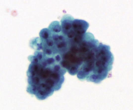
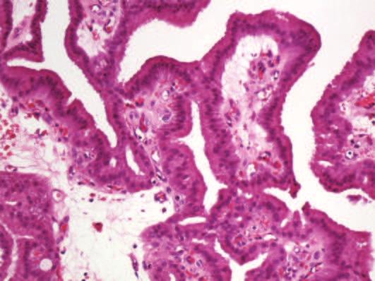
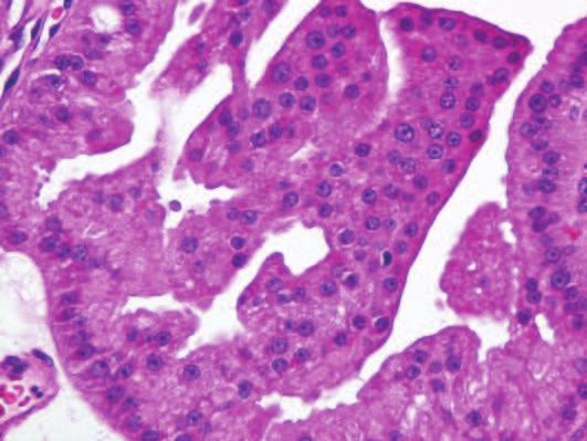
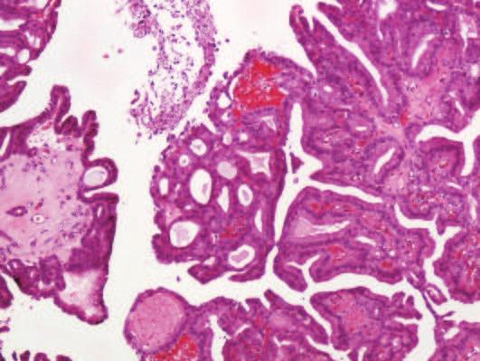
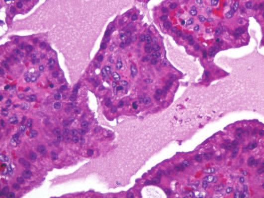
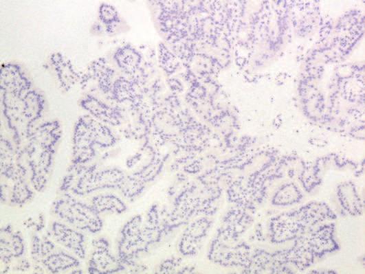
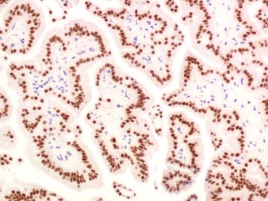
5 4 Pathological findings Grossly, the translucent cyst had a smooth inner surface and included three papillary yellow nodules measuring 1.0, 0.9, and 0.3 cm in diameter, respectively. Cytologically, the cyst contents included some three-dimensional papillary clusters in a background of scattered foamy macrophages (Fig. 1A). The cells of these clusters had relatively rich cytoplasm, large nuclei, and prominent macronucleoli. On the other hand, some clusters also included cells with abundant and granular cytoplasm, which were identical to benign apocrine cells (Fig. 1B). The cells were generally cohesive and the borders of the cell membranes were distinct. These findings suggested intracystic apocrine neoplasia, but were inconclusive for malignancy. Histologically, epithelial papillary lesions were present in the cystically dilated duct (Fig. 2A). Branching fibrovascular stalks were relatively broad with occasional edematous areas, and were covered by cells with abundant eosinophilic granular to pale cytoplasm (Fig. 2B). No myoepithelial cells were found. The nuclei were prominent with distinctive nucleoli (Fig. 2C). Although cribriform-like patterns were observed focally (Fig. 2D and E), more detailed examination revealed the presence of a delicate interstitium between glands (Fig. 2E). We confirmed that each gland was surrounded by type IV collagen immunohistochemically (Fig. 2F). Mitotic figures were sparsely observed (Fig. 2G). The cyst wall was covered with atrophic apocrine epithelium (Fig. 2H). In an immunohistochemical study, the tumor cells showed diffuse and strong cytoplasmic positivity for 15-kDa glycoprotein of gross cystic disease (GCDFP-15). Myoepithelial markers such as p63 (Fig. 2I), calponin, and CD10 were negative in both
6 5 the papillary nodules and cyst wall. High molecular weight cytokeratins (HMWCs) such as CK5/6 (Fig. 2J) and 34βE12 were also negative in the nodules. Androgen receptor was entirely positive (Fig. 2K), while estrogen receptor, progesterone receptor, and Her-2 were completely negative. Proliferative activity measured with Ki-67 was less than 1%. Discussion An intracystic/intraductal papillary tumor composed of an entirely apocrine epithelium is rare. Seal et al. [7] reported five cases with this lesion as encapsulated apocrine papillary carcinoma for the first time. According to their descriptions, all of their cases showed varied complexity of apocrine cell proliferation and had pseudopapillary and/or cribriform architectures. The cytological atypia were also varied in their cases. They described that it was not yet known whether this lesion is a form of papillary hyperplasia of apocrine cells or a true neoplasm [7]. Laforga et al. [3] reported a case with an apocrine type of EPC, and described that their case showed features fitting with low-grade carcinoma according to the criteria of O Malley and Bane [6], such as cell size, atypical nuclei with macronucleoli, architecture showing papillary and cribriform patterns of growth, and so on. Cserni [2] reported a case of intracystic papillary apocrine proliferation. He mentioned the difficulty associated with determination of malignancy because of the short-term follow-up, but supported benign biologic behavior. One of the most challenging issues is to assess apocrine atypia, and mainly differentiate between atypical apocrine proliferations and low-grade apocrine ductal carcinoma in situ (DCIS). O Malley et al. [6] summarized the criteria that have been
7 6 proposed thus far. Atypical apocrine lesions show three-fold nuclear enlargement with nucleolar enlargement, slightly irregular nuclear membranes, and fine chromatin. Nuclear stratification or tufting of the epithelium may be present in atypical apocrine lesions. Low-grade DCIS shows irregular nuclear membranes and coarse chromatin, and may exhibit a characteristic cribriform architecture. However, there are no broadly accepted criteria for distinguishing these lesions to date. For the differentiation, it may be difficult to use elements such as irregular nuclear membranes or coarse chromatin because a borderline apocrine lesion shows borderline nuclear atypia. These nuclear findings are relatively vague and noncommittal, especially for cases in which pathologists are irresolute. Among the criteria summarized by O Malley et al. [6], the cribriform architecture might be a relatively identifiable feature because of its distinct pattern. In the present case, the apocrine papillary epithelium revealed relatively uniform nuclei with nucleolar enlargement, smooth nuclear membranes, and fine chromatin. Architecturally, an indistinct cribriform pattern was seen in part of the lesion. Coincident with this area, some atypical features such as rough chromatin and increased nuclear-cytoplasmic ratio were discernible. These findings might suggest malignant potential for this lesion. At first sight, this area appeared to be a true cribriform pattern with punched-out regular spaces. However, as a result of more detailed observations, an interstitium intervened delicately among the glands and there was no solid growth pattern. We therefore judged it not to be a true cribriform architecture, but a tubular architecture. Although a lack of myoepithelial markers is commonly used as a hallmark for papillary carcinoma, it has reported that apocrine lesions, either benign or malignant,
8 7 can show reduction and occasional complete loss of myoepithelial cells [1,8]. Tramm et al. [8] described that a malignant diagnosis with apocrine changes of the breast cannot yet solely rely on the presence or absence of myoepithelial cells, and should be based on the overall morphology, i.e., the cytologic and architectural features. HMWCs such as 34βE12 and CK5/6 are widely used for distinguishing benign proliferative lesions from DCIS, since the former characteristically show a mosaic pattern and the latter is negative for these antigens [5]. However, these antibodies are often useless for apocrine proliferative lesions because an apocrine metaplastic epithelium is included within false-negative groups [5]. These peculiar immunohistochemical panels of the apocrine epithelium make the diagnosis of borderline lesions more difficult. The present case also showed complete negativity for myoepithelial markers and HMWCs. In summary, an intracystic papillary tumor composed of an entirely apocrine epithelium is rare. Although its monotonous appearance and florid growth might confuse pathologists, there are no distinct criteria to distinguish an atypical apocrine lesion from low-grade apocrine DCIS to date. Moreover, immunohistochemical panels such as myoepithelial markers and HMWCs may not be helpful for the diagnosis of this lesion. An apocrine epithelium has a broad cytoplasm and therefore its relatively dense proliferation mimics solid growth or a cribriform architecture. Consequently, pathologists should be cautious in the evaluation of malignancy for apocrine variants of IPC or EPC.
9 8 References 1. G. Cserni, Lack of myoepithelium in apocrine glands of the breast does not necessarily imply malignancy. Histopathology 52 (2008) G. Cserni, Benign apocrine papillary lesions of the breast lacking or virtually lacking myoepithelial cells-potential pitfalls in diagnosing malignancy. APMIS 120 (2012) J.B. Laforga, J.M. Gasent, I. Sánchez, Encapsulated apocrine papillary carcinoma of the breast: case report with clinicopathologic and immunohistochemical study. Diagn Cytopathol 39 (2011) G. MacGrogan, G. Tse, L. Collins, P.H. Tan, B. Chaiwun, J.S. Reis-Filho, Intraductal papillary carcinoma, In: S.R. Lakhani, I.O. Ellis, S.J. Schnitt, P.H. Tan, M.J. van de Vijver (Eds.), WHO classification of tumours of the breast, 4th ed., IARC, Lyon, (2012), pp T. Moriya, Y. Kozuka, N. Kanomata, G.M. Tse, P.H. Tan, The role of immunohistochemistry in the differential diagnosis of breast lesions. Pathology 41 (2009) F.P. O Malley, A.L. Bane, The spectrum of apocrine lesions of the breast. Adv Anat Pathol 11 (2004) M. Seal, C. Wilson, G.J. Naus, S. Chia, T.C. Bainbridge, M.M. Hayes, Encapsulated apocrine papillary carcinoma of the breast a tumour of uncertain malignant potential: report of five cases. Virchow Arch 455 (2009) T. Tramm, J.Y. Kim, F.A. Tavassoli, Diminished number or complete loss of myoepithelial cells associated with metaplastic and neoplastic apocrine lesions of the breast. Am J Surg Pathol 35 (2011)
10 9 Figure legends Fig. 1 Cytology of the contents of the cyst (Papanicolaou 400 ): (A) Three-dimensional papillary cluster of cells with relatively rich cytoplasm, large nuclei, and prominent macronucleoli. (B) Some clusters had a sheet-like appearance, and were composed of obvious benign apocrine cells with abundant and granular cytoplasm. Fig. 2 Histological features: (A) Papillary nodule in a cystically dilated duct (H&E 20 ). (B) Papillary architecture with an edematous stalk lined by apocrine cells (H&E 200 ). (C) Relatively monotonous cells with abundant, eosinophilic, and granular cytoplasm (H&E 400 ). (D) Focal cribriform-like architecture (H&E 100 ). (E) High magnification of a cribriform-like portion showing a tubular pattern with intervention of a delicate interstitium (H&E 400 ). (F) Type IV collagen surrounded each gland (400 ). (G) Scattered mitoses were discernible (H&E 400 ). (H) Atrophic apocrine cells lined the cyst wall (H&E 400 ). (I) Both papillary nodules and the cyst wall had no cells positive for p63 (100 ). (J) Proliferative cells were entirely negative for CK5/6 (100 ). (K) Strong and diffuse immunoreactivity for androgen receptor (200 ).

11 A B Fig. 1

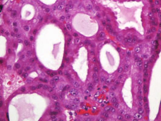

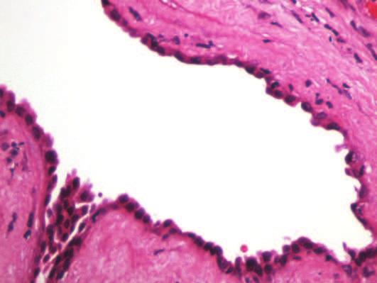
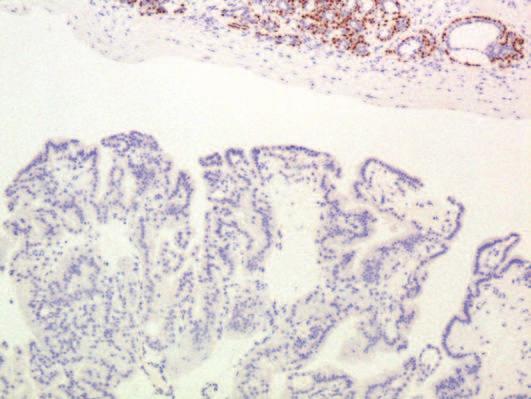
12 A B C D E F G H I J K Fig. 2
CASE REPORT Malignant transformation of breast ductal adenoma: a diagnostic pitfall
 Malaysian J Pathol 2015; 37(3) : 281 285 CASE REPORT Malignant transformation of breast ductal adenoma: a diagnostic pitfall Hiroko HAYASHI, Hiroshi OHTANI,* Junzo YAMAGUCHI,** and Isao SHIMOKAWA Department
Papillary Lesions of the Breast A Practical Approach to Diagnosis. (Arch Pathol Lab Med. 2016;140: ; doi: /arpa.
 Papillary Lesions of the Breast A Practical Approach to Diagnosis (Arch Pathol Lab Med. 2016;140:1052 1059; doi: 10.5858/arpa.2016-0219-RA) Papillary lesions of the breast Span the spectrum of benign,
Proliferative Epithelial lesions of the Breast. Sami Shousha, MD, FRCPath Charing Cross Hospital & Imperial College, London
 Proliferative Epithelial lesions of the Breast Sami Shousha, MD, FRCPath Charing Cross Hospital & Imperial College, London Amman, November2013 Proliferative Epithelial Lesions of the Breast Usual type
04/10/2018. Intraductal Papillary Neoplasms Of Breast INTRADUCTAL PAPILLOMA
 Intraductal Papillary Neoplasms Of Breast Savitri Krishnamurthy MD Professor of Pathology Deputy Division Head The University of Texas MD Anderson Cancer Center 25 th Annual Seminar in Pathology Pittsburgh,
1 NORMAL HISTOLOGY AND METAPLASIAS
 1 NORMAL HISTOLOGY AND METAPLASIAS, MD Anatomy and Histology 1 Metaplasias 2 ANATOMY AND HISTOLOGY The female breast is composed of a branching duct system, which begins at the nipple with the major lactiferous
Papillary Lesions of the breast
 Papillary Lesions of the breast Emad Rakha Professor of Breast Pathology The University of Nottingham Papillary lesions of the breast are a heterogeneous group of disease, which are characterised by neoplastic
ARTHUR PURDY STOUT SOCIETY COMPANION MEETING: DIFFICULT NEW DIFFERENTIAL DIAGNOSES IN PROSTATE PATHOLOGY. Jonathan I. Epstein.
 1 ARTHUR PURDY STOUT SOCIETY COMPANION MEETING: DIFFICULT NEW DIFFERENTIAL DIAGNOSES IN PROSTATE PATHOLOGY Jonathan I. Epstein Professor Pathology, Urology, Oncology The Reinhard Professor of Urological
Papillary Lesions of the Breast: WHO Update
 Papillary Lesions of the Breast: WHO Update Stuart J. Schnitt, M.D. Department of Pathology Beth Israel Deaconess Medical Center and Harvard Medical School Boston, MA, USA Papillary Lesions of the Breast
Papillary Lesions in Breast Pathology Practice: Diagnostic Challenges and Practical Approach. A Six- Year Experience from a Tertiary Care Hospital
 Open Access Journal Research Article DOI: 10.23958/ijirms/vol02-i05/12 Papillary Lesions in Breast Pathology Practice: Diagnostic Challenges and Practical Approach. A Six- Year Experience from a Tertiary
Epithelial Columnar Breast Lesions: Histopathology and Molecular Markers
 29th Annual International Conference Advances in the Application of Monoclonal Antibodies in Clinical Oncology and Symposium on Cancer Stem Cells 25 th -27t h June, 2012, Mykonos, Greece Epithelial Columnar
Proliferative Breast Disease: implications of core biopsy diagnosis. Proliferative Breast Disease
 Proliferative Breast Disease: implications of core biopsy diagnosis Jean F. Simpson, M.D. Breast Pathology Consultants, Inc. Nashville, TN Proliferative Breast Disease Must be interpreted in clinical and
Diseases of the breast (1 of 2)
") Diseases of the breast (1 of 2) Introduction A histology introduction Normal ducts and lobules of the breast are lined by two layers of cells a layer of luminal cells overlying a second layer of myoepithelial
Diseases of the breast (1 of 2) Introduction A histology introduction Normal ducts and lobules of the breast are lined by two layers of cells a layer of luminal cells overlying a second layer of myoepithelial
BREAST PATHOLOGY. Fibrocystic Changes
 BREAST PATHOLOGY Lesions of the breast are very common, and they present as palpable, sometimes painful, nodules or masses. Most of these lesions are benign. Breast cancer is the 2 nd most common cause
Synchronous squamous cell carcinoma of the breast. and invasive lobular carcinoma
 Sentani K et al. 1 Letter to the editor Synchronous squamous cell carcinoma of the breast and invasive lobular carcinoma Kazuhiro Sentani, 1 Takashi Tashiro, 2 Naohide Oue, 1 Wataru Yasui 1 1 Department
Cytyc Corporation - Case Presentation Archive - March 2002
 FirstCyte Ductal Lavage History: 68 Year Old Female Gail Index: Unknown Clinical History: Negative Mammogram in 1995 6 yrs. later presents with bloody nipple discharge Subsequent suspicious mammogram Suspicious
Pancreatitis: A Potential Pitfall in Endoscopic Ultrasound Guided Pancreatic FNA
 Pancreatitis: A Potential Pitfall in Endoscopic Ultrasound Guided Pancreatic FNA Jack Yang, MD Department of Pathology, Medical University of South Carolina Objectives Understand the indication of EUS
Columnar Cell Lesions. Columnar Cell Lesions and Flat Epithelial Atypia
 Columnar Cell Lesions and Stuart J. Schnitt, M.D. Beth Israel Deaconess Medical Center and Harvard Medical School Boston, MA, USA Columnar Cell Lesions Lesions characterized by columnar epithelial cells
Flat Epithelial Atypia
 Flat Epithelial Atypia Richard Owings, M.D. University of Arkansas for Medical Sciences Department of Pathology Flat epithelial atypia can be a difficult lesion May be a subtle diagnosis Lots of changes
Ductal Proliferations of the Breast: The Good, the Bad, and the Ugly
 Ductal Proliferations of the Breast: The Good, the Bad, and the Ugly Melinda F. Lerwill, MD CRITERIA FOR DISTINGUISHING LOW-GRADE DUCTAL CARCINOMA IN SITU FROM USUAL DUCTAL HYPERPLASIA CYTOLOGY Low-grade
PROSTATIC ADENOCARCINOMA: DIAGNOSTIC CRITERIA AND IMPORTANT MIMICKERS PROSTATIC ADENOCARCINOMA: DIAGNOSTIC CRITERIA
 PROSTATIC ADENOCARCINOMA: DIAGNOSTIC CRITERIA AND IMPORTANT MIMICKERS PROSTATIC ADENOCARCINOMA: DIAGNOSTIC CRITERIA 1 A good H & E helps! ADENOCARCINOMA DIAGNOSTIC CRITERIA Relatively uniform proliferation
Protocol for the Examination of Biopsy Specimens From Patients With Invasive Carcinoma of the Breast
 Protocol for the Examination of Specimens From Patients With Invasive Carcinoma of the Breast Version: BreastInvasive 1.0.0.0 Protocol Posting Date: February 2019 Accreditation Requirements The use of
Columnar Cell Lesions
 Columnar Cell Lesions Laura C. Collins, M.D. Department of Pathology Beth Israel Deaconess Medical Center and Harvard Medical School Boston, MA Question? Columnar cell lesions are: a) Annoying lesions
Papillary Lesions of the Breast
 Papillary Lesions of the Breast Laura C. Collins, M.D. Associate Professor of Pathology Associate Director, Division of Anatomic Pathology Beth Israel Deaconess Medical Center and Harvard Medical School
CLINICAL SIGNIFICANCE OF BENIGN EPITHELIAL CHANGES
 Papillomas. Papillomas are composed of multiple branching fibrovascular cores, each having a connective tissue axis lined by luminal and myoepithelial cells ( Fig. 23-11 ). Growth occurs within a dilated
Salivary Glands 3/7/2017
 Salivary Glands 3/7/2017 Goals and objectives Focus on the entities unique to H&N Common board type facts Information for your future practice Salivary Glands Salivary Glands Major gland. Paratid. Submandibular.
Original Article Clinicopathologic features of intraductal papillary neoplasm of breast: analyses of three cases
 Int J Clin Exp Pathol 2017;10(9):9575-9582 www.ijcep.com /ISSN:1936-2625/IJCEP0058752 Original Article Clinicopathologic features of intraductal papillary neoplasm of breast: analyses of three cases Yan
Salivary gland tumor cytologic and histologic correlation: Algorithmic and risk stratification based approaches
 Salivary gland tumor cytologic and histologic correlation: Algorithmic and risk stratification based approaches Christopher C. Griffith, MD, PhD Raja R. Seethala, MD 1. Salivary gland tumor cytology: A
University of Zurich. Histology and Immunophenotype of Invasive Lobular Breast Cancer. Daily Practice and Pitfalls. Zurich Open Repository and Archive
 University of Zurich Zurich Open Repository and Archive Winterthurerstr. 190 CH-8057 Zurich http://www.zora.uzh.ch Year: 2009 Histology and Immunophenotype of Invasive Lobular Breast Cancer. Daily Practice
Citation American Journal of Surgery, 196(5)
") NAOSITE: Nagasaki University's Ac Title Author(s) Multifocal branch-duct pancreatic i neoplasms Tajima, Yoshitsugu; Kuroki, Tamotsu Amane; Adachi, Tomohiko; Mishima, T Kanematsu, Takashi Citation American
NAOSITE: Nagasaki University's Ac Title Author(s) Multifocal branch-duct pancreatic i neoplasms Tajima, Yoshitsugu; Kuroki, Tamotsu Amane; Adachi, Tomohiko; Mishima, T Kanematsu, Takashi Citation American
Enterprise Interest None
 Enterprise Interest None B3 lesions of the breast What are they at surgery? Case 4 Edi Brogi MD PhD Attending Pathologist - Director of Breast Pathology Memorial Sloan Kettering Cancer Center New York
CYSTIC TUMORS OF THE KIDNEY JOHN N. EBLE, M.D. CYSTIC NEPHROMA
 Page 1 CYSTIC TUMORS OF THE KIDNEY JOHN N. EBLE, M.D. Department of Pathology & Laboratory Medicine Phone (317) 274-4806 Medical Science A-128 FAX: (317) 278-2018 635 Barnhill Drive jeble @iupui.edu Indianapolis,
Outline 11/2/2017. Pancreatic EUS-FNA general aspects. Cytomorphologic features of solid neoplasms/lesions of the pancreas
 ENDOSCOPIC ULTRASOUND GUIDED-FINE NEEDLE ASPIRATION CYTOLOGY OF PANCREAS Khalid Amin M.D. Assistant Professor Department of Laboratory Medicine and Pathology University of Minnesota Outline Pancreatic
Breast pathology. 2nd Department of Pathology Semmelweis University
 Breast pathology 2nd Department of Pathology Semmelweis University Breast pathology - Summary - Benign lesions - Acute mastitis - Plasma cell mastitis / duct ectasia - Fat necrosis - Fibrocystic change/
Papillary Lesions of the Breast
 Papillary Lesions of the Breast Texas Society of Pathologists 2013 Laura C. Collins, M.D. Associate Professor of Pathology Associate Director, Division of Anatomic Pathology Beth Israel Deaconess Medical
3/28/2017. Disclosure of Relevant Financial Relationships. GU Evening Subspecialty Case Conference. Differential Diagnosis:
 GU Evening Subspecialty Case Conference Rajal B. Shah, M.D. VP, Medical Director, Urologic Pathology Miraca Life Sciences, Irving, Texas Clinical Associate Professor of Pathology Baylor College of Medicine,
Various hereditary, acquired and neoplastic conditions can lead to cyst formation in the kidney.
 Dr. Fatima AlAl-Hashimi Hashimi,, MD, FRCPath Salmaniya Medical Complex, Bahrain Various hereditary, acquired and neoplastic conditions can lead to cyst formation in the kidney. The most frequently encountered
COMPANION MEETING BREAST. Auditorium 11:15 1:00 am. Convenor: A/Professor Gelareh Farshid, SA Pathology, SA
 Australasian Division of the International Academy of Pathology Limited ABN 73 008 593 815 36 TH Annual Scientific Meeting Darling Harbour Convention Centre, Sydney, Australia June 3-5, 2011 COMPANION
ACCME/Disclosures. Cribriform Lesions of the Prostate. Case
 Cribriform Lesions of the Prostate Ming Zhou, MD, PhD Departments of Pathology and Urology New York University Langone Medical Center New York, NY Ming.Zhou@NYUMC.ORG ACCME/Disclosures The USCAP requires
Macro- and microacinar proliferations of the prostate
 Macro- and microacinar proliferations of the prostate (with emphasis on cancer mimics) Rodolfo Montironi, MD (IT), FRCPath (UK), IFCAP (USA) Polytechnic University of Marche Region (Ancona) School of Medicine,
Breast Pathology. Breast Development
 Breast Pathology Lecturer: Hanina Hibshoosh, M.D. Reading: Kumar, Cotran, Robbins, Basic Pathology, 6th Edition, pages 623-635 Breast Development 5th week - thickening of the epidermis - milk line 5th
Papillary lesions of the breast: selected diagnostic and management issues
 Histopathology 2008, 52, 20 29. DOI: 10.1111/j.1365-2559.2007.02898.x REVIEW Papillary lesions of the breast: selected diagnostic and management issues L C Collins & S J Schnitt Department of Pathology,
In situ lobular neoplasia of the breast with marked myoepithelial proliferation
 In situ lobular neoplasia of the breast with marked myoepithelial proliferation Sami Shousha To cite this version: Sami Shousha. In situ lobular neoplasia of the breast with marked myoepithelial proliferation.
Demystifying Endometrial Hyperplasia
 Demystifying Endometrial Hyperplasia A review from Diagnostic Histopathology 19:7 Dr R Hadden ST5 Histopathology Derriford Hospital Plymouth Endometrium Target for sex-steroid hormones Glands Stroma Proliferate
Fine-Needle Aspiration Cytology of Low-Grade Cribriform Cystadenocarcinoma with Many Psammoma Bodies of the Salivary Gland
 The Korean Journal of Pathology 2013; 47: 481-485 CASE STUDY Fine-Needle Aspiration Cytology of Low-Grade Cribriform Cystadenocarcinoma with Many Psammoma Bodies of the Salivary Gland Ji Yun Jeong Dongbin
Invasive Papillary Breast Carcinoma
 410 This is an Open Access article licensed under the terms of the Creative Commons Attribution- NonCommercial-NoDerivs 3.0 License (www.karger.com/oa-license), applicable to the online version of the
Benign Mimics of Malignancy in Breast Pathology
 Arthur Purdy Stout Society of Surgical Pathologists Companion Meeting Benign Mimics of Malignancy in Breast Pathology Stuart J. Schnitt, M.D. Beth Israel Deaconess Medical Center and Harvard Medical School,
Can True Papillary Neoplasms of Breast and Their Mimickers Be Accurately Classified by Cytology?
 92 CANCER CYTOPATHOLOGY Can True Papillary Neoplasms of Breast and Their Mimickers Be Accurately Classified by Cytology? Claire W. Michael, M.D. 1 Bruce Buschmann, C.T. 2 1 University of Michigan, Department
Biliary tract tumors
 Short Course 2010 Annual Fall Meeting of the Korean Society for Pathologists Biliary tract tumors Joon Hyuk Choi, M.D., Ph.D. Professor, Department of Pathology, Yeungnam Univ. College of Medicine, Daegu,
Select problems in cystic pancreatic lesions
 Disclosure Select problems in cystic pancreatic lesions Five Prime Therapeutics shareholder Adicet Bio shareholder Bristol-Meyer Squibb advisory board grace.kim@ucsf.edu Pancreatic cystic lesions Intraductal
Interpretation of Breast Pathology in the Era of Minimally Invasive Procedures
 Shahla Masood, M.D. Professor and Chair Department of Pathology and Laboratory Medicine University of Florida College of Medicine Jacksonville Medical Director, UF Health Breast Center Chief of Pathology
6/3/2010. Outline of Talk. Lobular Breast Cancer: Definition of lobular differentiation. Common Problems in Diagnosing LCIS in Core Biopsies
 Outline of Talk Lobular Breast Cancer: Common Problems in Diagnosing LCIS in Core Biopsies Definition of lobular differentiation Variants of LCIS that: carry risk for unsampled invasive cancer mimic DCIS
Objectives. Atypical Glandular Cells. Atypical Endocervical Cells. Reactive Endocervical Cells
 2013 California Society of Pathologists 66 th Annual Meeting San Francisco, CA Atypical Glandular Cells to Early Invasive Adenocarcinoma: Cervical Cytology and Histology Christina S. Kong, MD Associate
Disclosures. Parathyroid Pathology. Objectives. The normal parathyroid 11/10/2012
 Disclosures Parathyroid Pathology I have nothing to disclose Annemieke van Zante MD/PhD Assistant Professor of Clinical Pathology Associate Chief of Cytopathology Objectives 1. Review the pathologic features
Synonyms. Nephrogenic metaplasia Mesonephric adenoma
 Nephrogenic Adenoma Synonyms Nephrogenic metaplasia Mesonephric adenoma Definition Benign epithelial lesion of urinary tract with tubular, glandular, papillary growth pattern Most frequently in the urinary
Case study 1. Rie Horii, M.D., Ph.D. Division of Pathology Cancer Institute Hospital, Japanese Foundation for Cancer Research
 NCCN/JCCNB Seminar in Japan April 15, 2012 Case study 1 Rie Horii, M.D., Ph.D. Division of Pathology Cancer Institute Hospital, Japanese Foundation for Cancer Research Present illness: A 50y.o.premenopausal
Low-grade Adenosquamous Carcinoma Coexisting with Sclerosing Adenosis of the Breast: A Case Report
 31 Case Report J. St. Marianna Univ. Vol. 8, pp. 31 35, 2017 Low-grade Adenosquamous Carcinoma Coexisting with Sclerosing Adenosis of the Breast: A Case Report Ryoko Oi 1, 2, Ichiro Maeda 1, Yoshio Aida
5/21/2018. Difficulty in Underdiagnosing Prostate Cancer. Diagnosis of Prostate Cancer. Evaluation of Prostate Cancer and Atypical on Needle Biopsy
 Evaluation of Prostate Cancer and Atypical on Needle Biopsy Jonathan I. Epstein Difficulty in Underdiagnosing Prostate Cancer Limited tissue on needle biopsy (1 cm. x
Intraductal carcinoma of the prostate on needle biopsy: histologic features and clinical significance
 & 2006 USCAP, Inc All rights reserved 0893-3952/06 $30.00 www.modernpathology.org Intraductal carcinoma of the prostate on needle biopsy: histologic features and clinical significance Charles C Guo 1 and
Case Report Synchronous Bilateral Solid Papillary Carcinomas of the Breast
 Case Reports in Surgery Volume 2013, Article ID 812129, 4 pages http://dx.doi.org/10.1155/2013/812129 Case Report Synchronous Bilateral Solid Papillary Carcinomas of the Breast Noriko Yoshimura, 1 Shigeru
Notice of Faculty Disclosure
 California Society of Pathology Diagnostic Problems in Surgical Pathology December 2015 Case 2 Laura C. Collins, M.D. Associate Professor of Pathology Associate Director of Anatomic Pathology Beth Israel
Endometrial Metaplasia, Hyperplasia & Other Cancer Mimics: a Consultant s Experience
 Endometrial Metaplasia, Hyperplasia & Other Cancer Mimics: a Consultant s Experience Pacific Northwest Society of Pathologists Vancouver, B.C. September 26, 2015 Teri A. Longacre, M.D. longacre@stanford.edu
Pleomorphic adenoma of breast - a case report and distinction with metaplastic carcinoma D Gupta, S Agrawal, N Trivedi, A Tewari
 of breast - a case report and distinction with metaplastic carcinoma D Gupta, S Agrawal, N Trivedi, A Tewari Introduction, also known as mixed tumour, is a benign tumour which typically presents as a painless,
Flat Epithelial Atypia: A Review of Current Concepts
 90 The Open Breast Cancer Journal, 2010, 2, 90-94 Flat Epithelial Atypia: A Review of Current Concepts Amy L. Adams * Open Access Department of Pathology and Laboratory Medicine, Emory University School
American Journal of Cancer Case Reports. Invasive Papillary Carcinoma of Male Breast: A Rare Case Report
 American Journal of Cancer Case Reports http://ivyunion.org/index.php/ajccr SantraAetal. American Journal of Cancer Case Reports 2014, 3:56-61 Page 1 of 6 Vol 3 Article ID 20140617, 6 pages Case Report
Histological Type. Morphological and Molecular Typing of breast Cancer. Nottingham Tenovus Primary Breast Cancer Study. Survival (%) Ian Ellis
 Ian Ellis") Morphological and Molecular Typing of breast Cancer Ian Ellis Molecular Medical Sciences, University of Nottingham Department of Histopathology, Nottingham University Hospitals NHS Trust Histological Type
Morphological and Molecular Typing of breast Cancer Ian Ellis Molecular Medical Sciences, University of Nottingham Department of Histopathology, Nottingham University Hospitals NHS Trust Histological Type
Thyroid follicular neoplasms in cytology. Ulrika Klopčič Institute of Oncology, Department of Cytopathology, Ljubljana, Slovenia
 Thyroid follicular neoplasms in cytology Ulrika Klopčič Institute of Oncology, Department of Cytopathology, Ljubljana, Slovenia Lecture overview importance of FNAB in assessing thyroid lesions follicular
Good afternoon everyone. First of all many thanks to Dr. Bonaventura and Dr. Arn for inviting
 PATHOLOGY IN-SITU CARCINOMA, ROHIT BHARGAVA, MD 1 Good afternoon everyone. First of all many thanks to Dr. Bonaventura and Dr. Arn for inviting me here, it s great to be here and I m going to talk about
NODULAR CYSTIC HIDRADENOMA OVER THE GLUTEAL REGION: A RARE CYTOMORPHOLOGICAL DIAGNOSIS
 NODULAR CYSTIC HIDRADENOMA OVER THE GLUTEAL REGION: A RARE CYTOMORPHOLOGICAL DIAGNOSIS Abstract: The primary as well as metastatic tumours of the skin can be diagnosed by fine needle aspiration cytology
Gross appearance of nodular hyperplasia in material obtained from suprapubic prostatectomy. Note the multinodular appearance and the admixture of
 Tiền liệt tuyến Tiền liệt tuyến Gross appearance of nodular hyperplasia in material obtained from suprapubic prostatectomy. Note the multinodular appearance and the admixture of solid and microcystic areas.
Case Report Basaloid ductal carcinoma in situ arising in salivary gland metaplasia of the breast: a case report
 Int J Clin Exp Pathol 2014;7(9):6370-6374 www.ijcep.com /ISSN:1936-2625/IJCEP0001480 Case Report Basaloid ductal carcinoma in situ arising in salivary gland metaplasia of the breast: a case report Eun
Update in Salivary Gland Pathology. Benjamin L. Witt University of Utah/ARUP Laboratories February 9, 2016
 Update in Salivary Gland Pathology Benjamin L. Witt University of Utah/ARUP Laboratories February 9, 2016 Objectives Review the different appearances of a selection of salivary gland tumor types Establish
Incidence of ductal lesions
 Ductal Proliferative Lesions of the Breast: From FEA to ADH to DCIS Incidence of ductal lesions Pre-mammography: DCIS < 3% of breast cancers, large palpable masses, with invasion Mammography: DCIS 25%
Disclosures 5/27/2012. Outline of Talk. Outline of Talk. When Is LCIS Clinically Significant? Classic LCIS. Classic LCIS
 When Is LCIS Clinically Significant? Disclosures I have nothing to disclose Yunn-Yi Chen, MD, PhD Professor Outline of Talk Outline of Talk Classic LCIS Classic LCIS Definition of lobular differentiation
DIAGNOSTIC SLIDE SEMINAR: PART 1 RENAL TUMOUR BIOPSY CASES
 DIAGNOSTIC SLIDE SEMINAR: PART 1 RENAL TUMOUR BIOPSY CASES Dr. Andrew J. Evans MD, PhD, FACP, FRCPC Consultant in Genitourinary Pathology University Health Network, Toronto, ON Case 1 43 year-old female,
Malignant Phyllodes tumor with necrosis a rare case report
 Quest Journals Journal of Medical and Dental Science Research Volume 1 ~ Issue 1 (2014) pp: 01-06 ISSN(Online) : 2394-076X ISSN (Print):2394-0751 www.questjournals.org Research Paper Malignant Phyllodes
Evaluation of Breast Specimens Removed by Needle Localization Technique
 Evaluation of Breast Specimens Removed by Needle Localization Technique Specimen Handling: The breast specimen when received should be measured and grossly inspected for any orientation designated by the
Thyroid Nodules: Understanding FNA Cytology (The Bethesda System for Reporting of Thyroid Cytopathology) Shamlal Mangray, MB, BS
 Shamlal Mangray, MB, BS") Thyroid Nodules: Understanding FNA Cytology (The Bethesda System for Reporting of Thyroid Cytopathology) Shamlal Mangray, MB, BS Attending Pathologist Rhode Island Hospital, Providence, RI DISCLOSURE:
Thyroid Nodules: Understanding FNA Cytology (The Bethesda System for Reporting of Thyroid Cytopathology) Shamlal Mangray, MB, BS Attending Pathologist Rhode Island Hospital, Providence, RI DISCLOSURE:
The Hot Topic for today is a biopsy from a 58-year-old woman who had worrisome mammographic calcifications on screening.
 The Hot Topic for today is a biopsy from a 58-year-old woman who had worrisome mammographic calcifications on screening. 1 My name is Dan Visscher; I am a consultant in the Division of Anatomic Pathology
04/10/2018 HIGH RISK BREAST LESIONS. Pathology Perspectives of High Risk Breast Lesions ELEVATED RISK OF BREAST CANCER HISTORICAL PERSPECTIVES
 Pathology Perspectives of High Risk Breast Lesions Savitri Krishnamurthy MD Professor of Pathology Deputy Division Head Director of Clinical Trials, Research and Development The University of Texas MD
International Society of Gynecological Pathologists Symposium 2007
 International Society of Gynecological Pathologists Symposium 2007 Anais Malpica, M.D. Department of Pathology The University of Texas M.D. Anderson Cancer Center Grading of Ovarian Cancer Histologic grade
Salivary Gland Cytology
 Salivary Gland Cytology Diagnostic challenges and potential pitfalls Tarik M. Elsheikh, MD Professor and Medical Director Anatomic Pathology Cleveland Clinic FNA Salivary Gland Lesions Indications Distinguish
Gynecologic Cytopathology: Glandular lesions
 Gynecologic Cytopathology: Glandular lesions Lin Wai Fung (MSc, MPH, CMIAC) 17/4/2014 Glandular lesions of the uterus Endocervix Endometrium Normal endocervical cells Sheets, strips well-preserved architecture:
Oncocytic-Appearing Salivary Gland Tumors. Oncocytic, Cystic, Mucinous, and High Grade Salivary Gland Tumors SALIVARY GLAND FNA: PART II
 William C. Faquin, MD, PhD Professor of Pathology Harvard Medical School Director of Head and Neck Pathology Massachusetts Eye and Ear Massachusetts General Hospital SALIVARY GLAND FNA: PART II Oncocytic,
Columnar Cell Lesions and Flat Epithelial Atypia
 Columnar Cell Lesions and Flat Epithelial Atypia Laura C. Collins, M.D. Department of Pathology Beth Israel Deaconess Medical Center and Harvard Medical School, Boston, MA Terminology for Columnar Cell
Mody. AIS vs. Invasive Adenocarcinoma of the Cervix
 Common Problems in Gynecologic Pathology Michael T. Deavers, M.D. Houston Methodist Hospital, Houston, Texas Common Problems in Gynecologic Pathology Adenocarcinoma in-situ (AIS) of the Cervix vs. Invasive
IBCM 2, April 2009, Sarajevo, Bosnia and Herzegovina
 Preoperative diagnosis and treatment planning in breast cancer The pathologist s perspective L. Mazzucchelli Istituto Cantonale di Patologia Locarno, Switzerland IBCM 2, 23-25 April 2009, Sarajevo, Bosnia
CPC 4 Breast Cancer. Rochelle Harwood, a 35 year old sales assistant, presents to her GP because she has noticed a painless lump in her left breast.
 CPC 4 Breast Cancer Rochelle Harwood, a 35 year old sales assistant, presents to her GP because she has noticed a painless lump in her left breast. 1. What are the most likely diagnoses of this lump? Fibroadenoma
Treatment options for the precancerous Atypical Breast lesions. Prof. YOUNG-JIN SUH The Catholic University of Korea
 Treatment options for the precancerous Atypical Breast lesions Prof. YOUNG-JIN SUH The Catholic University of Korea Not so benign lesions? Imaging abnormalities(10% recall) lead to diagnostic evaluation,
FNA of Thyroid. Toward a Uniform Terminology With Management Guidelines. NCI NCI Thyroid FNA State of the Science Conference
 FNA of Thyroid NCI NCI Thyroid FNA State of the Science Conference Toward a Uniform Terminology With Management Guidelines Thyroid Thyroid FNA Cytomorphology NCI Thyroid FNA State of the Science Conference
Benign Breast Disease and Breast Cancer Risk
 Benign Breast Disease and Breast Cancer Risk Jean F. Simpson, M.D. Vanderbilt University Nashville, Tennessee December 1, 2011 Nashville Nashville Lebanon 1 Cedars of Lebanon State Park The American University
The Korean Journal of Cytopathology 15(1) : 60-64, 2004
 : 60-64, 2004") 15 1 The Korean Journal of Cytopathology 15(1) : 60-64, 2004 : INTRODUCTION Papillary carcinoma of the thyroid gland has for long been traditionally diagnosed on the basis of the characteristic papillary
15 1 The Korean Journal of Cytopathology 15(1) : 60-64, 2004 : INTRODUCTION Papillary carcinoma of the thyroid gland has for long been traditionally diagnosed on the basis of the characteristic papillary
04/09/2018. Salivary Gland Pathology in the Molecular Era Old Friends, Old Foes, & New Acquaintances
 Salivary Gland Pathology in the Molecular Era Old Friends, Old Foes, & New Acquaintances Jennifer L. Hunt, MD, MEd Aubrey J. Hough Jr, MD, Endowed Professor of Pathology Chair of Pathology and Laboratory
Atypical Hyperplasia/EIN
 EIN Atypical Hyperplasia/EIN Based on scientific and diagnostic advances, in 2014 the WHO moved that the precursor lesion for endometrioid carcinoma be atypical hyperplasia/ein, rather than what was previously
Modern Pathology (2011) 24, & 2011 USCAP, Inc. All rights reserved /11 $
 24, & 2011 USCAP, Inc. All rights reserved /11 $") & 2011 USCAP, Inc. All rights reserved 0893-3952/11 $32.00 185 Luminal cytokeratin expression profiles of breast papillomas and papillary carcinomas and the utility of a cytokeratin 5/p63/cytokeratin 8/18
& 2011 USCAP, Inc. All rights reserved 0893-3952/11 $32.00 185 Luminal cytokeratin expression profiles of breast papillomas and papillary carcinomas and the utility of a cytokeratin 5/p63/cytokeratin 8/18
Disorders of Cell Growth & Neoplasia. Histopathology Lab
 Disorders of Cell Growth & Neoplasia Histopathology Lab Paul Hanna April 2010 Case #84 Clinical History: 5 yr-old, West Highland White terrier. skin mass from axillary region. has been present for the
"Atypical": Criteria and
 "Atypical": Criteria and Controversies Esther Rossi MD PhD MIAC Division of Anatomic Pathology and Cytology Catholic University of Sacred Heart Rome, Italy CASE HISTORY In 2015, 45 y/o woman underwent
Case Report Aggressive invasive micropapillary salivary duct carcinoma of the parotid gland
 Pathology International 2008; 58: 322 326 doi:10.1111/j.1440-1827.2008.02231.x Case Report Aggressive invasive micropapillary salivary duct carcinoma of the parotid gland Hidetaka Yamamoto, 1 Hideoki Uryu,
NEUROENDOCRINE DIFFERENTIATED BREAST CARCINOMA
 + NEUROENDOCRINE DIFFERENTIATED BREAST CARCINOMA + INTRODUCTION + NEUROENDOCRINE FEATURES IN BREAST CARCINOMA Incidence of 2-5% Seen in various histopathological types of breast carcinoma Seen in both
They Do Look Alike : Mimics of Prostate Cancer in Biopsy Samples
 They Do Look Alike : in Biopsy Samples Gladell P. Paner, MD Departments of Pathology and Surgery (Urology) University of Chicago, IL USA Gladell.paner@uchospitals.edu Benign in Needle Biopsy 1. Benign
AN AUTOPSY CASE OF PARATHYROID CARC. Matsumoto, Koji; Ito, Masahiro; Sek. Citation Acta medica Nagasakiensia. 1989, 34
 NAOSITE: Nagasaki University's Ac Title Author(s) AN AUTOPSY CASE OF PARATHYROID CARC Hsu, Chao-Tien; Naito, Shinji; Shik Matsumoto, Koji; Ito, Masahiro; Sek Citation Acta medica Nagasakiensia. 1989, 34