CNB vs Surgical Excision
|
|
|
- Elvin Greer
- 6 years ago
- Views:
Transcription
 less invasive,")
1 Update on Core Needle Biopsy of Non-palpable Breast Lesions Nour Sneige, M.D. UT MD Anderson Cancer Center Houston, Tx Image-Guided CNB of Breast Lesions An alternative to surgical biospy CNB vs Surgical Excision Same accuracy (1-2% delayed false neg) less invasive, less expensive Spares most patients (benign) unnecessary surgery Empowers patient to decide on treatment options 1
2 CNB of Breast Lesions 1. Technical considerations 2. Diagnostic problems 3. Controversies: to excise or not to excise Imaging Guidance Systems Ultrasound Stereotactic mammography Magnetic resonance (MRI) Ultrasound Guidance System System of choice (lesion imaged with US) Less expensive More readily available No radiation exposure Lesions not amenable to stereo (tail of axilla, close to chest wall) Less time consuming 2
3 Stereotactic Guidance System Can be used for wider range of lesions: masses and calcifications Breast and lesion are immobilized accurate sampling + 2 mm MRI Demonstrates cancers not detected on mammography or sonography Sensitivity 100% Specificity 37-97% Needle Biopsy Devices Automated (spring loaded gun) Directional Vacuum Assisted (DVAB) 3


4 Directional Vacuum Assisted Biopsy 1. Probe is placed into the breast adjacent to the tissue requiring sampling VACUUM Vacuum suction is applied, tissue is pulled into the cutting apeture A rotating mechanism cuts and encloses the tissue that has been pulled into the cutting aperture Advantages: DVAB vs Automated Faster (1.4 min. vs. 17 min.) Twice the weight (34 vs. 17 mg) Tissue shift by bleeding, avoided Contiguous sampling 4
 Calcs retrieval 99-100% Reynolds et al.")
5 Schematic Representation of Breast Biopsy Techniques Microcalcification Core biopsy Random sampling Contiguous sampling Advantages: DVAB vs Automated Underestimation of ADH 41% Automated needle 15% DVAB (Mammotome) Calcs retrieval % Reynolds et al. Am J Roentgenol 1998;171:
6 CNB Size Gauge: 18, 16, 14, 11, 8-g Calcs: 11-g V more accurate than 14-g V/A Optimal Numbers of CNB Solid masses: 4-5 cores Calcifications: > 10 cores % of Lesions 100% 98% 98% 99% 99% 95% 90% 91% 85% 84% 80% 75% >6 X-Core # Diagnostic yield from sequential core biopsy specimens for all masses (n=93) 6
: Cores")
7 % of Lesions 100% 90% 80% 70% 60% 50% 40% 30% 20% 10% 0% 92% 87% 79% 74% 62% 47% >6 X-Core # Diagnostic yield from sequential core biopsy specimens for calcifications without mass (n=53) Specimen Radiography - CNB Confirm Calcs Calcs must correspond to those on mammography Segregation of Cores with and without Calcs on Specimen Radiographs Diagnostic yield of malignancy (DVAB): Cores with calcs: 84% Cores without calcs: 71% Same diagnosis: atypical /malignant in 76% Equally careful attention should be given to cores with no calcs Margolin et al, Radiology 2004; 233: Easley et al, The Breast Journal 2007;13:

 Specimen radiograph 2)")
8 Radiologic-Pathologic Correlation Pathologist should review specimen radiograph (calcs) Pathologist should be provided with: 1) Specimen radiograph 2) BI-RADS category 8
9 Imaging-Histologic Discordance 0.9-6% of cases Discordance is an indication for surgical excision (24% malignant) Diagnostic Problems- CNB Tubular lesions (tubular CA vs. adenosis) Papillary lesions (pap CA vs. papilloma) Mucinous lesions (mucocele vs. muci CA) Fibroepithelial lesions (FA vs. PT) In situ vs invasive Ductal vs lobular carcinoma in situ Diagnostic Problems- CNB 1. Benign or malignant? 2. In situ or invasive? 3. In situ lobular or ductal? 9
 False Negative 20 (48%) False Positive 22 (52%) 21% of all breast bx claims involved CNB Troxel: Errors in Surgical Pathology, Am J Surg Pathol, 2004;28:1092-1095 Diagnostic Errors in")
10 Breast Biopsy Claims from Total Claims 42 (15.5%) False Negative 20 (48%) False Positive 22 (52%) 21% of all breast bx claims involved CNB Troxel: Errors in Surgical Pathology, Am J Surg Pathol, 2004;28: Diagnostic Errors in Stereotactic/ Palpable CNB of Breast 1. Misdiagnosis of DCIS, SA and adenosis as invasive ductal carcinoma. 2. Misdiagnosis of LCIS as low-grade DCIS. 3. Failure to recognize small, easily over-looked foci of invasive lobular carcinoma. Troxel: Int J Surg Pathol 8(4): ,
11 CNB misdiagnosed as IC 11


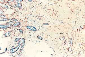
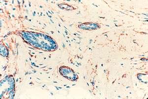
12 Carcinoma In Situ: lobular or Ductal? Diagnosis: 1. LCIS 2. Ca in situ 3. ADH/DCIS E-cadherin Deeper cut 12
13 Indeterminate Lesions at CNB Ancillary studies If in doubt, second opinion / defer to surgical excision Controversies: To Excise or Not to Excise? Lobular Neoplasia Papilloma Mucocele Radial Scar Flat epithelial atypia ADH Consensus Meeting on Image Detected Breast Cancer The American College of Surgeons 2005 Patients with high-risk lesions, including ADH, ALH and LCIS found on percutaneous biopsy may have DCIS or invasive cancer at the same site and should generally undergo surgical excision. This incidence of missing such important findings is markedly reduced with the use of vacuum assisted biopsy and large gauge needles 13
14 Consensus Meeting on Image Detected Breast Cancer The American College of Surgeons For some individuals with high-risk histologic findings, in whom careful correlation of imaging and histologic findings is concordant, or breast MRI is normal, follow-up without surgical excision may be reasonable. Lobular Neoplasia: To Excise or Not to Excise? 1% of all breast biopsies Marker of inc. risk in excisional biopsy Lobular Neoplasia Diagnosed at CNB Does Not Mandate Surgical Excision 328 Surgical Excision 504 Subjects (19 studies) 176 No Surgical Excision 23.2% upgrade. 7 studies included 101 HRL Exc HRL, 16.7% upgrade Bowman et al, Journal of Surgical Research 2007,
15 Outcomes for All Patients Undergoing Surgical Excision or Observation (504 Subjects) 328 Surgical Excision 176 No Surgical Excision 94 LCIS 124 ALH 76 HRL 34 LIN 62 F/U 111 No. F/U 3 Lost 6 DCIS 14 IC 21% 13 DCIS 5 IC 14.5% 34 CA 2 DCIS 2 IC CA 4.7% Bowman et al, Journal of Surgical Research 2007, Recommendations Regarding Management of LIN Diagnosed at CNB Bowman et al, J Surg Res 2007, Routine surgical excision 9 Surgical excision for specific circumstances 7 Only for LCIS (not ALH) 1 LIN with residual Calcs 2 LIN with synchronous mass 1 LIN with asso with a high risk lesion 1 Pleomorphic LCIS 1 Cases of diagnostic confusion 1 Clinical follow-up 1 No firm recommendations 2 LCIS and ALH on CNB Limitations of published reports 1) Marked variation in the clinical, methodological, and radiological details. 2) Inconsistency regarding inclusion criteria for surgical excision vs observation /selection bias 3) Inclusion of subjects with other high-risk lesions 4) Inclusion of cases with overlapping diagnoses (PLCIS). 5) Small numbers, the retrospective nature, and nearly half were nonconsecutive series. Bowman et al., Journal of Surgical Research 2007,
16 LN in CNB is Associated With a Low Risk of DCIS/IC 92 LCIS/ALH on CNB - 7 cancers on excision 3 (3%) in area of bx site (1 DCIS; 2 IDC)* 2 away from bx site 2 after negative bx site excision *One interpretive error (ADH on core) Renshaw et al. AJCP 2006; 126: LN in CNB is Associated With a Low Risk of DCIS/IC Rate of DCIS/IC found is well within the reported false-negative rate for NLB (1.2%- 9.1%). In centers with appropriate f/u info, routine excision of all biopsy sites for LN may not always be necessary. Renshaw et al. AJCP 2006; 126: CNB upgrade on excision 3(8%) 18 ALH 1 (6%) 20 LCIS 2 (10%) Problems: 8 associated with mass/density 9-11 V or 14 G auto (S or US) Cangiarella et al:arch Pathol Lab Med. 2008, 132:


17 LCIS/ALH on CNB- MDACC Multidisciplinary approach No excision required: if 1. Calcs only 2. Completely removed by DVAB 3. Classic LCIS 17


18 Post interventional imaging LCIS and CCC with calcs by imaging completely excised 18


19 Papillary lesions on CNB: to Excise or Not to Excise? < 5% of breast biopsies Include benign atypical and malignant 19
 with benign or atypical papillary lesions 138 (23%, R 6-39%) upgraded to carcinoma on excision Arora et al.")
20 Atypia in a Papilloma 10% focal, <30% atypia, 30-90% DCIS (Tavassoli) 3 mm area ADH/DCIS in papilloma (Page) Atypical papilloma on CNB require excision to R/O Ca Papillary lesions on CNB 1063 subjects (16 series) with benign or atypical papillary lesions 138 (23%, R 6-39%) upgraded to carcinoma on excision Arora et al. Am J Surg 194, , 2007 Reliability of CNB in the diagnosis of papilloma (no atypia)? 20
21 Reliability of CNB in the Diagnosis of Papilloma? Papilloma with no atypia 345 cases (15 series) 8 series: 0% upgrade to cancer 7 series: 2-20% upgrade to cancer Papillary lesions on CNB Limitations of published series: Small number of patients Selection bias for excision 2 largest studies used cases diagnosed on FNA Relationship of Mode of Biopsy of the Papillary Lesion With Malignancy at Surgical Excision Mode of percutaneous biopsy Stereotactic US- core US- FNA N Malignant % Valdes E, et al. Annals of Surg Onc. 2006; 13:
22 Relationship of Pathologic Characteristics of Papillary Lesions With Malignancy at Surgical Excision Pathologic Diagnosis bx Papilloma/papillomatosis Atypical papillomatosis/ papilloma with ADH Pure papillary lesion Papillary lesion with atypia Ca/cases 6/36 2/7 9/28 2/9 % Valdes, et al. Annals of Surg Oncol. 2006; 13(4): Department of surgery and radiology. Lesions Yielding a Benign, Concordant Diagnosis of Papilloma at Percutaneous Biopsy May Warrant Surgical Excision 35 papillomas on CNB 25 excised, 10 mammo F/U> 2 yrs Excision: 5 Cancers (14%) Liberman, et al AJR 186: Lesions Yielding Benign Papilloma at Percutaneous Biopsy and Cancer at Surgery Type Size (cm) Guidance/N eedle # of Cores Target Interval (mo) Surgical Pathology 1 Calcs 1.2 St/ 11 V 29 Excised 1 DCIS 1cm from bx site 2 Mass 1.2 St /14 A 4 Sampled 7 ICPC with DCIS at periphery of pap lesion. 3 Mass 0.8 US/ 14 A 3 Sampled 25 DCIS at periphery of papilloma 4 Mass 0.6 US/ 14 A 4 Sampled 21 DCIS in papilloma 5 Mass 0.8 St/ 11 V 14 Excised 22 IDC, gr 3 and DCIS 1.8cm; admixed with pap. 22
 DVAB samples Concordant imaging and histologic findings (size) Mucinous Lesions < 1% of CNB specimens Range: Mucocele-like to mucinous carcinoma")
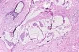
23 Lesions Diagnosed As Papilloma on CNB - MDACC No excision: Small (up to 1.5 cm) DVAB samples Concordant imaging and histologic findings (size) Mucinous Lesions < 1% of CNB specimens Range: Mucocele-like to mucinous carcinoma Mucocele-like Lesions Imaging: Indeterminate calcs (majority) Well-circumscribed lobulated mass 23


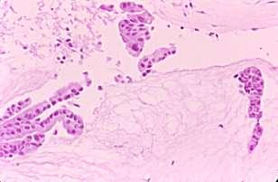
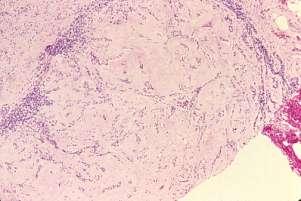
24 Mucocele- like lesion, CNB 24


")
25 CNB MLL with ADH and mucinous ca (20% of cases) 25


26 Mucocele-like Tumors- CNB Among 20 cases of MLL with no atypia, no carcinoma was found on excision. CNB is highly reliable for accurate Dx of mucinous lesions Wang et al AJCP 2007 Carder et al Histopath 2004 Renshaw AJCP 2002 Mucocele with no atypia on CNB, Calcs completely removed. Exicision is no required Radial Scars Stellate Radiating central fibroelastic stroma Nonpro/prolif.e pith. 26
27 Results of Surgical Excision of Radial Scars Without Atypia Diagnosed on CNB Authors Biopsy Technique # cases DCIS/IC at Excision Dershaw 14 A 1 0 Lee 14 A 4 1 DCIS Jackman 14 A 5 2 1IDC, 1 DCIS Philpotts 14 A; 11 M 8 0 Kirwan 14 A 30 0 Cawson 14 A 27 0 Total 75 3 (4%) Radial Scar on CNB Brenner et al. AJR 2002; 179: lesions (11 institutions) 157 lesions (102 excised, rest f/u 24 m) CA at excision: RS with ADH on CNB 28% RS no ADH 4% Radial Scar on CNB Dx of RS on CNB is likely to be reliable when: No associated ADH >12 cores obtained VAD used Brenner et al. AJR 2002; 179:
28 Radial Sclerosing Lesion 80 patients (9 or 11 SDVAB) 19 excised, 2 upgraded to atypia 61 mammographic surveillance Conclusions: more extensive sampling by 9/11-g device followed by meticulous correlation of radiological-pathological and close f/u could obviate surgical excision in the majority of RSL. Resetkova et al. Breast Cancer Res Treat:2008 ADH at CNB: MDACC Approach ADH limited to < 2 foci (DVAB) had no worse lesion on excision, provided that most of calcs are removed Ely et al., AJ S P 2001; 25, Sneige et al., AJCP 2003;119: ADH in DVAB of Microcalcifications 140 patients (86.4% excision) ADH associated with calcs in the absence of a mass can be categorized into 2 different risk groups. Nguyen, Albarracin, Whitman, Lopez, Sneige Ann Surg Oncol: (2011) 18:

 18: 752-761")
29 ADH in DVAB of Microcalcifications 1. ADH associated with significant atypia and/or necrosis are most likely to be associated with carcinoma and should excised Nguyen, Albarracin, Whitman, Lopez, Sneige Ann Surg Oncol: (2011) 18: ADH with significant cytologic atypia 29

 of carcinoma and may undergo mammographic")
 18: 752-761 ADH without significant atypia and /or")
30 ADH with necrosis ADH in DVAB of Microcalcifications 2. ADH without these features, regardless of extent of involvement, and with >95% removal of the targeted calcs, is associated with a minimal risk (<3%) of carcinoma and may undergo mammographic f/u only Nguyen, Albarracin, Whitman, Lopez, Sneige Ann Surg Oncol: (2011) 18: ADH without significant atypia and /or necrosis 30


31 Image-guided CNB of Breast Lesions- Conclusions Important for the diagnosis Awareness of the diagnostic problems associated with CNB Best managed using a multidisciplinary approach (clinician, radiologist, pathologist, and surgeon involved) 31

32 32
Controversies and Problematic Issues in Core Needle Biopsies (To excise or not to excise)
") Controversies and Problematic Issues in Core Needle Biopsies (To excise or not to excise) Laura C. Collins, M.D. Beth Israel Deaconess Medical Center and Harvard Medical School Boston, MA Schematic Representation
Controversies and Problematic Issues in Core Needle Biopsies (To excise or not to excise) Laura C. Collins, M.D. Beth Israel Deaconess Medical Center and Harvard Medical School Boston, MA Schematic Representation
Atypical Ductal Hyperplasia and Papillomas: A Comparison of Ultrasound Guided Breast Biopsy and Stereotactic Guided Breast Biopsy
 Atypical Ductal Hyperplasia and Papillomas: A Comparison of Ultrasound Guided Breast Biopsy and Stereotactic Guided Breast Biopsy Breast Cancer is the most common cancer diagnosed in women in the United
04/10/2018 HIGH RISK BREAST LESIONS. Pathology Perspectives of High Risk Breast Lesions ELEVATED RISK OF BREAST CANCER HISTORICAL PERSPECTIVES
 Pathology Perspectives of High Risk Breast Lesions Savitri Krishnamurthy MD Professor of Pathology Deputy Division Head Director of Clinical Trials, Research and Development The University of Texas MD
Treatment options for the precancerous Atypical Breast lesions. Prof. YOUNG-JIN SUH The Catholic University of Korea
 Treatment options for the precancerous Atypical Breast lesions Prof. YOUNG-JIN SUH The Catholic University of Korea Not so benign lesions? Imaging abnormalities(10% recall) lead to diagnostic evaluation,
3/27/2017. Disclosure of Relevant Financial Relationships. Papilloma???
 Management of Papillary Lesions Diagnosed at Rad Path Concordant Core Biopsy (CNB) Disclosure of Relevant Financial Relationships USCAP requires that all planners (Education Committee) in a position to
Image guided core biopsies:
 Recommendations on the Surgical, Radiologic and Pathologic Approaches to Breast Disease: Using best practices based on multidisciplinary methodologies developed through the Allina Breast Committee. Image
Excisional biopsy or long term follow-up results in breast high-risk lesions diagnosed at core needle biopsy
 Excisional biopsy or long term follow-up results in breast high-risk lesions diagnosed at core needle biopsy Poster No.: C-2515 Congress: ECR 2015 Type: Authors: Scientific Exhibit Ö. S. Okcu 1, A. Oktay
Diagnostic benefits of ultrasound-guided. CNB) versus mammograph-guided biopsy for suspicious microcalcifications. without definite breast mass
 versus mammograph-guided biopsy for suspicious microcalcifications. without definite breast mass") Volume 118 No. 19 2018, 531-543 ISSN: 1311-8080 (printed version); ISSN: 1314-3395 (on-line version) url: http://www.ijpam.eu ijpam.eu Diagnostic benefits of ultrasound-guided biopsy versus mammography-guided
Volume 118 No. 19 2018, 531-543 ISSN: 1311-8080 (printed version); ISSN: 1314-3395 (on-line version) url: http://www.ijpam.eu ijpam.eu Diagnostic benefits of ultrasound-guided biopsy versus mammography-guided
Advocating Nonsurgical Management of Patients With Small, Incidental Radial Scars at the Time of Needle Core Biopsy. A Study of 77 Cases
 Advocating Nonsurgical Management of Patients With Small, Incidental Radial Scars at the Time of Needle Core Biopsy A Study of 77 Cases Cathleen Matrai, MD; Timothy M. D Alfonso, MD; Lindsay Pharmer, MD;
Mammographic features and correlation with biopsy findings using 11-gauge stereotactic vacuum-assisted breast biopsy (SVABB)
") Original article Annals of Oncology 14: 450 454, 2003 DOI: 10.1093/annonc/mdh088 Mammographic features and correlation with biopsy findings using 11-gauge stereotactic vacuum-assisted breast biopsy (SVABB)
Original article Annals of Oncology 14: 450 454, 2003 DOI: 10.1093/annonc/mdh088 Mammographic features and correlation with biopsy findings using 11-gauge stereotactic vacuum-assisted breast biopsy (SVABB)
Guidance on the management of B3 lesions
 Guidance on the management of B3 lesions Lesion diagnosed on 14g or vacuumassisted biopsy (VAB) Risk of upgrade Recommended investigation Suggested approach for follow-up if no malignancy on VAE awaiting
Breast: Difficulties in Core Biopsies
 Breast: Difficulties in Core Biopsies Anna Marie Mulligan, MB, MSc, FRCPath University Health Network and University of Toronto E-mail: annamarie.mulligan@uhn.ca No conflicts of interest Role of Core Needle
Imaging-Guided Core Needle Biopsy of Papillary Lesions of the Breast
 Eric L. Rosen 1 Rex C. Bentley 2 Jay A. Baker 1 Mary Scott Soo 1 Received January 30, 2002; accepted after revision April 12, 2002. 1 Department of Radiology, Breast Imaging Division, Duke University Medical
Proliferative Breast Disease: implications of core biopsy diagnosis. Proliferative Breast Disease
 Proliferative Breast Disease: implications of core biopsy diagnosis Jean F. Simpson, M.D. Breast Pathology Consultants, Inc. Nashville, TN Proliferative Breast Disease Must be interpreted in clinical and
Macrobiopsy under X-Ray Guidance
 Macrobiopsy under X-Ray Guidance C. Balleyguier, B. Boyer Radiology Gustave Roussy, Villejuif, France Breast Intervention Imaging Major domain in breast imaging European guidelines recommend a pre surgical
Atypical proliferative lesions diagnosed on core biopsy - 6 year review
 Atypical proliferative lesions diagnosed on core biopsy - 6 year review Dr Angela Harris, Dr Julie Weigner & Dr Ricardo Vilain NSW Health Pathology Pathology North, Hunter Anatomical Pathology & Cytology
The management of B3 lesions with emphasis on lobular neoplasia
 The management of B3 lesions with emphasis on lobular neoplasia Abeer Shaaban Queen Elizabeth Hospital Birmingham NHSBSP core biopsy categories B1 - Normal B2 - Benign B3 Uncertain malignant potential
Enterprise Interest None
 Enterprise Interest None B3 lesions of the breast What are they at surgery? Case 4 Edi Brogi MD PhD Attending Pathologist - Director of Breast Pathology Memorial Sloan Kettering Cancer Center New York
Interpretation of Breast Pathology in the Era of Minimally Invasive Procedures
 Shahla Masood, M.D. Professor and Chair Department of Pathology and Laboratory Medicine University of Florida College of Medicine Jacksonville Medical Director, UF Health Breast Center Chief of Pathology
PURPOSE IMAGE-GUIDANCE MODALITIES IMAGE-GUIDED BREAST BIOPSY. US-Techniques. Ultrasound. US guided NLOBB. TH. Helbich
 IMAGE-GUIDED BREAST BIOPSY PURPOSE TH. Helbich Department of Radiology Division of Molecular & Gender Imaging Medical University of Vienna Imaging techniques Interventional procedures Quality management
Papillary Lesions of the Breast: WHO Update
 Papillary Lesions of the Breast: WHO Update Stuart J. Schnitt, M.D. Department of Pathology Beth Israel Deaconess Medical Center and Harvard Medical School Boston, MA, USA Papillary Lesions of the Breast
Quality ID #263: Preoperative Diagnosis of Breast Cancer National Quality Strategy Domain: Effective Clinical Care
 Quality ID #263: Preoperative Diagnosis of Breast Cancer National Quality Strategy Domain: Effective Clinical Care 2018 OPTIONS FOR INDIVIDUAL MEASURES: REGISTRY ONLY MEASURE TYPE: Process DESCRIPTION:
Disclosures 5/27/2012. Outline of Talk. Outline of Talk. When Is LCIS Clinically Significant? Classic LCIS. Classic LCIS
 When Is LCIS Clinically Significant? Disclosures I have nothing to disclose Yunn-Yi Chen, MD, PhD Professor Outline of Talk Outline of Talk Classic LCIS Classic LCIS Definition of lobular differentiation
Vacuum-assisted breast biopsy using computer-aided 3.0 T- MRI guidance: diagnostic performance in 173 lesions
 Vacuum-assisted breast biopsy using computer-aided 3.0 T- MRI guidance: diagnostic performance in 173 lesions Poster No.: C-2870 Congress: ECR 2017 Type: Scientific Exhibit Authors: A. Pozzetto, L. Camera,
Atypical ductal hyperplasia diagnosed at ultrasound guided biopsy of breast mass
 Atypical ductal hyperplasia diagnosed at ultrasound guided biopsy of breast mass Poster No.: C-1483 Congress: ECR 2014 Type: Authors: Keywords: DOI: Scientific Exhibit J. Cho, J. Chung, E. S. Cha, J. E.
Atypical papillary lesions after core needle biopsy and subsequent breast carcinoma
 Asian Biomedicine Vol. 5 No. 2 April 2011; 243-248 DOI: 10.5372/1905-7415.0502.031 Original article Atypical papillary lesions after core needle biopsy and subsequent breast carcinoma Tuenchit Khamapirad
Underestimation of Atypical Ductal Hyperplasia at Sonographically Guided Core Biopsy of the Breast
 Women s Imaging Original Research Jang et al. Sonographic Breast Biopsy Women s Imaging Original Research WOMEN S IMAGING Underestimation of Atypical Ductal Hyperplasia at Sonographically Guided Core Biopsy
Breast Lesion Excision System-Intact (BLES): A Stereotactic Method of Biopsy of Suspicius Non-Palpable Mammographic Lesions.
: A Stereotactic Method of Biopsy of Suspicius Non-Palpable Mammographic Lesions.") Breast Lesion Excision System-Intact (BLES): A Stereotactic Method of Biopsy of Suspicius Non-Palpable Mammographic Lesions. Poster No.: C-1595 Congress: ECR 2014 Type: Authors: Scientific Exhibit I. Georgiou
Breast Lesion Excision System-Intact (BLES): A Stereotactic Method of Biopsy of Suspicius Non-Palpable Mammographic Lesions. Poster No.: C-1595 Congress: ECR 2014 Type: Authors: Scientific Exhibit I. Georgiou
Management of B3 lesions
 Management of B3 lesions Pathological view Abeer Shaaban Queen Elizabeth Hospital Birmingham FEA AIDP B3 lesions In situ Lobular neoplasia Papilloma Radial scar Fibroaepithelial lesion Mucocoele like lesion
IBCM 2, April 2009, Sarajevo, Bosnia and Herzegovina
 Preoperative diagnosis and treatment planning in breast cancer The pathologist s perspective L. Mazzucchelli Istituto Cantonale di Patologia Locarno, Switzerland IBCM 2, 23-25 April 2009, Sarajevo, Bosnia
Stereotactic 11-Gauge Vacuum- Assisted Breast Biopsy: A Validation Study
 Georg Pfarl 1 Thomas H. Helbich 1 Christopher C. Riedl 1 Teresa Wagner 2 Michael Gnant 3 Margaretha Rudas 4 Laura Liberman 5 Received March 11, 2002; accepted after revision May 17, 2002. 1 Department
Multicenter Evaluation of the Breast Lesion Excision System, a Percutaneous, Vacuum-Assisted, Intact-Specimen Breast Biopsy Device
 945 Multicenter Evaluation of the Breast Lesion Excision System, a Percutaneous, Vacuum-Assisted, Intact-Specimen Breast Biopsy Device Angela Sie, MD 1 David C. Bryan, MD 2 Victor Gaines, MD 3 Larry K.
Flat Epithelial Atypia
 Flat Epithelial Atypia Richard Owings, M.D. University of Arkansas for Medical Sciences Department of Pathology Flat epithelial atypia can be a difficult lesion May be a subtle diagnosis Lots of changes
High risk lesions of the breast : Review of the current diagnostic and management strategies
 High risk lesions of the breast : Review of the current diagnostic and management strategies Poster No.: C-1204 Congress: ECR 2016 Type: Educational Exhibit Authors: P. Jagmohan, F. J. Pool, P. G. Pillay,
Benign Breast Disease and Breast Cancer Risk
 Benign Breast Disease and Breast Cancer Risk Jean F. Simpson, M.D. Vanderbilt University Nashville, Tennessee December 1, 2011 Nashville Nashville Lebanon 1 Cedars of Lebanon State Park The American University
Mammographic imaging of nonpalpable breast lesions. Malai Muttarak, MD Department of Radiology Chiang Mai University Chiang Mai, Thailand
 Mammographic imaging of nonpalpable breast lesions Malai Muttarak, MD Department of Radiology Chiang Mai University Chiang Mai, Thailand Introduction Contents Mammographic signs of nonpalpable breast cancer
Anatomic Pathology / Mucocele-like Lesions on Breast Core Biopsy. Mucocele-like Lesions Diagnosed on Breast Core Biopsy
 Anatomic Pathology / Mucocele-like Lesions on Breast Core Biopsy Mucocele-like Lesions Diagnosed on Breast Core Biopsy Assessment of Upgrade Rate and Need for Surgical Excision Brian Sutton, MD, 1 Simone
Women s Imaging Original Research
 Women s Imaging Original Research Villa et al. Biopsy of Atypical Ductal Hyperplasia Women s Imaging Original Research WOMEN S IMAGING Alessandro Villa 1 Alberto Tagliafico 2 Fabio Chiesa 1 Maurizio Chiaramondia
Controversies on the Management of High Risk Breast Lesions on Core Biopsy: An Update on the Literature
 Controversies on the Management of High Risk Breast Lesions on Core Biopsy: An Update on the Literature Dianne Georgian- Smith MD Brigham and Women s Hospital Associate Professor of Radiology, Harvard
Poster No.: C-0466 Congress: ECR 2010 Scientific Exhibit
 Up-right stereotactic vacuum-assisted biopsy (UP-VAB) of non palpable breast lesions: Results and correlations with radiological suspicion (BI-RADS classification) Poster No.: C-0466 Congress: ECR 2010
Consensus Guideline on Image-Guided Percutaneous Biopsy of Palpable and Nonpalpable Breast Lesions
 Consensus Guideline on Image-Guided Percutaneous Biopsy of Palpable and Nonpalpable Breast Lesions Purpose: To outline the use of minimally invasive biopsy techniques (MIBT) for palpable and nonpalpable
Original Report. Mucocele-Like Tumors of the Breast: Mammographic and Sonographic Appearances. Katrina Glazebrook 1 Carol Reynolds 2
 Katrina Glazebrook 1 Carol Reynolds 2 Received January 2, 2002; accepted after revision August 28, 2002. 1 Department of Radiology, Mayo Clinic, 200 First St. S.W., Rochester, MN 55905. Address correspondence
Diagnostic accuracy of ultrasonography-guided core needle biopsy for breast lesions
 Singapore Med J 01; 5(1) 40 Diagnostic accuracy of ultrasonography-guided core needle biopsy for breast lesions Wiratkapun Cl, MD, Treesit T1, MD, Wibulpolprasert E1, MD, Lertsithichai P, MD, MSc INTRODUCTION
BI-RADS CATEGORIZATION AND BREAST BIOPSY categorization in the selection of appropriate breast biopsy technique is also discussed. Patients and method
 Original Article Positive Predictive Value of BI-RADS Categorization in an Asian Population Yah-Yuen Tan, Siew-Bock Wee, Mona P.C. Tan and Bee-Kiang Chong, 1 Departments of General Surgery and 1Diagnostic
Effective Health Care Program
 Comparative Effectiveness Review Number 19 Effective Health Care Program Comparative Effectiveness of Core-Needle and Open Surgical Biopsy for the Diagnosis of Breast Lesions Executive Summary Background
Women s Imaging Original Research
 Women s Imaging Original Research Linda et al. Biopsy of Radial Scars Without Atypia Women s Imaging Original Research WOMEN S IMAGING Anna Linda 1 Chiara Zuiani 1 Alessandro Furlan 1 Viviana Londero 1
Utility of Adequate Core Biopsy Samples from Ultrasound Biopsies Needed for Today s Breast Pathology
 Utility of Adequate Core Biopsy Samples from Ultrasound Biopsies Needed for Today s Breast Pathology Ugur Ozerdem, M.D. 1 Abstract Background: There is a paradigm shift in breast biopsy philosophy. In
CURRENT METHODS IN IMAGE GUIDED BREAST BIOPSY
 CURRENT METHODS IN IMAGE GUIDED BREAST BIOPSY Stuart Silver April 24, 2004 OBJECTIVES Review development of current techniques Discuss stereotactic breast biopsy Discuss US guided breast biopsy 1 OBJECTIVES
Papillary Lesions of the Breast
 Papillary Lesions of the Breast Laura C. Collins, M.D. Associate Professor of Pathology Associate Director, Division of Anatomic Pathology Beth Israel Deaconess Medical Center and Harvard Medical School
Michael J. Wagoner, MD, 1 Christine Laronga, MD, 2 and Geza Acs, MD, PhD 1-3. Abstract
 Anatomic Pathology / ADH in Breast Core Needle Biopsy Specimens Extent and Histologic Pattern of Atypical Ductal Hyperplasia Present on Core Needle Biopsy Specimens of the Breast Can Predict Ductal Carcinoma
The Ratio of Atypical Ductal Hyperplasia Foci to Core Numbers in Needle Biopsy: A Practical Index Predicting Breast Cancer in Subsequent Excision
 The Korean Journal of Pathology 2012; 46: 15-21 ORIGINAL ARTICLE The Ratio of Atypical Ductal Hyperplasia Foci to Core Numbers in Needle Biopsy: A Practical Index Predicting Breast Cancer in Subsequent
STEREOTACTIC BREAST BIOPSY: CORRELATION WITH HISTOLOGY
 3-rd Baltic Congress of Radiology, October 8-9, 2010 Riga Rūta Briedienė, Rūta Grigienė, Raimundas Meškauskas Institute of Oncology Vilnius University, National Centre of Pathology STEREOTACTIC BREAST
Surgical Pathology Issues of Practical Importance
 Surgical Pathology Issues of Practical Importance Anne Moore, MD Medical Oncology Syed Hoda, MD Surgical Pathology The pathologist is central to the team approach needed to manage the patient with breast
Sonographically Guided Core Biopsy of the Breast: Comparison of 14-Gauge Automated Gun and 11-Gauge Directional Vacuum-Assisted Biopsy Methods
 Sonographically Guided Core Biopsy of the Breast: Comparison of 14-Gauge Automated Gun and 11-Gauge Directional Vacuum-Assisted Biopsy Methods Nariya Cho, MD 1 Woo Kyung Moon, MD 1 Joo Hee Cha, MD 1 Sun
Breast ultrasound appearances after Mammotome vacuumassisted
 Breast ultrasound appearances after Mammotome vacuumassisted biopsy. Poster No.: C-1924 Congress: ECR 2011 Type: Educational Exhibit Authors: R. Patel 1, G. R. Kaplan 2 ; 1 London/UK, 2 Herts/UK Keywords:
Papillary Lesions of the Breast
 Papillary Lesions of the Breast Texas Society of Pathologists 2013 Laura C. Collins, M.D. Associate Professor of Pathology Associate Director, Division of Anatomic Pathology Beth Israel Deaconess Medical
Atypical Ductal Hyperplasia and Ductal Carcinoma In Situ as Revealed by Large-Core Needle Breast Biopsy: Results of Surgical Excision
 Marla L. Rosenfield Darling 1 Darrell N. Smith 1 Susan C. Lester 2 Carolyn Kaelin 3 Donna-Lee G. Selland 1 Christine M. Denison 1 Pamela J. DiPiro 1 David I. Rose 1 Esther Rhei 3 Jack E. Meyer 1 Received
Non-mass Enhancement on Breast MRI. Aditi A. Desai, MD Margaret Ann Mays, MD
 Non-mass Enhancement on Breast MRI Aditi A. Desai, MD Margaret Ann Mays, MD Breast MRI Important screening and diagnostic tool, given its high sensitivity for breast cancer detection Breast MRI - Indications
Sonographically-Guided 14-Gauge Core Needle Biopsy for Papillary Lesions of the Breast
 Sonographically-Guided 14-Gauge Core Needle Biopsy for Papillary Lesions of the Breast Eun Sook Ko, MD Nariya Cho, MD Joo Hee Cha, MD Jeong Seon Park, MD Sun Mi Kim, MD Woo Kyung Moon, MD Index terms:
Percutaneous Biopsy of the Breast
 Percutaneous Biopsy of the Breast Expires Tuesday, March 31, 2020 Radiology Brooke A. Caldwell, M.D. Objectives 1. Describe the pros and cons of surgical biopsy and the reasoning behind the development
Surgical Management of High- Risk Breast Lesions
 Surgical Management of High- Risk Breast Lesions Amy C. Degnim, MD a, Tari A. King, MD b, * KEYWORDS High-risk lesion Atypical hyperplasia Lobular carcinoma in situ Percutaneous breast biopsy Breast cancer
HHS Public Access Author manuscript Am J Surg Pathol. Author manuscript; available in PMC 2016 September 06.
 Radial Scar at Image-guided Needle Biopsy: Is Excision Necessary? Niamh Conlon, MB, FRCPath *, Clare D Arcy, MB, FRCPath *, Jennifer B. Kaplan, MD, Zenica L. Bowser, MS *, Anibal Cordero, BS *, Edi Brogi,
The Hot Topic for today is a biopsy from a 58-year-old woman who had worrisome mammographic calcifications on screening.
 The Hot Topic for today is a biopsy from a 58-year-old woman who had worrisome mammographic calcifications on screening. 1 My name is Dan Visscher; I am a consultant in the Division of Anatomic Pathology
Pitfalls and Limitations of Breast MRI. Susan Orel Roth, MD Professor of Radiology University of Pennsylvania
 Pitfalls and Limitations of Breast MRI Susan Orel Roth, MD Professor of Radiology University of Pennsylvania Objectives Review the etiologies of false negative breast MRI examinations Discuss the limitations
Current Status of Supplementary Screening With Breast Ultrasound
 Current Status of Supplementary Screening With Breast Ultrasound Stephen A. Feig, M.D., FACR Fong and Jean Tsai Professor of Women s Imaging Department of Radiologic Sciences University of California,
Journal of Breast Cancer
 Journal of Breast Cancer ORIGINAL ARTICLE J Breast Cancer 2014 September; 17(3): 265-269 Absence of Residual Microcalcifications in Atypical Ductal Hyperplasia Diagnosed via Stereotactic Vacuum-Assisted
Classic lobular neoplasia on core biopsy: a clinical and radio-pathologic correlation study with follow-up excision biopsy
 762 & 2013 USCAP, Inc All rights reserved 0893-3952/13 $32.00 Classic lobular neoplasia on core biopsy: a clinical and radio-pathologic correlation study with follow-up excision biopsy Shweta Chaudhary
Underestimation of cancer in case of diagnosis of atypical ductal hyperplasia (ADH) by vacuum assisted core needle biopsy
 by vacuum assisted core needle biopsy") reports of practical oncology and radiotherapy 1 7 (2 0 1 2) 129 133 Available online at www.sciencedirect.com journal homepage: http://www.elsevier.com/locate/rpor Original research article Underestimation
reports of practical oncology and radiotherapy 1 7 (2 0 1 2) 129 133 Available online at www.sciencedirect.com journal homepage: http://www.elsevier.com/locate/rpor Original research article Underestimation
Epithelial Columnar Breast Lesions: Histopathology and Molecular Markers
 29th Annual International Conference Advances in the Application of Monoclonal Antibodies in Clinical Oncology and Symposium on Cancer Stem Cells 25 th -27t h June, 2012, Mykonos, Greece Epithelial Columnar
EARLY DETECTION: MAMMOGRAPHY AND SONOGRAPHY
 EARLY DETECTION: MAMMOGRAPHY AND SONOGRAPHY Elizabeth A. Rafferty, M.D. Avon Comprehensive Breast Center Massachusetts General Hospital Harvard Medical School Breast Cancer Screening Early detection of
Note: This copy is for your personal, non-commercial use only. To order presentation-ready copies for distribution to your colleagues or clients, cont
 Note: This copy is for your personal, non-commercial use only. To order presentation-ready copies for distribution to your colleagues or clients, contact us at www.rsna.org/rsnarights. Risk of Upgrade
Ductal Carcinoma in Situ. Laura C. Collins, M.D. Department of Pathology Beth Israel Deaconess Medical Center and Harvard Medical School Boston, MA
 Ductal Carcinoma in Situ Laura C. Collins, M.D. Department of Pathology Beth Israel Deaconess Medical Center and Harvard Medical School Boston, MA Definition of DCIS WHO 2012 A neoplastic proliferation
Stereotactic Core-Needle Biopsy of Non-Mass Calcifications: Outcome and Accuracy at Long-Term Follow-Up
 Stereotactic Core-Needle Biopsy of Non-Mass Calcifications: Outcome and Accuracy at Long-Term Follow-Up Boo-Kyung Han, MD 1 Yeon Hyeon Choe, MD 1 Young-Hyeh Ko, MD 2 Seok-Jin Nam, MD 3 Jung-Han Kim, MD
Original Article Breast Imaging
 Original Article Breast Imaging http://dx.doi.org/10.3348/kjr.2014.15.6.697 pissn 1229-6929 eissn 2005-8330 Korean J Radiol 2014;15(6):697-703 Percutaneous Ultrasound-Guided Vacuum-Assisted Removal versus
EARLY DETECTION: MAMMOGRAPHY AND SONOGRAPHY
 EARLY DETECTION: MAMMOGRAPHY AND SONOGRAPHY Elizabeth A. Rafferty, M.D. Avon Comprehensive Breast Center Massachusetts General Hospital Harvard Medical School Breast Cancer Screening Early detection of
Case study 1. Rie Horii, M.D., Ph.D. Division of Pathology Cancer Institute Hospital, Japanese Foundation for Cancer Research
 NCCN/JCCNB Seminar in Japan April 15, 2012 Case study 1 Rie Horii, M.D., Ph.D. Division of Pathology Cancer Institute Hospital, Japanese Foundation for Cancer Research Present illness: A 50y.o.premenopausal
Jeddah Breast Cancer Pilot Screening Program, KSA
 Jeddah Breast Cancer Pilot Screening Program, KSA 7 th Global Summit on Cancer Therapy, Oct 5-7, 2015 Dubai, Crown Plaza Hotel Muna Baslaim, MD Consultant Surgeon Head of the Breast Unit, King Fahd General
Prone table stereotactic breast biopsy
 JYH Hui LK Chan RLM Chan AWL Lau J Lo JCS Chan HS Lam MEDICAL PRACTICE Prone table stereotactic breast biopsy!"#$%&'()*+, The prone table machine is a mammographic X-ray system specially designed for use
RSNA, /radiol Appendix E1. Methods
 RSNA, 2016 10.1148/radiol.2016151097 Appendix E1 Methods US and Near-infrared Data Acquisition Four optical wavelengths (740 nm, 780 nm, 808 nm, and 830 nm) were used to sequentially deliver the light
Stereotactic vacuum-assisted breast biopsy under lateral decubitus position
 ORIGINAL ARTICLE pissn 2288-6575 eissn 2288-6796 http://dx.doi.org/10.4174/astr.2016.90.1.16 Annals of Surgical Treatment and Research Stereotactic vacuum-assisted breast biopsy under lateral decubitus
Sonographic Detection and Sonographically Guided Biopsy of Breast Microcalcifications
 Sonographic Detection and Sonographically Guided Biopsy of Breast Microcalcifications Mary Scott Soo 1 Jay A. Baker Eric L. Rosen OBJECTIVE. The purpose of this study was to evaluate the ability of sonography
ISSN X (Print) Research Article. *Corresponding author Dr. Amlendu Nagar
 Research Article. *Corresponding author Dr. Amlendu Nagar") Scholars Journal of Applied Medical Sciences (SJAMS) Sch. J. App. Med. Sci., 2015; 3(3A):1069-1073 Scholars Academic and Scientific Publisher (An International Publisher for Academic and Scientific Resources)
Scholars Journal of Applied Medical Sciences (SJAMS) Sch. J. App. Med. Sci., 2015; 3(3A):1069-1073 Scholars Academic and Scientific Publisher (An International Publisher for Academic and Scientific Resources)
Image-guided core needle biopsy has become a standard
 Pathologic Review of Atypical Hyperplasia Identified by Image-Guided Breast Needle Core Biopsy Correlation With Excision Specimen I-Tien Yeh, MD; Diana Dimitrov, MD; Pamela Otto, MD; Alexander R. Miller,
Journal of Breast Cancer
 Journal of Breast Cancer ORIGINAL ARTICLE J Breast Cancer 2012 December; 15(4): 407-411 Validation of a Scoring System for Predicting Malignancy in Patients Diagnosed with Atypical Ductal Hyperplasia Using
Breast Pathology. Breast Development
 Breast Pathology Lecturer: Hanina Hibshoosh, M.D. Reading: Kumar, Cotran, Robbins, Basic Pathology, 6th Edition, pages 623-635 Breast Development 5th week - thickening of the epidermis - milk line 5th
04/10/2018. Intraductal Papillary Neoplasms Of Breast INTRADUCTAL PAPILLOMA
 Intraductal Papillary Neoplasms Of Breast Savitri Krishnamurthy MD Professor of Pathology Deputy Division Head The University of Texas MD Anderson Cancer Center 25 th Annual Seminar in Pathology Pittsburgh,
6/3/2010. Outline of Talk. Lobular Breast Cancer: Definition of lobular differentiation. Common Problems in Diagnosing LCIS in Core Biopsies
 Outline of Talk Lobular Breast Cancer: Common Problems in Diagnosing LCIS in Core Biopsies Definition of lobular differentiation Variants of LCIS that: carry risk for unsampled invasive cancer mimic DCIS
Pathology of Lobular & Ductal Preneoplasia. Syed A Hoda, MD Weill-Cornell, New York, NY
 Pathology of Lobular & Ductal Preneoplasia Syed A Hoda, MD Weill-Cornell, New York, NY Proliferative Epithelial Changes in Breast A wide range of proliferative epithelial changes occur in the breast There
Department of Radiology, Research Institute of Radiological Science, Yonsei University, College of Medicine, Seodaemun-gu, Seoul, Republic of Korea
 The British Journal of Radiology, 85 (2012), e349 e356 Comparison of the underestimation rate in cases with ductal carcinoma in situ at ultrasound-guided core biopsy: 14-gauge automated core-needle biopsy
Performance Indices of Needle Biopsy Procedures for the Assessment of Screen Detected Abnormalities in Services Accredited by BreastScreen Australia
 DOI:http://dx.doi.org/10.7314/APJCP.2014.15.24.10665 RESEARCH ARTICLE Performance Indices of Needle Biopsy Procedures for the Assessment of Screen Detected Abnormalities in Services Accredited by BreastScreen
Management of Patients Diagnosed With Lobular Carcinoma in Situ at Needle Core Biopsy at a Community-Based Outpatient Facility
 Women s Imaging Original Research Destounis et al. Management of LCIS Diagnosed at Core Needle Biopsy Women s Imaging Original Research FOCUS ON: Stamatia V. Destounis 1 Philip F. Murphy Posy J. Seifert
Can magnetic resonance imaging obviate the need for biopsy for microcalcifications?
 Original Article Can magnetic resonance imaging obviate the need for biopsy for microcalcifications? Shinya Yamamoto, Takashi Chishima Department of Breast Surgery, Yokohama Rosai Hospital, Yokohama 222-0036,
Controversies on the Management of High-Risk Lesions at Core Biopsy from a Radiology/ Pathology Perspective
 Controversies on the Management of High-Risk Lesions at Core Biopsy from a Radiology/ Pathology Perspective Dianne Georgian-Smith, MD a, *, Thomas J. Lawton, MD b,c KEYWORDS Breast high-risk lesion Lobular
Columnar Cell Lesions
 Columnar Cell Lesions Laura C. Collins, M.D. Department of Pathology Beth Israel Deaconess Medical Center and Harvard Medical School Boston, MA Question? Columnar cell lesions are: a) Annoying lesions
Proliferative Epithelial lesions of the Breast. Sami Shousha, MD, FRCPath Charing Cross Hospital & Imperial College, London
 Proliferative Epithelial lesions of the Breast Sami Shousha, MD, FRCPath Charing Cross Hospital & Imperial College, London Amman, November2013 Proliferative Epithelial Lesions of the Breast Usual type
Ductal carcinoma in situ, underestimation, ultrasound-guided core needle biopsy
 Ductal carcinoma in situ diagnosed after an ultrasoundguided 14-gauge core needle biopsy of breast masses: Can underestimation be predicted preoperatively? Poster No.: C-0442 Congress: ECR 2010 Type: Scientific
Breast pathology. 2nd Department of Pathology Semmelweis University
 Breast pathology 2nd Department of Pathology Semmelweis University Breast pathology - Summary - Benign lesions - Acute mastitis - Plasma cell mastitis / duct ectasia - Fat necrosis - Fibrocystic change/
Tips and Tricks to performing Magnetic Resonance Imaging Guided Breast Interventional Procedures Habib Rahbar, MD, FSBI October 23, 2018, 7:00pm ET
 Tips and Tricks to performing Magnetic Resonance Imaging Guided Breast Interventional Procedures Habib Rahbar, MD, FSBI October 23, 2018, 7:00pm ET SAM Questions/Answers/Rationales/References 1. Below
Breast calcifications are important mammographic features in the
 Diagn Interv Radiol 2012; 18:354 359 Turkish Society of Radiology 2012 BREAST IMAGING ORIGINAL ARTICLE Causes of failure in removing calcium in microcalcification-only lesions using 11-gauge stereotactic
COMMON BENIGN DISORDERS AND DISEASES OF THE BREAST
 COMMON BENIGN DISORDERS AND DISEASES OF THE BREAST Aberrations of Normal Development and Involution (ANDI). The basic principles underlying the aberrations of normal development and involution (ANDI) classification