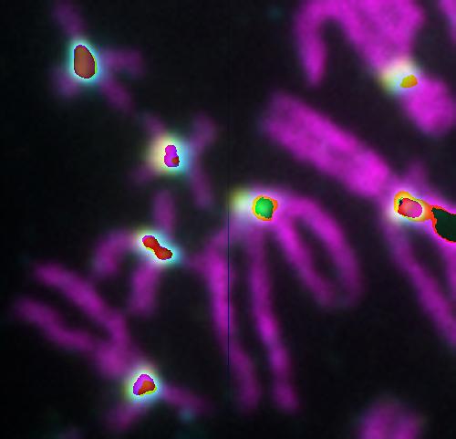
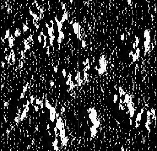
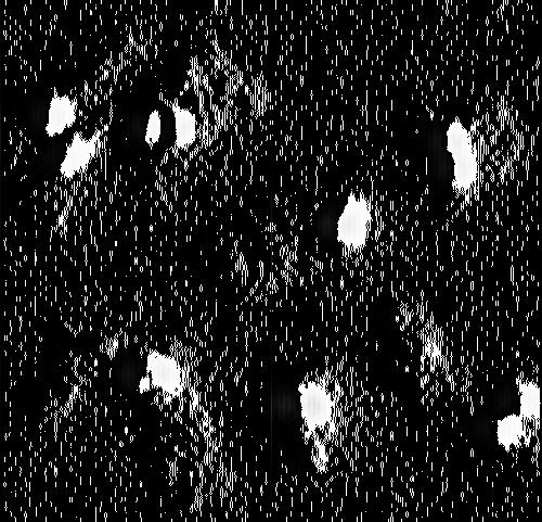
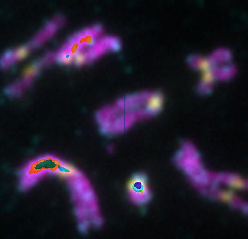
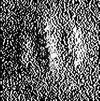
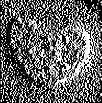
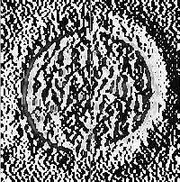
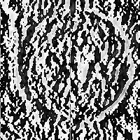
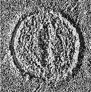
Figure S1. HP1α localizes to centromeres in mitosis and interacts with INCENP. (A&B) HeLa
|
|
|
- Helena Crawford
- 6 years ago
- Views:
Transcription
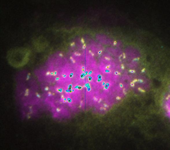
1 SUPPLEMENTARY FIGURES Figure S1. HP1α localizes to centromeres in mitosis and interacts with INCENP. (A&B) HeLa tet-on cells that stably express HP1α-CFP, HP1β-CFP, or HP1γ-CFP were monitored with livecell imaging. Arrow denotes the midbody localization of HP1α-CFP at telophase. (C) Metaphase spread of HeLa tet-on cells was stained with DAPI (blue in overlay), α-hp1α (green in overlay), and CREST (red in overlay). (D) Lysates and Myc IP of HeLa tet-on cells transfected with the indicated plasmids were blotted with α-myc and α-ha. Figure S2. (A) The endogenous INCENP and HP1α interact in mitosis. HeLa tet-on cells were arrested in G1 or mitosis with thymidine or nocodazole, respectively. Cell lysates, IgG control, or α-incenp IP were blotted with α-incenp, α-aurora B, and α-hp1α. (B) Quantification of the mitotic chromosome signals of GFP-HP1α of cells in Figure 2A. The average intensity of GFP-HP1α at the centromeres of five chromosomes was divided by its average intensity at chromosome arms, and these ratios were plotted (N = 8 cells). Figure S3. Expression of INCENP Δ125 rescues the cell-cycle and spindle-checkpoint defects of INCENP RNAi Cells. (A) HeLa tet-on cells were first transfected with vector, mcherry- INCENP WT or Δ125 plasmids 6 hr and then with INCENP sirna for 24 hr, and were treated with Taxol (100 nm) for another 18 hr. Cell lysates were blotted α-incenp and α-tubulin (loading control). (B) FACS analysis of HeLa tet-on cells transfected as described (A) except that the cells were not treated with Taxol. The populations of G1 (2N), G2 (4N), and mitotic (4N and MPM2) cells are indicated by blue, brown, and red arrowheads, respectively. 10,000 1
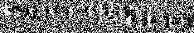
2 events were counted in each sample. (C) Cells in (A) were analyzed by FACS as described in (B). The mitotic index of these cells is plotted (mean ± SD of two independent experiments). Figure S4. INCENP depletion causes diffusive chromosome localization of Sgo1. (A) HeLa tet-on cells were transfected with INCENP sirna for 48 hr. Metaphase chromosome spread was prepared from these cells and stained with DAPI, CREST and α-sgo1. DAPI, Sgo1 staining, and CREST staining were colored blue, green, and red, respectively, in the overlay. (B) The average intensity of Sgo1 at the centromeres of five chromosomes was divided by its average intensity at chromosome arms, and these ratios were plotted (N = 10 cells). Figure S5. The INCENPHP1 interaction is dispensable for mitotic progression. (A) HeLa teton cells that stably express mcherry-incenp WT (clone #4) or Δ125 (clone #15) were transfected with INCENP sirna for 24 hr and then arrested at the G1/S boundary by thymidine treatment for 24 hr. Live-cell imaging was performed after the cells were released into fresh media. The cumulative percentage of mitotic cells was plotted against the mitotic duration (as defined by the time from nuclear envelope breakdown to the appearance of INCENP midbody staining at telophase) (N = 25 cells). (B) The mitotic duration of cells in (A) were blotted in a scatter plot. (C) Representative cells in (A) showing the dynamic localization patterns of mcherry-incenp or Δ125. Figure S6. The Sgo1HP1 interaction is dispensable for sister-chromatid cohesion. (A) HeLa tet-on cells that stably express Myc-Sgo1 WT (clone #8) or P1A (clones #3 and #5) under the control of doxycycline were cultured in the absence () or presence () doxycycline (Dox) and 2
3 transfected with Sgo1 sirna for 24 hr. Cell lysates were blotted with α-myc and α-tubulin. (B) The mitotic index of cells in (A). Cells were stained with propidium iodide and α-h3-ps10 and analyzed by FACS. 10,000 events were counted for each sample. Mitotic cells have 4N DNA content and are H3-pS10-positive. (C) The extent of sister-chromatid separation of cells in (A) as determined by metaphase spread with Giemsa staining (N = 50 cells). Figure S7. The Sgo1HP1 interaction does not regulate mitotic progression or the timing of Sgo1 localization to mitotic centromeres. HeLa tet-on cells were first transfected with GFP- Sgo1 WT or P1A for 6 hr and then with Sgo1 sirna for 24 hr. After that, cells were arrested at G2 with an 18-hr treatment of the Cdk1 inhibitor RO-3306 and then released into fresh media for live-cell imaging. 3
4 Table S1. Crystallographic and Refinement Data and Statistics of HP1β CSDSgo1P1 Parameter Value Space Group P Unit cell dimensions a, b, c (Å) 76.8, 108.9, 41.9 α, β, γ ( ) 90, 90, 90 Resolution (Å) ( )* Completeness (%) 99.9 (100) Multiplicity 8.4 (8.3) Unique reflections 26,940 (1,314) R sym (%) 9.7 (96.7) I/σ(I) 16.5 (2.2) Wilson B (Å 2 ) 28.5 Refinement Resolution (Å) No. non-solvent atoms 2,520 No. solvent atoms 162 Cutoff F o /σ Fo 0 Avg. B-factors non-solvent (Å 2 ) 37.0 solvent 40.3 R-values R work (%) 18.9 R free (%) 23.0 Ramachandran statistics outliers (%) 0.0 most favored region (%) 99.3 r.m.s. deviations Bond lengths (Å) Bond angles ( ) 1.2 4
5 *Values in parentheses are for the highest resolution shell. where the outer sum (h) is over the unique reflections and the inner sum (i) is over the set of independent observations of each unique reflection. From MolProbity (Chen et al., 2010). 5
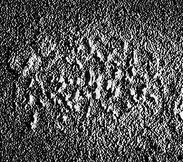
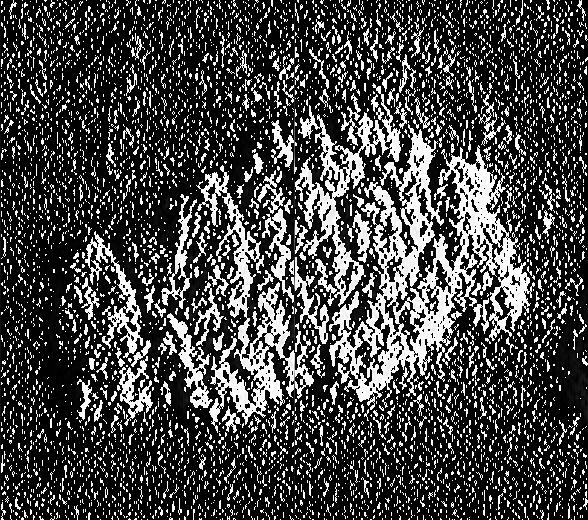
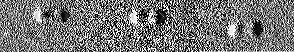
6 A B Interphase Prometaphase Interphase Prometaphase Telophase HP1β-CFP HP1α-CFP HP1γ-CFP C DAPI CREST Anti-HP1α D IP by Myc Total HA-HP1α HA-HP1β Overlay HA-HP1γ HA-HP1α HA-HP1β HA-HP1γ Myc-INCENP 125 Myc-INCENP WT Anti-myc Anti-HA Figure S1

7 A Total IP by Anti-INCENP IP by IgG Thymidine Nocodazole Anti-INCENP Anti-Aurora B Anti-HP1α B Ratio of centromere/arm staining GFP-HP1α on chromosomes P<0.001 Mock RNAi INCENP RNAi Figure S2


8 A INCENP RNAi mch-incenp 125 mch-incenp WT Anti-INCENP Anti-Tubulin B DNA Content Vector Mock RNAi 26.3% 3.5% DNA Content Vector INCENP RNAi 67.6% 2.5% 50.7% 4.7% C Mitotic Index(%) nm Taxol DNA Content MPM2 INCENP WT INCENP RNAi 3.2% DNA Content 31.9% 36.4% MPM2 INCENP 125 INCENP RNAi 4.1% 0 INCENP RNAi mch-incenp 125 mch-incenp WT 41.2% MPM2 35.7% MPM2 Figure S3


9 A DAPI Sgo1 CREST Overlay Mock RNAi INCENP RNAi B Ratio of centromere/arm staining Sgo1 on chromosomes P<0.001 Mock RNAi INCENP RNAi Figure S4
10 A Cumulative percentage (%) Mitotic Duration (min) WT #4 125 #15 B Mitotic duration (min) INCENP WT #4 P=0.306 INCENP 125 #15 C 0 min 10 min 20 min 30 min 40 min 50 min mcherry INCENP WT #4 DIC 0 min 10 min 20 min 30 min 40 min 50 min mcherry INCENP 125 #15 DIC Figure S5
 15 10 5 0 Sgo1 RNAi Dox WT #8")

11 A WT #8 P1A #3 P1A #5 Dox Anti-Myc-Sgo1 Anti-Tubulin B Mitotic index (%) Sgo1 RNAi Dox WT #8 P1A #3 P1A #5 C Separated chromsomes (%) Sgo1 RNAi Dox WT #8 P1A #3 P1A #5 Figure S6


12 GFP- Sgo1 WT GFP DIC GFP- Sgo1 P1A GFP DIC Control DIC Figure S7
Nature Structural and Molecular Biology: doi: /nsmb Supplementary Figure 1
 Supplementary Figure 1 Mutational analysis of the SA2-Scc1 interaction in vitro and in human cells. (a) Autoradiograph (top) and Coomassie stained gel (bottom) of 35 S-labeled Myc-SA2 proteins (input)
Effects of UBL5 knockdown on cell cycle distribution and sister chromatid cohesion
 Supplementary Figure S1. Effects of UBL5 knockdown on cell cycle distribution and sister chromatid cohesion A. Representative examples of flow cytometry profiles of HeLa cells transfected with indicated
 a Control IgG Intestine c Testis b Thymus 1 3 2 S S 2 1 3 4 4 Figure S1 The wild-type mouse (C57BL/6J) organs (intestine, thymus and testis) were frozen in liquid nitrogen and sectioned at 5 µm on a cryostat.
T H E J O U R N A L O F C E L L B I O L O G Y
 T H E J O U R N A L O F C E L L B I O L O G Y Supplemental material Dunsch et al., http://www.jcb.org/cgi/content/full/jcb.201202112/dc1 Figure S1. Characterization of HMMR and CHICA antibodies. (A) HeLa
SUPPLEMENTARY INFORMATION
 DOI: 10.1038/ncb3076 Supplementary Figure 1 btrcp targets Cep68 for degradation during mitosis. a) Cep68 immunofluorescence in interphase and metaphase. U-2OS cells were transfected with control sirna
Supplementary table 1
 Supplementary table 1 S. pombe strain list Fig. 1A JX38 h + ade6-m216 nda3-km311 PX476 PW775 PX545 PX546 h- ade6-m216 sgo2::ura4 + nda3-km311 h 9 mad2::ura4 + nda3-km311 h + ade6-m21 nda3-km311 rad21 +
Supplementary Figure 1. Mother centrioles can reduplicate while in the close association
 C1-GFP distance (nm) C1-GFP distance (nm) a arrested HeLa cell expressing C1-GFP and Plk1TD-RFP -3 s 1 2 3 4 5 6 7 8 9 11 12 13 14 16 17 18 19 2 21 22 23 24 26 27 28 29 3 b 9 8 7 6 5 4 3 2 arrested HeLa
 Supplementary Figure Legends Supplementary Figure S1. Aurora-A is essential for SAC establishment in early mitosis. (a-c) RPE cells were treated with DMSO (a), MLN8237 (b) or BI2536 (c) for Two hours.
Supplementary information. The Light Intermediate Chain 2 Subpopulation of Dynein Regulates Mitotic. Spindle Orientation
 Supplementary information The Light Intermediate Chain 2 Subpopulation of Dynein Regulates Mitotic Spindle Orientation Running title: Dynein LICs distribute mitotic functions. Sagar Mahale a, d, *, Megha
T H E J O U R N A L O F C E L L B I O L O G Y
 T H E J O U R N A L O F C E L L B I O L O G Y Supplemental material Posch et al., http://www.jcb.org/cgi/content/full/jcb.200912046/dc1 Figure S1. Biochemical characterization of the interaction between
Mislocalization of centromeric histone H3 variant CENP-A contributes to chromosomal instability (CIN) in human cells
 in human cells") /, 2017, Vol. 8, (No. 29), pp: 46781-46800 Mislocalization of centromeric histone H3 variant CENP-A contributes to chromosomal instability (CIN) in human cells Roshan L. Shrestha 1, Grace S. Ahn 1, Mae
/, 2017, Vol. 8, (No. 29), pp: 46781-46800 Mislocalization of centromeric histone H3 variant CENP-A contributes to chromosomal instability (CIN) in human cells Roshan L. Shrestha 1, Grace S. Ahn 1, Mae
SUPPLEMENTARY INFORMATION
 DOI: 1.138/ncb222 / b. WB anti- WB anti- ulin Mitotic index (%) 14 1 6 2 T (h) 32 48-1 1 2 3 4 6-1 4 16 22 28 3 33 e. 6 4 2 Time (min) 1-6- 11-1 > 1 % cells Figure S1 depletion leads to mitotic defects
T H E J O U R N A L O F C E L L B I O L O G Y
 T H E J O U R N A L O F C E L L B I O L O G Y Supplemental material Lu et al., http://www.jcb.org/cgi/content/full/jcb.201012063/dc1 Figure S1. Kinetics of nuclear envelope assembly, recruitment of Nup133
Mitotic Centromeric Targeting of HP1 and Its Binding to Sgo1 Are Dispensable. for Sister-Chromatid Cohesion in Human Cells
 Mitotic Centromeric Targeting of HP1 and Its Binding to Sgo1 Are Dispensable for Sister-Chromatid Cohesion in Human Cells Jungseog Kang 1, Jaideep Chaudhary 1, Hui Dong 1, Soonjoung Kim 1, Chad A. Brautigam
(a) Reproduction. (b) Growth and development. (c) Tissue renewal
 Reproduction. (b) Growth and development. (c) Tissue renewal") 100 µm 200 µm 20 µm (a) Reproduction (b) Growth and development (c) Tissue renewal 1 20 µm 2 0.5 µm Chromosomes DNA molecules Chromosome arm Centromere Chromosome duplication (including DNA synthesis)
100 µm 200 µm 20 µm (a) Reproduction (b) Growth and development (c) Tissue renewal 1 20 µm 2 0.5 µm Chromosomes DNA molecules Chromosome arm Centromere Chromosome duplication (including DNA synthesis)
Supporting Information
 Supporting Information ou et al..73/pnas.08791112 dd Thymidine Release & transfection dd Thymidine Release dd MG132 Fix and IF -14 h 0 h 8 h 24 h 34 h 36 h siontrol simps1-1 simps1-1 simps1-1 simps1-2
Supplementary Figure S1
 Supplementary Figure S1 Supplementary Figure S1. PARP localization patterns using GFP-PARP and PARP-specific antibody libraries GFP-PARP localization in non-fixed (A) and formaldehyde fixed (B) GFP-PARPx
T H E J O U R N A L O F C E L L B I O L O G Y
 T H E J O U R N A L O F C E L L B I O L O G Y Supplemental material Krenn et al., http://www.jcb.org/cgi/content/full/jcb.201110013/dc1 Figure S1. Levels of expressed proteins and demonstration that C-terminal
EMBO. PICH and BLM limit histone association with anaphase centromeric DNA threads and promote their resolution EMBO. open
 The EMBO Journal (2011) 30, 3309 3321 & 2011 European Molecular Biology Organization Some Rights Reserved 0261-4189/11 www.embojournal.org PICH and BLM limit histone association with anaphase centromeric
Supplementary figures
 Supplementary figures Supplementary Figure 1. B cells stimulated with pokeweed mitogen display normal mitotic figures but not cells infected with B95-8. The figures show cells stimulated with pokeweed
Chapter 8: Cellular Reproduction
 Chapter 8: Cellular Reproduction 1. The Cell Cycle 2. Mitosis 3. Meiosis 2 Types of Cell Division 2n 1n Mitosis: occurs in somatic cells (almost all cells of the body) generates cells identical to original
Supplemental Materials. STK16 regulates actin dynamics to control Golgi organization and cell cycle
 Supplemental Materials STK16 regulates actin dynamics to control Golgi organization and cell cycle Juanjuan Liu 1,2,3, Xingxing Yang 1,3, Binhua Li 1, Junjun Wang 1,2, Wenchao Wang 1, Jing Liu 1, Qingsong
A. List of selected proteins with high SILAC (H/L) ratios identified in mass
 ratios identified in mass") Supplementary material Figure S1. Interaction between UBL5 and FANCI A. List of selected proteins with high SILAC (H/L) ratios identified in mass spectrometry (MS)-based analysis of UBL5-interacting proteins,
Supplementary material Figure S1. Interaction between UBL5 and FANCI A. List of selected proteins with high SILAC (H/L) ratios identified in mass spectrometry (MS)-based analysis of UBL5-interacting proteins,
(a) Schematic diagram of the FS mutation of UVRAG in exon 8 containing the highly instable
 Schematic diagram of the FS mutation of UVRAG in exon 8 containing the highly instable") Supplementary Figure 1. Frameshift (FS) mutation in UVRAG. (a) Schematic diagram of the FS mutation of UVRAG in exon 8 containing the highly instable A 10 DNA repeat, generating a premature stop codon
Supplementary Figure 1. Frameshift (FS) mutation in UVRAG. (a) Schematic diagram of the FS mutation of UVRAG in exon 8 containing the highly instable A 10 DNA repeat, generating a premature stop codon
a" b" 2N c" d" e" f" !!Aurora!A!!!CP110!
 DLD1/Reference a" 2N 2N 2N/DLD1 2N/ /DLD1 c" d" e" f" TargetID 2N.AVG_Sig 2N.Det Pval.AVG_Sig.Det Pval Diff Pval DiffScore SYMBOL ILMN_26396 18.35238 0.0080058 44.81118 0.0021834 0.000323 34.90542 KRTHA4
Cell Cycle, Mitosis, and Microtubules. LS1A Final Exam Review Friday 1/12/07. Processes occurring during cell cycle
 Cell Cycle, Mitosis, and Microtubules LS1A Final Exam Review Friday 1/12/07 Processes occurring during cell cycle Replicate chromosomes Segregate chromosomes Cell divides Cell grows Cell Growth 1 The standard
T R L J. Version 2, 2018 NAME: OPTION GROUP: CELL DIVISION MITOSIS WORKBOOK
 NAME: OPTION GROUP: CELL DIVISION MITOSIS WORKBOOK 1 STUDY CHECKLIST AND ASSESSMENT OBJECTIVES Instructions Regular revision throughout the year is essential. It s vital you keep a track of what you understand
The Cell Cycle. Dr. SARRAY Sameh, Ph.D
 The Cell Cycle Dr. SARRAY Sameh, Ph.D Overview When an organism requires additional cells (either for growth or replacement of lost cells), new cells are produced by cell division (mitosis) Somatic cells
Supplementary Information POLO-LIKE KINASE 1 FACILITATES LOSS OF PTEN-INDUCED PROSTATE CANCER FORMATION
 Supplementary Information POLO-LIKE KINASE 1 FACILITATES LOSS OF PTEN-INDUCED PROSTATE CANCER FORMATION X. Shawn Liu 1, 3, Bing Song 2, 3, Bennett D. Elzey 3, 4, Timothy L. Ratliff 3, 4, Stephen F. Konieczny
Supplementary Information for. Shi and King, Chromosome Nondisjunction Yields Tetraploid Rather than Aneuploid Cells in Human Cell Lines.
 Supplementary Information for Shi and King, Chromosome Nondisjunction Yields Tetraploid Rather than Aneuploid Cells in Human Cell Lines Contains Supplementary Methods Supplementary Figures 1-7 Supplementary
Nature Methods: doi: /nmeth.4257
 Supplementary Figure 1 Screen for polypeptides that affect cellular actin filaments. (a) Table summarizing results from all polypeptides tested. Source shows organism, gene, and amino acid numbers used.
Yan Ma 1.,XiYuan 1., William R. Wyatt 2, Joseph R. Pomerening 1 * Abstract. Introduction
 Expression of Constitutively Active CDK1 Stabilizes APC- Cdh1 Substrates and Potentiates Premature Spindle Assembly and Checkpoint Function in G1 Cells Yan Ma 1.,XiYuan 1., William R. Wyatt 2, Joseph R.
How Cells Divide. Chapter 10
 How Cells Divide Chapter 10 Bacterial Cell Division Bacteria divide by binary fission. -the single, circular bacterial chromosome is replicated -replication begins at the origin of replication and proceeds
Cellular Reproduction, Part 2: Meiosis Lecture 10 Fall 2008
 Mitosis & 1 Cellular Reproduction, Part 2: Lecture 10 Fall 2008 Mitosis Form of cell division that leads to identical daughter cells with the full complement of DNA Occurs in somatic cells Cells of body
Cell Cycle. Interphase, Mitosis, Cytokinesis, and Cancer
 Cell Cycle Interphase, Mitosis, Cytokinesis, and Cancer Cell Division One cell divides into 2 new identical daughter cells. Chromosomes carry the genetic information (traits) of the cell How many Chromosomes
Cell Division Mitosis Notes
 Cell Division Mitosis Notes Cell Division process by which a cell divides into 2 new cells Why do cells need to divide? 1.Living things grow by producing more cells, NOT because each cell increases in
Mitosis Flap Book Excludes Prometaphase
 Mitosis Flap Book Excludes Prometaphase TEACHER S INSTRUCTIONS 1) Choose one of the foldables from the choices below. Three Color Choices Black & White Cells without Chromosomes Choose this option if you
基醫所. The Cell Cycle. Chi-Wu Chiang, Ph.D. IMM, NCKU
 基醫所 The Cell Cycle Chi-Wu Chiang, Ph.D. IMM, NCKU 1 1 Introduction to cell cycle and cell cycle checkpoints 2 2 Cell cycle A cell reproduces by performing an orderly sequence of events in which it duplicates
-The cell s hereditary endowment of DNA -Usually packaged into chromosomes for manageability
 Binary Fission-Bacterial Cell Division -Asexual reproduction of prokaryotes -No mitosis -Circular DNA and organelles replicate, the copies migrate to opposite sides of the elongating cell, and the cell
Supplementary Materials
 Supplementary Materials Supplementary Figure S1 Regulation of Ubl4A stability by its assembly partner A, The translation rate of Ubl4A is not affected in the absence of Bag6. Control, Bag6 and Ubl4A CRISPR
BIOLOGY - CLUTCH CH.12 - CELL DIVISION.
 !! www.clutchprep.com CONCEPT: CELL DIVISION Cell division is the process by which one cell splits into two or more daughter cells. Cell division generally requires that cells produce enough materials,
10-2 Cell Division. Chromosomes
 Cell Division In eukaryotes, cell division occurs in two major stages. The first stage, division of the cell nucleus, is called mitosis. The second stage, division of the cell cytoplasm, is called cytokinesis.
The Cell Cycle. Packet #9. Thursday, August 20, 2015
 1 The Cell Cycle Packet #9 2 Introduction Cell Cycle An ordered sequence of events in the life of a dividing eukaryotic cell and is a cellular asexual reproduction. The contents of the parent s cell nucleus
Cell Cycle Phase. Interphase (G 1, S, G 2 ) Mitotic Phase (M phase) Prophase. Metaphase. Anaphase. Telophase
 Mitotic Phase (M phase) Prophase. Metaphase. Anaphase. Telophase") Part I: The Cell Cycle Use your resources at hand and the Explore Student Guide to outline what occurs within the cell during each stage of the cell cycle. Record this information in Table 1 below. Cell
Part I: The Cell Cycle Use your resources at hand and the Explore Student Guide to outline what occurs within the cell during each stage of the cell cycle. Record this information in Table 1 below. Cell
Expanded View Figures
 PEX13 functions in selective autophagy Ming Y Lee et al Expanded View Figures Figure EV1. PEX13 is required for Sindbis virophagy. A, B Quantification of mcherry-capsid puncta per cell (A) and GFP-LC3
10-2 Cell Division mitosis. cytokinesis. Chromosomes chromosomes Slide 1 of 38
 In eukaryotes, cell division occurs in two major stages. The first stage, division of the cell nucleus, is called mitosis. The second stage, division of the cell cytoplasm, is called cytokinesis. Chromosomes
Creating Identical Body Cells
 Creating Identical Body Cells 5.A Students will describe the stages of the cell cycle, including DNA replication and mitosis, and the importance of the cell cycle to the growth of organisms 5.D Students
Cellular Reproduction
 Section 1: Cellular Growth Section 2: Mitosis and Cytokinesis Section 3: Cell Cycle Regulation Click on a lesson name to select. Section 1 Cellular Growth Ratio of Surface Area to Volume Section 1 Cellular
Regulators of Cell Cycle Progression
 Regulators of Cell Cycle Progression Studies of Cdk s and cyclins in genetically modified mice reveal a high level of plasticity, allowing different cyclins and Cdk s to compensate for the loss of one
The Cell Cycle and Cell Division
 Content Vocabulary Directions: On each line, write the term from the word bank that correctly replaces the underlined words in each sentence. NOTE: You may need to change a term to its plural form. cell
Omnis cellula e cellula
 Chapter 12 The Cell Cycle Omnis cellula e cellula 1855- Rudolf Virchow German scientist all cells arise from a previous cell Every cell from a cell In order for this to be true, cells must have the ability
Prolonged mitotic arrest induces a caspase-dependent DNA damage
 SUPPLEMENTARY INFORMATION Prolonged mitotic arrest induces a caspase-dependent DNA damage response at telomeres that determines cell survival Karolina O. Hain, Didier J. Colin, Shubhra Rastogi, Lindsey
MBoC ARTICLE. Sushama Sivakumar a,b, John R. Daum a, Aaron R. Tipton a, Susannah Rankin a,b, and Gary J. Gorbsky a,b a
 MBoC ARTICLE The spindle and kinetochore associated (Ska) complex enhances binding of the anaphasepromoting complex/cyclosome (APC/C) to chromosomes and promotes mitotic exit Sushama Sivakumar a,b, John
Mitosis/Meiosis Simulation Activities
 Mitosis/Meiosis Simulation Activities In this simulation, you will demonstrate an understanding of mitosis, meiosis, segregation, independent assortment, and crossing over, all processes involved with
Prentice Hall Biology Slide 1 of 38
 Prentice Hall Biology 1 of 38 2 of 38 In eukaryotes, cell division occurs in two major stages. The first stage, division of the cell nucleus, is called mitosis. The second stage, division of the cell cytoplasm,
CELL DIVISION! Genes, Mitosis and Cytokinesis 12/17/14. G. Podgorski, Biol Mitosis!
 Genes, Mitosis and Cytokinesis 12/17/14 CELL DIVISION! Mitosis! ü Mitotic division results in genetically identical eukaryotic cells or a clone ü Mitosis is the basis of asexual! reproduction G. Podgorski,
The Cell Cycle CHAPTER 12
 The Cell Cycle CHAPTER 12 The Key Roles of Cell Division cell division = reproduction of cells All cells come from pre-exisiting cells Omnis cellula e cellula Unicellular organisms division of 1 cell reproduces
Cell Division and Mitosis
 Chromatin-Uncoiled DNA during interphase Cell Division and Mitosis Chromosomes-Tightly coiled DNA Chromatid-One half of a duplicated chromosome. Each is identical and called sister chromatids Centromere-The
To segregate sister chromatids evenly into daughter cells
 Human Bub1 protects centromeric sister-chromatid cohesion through Shugoshin during mitosis Zhanyun Tang*, Yuxiao Sun, Sara E. Harley*, Hui Zou, and Hongtao Yu* Departments of *Pharmacology and Molecular
Bacterial cell. Origin of replication. Septum
 Bacterial cell Bacterial chromosome: Double-stranded DNA Origin of replication Septum 1 2 3 Chromosome Rosettes of Chromatin Loops Scaffold protein Chromatin Loop Solenoid Scaffold protein Chromatin loop
Cell Division. During interphase, a cell s DNA is in a loose form called. It condenses into tightly coiled structures called chromosomes during.
 Cell Division The is a cell s total DNA. Prokaryotes DNA is found mostly in a single called the and also in small circles called. Eukaryotes have several DNA double helices packaged into. During interphase,
The questions below refer to the following terms. Each term may be used once, more than once, or not at all.
 The questions below refer to the following terms. Each term may be used once, more than once, or not at all. a) telophase b) anaphase c) prometaphase d) metaphase e) prophase 1) DNA begins to coil and
Supplementary Figure 1.TRIM33 binds β-catenin in the nucleus. a & b, Co-IP of endogenous TRIM33 with β-catenin in HT-29 cells (a) and HEK 293T cells
 and HEK 293T cells") Supplementary Figure 1.TRIM33 binds β-catenin in the nucleus. a & b, Co-IP of endogenous TRIM33 with β-catenin in HT-29 cells (a) and HEK 293T cells (b). TRIM33 was immunoprecipitated, and the amount of
Supplementary Figure 1.TRIM33 binds β-catenin in the nucleus. a & b, Co-IP of endogenous TRIM33 with β-catenin in HT-29 cells (a) and HEK 293T cells (b). TRIM33 was immunoprecipitated, and the amount of
Essential Questions. Why are cells relatively small? What are the primary stages of the cell cycle? What are the stages of interphase?
 Essential Questions Why are cells relatively small? What are the primary stages of the cell cycle? What are the stages of interphase? Cellular Growth Vocabulary Review selective permeability New cell cycle
Cell Division Mitosis Notes
 Cell Division Mitosis Notes Cell Division process by which a cell divides into 2 new cells Why do cells need to divide? 1.Living things grow by producing more cells, NOT because each cell increases in
SUPPLEMENTARY FIGURE LEGENDS. atypical adenomatous hyperplasias (AAH); Grade II: adenomas; Grade III: adenocarcinomas;
; Grade II: adenomas; Grade III: adenocarcinomas;") SUPPLEMENTARY FIGURE LEGENDS Supplementary Figure S1: Tumor grades in Ras G12D ; p53 / lung tumors. Representative histology (H&E) of K-Ras G12D ; p53 / lung tumors 13 weeks after tumor initiation. Grade
SUPPLEMENTARY FIGURE LEGENDS Supplementary Figure S1: Tumor grades in Ras G12D ; p53 / lung tumors. Representative histology (H&E) of K-Ras G12D ; p53 / lung tumors 13 weeks after tumor initiation. Grade
Monday, October 6 Put these items into the appropriate category:
 Monday, October 6 Put these items into the appropriate category: Active Transport Facilitated Diffusion Osmosis Simple Diffusion The smell of rotten eggs spreading through the room Requires ATP expenditure
Biology 4A Laboratory MITOSIS Asexual Reproduction OBJECTIVE
 Biology 4A Laboratory MITOSIS Asexual Reproduction OBJECTIVE To study the cell cycle and understand how, when and why cells divide. To study and identify the major stages of cell division. To relate the
172R 172K TAM-2/172R TAM-2/172K. AZT concentration [nm] AZT concentration [nm] MgCl 2 2.5K 2.5K 5K 2.5K 5K 2.5K K 5K 2.5K 5K 2.5K 50 2.
![172R 172K TAM-2/172R TAM-2/172K. AZT concentration [nm] AZT concentration [nm] MgCl 2 2.5K 2.5K 5K 2.5K 5K 2.5K K 5K 2.5K 5K 2.5K 50 2.](/thumbs/82/85138523.jpg "172R 172K TAM-2/172R TAM-2/172K. AZT concentration [nm] AZT concentration [nm] MgCl 2 2.5K 2.5K 5K 2.5K 5K 2.5K K 5K 2.5K 5K 2.5K 50 2.") 5 5 5 5 A MgCl 2 172R 172K TAM-2/172R TAM-2/172K AZT concentration [nm] B 172R 172K TAM-2/172R TAM-2/172K AZT concentration [nm] ATP + ATP - Supplemental Figure 1. Primer extension of HIV-1 RT polymorphisms
5 5 5 5 A MgCl 2 172R 172K TAM-2/172R TAM-2/172K AZT concentration [nm] B 172R 172K TAM-2/172R TAM-2/172K AZT concentration [nm] ATP + ATP - Supplemental Figure 1. Primer extension of HIV-1 RT polymorphisms
SUPPLEMENTARY INFORMATION
 DOI: 0.038/ncb33 a b c 0 min 6 min 7 min (fixed) DIC -GFP, CenpF 3 µm Nocodazole Single optical plane -GFP, CenpF Max. intensity projection d µm -GFP, CenpF, -GFP CenpF 3-D rendering e f 0 min 4 min 0
SUPPLEMENTARY INFORMATION
 DOI:.38/ncb2822 a MTC02 FAO cells EEA1 b +/+ MEFs /DAPI -/- MEFs /DAPI -/- MEFs //DAPI c HEK 293 cells WCE N M C P AKT TBC1D7 Lamin A/C EEA1 VDAC d HeLa cells WCE N M C P AKT Lamin A/C EEA1 VDAC Figure
Chapter 10. Cell Growth and Division
 Chapter 10 Cell Growth and Division Cell Growth A. Limits to Cell Growth 1. Two main reasons why cells divide: a. Demands on DNA as the cell get too large Cell Growth b. Moving nutrients and waste across
Chapter 10 Cell Cycle
 Chapter 10 Cell Cycle Chapter 10 Cell Cycle Grade:«grade» Subject:Biology Date:«date» 1 As a cell becomes larger, its surface area increases faster than its volume. 2 As a cell becomes larger, its volume
Cell Cycle and Mitosis
 Cell Cycle and Mitosis Name Period A# THE CELL CYCLE The cell cycle, or cell-division cycle, is the series of events that take place in a eukaryotic cell between its formation and the moment it replicates
Cellular Reproduction, Part 1: Mitosis Lecture 10 Fall 2008
 Cell Theory 1 Cellular Reproduction, Part 1: Mitosis Lecture 10 Fall 2008 Cell theory: All organisms are made of cells All cells arise from preexisting cells How do new cells arise? Cell division the reproduction
CHAPTER 8: CELL GROWTH AND DIVISION 8-1: CELL GROWTH 8-2: CELL DIVISION: MITOSIS AND CYTOKINESIS
 CHAPTER 8: CELL GROWTH AND DIVISION 8-1: CELL GROWTH 8-2: CELL DIVISION: MITOSIS AND CYTOKINESIS 1 LEARNING OBJECTIVES You should be able to: Give two physical reasons why mitosis must occur. Draw a chromosome
Why do cells reproduce?
 Outline Cell Reproduction 1. Overview of Cell Reproduction 2. Cell Reproduction in Prokaryotes 3. Cell Reproduction in Eukaryotes 1. Chromosomes 2. Cell Cycle 3. Mitosis and Cytokinesis Examples of Cell
Functional impact of Aurora A-mediated phosphorylation of HP1γ at serine 83 during cell cycle progression
 Grzenda et al. Epigenetics & Chromatin 2013, 6:21 RESEARCH Open Access Functional impact of Aurora A-mediated phosphorylation of HP1γ at serine 83 during cell cycle progression Adrienne Grzenda 1, Phoebe
Outline Interphase Mitotic Stage Cell Cycle Control Apoptosis Mitosis Mitosis in Animal Cells Cytokinesis Cancer Prokaryotic Cell Division
 The Cell Cycle and Cellular Reproduction Chapter 9 Outline Interphase Mitotic Stage Cell Cycle Control Apoptosis Mitosis Mitosis in Animal Cells Cytokinesis Cancer Prokaryotic Cell Division 1 2 Interphase
10-2 Cell Division. Copyright Pearson Prentice Hall
 10-2 Cell Division Copyright Pearson Prentice Hall Cell Growth and Division In multicellular organisms, cell division makes new cells To replace old or damaged ones So organisms can grow In single-celled
Mitosis. AND Cell DiVISION
 Mitosis AND Cell DiVISION Cell Division Characteristic of living things: ability to reproduce their own kind. Cell division purpose: When unicellular organisms such as amoeba divide to form offspring reproduction
Why do cells divide? Cells divide in order to make more cells they multiply in order to create a larger surface to volume ratio!!!
 Why do cells divide? Cells divide in order to make more cells they multiply in order to create a larger surface to volume ratio!!! Chromosomes Are made of chromatin: a mass of genetic material composed
Cell Division. Learning Objectives: Introduction. Revised Fall 2018
 Revised Fall 2018 Cell Division Learning Objectives: 1. Define cell cycle and the ordered sequence of events in the cell cycle (Interphase and The divisional phase or M phase) 2. Explain the stages in
Multiple Choice Identify the letter of the choice that best completes the statement or answers the question.
 Biology Mo Test: Q3 Mr. Rellinger Multiple Choice Identify the letter of the choice that best completes the statement or answers the question. 1. Which event occurs during interphase? The cell carries
Unit 6: CELL DIVISION PACKET
 Unit 6: CELL DIVISION PACKET This packet is designed to help you understand several concepts about Cell Division. As you practice the exercises on each handout, you will be able to: Use a model to illustrate
Cell division functions in 1. reproduction, 2. growth, and 3. repair
 Cell division functions in 1. reproduction, 2. growth, and 3. repair What do you think you are looking at here??? Can something like you or I do this??? Fig. 12.1 How did you start out? How did you grow?
Cell Division (Mitosis)
") Cell Division (Mitosis) Chromosomes The essential part of a chromosome is a single very long strand of DNA. This DNA contains all the genetic information for creating and running the organism. Each chromosome
Cell Division (Mitosis) Chromosomes The essential part of a chromosome is a single very long strand of DNA. This DNA contains all the genetic information for creating and running the organism. Each chromosome
10-2 Cell Division. Slide 1 of 38. End Show. Copyright Pearson Prentice Hall
 1 of 38 Cell Division In eukaryotes, cell division occurs in two major stages. The first stage, division of the cell nucleus, is called mitosis. The second stage, division of the cell cytoplasm, is called
Cell Cycle - Introduction
 Cell Cycle - Introduction Key Concepts Cell division results in two identical cells During cell division the ability to organize DNA in time and space (location in the cell) is critical! The mitotic phase
Genetics. Instructor: Dr. Jihad Abdallah Lecture 2 The cell cycle and Cell Division
 Genetics Instructor: Dr. Jihad Abdallah Lecture 2 The cell cycle and Cell Division 1 The cell cycle Living cells go through a series of stages known as the cell cycle. They undergo a continuous alternation
Mitosis THE CELL CYCLE. In unicellular organisms, division of one cell reproduces the entire organism Multicellular organisms use cell division for..
 Mitosis THE CELL CYCLE In unicellular organisms, division of one cell reproduces the entire organism Multicellular organisms use cell division for.. Development from a fertilized cell Growth Repair Cell
SUPPLEMENTAL FIGURE LEGENDS
 SUPPLEMENTAL FIGURE LEGENDS Supplemental Figure S1: Endogenous interaction between RNF2 and H2AX: Whole cell extracts from 293T were subjected to immunoprecipitation with anti-rnf2 or anti-γ-h2ax antibodies
CELL CYCLE INTRODUCTION PART I ANIMAL CELL CYCLE INTERPHASE
 CELL CYCLE INTRODUCTION The nuclei in cells of eukaryotic organisms contain chromosomes with clusters of genes, discrete units of hereditary information consisting of double-stranded DNA. Structural proteins
Cellular Reproduction
 Chapter Test A CHAPTER 9 Cellular Reproduction Part A: Multiple Choice In the space at the left, write the letter of the term or phrase that best answers each question. Part B: Matching 1. What can limit
CELL CYCLE REGULATION AND CANCER. Cellular Reproduction II
 CELL CYCLE REGULATION AND CANCER Cellular Reproduction II THE CELL CYCLE Interphase G1- gap phase 1- cell grows and develops S- DNA synthesis phase- cell replicates each chromosome G2- gap phase 2- cell
WDR62 is associated with the spindle pole and mutated in human microcephaly
 WDR62 is associated with the spindle pole and mutated in human microcephaly Adeline K. Nicholas, Maryam Khurshid, Julie Désir, Ofélia P. Carvalho, James J. Cox, Gemma Thornton, Rizwana Kausar, Muhammad
Origin of replication. Septum
 Bacterial cell Bacterial chromosome: Double-stranded DNA Origin of replication Septum 1 2 3 Chromosome Rosettes of Chromatin Loops Chromatin Loop Solenoid Scaffold protein Scaffold protein Chromatin loop
Section Cell Growth. A. Limits to Cell Growth 1. DNA Overload 2. Exchanging Materials 3. Ratio of Surface Area to Volume 4.
 Getting Through Materials move through cells by diffusion. Oxygen and food move into cells, while waste products move out of cells. How does the size of a cell affect how efficiently materials get to all
Section 10 1 Cell Growth (pages )
") Chapter 10 Cell Growth and Division Section 10 1 Cell Growth (pages 241 243) Key Concept What problems does growth cause for cells? Limits to Cell Growth (pages 241 243) 1 What are two reasons why cells
Chapter 10 Cell Growth and Division Section 10 1 Cell Growth (pages 241 243) Key Concept What problems does growth cause for cells? Limits to Cell Growth (pages 241 243) 1 What are two reasons why cells
SUPPLEMENTARY INFORMATION
 DOI: 10.1038/ncb2566 Figure S1 CDKL5 protein expression pattern and localization in mouse brain. (a) Multiple-tissue western blot from a postnatal day (P) 21 mouse probed with an antibody against CDKL5.