p16 Cervical HISTOLOGY Histology Compendium & Staining Atlas
|
|
|
- Sheila Hunt
- 6 years ago
- Views:
Transcription
1 p16 Cervical HISTOLOGY Histology Compendium & Staining Atlas
2 Chapter 1: An Introduction to p Normal Cervical Epithelium and the Cell Cycle....4 HPV Infection and Cervical Disease p16: A Biomarker for Cervical Disease Chapter 2: Interpreting p16 Staining Patterns Squamous Epithelium p16 Positive p16 Negative Endocervical Epithelium p16 Positive p16 Negative Chapter 3: Clinical Utility of p16 for High-Grade CIN Confirmation of CIN2/ Differential Diagnosis of High-Grade Dysplasia Squamous Atrophy Atypical Squamous Metaplasia Chronic Cervicitis with Reactive Atypia Chapter 4: Clinical Utility of p16 for Low-Grade CIN Chapter 5: Clinical Utility of p16 for Endocervical Neoplasia

3 contents CINtec Histology Staining Case Examples Case 1: High-grade squamous dysplasia and AIS Case 2: CIN Case 3: Inflammatory atypia versus CIN Case 4: Glandular atypia, differential diagnosis tubo-endometrial metaplasia versus AIS Case 5: Inflammatory atypia versus CIN Case 6: Atypical immature metaplasia versus CIN Case 7: Atypical immature metaplasia versus CIN2/ Case 8: Atrophic cervicitis versus high-grade dysplasia Case 9: Chronic cervicitis with reactive atypia versus CIN2/ Case 10: Reserve cell hyperplasia versus CIN Case 11: Reactive atypia versus CIN Case 12: Reactive atypia versus CIN Case 13: Disrupted fragments of cervical mucosa, tangentially sectioned, atrophy versus CIN2/ Case 14: CIN 2/3, small lesion Case 15: CIN 2/3, small lesion, disrupted mucosa Case 16: CIN1, small lesion, endocervical glandular involvement Bibliography
4
-mediated viral cytopathic effects.")


5 An Introduction to p16 Chapter 1 An Introduction to p16 The histologic diagnosis of Cervical Intraepithelial Neoplasia (CIN) has historically been based only on the morphologic evaluation of disordered squamous maturation and the identification of Human Papilloma Virus (HPV)-mediated viral cytopathic effects. While the differential diagnosis of high-grade dysplasia (CIN2/3) versus normal squamous mucosa may be definitive in optimal cases, the subjective nature of the diagnostic processes and the inconsistent reproducibility in histologically subtle or morphologically equivocal cases may lead to false positive or false negative diagnoses. Published clinical research studies have shown that the accuracy of community-based pathologists for diagnosing high-grade CIN on cervical biopsy specimens can be significantly increased by the conjunctive interpretation of p16-immunostained slides along with the routine H&E-stained slide interpretation. 1 And, the agreement of community-based pathologists for categorizing lesions as highgrade CIN vs CIN 1 or Negative for dysplasia significantly improved with H&E- plus p16-stained slides. 1 An additional series of reports have suggested that the detection of the overexpression of p16 in cervical lesions may allow the progression risk of low-grade cervical dysplasia to be predicted. 3,17
6 Normal Cervical Epithelium and the Cell Cycle Extensive research over the past 20 years has provided strong evidence that persistent infections with high-risk types of human papillomaviruses (HR-HPV) may cause cervical cancer.16 Normal ectocervical mucosa includes a basal cell layer, parabasal cells, intermediate cells, and superficial cells. As squamous cells mature, they migrate toward the mucosal surface (Fig. 1-1). Superficial cells Terminally differentiated cells Largeintermediate glycogenatedcells Differentiating cells Smallintermediate cells Commitment to terminal differentiation Parabasal cells Actively replicating cells Basal cells Stem cell compartment Fig. 1-1 Normal Squamous Maturation. Proliferation is limited to the basal and parabasal cells. Maturation occurs in the intermediate zone and the superficial layer contains terminally differentiated squamous cells that eventually exfoliate. Cell replication is controlled through a complex mechanism involving many regulatory pathways. One of these is the retinoblastoma protein (prb) pathway which controls cell cycle progression and cellular proliferation. Under normal conditions, the bound complex of transcription factor E2F and prb is a critical control mechanism that prevents cells from continuously replicating their genomes and proliferating (Fig 1-2A). p16 normally serves as a negative regulator of cell cycle progression that blocks proliferation by promoting the interaction between E2F and prb (Fig 1-2B). 2
7 HISTOLOGY A Fig. 1-2, Panel A Normal regulation of cell cycle; Arrest. prb binds to the transcription factor E2F. This prb-e2f protein complex blocks the transcription of the genes that promote cell cycle progression and proliferation, as well as the gene that encodes for p16, a negative regulator of cell cycle progression. B Progression Arrest Fig. 1-2, Panel B Normal regulation of cell cycle; Progression. The release of E2F from prb results in cell cycle progression, mitotic replication, and activation of the p16 gene, enabling p16 protein production. p16 protein in turn facilitates the re-binding of prb to E2F, leading to cell cycle arrest. This feedback control mechanism is key to maintaining the balance between cell cycle progression/ proliferation and cell cycle arrest.

. Fig.")
8 The onset of HPV mediated cervical disease occurs when HR-HPV types infect the basal cells of the epithelium (Fig 1-3). The vast majority of HPV infections are transient and typically clear within 6-12 months. When transient, an HR-HPV infection does not significantly affect the regulatory mechanisms and cells undergo normal cell cycle (Fig 1-4). Fig. 1-3 A breach in the cervical mucosa may allow viral particles to reach the basal layer where they may establish a productive infection. 4
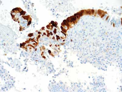
9 HISTOLOGY Progression Arrest Fig 1-4 Although transient HPV infection may result in increased cell proliferation, these infections do not disrupt the balance between prb and E2F or the control of p16 expression. HPV Infection and Cervical Disease Some HR-HPV infections, however, persist and produce levels of viral E6 and E7 oncoproteins that can mediate oncogenic transformation through the disruption of the cell cycle regulatory mechanism (Fig 1-5). This transformation can lead to the development of high-grade cervical dysplasia (CIN 2/3), adenocarcinoma in situ (AIS), and invasive cervical carcinomas. The cellular protein p16 is a useful marker to identify high-grade CIN lesions, as it is over-expressed in cervical cells undergoing cellular transformation in response to the expression of the HR-HPV E7 oncoprotein. 16,15,9,26 (Fig 1-6).

10 A E6 Fig. 1-5, Panel A HR-HPV E6 and E7 mediate oncogenic transformation. B Fig. 1-5, Panel B Fully transformed cells are characterized by unregulated cell cycle progression, disruptedmaturation,andtheabilitytoinvadeunderlying cervical stroma. Cellular evidence of transformation includes an increase in nuclear/cytoplasmic ratios, anisonucleosis, and hyperchromasia.2 p16: A Biomarker for Cervical Disease DNA testing for HPV cannot differentiate transient infections, which make up a majority of cases, from transforming infections, which occur less frequently. Therefore, testing for HPV is of limited use in pinpointing clinically significant disease, especially in younger women where the prevalence of HPV infection can be as high as 30%. 6
11 Diffuse p16 staining; however, results from E7-mediated inactivation of the prb-e2f control mechanism, which is a central hallmark of the oncogenic process. Thus, in contrast to HPV DNA detection, the immunohistochemical detection of a diffuse pattern of p16 over-expression identifies lesions of cervical mucosa in which molecular mechanisms leading to oncogenic 28, 8, 27, 11 transformation are active. Fig. 1-6 In cells with transforming HPV infections, HPV viral oncoprotein E7 impairs the function of prb, disrupting its ability to bind to transcription factor E2F. This leads to deregulated cell proliferation, genetic instability and p16 protein over-expression whichisdetectablebyimmunohistochemistry.
12

13 Chapter 2 Interpreting p16 Staining Patterns Interpreting p16 Staining Patterns
14 Squamous Epithelium p16 is a nuclear protein under normal physiological conditions, however, typically both nuclear and a cytoplasmic expression are observed when p16 is over-expressed in cervical neoplasia. 9, 24, 19, 10 In few cases, nuclear expression may be faint or undetectable, but nuclear staining is not required to interpret the p16 staining. Therefore, both nuclear and/or cytoplasmic immunostaining should be taken into account when assessing the p16 staining pattern. As with any immunohistochemical procedure, it is important to standardize the interpretation criteria for p16 by designating specific immunostaining patterns. The p16 rating system not only reflects the molecular pathogenesis of cervical dysplasia, but has also been validated by correlation with the progression of CIN in numerous studies. 1,28,7,18, 24 Interpretation of p16 staining results must take into consideration the fact that p16 at the individual cell level may be expressed in highgrade dysplastic cervical lesions and cervical cancers as well as in some conditions not associated with cervical dysplasia. Furthermore, it is important to note that p16 test results should be used only to provide additional information that must be interpreted in conjunction with the morphological classification of the lesion. Specimens stained with the CINtec Histology Kit are evaluated according to a binary rating system composed of the ratings positive and negative. The rating of a slide as positive or negative considering both nuclear and/or cytoplasmic staining is assigned as follows: 10


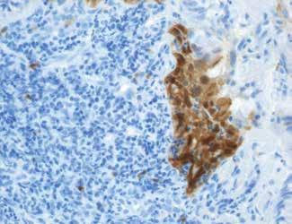
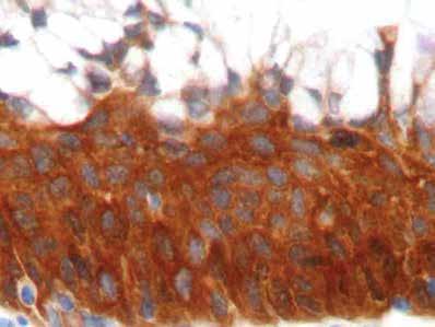
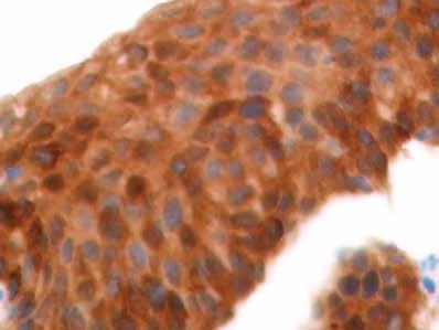
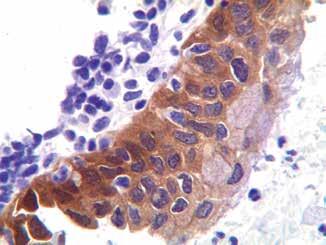
15 HISTOLOGY p16 Positive: A p16 test result is positive if the staining pattern is Diffuse, showing a continuous staining of cells of the basal and parabasal cell layers of the squamous cervical epithelium, with or without staining of cells of superficial cell layers. Fig 2-1 Diffuse p16 staining pattern. A B Panel A Magnification 20X Panel B Magnification 40X A diffuse staining pattern is sometimes also referred to as a confluent staining, band staining, or block staining, where typically the staining intensity of the cells within the lesion is homogeneous. Virtually all CIN3 lesions are positive for p16 28,7 and a positive test result provides high positive predictive value for high-grade CIN. 20 Although high-grade lesions typically show diffuse staining, 40-60% of CIN1 lesions can also show diffuse staining, which may indicate the onset of transforming HPV infections. 4,22,7 Therefore, the assessment of the extent of the diffuse staining within the squamous cervical epithelium itself should not be used as an independent criterion for the interpretation of cervical lesions.


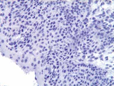
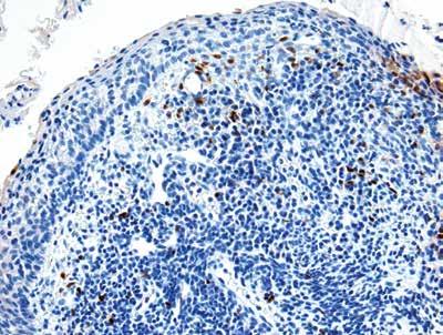
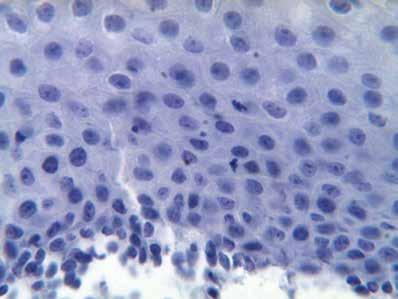
16 p16 Negative: A test result is rated negative if a p16 stained slide shows either a negative staining reaction in the squamous epithelium ( negative staining pattern Fig 2-2A), or a staining of isolated cells or small cell clusters; i.e., a noncontinuous staining, particularly not of the basal and parabasal cells ( focal staining pattern Fig 2-2B). 1, 8 Fig. 2-2 Negative p16 results. A Panel A No p16 staining; magnification 20X B Panel B Focal p16 staining pattern; magnification 20X 12
17 HISTOLOGY A focal staining pattern is sometimes also referred to as patchy staining, scattered staining, or checkerboard staining. The staining intensity of the cells of the same morphological grade within the respective lesion may show substantial variability. Focal p16 staining is occasionally seen in squamous metaplasia, atypical immature metaplasia, reactive atypia, and is frequently detected in CIN1. Neg Negative: Focal p16 S Columnar cells Negative: Focal p16 Staining Columnar cells Normal Negative: Focal p16 Staining Squamous MetaplasiaColumnar cells Normal CIN1 HPV infected HPV infected p16 over-expre Negative: Focal p16 Staining Columnar cells Columnar cells Normal Normal Normal p16over-expression HPV infected HPV infected p16 over-expression Fig. 2-3 Negative p16 results. Squamous metaplasia, CIN1 (focal staining pattern) HPV infected p16 over-expression p16 over-expression

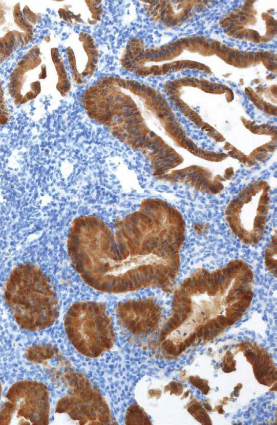
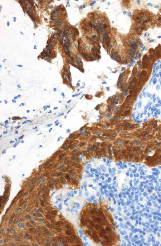
18 Endocervical Epithelium When interpreting glandular cervical epithelium, an analogous rating system may be used for the evaluation of p16 staining patterns. p16 Positive: As in squamous tissue, a diffuse staining pattern in endocervical tissue is rated as positive. A diffuse pattern requires all atypical cells in the gland to stain for p16. Diffuse p16 staining in the glands is typically observed in adenocarcinoma in situ (AIS). (Fig. 2-4, 2-5) as well as in invasive adenocarcinomas and should 8, 23 be rated as a positive test result. Fig. 2-4 Diffuse p16 staining as seen in AIS Fig. 2-5 Positive p16 test result, AIS. Note diffuse staining in atypical glandular epithelial cells and absence of staining in adjacent benign glandular mucosa (arrow) Magnification 10X 14

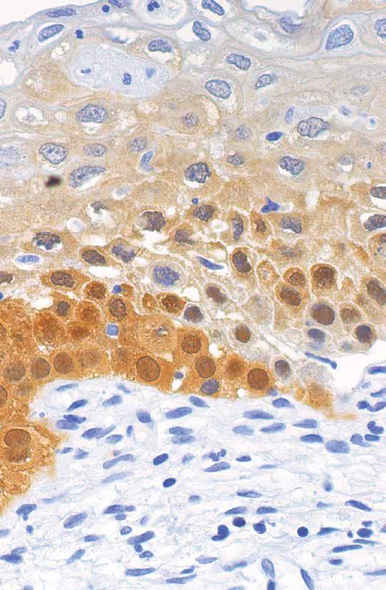
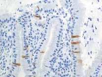
19 HISTOLOGY Negative: Both no p16 reactivity (Fig 2-6) or a focal staining pattern (Fig 2-7) are rated as negative. Focal p16 staining can be observed occasionally in normal glandular epithelium where individual cells or small groups of neighboring glandular cells may show p16 immuno-reactivity. 8 Fig. 2-6 Negative p16 result. No staining. Fig. 2-7 Negative p16 result. Focal staining pattern as can be seen in tubal metaplasia and tubo-endometrial metaplasia Fig. 2-8: Although benign mucinous glandular cells don t typically stain for p16, staining may be detected in scattered, non-mucinous/secretory cells (with or without cilia) as well as in areas of tubo-endometrial metaplasia producing a focal 8, 23, 12, 13 staining pattern. Magnification 40X
20


21 Chapter 3 Clinical Utility of p16 for High-Grade CIN Clinical Utility of p16 for High-Grade CIN

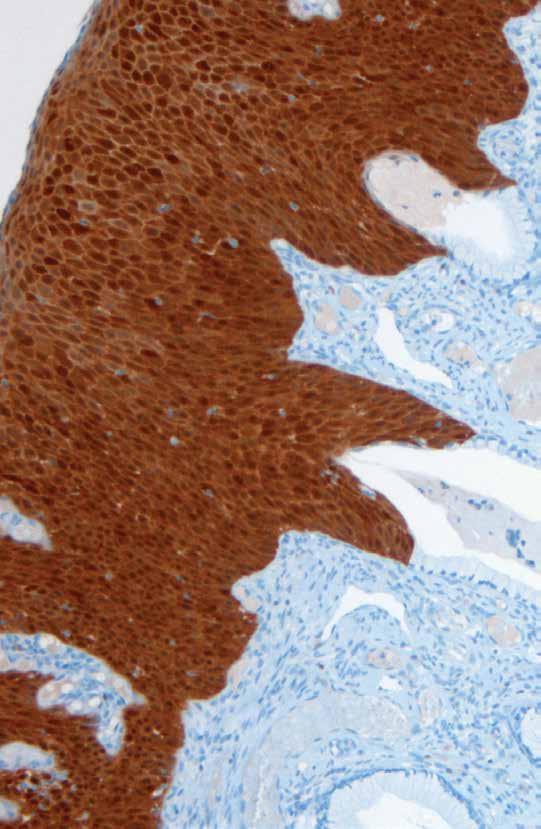
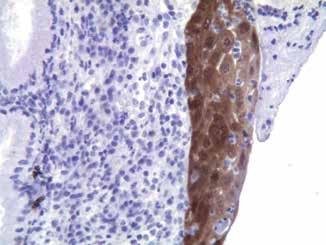
22 Confirmation of CIN2/3 The primary role of the conjunctive use of p16 immunohistochemical staining is to confirm or rule out highgrade dysplasia (CIN2/3) in lesions with equivocal morphology. p16 has been found to be strongly over-expressed in over 99% of cases of high-grade cervical dysplasia. 1,4,15 In addition, a recent study has reported a high level of inter-observer reproducibility in the scoring of p16 test results. 1 A B Fig. 3-1 CIN3. Note dysplastic cervical mucosa stains diffusely for p16 indicating a positive p16 test result. The diagnosis of CIN3 is confirmed. Magnification 2.5X Panel A H&E; Panel B p16 18


23 HISTOLOGY A B Fig. 3-2 CIN3 at squamo-columnar junction. Note diffuse p16 staining throughout all layers of dysplastic cervical mucosa and absence of staining in adjacent benign glandular cells. The p16 test result is rated positive and the diagnosis of CIN3 is confirmed. Magnification 40X Panel A H&E; Panel B p16 A B Fig. 3-3 CIN2. Note the continuous p16 staining of the basal and parabasal cell layers of the dysplastic cervical mucosa. The p16 staining pattern is diffuse and rated as a positive p16 test result. Magnification 20X Panel A H&E; Panel B p16


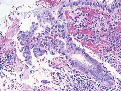
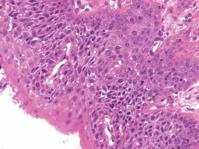
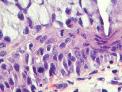
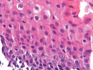
24 Differential Diagnosis of High-Grade Dysplasia Some benign conditions, including squamous atrophy and atypical squamous metaplasia, can mimic high-grade dysplasia and represent diagnostic challenges. p16 staining in these cases can provide important data to support or exclude the final diagnosis of high-grade dysplasia, as highlighted in the 4, 8, 29 following examples. 1.Squamous Atrophy: CIN2/3 and squamous atrophy are both characterized by an arrest of normal squamous differentiation with nuclear crowding and high nuclear to cytoplasmic ratios. Whereas CIN2/3 is a precursor of cervical cancer, squamous atrophy has no premalignant potential and shows only limited focal staining or is completely negative for p16 staining. Thus, p16 staining, especially in biopsies from postmenopausal women can provide important information that can help solve the differential diagnosis of CIN2/3 versus squamous atrophy. 11 A B Fig. 3-4 Squamous atrophy versus high-grade dysplasia. Note nuclear crowding and disordered maturation, however p16 shows no immuno-reactivity. The result of the p16 test is negative helping to rule out high-grade dysplasia. Magnification 20X Panel A H&E; Panel B p16 20
, 29 whereas a negative p16")

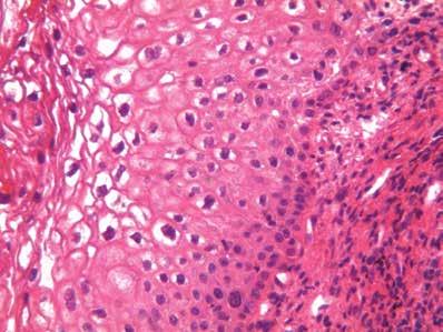
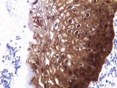
25 HISTOLOGY 2. Atypical Squamous Metaplasia Atypical squamous metaplasia (ASM) encompasses the replacement of benign glandular cells with squamous metaplastic cells that may mimic high-grade disease. p16 is a useful tool to aid in differential diagnosis, a diffuse stain indicating the presence of transforming HPV infection supports the diagnosis of CIN2/3 (Fig 3-5), 29 whereas a negative p16 result favors the diagnosis of ASM (Fig 3-6). It is important to note that both focal staining and the absence of staining should be rated as a negative p16 test result. A B Fig. 3-5 ASM versus CIN3. The mucosal surface appears attenuated and could be interpreted based on H&E staining to reflect either ASM or a high-grade squamous lesion. Diffuse p16 staining is rated as a positive p16 test result and supports the diagnosis of CIN3. Magnification 40X Panel A H&E; Panel B p16


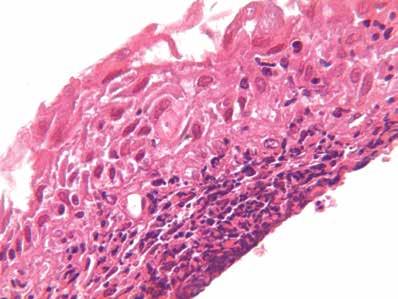
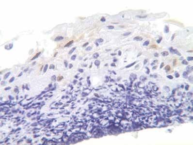
26 A B Fig. 3-6 Atypical squamous metaplasia (ASM) versus high-grade dysplasia. Focal nuclear crowding and disrupted maturation but with minimal nuclear changes is suggestive of ASM versus CIN2/3. Focal staining for p16, indicating a negative p16 test, helps to exclude the diagnosis of CIN2/3 and supports the final diagnosis of ASM. Magnification 40X Panel A H&E; Panel B p16 3. Chronic Cervicitis with Reactive Atypia Acute and/or chronic cervicitis may lead to reactive atypia and may also obscure underlying high-grade dysplasia. In these cases, p16 immunostaining may help to confirm or rule out the presence of underlying CIN2/3 lesions that are not readily apparent on H&E stained sections. 22


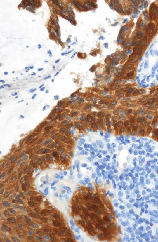
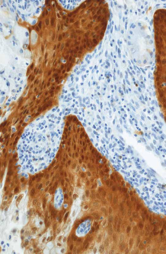
27 HISTOLOGY A B Fig. 3-7 Chronic cervicitis with reactive atypia versus CIN2/3. Intense chronic inflammation of the cervical stroma with reactive atypia of the overlying squamous epithelium, suggestive but not diagnostic for high-grade dysplasia. p16 shows no immuno-reactivity and is rated a negative test result, helping to rule out CIN2/3. Magnification 10X Panel A H&E; Panel B p16 A B Fig. 3-9 Atypical squamous metaplasia versus CIN2/3 in a background of severe chronic inflammation. Note diffuse staining for p16 in lesional epithelium. The p16 test result is positive and consistent with the final conclusion that this is a case of high-grade squamous dysplasia (CIN2/3). Magnification 20X Panel A H&E; Panel B p16
28

29 Chapter 4 Clinical Utility of p16 for Low-Grade CIN Clinical Utility of p16 for Low-Grade CIN

, as well as")
30 The primary role of the conjunctive use of p16 immunohistochemical staining in CIN1 is to provide evidence to help rule out high-grade disease with a negative test result. Tissue biopsies that are classified as CIN1 include those that represent productive HPV infections and in some cases, those that have similar morphologic characteristics but are HPV negative. Cases that have definitive features of HPV infection, including unequivocal koilocytotic atypia, may show positive or negative p16 test results. p16 negative test results are commonly seen in cases that have some degree of disordered maturation but lack unequivocal morphologic evidence of HPVassociated cytopathic effect (Fig 4-1), as well as in some cases that are morphologically definitive for HPV cytopathic effect. Nevertheless, the majority of cases that have unequivocal koilocytotic atypia show either focal staining (Fig 4-2) or diffuse staining (Fig 4-3) for p A B Fig. 4-1 CIN1 versus tangential sectioning. Crowding of nuclei in the lower third that couldhavebeeninterpretedasevidenceofcin1versustangentialsectioningofnormal mucosa. p16 shows no immuno-reactivity and the test result is rated as negative. Magnification 20X Panel A: H&E; Panel B: p16 26


31 HISTOLOGY A B Fig. 4-2 This case was classified as CIN1 based on the presence of cells with perinuclear clearing (koilocytosis). p16 staining is focal, p16 test result is rated negative. In retrospect, however, the areas of perinuclear clearing may have resulted from glycogen accumulation rather than HPV infection, which is a commonly encountered pitfall in the diagnosis of CIN1. Magnification 20X Panel A: H&E; Panel B: p16 A B Fig. 4-3 Koilocytotic atypia and dyskeratosis with crowding of nuclei in the lower third, consistent with CIN1. p16 staining is diffuse and this cases is rated as a positive test result. Magnification 40X Panel A: H&E; Panel B: p16 Several published reports indicate that CIN1 lesions that show no p16 reactivity or stain in a focal pattern may persist or regress but those that show a diffuse p16 staining pattern are at an increased risk for progression to CIN2/3 over a 2-3 year period. 3,17,5,20 More clinical research is required to validate these interesting but still preliminary findings.
32

33 Chapter 5 Clinical Utility of p16 for Endocervical Neoplasia Clinical Utility of p16 for Endocervical Neoplasia
. In contrast, most preneoplastic and neoplastic endocervical glandular lesions, as a result of their association with high-risk HPV, exhibit diffuse p16 positivity (Fig 5-2,5-3) 8, 23 Although")
.")
34 It has been reported that normal endocervical glands are usually p16 negative (Fig 5-1) with an occasional case exhibiting focal weak staining that is also rated as a negative test result (Fig 5-5, 5-6). In contrast, most preneoplastic and neoplastic endocervical glandular lesions, as a result of their association with high-risk HPV, exhibit diffuse p16 positivity (Fig 5-2,5-3) 8, 23 Although benign mucinous columnar cells, including those in areas of microglandular hyperplasia (Fig 5-4), are consistently negative for p16, benign non-mucinous/secretory cells, tubal metaplasia, tubo-endometrial metaplasia, cervical endometriosis, and endometrial glandular cells, including lower uterine segment mucosal cells sometimes show extensive p16 immuno-reactivity (Fig 5-5, 5-6). A B Fig. 5-1 Benign endocervical mucinous glandular epithelium, negative for p16. Magnification 40X Panel A: H&E; Panel B: p16 It is very important to recognize that staining in benign glandular cells is a normal manifestation and does not denote premalignant potential. Thus, p16 staining results should be interpreted in correlation with tissue cellular morphology to avoid potential misclassification of benign cellular changes as glandular neoplasia. 30

.")


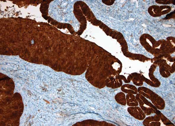
35 HISTOLOGY A B Fig. 5-2 AIS. Note intense nuclear and cytoplasmic staining for p16 in all neoplastic cells and the absenceofstaininginbenignglandularcells (arrow).thep16stainingpatternisdiffuseindicating a positive p16 test result and confirming AIS. Magnification 40X Panel A: H&E; Panel B: p16 A B Fig. 5-3 Invasiveadenocarcinoma.Intensenuclearandcytoplasmicstainingforp16isretained in areas of invasion. The p16 staining pattern is diffuse indicating a positive p16 test result and confirming Invasive adenocarcinoma. Magnification 10X Panel A: H&E; Panel B: p16


36 A B Fig. 5-4 Microglandular hyperplasia, p16 shows no immuno-reactivity and the test result is rated negative. Magnification 40X Panel A: H&E; Panel B: p16 A B Fig. 5-5 Normal endocervical glands with mucinous and non-mucinous/secretory columnar cells.notefocalstaining,themucinouscellsarep16negativebutthenon-mucinous/secretory cells show strong staining for p16. The p16 test result is rated negative. Magnification 40X Panel A: H&E; Panel B: p16 32


37 HISTOLOGY A B Fig. 5-6 Tubal metaplasia with focal staining for p16. Normal endocervical glands with mucinous and non-mucinous/secretory columnar cells. Note that the mucinous cells are p16 negative but that the non-mucinous/ secretory cells show strong staining for p16. The p16 test result is rated negative. Magnification 20X Panel A: H&E; Panel B: p16
38


39 CINtec Histology Staining Case Examples Histology Staining Case Examples
40 36
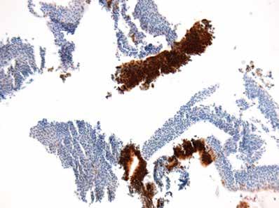
41 HISTOLOGY CASE 1: High-grade squamous dysplasia and AIS Diffuse p16 staining pattern in both squamous and glandular components are interpreted as a positive p16 result. Final diagnosis: CIN3 with endocervical glandular involvement and AIS Magnification 20X
42 A B 38
43 HISTOLOGY CASE 2: CIN 1 Note disordered maturation in the lower third of the squamous mucosa with morphologic evidence of a productive HPV infection, including koilocytosis and hyperkeratosis. p16 shows diffuse staining throughout the full thickness of the lesion and the test result is rated positive. Magnification 40X Panel A H&E Panel B p16
44 A B 40
45 HISTOLOGY CASE 3: Inflammatory atypia versus CIN1 Histologic features include nuclear enlargement and disordered maturation but without koilocytosis. Note underlying acute inflammatory cells that extend into the overlying squamous mucosa. p16 staining shows a focal pattern and the test is rated negative. Magnification 40X Panel A H&E Panel B p16
46 A B 42
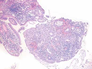
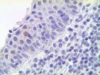
47 HISTOLOGY CASE 4: Glandular atypia Glandular atypia, differential diagnosis tubo-endometrial metaplasia versus AIS. The focal p16 staining pattern is rated as a negative test result and is consistent with tubo-endometrial metaplasia. Magnification 20X Panel A H&E Panel B p16


48 A C CASE 5: Inflammatory atypia versus CIN The mucosa shows dense acute and chronic inflammation with disordered maturation and nuclear crowding of the squamous epithelial cells. The differential diagnosis includes inflammatory atypia versus low-grade or high-grade CIN. p16 staining is diffuse and rated as a positive test result, supporting the final diagnosis of CIN 2/3. Note diffuse staining involving disrupted nests of squamous cells. Magnification 20X Panel A H&E Panel C p16 Magnification 40X Panel B H&E Panel D p16 44


49 HISTOLOGY B D
50 A B 46
51 HISTOLOGY CASE 6: Atypical immature metaplasia versus CIN2 The lesion shows nuclear crowding without enlargement or mitotic activity that was considered suggestive but not diagnostic of CIN2. p16 shows no immuno-reactivity and the test result is rated as negative, confirming that this is not high-grade dysplasia. Magnification 20X Panel A H&E Panel B p16
52 A B 48
53 HISTOLOGY CASE 7: Atypical immature metaplasia versus CIN2/3 Note squamous cells with nuclear crowding and abnormal mitotic activity and overlying mucinous columnar cells. The squamous cells are diffusely immunoreactive for p16, resulting in a positive test rating which confirms the impression of a high-grade (CIN2/3) lesion. Magnification 40X Panel A H&E Panel B p16
54 A B 50
55 HISTOLOGY CASE 8: Atrophic cervicitis versus high-grade dysplasia Immature basal-like cells are present through all layers of the squamous mucosa. The loss of maturation raises a differential diagnosis that includes high-grade dysplasia. The p16 staining pattern is focal and rated as a negative result. Focal staining is located primarily in superficial epithelial cells and in scattered inflammatory cells within the cervical stroma, helping to rule out highgrade dysplasia. Magnification 20X Panel A H&E Panel B p16
56 A B 52
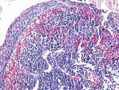
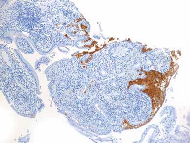
57 HISTOLOGY CASE 9: Chronic cervicitis with reactive atypia versus CIN2/3 Intense chronic inflammation in the cervical stroma and associated reactive atypia of the overlying squamous epithelium largely obscured the presence of concurrent highgrade dysplasia that, however, is clearly detected by diffuse p16 staining. Magnification 2.5X Panel A H&E Panel B p16


58 A C CASE 10: Reserve cell hyperplasia versus CIN The mucosa is composed predominantly of basal/parabasal-like cells with nuclear crowding. p16 shows no immuno-reactivity, helping to exclude the diagnosis of CIN2/3. Magnification 20X Panel A H&E Panel C p16 Magnification 40X Panel B H&E Panel D p16 54

59 HISTOLOGY B D
60 A B 56
61 HISTOLOGY CASE 11: Reactive atypia versus CIN This section is cut too thick and shows some fracture artifact. Some cells show perinuclear clearing that raised suspicion for koilocytosis. The lack of p16 immunoreactivity confirms a negative p16 test result and helps to exclude the diagnosis of CIN2/3. Magnification 40X Panel A H&E Panel B p16
62 A B 58
63 HISTOLOGY CASE 12: Reactive atypia versus CIN3 The lesion shows moderate nuclear crowding, extending into the upper layers of the squamous mucosa. Diffuse p16 staining is graded as a positive result and confirms the impression of CIN3. Magnification 40X Panel A H&E Panel B p16
64 A B 60
65 HISTOLOGY CASE 13: Disrupted fragments of cervical mucosa, tangentially sectioned, atrophy versus CIN2/3 Note diffuse p16 staining, rated as positive p16 test result. Final diagnosis: CIN2/3. Magnification 10X Panel A H&E Panel B p16
 serve to highlight nests of high-grade CIN.")
66 A C CASE 14: CIN 2/3, small lesion Note the complex architectural pattern, including crowded benign endocervical glands, and minute foci of metaplastic squamous cells, in a background of acute and chronic cervicitis. p16 positive results (diffuse staining pattern) serve to highlight nests of high-grade CIN. Magnification 4X Panel A H&E Panel C p16 Magnification 40X Panel B H&E Panel D p16 62

67 HISTOLOGY B D


68 A C CASE 15: CIN 2/3, small lesion, disrupted mucosa This case shows predominantly benign glandular mucosa with few squamous cells. p16 positive test results (diffuse pattern), aided in the detection of a minute focus of CIN2/3. Magnification 10X Panel A H&E Panel C p16 Magnification 40X Panel B H&E Panel D p16 64

69 HISTOLOGY B D


70 A C CASE 16: CIN1, small lesion, endocervical glandular involvement Note minute focus of squamous metaplasia adjacent to benign endocervical glands. Diffuse p16 positive test result (arrow), highlights endocervical gland involvement by CIN1. Magnification 20X Panel A H&E Panel C p16 Magnification 40X Panel B H&E Panel D p16 66


71 HISTOLOGY B D

72 1..Bergeron C, Ordi J, Schmidt D, et al. Conjunctive p16 INK4a Testing Significantly Increases Accuracy in Diagnosing High-Grade Cervical Intraepithelial Neoplasia. Am J Clin Pathol. 2010;133: Cameron RI, Maxwell P, Jenkins D, et al. Immunohistochemical staining with MIB1, bcl2 and p16 assists in the distinction of cervical glandular intraepithelial neoplasiafromtubo-endometrialmetaplasia,endometriosisandmicroglandular hyperplasia. Histopathology 2002;41: Cuschieri K, Wentzensen N. Human papillomavirus mrna and p16 detection as biomarkers for the improved diagnosis of cervical neoplasia. Cancer Epidemiol Biomarkers Prev. 2008;17: Dallenbach-Hellweg G, Trunk MJ, von Knebel Doeberitz Traditional and new molecular methods for early detection of cervical cancer. M.Arkh Patol. 2004;66: del Pino M, Garcia S, Fusté V, et al. Value of p16 INK4a as a marker of progression/ regression in cervical intraepithelial neoplasia grade 1. Am J Obstet Gynecol. 2009; 201:488.e Dijkstra MG, Heideman DA, de Roy SC, et al. p16 INK4a immunostaining as an alternative to histology review for reliable grading of cervical intraepithelial lesions. J Clin Pathol. 2010;63: Evangelou K, Bramis J, Peros I, et al. Electron microscopy evidence that cytoplasmic localization of the p16 INK4a nuclear cyclin-dependent kinase inhibitor (CKI) in tumor cells is specific and not an artifact. A study in non-small cell lung carcinomas. Biotech Histochem. 2004;79: Galgano M, Castle P, Atkins K, et al. Using Biomarkers as Objective Standards in the Diagnosis of Cervical Biopsies. Am J Surg Pathol 2010;34: Ghiorzo P, Villaggio B, Sementa AR, et al. Expression and localizationofmutantp16proteinsinmelanocyticlesions from familial melanoma patients. Hum Pathol. 2004;35:25-33.

73 bibliography 10. Hariri J, Oster A. The negative predictive value of p16 INK4a to assess the outcome of cervical intraepithelial neoplasia 1 in the uterine cervix. Int J Gynecol Pathol. 2007;26: Horn LC, Reichert A, Oster A, et al. Immunostaining for p16 INK4a used as a conjunctivetoolimprovesinterobserveragreementofthehistologicaldiagnosis of cervical intraepithelial neoplasia. Am J Surg Pathol. 2008;32: Keating JT, Cviko A, Riethdorf S, et al. Ki-67, cyclin E, and p16 INK4a are complimentarysurrogatebiomarkersforhumanpapillomavirus-relatedcervical neoplasia. Am J Surg Pathol. 2001;25: Klaes R, Benner A, Friedrich T, et al. Overexpression of p16 INK4a as a specific marker for dysplastic and neoplastic epithelial cells of the cervix uteri. Int J Cancer 2001;92: Negri G, Egarter-Vigl E, Kasal A, et al. p16 INK4a Is a Useful Marker for the Diagnosis of Adenocarcinoma of the Cervix Uteri and Its Precursors. Am J Surg Pathol. 2003;27: Negri G, Vittadello F, Romano F, et al. p16 INK4a expression and progression risk of low-grade intraepithelial neoplasia of the cervix uteri. Virchows Arch. 2004;445: NilssonK,LandbergG.Subcellularlocalization,modificationandproteincomplex formation of the cdk-inhibitor p16 in Rb-functional and Rb-inactivated tumor cells. Int J Cancer. 2006;118: O Neill CJ, McCluggage WG. p16 expression in the female genital tract and its value in diagnosis. Adv Anat Pathol. 2006;13: Ordi J, Garcia S, del Pino M, et al. p16 INK4a immunostaining identifies occult CIN lesions in HPV-positive women. Int J Gynecol Pathol. 2008;28: Qiao X, Bhuiya TA, et al. Differentiating high-grade cervical intraepithelial lesion from atrophy in postmenopausal women using Ki-67, cyclin E, and p16 immunohistochemical analysis. J Low Genit Tract Dis. 2005;9:100-7.
 immature squamous metaplasia from high-grade cervical intraepithelial neoplasia (CIN III).")
74 20. Redman R, Rufforny I, et al.the utility of p16 INK4a in discriminating between cervical intraepithelial neoplasia 1 and nonneoplastic equivocal lesions of the cervix. Arch Pathol Lab Med. 2008;132: Regauer S, Reich O. CK17 and p16 expression patterns distinguish (atypical) immature squamous metaplasia from high-grade cervical intraepithelial neoplasia (CIN III). Histopathology 2007;50: Sano T, Oyama T, Kashiwabara K, et al. Expression status of p16 protein is associatedwithhumanpapillomavirusoncogenicpotentialincervicalandgenital lesions. Am J Pathol. 1998;153: Sayed K, Korourian S, Ellison DA, et al. Diagnosing cervical biopsies in adolescents: the use of p16 immunohistochemistry to improve reliability and reproducibility. J Lower Genital Tract Dis. 2007;11: Tringler B, Gup CJ, Singh M, et al. Evaluation of p16 INK4a and prb expression in cervical squamous and glandular neoplasia. Hum Pathol. 2004;35: Tsoumpou I, Arbyn M, Kyrgiou M, et al. p16 INK4a immunostaining in cytological and histological specimens from the uterine cervix: a systematic review and meta-analysis. Cancer Treatment Rev. 2009;35: von Knebel Doeberitz. New markers for cervical dysplasia to visualise the genomic chaos created by aberrant oncogenic papillomavirus infections. Eur J Cancer 2002;38: von Knebel Doeberitz. Principles and key findings of the p16 test. HPV Today. No 08; Feb 2006.

75 bibliography 28. Wang SS, Trunk M, Schiffman M et al. Validation of p16 INK4a as a marker of oncogenic human papillomavirus infection in cervical biopsiesfromapopulation-basedcohortincostarica.cancerepidemiol Biomarkers Prev. 2004;13: Wentzensen N, von Knebel Doeberitz M. Biomarkers in cervical cancer screening. Dis Markers 2007;23: Roche greatly acknowledges the work of the authors and contributors of this Compendium & Staining Atlas: Kenneth R. Shroyer MD, PhD, Stony Brook University Medical Center Mamatha Chivukula MD, Magee Women s Hospital of University of Pittsburgh Medical Center Brigitte Ronnett MD, Johns Hopkins University School of Medicine Terry Morgan MD, PhD, Oregon State Health and Science University
76 For further information please visit us at CINtecP16.com This Compendium & Staining Atlas may not be distributed in or to the United States of America. Roche Diagnostics International Ltd. CH-6343 Rotkreuz Switzerland Roche Diagnostics International Ltd. G A G A EN MP ITREV2
Table of Contents. 1. Overview. 2. Interpretation Guide. 3. Staining Gallery Cases Negative for CINtec PLUS
 Staining Atlas Table of Contents 1. Overview 1.1 Introduction 1.2 Role of p16 INK4a 1.3 Role of Ki-67 1.4 Molecular Pathogenesis 1.5 p16 INK4a Expression in Cervical Dysplasia 1.6 The Concept of CINtec
CINtec p16 INK4a Staining Atlas
 CINtec p16 INK4a Staining Atlas Rating Rating Positive The rating positive will be assigned if the p16 INK4a -stained slide shows a continuous staining of cells of the basal and parabasal cell layers of
CINtec PLUS Cytology. Interpretation training
 CINtec PLUS Cytology Interpretation training Objectives After reviewing this learning module, you will have a basic understanding of how to interpret CINtec PLUS Cytology, including: The mechanism of action
chapter 4. The effect of oncogenic HPV on transformation zone epithelium
 chapter 4. The effect of oncogenic HPV on transformation zone epithelium CHAPTER 1 All squamous cervical cancer (and probably all cervical adenocarcinoma) is associated with oncogenic HPV, and the absence
New Diagnoses Need New Approaches: A Glimpse into the Near Future of Gynecologic Pathology
 New Diagnoses Need New Approaches: A Glimpse into the Near Future of Gynecologic Pathology United States and Canadian Academy of Pathology 102 nd Annual Meeting Baltimore, Maryland Christina S. Kong, M.D.
CK17 and p16 expression patterns distinguish (atypical) immature squamous metaplasia from high-grade cervical intraepithelial neoplasia (CIN III)
 immature squamous metaplasia from high-grade cervical intraepithelial neoplasia (CIN III)") Histopathology 2007, 50, 629 635. DOI: 10.1111/j.1365-2559.2007.02652.x CK17 and p16 expression patterns distinguish (atypical) immature squamous metaplasia from high-grade cervical intraepithelial neoplasia
Histopathology 2007, 50, 629 635. DOI: 10.1111/j.1365-2559.2007.02652.x CK17 and p16 expression patterns distinguish (atypical) immature squamous metaplasia from high-grade cervical intraepithelial neoplasia
Objectives. Atypical Glandular Cells. Atypical Endocervical Cells. Reactive Endocervical Cells
 2013 California Society of Pathologists 66 th Annual Meeting San Francisco, CA Atypical Glandular Cells to Early Invasive Adenocarcinoma: Cervical Cytology and Histology Christina S. Kong, MD Associate
EU guidelines for reporting gynaecological cytology
 EU guidelines for reporting gynaecological cytology Amanda Herbert Guy s & St Thomas Foundation NHS Trust 5th EFCS Annual Tutorial, Trondheim, Norway 28 th May 1 st June 2012 EU guidelines aim to harmonize
SQUAMOUS CELLS: Atypical squamous cells (ASC) - of undetermined significance (ASC-US) - cannot exclude HSIL (ASC-H)
 - of undetermined significance (ASC-US) - cannot exclude HSIL (ASC-H)") SQUAMOUS CELLS: Atypical squamous cells (ASC) - of undetermined significance (ASC-US) - cannot exclude HSIL (ASC-H) ASC refers to cytologic changes suggestive of SIL, which are qualitativley or quantitatively
SQUAMOUS CELLS: Atypical squamous cells (ASC) - of undetermined significance (ASC-US) - cannot exclude HSIL (ASC-H) ASC refers to cytologic changes suggestive of SIL, which are qualitativley or quantitatively
Histopathology: Cervical HPV and neoplasia
 Histopathology: Cervical HPV and neoplasia These presentations are to help you identify basic histopathological features. They do not contain the additional factual information that you need to learn about
HPV and Lower Genital Tract Disease. Simon Herrington University of Edinburgh, UK Royal Infirmary of Edinburgh, UK
 HPV and Lower Genital Tract Disease Simon Herrington University of Edinburgh, UK Royal Infirmary of Edinburgh, UK Conflict of interest/funding X None Company: Product royalties Paid consultant Research
Interpretation guide. Abnormal cytology can t hide anymore
 Interpretation guide Abnormal cytology can t hide anymore Unique dual-biomarker technology makes you certain about the presence of transforming HPV infection. The science that creates certainty. Table
p16ink4a expression in cervical intraepithelial neoplasia and cervical cancer
 Original Article Brunei Int Med J. 2013; 9 (3): 165-171 p16ink4a expression in cervical intraepithelial neoplasia and cervical cancer Kalpana KUMARI 1 and Akhila Arcot VADIVELAN 2 1 Department of Pathology,
P16 INK4A EXPRESSION AS A POTENTIAL PROGNOSTIC MARKER IN CERVICAL PRECANCEROUS AND CANCEROUS LESIONS IN MOROCCO
 P16 INK4A EXPRESSION AS A POTENTIAL PROGNOSTIC MARKER IN CERVICAL PRECANCEROUS AND CANCEROUS LESIONS IN MOROCCO Yassine Zouheir Laboratory of histo-cytopathology of Institut Pasteur of Morocco, Casablanca,
When Immunostains Can Get You in Trouble: Gynecologic Pathology p16: Panacea or Pandora s Box?
 When Immunostains Can Get You in Trouble: Gynecologic Pathology p16: Panacea or Pandora s Box? Teri A. Longacre, MD Stanford Medicine Stanford California pi6 in Gynecologic Pathology: Panacea or Pandora
Thin HSIL of the Cervix: Detecting a Variant of High-grade Squamous Intraepithelial Lesions With a p16 INK4a Antibody
 International Journal of Gynecological Pathology 00:1 5, Lippincott Williams & Wilkins, Baltimore r 2016 International Society of Gynecological Pathologists Original Article Thin HSIL of the Cervix: Detecting
Workshop for O& G trainees and paramedics 17 Dec 2011 Cytological Interpretation
 Workshop for O& G trainees and paramedics 17 Dec 2011 Cytological Interpretation May Yu Director of Cytology Laboratory Service Department of Anatomical & Cellular Pathology Prince of Wales Hospital Cervical
Biomarkers for cervical cancer screening: the role of p16 INK4a to highlight transforming HPV infections
 Review For reprint orders, please contact reprints@expert-reviews.com Biomarkers for cervical cancer screening: the role of p16 INK4a to highlight transforming HPV infections Expert Rev. Proteomics 9(2),
P16 et Ki67 Biomarkers: new tool for risk management and low grade intraepithelial lesions (LGSIL): be ready for the future.
: be ready for the future.") P16 et Ki67 Biomarkers: new tool for risk management and low grade intraepithelial lesions (LGSIL): be ready for the future. Mark H Stoler, MD University of Virginia Health System, Charlottesville, VA,
P16 et Ki67 Biomarkers: new tool for risk management and low grade intraepithelial lesions (LGSIL): be ready for the future. Mark H Stoler, MD University of Virginia Health System, Charlottesville, VA,
Intravascular Endometrium Mimicking Vascular Invasion
 ISPUB.COM The Internet Journal of Pathology Volume 12 Number 1 A Papanicolau, G Lin Citation A Papanicolau, G Lin.. The Internet Journal of Pathology. 2010 Volume 12 Number 1. Abstract Intravascular endometrium
South Afr J Gynaecol Oncol RESEARCH
 Southern African Journal of Gynaecological Oncology 2017; 9(2):25 29 https://doi.org/10.1080/20742835.2017.1370841 Open Access article distributed under the terms of the Creative Commons License [CC BY-NC
Colposcopy. Attila L Major, MD, PhD
 Colposcopy Attila L Major, MD, PhD Histology Colposcopy Cytology It has been estimated that annual Pap smear testing reduces a woman s chance of dying of cervical cancer from 4 in 1000 to about 5 in 10,000
1.Acute and Chronic Cervicitis - At the onset of menarche, the production of estrogens by the ovary stimulates maturation of the cervical and vaginal
 Diseases of cervix I. Inflammations 1.Acute and Chronic Cervicitis - At the onset of menarche, the production of estrogens by the ovary stimulates maturation of the cervical and vaginal squamous mucosa
THE ROLE OF HUMAN PAPILLOMA VIRUS IN THE DEVELOPMENT OF ORAL LEUKOPLAKIA
 Oral biology THE ROLE OF HUMAN PAPILLOMA VIRUS IN THE DEVELOPMENT OF ORAL LEUKOPLAKIA Yu. G. KOLENKO 1 1 Associate Prof., PhD, Dept. Operative Dentistry, Faculty of Medical Dentistry, Bogomolets National
In situ and Invasive Endocervical Carcinoma: Problems and Pitfalls in Diagnosis
 In situ and Invasive Endocervical Carcinoma: Problems and Pitfalls in Diagnosis Rouba Ali-Fehmi,MD The Karmanos Cancer Institute, Wayne State University School of Medicine Global incidence of cervical
The concept that invasive squamous cell carcinoma of
 REVIEW ARTICLE Redefining Early Cervical Neoplasia: Recent Progress Marisa R. Nucci, MD and Christopher P. Crum, MD Abstract: The classification of cervical precancers has evolved over the past 40 years
CASE 4 21/07/2017. Ectopic Prostatic Tissue in Cervix. Female 31. LLETZ for borderline nuclear abnormalities
 Female 31 CASE 4 LLETZ for borderline nuclear abnormalities PSA Ectopic Prostatic Tissue in Cervix AJSP 2006;30;209-215 usually incidental microscopic finding usually in ectocervical stroma? developmental
Case 3 - GYN. History: 66 year old, routine Pap test. Dr. Stelow
 Case 3 - GYN History: 66 year old, routine Pap test Dr. Stelow Case 3 66 year year old woman Routine Pap Test Cytologic Features 3 dimensional clusters of cells with small to moderate amount of
Cervical Dysplasia and HPV
 Cervical Dysplasia and HPV J. Anthony Rakowski D.O., F.A.C.O.O.G. MSU SCS Board Review Coarse HPV Double stranded DNA virus The HPV infect epithelial cells of the skin and mucous membranes Highest risk
Immunohistochemical Expression of Cell Proliferating Nuclear Antigen (PCNA) and p53 Protein in Cervical Cancer
 and p53 Protein in Cervical Cancer") DOI 10.1007/s13224-012-0180-6 ORIGINAL ARTICLE Immunohistochemical Expression of Cell Proliferating Nuclear Antigen (PCNA) and p53 Protein in Cervical Cancer Madhumati Goel Kavita Somani Anju Mehrotra
DOI 10.1007/s13224-012-0180-6 ORIGINAL ARTICLE Immunohistochemical Expression of Cell Proliferating Nuclear Antigen (PCNA) and p53 Protein in Cervical Cancer Madhumati Goel Kavita Somani Anju Mehrotra
CERVIX. MLS Basic histological diagnosis MLS HIST 422 Semester 8- batch 7 L12 : Dr. Ali Eltayb.
 CERVIX MLS Basic histological diagnosis MLS HIST 422 Semester 8- batch 7 L12 : Dr. Ali Eltayb. CERVIX Most cervical lesions are: Most are Cervicitis. cancers ( common in women worldwide). CERVICITIS Extremely
Endometrial Metaplasia, Hyperplasia & Other Cancer Mimics: a Consultant s Experience
 Endometrial Metaplasia, Hyperplasia & Other Cancer Mimics: a Consultant s Experience Pacific Northwest Society of Pathologists Vancouver, B.C. September 26, 2015 Teri A. Longacre, M.D. longacre@stanford.edu
Gynecologic Cytopathology: Glandular lesions
 Gynecologic Cytopathology: Glandular lesions Lin Wai Fung (MSc, MPH, CMIAC) 17/4/2014 Glandular lesions of the uterus Endocervix Endometrium Normal endocervical cells Sheets, strips well-preserved architecture:
Cytology/Biopsy/Leep Gynecologic Correlation: Practical Considerations and Approaches.
 Cytology/Biopsy/Leep Gynecologic Correlation: Practical Considerations and Approaches. Fadi W. Abdul-Karim MD MEd. Professor of Pathology. Vice chair for education. Robert Tomsich Pathology and Lab Med
Hyperchromatic Crowded Groups: What is Your Diagnosis? Session 3000
 Hyperchromatic Crowded Groups: What is Your Diagnosis? Session 3000 Thomas A. Bonfiglio, M.D. Professor Emeritus, Pathology and Laboratory Medicine University of Rochester Disclosures In the past 12 months,
PAP SMEAR by Dr.Shantha Krishnamurthy MD Senior Consultant Pathology Fortis Hospitals
 PAP SMEAR by Dr.Shantha Krishnamurthy MD Senior Consultant Pathology Fortis Hospitals Historical Named after George Papanicolaou, a Greek American Studied cervical epithelium in menstrual cycle of guinea
Cervical cancer prevention: Advances in primary screening and triage system
 Cervical cancer prevention: Advances in primary screening and triage system Dr Farid Hadi Regional Medical and Scientific Affairs Roche Diagnostics Asia-Pacific, Singapore Cervical cancer is highly preventable
Cytyc Corporation - Case Presentation Archive - July 2002
 ThinPrep Pap Test History: 34 Year Old Female LMP: Day 20 Specimen Type: Cervical/Vaginal Case provided by Mark Tulecke, M.D. and Gabrielle Trawinski CT (ASCP), Mount Auburn Hospital, Cambridge, Massachusetts.
Papillary Lesions of the Breast A Practical Approach to Diagnosis. (Arch Pathol Lab Med. 2016;140: ; doi: /arpa.
 Papillary Lesions of the Breast A Practical Approach to Diagnosis (Arch Pathol Lab Med. 2016;140:1052 1059; doi: 10.5858/arpa.2016-0219-RA) Papillary lesions of the breast Span the spectrum of benign,
Case 1. Pathology of gynecological cancer. What do we need to know (Case 1) Luca Mazzucchelli Istituto cantonale di patologia Locarno
 Luca Mazzucchelli Istituto cantonale di patologia Locarno") Case 1 Pathology of gynecological cancer. What do we need to know (Case 1) Luca Mazzucchelli Istituto cantonale di patologia Locarno SAMO Interdisciplinary Workshop on Gynecological Tumors Lucern, October
Case 1 Pathology of gynecological cancer. What do we need to know (Case 1) Luca Mazzucchelli Istituto cantonale di patologia Locarno SAMO Interdisciplinary Workshop on Gynecological Tumors Lucern, October
Cytology Report Format
 Squamous Precursor Lesions and Malignancies In Pap Test Dina R. Mody, MD, FCAP Director of Cytology The Methodist Hospital, Houston, TX Professor of Pathology and Laboratory Medicine Weill Medical College
International Society of Gynecological Pathologists Symposium 2007
 International Society of Gynecological Pathologists Symposium 2007 Anais Malpica, M.D. Department of Pathology The University of Texas M.D. Anderson Cancer Center Grading of Ovarian Cancer Histologic grade
Prepared By Jocelyn Palao and Layla Faqih
 Prepared By Jocelyn Palao and Layla Faqih The structure of the suspected atypical cell should always be compared to the structure of other similar, benign, cells which are present in the smears. The diagnosis
A Study on Diagnostic Accuracy of Cervical Pap Smear by Correlating with Histopathology in a Tertiary Care Centre
 Original Article DOI: 10.21276/APALM.1878 A Study on Diagnostic Accuracy of Cervical Pap Smear by Correlating with Histopathology in a Tertiary Care Centre Rachana L Y, S.S. Hiremath*, Prabhu M H, S.S
Ph.D. THESIS ENDOMETRIAL HYPERPLASIAS IN PERIMENOPAUSE SUMMARY
 UNIVERSITY OF MEDICINE AND PHARMACY OF CRAIOVA FACULTY OF MEDICINE Ph.D. THESIS ENDOMETRIAL HYPERPLASIAS IN PERIMENOPAUSE SUMMARY SCIENTIFIC COORDINATOR: PROF. DR. MIHAI B. BRĂILA, Ph.D. Ph.D. Graduand:
JMSCR Vol 05 Issue 01 Page January 2017
 www.jmscr.igmpublication.org Impact Factor 5.244 Index Copernicus Value: 83.27 ISSN (e)-2347-176x ISSN (p) 2455-0450 DOI: https://dx.doi.org/10.18535/jmscr/v5i1.37 Immuno-Histochemical Study of P16INK4A
New molecular tools for efficient screening of cervical cancer
 123 New molecular tools for efficient screening of cervical cancer Magnus von Knebel Doeberitz Division of Molecular Diagnostics & Therapy, Department of Surgery, University of Heidelberg, Im Neuenheimer
Clinically Microscopically Pathogenesis: autoimmune not lifetime
 Vulvar Diseases: Can be divided to non-neoplastic and neoplastic diseases. The neoplastic diseases are much less common. Of those, squamous cell carcinoma is the most common. most common in postmenopausal
The LAST Guidelines in Clinical Practice. Implementing Recommendations for p16 Use
 AJCP / Original Article The LAST Guidelines in Clinical Practice Implementing Recommendations for p16 Use Lani K. Clinton, MD, PhD, 1,2 Kyle Miyazaki, 1 Asia Ayabe, 1 James Davis, PhD, 2 Pamela Tauchi-Nishi,
Cytopathology. Robert M Genta Pathologie Clinique Université de Genève
 Cytopathology Robert M Genta Pathologie Clinique Université de Genève Learning objectives At the end of this hour you will know: 1. What cytopathology is 2. How specimens are collected, processed, and
Normal Morphology. Anatomic Considerations. Normal Urothelial Histology and Cytology
 1 Normal Morphology Anatomic Considerations The urinary tract can be divided into three regions: the kidney; the calyces, pelves and ureters (upper collecting system or upper tract); and the bladder and
Lessons From Cases of Screened Women Who Developed Cervical Carcinoma
 Lessons From Cases of Screened Women Who Developed Cervical Carcinoma R. Marshall Austin MD,PhD Magee-Womens Hospital of University of Pittsburgh Medical Center raustin@magee.edu Why Focus Study On Cases
5/21/2018. Difficulty in Underdiagnosing Prostate Cancer. Diagnosis of Prostate Cancer. Evaluation of Prostate Cancer and Atypical on Needle Biopsy
 Evaluation of Prostate Cancer and Atypical on Needle Biopsy Jonathan I. Epstein Difficulty in Underdiagnosing Prostate Cancer Limited tissue on needle biopsy (1 cm. x
Squamous Cell Neoplasia and Precursor Lesions
 Squamous Cell Neoplasia and Precursor Lesions Jennifer L. Hunt, MD, MEd Aubrey J. Hough Jr, MD, Endowed Professor of Pathology Chair of Pathology and Laboratory Medicine University of Arkansas for Medical
Diagnostic difficulties with lesions of the oral mucosa
 BDIAP London, November 2010 School of Clinical Dentistry University of Sheffield Diagnostic difficulties with lesions of the oral mucosa Paul M Speight Dept Oral & Maxillofacial Pathology University of
3/28/2017. Disclosure of Relevant Financial Relationships. GU Evening Subspecialty Case Conference. Differential Diagnosis:
 GU Evening Subspecialty Case Conference Rajal B. Shah, M.D. VP, Medical Director, Urologic Pathology Miraca Life Sciences, Irving, Texas Clinical Associate Professor of Pathology Baylor College of Medicine,
Utilization of the Biomarkers to Improve Cervical Cancer Screening
 Utilization of the Biomarkers to Improve Cervical Cancer Screening Elena BERNAD Victor Babes University of Medicine and Pharmacy Timisoara, Romania Cervical cancer is at the second most common cancer in
Abstract. Anatomic Pathology / p16 Cytology for ASC-US and LSIL Triage
 Anatomic Pathology / p16 Cytology for ASC-US and LSIL Triage The Sensitivity and Specificity of p16 INK4a Cytology vs HPV Testing for Detecting High-Grade Cervical Disease in the Triage of ASC-US and LSIL
Neoplasia 2018 Lecture 2. Dr Heyam Awad MD, FRCPath
 Neoplasia 2018 Lecture 2 Dr Heyam Awad MD, FRCPath ILOS 1. List the differences between benign and malignant tumors. 2. Recognize the histological features of malignancy. 3. Define dysplasia and understand
Appropriate Use of Cytology and HPV Testing in the New Cervical Cancer Screening Guidelines
 Appropriate Use of Cytology and HPV Testing in the New Cervical Cancer Screening Guidelines Tim Kremer, MD Ralph Anderson, MD 1 Objectives Describe the natural history of HPV particularly as it relates
New Developments in Immunohistochemistry for Gynecologic Pathology
 New Developments in Immunohistochemistry for Gynecologic Pathology Michael T. Deavers, M.D. Professor, Departments of Pathology and Gynecologic Oncology Immunohistochemistry in Gynecologic Pathology Majority
Cervical Cancer : Pap smear
 Taking a PAP SMEAR Cervical Cancer : Pap smear George N Papanicolaou introduced cervical cytology in clinical practice in 1940 In 1945, PAP smear was endorsed by American cancer society as an effective
Index 179. Genital tract contaminants, 17, 20, 22, 150 papilloma virus-infected cells, 47 squamous cells, sources of, 7
 Index Accuracy of urinary cytology, 166 Acute inflammatory cells, 38 catheter sample, 39 herpes simplex infections, 44 carcinomas, 104, 105 non-viral inclusions, 52, 53 voided urine, 17 Adenocarcinoma
Mody. AIS vs. Invasive Adenocarcinoma of the Cervix
 Common Problems in Gynecologic Pathology Michael T. Deavers, M.D. Houston Methodist Hospital, Houston, Texas Common Problems in Gynecologic Pathology Adenocarcinoma in-situ (AIS) of the Cervix vs. Invasive
Dysplasia, Mimics and Other Controversies
 Dysplasia, Mimics and Other Controversies Mary S. Richardson, MD Dept. of Pathology Medical University of South Carolina Charleston, SC Notice of Faculty Disclosure In accordance with ACGME guidelines,
p16 INK4a immunostaining as an alternative to histology review for reliable grading of cervical intraepithelial lesions
 Chapter 6 p16 INK4a immunostaining as an alternative to histology review for reliable grading of cervical intraepithelial lesions M.G. Dijkstra D.A.M. Heideman S. C. de Roy L. Rozendaal J. Berkhof K. van
Glandular lesions in cervical cytology. Margareta Strojan Fležar Institute of Pathology Faculty of Medicine University of Ljubljana Slovenia
 Glandular lesions in cervical cytology Margareta Strojan Fležar Institute of Pathology Faculty of Medicine University of Ljubljana Slovenia 2nd PANNONIA CONGRESS OF PATHOLOGY, SIÓFOK, HUNGARY, 17-19 MAY
Cervical Cancer Screening. David Quinlan December 2013
 Cervical Cancer Screening David Quinlan December 2013 Cervix Cervical Cancer Screening Modest variation provincially WHO and UK begin at 25 stop at 60 Finland begin at 30 stop at 60 Rationale for
The Pathologist s Role in the Diagnosis and Management of Neoplasia in Barrett s Oesophagus Cian Muldoon, St. James s Hospital, Dublin
 The Pathologist s Role in the Diagnosis and Management of Neoplasia in Barrett s Oesophagus Cian Muldoon, St. James s Hospital, Dublin 24.06.15 Norman Barrett Smiles [A brief digression - Chair becoming
LARYNGEAL DYSPLASIA. Tomas Fernandez M; 3 rd year ENT resident, Son Espases University Hospital
 LARYNGEAL DYSPLASIA Tomas Fernandez M; 3 rd year ENT resident, Son Espases University Hospital INTRODUCTION Laryngeal cancer constitutes 1-2% of all malignancies diagnosed worldwide Survival is related
6/5/2010. Outline of Talk. Endometrial Alterations That Mimic Cancer & Vice Versa: Metaplastic / reactive changes. Problems in Biopsies/Curettages
 Outline of Talk Endometrial Alterations That Mimic Cancer & Vice Versa: Problems in Biopsies/Curettages Metaplastic / reactive changes Mucinous change Microglandular hyperplasia-like change Squamous metaplasia
Cervical FISH Testing for Triage and Support of Challenging Diagnoses: A Case Study of 2 Patients
 Cervical FISH Testing for Triage and Support of Challenging Diagnoses: A Case Study of 2 Patients Richard Hopley, MD, Alexandra Gillespie, MD* Laboratory Medicine 47:1:52-56 CLINICAL HISTORY Patients:
04/09/2018. Squamous Cell Neoplasia and Precursor Lesions. Agenda. Squamous Dysplasia. Squamo-proliferative lesions. Architectural features
 Squamous Cell Neoplasia and Precursor Lesions Jennifer L. Hunt, MD, MEd Aubrey J. Hough Jr, MD, Endowed Professor of Pathology Chair of Pathology and Laboratory Medicine University of Arkansas for Medical
Cytyc Corporation - Case Presentation Archive - June 2003
 ThinPrep General Cytology History: Asymptomatic 35 Year Old Male Specimen type: Anal Cytology - This specimen was collected using a Dacron swab under proctoscopic visualization. This case was provided
Estimation of Prognoses for Cervical Intraepithelial Neoplasia 2 by p16 INK4a Immunoexpression and High-Risk HPV In Situ Hybridization Signal Types
 Anatomic Pathology / CIN PROGNOSES ESTIMATION Estimation of Prognoses for Cervical Intraepithelial Neoplasia by p16 INK4a Immunoexpression and High-Risk HPV In Situ Hybridization Signal Types Makiko Omori,
Colposcopic Principles. Simon Leeson Consultant Obstetrician/ Gynaecologist Betsi Cadwaladr University Health Board UK
 Colposcopic Principles Simon Leeson Consultant Obstetrician/ Gynaecologist Betsi Cadwaladr University Health Board UK The cervix topics discussed are: original squamous epithelium endocervical epithelium
HPV and Cervical Cancer, Screening and Prevention. John Ragsdale, MD July 12, 2018 CME Lecture Series
 HPV and Cervical Cancer, Screening and Prevention John Ragsdale, MD July 12, 2018 CME Lecture Series We have come a long Way Prevalence HPV in Young Adults in U.S HPV genotypes 55-60% of All cancers 20%
It depends on the site: In Cervix 99%, in Anus ~ 85-90% and in Vulva, Penis ~ 40-50%. True.
 Are all high grade lesions caused by HPV, or are there other etiologies? The issue is not if you are infected with HPV high risk, but which of the patients infected with HR hpv would go into progressive
Gynaecological Malignancies
 Gynaecological Malignancies Dr Rodney Itaki Lecturer Anatomical Pathology Discipline University of Papua New Guinea Division of Pathology School of Medicine & Health Sciences Overview Genital tract tumors
ENODMETRIAL CARCINOMA: SPECIAL & NOT SO SPECIAL VARIANTS
 ENODMETRIAL CARCINOMA: SPECIAL & NOT SO SPECIAL VARIANTS Pacific Northwest Society of Pathologists Vancouver, B.C. September 26, 2015 Teri A. Longacre, M.D. longacre@stanford.edu Stanford University, Stanford,
Preface to the Second Edition
 Preface to the Second Edition This second edition of Diagnosis of Endometrial Biopsies and Curettings: A Practical Approach follows a number of favorable comments we received about the first edition. As
Page # 1. Endometrium. Cellular Components. Anatomical Regions. Management of SIL Thomas C. Wright, Jr. Most common diseases:
 Endometrium Pathology of the Endometrium Thomas C. Wright Columbia University, New York, NY Most common diseases: Abnormal uterine bleeding Inflammatory conditions Benign neoplasms Endometrial cancer Anatomical
Cellular Pathology Of Glandular Lesions And Uncommon Neoplasms Of The Cervix By Glenn McCluggage;John Tidy;John Smith READ ONLINE
 Cellular Pathology Of Glandular Lesions And Uncommon Neoplasms Of The Cervix By Glenn McCluggage;John Tidy;John Smith READ ONLINE If looking for a book by Glenn McCluggage;John Tidy;John Smith Cellular
The ABCs of TBS. A Novice's Guide to the Bethesda System
 CE U P D A T E W O M E N ' S HEALTH III Julia Woodruff Wildes, MD The ABCs of TBS A Novice's Guide to the Bethesda System This is the third and final article in a three-part series on women's health. The
A cyto-histopathological correlation study of lesions of uterine cervix
 Original Research Article Mandakini B. Tengli 1,*, Mohammed Mateen Ahmed 2 1 Associate Professor, 2 Assistant Professor, Dept. of Pathology, KBNIMS, Gulbarga *Corresponding Author: Email: mandakinibt@gmail.com
Done by khozama jehad. Neoplasia of the cervix
 Done by khozama jehad Neoplasia of the cervix An overview of cervical neoplasia very import. Most tumors of the cervix are of epithelial origin and are caused by oncogenic strains of human papillomavirus
Current Concept in Ovarian Carcinoma: Pathology Perspectives
 Current Concept in Ovarian Carcinoma: Pathology Perspectives Rouba Ali-Fehmi, MD Professor of Pathology The Karmanos Cancer Institute, Wayne State University School of Medicine Current Concept in Ovarian
GYN (Glandulars) Still Difficult After All These Years! Dina R Mody, MD Director of Cytology Laboratories and fellowship Program Methodist Hospital
 Still Difficult After All These Years! Dina R Mody, MD Director of Cytology Laboratories and fellowship Program Methodist Hospital") GYN (Glandulars) Still Difficult After All These Years! Dina R Mody, MD Director of Cytology Laboratories and fellowship Program Methodist Hospital and Bioreference Labs (Houston) Department of Pathology
GYN (Glandulars) Still Difficult After All These Years! Dina R Mody, MD Director of Cytology Laboratories and fellowship Program Methodist Hospital and Bioreference Labs (Houston) Department of Pathology
ARTHUR PURDY STOUT SOCIETY COMPANION MEETING: DIFFICULT NEW DIFFERENTIAL DIAGNOSES IN PROSTATE PATHOLOGY. Jonathan I. Epstein.
 1 ARTHUR PURDY STOUT SOCIETY COMPANION MEETING: DIFFICULT NEW DIFFERENTIAL DIAGNOSES IN PROSTATE PATHOLOGY Jonathan I. Epstein Professor Pathology, Urology, Oncology The Reinhard Professor of Urological
Endometrial hyperplasia vs. Intraepithelial neoplasia. Martin Chang, MD PhD FRCPC Pathology Update Friday November 9, 2012
 Endometrial hyperplasia vs. Intraepithelial neoplasia Martin Chang, MD PhD FRCPC Pathology Update Friday November 9, 2012 Disclosure No relevant financial conflicts to declare. Case 1 Gland crowding Gland
Enterprise Interest None
 Enterprise Interest None Invasive Stratified Mucin-producing Carcinoma (ismile) of the Cervix A Study in Morphology, Immunohistochemistry and Human Papillomavirus Status Kay J. Park, MD Memorial Sloan
intraepithelial neoplasia: a premalignant lesion?
 J Clin Pathol 1986;39:22-28 Cervical glandular atypia associated with squamous intraepithelial neoplasia: a premalignant lesion? LJR BROWN, M WELLS From the Department of Pathology, University ofleeds,
Immunohistochemical Overexpression of p16 Protein Associated with Cervical Cancer in Thailand
 Overexpression of p16 Protein in Cervical Cancer in Thailand RESEARCH COMMUNICATION Immunohistochemical Overexpression of p16 Protein Associated with Cervical Cancer in Thailand Adisorn Jedpiyawongse,
Synonyms. Nephrogenic metaplasia Mesonephric adenoma
 Nephrogenic Adenoma Synonyms Nephrogenic metaplasia Mesonephric adenoma Definition Benign epithelial lesion of urinary tract with tubular, glandular, papillary growth pattern Most frequently in the urinary
ACGME Competency / Milestone Assessment. The Pap Test. Ricardo R. Lastra, MD Zubair W. Baloch, MD, PhD
 1 ACGME Competency / Milestone Assessment The Pap Test Ricardo R. Lastra, MD Zubair W. Baloch, MD, PhD Department of Pathology & Laboratory Medicine University of Pennsylvania, Perelman School of Medicine
Key Words: SurePath. CINtec; p16; Ki-67, MIB1; sensitivity; specificity;
 Evaluation of CINtec PLUS 1 Testing as an Adjunctive Test in ASC-US Diagnosed Surepath 1 Preparations Neil Edgerton, M.D., Cynthia Cohen, M.D., andmomint. Siddiqui,M.D., F.I.A.C.* The CINtec PLUS 1 system
Epithelial Columnar Breast Lesions: Histopathology and Molecular Markers
 29th Annual International Conference Advances in the Application of Monoclonal Antibodies in Clinical Oncology and Symposium on Cancer Stem Cells 25 th -27t h June, 2012, Mykonos, Greece Epithelial Columnar
Faculty Pap Smear Guidelines: Family Planning Update 2008 Part Two
 Faculty Pap Smear Guidelines: Family Planning Update 2008 Part Two Seshu P. Sarma, MD, FAAP Emory University Regional Training Center Atlanta, Georgia Produced by the Alabama Department of Public Health