77R (REPEAT) Flow Cytometry: Basic Principles and Case Analysis. Charles Goolsby PhD Kristy Wolniak MD, PhD
|
|
|
- Victor Hopkins
- 6 years ago
- Views:
Transcription
1 77R (REPEAT) Flow Cytometry: Basic Principles and Case Analysis Charles Goolsby PhD Kristy Wolniak MD, PhD 011 Annual Meeting Las Vegas, NV AMERICAN SOCIETY FOR CLINICAL PATHOLOGY 33 W. Monroe, Ste Chicago, IL 60603
2 77R (REPEAT) Flow Cytometry: Basic Principles and Case Analysis This session will present basic principles of flow cytometry analysis of hematopoietic malignancies through short lecture and mutlitple case presentations including demonstrated listmode analysis of the cases. Although both acute and chronic processes will be presented, there will be an emphasis on chronic lymphoid and lymphoma cases. The case presentations will also stress the analysis of the flow cytometry results in the context of the morphology and patient presentation. Appreication of the variety of flow cytometry analysis approaches necessary for sensitive and specific analysis of hematopoietic malignancies. Understand basic technical aspects/prinicples including epitope deletion/masking and defining positive and negative staining including in cases of dim staining. Understand the goals and basic guidelines of flow cytometric analysis of hematopoietic malignancies. FACULTY: Charles Goolsby PhD Kristy Wolniak MD, PhD Practicing Pathologists Hematopathology Hematopathology.0 CME/CMLE Credits Accreditation Statement: The American Society for Clinical Pathology (ASCP) is accredited by the Accreditation Council for Continuing Medical Education to provide continuing medical education (CME) for physicians. This activity has been planned and implemented in accordance with the Essential Areas and Policies of the Accreditation Council for Continuing Medical Education (ACCME). Credit Designation: The ASCP designates this enduring material for a maximum of AMA PRA Category 1 Credits. Physicians should only claim credit commensurate with the extent of their participation in the activity. ASCP continuing education activities are accepted by California, Florida, and many other states for relicensure of clinical laboratory personnel. ASCP designates these activities for the indicated number of Continuing Medical Laboratory Education (CMLE) credit hours. ASCP CMLE credit hours are acceptable to meet the continuing education requirements for the ASCP Board of Registry Certification Maintenance Program. All ASCP CMLE programs are conducted at intermediate to advanced levels of learning. Continuing medical education (CME) activities offered by ASCP are acceptable for the American Board of Pathology s Maintenance of Certification Program.
3 Flow cytometry: Basic analysis of hematopoietic malignancies Charles Goolsby Kristy Wolniak Northwestern University Medical School Flow cytometry: Immunphenotyping in diagnosis of hematopoietic malignancies Key, critical component of primary diagnostic workup Adjunctive: Critical is correlation with Morphology Other laboratory/pathology data Clinical data/presentation Cytogenetics/molecular History Flow cytometry: Lineage determination B versus T Lymphoid versus myeloid Establishing B cell clonality Benign (polyclonal) versus malignant (clonal) Clonal implies malignant??? Diagnostic classification CLL versus mantle cell lymphoma (MCL) Follicular lymphoma HCL Prognostic ZAP-70 8 CD3 Integral component WHO classification Detection of bone marrow involvement in lymphoma Sensitive methodology (< 1%) Detection of circulating lymphoma cells Residual disease detection 1


4 Flow cytometry: Flow cytometry: Potpourri of general analysis comments +/- Intensity Epitope deletion/masking Dim staining General analysis guidelines Example case analyses : What is + and -? Internal Cellular Controls 4 Intensity 3 Intensity 0 ed Light Intensity ed Light Intensity Intensity CD7 Intensity
5 : Staining Intensity CD0 DIM CD0 MODERATE CD0 BRIGHT Comparison to normal staining intensity Quantitative number of bound antibodies : Dim staining comments Dim staining = dim antigen? Establish staining level where confident Below that, what to do Competitive inhibition Epitope not practical, cost prohibitive Unlabeled antibody, expensive : Dim staining comments Dim staining = dim antigen? Establish staining level where confident Below that, what to do Competitive inhibition Epitope not practical, cost prohibitive Unlabeled antibody, expensive Independent antibodies, brighter fluorochrome No staining not necessarily informative Increase or change blocking reagents Routine 30% FBS 50% up to 100% NMS other 3

6 : Dim staining comments 30% FBS 80% NMS CD45 CD45 Surface Surface : Dim staining With 50% normal mouse serum With 100% normal mouse serum S/N ~ 1.3 S/N ~ 3.1 CD10 CD10 : Epitope deletion/masking No staining=no / antigen Kappa Lambda Kappa Lambda -Frequent with Kappa/lambda -Can happen with any antigen 4
7 : Epitope deletion/masking CD1 19 CD PE A CD3* 10 5 PE A CD3* *Independent anti-cd3 antibodies : Epitope deletion/masking In ntensity In ntensity CD7 Intensity* CD7 Intensity* * Independent anti-cd7 antibodies General guidelines Analysis Goals Pattern recognition abnormal patterns Subjective, experience needed Scatter characteristics of a subset Scatter/antigen ti pattern Multiple antigen pattern Are unusual /aberrant patterns abnormal or reactive change? Pattern of reactivity with a panel antigens Characteristic immunophenotypic signatures CLL vs mantle cell Follicular lymphoma/burkitt s 5

8 General guidelines Account for all cells Are they normal/abnormal? Positive identification of cells of interest Minimally two + antigens Minimally one antigen More the better Pan restricted vs associated antigens Boolean gating virtually all cases Scatter gate to remove debris/dead cells, etc Antigen sets to identify subsets Linked to assess other antigen expression Multiple strategies will be needed Always assess un-gated for all antigens FS vs SS, In way best attuned to note aberrant patterns Color eventing/gating Powerful if done to add information content General guidelines CD Forward Sc catter CD General guidelines atter Forward Sca CD5 CD5 6


9 General guidelines 4 Intensity 3 Side Kappa Scattered Intensity Light Intensity ed Light Forward Lambda Scattered Intensity Light CD0 Intensity Intensity Intensity Intensity CD5 Intensity CD103 Intensity CD11c Intensity General guidelines Intensity Kappa Intensity Kappa Intensity ed Light Lambda Intensity Lambda Intensity Kappa Intensity Intensity Intensity Lambda Intensity Kappa Intensity Lambda Intensity General guidelines Intensity Intensity Kappa Intensity CD10 Intensity CD5 Intensity Lambda Intensity Intensity Intensity CD0 Intensity CD79b Intensity 7
10 General guidelines Intensity Gate 5 Gate 3 Gate 4 Kappa Intensity CD10 Intensity Lambda Intensity Kappa Intensity Kappa Intensity Kappa Intensity Lambda Intensity Lambda Intensity Lambda Intensity Patient #1: Case History 78 year old female with a relative lymphocytosis Asymptomatic Laboratory values WBC 11,300/uL Hemoglobin 1.9 g/dl Platelets 195,000/uL Peripheral blood Patient #1 Forward Scatter CD45 - Debri 81% 1% 17% Lymphocytes +: 41% CD5 Lymphocytes Lymphocytes + cells +: 55% CD5 NK: 5% CD CD5 CD16/56/57 CD8 8


11 Patient #1 + cells Lymphocytes Kappa Lambda CD3 Lymphocytes Lymphocytes Lymphocytes FMC7 CD79b CD0 Peripheral blood smear Mantle cell lymphoma + Cells Kappa Lambda CD5 CD3 FMC7 CD79b CD0 9
 - (+) - (+) + -(dim) CD5 - (+) - - + - CD103 - - - + - @ Distinctly increased scatter light intensity CLL variability in archetypical pattern ~40% of cases aytpical (ie moderate or")
12 Mature B cell malignancies-panel of antigens +/ CLL Mantle Follicular Marginal CD CD sig dim CD (+) - CD0 dim (bright) + FMC CD79b CD11c - (+) - (+) - (+) + -(dim) CD5 - (+) CD Distinctly increased scatter light intensity CLL variability in archetypical pattern ~40% of cases aytpical (ie moderate or bright) staining one or more of the pan B cell antigens* 36% CD0 19% CD79b 7% FMC7 No correlation atypical pan B cell antigen staining intensity and morphology (CLL/PL or transformed) or extent of BM involvement* CD3 generally moderate to bright Can be variable Subset mantle cell lymphoma CD3+ * Monaghan SA et al, Clinical Cytometry, 003 Mantle cell lymphoma Intensity Intensity CD5 Intensity CD3 Intensity Kappa Intensity Lambda Intensity Intensity FMC7 Intensity Cyclin D1 + t(11;14) + 10
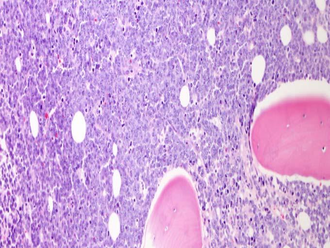
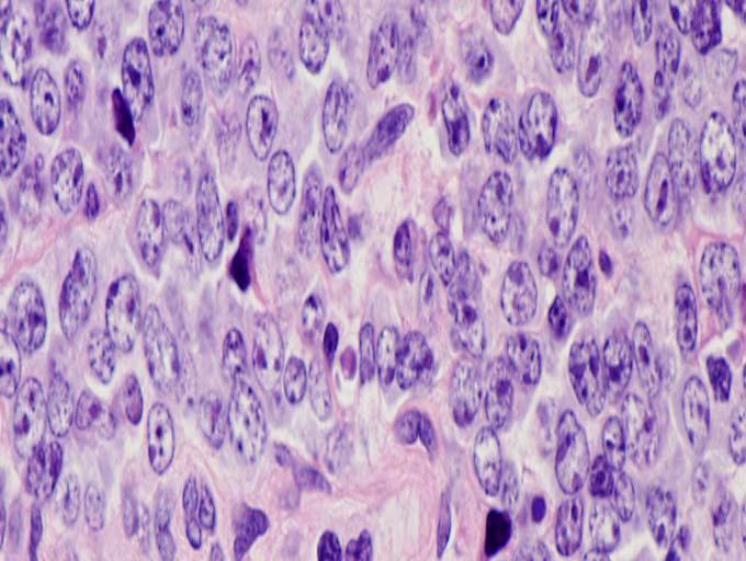
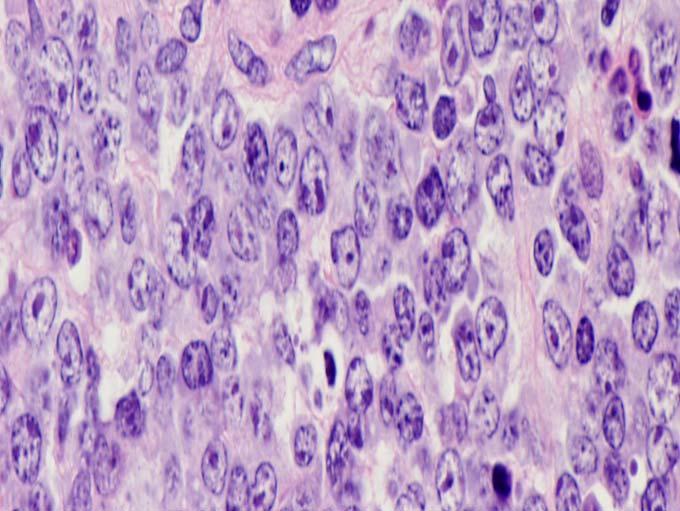
13 100 Mantle cell lymphoma Event-free, CD3-negative Event-free CD3-positive Percent survival Percent survival Time in Months Overall, CD3-negative Overall, CD3-positive Time in Months 30-40% CD3 + 8% all sites 11% discordant between sites Pertinent clinical differences Bulky disease (13% vs 39%) Splenomegaly (73% vs 4%) 4 year EFS: 47% vs 18%, p=0.0 4 year OS: 76% vs 5%, p=0.05 Multivariate Cox regression CD3, LDH, and HSCT Hazards ratio (EFS) CD3: 0.31, p=0.06 LDH: 0.5, p=0.18 HSCT: 0.99, p=0.99 Keleman et al, AJCP, 008 Patient #: Case History 41 year old male with a rectal mass The patient has a history of HIV and recent left lower quadrant pain Laboratory values WBC 1,500/uL (low) Hemoglobin 8.7 g/dl (low) Platelets 3,000/uL (low) Bone marrow aspirate Patient # 04 + cells Forward Scatter Kappa Lambda Normal cells BM 10 #1 4 # CD10 # Gate 5 # PE C 7 A CD0 Kappa Lambda Kappa Lambda 11


14 Normal bone marrow Bone marrow Bone marrow 1
15 Burkitt Lymphoma Rapidly proliferating B cell lymphoma Frequently associated with translocation of MYC Three types of Burkitt lymphoma Endemic Children ages 4-7, predominantly males Equatorial Africa Most are associated with EBV Sporadic Mainly in children and young adults Low incidence world-wide Immunodeficiency-associated Particularly associated with HIV Often occurs before CD4+ counts are reduced Burkitt Lymphoma Diagnosis Clinical features, morphology, immunophenotype, and genetics Clinical features Extranodal sites often involved Endemic form jaw and orbital lesions Majority of patients present at an advanced stage Highly aggressive, but can be cured Characteristic morphology Medium-sized cells, basophilic cytoplasm, small round nuclei Starry sky pattern in solid tissue with admixed tingible body macrophages Frequent mitoses Genetics t(8;14)(q4;q3) found in a majority of cases (c-myc and IgH fusion) CD10+ differential CD10+, sig+, clonal/monotypic B cell population Follic lar Follicular Large cell lymphoma Burkitt s Rare immature acute B cell lymphoblastic leukemia 13
16 Patient #3: Case History 79 year old female with an incidental finding of lymphadenopathy on imaging In good health and asymptomatic Laboratory values WBC 7,000/uL Hemoglobin 13. g/dl Platelets 30,000/uL Lymph node Patient #3 Forward Scatter CD5 Kappa Kappa CD10 Lambda Lambda Patient #3 CD0 CD5 CD0 CD10 CD10 Kappa Side Lambda Scatter CD79b CD3 14
17 Patient #3 Two monotypic (surface Kappa) + populations CD5+, CD10- CD3+ FMC7-, dim CD79b+ Dimmer CD0+ CD10+, CD5- Brighter CD0+ Increased scattered light intensity FMC7+, CD79b+ Lymph node Patient #4: Case History 68 year old male with skin lesions Multiple erythematous plaque lesions on thighs and upper extremities. No other symptoms. Laboratory values WBC 6700/uL Hemoglobin 14.5 g/dl Platelets 0,000/uL Peripheral blood 15


18 0 0 10/8/011 Patient # CD4 CD8 CD CD5 CD CD6 Peripheral blood Peripheral blood 16
19 Mycosis Fungoides/Sezary Syndrome: Pan T cell antigen loss/alteration Mycosis Fungoides (MF) Most common CTCL (~50% of primary cutaneous lymphomas) Epidermotropic clonal T cell malignancy Skin lesions (patches, plaques, etc) Cells with characteristic cerebriform nuclei Primarily older adults Slight male predominance (1.5 to :1) Indolent 85-90% 5 year survival Sezary syndrome Many common pathology/immunophenotypic features with MF Disease of adults Skin lesions and generalized lymphadenopathy Malignant cells in skin, lymph node, and peripheral blood Refer to Blood 105:3768, 005 for diagnostic criteria Aggressive disease ~5% 5 year survival Mycosis Fungoides/Sezary Syndrome Immunophenotype Mature T cell malignancy (surface +) Most frequently, T helper immunophenotype +CD4+ Characteristically, CD7- May be partial Can be CD7+ Typically, CD+, CD5+ But deletion can be seen Altered expression of any of the pan T cell antigens can be seen Memory cell immunphenotype (CD45RO+, CD9+, etc) Most frequently, CD5- CD5+ in ~10-0% of cases CD7 Modulation in Reactive T cells Reactive T cells can modulate pan T cell antigen expression Most temporally short CD7 can be significant and persistent Dim CD7 to CD7- reactive T cells can be CD4+ CD5+, CD6+ (activated immunophenotype) Overlaps with MF/Sezary and ATLL immunophenotype Pattern of CD7 staining can be helpful 17

 Reported to be useful")


20 CD7 Modulation in Reactive T cells CD4 CD7 CD8 Mycosis Fungoides/Sezary Syndrome CD6 T cell activation antigen Membrane and secreted protein Modulates chemokine activity Co-stimulatory molecule (both and CD pathways) Reported to be useful differentiating Sezary cells from reactive T cells Jones et al, Am J Clin Path 115: 885, 001 Bernengo et al, Br J Dermatol 144(#1):15, 001 Circulating Sezary cells frequently CD6- Reactive Dim CD7 to CD7- cells which would be CD6+ Mycosis Fungoides/Sezary Syndrome CD7 CD6 CD7 CD6 18
21 Mycosis Fungoides/Sezary Syndrome How useful? % % 73% 69% 43% CD6 expression (%) CD4/CD8 >10 >5% convoluted lymphocytes >1000/mm 3 convoluted lymphocytes Positive TCR gene rearrangement Overall, ~64% of cases show CD4+CD6- Keleman et al, AJCP, in press, 007 Lymphoblastic Lymphoma/Leukemia 14 year old male Shortness of breath and cough Chest discomfort Chest x-ray revealed a large mediastinal mass and left pleural effusion CBC: normal Received mediastinal mass biopsy Lymphoblastic Lymphoma/Leukemia CD4 CD4 CD CD8 CD1 CD5 CD5 CD5 TdT s c 19


22 Acute Lymphoblastic Leukemia Bone marrow aspirate Bone marrow core biopsy Lymphoblastic Lymphoma/Leukemia Proliferation of malignant blast cells T-LBL and T-ALL same disease T-LBL ~80-85% of lymphoblastic lymphomas Primarily, young males Most frequently presents with mediastinal mass Can involve lymph node, spleen, skin, liver Pleural effusions common Peripheral blood/bone marrow (if extensive T- ALL) Lymphoblastic Lymphoma/Leukemia Immunophenotype Most frequent is common thymic T cell immunophenotype s-, c+, CD+, CD5+, CD7+ Less often dim s or s+ staining can be seen CD4+/CD8+, CD1a+ TdT+, 4- Frequently, CD10+ Loss or modulation of CD, CD5, or CD7 can be seen Aberrant expression of CD13 and/or 3 can be seen CD79a rarely Although rare, more immature immunphenotypes can be seen 0
23 Patient #5: Case History 60 year old male with incidental finding of a mediastinal mass on pre-op evaluation Asymptomatic Laboratory values WBC 3500/uL Hemoglobin 14. g/dl Platelets 9,000/uL Patient #5 Forward Scatter CD7 Gate 1 Surface Surface CD5 CD4 Cytoplasmic CD5 CD CD1a CD8 Mediastinal mass 1
 can overlap with normal immature")


24 Mediastinal mass Cytokeratin immunohistochemistry Lymphoblastic Lymphoma/Leukemia Frequent differential in patient with mediastinal mass Thymoma vs T-LBL/T-ALL Immunophenotype of T-LBL/T-ALL (s-, c+, CD+, CD5+, CD7+, CD4+/CD8+, CD1a+, TdT+) can overlap with normal immature thymic T cells T cells differentiate in the thymus Normal thymus mixture of T and non-hematopoietic cells Immature, common thymocyte T cells More mature, medullary thymocytes Mature T cells exiting to the periphery Lymphoblastic Lymphoma/Leukemia Immature Thymus Peripheral 4 CD CD CD CD5 TdT CD CD7 CD5 CD1 TdT CD7 CD5 CD7 CD5 CD7 CD4/CD8 CD4 or CD8 CD4 or CD8 Common Thymocyte Medullary Thymocyte Normal is a process of differentiation Common and medullary populations Transition immunphenotypes as well Pattern of staining can be helpful in differentiating normal vs abnormal


 Hemoglobin 8.")
25 Pattern of expression s CD4 CD1a CD8 Pattern of expression Normal BM Archetypical B lymphoblastic leukemia CD10 CD10 CD0 CD0 Patient #6: Case History 7 year old female with diffuse lymphadenopathy and hepatosplenomegaly Anorexia, fatigue, dyspnea, and fevers Laboratory values WBC 13,100/uL (high) Hemoglobin 8.4 g/dl (low) Platelets 138,000/uL Lymph node 3


26 Patient #6 4 Forward Scatter 8 6 CD45 % 77% 0 CD T cells 81% T cells CD4 18% CD5 CD CD8 Patient # CD10 CD All T CD4 0 0 CD10 CD8 Lymph node 4

 Denies passing out H/O anemia (refused w/u, takes Fe) Fatigue -3 wks, Myalgias,")
27 Lymph node Patient #7: Case History 81 y.o. female Found on floor at home, too weak to walk/stand (spot easy access to bathroom) Denies passing out H/O anemia (refused w/u, takes Fe) Fatigue -3 wks, Myalgias, chills, fever (100.4F), drenching night sweats No appetite or regular meals for ~10 days Patient #7: Case History cont. Treated with antibiotics, mild improvement Thought she had the flu Labs WBC: 33.1 (~11.0 a few months prior), HGB: 8., HCT: 5., MCV: 77, RDW: 19.7, PLT:117 BP 97/5, T 98.6, H 103, RR 16 PE: Alert, no sign of leukostasis Heme/Onc fellow evaluation Blood smear: mature appearing lymphocytes No hemolysis Peripheral blood sample received by flow lab 5


28 Patient #7 Forward Scatter F CD Forward Scatter Lymphoblast CD45 Myeloblast Immature Granulocyte


29 Patient #7 Forward Scatter CD CD CD CD CD7 Patient #7 5 5 CD CD CD CD CD CD13 3 Patient #7 CD45 MPO Cytoplasmic CD79a TdT Cytoplasmic CD CD7 CD5 CD64 7
 Immature antigens 4+, CD117+ TdT- Myeloid associated antigens Myeloperoxidase+, CD13+, 3+ CD64-,")

30 Patient #7 ~97% dim CD45, low to intermediate SS intensity cells (<0.1% B cells) Immature antigens 4+, CD117+ TdT- Myeloid associated antigens Myeloperoxidase+, CD13+, 3+ CD64-, CD11b- T cell associated antigens Dim aberrant CD7 and CD5 - (cytoplasmic and surface), CD- B cell associated antigens -, CD0-, CD79a- Other HLADR+ Myeloperoxidase Patient #7: Follow-up Follow up Diagnosis AML Not treated per family decision Transferred to Hospice 8
31 AML Flow Cytometric Analysis AML Lineage Sub-classification No Uitility Correlations Aberrant antigen expression CD7: ~30 to 35% CD: ~0% CD5: rarer : ~0% TdT: ~30% Helpful in confirming abnormal Summary Analysis of a panel of antigens Patterns of staining with multiple antigens Recognition of normal and abnormal Account for all cells + and antigens for all populations Recognition of abnormal (normal) is subjective and pattern recognition driven Experience needed +/-, intensity, scattered light characteristics Multiple gating/data presentation approaches required Technical issues including Dim staining vs antigen expression Lack of staining vs lack of antigen (epitope alteration) Careful correlation with morphology, pathology, molecular, cytogenetics,. Flow cytometry: Northwestern Memorial Clinical Flow Cytometry Laboratory Laura Marzalek Adella Khong Janet McLaughlin Keisha Hughes Carolina Ostiguinin Maybelle Tiongson Hematopathology Team LoAnn Peterson Beverly Nelson Diana Variakojis Amy Chadburn Yihua Chen Bill Karpus 9
32 Flow cytometry: Basic analysis of hematopoietic malignancies Discussion i and Questions 30
T CELL LYMPHOMA ANALYSIS
 T CELL LYMPHOMA ANALYSIS Charles Goolsby, Ph.D. Floyd E. Patterson Research Professor of Pathology Northwestern Feinberg School of Medicine c-goolsby@northwestern.edu 1 T CELL LYMPHOMA ANALYSIS Diverse
81 Stump the Stars 2011: Problem Cases in Hematopathology. Steven Kroft MD Russell Brynes MD LoAnn Peterson MD Eric Hsi MD Kaaren Reichard MD
 81 Stump the Stars 2011: Problem Cases in Hematopathology Steven Kroft MD Russell Brynes MD LoAnn Peterson MD Eric Hsi MD Kaaren Reichard MD 2011 Annual Meeting Las Vegas, NV AMERICAN SOCIETY FOR CLINICAL
7 Omar Abu Reesh. Dr. Ahmad Mansour Dr. Ahmad Mansour
 7 Omar Abu Reesh Dr. Ahmad Mansour Dr. Ahmad Mansour -Leukemia: neoplastic leukocytes circulating in the peripheral bloodstream. -Lymphoma: a neoplastic process in the lymph nodes, spleen or other lymphatic
Differential diagnosis of hematolymphoid tumors composed of medium-sized cells. Brian Skinnider B.C. Cancer Agency, Vancouver General Hospital
 Differential diagnosis of hematolymphoid tumors composed of medium-sized cells Brian Skinnider B.C. Cancer Agency, Vancouver General Hospital Lymphoma classification Lymphoma diagnosis starts with morphologic
Non-Hodgkin lymphomas (NHLs) Hodgkin lymphoma )HL)
 Hodgkin lymphoma )HL)") Non-Hodgkin lymphomas (NHLs) Hodgkin lymphoma )HL) Lymphoid Neoplasms: 1- non-hodgkin lymphomas (NHLs) 2- Hodgkin lymphoma 3- plasma cell neoplasms Non-Hodgkin lymphomas (NHLs) Acute Lymphoblastic Leukemia/Lymphoma
Non-Hodgkin lymphomas (NHLs) Hodgkin lymphoma )HL) Lymphoid Neoplasms: 1- non-hodgkin lymphomas (NHLs) 2- Hodgkin lymphoma 3- plasma cell neoplasms Non-Hodgkin lymphomas (NHLs) Acute Lymphoblastic Leukemia/Lymphoma
The spectrum of flow cytometry of the bone marrow
 The spectrum of flow cytometry of the bone marrow Anna Porwit Lund University Faculty of Medicine Dept. of Clinical Sciences Div. Oncology and Pathology anna.porwit@med.lu.se Disclosure of speaker s interests
78 The Power of Peripheral Blood Smears-Apparent Diagnostic Clues (Part 1) Gene Gulati PhD, SH(ASCP)
 Gene Gulati PhD, SH(ASCP)") 78 The Power of Peripheral Blood Smears-Apparent Diagnostic Clues (Part 1) Gene Gulati PhD, SH(ASCP) 2011 Annual Meeting Las Vegas, NV AMERICAN SOCIETY FOR CLINICAL PATHOLOGY 33 W. Monroe, Ste. 1600 Chicago,
78 The Power of Peripheral Blood Smears-Apparent Diagnostic Clues (Part 1) Gene Gulati PhD, SH(ASCP) 2011 Annual Meeting Las Vegas, NV AMERICAN SOCIETY FOR CLINICAL PATHOLOGY 33 W. Monroe, Ste. 1600 Chicago,
FLOW CYTOMETRY PRINCIPLES AND PRACTICE. Toby Eyre Consultant Haematologist Oxford University Hospitals NHS Foundation Trust June 2018
 FLOW CYTOMETRY PRINCIPLES AND PRACTICE Toby Eyre Consultant Haematologist Oxford University Hospitals NHS Foundation Trust June 2018 Aims and Objectives Principles of flow cytometry Preparation Steps involved
Lymphoma/CLL 101: Know your Subtype. Dr. David Macdonald Hematologist, The Ottawa Hospital
 Lymphoma/CLL 101: Know your Subtype Dr. David Macdonald Hematologist, The Ottawa Hospital Function of the Lymph System Lymph Node Lymphocytes B-cells develop in the bone marrow and influence the immune
Flow cytometric analysis of B-cell lymphoproliferative disorders
 Flow cytometric analysis of B-cell lymphoproliferative disorders David M. Dorfman, M.D., Ph.D. Department of Pathology Brigham and Women s Hospital and Harvard Medical School Boston, MA Objectives Review
Burkitt lymphoma. Sporadic Endemic in Africa associated with EBV Translocations involving MYC gene on chromosome 8
 Heme 8 Burkitt lymphoma Sporadic Endemic in Africa associated with EBV Translocations involving MYC gene on chromosome 8 Most common is t(8;14) Believed to be the fastest growing tumor in humans!!!! Morphology
The patient had a mild splenomegaly but no obvious lymph node enlargement. The consensus phenotype obtained from part one of the exercise was:
 Case History An 86 year old male was admitted to hospital with chest infection. Haematological examination subsequently revealed the following: Hb- 11.0 g/dl; WBC- 67.1 x 10^9/l; PLT- 99 x10^9/l; RBC-
Hematopathology Specialty Conference Case #1
 Hematopathology Specialty Conference Case #1 Robert (Bob) Ohgami, MD, PhD Assistant Professor Stanford University Disclosure of Relevant Financial Relationships Disclosure of Relevant Financial Relationships
ACCME/Disclosures 4/13/2016. Clinical History
 ACCME/Disclosures The USCAP requires that anyone in a position to influence or control the content of CME disclose any relevant financial relationship WITH COMMERCIAL INTERESTS which they or their spouse/partner
ADx Bone Marrow Report. Patient Information Referring Physician Specimen Information
 ADx Bone Marrow Report Patient Information Referring Physician Specimen Information Patient Name: Specimen: Bone Marrow Site: Left iliac Physician: Accession #: ID#: Reported: 08/19/2014 - CHRONIC MYELOGENOUS
HENATOLYMPHOID SYSTEM THIRD YEAR MEDICAL STUDENTS- UNIVERSITY OF JORDAN AHMAD T. MANSOUR, MD. Parts 2 and 3
 HENATOLYMPHOID SYSTEM THIRD YEAR MEDICAL STUDENTS- UNIVERSITY OF JORDAN AHMAD T. MANSOUR, MD Parts 2 and 3 NEOPLASTIC LYMPHOID DISEASES Introduction o The bone marrow is the source of all cells in the
Lymphoma: What You Need to Know. Richard van der Jagt MD, FRCPC
 Lymphoma: What You Need to Know Richard van der Jagt MD, FRCPC Overview Concepts, classification, biology Epidemiology Clinical presentation Diagnosis Staging Three important types of lymphoma Conceptualizing
5000 International Clinical Cytometry Society: Practical Flow Cytometry in Hematopathology A Case-Based Approach
 5000 International Clinical Cytometry Society: Practical Flow Cytometry in Hematopathology A Case-Based Approach Joseph A DiGiuseppe, MD, PhD Hartford Hospital Disclosures In the past 12 months, I have
88-year-old Female with Lymphadenopathy. Faizi Ali, MD
 88-year-old Female with Lymphadenopathy Faizi Ali, MD Clinical History A 88-year-old caucasian female presented to our hospital with the complaints of nausea, vomiting,diarrhea, shortness of breath and
Beyond the CBC Report: Extended Laboratory Testing in the Evaluation for Hematologic Neoplasia Disclosure
 Beyond the CBC Report: Extended Laboratory Testing in the Evaluation for Hematologic Neoplasia Disclosure I am receiving an honorarium from Sysmex for today s presentation. 1 Determining the Etiology for
Case Presentation No. 075
 Case Presentation No. 075 Session 4. Myelodysplastic Syndrome Cristina Montalvo, MD Baylor College of Medicine Houston, Texas 2007 Workshop of Society for Hematopathology and European Association for Haematopathology
Successful flow cytometric immunophenotyping of body fluid specimens
 Successful flow cytometric immunophenotyping of body fluid specimens Fiona E. Craig, MD Division of Hematopathology Mayo Clinic Arizona 2017 MFMER slide-1 Financial disclosure No conflicts 2017 MFMER slide-2
WBCs Disorders 1. Dr. Nabila Hamdi MD, PhD
 WBCs Disorders 1 Dr. Nabila Hamdi MD, PhD ILOs Compare and contrast ALL, AML, CLL, CML in terms of age distribution, cytogenetics, morphology, immunophenotyping, laboratory diagnosis clinical features
Classification of Hematologic Malignancies. Patricia Aoun MD MPH
 Classification of Hematologic Malignancies Patricia Aoun MD MPH Objectives Know the basic principles of the current classification system for hematopoietic and lymphoid malignancies Understand the differences
Large cell immunoblastic Diffuse histiocytic (DHL) Lymphoblastic lymphoma Diffuse lymphoblastic Small non cleaved cell Burkitt s Non- Burkitt s
 Lymphoblastic lymphoma Diffuse lymphoblastic Small non cleaved cell Burkitt s Non- Burkitt s") Non Hodgkin s Lymphoma Introduction 6th most common cause of cancer death in United States. Increasing in incidence and mortality. Since 1970, the incidence of has almost doubled. Overview The types of
Non Hodgkin s Lymphoma Introduction 6th most common cause of cancer death in United States. Increasing in incidence and mortality. Since 1970, the incidence of has almost doubled. Overview The types of
WHO Classification. B-cell chronic lymphocytic leukemia/small T-cell granular lymphocytic leukemia
 Blood Malignancies-II Prof. Dr. Herman Hariman, a Ph.D, SpPK (KH). Prof. Dr. Adikoesoema Aman, SpPK (KH) Dept. of Clinical Pathology, School of Medicine, University of North Sumatra WHO classification
2007 Workshop of Society for Hematopathology & European Association for Hematopathology Indianapolis, IN, USA Case # 228
 2007 Workshop of Society for Hematopathology & European Association for Hematopathology Indianapolis, IN, USA Case # 228 Vishnu V. B Reddy, MD University of Alabama at Birmingham Birmingham, AL USA 11/03/07
2013 AAIM Pathology Workshop
 2013 AAIM Pathology Workshop John Schmieg, M.D., Ph.D. None Disclosures 1 Pathology Workshop Objectives Define the general philosophy of reviewing pathology reports Review the various components of Bone
Lymphoma and Pseudolymphoma
 Lymphoma and Pseudolymphoma Laura B. Pincus, MD Co-Director, Cutaneous Lymphoma Clinic Associate Professor Dermatology and Pathology University of California, San Francisco I HAVE NO RELEVANT RELATIONSHIPS
Lymphoma: The Basics. Dr. Douglas Stewart
 Lymphoma: The Basics Dr. Douglas Stewart Objectives What is lymphoma? How common is it? Why does it occur? How do you diagnose it? How do you manage it? How do you follow patients after treatment? What
LEUKAEMIA and LYMPHOMA. Dr Mubarak Abdelrahman Assistant Professor Jazan University
 LEUKAEMIA and LYMPHOMA Dr Mubarak Abdelrahman Assistant Professor Jazan University OBJECTIVES Identify etiology and epidemiology for leukemia and lymphoma. Discuss common types of leukemia. Distinguish
Immunopathology of Lymphoma
 Immunopathology of Lymphoma Noraidah Masir MBBCh, M.Med (Pathology), D.Phil. Department of Pathology Faculty of Medicine Universiti Kebangsaan Malaysia Lymphoma classification has been challenging to pathologists.
Integrated Hematopathology. Morphology and FCI with IHC
 Integrated Hematopathology Morphology and FCI with IHC FrontMatter.indd i 9/6/2009 9:30:12 PM FrontMatter.indd ii 9/6/2009 9:30:18 PM Integrated Hematopathology Morphology and FCI with IHC Cherie H Dunphy,
Lymphoid Neoplasms. Sylvie Freeman Department of Clinical Immunology, University of Birmingham
 Lymphoid Neoplasms Sylvie Freeman Department of Clinical Immunology, University of Birmingham Incidence of Haematological Malignancies UK2001 (CRUK) Malignancy New Cases All Cancers 271,000 Leukaemia 6,760
WBCs Disorders. Dr. Nabila Hamdi MD, PhD
 WBCs Disorders Dr. Nabila Hamdi MD, PhD ILOs Compare and contrast ALL, AML, CLL, CML in terms of age distribution, cytogenetics, morphology, immunophenotyping, laboratory diagnosis clinical features and
Patterns of Lymphoid Neoplasia in Peripheral Blood. Leon F. Baltrucki, M.D. Leon F. Baltrucki, M.D. Disclosure
 Patterns of Lymphoid Neoplasia in Peripheral Blood Leon F. Baltrucki, M.D. Leon F. Baltrucki, M.D. Disclosure Dr Baltrucki has received an honorarium for his participation as a faculty presenter in this
V. Acute leukemia. Flow cytometry in evaluation of hematopoietic neoplasms: A case-based approach
 V. Acute leukemia Evaluating a sample for an acute leukemia Acute leukemia is a neoplasm of immature myeloid or lymphoid cells characterized by a block in maturation, usually at the stage of an early progenitor
Leukocytosis - Some Learning Points
 Leukocytosis - Some Learning Points Koh Liang Piu Department of Hematology-Oncology National University Cancer Institute National University Health System Objectives of this talk: 1. To provide some useful
Test Utilization: Chronic Lymphocytic Leukemia
 Test Utilization: Chronic Lymphocytic Leukemia Initial Evaluation Diagnostic Criteria Selection of Tests for Prognosis Response to Therapy Challenges Assessment for persistent disease Paul J. Kurtin, M.D.
Mast Cell Disease Case 054 Session 7
 Mast Cell Disease Case 054 Session 7 Rodney R. Miles, M.D., Ph.D. Lauren B. Smith, M.D. Cem Akin, M.D. Diane Roulston,, Ph.D. Charles W. Ross, M.D. Departments of Pathology and Internal Medicine University
Case 3. Ann T. Moriarty,MD
 Case 3 Ann T. Moriarty,MD Case 3 59 year old male with asymptomatic cervical lymphadenopathy. These images are from a fine needle biopsy of a left cervical lymph node. Image 1 Papanicolaou Stained smear,100x.
Bone Marrow. Procedures Blood Film Aspirate, Cell Block Trephine Biopsy, Touch Imprint
 Bone Marrow Protocol applies to acute leukemias, myelodysplastic syndromes, myeloproliferative disorders, chronic lymphoproliferative disorders, malignant lymphomas, plasma cell dyscrasias, histiocytic
Disclosures/COI. Cases in Hematopathology. Outline. Heme Path Findings Not to Miss. Normal Peripheral Smear 6/30/2016
 Disclosures/COI Cases in Hematopathology Vamsi Kota Assistant Professor Department of Hematology & Medical Oncology Leukemia/BMT I have no disclosures or conflicts of interest regarding this presentation.
Pathology #07. Hussein Al-Sa di. Dr. Sohaib Al-Khatib. Mature B-Cell Neoplasm. 0 P a g e
 Pathology #07 Mature B-Cell Neoplasm Hussein Al-Sa di Dr. Sohaib Al-Khatib 0 P a g e Thursday 18/2/2016 Our lecture today (with the next 2 lectures) will be about lymphoid tumors This is a little bit long
Chronic Lymphocytic Leukemia FISH Panel. Impact on Diagnosis
 Hematopathology / CLL, FISH, AND 14Q32 TRANSLOCATIONS Chronic Lymphocytic Leukemia FISH Panel Impact on Diagnosis Beverly P. Nelson, MD, 1 Rohit Gupta, MD, 1 Gordon W. Dewald, PhD, 2 Sarah F. Paternoster,
Low grade High grade , immune suppression chronic persistent inflammation viruses B-symptoms
 We've one category for lymphoid neoplasm which is the lymphoma in contrast to that of myeloid which has three categories; acute myeloid leukemias, myeloproliferative & myelodysplastic disorders. Lymphoma
DETERMINATION OF A LYMPHOID PROCESS
 Chapter 2 Applications of Touch Preparation Cytology to Intraoperative Consultations: Lymph Nodes and Extranodal Tissues for Evaluation of Hematolymphoid Disorders INTRODUCTION As discussed in Chap. 1,
Welcome. Welcome. Emerging Technologies in Flow Cytometry
 Emerging Technologies in Flow Cytometry Dr. William Dittman December 11, 2012 You may download a copy of the handout by clicking on the handout icon, located in the upper right hand corner of your screen
Follicular Lymphoma: the WHO
 Follicular Lymphoma: the WHO and the WHERE? Yuri Fedoriw, MD Associate Professor of Pathology and Laboratory Medicine Director of Hematopathology University of North Carolina Chapel Hill, NC Disclosure
Lymphatic system component
 Introduction Lymphatic system component Statistics Overview Lymphoma Non Hodgkin s Lymphoma Non- Hodgkin's is a type of cancer that originates in the lymphatic system. It is estimated to be the sixth most
Persistent lymphocytosis. Persistent lymphocytosis: are there prognostic indicators? Problem. Questions. Basic markers used to identify lymphocytes
 Persistent lymphocytosis Persistent lymphocytosis: are there prognostic indicators? Paul R. Avery VMD, PhD, DACVP Marjorie Williams, DVM Anne C. Avery VMD, PhD Clinical Immunology Laboratory Colorado State
NON HODGKINS LYMPHOMA: INDOLENT Updated June 2015 by Dr. Manna (PGY-5 Medical Oncology Resident, University of Calgary)
") NON HODGKINS LYMPHOMA: INDOLENT Updated June 2015 by Dr. Manna (PGY-5 Medical Oncology Resident, University of Calgary) Reviewed by Dr. Michelle Geddes (Staff Hematologist, University of Calgary) and Dr.
NON HODGKINS LYMPHOMA: INDOLENT Updated June 2015 by Dr. Manna (PGY-5 Medical Oncology Resident, University of Calgary) Reviewed by Dr. Michelle Geddes (Staff Hematologist, University of Calgary) and Dr.
Integrated Diagnostic Approach to the Classification of Myeloid Neoplasms. Daniel A. Arber, MD Stanford University
 Integrated Diagnostic Approach to the Classification of Myeloid Neoplasms Daniel A. Arber, MD Stanford University What is an integrated approach? What is an integrated approach? Incorporating all diagnostic
From Morphology to Molecular Pathology: A Practical Approach for Cytopathologists Part 1-Cytomorphology. Songlin Zhang, MD, PhD LSUHSC-Shreveport
 From Morphology to Molecular Pathology: A Practical Approach for Cytopathologists Part 1-Cytomorphology Songlin Zhang, MD, PhD LSUHSC-Shreveport I have no Conflict of Interest. FNA on Lymphoproliferative
Hepatic Lymphoma Diagnosis An Algorithmic Approach
 Hepatic Lymphoma Diagnosis An Algorithmic Approach Ryan M. Gill, M.D., Ph.D. University of California, San Francisco PLEASE TURN OFF YOUR CELL PHONES Disclosure of Relevant Financial Relationships USCAP
Lymphoma and Myeloma Kris3ne Kra4s, M.D.
 Lymphoma and Myeloma Kris3ne Kra4s, M.D. Hematologic Malignancies Leukemia Malignancy of hematopoie3c cells Starts in bone marrow, can spread to blood, nodes Myeloid or lymphoid Acute or chronic Lymphoma
Myelodysplastic Syndrome Case 158
 Myelodysplastic Syndrome Case 158 Dong Chen MD PhD Division of Hematopathology Mayo Clinic Clinical History 86 year old man Persistent borderline anemia and thrombocytopenia. His past medical history was
HEMATOPATHOLOGY SUMMARY REPORT RL;MMR;
 HEMATOPATHOLOGY SUMMARY REPORT RL;MMR; Page 1 of 1 05/15/20XX HP000000-20XX 05/21/20XX (212) 123-457 (51) 32-3455 (51) 123-457 Age: 78 DOB: 0/05/19XX SS#: 45-45-45 Clinical Information: 78 y/o female with
3/23/2017. Disclosure of Relevant Financial Relationships. Pitfalls in Immunohistochemistry in Hematopathology: CD20 and CD3 Can Let Me Down?!
 Pitfalls in Immunohistochemistry in Hematopathology: CD20 and CD3 Can Let Me Down?! Judith A. Ferry Massachusetts General Hospital Disclosure of Relevant Financial Relationships USCAP requires that all
Pathology of Hematopoietic and Lymphoid tissue
 Pathology of Hematopoietic and Lymphoid tissue Peerayut Sitthichaiyakul, M.D. Department of Pathology and Forensic Medicine Faculty of Medicine, Naresuan University CONTENTS White blood cells and lymph
GP CME. James Liang Consultant Haematologist. Created by: Date:
 GP CME James Liang Consultant Haematologist Date: Created by: Scenario 52 year old European male Fit and well Brother recently diagnosed with diabetes PMHx Nil Social Hx Ex-smoker stopped 5 years ago (20
HEMATOPATHOLOGY (SHANDS HOSPITAL AT THE UNIVERSITY OF FLORIDA): Rotation Director: Ying Li, M.D., Ph.D., Assistant Professor
: Rotation Director: Ying Li, M.D., Ph.D., Assistant Professor") HEMATOPATHOLOGY (SHANDS HOSPITAL AT THE UNIVERSITY OF FLORIDA): Rotation Director: Ying Li, M.D., Ph.D., Assistant Professor I. Description of the rotation: During this rotation, the resident will gain
HEMATOPATHOLOGY (SHANDS HOSPITAL AT THE UNIVERSITY OF FLORIDA): Rotation Director: Ying Li, M.D., Ph.D., Assistant Professor I. Description of the rotation: During this rotation, the resident will gain
Hematopathology Case Study
 www.medfusionservices.com Hematopathology Case Study CV3515-14 JUNE Clinical Presentation: Clinical Information: A 42 year old male with history of chronic myelogenous leukemia (CML) presents with an elevated
Acute myeloid leukemia. M. Kaźmierczak 2016
 Acute myeloid leukemia M. Kaźmierczak 2016 Acute myeloid leukemia Malignant clonal disorder of immature hematopoietic cells characterized by clonal proliferation of abnormal blast cells and impaired production
Instructions for Chronic Lymphocytic Leukemia Post-HSCT Data (Form 2113)
") Instructions for Chronic Lymphocytic Leukemia Post-HSCT Data (Form 2113) This section of the CIBMTR Forms Instruction Manual is intended to be a resource for completing the CLL Post-HSCT Data Form. E-mail
Instructions for Chronic Lymphocytic Leukemia Post-HSCT Data (Form 2113) This section of the CIBMTR Forms Instruction Manual is intended to be a resource for completing the CLL Post-HSCT Data Form. E-mail
Hematology 101. Rachid Baz, M.D. 5/16/2014
 Hematology 101 Rachid Baz, M.D. 5/16/2014 Florida 101 Epidemiology Estimated prevalence 8,000 individuals in U.S (compare with 80,000 MM patients) Annual age adjusted incidence 3-8/million-year 1 More
HIGH GRADE B-CELL LYMPHOMA DAVID NOLTE, MD (PGY-2) HUSSAM AL-KATEB, PHD, FACMG DEBORAH FUCHS, MD
 HUSSAM AL-KATEB, PHD, FACMG DEBORAH FUCHS, MD") HIGH GRADE B-CELL LYMPHOMA DAVID NOLTE, MD (PGY-2) HUSSAM AL-KATEB, PHD, FACMG DEBORAH FUCHS, MD OUTLINE High grade B-cell lymphoma with MYC and BCL2 and/or BCL6 rearrangements Patient presentation 2008/2016
HIGH GRADE B-CELL LYMPHOMA DAVID NOLTE, MD (PGY-2) HUSSAM AL-KATEB, PHD, FACMG DEBORAH FUCHS, MD OUTLINE High grade B-cell lymphoma with MYC and BCL2 and/or BCL6 rearrangements Patient presentation 2008/2016
Plasma cell myeloma (multiple myeloma)
") Plasma cell myeloma (multiple myeloma) Common lymphoid neoplasm, present at old age (70 years average) Remember: plasma cells are terminally differentiated B-lymphocytes that produces antibodies. B-cells
Plasma cell myeloma (multiple myeloma) Common lymphoid neoplasm, present at old age (70 years average) Remember: plasma cells are terminally differentiated B-lymphocytes that produces antibodies. B-cells
Extramedullary precursor T-lymphoblastic transformation of CML at presentation
 Extramedullary precursor T-lymphoblastic transformation of CML at presentation Neerja Vajpayee, Constance Stein, Bernard Poeisz & Robert E. Hutchison Clinical History 30 year old man presented to the emergency
PRECURSOR LYMHPOID NEOPLASMS. B lymphoblastic leukaemia/lymphoma T lymphoblastic leukaemia/lymphoma
 PRECURSOR LYMHPOID NEOPLASMS B lymphoblastic leukaemia/lymphoma T lymphoblastic leukaemia/lymphoma B lymphoblastic leukaemia/lymphoma Definition: B lymphoblastic leukaemia/lymphoma is a neoplasm of precursor
Pathology. #11 Acute Leukemias. Farah Banyhany. Dr. Sohaib Al- Khatib 23/2/16
 35 Pathology #11 Acute Leukemias Farah Banyhany Dr. Sohaib Al- Khatib 23/2/16 1 Salam First of all, this tafreegh is NOT as long as you may think. If you just focus while studying this, everything will
Aggressive B-cell Lymphoma 2013
 Aggressive B-cell Lymphoma 2013 Diffuse Large B-Cell Lymphoma Burkitt Lymphoblastic lymphoma Gray zone Intermediate DLBCL/HL Intermediate BL/DLBCL Diffuse Large B-cell lymphoma Common morphology: diffuse
CME/SAM. Mixed Phenotype Acute Leukemia
 AJCP / Original Article Mixed Phenotype Acute Leukemia A Study of 61 Cases Using World Health Organization and European Group for the Immunological Classification of Leukaemias Criteria Olga K. Weinberg,
Collected: , PM Sent: , PM Received: , PM Preliminary: , PM. Notification Status: COMPREHENSIVE DIAGNOSIS
 PATIENT Name:HIPAA, Compliant DOB: 03-25-1945 (60 yr) ID#: xxx-xx-0000 Sex: M Tel: xxx-xxx-xxxx SPECIMEN Your No:WS05-xxxx Case No:C05-00xxx Req. No:Txxxxx Collected: 06-08-05, PM Sent: 06-09-05, PM Received:
Prepared by: Dr.Mansour Al-Yazji
 C L L CLL Prepared by: Abd El-Hakeem Abd El-Rahman Abu Naser Ahmed Khamis Abu Warda Ahmed Mohammed Abu Ghaben Bassel Ziad Abu Warda Nedal Mostafa El-Nahhal Dr.Mansour Al-Yazji LEUKEMIA Leukemia is a form
SWOG ONCOLOGY RESEARCH PROFESSIONAL (ORP) MANUAL LEUKEMIA FORMS CHAPTER 16A REVISED: DECEMBER 2017
 MANUAL LEUKEMIA FORMS CHAPTER 16A REVISED: DECEMBER 2017") LEUKEMIA FORMS The guidelines and figures below are specific to Leukemia studies. The information in this manual does NOT represent a complete set of required forms for any leukemia study. Please refer
LEUKEMIA FORMS The guidelines and figures below are specific to Leukemia studies. The information in this manual does NOT represent a complete set of required forms for any leukemia study. Please refer
Peripheral blood Pleural effusion in a cat
 Tools for the Diagnosis of Lymphoproliferative Diseases When is it difficult to diagnose lymphoproliferative disease? Persistent lymphocytosis consisting of small Lymph node aspirates containing an excess
A 64yo female with tuberculous empyema not improving on treatment: A tribute and farewell to Dr. Alphonse Kayembe
 A 64yo female with tuberculous empyema not improving on treatment: A tribute and farewell to Dr. Alphonse Kayembe Continuing Medical Education Announcement Harvard Medical School RSS 3081: Monthly BOTSOGO
Case year old male with abdominal lymphadenopathy Treated with 8 cycles of R-CHOP One year later B-symptoms and progressive disease
 Codirectors Tsieh Sun, M.D., FASCP Francisco Vega, M.D., Ph.D. Department of Hematopathology UT MD Anderson Cancer Center Houston Texas There is no conflict of interest involved in the content and presentation
Hematopathology Case Study
 Hematopathology Case Study AMP Outreach Course 2009 AMP Annual Meeting John Greg Howe Ph.D. Department of Laboratory Medicine Yale University School of Medicine November 19, 2009 HISTORY Case History An
Morphology Case Study. Presented by Niamh O Donnell, BSc, MSc. Medical Scientist Haematology Laboratory Cork University Hospital
 Morphology Case Study Presented by Niamh O Donnell, BSc, MSc. Medical Scientist Haematology Laboratory Cork University Hospital 41 year old male presented to GP for routine check-up in May 2011. FBC Results:
MECHANISMS OF HUMAN DISEASE: LABORATORY SESSIONS LYMPHOMA. April 16, 2008
 MECHANISMS OF HUMAN DISEASE: LABORATORY SESSIONS LYMPHOMA April 16, 2008 FACULTY COPY GOAL: Learn the appearance of normal peripheral blood elements and lymph nodes. Recognize abnormal peripheral blood
LYMPHOMAS an overview of some subtypes of NHLs
 One of the confusing aspects of the lymphoid neoplasms concerns the use of the descriptive terms "leukemia" and "lymphoma." LYMPHOMAS an overview of some subtypes of NHLs Leukemia is used for lymphoid
Blood Cell Identification: 2011-B Mailing: Acute Myeloid Leukemia (AML)
") Please Note: To view the Figures and Images contained within this education activity in color, access the electronic version of the reading. CASE HISTORY This peripheral blood smear is from a 51-year-old
Please Note: To view the Figures and Images contained within this education activity in color, access the electronic version of the reading. CASE HISTORY This peripheral blood smear is from a 51-year-old
Pathology of Hematopoietic and Lymphoid tissue
 CONTENTS Pathology of Hematopoietic and Lymphoid tissue White blood cells and lymph nodes Quantitative disorder of white blood cells Reactive lymphadenopathies Infectious lymphadenitis Tumor metastasis
20/20 PATHOLOGY REPORTS
 20/20 PATHOLOGY REPORTS Improving the Physician Experience. Enhancing the Patient Experience. We are LabTest Diagnostics. LabTest Diagnostics has a full-service laboratory with state-of-the-art equipment
Small B-cell (Histologically Low Grade) Lymphoma
 Lymphoma") Frequency of Lymphoid Neoplasms Small B-cell (Histologically Low Grade) Lymphoma Stephen Hamilton-Dutoit Institute of Pathology Aarhus University Hospital B-cell neoplasms 88% Diffuse large B-cell lymphoma
Frequency of Lymphoid Neoplasms Small B-cell (Histologically Low Grade) Lymphoma Stephen Hamilton-Dutoit Institute of Pathology Aarhus University Hospital B-cell neoplasms 88% Diffuse large B-cell lymphoma
Easy Trick to Spot Leukemia for Pediatricians
 Easy Trick to Spot Leukemia for Pediatricians Piya Rujkijyanont, MD Division of Hematology-Oncology Department of Pediatrics Phramongkutklao Hospital Most Common Pediatric Cancers Age 0-14 Leukemia 32%
Classifications of lymphomas
 Classifications of lymphomas Lukes and Collins Kiel classification Working formulation REAL classification (1994) WHO classification (2000) WHO CLASSIFICATIONF OF NEOPLASMS HAEMATOPETIC AND LYMPHOID TISSUES
FOLLICULARITY in LYMPHOMA
 FOLLICULARITY in LYMPHOMA Reactive Follicular Hyperplasia Follicular Hyperplasia irregular follicles Follicular Hyperplasia dark and light zones Light Zone Dark Zone Follicular hyperplasia MIB1 Follicular
Aggressive B-Cell Lymphomas
 Aggressive B-cell Lymphomas Aggressive B-Cell Lymphomas Stephen Hamilton Dutoit Institute of Pathology Aarhus Kommunehospital B-lymphoblastic lymphoma Diffuse large cell lymphoma, NOS T-cell / histiocyte-rich;
Case Workshop of Society for Hematopathology and European Association for Haematopathology
 Case 148 2007 Workshop of Society for Hematopathology and European Association for Haematopathology Robert P Hasserjian Department of Pathology Massachusetts General Hospital Boston, MA Clinical history
CCND1-IGH Fusion-Amplification and MYC Copy Number Gain in a Case of Pleomorphic Variant Mantle Cell Lymphoma
 AJCP /CASE REPORT CCND1-IGH Fusion-Amplification and MYC Copy Number Gain in a Case of Pleomorphic Variant Mantle Cell Lymphoma Yuan Miao, MD, 1,2 Pei Lin, MD, 1 Wei Wang, MD, 1 L. Jeffrey Medeiros, MD,
Development of B and T lymphocytes
 Development of B and T lymphocytes What will we discuss today? B-cell development T-cell development B- cell development overview Stem cell In periphery Pro-B cell Pre-B cell Immature B cell Mature B cell
Non-Hodgkin lymphoma
 Non-Hodgkin lymphoma Non-Hodgkin s lymphoma Definition: - clonal tumours of mature and immature B cells, T cells or NK cells - highly heterogeneous, both histologically and clinically Non-Hodgkin lymphoma
Myelodysplastic Syndromes: Everyday Challenges and Pitfalls
 Myelodysplastic Syndromes: Everyday Challenges and Pitfalls Kathryn Foucar, MD kfoucar@salud.unm.edu Henry Moon lecture May 2007 Outline Definition Conceptual overview; pathophysiologic mechanisms Incidence,
Molecular Pathology of Lymphoma (Part 1) Rex K.H. Au-Yeung Department of Pathology, HKU
 Rex K.H. Au-Yeung Department of Pathology, HKU") Molecular Pathology of Lymphoma (Part 1) Rex K.H. Au-Yeung Department of Pathology, HKU Lecture outline Time 10:00 11:00 11:15 12:10 12:20 13:15 Content Introduction to lymphoma Review of lymphocyte biology
Molecular Pathology of Lymphoma (Part 1) Rex K.H. Au-Yeung Department of Pathology, HKU Lecture outline Time 10:00 11:00 11:15 12:10 12:20 13:15 Content Introduction to lymphoma Review of lymphocyte biology
Mixed Phenotype Acute Leukemias
 Mixed Phenotype Acute Leukemias CHEN GAO; AMY M. SANDS; JIANLAN SUN NORTH AMERICAN JOURNAL OF MEDICINE AND SCIENCE APR 2012 VOL 5 NO.2 INTRODUCTION Most cases of acute leukemia can be classified based
Overview of Cutaneous Lymphomas: Diagnosis and Staging. Lauren C. Pinter-Brown MD, FACP Health Sciences Professor of Medicine and Dermatology
 Overview of Cutaneous Lymphomas: Diagnosis and Staging Lauren C. Pinter-Brown MD, FACP Health Sciences Professor of Medicine and Dermatology Definition of Lymphoma A cancer or malignancy that comes from
CHAPTER:4 LEUKEMIA. BY Mrs. K.SHAILAJA., M. PHARM., LECTURER DEPT OF PHARMACY PRACTICE, SRM COLLEGE OF PHARMACY 8/12/2009
 LEUKEMIA CHAPTER:4 1 BY Mrs. K.SHAILAJA., M. PHARM., LECTURER DEPT OF PHARMACY PRACTICE, SRM COLLEGE OF PHARMACY Leukemia A group of malignant disorders affecting the blood and blood-forming tissues of