Hematology Essentials: A Foundation for Accurate Smear Reviews. Christine Hinz, MS, MLS(ASCP) CM
|
|
|
- Magdalen Cameron
- 6 years ago
- Views:
Transcription

1 Hematology Essentials: A Foundation for Accurate Smear Reviews Christine Hinz, MS, MLS(ASCP) CM
2 Differential Training Program Current Challenges System Wide Approach Standardization

3 Training Area

4 How does the program work? Training Material Trainer and Trainee Checklists Reference guides Actual patient slides Case Study Power Point Competency Checklist
5 Trainee Feedback I have to tell you I was dreading the diff training BUT it was AWESOME! Where I worked before I didn't have any formal diff training, you knew the basics and that was it. I love all the hand outs you provided. Things made more sense after the class.

6 Manager Feedback I was worried about the time commitment, but every employee came back saying how valuable the training was I was impressed to hear how excited my employees were about hematology after the training Employees feel they really have the tools now to provide great patient care
7 Post Training Follow up post training with Cellavision images for competency Ongoing competency assessment Adapted for smaller sites and/or affiliates
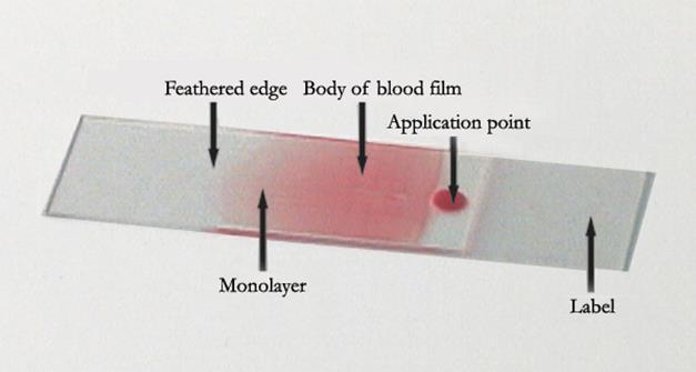
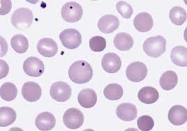
8 Proper Slide Preparation smooth, homogenous film 1/2 to 3/4 the slide length straight feather edge at least 1/4 inch examination area pink RBCs and appropriate WBC blues under gross examination (Rainbow feather edge)

9 Bad slide prep

10 Good slide prep

11 The Good and Bad
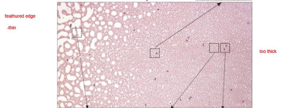
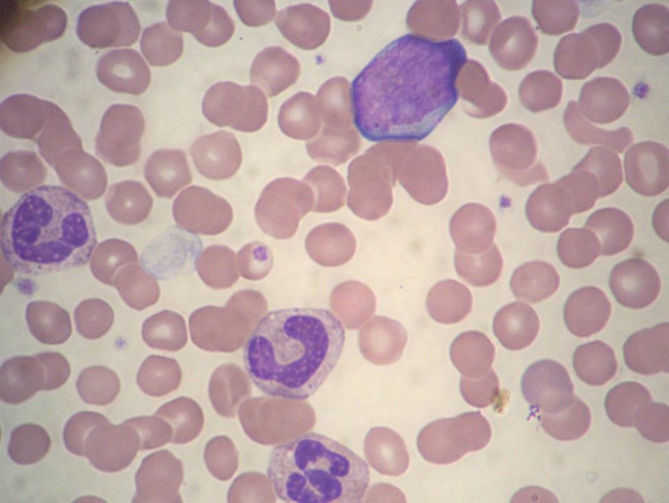
12 Starting your slide examination Examine on 10X: Check for good cell distribution, free of precipitate Examine extreme feather edge: Platelet clumps Look for abnormal cells: More dense and larger cells will be pushed to the feather edge
13 Starting your slide examination Area between extreme feather edge and Zone of Morphology is the cobblestone area. DON T do the morph or diff in this area. Zone of Morphology -area where cells evenly distributed, RBC s close but not touching. Diff and morphology should be performed here
14 Zone of morphology
15 WBC Estimate Make sure slide has been made correctly If the slide has been pushed too hard when making the slide, WBC s will be concentrated at extreme feather edge and estimate will not match instrument result.
16 WBC Estimate Estimate the white count under 10x or 40X/50x. Under low power 10X: 5 WBC's = 1,000/cumm Under 40X/50X: 1 WBC = 2,500/cumm The white count estimate may not be reported, but every manual differential white count is checked in this manner
17 Performing a manual differential In Zone of Morphology : Switch to 40x/50X or 100X to count 100 WBC cells. Note: Perception at 100x can be distorted Manual differential vs analyzer differential Must drop to 100X for RBC morphology and Platelet estimate. Platelet Estimate = (Total # of PLTs Counted in 10 Fields Using 100X ) X 15,000
18 Know appropriate morphology reporting Morphology not reported: Anisocytosis, Macrocytosis, Microcytosis, Poikilocytosis, Stomatocytes Morphology reported as present: Toxic Granulation, Dohle Bodies, Auer Rods, Hypersegmented Neutrophils, Hyposegmented Neutrophils, Vacuolated Neutrophils, Reactive Lymphocytes, Smudge Cells, Large Platelets, Agranular Platelets, Dwarf Megakaryocytes, Atypical Platelets, Basophillic Stippling, Pappenheimer Bodies, Howell Jolly Bodies, Sickle Cells, Rouleaux
19 Know appropriate morphology reporting Slight, Moderate, Marked: Hypochromasia, Polychromasia Few, Moderate, Many: Target, Acanthocytes, Echinocytes, Schistocytes, Spherocytes Platelet estimate choices: Decreased, Adequate, Increased, Clumped

20 Review Blood Maturation Chart
21 Myeloid Series-5 characteristics to look for N/C Ratio Chromatin pattern-clumped or fine Nucleoli Cytoplasm-Color of granules, inclusions Size of cell
22 Normal Slide PMN- coarse chromatin Lymph-N/C ratio 5:1 to 2:1 chromatin pattern clumped. Sky blue cytoplasm Large Lymph nucleus off center/clear cytoplasm (size determined by type of lymph, B,T,Killer) Basophil-large purple granules-see increase in reactive conditions such as MPD(myloproliferative disease.) Monocyte Eosinophil-contain bright orange-red granules evenly distributed in the cytoplasm-rarely overlie the nucleus. Band-narrowing of nucleus by 50%


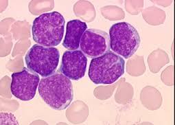
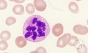
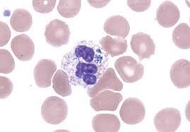
23 Neutrophil Maslak, P. ASH Image Bank 2002;2002: Copyright 2002 American Society of Hematology. Copyright restrictions may apply.

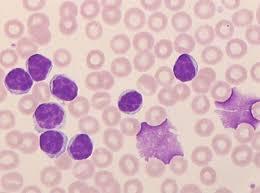
24 Lymphocyte 7-16 µm, nucleus is the size of a normal RBC, condensed chromatin, granules may be present

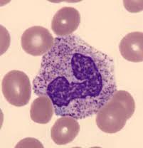
25 Monocyte µm, folded nucleus, lacy chromatin, blue-gray cytoplasm, fine granules

26 Eosinophil and Basophil µm, 2-3 lobed nucleus, prominent reddish-orange granules µm, segmented nucleus, prominent blue granules Slides courtesy of EME003.html

27 Band Neutrophil 9-15 µm, horseshoe shaped nucleus, chromatin present in any filaments
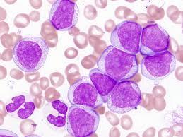
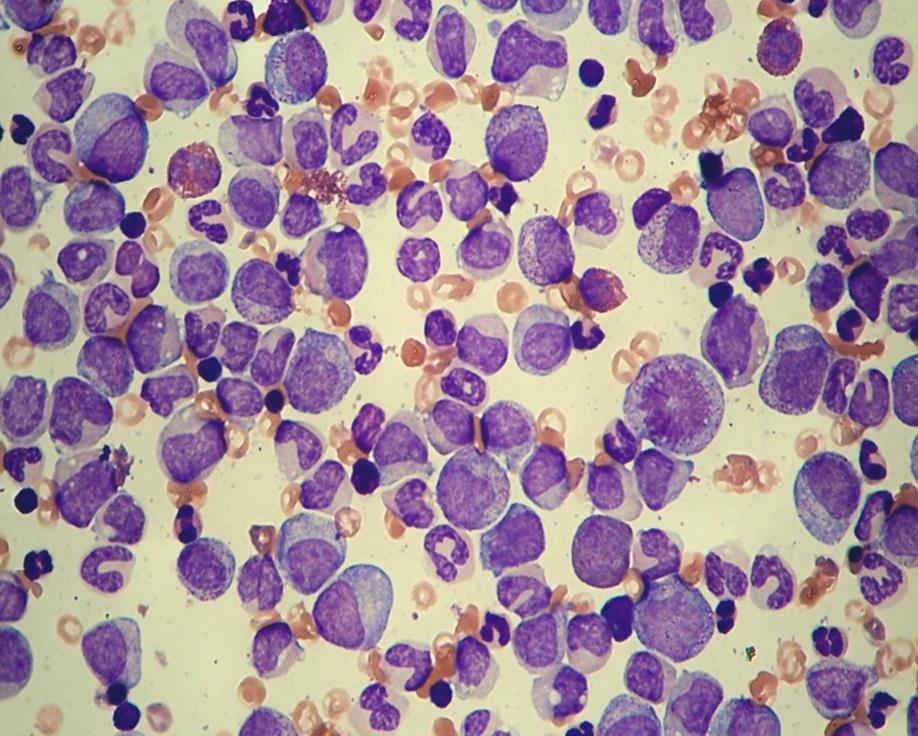
28 Chronic Myeloid Leukemia Leukemia is the uncontrollable growth of cells. Demonstrates a variety of immature cells, including blasts Basophilia and a left shift can be some of the first signs of CML Cells to be identified on slide: Myelocyte Metamylocyte-Nucleus kidney bean shaped Promyelocyte-(granules can overlap nucleus) Basophilic cytoplasm-chromatin pattern is fine 1-2 nucleoli NRBC Myeloblast-Most immature cell in the myeloid series, N/C ratio high-fine chromatin pattern, basophilic cytoplasm
29 CML
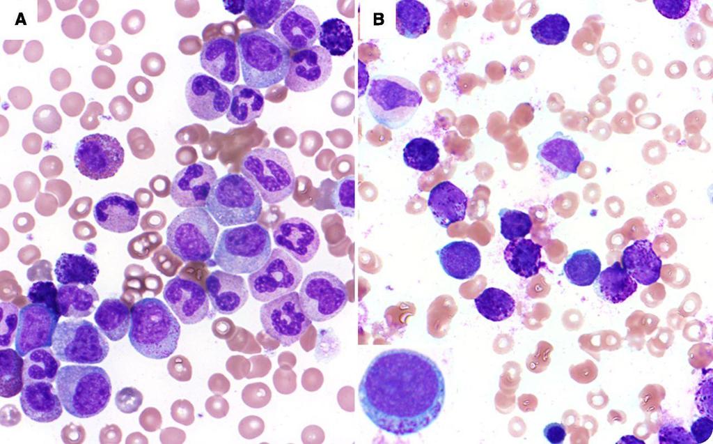
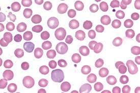
30 Acute Myeloid Leukemia Mononuclear cells seen on slide Not seeing RBC s overlapping on slide Not seeing many platelets Pancytopenia-All three cell lines are affected Don t see many neutrophils (neutropenia) Large lymphs (clear cytoplasm/offset Nucleus) Blasts: Note-If you see Auer Rods this indicates cell is in the myeloid lineage
31 Acute Myeloid Leukemia RBC morphology sometimes seen on slide: Basophilic stippling Polychromatic Elliptocytes (Ovalocytes) Teardrops NRBCs
32 AML
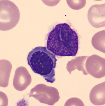
33 AML

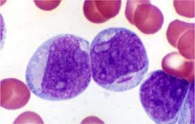
34 Blast vs Lymph
35 ALL 4yr old, cough, fatigue High WBC count, low Hgb-3.8g/dl, low Plt-20,000 Mononuclear cells with high N/C ratio, fine very fine, smooth chromatin pattern Slide full of Blasts
36 ALL
37 CLL Affects B-cell lymphocytes Typical Lymphocytosis >5.0 absolute Characteristic nucleus that looks like cracked earth or a soccer ball Cells are fragile, resulting in smudge cells present on smear Albumin slides made to reduce smudge cells, diff should be performed on albumin slide, RBC/WBC morphology should be performed on the original slide
38 CLL

39 CLL vs ALL
40 Reactive Lymphs Variability of cellular size and shape as well as nuclear size, shape and chromatin pattern Seen in many viral illnesses-infectious mononucleosis Nucleus attached to cell wall Cytoplasm surrounding RBC s Reactive lymph vs Monocyte

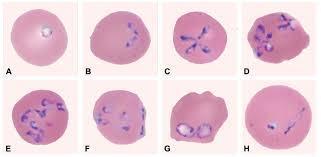
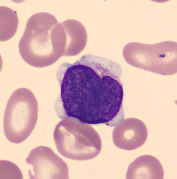
41 Reactive Lymphs

42 Reactive Lymph
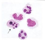
43 GCSF: Neulasta, Neupogen Used to boost WBC following chemo Toxic granulation Dohle Bodies-sometimes Immature cells
44 Toxic gran, Dohle Bodies, Vacuolated Neutrophils Toxic Granulation-Large, purple or dark blue azurophilic granules, resembling the primary granules of promyelocytes, in the cytoplasm of neutrophils, bands and metamylocytes. Seen in severe infection, chemical poisoning, and other toxic states Dohle Bodies-Appear as single or multiple light blue or gray staining area in the cytoplasm of neutrophil. RNA and represent failure of cytoplasm to mature. Seen in infections, poisoning, burns and following chemotheraphy Vacuolated Neutrophils-seen in cytoplasm of neutrophils and bands and represent the sites of phagocytosed material. Seen in association with toxic granulation
45 Toxic Granulation

46 Toxic Granules with Vacuoles

47 Toxic Granules + Dohle Bodies
48 Hypersegmented Neutrophils Neutrophil with 5 or more lobes Need to see a # of them to call Seen in megaloblastic anemia, B12/Folate deficiency Seeing macrocytosis-mcv is 130 on this patient
49 Hypersegmented Neutrophil
50 Pelger Huet Unilobed neutrophil Genetic Disorder (benign) Cells will function fine Pelger vs pseudo Pelger vs pyknotic

51 Pelger Huet vs Pyknotic

52 True hypogranular, hypolobulated neutrophils
53 Case Study Time!
54 Case Study #1 22 yr old female presents at college health services Patient complains of sore throat, fever, and swollen glands
55 Case Study #1 CBC results: Differential results: WBC 16.0 thou/cu mm Neutrophils 26 RBC 4.22 mil/cu mm Lymphocytes 63 HGB 12.8 g/dl Monocytes 10 HCT 37.5 % Eos 1 MCV 89 fl MCH 30.4 pg MCHC 34.2 % RDW 12.6 % PLT 213 thou/cu mm

56 Case Study #1

57 Case Study #1
58 Case Study #1 Manual Differential reveals 3+ reactive lymphs Heterophile Antibody Test confirms infectious mononucleosis diagnosis
59 Case Study #2 63 yr old female presents in ED Left lower quadrant pain, fever, chills History of diverticulitis, breast cancer Patient is quadriplegic due to the effects of polio as a child
60 Case Study #2 CBC results: Differential results: WBC thou/cu mm Neutrophils 48 RBC 4.31 mil/cu mm Lymphocytes 10 HGB 13.3 g/dl Monocytes 5 HCT 39.9 % Eos 2 MCV 93 fl Baso 3 MCH 30.9 pg Bands 14 MCHC 33.3 % Meta 7 RDW 17.1 % Myelo 11 PLT 189 thou/cu mm

61 Case Study #2
62 Case Study #2 Blast-peripheral blood Bone marrow-me slide

63 Case Study #2 Initial Hematology/Oncology consult determined increase in WBC was due to infection since Hgb and Plts were normal Next step?
64 Case Study #2 Smear was referred to pathologist Pathologist sent blood for BCR/ABL gene Specific for Chronic Myelogenous Leukemia (CML) Results are positive Second Oncology consult results in bone marrow biopsy Bone marrow confirms CML diagnosis
65 Case Study #3 Child Presented to clinic with cough and fatigue Pediatrician ordered CBC/Differential CBC results revealed the following: WBC 32,000 Hgb 3.8 g/dl Plt 19,000
66 Case Study #3 Peripheral smear review: High % mononuclear WBC s Irregular, clefted nuclei Vacuoles present Pediatrician informed of possible abnormal cells; requires confirmation by Pathologist Slide sent STAT to hospital Blasts confirmed by Pathology

67 Case Study #3

68 Case Study #3
69 Case Study #3 Pediatrician notified by Pathologist Flow Cytometry: Lymphoid B Cell ALL Cytogenetics t(12;21) Prognosis: favorable 5-year overall survival rate for childhood ALL 89% Treatment: Induction/Consolidation
70 Case Study #4 Pre-op for total knee replacement Routine labs included urinalysis, BMP, and CBC CBC revealed low platelet count =86 Slide reviewed No abnormalities revealed Next day platelet count low Slide reviewed (rule, Blast flag)


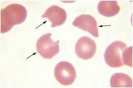
71 Case Study #4 Images Blast w/auer Rod Blast w/ prominent nucleolus
72 Case Study #4 Slide review revealed 2-3 blast type cells with possible auer rods Pathologist reviewed, contacted physician for further workup Initial slide reviewed to see if we missed anything Surgery delayed Patient had bone marrow biopsy
73 Case Study #4 Morphology Large blast cells Basophillic cytoplasm/granules Auer rods
74 Case Study #4 AML with t(8;21) Prevalence ~25% adult AMLs Prognosis: Good, 70% 5 year survival rate Treatment: Patient starts induction chemo followed by consolidation therapy
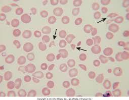
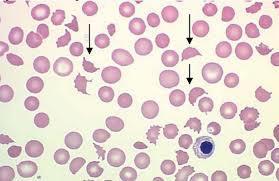
75 Break!
76 Normal Red Blood Cells Function Size Color Central Palor Note area of review Stain quality


77 Polychromasia Maslak, P. ASH Image Bank 2004;2004: Acute/chronic bleed Hemolysis Newborns Hypochromasia IDA Thalassemias Schrier, S. ASH Image Bank 2001;2001:100208
78 Hereditary Spherocytosis Spherocytes and many times, polychromasia Inherited hemolytic anemia Defect in the protein that forms the outer membrane of RBC RBC s become spherical and lose central palor Cells break down more quickly and are destroyed in spleen Bone marrow will start producing more RBC
79 Spherocytes
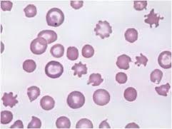
80 Hereditary Spherocytosis Same patient as previous slide after spleen removed Seeing Howell-Jolly bodies in RBC s Round, purple nuclear fragments composed of DNA Seen following splenectomy Notice not seeing polychromasia because bone marrow doesn t have to work as hard
81 Post splenectomy
82 Fragmented RBCs Marked increase in fragmented RBC (schistocytes) May be of any size or shape including helmet cells, keratocyte and other irregular, unusual shapes Look sheared or cut
83 Fragmented RBCs
84 Fragmented RBCs Clinically significant and often seen in 3 conditions Mechanical heart valve shearing RBCs Burn victims Microangiopathic anemias that includes disseminated intravascular coagulation (DIC), Hemolytic Uremic Syndrome (HUS), or Thrombotic thrombocytopenic Purpura (TTP)-these are heme emergencies. A physician and/or pathologist should be notified immediately.
85 TTP Extensive microscopic clots are formed in small blood vessels Caused primarily by autoimmune inhibition of the ADAMTS13 enzyme that cleaves Von Willebrand factor. The increase in vwf increases platelet adhesion Treatment is plasma exchange to reduce circulating antibodies and increase the ADAMTS13 enzyme
86 TTP
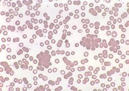
87 Acanthocytes (Spur Cells) RBC s that lack central pallor with multiple oblong projections (rounded ends) Form due to alteration in the lipid content of the RBC membrane Seen in abetalipoproteinemia (genetic and rare disease) Also seen in severe liver disease
88 Acanthocytes

89 Tear Drops Myelofibrosis Thalassemias Maslak, P. ASH Image Bank 2002;2002:100453

90 Basophillic Stippling Lazarchick, J. ASH Image Bank 2007;2007: Numerous fine or coarse granules Evenly distributed Composed of RNA Lead Poisoning Thalassemias

91 Pappenheimer Bodies Lazarchick, J. ASH Image Bank 2007;2007: Fine, irregular granules Usually in clusters Composed of Iron Splenectomy Hemoglobinopathies Hemolytic anemia Sideroblastic anemia
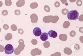
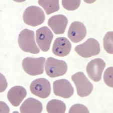
92 Sickle Cell RBC s appearing in the shape of a sickle with two pointed ends Can also appear as crescent-shaped, boat shaped and lack central pallor Also see many target cells on this slide

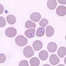
93 Sickle cell

94 Rouleaux RBCs stack like coins Due to increased protein concentration Multiple Myeloma Blue slide Maslak, P. ASH Image Bank 2004;2004:101153
95 RBC Agglutination See clumps of RBC s Caused by cold agglutinins
96 RBC Agglutination
97 Plasma Cell Leukemia Note Rouleaux (as compared to agglutination) Plasma cells have eccentric nucleus, clockface nuclei Plasma vs reactive lymphs

98 Plasma cells
99 Malaria
100 Babesia Look for maltase cross, often in rings Unlike malaria, can have extracellular ring forms
101 Babesia
102 Anaplasma
103 Hairy Cell Abnormal B Lymphocytes Hair-like cytoplasmic projections TRAP stain can identify hairy cells

104 Hairy Cells
105 Case Study Time!
106 Case Study #1 47 yr old female presents at clinic History of gastric bypass surgery 3 week history of fever with unknown origin F Experiencing sweats and chills 1-2 times/day Recently was treated with amoxicillin for strep throat Peripheral smear referred
107 Case Study #1 CBC results: Differential results: WBC 4.3 thou/cu mm Neutrophils 33 RBC 3.84 mil/cu mm Lymphocytes 57 HGB 9.7 g/dl Monocytes 7 HCT 31.8 % Eosinophils 2 MCV 83 fl Myelocytes 1 MCH 25.3 pg NRBC 2 MCHC 30.5 % RDW 16.7 % PLT 306 thou/cu mm
108 Case Study #1 Additional history reveals Patient underwent gastric bypass surgery 17 yrs ago that was unsuccessful Patient had corrective surgery but developed short bowel syndrome and subsequent chronic malnutrition Patient had a Hickman catheter placed to receive nutrition (TPN) at night

109 Case Study #1 Peripheral smear: Neutrophil- What s in it?


110 Case Study #1-Examine the entire slide Don t forget the feathered edge! Peripheral smear
111 Case Study #1 Yeast! Pathologist notified and primary physician called immediately Patient admitted Catheter removed Blood and catheter cultures revealed Rhodotorula Species
112 Case Study #2 68 yr old male presents in ER 2 week history of nausea, diarrhea, chills, weight loss, and mild confusion Right upper quadrant pain
113 Case Study #2 CBC results: Differential results: WBC 5.37 thou/cu mm Neutrophils 27 RBC 3.64 mil/cu mm Lymphocytes 3 HGB 11.1 g/dl Monocytes 4 HCT 31.1 % Bands 62 MCV 85.4 fl Metas 4 MCH 30.5 pg MCHC 35.7 % RDW 13.5 % PLT 18 thou/cu mm

114 Case Study #2

115 Case Study #2 Additional history reveals One week prior to this episode he spent time at his cabin in Western Wisconsin with his wife

116 Case Study #2 Human Anaplasmosis revealed on buffy coat smear Present in neutrophils Physician alerted immediately Patient started on IV doxycycline DNA by PCR was positive
117 Case Study #3 34 yr old male presents to the ED with the following: Sternal chest pain Back Pain Groin Pain Mild Shortness of Breath
118 CBC: WBC 13.8 HGB 8.1 PLT 370 Other labs: Total Bilirubin: 15.3 ALT 52 AST 58 Case Study #3

119 Case Study #3

120 Case Study #3
121 Case Study #3 Sickle Cell Crisis Homozygous Hemoglobin S Disease Present in % of African Americans Sickle Cell Trait: 8-10% Deoxygenated state produces sickled cells Sickled cells jam in capillaries causing pain Anemia caused by hemolysis Lifespan of a sickle cell: 14 days Treatment: Hydroxyurea
122 Case Study #4 84 yr old female presents with the following: Left hip fracture after a fall Moderate fatigue History of CAD
123 Case Study #4 CBC: HGB 10.5 MCV 73 MCH 18.5 MCHC 28.3 RDW 18.2 PLT 320 Other labs: Ferritin 31(normal ) Soluable Transferrin Receptor 14.5 (normal )

124 Case Study #4
125 Case Study #4 Advanced Stage Iron Deficiency Anemia HGB decreased MCV <75 Ferritin 31 <15 is diagnostic Increased STfR Microcytic, hypochromic (MCH, MCHC) Target cells Therapy: Iron replacement
126 Case Study #5 67 yr old male presents with: Fatigue Shortness of Breath Post aortic valve replacement
127 Case Study #5 CBC: WBC 8.9 HGB 7.8 Retics 6.3% Other labs: LDH increased Haptoglobin decreased

128 Case Study #5
129 Case Study #5 Microangiopathic Hemolytic Anemia (MAHA) Secondary to a poorly functioning heart valve Schistocytes present Will probably have to have valve replaced
130 Case #6 34 yr old male presents with: Fever Fatigue Chills Sweats

131 Case Study #6

132 Case Study #6
133 Case Study #6 Babesia microti Transmitted by the tick Ixodus scapularis (deer tick) present in the Minnesota Important to distinguish babesia from other RBC inclusions or malaria Often found in tetrads, vary in size, Treatment: Clindamycin and quinine
134 Case Study #7 Middle aged patient Symptoms-fatigue, general ill feelings CBC results: WBC 25.6, RBC 5.90, HCT 58, RDW 26, PLT >750,000
135 Case Study #7 Polycythemia vera WHO: Chronic Myeloproliferative Disease Molecular on PB-JAK2 Treatment-hydroxyurea
136 Case Study #7 Same patient, 3 years later, presents with bone pain CBC revealed pancytopenia, bizarre platelet morphology Bone marrow biopsy-reticulin stain Addition of chromosome 9 CMPD-Myelofibrosis Treatment Splenectomy Continued hydroxyurea

137 Case Study #7

138 Case Study #7

139 Case Study #7

140 Case Study #7
141 Case Study #7 Same patient, 13 years later presents with continued bone pain, poor quality of life CBC and Differential-PB, WBC >100,000, increased blasts ~20% No bone marrow biopsy, confirmed by flow for CD34+ cells Transformation to Acute Leukemia WHO: AML w/multilineage displasia (w/prior MDS)
142 Thank you
Proper Slide Preparation
 Hematology Essentials: A Foundation for WBC Review Using Case Studies Christine Hinz, MS, MLS(ASCP) CM Proper Slide Preparation smooth, homogenous film 1/2 to 3/4 the slide length straight feather edge
HEMATOLOGIC MORPHOLOGY- AECOM HEMATOLOGY COURSE
 Log Out Help current login :lcytryn@montefiore.org HEMATOLOGIC MORPHOLOGY- AECOM HEMATOLOGY COURSE Lawrence Cytryn, M.D. - Course Director 1998 Edward Burns, M.D. Images used by permission within AECOM
NEW YORK STATE CYTOHEMATOLOGY PROFICIENCY TEST PROGRAM Glass Slide - November 2016
 NEW YORK STATE CYTOHEMATOLOGY PROFICIENCY TEST PROGRAM Glass Slide - November 2016 Results from this proficiency test event are available at: http://www.wadsworth.org/regulatory/clep/pt/summaries SLIDE
Blood Cell Identification Graded
 BCP-21 Blood Cell Identification Graded Case History The patient is a 37-year-old female with a history of multiple sickle cell crises. She now presents with avascular necrosis of the left hip. Laboratory
EDUCATIONAL COMMENTARY BLOOD CELL IDENTIFICATION
 EDUCATIONAL COMMENTARY BLOOD CELL IDENTIFICATION Educational commentary is provided through our affiliation with the American Society for Clinical Pathology (ASCP). To obtain FREE CME/CMLE credits click
Hematopathology Lab. Third year medical students
 Hematopathology Lab Third year medical students Objectives Identify the lesion Know the specific name of the lesion Know associated disease Know relevant pathologic background Spherocytes: appear small,
Blood Cell Identification Graded
 Blood Cell Identification Graded Case History The patient was a five-day-old girl with an elevated unconjugated bilirubin and a weakly positive direct antiglobulin test (DAT). Her CBC showed: WBC = 11.0
Interpreting the CBC. Robert Miller PA Assistant Professor of Clinical Pediatrics and Family Medicine USC Keck School of Medicine Retired
 Interpreting the CBC Robert Miller PA Assistant Professor of Clinical Pediatrics and Family Medicine USC Keck School of Medicine Retired The CBC 3 Cell Lines RBCs WBCs Platelets Assess general health Make
Blood Cell Identification Graded
 Blood Cell Identification Graded Case History The patient is a 20-year-old female with sickle cell disease who presents with bilateral leg pain for 3 days. She is scheduled to have bilateral hip and leg
Lymphoma Tumor Board Quiz! Laboratory Hematology: Basic Cell Morphology
 Lymphoma Tumor Board Quiz! Laboratory Hematology: Basic Cell Morphology CABOT RINGS Cabot rings in a patient with hemolytic anemia. Cabot ring (red arrow) and Howell-Jolly body (blue arrow). Observed in
Indication of peripheral blood smear exmination:
 Indication of peripheral blood smear exmination: 1. For carried out differential WBC count. 2. For differential diagnosis of anemia. 3. For detection of parasites. 4. For diagnosis of leucemoid reaction.
EDUCATIONAL COMMENTARY MORPHOLOGIC CHANGES IN PERIPHERAL BLOOD CELLS
 EDUCATIONAL COMMENTARY MORPHOLOGIC CHANGES IN PERIPHERAL BLOOD CELLS Educational commentary is provided through our affiliation with the American Society for Clinical Pathology (ASCP). To obtain FREE CME/CMLE
Drop of Blood Unravels Mysteries. Prof. Salma Afrose Department of Hematology Dhaka Medical College
 Drop of Blood Unravels Mysteries Prof. Salma Afrose Department of Hematology Dhaka Medical College Peripheral Blood Film (PBF) PBF is a laboratory workup that involves cytology of Peripheral blood cell
EDUCATIONAL COMMENTARY DISTINGUISHING MORPHOLOGIC LOOK-ALIKES
 EDUCATIONAL COMMENTARY DISTINGUISHING MORPHOLOGIC LOOK-ALIKES Educational commentary is provided through our affiliation with the American Society for Clinical Pathology (ASCP). To obtain FREE CME/CMLE
Differential Blood Smear H3
 Verein für Association pour le Associazione per il medizinische Qualitätskontrolle contrôle de qualité médical controllo di qualità medico Report Differential Blood Smear H3 MQ 2015-4 MQ, Institut für
Contents. Section Editor David Blomberg, MD
 Contents A Closer Look At Discussions...viii Heme CAPsules Video...ix Foreword...x Preface to First Edition...xii Preface to Second Edition...xiii Contributors...xiv Current and Past HCMRC Members...xv
Participants Identification No. % Evaluation. Mitotic figure Educational Erythrocyte precursor, abnormal 1 0.
 Cell Identification Mitotic figure 212 99.5 Educational Erythrocyte precursor, abnormal BMD-02 The arrowed cell is a mitotic figure. It was correctly identified by 99.5% of the participants. A cell containing
Hematology Unit Lab 1 Review Material
 Hematology Unit Lab 1 Review Material - 2018 Objectives Laboratory instructors: 1. Assist students during lab session Students: 1. Review the introductory material 2. Study the case histories provided
MECHANISMS OF HUMAN DISEASE: LABORATORY SESSIONS LYMPHOMA. April 16, 2008
 MECHANISMS OF HUMAN DISEASE: LABORATORY SESSIONS LYMPHOMA April 16, 2008 FACULTY COPY GOAL: Learn the appearance of normal peripheral blood elements and lymph nodes. Recognize abnormal peripheral blood
EDUCATIONAL COMMENTARY DIFFERENTIATING IMMATURE PERIPHERAL BLOOD CELLS
 Educational commentary is provided through our affiliation with the American Society for Clinical Pathology (ASCP). To obtain FREE CME/CMLE credits click on Continuing Education on the left side of the
Year 2003 Paper two: Questions supplied by Tricia
 QUESTION 65 A 36-year-old man presents in a post-ictal state after an observed generalised seizure. Full blood investigation shows: haemoglobin 0 g/l [128-175] mean corpuscular volume (MCV) 106 fl [80-7]
namib la UnIVERSITY OF SCIEnCE AnD TECHnOLOGY FACULTY OF HEALTH AND APPLIED SCIENCES DEPARTMENT OF HEALTH SCIENCES
 namib la UnIVERSITY OF SCIEnCE AnD TECHnOLOGY FACULTY OF HEALTH AND APPLIED SCIENCES DEPARTMENT OF HEALTH SCIENCES QUALIFICATION: BACHELOR OF BIOMEDICAL SCIENCES QUALIFICATION CODE: SOBBMS LEVEL: 6 COURSE
Peripheral Blood Smear Examination. Momtazmanesh MD. Ped. Hematologist & Oncologist Loghman General Hospital
 1395 Peripheral Blood Smear Examination Momtazmanesh MD. Ped. Hematologist & Oncologist Loghman General Hospital Peripheral Blood Smear A peripheral blood smear is a snapshot of the cells that are present
VETERINARY HEMATOLOGY ATLAS OF COMMON DOMESTIC AND NON-DOMESTIC SPECIES COPYRIGHTED MATERIAL SECOND EDITION
 VETERINARY HEMATOLOGY ATLAS OF COMMON DOMESTIC AND NON-DOMESTIC SPECIES SECOND EDITION COPYRIGHTED MATERIAL CHAPTER ONE HEMATOPOIESIS GENERAL FEATURES All blood cells have a finite life span, but in normal
Disclosures/COI. Cases in Hematopathology. Outline. Heme Path Findings Not to Miss. Normal Peripheral Smear 6/30/2016
 Disclosures/COI Cases in Hematopathology Vamsi Kota Assistant Professor Department of Hematology & Medical Oncology Leukemia/BMT I have no disclosures or conflicts of interest regarding this presentation.
Does Morphology Matter in 2017
 Does Morphology Matter in 2017 ISLH May 2017 Kathryn Foucar Distinguished Professor Emerita kfoucar@salud.unm.edu Objectives Recognize unique RBC and WBC abnormalities in non-neoplastic disorders Learn
Hematology Unit Lab 2 Review Material
 Objectives Hematology Unit Lab 2 Review Material - 2018 Laboratory Instructors: 1. Assist students during lab session Students: 1. Review the introductory material 2. Study the case histories provided
Differential Blood Smear H3
 Verein für Association pour le Associazione per il medizinische Qualitätskontrolle contrôle de qualité médical controllo di qualità medico Report Differential Blood Smear H3 MQ 2018-1 MQ, Institut für
Peripheral Blood Smear: Diagnostic Clues and Algorithms
 Transcript Details This is a transcript of a continuing medical education (CME) activity accessible on the ReachMD network. Additional media formats for the activity and full activity details (including
Differential Blood Smear H3
 Verein für Association pour le Associazione per il medizinische Qualitätskontrolle contrôle de qualité médical controllo di qualità medico Report Differential Blood Smear H3 MQ 2014-2 MQ, Institut für
Kathleen Finnegan MS MT(ASCP)SHCM
SHCM") Kathleen Finnegan MS MT(ASCP)SHCM Discuss the history of hematology automation and digital differentials. Discuss the HemoFAXS Hematology Analysis System by Tissue Gnostics. Review automated microscopy
Kathleen Finnegan MS MT(ASCP)SHCM Discuss the history of hematology automation and digital differentials. Discuss the HemoFAXS Hematology Analysis System by Tissue Gnostics. Review automated microscopy
EDUCATIONAL COMMENTARY MORPHOLOGIC ABNORMALITIES IN LEUKOCYTES
 EDUCATIONAL COMMENTARY MORPHOLOGIC ABNORMALITIES IN LEUKOCYTES Educational commentary is provided through our affiliation with the American Society for Clinical Pathology (ASCP). To obtain FREE CME/CMLE
r). SUPPLEMENTARY/SECOND OPPORTUNITY EXAMINATION PAPER nnmlbih UNIVERSITY Sophia Blaauw INSTRUCTIONS FACULTY OF HEALTH AND APPLIED SCIENCES
. SUPPLEMENTARY/SECOND OPPORTUNITY EXAMINATION PAPER nnmlbih UNIVERSITY Sophia Blaauw INSTRUCTIONS FACULTY OF HEALTH AND APPLIED SCIENCES") r). nnmlbih UNIVERSITY OF SCIEFICE nnd TECHNOLOGY FACULTY OF HEALTH AND APPLIED SCIENCES DEPARTMENT OF HEALTH SCIENCES QUALIFICATION: BACHELOR OF MEDICAL LABORATORY SCIENCES QUALIFICATION CODE: 08BMLS
r). nnmlbih UNIVERSITY OF SCIEFICE nnd TECHNOLOGY FACULTY OF HEALTH AND APPLIED SCIENCES DEPARTMENT OF HEALTH SCIENCES QUALIFICATION: BACHELOR OF MEDICAL LABORATORY SCIENCES QUALIFICATION CODE: 08BMLS
Beyond the CBC Report: Extended Laboratory Testing in the Evaluation for Hematologic Neoplasia Disclosure
 Beyond the CBC Report: Extended Laboratory Testing in the Evaluation for Hematologic Neoplasia Disclosure I am receiving an honorarium from Sysmex for today s presentation. 1 Determining the Etiology for
(anemia) ก hemoglobin concentration, hematocrit deviation 1 1 ก hemoglobin, hematocrit mean corpuscular volume (MCV) 2
 ก hemoglobin concentration, hematocrit deviation 1 1 ก hemoglobin, hematocrit mean corpuscular volume (MCV) 2") ก ก. ก ก.. ก (anemia) ก hemoglobin concentration, hematocrit ก ก ก 2 Standard deviation 1 1 ก hemoglobin, hematocrit mean corpuscular volume (MCV) 2 Hemoglobin hematocrit MCV (g/dl) (%) (fl) ( ) 0.5-1.9
ก ก. ก ก.. ก (anemia) ก hemoglobin concentration, hematocrit ก ก ก 2 Standard deviation 1 1 ก hemoglobin, hematocrit mean corpuscular volume (MCV) 2 Hemoglobin hematocrit MCV (g/dl) (%) (fl) ( ) 0.5-1.9
Standard Operating Procedure
 Subject Differential: Counting and Morphology Index Number Lab-5074 Section Laboratory Subsection Hematology Automated Laboratory Category Departmental Contact Moldenhauer, Emily C Last Revised 11/28/2017
Deconstructing the CBC
 Deconstructing the CBC Dr. Ann M. Wexler Solano Hematology Oncology September 10, 2017 What Are the Major Components of Blood? Red Blood Cells (also called erythrocytes) White Blood Cells (also called
Interpreting Hematology Scatter-Plots; One Cancer Center s Keys to Seeing the BIG Picture
 Interpreting Hematology Scatter-Plots; One Cancer Center s Keys to Seeing the BIG Picture Barbara L. Burch, MHA MT (ASCP) Laboratory Manager New York University Clinical Cancer Center Disclosure Ms Burch
Collect and label sample according to standard protocols. Gently invert tube 8-10 times immediately after draw. DO NOT SHAKE. Do not centrifuge.
 Complete Blood Count CPT Code: CBC with Differential: 85025 CBC without Differential: 85027 Order Code: CBC with Differential: C915 Includes: White blood cell, Red blood cell, Hematocrit, Hemoglobin, MCV,
ADx Bone Marrow Report. Patient Information Referring Physician Specimen Information
 ADx Bone Marrow Report Patient Information Referring Physician Specimen Information Patient Name: Specimen: Bone Marrow Site: Left iliac Physician: Accession #: ID#: Reported: 08/19/2014 - CHRONIC MYELOGENOUS
Disorders of Blood Cells & Blood Coagulation
 Disorders of Blood Cells & Blood Coagulation HIHIM 409 WBC count RBC count WBC differential Hemoglobin (HGB) Hematocrit (HCT) % of volume occupied by RBCs CBC Red cell indices Mean cell volume (MCV) average
Evaluation of Anemia. Md. Shafiqul Bari Associate professor (Medicine) SOMC
 SOMC") Evaluation of Anemia Md. Shafiqul Bari Associate professor (Medicine) SOMC Definition Anemia is operationally defined as a reduction in one or more of the major RBC measurements Hemoglobin concentration
Evaluation of Anemia Md. Shafiqul Bari Associate professor (Medicine) SOMC Definition Anemia is operationally defined as a reduction in one or more of the major RBC measurements Hemoglobin concentration
Hemopoiesis and Blood
 Hemopoiesis and Blood Blood Cells o o o Erythrocytes Leukocytes Thrombocytes Function o Transport nutrients and wastes throughout the bloodstream, fight foreign antigens and blood coagulation. Location
Hematopathology Case Study
 www.medfusionservices.com Hematopathology Case Study CV3515-14 JUNE Clinical Presentation: Clinical Information: A 42 year old male with history of chronic myelogenous leukemia (CML) presents with an elevated
Hematology 101. Cindy Rogers, MT(ASCP) Diagnostics System Specialist
 Diagnostics System Specialist") Hematology 101 Cindy Rogers, MT(ASCP) Diagnostics System Specialist More Acronyms...» CBC» RBC» HGB» HCT» WBC» MPV» PLT» RDW» DIFF» H&H» Complete Blood Count» Red Blood Cell» Hemoglobin» Hematocrit» White
Hematology 101 Cindy Rogers, MT(ASCP) Diagnostics System Specialist More Acronyms...» CBC» RBC» HGB» HCT» WBC» MPV» PLT» RDW» DIFF» H&H» Complete Blood Count» Red Blood Cell» Hemoglobin» Hematocrit» White
Things to never miss in the office. Brett Houston MD FRCPC (PYG-5, hematology) Leonard Minuk MD FRCPC
 Leonard Minuk MD FRCPC") Things to never miss in the office Brett Houston MD FRCPC (PYG-5, hematology) Leonard Minuk MD FRCPC Presenter Disclosure Faculty / Speaker s name: Brett Houston / Leonard Minuk Relationships with commercial
Things to never miss in the office Brett Houston MD FRCPC (PYG-5, hematology) Leonard Minuk MD FRCPC Presenter Disclosure Faculty / Speaker s name: Brett Houston / Leonard Minuk Relationships with commercial
By Dr. Mohamed Saad Daoud
 By Dr. Mohamed Saad Daoud Part I Introduction Types of White Blood Cells Genesis of the White Blood Cells Life Span of the White Blood Cells Dr. Mohamed Saad Daoud 2 Leucocytes Introduction: Infectious
VPBS-02 Participants Identification No. % Evaluation
 Cell Identification VPBS-02 nrbc, norm/abn morphology 833 85.5 Educational Lymphocyte 118 12.1 Educational Lymphocyte, reactive 19 2.0 Educational The image is that of a nucleated RBC, as correctly identified
Myelodysplastic Syndromes: Everyday Challenges and Pitfalls
 Myelodysplastic Syndromes: Everyday Challenges and Pitfalls Kathryn Foucar, MD kfoucar@salud.unm.edu Henry Moon lecture May 2007 Outline Definition Conceptual overview; pathophysiologic mechanisms Incidence,
MORPHOLOGY IN ACTION. Description MINI-CASE ONE OBJECTIVES. Differential Diagnosis. Laboratory Results
 MORPHOLOGY IN ACTION Mini-case studies using morphology Bernadette Rodak, MS, MT, SH(ASCP) Professor emeritus Indiana University brodak@iupui.edu Description Mini-case studies will be used to integrate
Taking The Fear Out of Abnormal CBC s Problems of Production, Destruction or loss
 Taking The Fear Out of Abnormal CBC s Problems of Production, Destruction or loss Joanne Eddington, MN, FNP, AOCN Providence Oncology and Hematology Care Clinic - Eastside Blood Cell Abnormalities Abnormalities
HENATOLYMPHOID SYSTEM THIRD YEAR MEDICAL STUDENTS- UNIVERSITY OF JORDAN AHMAD T. MANSOUR, MD. Part 4 MYELOID NEOPLASMS
 HENATOLYMPHOID SYSTEM THIRD YEAR MEDICAL STUDENTS- UNIVERSITY OF JORDAN AHMAD T. MANSOUR, MD Part 4 MYELOID NEOPLASMS Introduction: o Myeloid neoplasms are divided into three major categories: o Acute
HAEMATOLOGICAL EVALUATION OF ANEMIA. Sitalakshmi S Professor and Head Department of Clinical Pathology St John s medical College, Bangalore
 HAEMATOLOGICAL EVALUATION OF ANEMIA Sitalakshmi S Professor and Head Department of Clinical Pathology St John s medical College, Bangalore Learning Objectives Laboratory tests for the evaluation of anemia
Hematology 101. Blanche P Alter, MD, MPH, FAAP Clinical Genetics Branch Division of Cancer Epidemiology and Genetics Bethesda, MD
 Hematology 101 Blanche P Alter, MD, MPH, FAAP Clinical Genetics Branch Division of Cancer Epidemiology and Genetics Bethesda, MD Hematocrits Plasma White cells Red cells Normal, Hemorrhage, IDA, Leukemia,
LESSON ASSIGNMENT. After completing this lesson, you should be able to:
 LESSON ASSIGNMENT LESSON 4 Morphology of Blood Cells. TEXT ASSIGNMENT Paragraphs 4-1 through 4-13. LESSON OBJECTIVES After completing this lesson, you should be able to: 4-1. Select the statement that
Guide to the 1-3 Minute Blood Film Microscopic Review: Why and How?
 Guide to the 1-3 Minute Blood Film Microscopic Review: Why and How? Dennis B. DeNicola, DVM, PhD, DACVP Chief Veterinary Educator IDEXX Laboratories, Inc. Westbrook, ME USA Adjunct Professor of Veterinary
Blood Cell Identification Graded
 Blood Cell Identification Graded Case History A 51-year-old female presented with dyspnea on exertion. Laboratory results were as follows: WBC=5.5 X 10 9 /L; Hgb=4.2 g/dl; Hct=13.9%; MCV=78.7fL; RDW=30;
The Power of Peripheral Blood Smears: Apparent Diagnostic Clues (Part 1) (Wednesday, October 19, 2011)
 (Wednesday, October 19, 2011)") The Power of Peripheral Blood Smears: Apparent Diagnostic Clues (Part 1) (Wednesday, October 19, 2011) By Gene Gulati, Ph.D., SH(ASCP) Conflict of Interest None Plan for the Course Review blood smears,
The Power of Peripheral Blood Smears: Apparent Diagnostic Clues (Part 1) (Wednesday, October 19, 2011) By Gene Gulati, Ph.D., SH(ASCP) Conflict of Interest None Plan for the Course Review blood smears,
I. Definitions. V. Evaluation A. History B. Physical Exam C. Laboratory evaluation D. Bone marrow examination E. Specialty referrals
 I. Definitions II. III. Red blood cell life cycle Iron metabolism IV. Causes of anemia A. Kinetic approach 1. decreased production 2. increased destruction 3. blood loss B. Morphologic approach 1. normocytic
Myeloid neoplasms. Early arrest in the blast cell or immature cell "we call it acute leukemia" Myoid neoplasm divided in to 3 major categories:
 Myeloid neoplasms Note: Early arrest in the blast cell or immature cell "we call it acute leukemia" Myoid neoplasm divided in to 3 major categories: 1. AML : Acute myeloid leukemia(stem cell with myeloid
Faculty of Medicine Dr. Tariq Aladily
 Iron deficiency anemia The most common anemia worldwide Only 10% of ingested iron is absorbed Most dietary iron occurs in meat products Absorbed in duodenum Hepcidin By inhibiting ferroportin, hepcidin
Blood DLC, Retic count, PCV, Hb and ESR. Dr. Tamara Alqudah
 Blood DLC, Retic count, PCV, Hb and ESR Dr. Tamara Alqudah Differential Leukocyte Count (DLC) There are 5 main types of WBCs: 1. Neutrophils: 40-80% 2. Eosinophils: 1-6 % 3. Basophils: < 1-2% 4. Lymphocytes:
9/23/2018. Investigation of Hemolysis in the Clinical Laboratory. Objectives. What is hemolysis?
 Investigation of Hemolysis in the Clinical Laboratory Jason Anderson, MPH, MT(ASCP) Field Product Specialist Objectives 1. Define hemolysis. 2. Distinguish between intrinsic and extrinsic hemolysis 3.
Notes for the 2 nd histology lab
 Notes for the 2 nd histology lab Note : Please refer to the slides and see the morphological characteristics of each cell, as the practical exam will be in the form of figures. SLIDE #2 Erythropoiesis
3/5/2013. Hematopoiesis: Red and white marrow. Hematopoiesis. Bone marrow aspirate and core biopsy. Gartner, Color Textbook of Histology, 3 rd Edition
 Hematopoiesis Hematopoiesis: Red and white marrow Bone marrow aspirate and core biopsy Gartner, Color Textbook of Histology, 3 rd Edition 1 reticulocytes elliptocytes schistocytes spheroctyes target cells
MYELOPROLIFERATIVE DISEASE. Dr Mere Kende MBBS (UPNG), MMED (Path),MAACB, MACTM, MACRRM (Aus) Lecturer-SMHS UPNG
, MMED (Path),MAACB, MACTM, MACRRM (Aus) Lecturer-SMHS UPNG") MYELOPROLIFERATIVE DISEASE Dr Mere Kende MBBS (UPNG), MMED (Path),MAACB, MACTM, MACRRM (Aus) Lecturer-SMHS UPNG Myeloproliferative Diseases Essential to diagnosis Acquired clonal abnormalities of the hematopoietic
MYELOPROLIFERATIVE DISEASE Dr Mere Kende MBBS (UPNG), MMED (Path),MAACB, MACTM, MACRRM (Aus) Lecturer-SMHS UPNG Myeloproliferative Diseases Essential to diagnosis Acquired clonal abnormalities of the hematopoietic
Outline: Objectives RED BLOOD CELL. Pathology of RBC and WBC. Morphology and normal value of red blood cell
 SCBM343: Clinical Pathology Pathology of RBC and WBC RED BLOOD CELL Niwat Kangwanrangsan, Ph.D. [niwat.kan@mahidol.ac.th] Department of Pathobiology, Faculty of Science, Mahidol University 272 Rama VI
2007 Workshop of Society for Hematopathology & European Association for Hematopathology Indianapolis, IN, USA Case # 228
 2007 Workshop of Society for Hematopathology & European Association for Hematopathology Indianapolis, IN, USA Case # 228 Vishnu V. B Reddy, MD University of Alabama at Birmingham Birmingham, AL USA 11/03/07
Anemia 1: Fourth year Medical Students/ October/21/ 2015/ Abdallah Abbadi.MD.FRCP Professor
 Anemia 1: Fourth year Medical Students/ October/21/ 2015/ Abdallah Abbadi.MD.FRCP Professor Email: abdalla.awidi@gmail.com Main Hematological diseases A- Benign Hematology 1- Anemias 2- Bleeding disorders
CASE STUDIES PERIPHERAL BLOOD AND BODY FLUIDS
 CASE STUDIES PERIPHERAL BLOOD AND BODY FLUIDS WHERE TO START anemias hemoglobinopathies new and old parameters uncommon things fluids.benign and malignant.yuk! really annoying stuff and maybe some entertainment
Laboratory for diagnosis of THALASSEMIA
 SCBM343 CLINICAL PATHOLOGY 2(1-2-3) Laboratory for diagnosis of THALASSEMIA PORNTHIP CHAICHOMPOO pornthip.chh@mahidol.ac.th Acknowledgements Dr. Pranee Winichagoon Fucharoen Ms. Pornnapa Khampan Thalassemia
Good CBC Morphological Assessment
 Good CBC Morphological Assessment NAZAR AHMED MOHAMED ABD-ALLA BSC - OMDURMAN AHLIA HIGH DOPLOMA DGREE - ELZAEM EL-AZHARY FORMER HEAD OF HEMATOLOGY & BLOOD BANK MINISTRY OF HEALTH LABORATORY ADMINISTRATION
Blood smear analysis in the emergency veterinary patient
 Vet Times The website for the veterinary profession https://www.vettimes.co.uk Blood smear analysis in the emergency veterinary patient Author : Ashley Wemple Categories : RVNs Date : April 1, 2010 Ashley
Incorporating Differentials Into Every Complete Blood Count. Paige Flowers, LVT Dogwood Veterinary Internal Medicine
 Incorporating Differentials Into Every Complete Blood Count Paige Flowers, LVT Dogwood Veterinary Internal Medicine Complete Blood Count Diagnostic performed to evaluate the quantity and morphology of
COMPANY OR UNIVERSITY
 CONTRIBUTOR NAME Daniel Heinrich, DVM CONTRIBUTOR EMAIL dheinric@umn.edu COAUTHORS Jed Overmann, DVM, DACVP; Davis Seelig DVM, PhD, DACVP & Matthew Sturos, DVM COMPANY OR UNIVERSITY University of Minnesota
Participants Identification No. % Evaluation. Mitotic figure Educational Erythrocyte precursor, abnormal/
 Cell Identification BMD-09 Participants Identification No. % Evaluation Mitotic figure 233 96.7 Educational Erythrocyte precursor, abnormal/ 4 1.7 Educational dysplastic nuclear features Erythrocyte precursor
Tim R. Randolph. PhD, MT(ASCP) Chair and Associate Professor Department of Biomedical Laboratory Science Saint Louis University
 Chair and Associate Professor Department of Biomedical Laboratory Science Saint Louis University") Tim R. Randolph. PhD, MT(ASCP) Chair and Associate Professor Department of Biomedical Laboratory Science Saint Louis University Anemias Over 30 types Myeloproliferative Neoplasm Polycythemia Leukemia AML:M6
Tim R. Randolph. PhD, MT(ASCP) Chair and Associate Professor Department of Biomedical Laboratory Science Saint Louis University Anemias Over 30 types Myeloproliferative Neoplasm Polycythemia Leukemia AML:M6
Myeloproliferative Disorders: Diagnostic Enigmas, Therapeutic Dilemmas. James J. Stark, MD, FACP
 Myeloproliferative Disorders: Diagnostic Enigmas, Therapeutic Dilemmas James J. Stark, MD, FACP Medical Director, Cancer Program and Palliative Care Maryview Medical Center Professor of Medicine, EVMS
Approach to the abnormal CBC
 Approach to the abnormal CBC Robert T. Means, Jr., M.D. Hematology and Blood & Marrow Transplant Division University of Kentucky and VA Medical Center Lexington KY General Considerations Always repeat
XN-SERIES. XN Technology and Case Studies
 XN Technology and Case Studies Karen Hoffman MT(ASCP) Clinical Applications Specialist OBJECTIVES Explain how scattergrams and histogram pictures can provide great insight into abnormal hematology samples
Combining. and New Diagnostic. to Help Clinicians Achieve. Patient Outcomes at. per Healthcare Encounter
 Combining and New Diagnostic to Help Clinicians Achieve Patient Outcomes at per Healthcare Encounter Holly McDaniel, MD hmcdaniel@clinpath.com Holly.mcdaniel@bannerhealth.com Holly McDaniel, MD AP/CP and
Megakaryocyte or Precursor, Normal
 Precursor, Normal SYNONYMS none VITAL STATISTICS size...20-160 µm in diameter N:C ratio...varible, depending on maturation of cell; early forms have a high N:C rato which decreases as cell matures and
3 Ruba hussein Dr. ahmad Dr. ahmad
 3 Ruba hussein Dr. ahmad Dr. ahmad The arrangement of this sheet differs from that of the record. Anemia of peripheral removal in which we are losing hemoglobin and RBCs mass and the two major Causes are:
Blood Cells. Dr. Sami Zaqout. Dr. Sami Zaqout Faculty of Medicine IUG
 Blood Cells Dr. Sami Zaqout Blood Blood Blood cells (45%) Erythrocytes Platelets Leukocytes Plasma (55%) Hematocrit tubes with blood Composition of Plasma Plasma Aqueous solution (90%) Substances (10%)
The LaboratoryMatters
 Laboratory Medicine Newsletter for clinicians, pathologists & clinical laboratory technologists. A Initiative. Complete Blood Count This issue highlights: CBC, while ubiquitous, is an excellent diagnostic
3. Blood Cell Histograms:
 LECTURE MODULE 6c: ELECTRONIC CELL COUNTING PART III 3. Blood Cell Histograms: a. The Coulter cell counters today provides size distributions of the cellular content: 1) volume given in µm 3 or fl vs relative
Symptoms and Signs in Hematology (2)/ 2013
/ 2013") Symptoms and Signs in Hematology (2)/ 2013 Abdallah Abbadi.MD.FRCP Professor of Medicine,Hematology & Oncology University of Jordan & JUH Email: abdalla.awidi@gmail.com Case one: A 24 yr old female complains
Symptoms and Signs in Hematology (2)/ 2013 Abdallah Abbadi.MD.FRCP Professor of Medicine,Hematology & Oncology University of Jordan & JUH Email: abdalla.awidi@gmail.com Case one: A 24 yr old female complains
General Characterisctics
 Anemia General Characterisctics Definition: anemia is a decrease in red blood cells. Happens due to underproduction, increased destruction or loss of red cells. Diagnosis of anemia: Hgb < 135 (men) Hgb
Anemia. A case-based approach. David B. Sykes, MD, PhD Hematology, MGH Cancer Center June 8, 2017
 Anemia A case-based approach David B. Sykes, MD, PhD Hematology, MGH Cancer Center June 8, 2017 Recognizing trends Learning Objectives MCV, RDW, Ferritin, LDH, Reticulocytes Managing complex patients 1.
9/23/2018. Hematology Case Studies Jason Anderson, MPH, MT(ASCP) Field Product Specialist OBJECTIVES FLUORESCENT FLOW CYTOMETRY
 Field Product Specialist OBJECTIVES FLUORESCENT FLOW CYTOMETRY") Hematology Case Studies Jason Anderson, MPH, MT(ASCP) Field Product Specialist OBJECTIVES Discuss how scattergram and histogram pictures can provide insight into abnormal hematology samples Utilize case
Hematology Case Studies Jason Anderson, MPH, MT(ASCP) Field Product Specialist OBJECTIVES Discuss how scattergram and histogram pictures can provide insight into abnormal hematology samples Utilize case
LAB TIME/DATE. 1. most numerous leukocyte. 3. also called an erythrocyte; anucleate formed element. 6. ancestral cell of platelets
 ighapmlre29apg245_250 5/12/04 2:46 PM Page 245 impos03 302:bjighapmL:ighapmLrevshts:layouts: NAME Blood LAB TIME/DATE REVIEW SHEET exercise 29A Composition of Blood 1. What is the blood volume of an average-size
Blood & Blood Formation
 Module IB Blood & Blood Formation Histology and Embryology Martin Špaček, MD (m.spacek@centrum.cz) http://www.lf3.cuni.cz/histologie Approximately 7% of a person's weight is blood (about 5 L) Blood consists
11. An acute leukemia causing. 12. An adult patient presents with acute. 13. Anemia due to renal failure may be
 Hematology Study online at 1. A 23 year old white female has weakness, fatigue and has developed a habit of chewing ice. What are the expected findings in regard to TIBC and Ferritin? 2. A 25 year old
Myeloproliferative Disorders - D Savage - 9 Jan 2002
 Disease Usual phenotype acute leukemia precursor chronic leukemia low grade lymphoma myeloma differentiated Total WBC > 60 leukemoid reaction acute leukemia Blast Pro Myel Meta Band Seg Lymph 0 0 0 2
Evaluation of Bone Marrow Biopsies and Aspirates ANNA PORWIT DEPARTMENT OF PATHOLOGY, LUND UNIVERSITY
 Evaluation of Bone Marrow Biopsies and Aspirates ANNA PORWIT DEPARTMENT OF PATHOLOGY, LUND UNIVERSITY DISCLOSURES NONE Learning objectives To review the rules of BMA evaluation To review the main issues
Diagnostic Approach to Patients with Anemia
 J KMA Special Issue Diagnostic Approach to Patients with Anemia Seonyang Park, MD Department of Internal Medicine, Seoul National University College of Medicine E mail : seonpark@snu.ac.kr J Korean Med
3/31/2017 OBJECTIVES CASE STUDY #1 MANUAL REVIEW. Hematology Case Studies: Every Picture Tells a Story
 OBJECTIVES Hematology Case Studies: Every Picture Tells a Story Jason Anderson, MPH, MT(ASCP) Field Product Specialist Discuss how scattergram and histogram pictures can provide insight into abnormal hematology
Full Blood Count analysis Is a 3 part-diff good enough? Dr Marion Münster, Sysmex South Africa
 Full Blood Count analysis Is a 3 part-diff good enough? Dr Marion Münster, Sysmex South Africa The Role of the FBC in clinical decision making History Examination Investigations Decision 70% FBC Laboratory
Pathological WBC counts
 B Pathological counts Pathological counts Basophilia Diagnosis: Chronic myelogenous leukemia Case B13 This 70-year old woman was earlier operated for melanoma and breast cancer in situ. In 2001 she was
ACCME/Disclosures. History. Hematopathology Specialty Conference Case #4 4/13/2016
 Hematopathology Specialty Conference Case #4 Sherrie L. Perkins MD, PhD University of Utah ACCME/Disclosures The USCAP requires that anyone in a position to influence or control the content of CME disclose