Clinical Utility of Positron Emission Tomography Scanning in Breast Cancer Management
|
|
|
- Barnard Harper
- 6 years ago
- Views:
Transcription


1 Clinical Utility of Positron Emission Tomography Scanning in Breast Cancer Management David Schuster, MD Director, Division of Nuclear Medicine and Molecular Imaging U N I V E R S I T Y S C H O O L O F M E D I C I N E In Memory: Edward V. Staab, M.D., co-author
2 You can find talk at radiology.emory.edu

3
4

5 Objectives Our shared objective is to decrease breast cancer morbidity and mortality Review current data supporting use of FDG PET in patients with breast cancer Understand the role of molecular imaging in diagnosis, staging, and therapy
6 Take Aways FDG PET: Whole body imaging has limited value in detection and initial nodal staging Useful for high risk, recurrence and restaging Useful monitoring and predicting outcome New devices and agents are promising
7 Good Review Papers Also more recent Lee, at al. J Nucl Med 2009;50:569 (Part 1) AND Lee, at al. J Nucl Med 2009;50:738 (Part 2)
8 18 F-FDG Concentration in the Cell Is Proportional to Glucose Metabolism Glucose GLUT Glucose Hexokinase II Glycogen Glk-6-P Glycolysis Pyruvate Oxidation FDG FDG Hexokinase II x FDG-6-P x
9 Malignant Versus Benign FDG is nonspecific Normal cells utilize glucose Malignant cells use more glucose than benign cells for energy
10 Biologic Correlates of FDG Uptake In Human Breast Cancer Measured by PET Glut-1 expression (FDG transportation) Hexokinase expression (enter metabolic pathway) Mitotic activity index Number of lymphocytes Tumor cells/volume Microvessel density Amount of necrosis Bos et al. J Clin Oncol 2002;20:379
11 Biologic Correlates of FDG Uptake In Human Breast Cancer Measured by PET Most significantly related to SUV: Tumor size Histological grade and type Ki-67 Estrogen receptor (negative) Gil-Rendo et al. Br J Surg 2009;96:166
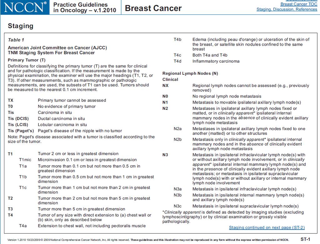
12 NCCN 2009 PET/CT noted only for: Invasive stage 3 Recurrent Stage 4 Generally discouraged except when other studies equivocal or suspicious; biopsy more useful Not our experience And what do others say.?
13 CMS Approved Staging of patients with distant metastasis Restaging of patients with locoregional recurrence or metastasis For monitoring response to therapy When a change in therapy is contemplated Not Approved Initial diagnosis of breast cancer Axillary lymph node staging and surgical planning
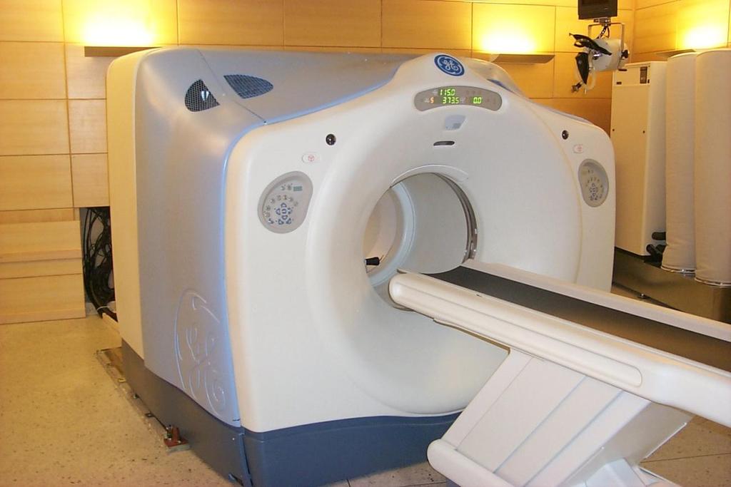
14 PET/CT
15 Patient Preparation Fasting: at least 4-6 hours Make sure no IV dextrose is being given Check glucose (< ) Increased insulin, decreased sensitivity Full history Last surgery, chemo, radiation Old or correlative studies
16 Protocol mci of FDG IV Contralateral to breast lesion Wait quietly in a room for min Image after emptying bladder Most image supine with arms up Typical skull base to mid-thigh PET-CT imaging: minutes
17 Novel PET-CT Techniques Dual time point imaging Mavi et al. J Nucl Med 2006;47:1440 Cancer: slightly increased uptake with time Normal and inflammation: no change to slightly decreased Change is 10% Whole body PET/CT Mammography Heusner et al. J Nucl Med 2008;49:1215 Prone imaging in special cradle after WB PET
18 Interpretation Non-physiologic uptake over background The hotter it is, the more likely cancer Beware false pos/neg Must integrate all data Cannot just look at images for what is hot SUV Variability Time from injection to image Body composition weight/fat Blood glucose/insulin Lesion size Partial volume Technical factors Not just SUV max but extent of uptake
19 FDG Uptake in Breast Variants False positives: Dysplasia 10% fibroadenomas Ductal ectasia Inflammation/infection Post-surgical Silicon leak Fat necrosis Even a bee sting False negatives: Lesions < 1 cm Tubular carcinoma Lobular carcinoma Carcinoma in-situ Diffuse Uptake Dense breasts Menstrual cycle Lactating breasts

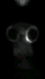
20 Normal Breasts FDG Uptake in Breast Benign Variants Breast tissue uptake young adult


21 FDG Uptake in Breast Benign Variants Inflammation Breast Implants

22 FDG Uptake in Breast Benign Variants Post-surgical inflammatory changes

23 FDG Uptake in Breast CA Benign Variants Benign brown fat uptake


24 FDG Uptake in Breast CA Benign Variants Lymph node uptake from dose infiltration
25 Breast Cancer Initial Assessment 1) Clinical examination 2) Imaging Mammography Ultrasound MRI 3) Needle biopsy 4) Staging PET plays biggest role in staging and restaging
26
27
28
29 FDG PET in Breast Cancer Clinical Applications Detection of the Primary Lesion Initial Lymph Node Assessment Evaluation of Distant Metastasis / Bony Metastasis Monitoring Response to Chemotherapy Monitoring Response to Hormonal Therapy Recurrence


30 FDG PET 50 y/o woman, recently diagnosed right breast ductal carcinoma. No adenopathy or distant metastasis
31 Primary Lesion Avril, et al. J Clin Oncol 2000;18: patients with 185 breast tumors pt1, only 30/44 (68%) breast carcinomas were detected, compared with 57/62 (92%) at stage pt2 65% lobular carcinomas false-negative (65%) compared with ductal carcinomas (24%) PET scans: high PPV (97%) for breast cancer Scheidhauer et al. EJNM 2004;31:S70 Overall PET for primary lesion 64-96% sensitivity % specificity

 Low-grade")
 Diffuse growth")

32 Detection of Primary Breast Cancer Most false negatives Small size (<1cm) Low-grade malignancies (tubular or lobular Ca) Diffuse growth pattern Avril, et al. JNM 2001;42:9 Kumar, et al. Br Ca Res Treat 2006;20:379


33 But may be good for problem cases such as implants and dense breasts
34 Diagnosis: Summary Initial promise in the primary diagnosis of breast cancer Later studies pointed out limitations Lack of sensitivity with small lesions Role to play in a select group of patients Dense breasts or with implants and other surgery Localizing primary tumor in patients with metastases of breast origin when mammography/mri is indeterminate Patients in which biopsy is not a desirable option Any incidental FDG avid breast lesion merits evaluation Discussion so far is for whole body scanners

35 But Now on the Horizon PEM
36 Duke PEM Device Rosen et al. Radiology 2005;234: patients, 24 lesions 18 TP 1 TN 2 FP (fat necrosis) 3 FN (1 DCIS, 2 IDC)


37 Malignant: Invasive ductal Ca, Grade 3 Apocrine features Focal mucinous differentiation 1.0 X.8 X.7cm

38 Guiding Surgery: Multifocality Courtesy Mary Beth Lobrano M.D., East Jefferson General Hospital, Metairie LA
39 PEM Multicenter Trial Berg at al. Breast J 2006;12: patients w/ known or suspected lesions 90% sens, 86% spec, 88% accuracy 1/2 T1a, 4/6 T1b, 7/7 T1c 3/4 invasive lobular But had mammos, knew index lesion 9/15 non-index positive were benign Partially funded by Naviscan which makes FDA approved PEM device
40 PEM Higher resolution, less attenuation Shorter imaging time Can correlate with mammography Biopsy guiding device now available Phys Med Biol 2008;53;637 FDG still issues with nonspecificity More work and multi-center trials required
41 Scintimammography Single photon radiotracer Tc99m Sestamibi Localizes to cancer through various mechanisms Whole body scanner limited resolution False positive: Benign lesions Inflammation Fat necrosis
42 Scintimammography Now like PEM, high resolution small FOV gamma cameras Early results promising Bern et al. Radiology 2008;247:651 Large comparative trials needed New device from Duke combines SPECT and CT for 3D volumetric imaging of the breast
43 FDG PET in Breast Cancer Clinical Applications Detection of the Primary Lesion Initial Lymph Node Assessment Evaluation of Distant Metastasis / Bony Metastasis Monitoring Response to Chemotherapy Monitoring Response to Hormonal Therapy Recurrence
44 Can PET take Place of Axillary Nodal Dissection/SLN? Early studies weighted to advanced breast cancer Sensitivity depends on axillary tumor burden and uptake in primary tumor Wahl et al. J Clin Onc 2004;22:277 Prospective multicenter, 360 patients (not PET/CT) 61% sensitivity; 80% specificity Small and fewer axillary nodes, more false negative

45 51 y/o Woman, Ductal Adenocarcinoma Baseline FDG PET: Initial Staging
46 Consensus is NO PET Versus SLN? Veronesi et al. Annals Onc 2007;18: patients; PET-CT Interpretation geared for highest sensitivity All SLN; full ALND if PET or SLN positive 37% sensitivity; 96% specificity
47 Staging Lymph Nodes Sentinel lymph node dissection High sensitivity and specificity to avoid unnecessary full axillary dissections FDG PET is not of sufficient sensitivity To take the place of fine sectioning and immunohistochemical lymph node evaluation Because of high positive predictive value, PET can obviate a sentinel lymph node procedure Backed up by US or image guided sampling
48 Staging Lymph Nodes Suspected high-risk disease (e.g. LABC) PET can detect IM and supraclavicular nodes as well as distant metastases May be especially useful for inner quadrant lesions J Nucl Med Sep;46(9): x incidence on PET of isolated extra-axillary mets Triple risk for disease progression Nason et al, Cancer 2000;89:2187 Increased false negative SLN rate after neoadjuvant chemotherapy for LABC
49 FDG to Detect Mediastinal or Internal Mammary Metastases Eubank et al; J Clin Oncol 2001;19:3516 Retrospective 92 patients High frequency advanced disease PET: 85% sens; 90% spec; 88% accuracy CT: 50% sens; 83% spec; 70% accuracy Upstaged 10/33 May help guide decision and field for radiation therapy in high risk disease


50 Breast Cancer with IM Node on PET (also axillary nodes) Correlated with MR as well

51 Mediastinal and Liver Metastases
52 FDG PET in Breast Cancer Clinical Applications Detection of the Primary Lesion Lymph Node Assessment Evaluation of Distant Metastasis / Bony Metastasis Monitoring Response to Chemotherapy Monitoring Response to Hormonal Therapy Recurrence
53 Distant Disease Systemic staging not useful with early stage breast cancer (unless suspicion) Low incidence metastases Possibility false positives But with high risk such as LABC and IBC and Stage 2 and 3, evidence for use Chia et al. J Clin Onc 2008;26:786 Groheux et al. Int J Radiation Oncol Biol Phys 2008
54 PET Excellent for WB Staging Schirrmeister H et al. Eur J Nucl Med 2001;28: pre-op patients: PET more accurate for LN and distant Mahner et al. Ann Oncol 2008;19: patients with new LABC Sens 93%; Spec 85% Contrast CT better than PET for liver PET better for other organs, regions Above for PET alone
55 PET-CT for Distant Staging Carkaci et al. JNM 2009;50: patients with inflammatory breast cancer PET-CT found 20 patients with distant disease (7 unsuspected) Alberini et al. Cancer ;115: patients with inflammatory breast cancer PET-CT useful for nodes and distant disease 31% distant lesions Prognostic information
56 PET-CT for Distant Staging Dirisamer et al. Eur J Rad 2009 Jan 30 PET/CT +c in 52 patients with suspected recurrence 42/52 had metastases CT: 28/42; PET:34/42; PET/CT: 40/42 most improvement of PET/CT over PET due to lung

, s/p")

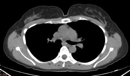
57 FDG PET-CT Staging 70 y/o woman, infiltrating ductal carcinoma grade III (7x6x5 cm), s/p recent mastectomy and axillary dissection (+2/14 nodes)


58 Multifocal Breast Carcinoma with Axillary Nodes Unexpected sternal met and IM node

59 Bone Scan 33 y/o woman, infiltrating ductal carcinoma, s/p partial right mastectomy, axillary dissection, chemotherapy and radiation therapy
60 FDG PET


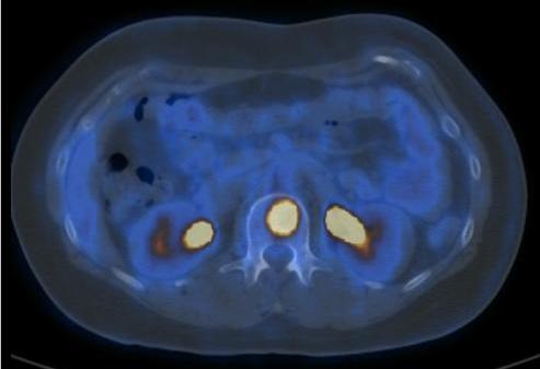
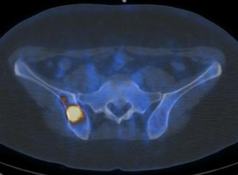
61 Unsuspected Disease Extensive malignant lymphadenopathy Skeletal metastasis unsuspected on CT
62 Controversy: Detection of Bone Metastases in Breast Cancer FDG PET has superior accuracy in detecting bone metastases (in comparison to Tc99m MDP) Nakamoto Clin Nuc Med 2003;28: PET is superior to bone scan in detecting bone metastases American Society of Clinical Oncology Tc99m MDP bone scan is much more sensitive than FDG PET in breast cancer patients. Uematsu AJR 2005;184:1266
63 Bone Metastases in Breast Cancer Nakai et al, EJNM 2005;32: patients both FDG and MDP (Planar + SPECT) 55 with bone metastases Relative visualization rate FDG MDP Osteoblastic 55.6% 100% Osteolytic 100% 70% Mixed 94.7% 84.2% Invisible 87.5% 25% Overall: PET 83.1% accuracy MDP 79.8%
64 18 F PET 18 F Fluoride PET and PET-CT more accurate than bone scanning Even-Sapir et al. JNM 2004;45:272 Iaguru et al. JNM 2009;50:501 Cocktail of 18 F-FDG and 18 F One session PET-CT Good pilot data (But response criteria may be confusing)
65 Natural History of Treated Bone Metastases in Breast Cancer Tateishi et al. Radiology 2008;247:189 Increased density on CT and decreased SUV Predictors of response duration Du et al. J Clin Oncol 2007;25:3440 FDG PET/CT better reflects true tumor activity FDG avidity predicts survival difference


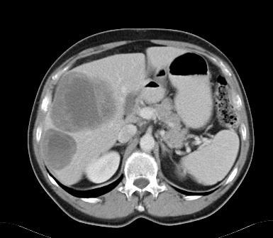
66 Incidental Cancers Incidental bilateral ovarian cancer found during staging for breast cancer
67 Staging - Metastases PET is superlative in this group of patients to detect distant disease Bone scan is sensitive for purely blastic lesion, while PET more sensitive for lytic PET/CT may be optimal since sclerotic lesions will be seen on CT portion More studies Start with PET/CT If negative, and suspect bone, obtain bone scan
68 PET/CT and Whole-Body MRI Complementary Antoch et al. JAMA 2003;290:3199 PET correct TNM 75/98; WBMRI correct 53/98 MRI more sensitive for bone and liver PET/CT more sensitive for lung and nodes Schmidt et al. Eur J Rad 2008;65:47 WBMRI: 93% sens, 86% spec, 91% accuracy PET-CT: 91% sens, 90% spec, 91% accuracy PET-CT superior for nodal disease Another study with bone metastases by the same group showed higher sensitivity for WBMRI but specificity for PET-CT

69 Fused PET MRI
70 FDG PET in Breast Cancer Clinical Applications Detection of the Primary Lesion Lymph Node Assessment Evaluation of Distant Metastasis / Bony Metastasis Monitoring Response to Chemotherapy Monitoring Response to Hormonal Therapy Recurrence


 Correlation between")
71 Prognostic Significance of FDG- PET in Breast Cancer Oshida et al. Cancer 1998;82: women with primary breast cancer Multivariate analysis: SUV is an independent predictor of disease-free survival (inversely) Correlation between SUV and microvessel density

72 Clinical Need: Determine the Response to Therapy Histopathologic response gold standard Anatomic criteria (WHO, RECIST) inadequate Questions: Is the tumor responding? Can one provide an early assessment of response? Can one determine residual disease post-therapy?

73 July 2000 September 2000
74 FDG PET for Evaluating Chemotherapy Response Most PET series with neoadjuvent therapy Most studies at mid-therapy Primary tumor decline of 50% baseline SUV predicts good response Some studies with early PET (1-2 cycles) predict better discrimination Presence of uptake on PET after chemotherapy predictive of relapse But absence of uptake is not sensitive for pcr

75 p<.001 P=.9
76 Early PET to Monitor Response Schelling M et al. J Clin Onc 2000;18: 1689 PET after first and second course chemotherapy Compared with baseline scan in 22 patients Histopathology gold standard After the first course of chemotherapy All responders correctly identified (SUV 55% baseline) Sensitivity 100%, specificity 85%
77 Early PET to Monitor Response Rousseau et al. J Clin Onc 2006;24: patients stage 2 and 3 60% decrease SUV predicts responders One course: 61% sens, 96% spec Two courses: 89% sens, 95% spec Studies depend on how close one looks Combined total and near total in pcr group But are a few cells important?
78 Metastases: Response to Therapy Dose-Schwarz et al. J Nucl Med 2005;46:1144 After chemotherapy, 17 metastatic lesions responded. In those lesions SUV decreased to 72% +/- 21% after the first cycle 54% +/- 16% after the second cycle. Uptake in lesions not responding (n = 9) Declined only to 94% +/- 19% after first cycle 79% +/- 9% after the second cycle. Differences between responding and nonresponding lesions statistically significant after the first (P = 0.02) and second (P = 0.003) cycles.


79 FDG PET Extensive breast cancer pleural implants in the left chest, and after one dose of kinase inhibitor after which the implants resolved.
80 PET and MRI are Complementary Chen, et al. Acad Radiol 2004;11: lesions in 15 women LABC before and after neoadjuvant chemotherapy PET correctly predicted lack of response (5/6) MRI predicted non response correctly (0/6) MRI predicted complete response 100% PET more accurate in predicting pathologic NR Complete response by MRI correlated well with macroscopic pathologic complete response
81 FDG PET in Breast Cancer Clinical Applications Detection of the Primary Lesion Lymph Node Assessment Evaluation of Distant Metastasis / Bony Metastasis Monitoring Response to Chemotherapy Monitoring Response to Hormonal Therapy Recurrence
82 Metabolic Flare: Indicator of Hormone Responsiveness in Advanced Breast Cancer Mortimer JE et al: J Clin Oncol 2001;19: women with advanced ER-positive (ER+) PET before and 7 to 10 days after tamoxifen therapy Responders: Tumor FDG uptake increased (28.4% +/- 23.3%) Nonresponders: No significant change (10.1% +/- 16.2%) Conclusion: Results of PET are predictive of responsiveness to tamoxifen therapy in patients with advanced ER+ breast cancer.
83 Summary: Monitoring Tumor Response with FDG/PET Valuable for prognostic information and following response to therapy Minimal residual tumor cannot be reliably detected Residual disease present if focal uptake after therapy Responding tumors show greater decrease in relative uptake to baseline Identify nonresponders earlier Use a combination of 50% SUV decline and visual analysis, but no well defined universal criteria
84 Summary: Monitoring Tumor Response with FDG/PET Timing of scan relative to treatment is important Early versus midpoint versus completion Careful attention to detail of PET scans is needed Same scanner, same uptake time, partial volume Standardized multi-center trials needed Other PET probes?
85 FDG PET in Breast Cancer Clinical Applications Detection of the Primary Lesion Lymph Node Assessment Evaluation of Distant Metastasis / Bony Metastasis Monitoring Response to Chemotherapy Monitoring Response to Hormonal Therapy Recurrence
86 Recurrence Detection of early recurrence may have important survival benefit Increase in serum tumor markers Signs and symptoms suggesting recurrence, but negative or equivocal conventional studies Difficult to differentiate true recurrence from post-surgical and radiation sequelae
87 Useful to Detect Metastases Radan et al. Cancer 2006;107: patients with increased tumor markers Sens 90%; Spec 71%; Accuracy 83% Performed significantly better than CT Changed management in 51% Kamal et al. J Cancer Res Clin Onc 2003;129: patients with suspected recurrence Locoregional: Sens 89%; Spec 84%; Accuracy 87% Distant: Sens 100%; Spec 97%; Accuracy 98%


88 Meta-analysis of FDG-PET for Breast Cancer Recurrence and Metastases Isasi et al. Cancer Res Treat 2005:90: patients 1013 lesions Results Sensitivity 92.7% Specificity 81.6% False positive 11%


89 Patient with cancer recurrence in the right breast and skin implants and an unexpected vertebral body metastasis
90 Summary: Recurrence and PET Great efficacy with suspected recurrence Surpasses utility of conventional imaging modalities for whole body evaluation Complementary to MR False positives can be problematic Experience and PET(CT) help Fueger et al. Mol Imaging Biol 2005;7:369 PET-CT restaged 89.7% correctly
91 On the Horizon How can PET and other imaging correlate and integrate with novel biomarkers such as circulating tumor cells for therapy response De Giorgi et al. J Clin Onc 2009;27:3303 Mid-therapy CTC counts and PET predicted overall survival CTC better in multivariate analysis More research needed
92 Other New (and Old) Agents Tumor perfusion and angiogenesis 15 O H 2 O Dynamic FDG PET High metabolism relative to perfusion associated with poor response Groves et al. Eur J Nucl Med 2009;36:416 Combined PET/dynamic contrast CT Metabolism and perfusion
93 Other New (and Old) Agents Receptor Imaging 18 F FES (Fluoroestradiol) ER expression, predicts response to therapy Radiolabelled trastuzumab (HER2) Tumor biosynthesis Lipid: 18 F FCH, 11 C Choline, 11 C-Actetate Cellular proliferation 18 F FLT (fluorothymidine) Cell death (apoptosis) 99m Tc-Annexin
94 In Conclusion PET has utility in patients with: Suspected distant metastases Evaluate locoregional extent in the high-risk patient Detect recurrence and monitor response to therapy Changed management in 10-50% of selected patients WB PET does not have sufficient sensitivity as a primary screening or initial staging modality Useful as a problem solving tool Initial staging in high-risk disease due to its high positive predictive value
95 In Conclusion FDG PET more accurate for lymph node and distant metastasis compared to conventional imaging Does not take the place of SLN Differentiates responders from non-responders early in the course of chemotherapy Prognostic accuracy superior to conventional imaging studies More sensitive than bone scan for lytic bone marrow metastases (bone scan is more sensitive for sclerotic lesions) PET/CT may make this point moot


96 And.. Don t forget, breast cancer in men 60 year old veteran with right lumpectomy positive for cancer Negative mammo left PET performed Unsuspected contralateral cancer
97
Clinical Utility of Positron Emission Tomography Scanning in Breast Cancer Management
 Clinical Utility of Positron Emission Tomography Scanning in Breast Cancer Management David Schuster, MD Director, Division of Nuclear Medicine and Molecular Imaging Department of Radiology and Imaging
PET/CT in Breast Cancer
 PET/CT in Breast Cancer Rodolfo Núñez Miller, M.D. Nuclear Medicine and Diagnostic Imaging Section Division of Human Health International Atomic Energy Agency Vienna, Austria Overview Introduction Locorregional
PET/CT in breast cancer staging
 PET/CT in breast cancer staging Anni Morsing Consultant, PhD, DMSc Rigshospitalet 1 18F- FDG PET/CT for breastcancer staging Where is the clinical impact? To which women should 18F- FDG PET/CT be offered?
ROLE OF PET-CT IN BREAST CANCER, GUIDELINES AND BEYOND. Prof Jamshed B. Bomanji Institute of Nuclear Medicine UCL Hospitals London
 ROLE OF PET-CT IN BREAST CANCER, GUIDELINES AND BEYOND Prof Jamshed B. Bomanji Institute of Nuclear Medicine UCL Hospitals London CANCER Key facts Estimated 15.2 million new cases per year in 2015 worldwide
Breast Cancer. Most common cancer among women in the US. 2nd leading cause of death in women. Mortality rates though have declined
 Breast Cancer Most common cancer among women in the US 2nd leading cause of death in women Mortality rates though have declined 1 in 8 women will develop breast cancer Breast Cancer Breast cancer increases
Breast Cancer. Saima Saeed MD
 Breast Cancer Saima Saeed MD Breast Cancer Most common cancer among women in the US 2nd leading cause of death in women 1 in 8 women will develop breast cancer Incidence/mortality rates have declined Breast
When do you need PET/CT or MRI in early breast cancer?
 When do you need PET/CT or MRI in early breast cancer? Elizabeth A. Morris MD FACR Chief, Breast Imaging Service Memorial Sloan-Kettering Cancer Center NY, NY Objectives What is the role of MRI in initial
PET/CT in Breast Cancer
 PET/CT in Breast Cancer Hossein Jadvar, MD, PhD, MPH, MBA Associate Professor of Radiology and Vice Chair of Research Associate Professor of Biomedical Engineering President, Society of Nuclear Medicine
FDG PET/CT STAGING OF LUNG CANCER. Dr Shakher Ramdave
 FDG PET/CT STAGING OF LUNG CANCER Dr Shakher Ramdave FDG PET/CT STAGING OF LUNG CANCER FDG PET/CT is used in all patients with lung cancer who are considered for curative treatment to exclude occult disease.
PET/CT Frequently Asked Questions
 PET/CT Frequently Asked Questions General Q: Is FDG PET specific for cancer? A: No, it is a marker of metabolism. In general, any disease that causes increased metabolism can result in increased FDG uptake
Breast Cancer Staging. Physiology Trumps Anatomy Author: Maxine Jochelson, MD, FSBI
 Breast Cancer Staging. Physiology Trumps Anatomy Author: Maxine Jochelson, MD, FSBI The purpose of this paper is to address the importance of physiologic imaging for the staging and follow up of patients
Dr Sneha Shah Tata Memorial Hospital, Mumbai.
 Dr Sneha Shah Tata Memorial Hospital, Mumbai. Topics covered Lymphomas including Burkitts Pediatric solid tumors (non CNS) Musculoskeletal Ewings & osteosarcoma. Neuroblastomas Nasopharyngeal carcinomas
FDG-PET/CT in Gynaecologic Cancers
 Friday, August 31, 2012 Session 6, 9:00-9:30 FDG-PET/CT in Gynaecologic Cancers (Uterine) cervical cancer Endometrial cancer & Uterine sarcomas Ovarian cancer Little mermaid (Edvard Eriksen 1913) honoring
Using PET/CT in Prostate Cancer
 Using PET/CT in Prostate Cancer Legal Disclaimer These materials were prepared in good faith by MITA as a service to the profession and are believed to be reliable based on current scientific literature.
Role of PEM in Breast Cancer Management. Judy Kalinyak, MD, PhD Chief Medical Officer Naviscan, Inc (San Diego, CA)
") Role of PEM in Breast Cancer Management Judy Kalinyak, MD, PhD Chief Medical Officer Naviscan, Inc (San Diego, CA) Role of PEM in Breast Cancer Management Introduction to Positron Emission Mammography
Role of PEM in Breast Cancer Management Judy Kalinyak, MD, PhD Chief Medical Officer Naviscan, Inc (San Diego, CA) Role of PEM in Breast Cancer Management Introduction to Positron Emission Mammography
Colorectal Cancer and FDG PET/CT
 Hybrid imaging in colorectal & esophageal cancer Emmanuel Deshayes IAEA WorkShop, November 2017 Colorectal Cancer and FDG PET/CT 1 Clinical background Cancer of the colon and rectum is one of the most
An Introduction to PET Imaging in Oncology
 January 2002 An Introduction to PET Imaging in Oncology Janet McLaren, Harvard Medical School Year III Basics of PET Principle of Physiologic Imaging: Allows in vivo visualization of structures by their
ROLE OF MRI IN SCREENING, DIAGNOSIS AND MANAGEMENT OF BREAST CANCER. B.Zandi Professor of Radiology
 ROLE OF MRI IN SCREENING, DIAGNOSIS AND MANAGEMENT OF BREAST CANCER B.Zandi Professor of Radiology Introduction In the USA, Breast Cancer is : The Most Common Non-Skin Cancer The Second Leading cause of
New Visions in PET: Surgical Decision Making and PET/CT
 New Visions in PET: Surgical Decision Making and PET/CT Stanley J. Goldsmith, MD Director, Nuclear Medicine Professor, Radiology & Medicine New York Presbyterian Hospital- Weill Cornell Medical Center
PET CT for Staging Lung Cancer
 PET CT for Staging Lung Cancer Rohit Kochhar Consultant Radiologist Disclosures Neither I nor my immediate family members have financial relationships with commercial organizations that may have a direct
Molecular Imaging and Breast Cancer
 Molecular Imaging and Breast Cancer Breast cancer forms in tissues of the breast usually in the ducts, tubes that carry milk to the nipple, and lobules, the glands that make milk. It occurs in both men
The Role of Radiotracer Imaging in the Diagnosis and Management of Patients with Breast Cancer: Part 1 Overview, Detection, and Staging*
 CONTINUING EDUCATION The Role of Radiotracer Imaging in the Diagnosis and Management of Patients with Breast Cancer: Part 1 Overview, Detection, and Staging* Jean H. Lee 1, Eric L. Rosen 2, and David A.
Los Angeles Radiological Society 62 nd Annual Midwinter Radiology Conference January 31, 2010
 Los Angeles Radiological Society 62 nd Annual Midwinter Radiology Conference January 31, 2010 Self Assessment Module on Nuclear Medicine and PET/CT Case Review FDG PET/CT IN LYMPHOMA AND MELANOMA Submitted
Surgical Considerations in Breast Cancer treated with Neoadjuvant Therapy
 Surgical Considerations in Breast Cancer treated with Neoadjuvant Therapy Rebecca Warburton MD Department of Surgery, University of British Columbia Mount Saint Joseph Hospital, Providence Health Care
Hybrid Imaging SPECT/CT PET/CT PET/MRI. SNMMI Southwest Chapter Aaron C. Jessop, MD
 Hybrid Imaging SPECT/CT PET/CT PET/MRI SNMMI Southwest Chapter 2014 Aaron C. Jessop, MD Assistant Professor, Department of Nuclear Medicine UT MD Anderson Cancer Center, Houston, Texas Complimentary role
Case Scenario 1 History and Physical 3/15/13 Imaging Pathology
 Case Scenario 1 History and Physical 3/15/13 The patient is an 84 year old white female who presented with an abnormal mammogram. The patient has a five year history of refractory anemia with ringed sideroblasts
F NaF PET/CT in the Evaluation of Skeletal Malignancy
 F NaF PET/CT in the Evaluation of Skeletal Malignancy Andrei Iagaru, MD September 26, 2013 School of of Medicine Ø Introduction Ø F NaF PET/CT in Primary Bone Cancers Ø F NaF PET/CT in Bone Metastases
Esophageal Cancer. What is the value of performing PET scan routinely for staging of esophageal cancers
 Esophageal Cancer What is the value of performing PET scan routinely for staging of esophageal cancers What is the sensitivity and specificity of PET scan for metastatic lesions When should PET scan be
Staging and restaging for distant metastatic disease in breast cancer: Has anything changed?
 Staging and restaging for distant metastatic disease in breast cancer: Has anything changed? Sarah J Vinnicombe Clinical Senior Lecturer in Cancer Imaging Dundee Cancer Centre s.vinnicombe@dundee.ac.uk
1 Introduction. 2 Materials and methods. LI Na 1 LI Yaming 1,* YANG Chunming 2 LI Xuena 1 YIN Yafu 1 ZHOU Jiumao 1
 Nuclear Science and Techniques 20 (2009) 354 358 18 F-FDG PET/CT in diagnosis of skeletal metastases LI Na 1 LI Yaming 1,* YANG Chunming 2 LI Xuena 1 YIN Yafu 1 ZHOU Jiumao 1 1 Department of Nuclear Medicine,
FLUCICLOVINE: 1 ST FDA APPROVED F-18 PET IMAGING AGENT FOR RECURRENT PROSTATE CANCER
 FLUCICLOVINE: 1 ST FDA APPROVED F-18 PET IMAGING AGENT FOR RECURRENT PROSTATE CANCER KEVIN P BANKS, MD SAN ANTONIO MILITARY MEDICAL CENTER ASSISTANT PROFESSOR OF RADIOLOGY, USU I HAVE NO FINANCIAL DISCLOSURES.
Lung Cancer Imaging. Terence Z. Wong, MD,PhD. Department of Radiology Duke University Medical Center Durham, NC 9/9/09
 Lung Cancer Imaging Terence Z. Wong, MD,PhD Department of Radiology Duke University Medical Center Durham, NC 9/9/09 Acknowledgements Edward F. Patz, Jr., MD Jenny Hoang, MD Ellen L. Jones, MD, PhD Lung
Whole body F-18 sodium fluoride PET/CT in the detection of bone metastases in patients with known malignancies: A pictorial review
 Whole body F-18 sodium fluoride PET/CT in the detection of bone metastases in patients with known malignancies: A pictorial review Poster No.: C-1196 Congress: ECR 2014 Type: Educational Exhibit Authors:
Breast Imaging: Multidisciplinary Approach. Madelene Lewis, MD Assistant Professor Associate Program Director Medical University of South Carolina
 Breast Imaging: Multidisciplinary Approach Madelene Lewis, MD Assistant Professor Associate Program Director Medical University of South Carolina No Disclosures Objectives Discuss a multidisciplinary breast
THE ROLE OF CONTEMPORARY IMAGING AND HYBRID METHODS IN THE DIAGNOSIS OF CUTANEOUS MALIGNANT MELANOMA(CMM) AND MERKEL CELL CARCINOMA (MCC)
 AND MERKEL CELL CARCINOMA (MCC)") THE ROLE OF CONTEMPORARY IMAGING AND HYBRID METHODS IN THE DIAGNOSIS OF CUTANEOUS MALIGNANT MELANOMA(CMM) AND MERKEL CELL CARCINOMA (MCC) I.Kostadinova, Sofia, Bulgaria CMM some clinical facts The incidence
THE ROLE OF CONTEMPORARY IMAGING AND HYBRID METHODS IN THE DIAGNOSIS OF CUTANEOUS MALIGNANT MELANOMA(CMM) AND MERKEL CELL CARCINOMA (MCC) I.Kostadinova, Sofia, Bulgaria CMM some clinical facts The incidence
Role of positron emission mammography (PEM) for assessment of axillary lymph node status in patients with breast cancer
 for assessment of axillary lymph node status in patients with breast cancer") Role of positron emission mammography (PEM) for assessment of axillary lymph node status in patients with breast cancer Poster No.: C-1260 Congress: ECR 2011 Type: Scientific Paper Authors: K. M. Kulkarni,
Role of positron emission mammography (PEM) for assessment of axillary lymph node status in patients with breast cancer Poster No.: C-1260 Congress: ECR 2011 Type: Scientific Paper Authors: K. M. Kulkarni,
Bone and CT Scans Are Complementary for Diagnoses of Bone Metastases in Breast Cancer When PET Scans Findings Are Equivocal: A Case Report
 Bone and CT Scans Are Complementary for Diagnoses of Bone Metastases in Breast Cancer When Scans Findings Are Equivocal: A Case Report Yuk-Wah Tsang 1, Jyh-Gang Leu 2, Yen-Kung Chen 3, Kwan-Hwa Chi 1,4
Breast Cancer Diagnosis, Treatment and Follow-up
 Breast Cancer Diagnosis, Treatment and Follow-up What is breast cancer? Each of the body s organs, including the breast, is made up of many types of cells. Normally, healthy cells grow and divide to produce
Breast Cancer. Dr. Andres Wiernik 2017
 Breast Cancer Dr. Andres Wiernik 2017 Agenda: The Facts! (Epidemiology/Risk Factors) Biological Classification/Phenotypes of Breast Cancer Treatment approach Local Systemic Agenda: The Facts! (Epidemiology/Risk
CT PET SCANNING for GIT Malignancies A clinician s perspective
 CT PET SCANNING for GIT Malignancies A clinician s perspective Damon Bizos Head, Surgical Gastroenterology Charlotte Maxeke Johannesburg Academic Hospital Case presentation 54 year old with recent onset
PET/CT in lung cancer
 PET/CT in lung cancer Andrei Šamarin North Estonia Medical Centre 3 rd Baltic Congress of Radiology 08.10.2010 Imaging in lung cancer Why do we need PET/CT? CT is routine imaging modality for staging of
Neoadjuvant Treatment of. of Radiotherapy
 Neoadjuvant Treatment of Breast Cancer: Role of Radiotherapy Neoadjuvant Chemotherapy Many new questions for radiation oncology? lack of path stage to guide indications should treatment response affect
Radionuclide detection of sentinel lymph node
 Radionuclide detection of sentinel lymph node Sophia I. Koukouraki Assoc. Professor Department of Nuclear Medicine Medicine School, University of Crete 1 BACKGROUND The prognosis of malignant disease is
Lesion Imaging Characteristics Mass, Favoring Benign Circumscribed Margins Intramammary Lymph Node
 Lesion Imaging Characteristics Mass, Favoring Benign Circumscribed Margins Intramammary Lymph Node Oil Cyst Mass, Intermediate Concern Microlobulated Margins Obscured Margins Mass, Favoring Malignant Indistinct
Index. Surg Oncol Clin N Am 16 (2007) Note: Page numbers of article titles are in boldface type.
 Note: Page numbers of article titles are in boldface type.") Surg Oncol Clin N Am 16 (2007) 465 469 Index Note: Page numbers of article titles are in boldface type. A Adjuvant therapy, preoperative for gastric cancer, staging and, 339 B Breast cancer, metabolic
Surg Oncol Clin N Am 16 (2007) 465 469 Index Note: Page numbers of article titles are in boldface type. A Adjuvant therapy, preoperative for gastric cancer, staging and, 339 B Breast cancer, metabolic
Pitfalls and Limitations of Breast MRI. Susan Orel Roth, MD Professor of Radiology University of Pennsylvania
 Pitfalls and Limitations of Breast MRI Susan Orel Roth, MD Professor of Radiology University of Pennsylvania Objectives Review the etiologies of false negative breast MRI examinations Discuss the limitations
Molecular Imaging and Cancer
 Molecular Imaging and Cancer Cancer causes one in every four deaths in the United States, second only to heart disease. According to the U.S. Department of Health and Human Services, more than 512,000
Prof. Dr. NAGUI M. ABDELWAHAB,M.D.; MARYSE Y. AWADALLAH, M.D. AYA M. BASSAM, Ms.C.
 Role of Whole-body Diffusion MR in Detection of Metastatic lesions Prof. Dr. NAGUI M. ABDELWAHAB,M.D.; MARYSE Y. AWADALLAH, M.D. AYA M. BASSAM, Ms.C. Cancer is a potentially life-threatening disease,
Breast Cancer Imaging
 Breast Cancer Imaging I. Policy University Health Alliance (UHA) will cover breast imaging when such services meet the medical criteria guidelines (subject to limitations and exclusions) indicated below.
Results of the ACOSOG Z0011 Trial
 DCIS and Early Breast Cancer Symposium JUNE 15-17 2012 CAPPADOCIA Results of the ACOSOG Z0011 Trial Kelly K. Hunt, M.D. Professor of Surgery Axillary Node Dissection Staging, Regional control, Survival
The Diagnostic Value of PET/CT in Breast Cancer Recurrence and Metastases
 Original Paper, Oncology. The Diagnostic Value of PET/CT in Breast Cancer Recurrence and Metastases Taalab, Kh. 1 ; Abutaleb, AS 1 ; Moftah, SG 2 ; Abdel-Mutaleb, MG 2 and Abdl-Mawla, YA 2. 1 Military
The Use of PET Scanning in Urologic Oncology
 The Use of PET Scanning in Urologic Oncology Dr Nicholas C. Buchan Uro-oncology Fellow 1 2 Aims To understand the basic concepts underlying PET scanning. Understand the emerging role of PET Scanning for
PET/CT in Gynaecological Cancers. Stroobants Sigrid, MD, PhD Departement of Nuclear Medicine University Hospital,Antwerp
 PET/CT in Gynaecological Cancers Stroobants Sigrid, MD, PhD Departement of Nuclear Medicine University Hospital,Antwerp Cervix cancer Outline of this talk Initial staging Treatment monitoring/guidance
The Role of PET / CT in Lung Cancer Staging
 July 2004 The Role of PET / CT in Lung Cancer Staging Vlad Vinarsky, Harvard Medical School Year IV Patient AM HPI: 81 yo F p/w hemoptysis x 1 month LLL lesion on CXR, not responsive to Abx 35 pack-year
STAGE CATEGORY DEFINITIONS
 CLINICAL Extent of disease before any treatment y clinical staging completed after neoadjuvant therapy but before subsequent surgery TX Tis Tis (DCIS) Tis (LCIS) Tis (Paget s) T1 T1mi T1a T1b T1c a b c
POSITRON EMISSION TOMOGRAPHY (PET)
") Status Active Medical and Behavioral Health Policy Section: Radiology Policy Number: V-27 Effective Date: 08/27/2014 Blue Cross and Blue Shield of Minnesota medical policies do not imply that members should
Status Active Medical and Behavioral Health Policy Section: Radiology Policy Number: V-27 Effective Date: 08/27/2014 Blue Cross and Blue Shield of Minnesota medical policies do not imply that members should
Newly Diagnosed Breast Cancer: Preoperative Imaging and Localization
 Newly Diagnosed Breast Cancer: Preoperative Imaging and Localization Debra Monticciolo, MD Professor of Radiology Texas A&M University no disclosures Debra Monticciolo, MD Professor of Radiology Texas
PET IMAGING (POSITRON EMISSION TOMOGRAPY) FACT SHEET
 FACT SHEET") Positron Emission Tomography (PET) When calling Anthem (1-800-533-1120) or using the Point of Care authorization system for a Health Service Review, the following clinical information may be needed to
Positron Emission Tomography (PET) When calling Anthem (1-800-533-1120) or using the Point of Care authorization system for a Health Service Review, the following clinical information may be needed to
Radiology Pathology Conference
 Radiology Pathology Conference Sharlin Johnykutty,, MD, Cytopathology Fellow Sara Majewski, MD, Radiology Resident Friday, August 28, 2009 Presentation material is for education purposes only. All rights
Joint Comments on Positron Emission Tomography (NaF-18) to Identify Bone Metastasis of Cancer (CAG-00065R1)
 to Identify Bone Metastasis of Cancer (CAG-00065R1)") July 2, 2009 Tamara Syrek Jensen, J.D. Acting Director, Coverage and Analysis Group Centers for Medicare & Medicaid Services 7500 Security Blvd., Mail Stop C1-09-06 Baltimore, MD 21244 Re: Joint Comments
July 2, 2009 Tamara Syrek Jensen, J.D. Acting Director, Coverage and Analysis Group Centers for Medicare & Medicaid Services 7500 Security Blvd., Mail Stop C1-09-06 Baltimore, MD 21244 Re: Joint Comments
Case Scenario 1 Worksheet. Primary Site C44.4 Morphology 8743/3 Laterality 0 Stage/ Prognostic Factors
 CASE SCENARIO 1 9/10/13 HISTORY: Patient is a 67-year-old white male and presents with lesion located 4-5cm above his right ear. The lesion has been present for years. No lymphadenopathy. 9/10/13 anterior
Mammographic imaging of nonpalpable breast lesions. Malai Muttarak, MD Department of Radiology Chiang Mai University Chiang Mai, Thailand
 Mammographic imaging of nonpalpable breast lesions Malai Muttarak, MD Department of Radiology Chiang Mai University Chiang Mai, Thailand Introduction Contents Mammographic signs of nonpalpable breast cancer
Imaging Surveillance in Women with a History of Treated Breast Cancer. Wei Tse Yang, M.D.
 Imaging Surveillance in Women with a History of Treated Breast Cancer Wei Tse Yang, M.D. Breast Cancer 1. Extent 2. Response 3. Recurrence Surveillance Breast Cancer 1. Extent 2. Response Surveillance
Nuclear medicine in oncology. 1. Diagnosis 2. Therapy
 Nuclear medicine in oncology 1. Diagnosis 2. Therapy Diagnosis - Conventional methods - Nonspecific radiopharmaceuticals cumulating in tumours - Specific radiopharmaceuticals (receptor- and immunoscintigraphy)
performed to help sway the clinician in what the appropriate diagnosis is, which can substantially alter the treatment of management.
 Hello, I am Maura Polansky at the University of Texas MD Anderson Cancer Center. I am a Physician Assistant in the Department of Gastrointestinal Medical Oncology and the Program Director for Physician
ACRIN 6666 Therapeutic Surgery Form
 S1 ACRIN 6666 Therapeutic Surgery Form 6666 Instructions: Complete a separate S1 form for each separate area of each breast excised with the intent to treat a cancer (e.g. each lumpectomy or mastectomy).
Short summary of published results of PET with fluoromethylcholine (18F) in prostate cancer
 in prostate cancer") Short summary of published results of PET with fluoromethylcholine (18F) in prostate cancer JN TALBOT and all the team of Service de Médecine Nucléaire Hôpital Tenon et Université Pierre et Marie Curie,
Short summary of published results of PET with fluoromethylcholine (18F) in prostate cancer JN TALBOT and all the team of Service de Médecine Nucléaire Hôpital Tenon et Université Pierre et Marie Curie,
Nuclear Medicine in Thyroid Cancer. Phillip J. Koo, MD Division Chief of Diagnostic Imaging
 Nuclear Medicine in Thyroid Cancer Phillip J. Koo, MD Division Chief of Diagnostic Imaging Financial Disclosures Bayer Janssen Learning Objectives To learn the advantages and disadvantages of SPECT/CT
Clinical indications for positron emission tomography
 Clinical indications for positron emission tomography Oncology applications Brain and spinal cord Parotid Suspected tumour recurrence when anatomical imaging is difficult or equivocal and management will
Armed Forces Institute of Pathology.
 Armed Forces Institute of Pathology www.radpath.com Armed Forces Institute of Pathology Breast Disease www.radpath.org Armed Forces Institute of Pathology Interpretation of Breast MRI Leonard M. Glassman
Case Scenario 1. 2/15/2011 The patient received IMRT 45 Gy at 1.8 Gy per fraction for 25 fractions.
 Case Scenario 1 1/3/11 A 57 year old white female presents for her annual mammogram and is found to have a suspicious area of calcification, spread out over at least 4 centimeters. She is scheduled to
Completing the Puzzle AJCC TNM Staging Breast. Nicole Catlett, CTR 2017 Kentucky Cancer Registry Fall Conference, September 21 & 22, 2017
 Completing the Puzzle AJCC TNM Staging Breast Nicole Catlett, CTR 2017 Kentucky Cancer Registry Fall Conference, September 21 & 22, 2017 OBJECTIVES Understanding of Breast TNM staging Identify clinical
Feasibility of Preoperative Axillary Lymph Node Marking with a Clip in Breast Cancer Patients before Neoadjuvant Chemotherapy: A Preliminary Study
 [ABS-0078] GBCC 2018 Feasibility of Preoperative Axillary Lymph Node Marking with a Clip in Breast Cancer Patients before Neoadjuvant Chemotherapy: A Preliminary Study Eun Young Kim 1, Kwan Ho Lee 1, Yong
Positron Emission Tomography in Lung Cancer
 May 19, 2003 Positron Emission Tomography in Lung Cancer Andrew Wang, HMS III Patient DD 53 y/o gentleman presented with worsening dyspnea on exertion for the past two months 30 pack-year smoking Hx and
Detection of Internal Mammary Adenopathy in Patients With Breast Cancer by PET/CT and MRI
 Women s Imaging Original Research Jochelson et al. PET/CT and MRI for Internal Mammary Adenopathy Women s Imaging Original Research Maxine S. Jochelson 1 Lizza Lebron Stefanie S. Jacobs Junting Zheng Chaya
Ines Buccimazza 16 TH UP CONTROVERSIES AND PROBLEMS IN SURGERY SYMPOSIUM
 BILATERAL MASTECTOMY IS NOT ROUTINELY JUSTIFIED IN PATIENTS WITH BILATERAL AXILLARY LYMPHADENOPATHY AND ONLY ONE DETECTABLE PRIMARY BREAST CANCER LESION SURGERY SYMPOSIUM Ines Buccimazza Breast Unit Department
Evaluation of Lung Cancer Response: Current Practice and Advances
 Evaluation of Lung Cancer Response: Current Practice and Advances Jeremy J. Erasmus I have no financial relationships, arrangements or affiliations and this presentation will not include discussion of
Radiological staging of lung cancer. Shukri Loutfi,MD,FRCR Consultant Thoracic Radiologist KAMC-Riyadh
 Radiological staging of lung cancer Shukri Loutfi,MD,FRCR Consultant Thoracic Radiologist KAMC-Riyadh Bronchogenic Carcinoma Accounts for 14% of new cancer diagnoses in 2012. Estimated to kill ~150,000
Sentinel Lymph Node Biopsy for Breast Cancer
 Sentinel Lymph Node Biopsy for Breast Cancer Registrar Tutorial Adam Cichowitz Surgical Registrar The Royal Melbourne Hospital Sentinel Lymph Node Biopsy Axillary LN status important prognostic factor
Whole Body MRI. Dr. Nina Tunariu. Prostate Cancer recurrence, progression and restaging
 Whole Body MRI Prostate Cancer recurrence, progression and restaging Dr. Nina Tunariu Consultant Radiology Drug Development Unit and Prostate Targeted Therapies Group 12-13 Janeiro 2018 Evolving Treatment
Utility of 18 F-FDG PET/CT in metabolic response assessment after CyberKnife radiosurgery for early stage non-small cell lung cancer
 Utility of F-FDG PET/CT in metabolic response assessment after CyberKnife radiosurgery for early stage non-small cell lung cancer Ngoc Ha Le 1*, Hong Son Mai 1, Van Nguyen Le 2, Quang Bieu Bui 2 1 Department
Breast pathology. 2nd Department of Pathology Semmelweis University
 Breast pathology 2nd Department of Pathology Semmelweis University Breast pathology - Summary - Benign lesions - Acute mastitis - Plasma cell mastitis / duct ectasia - Fat necrosis - Fibrocystic change/
Debate Axillary dissection - con. Prof. Dr. Rodica Anghel Institute of Oncology Bucharest
 Debate Axillary dissection - con Prof. Dr. Rodica Anghel Institute of Oncology Bucharest Summer School of Oncology, third edition Updated Oncology 2015: State of the Art News & Challenging Topics Bucharest,
What is Cancer? Petra Ketterl, MD Medical Oncology and Functional Medicine
 What is Cancer? Petra Ketterl, MD Medical Oncology and Functional Medicine What is Cancer? Layman s terms: cancer starts when cells grow out of control (in any place in the body) and crowd out normal cells
objectives Pitfalls and Pearls in PET/CT imaging Kevin Robinson, DO Assistant Professor Department of Radiology Michigan State University
 objectives Pitfalls and Pearls in PET/CT imaging Kevin Robinson, DO Assistant Professor Department of Radiology Michigan State University To determine the regions of physiologic activity To understand
PAAF vs Core Biopsy en Lesiones Mamarias Case #1
 5/19/2014 PAAF vs Core Biopsy en Lesiones Mamarias Case #1 Fine Needle Aspiration Cytology of Breast: Correlation with Needle Core Biopsy 64-year-old woman Mass in breast Syed Hoda, MD CD31 Post-Radiation
Lymphoma Read with the experts
 Lymphoma Read with the experts Marc Seltzer, MD Associate Professor of Radiology Geisel School of Medicine at Dartmouth Director, PET-CT Course American College of Radiology Learning Objectives Recognize
The Role of Sentinel Lymph Node Biopsy and Axillary Dissection
 The Role of Sentinel Lymph Node Biopsy and Axillary Dissection Henry Mark Kuerer, MD, PhD, FACS Department of Surgical Oncology University of Texas MD Anderson Cancer Center SLN Biopsy Revolutionized surgical
Surgical Therapy: Sentinel Node Biopsy and Breast Conservation
 Surgical Therapy: Sentinel Node Biopsy and Breast Conservation Stephen B. Edge, MD Professor of Surgery and Oncology Roswell Park Cancer Institute University at Buffalo Dr. Roswell Park: Tradition in Cancer
16/09/2015. ACOSOG Z011 changing practice. Presentation outline. Nodal mets #1 prognostic tool. Less surgery no change in oncologic outcomes
 ACOSOG Z011 changing practice The end of axillary US/FNA? Preoperative staging of the axilla in the era of Z011 Adena S Scheer MD MSc FRCSC Surgical Oncologist, St. Michael s Hospital Assistant Professor,
131-I Therapy Planning in Thyroid Cancer: The role of diagnostic radioiodine scans
 131-I Therapy Planning in Thyroid Cancer: The role of diagnostic radioiodine scans Anca M. Avram, M.D. Associate Professor of Radiology Department of Nuclear Medicine University of Michigan Ann Arbor,
Diagnosis and staging of breast cancer and multidisciplinary team working
 1 Diagnosis and staging of breast cancer and multidisciplinary team working Common symptoms and signs Over 90% of breast cancers (BCs) are local or regional when first detected. At least 60% of patients
National Diagnostic Imaging Symposium 2013 SAM - Breast MRI 1
 National Diagnostic Imaging Symposium 2013 December 8-12, 2013 Disney s Yacht Club Resort Lake Buena Vista, Florida Self Assessment Module Questions, Answers and References Day SAM Title - Each SAM title
8/10/2016. PET/CT for Tumor Response. Staging and restaging Early treatment response evaluation Guiding biopsy
 PET/CT for Tumor Response Evaluation August 4, 2016 Wei Lu, PhD Department of Medical Physics www.mskcc.org Department of Radiation Oncology www.umaryland.edu FDG PET/CT for Cancer Imaging Staging and
ANNEX 1 OBJECTIVES. At the completion of the training period, the fellow should be able to:
 1 ANNEX 1 OBJECTIVES At the completion of the training period, the fellow should be able to: 1. Breast Surgery Evaluate and manage common benign and malignant breast conditions. Assess the indications
Targeting Surgery for Known Axillary Disease. Abigail Caudle, MD Henry Kuerer, MD PhD Dept. Surgical Oncology MD Anderson Cancer Center
 Targeting Surgery for Known Axillary Disease Abigail Caudle, MD Henry Kuerer, MD PhD Dept. Surgical Oncology MD Anderson Cancer Center Nodal Ultrasound at Diagnosis Whole breast and draining lymphatic
MP Magnetic Resonance Imaging for Detection and Diagnosis of Breast Cancer
 Medical Policy MP 6.01.29 BCBSA Ref. Policy: 6.01.29 Last Review: 09/19/2018 Effective Date: 09/19/2018 Section: Radiology Related Policies 6.01.45 Computer-Aided Evaluation of Malignancy With Magnetic
Page: 1 of 29. For this policy, PET scanning is discussed for the following 4 applications in oncology:
 Emission Tomography Scanning Page: 1 of 29 Last Review Status/Date: June 2015 Description Positron emission tomography (PET) scans are based on the use of positron-emitting radionuclide tracers coupled
Molecular imaging of breast cancer with 18 F-fluorodeoxyglucose
 Blackwell Publishing Inc ORIGINAL ARTICLE DOI: 10.1111/j.1075-122X.2006.00269.x High-Resolution Fluorodeoxyglucose Positron Emission Tomography with Compression ( Positron Emission Mammography ) is Highly
RSNA, /radiol Appendix E1. Methods
 RSNA, 2016 10.1148/radiol.2016151097 Appendix E1 Methods US and Near-infrared Data Acquisition Four optical wavelengths (740 nm, 780 nm, 808 nm, and 830 nm) were used to sequentially deliver the light