MRI in breast cancer: diagnosis and intervention. Dr Sue Barter Addenbrookes Hospital, Cambridge UK
|
|
|
- Hugh Sutton
- 6 years ago
- Views:
Transcription

1 MRI in breast cancer: diagnosis and intervention Dr Sue Barter Addenbrookes Hospital, Cambridge UK
2 Intervention will be discussed in High Risk Screening!
3 Indications UK and Europe: Breast MRI is well established as a tool for certain recognised indications which include: Mammographically occult lesions in dense breasts Discrepancy in size (mammography/ultrasound/clinical) Suspected multifocal disease Lobular carcinomas Axillary lymphadenopathy, primary unknown Evaluation of response to primary chemotherapy Assessment of breast implant integrity High risk screening EUSOBI 2008 Eusoma 2010: Evidence based
4 Indications US: USA 12 indications Lesion characterization Neoadjuvant chemotherapy Infiltrating lobular and ductal carcinoma Axillary adenopathy, primary unknown Postoperative tissue reconstruction Silicone and non-silicone breast augmentation Invasion deep to the fascia Contralateral breast examination in patients with breast malignancy Postlumpectomy for residual disease Surveillance of high-risk patients Recurrence of breast cancer

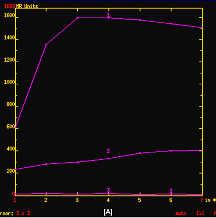
5 Dynamic CE MRI Angiogenesis closely correlated with invasive cancer Neo capillaries ++ Enlarged fenestrations Capillary leakage AV shunting


6 How we do it: Dedicated breast coil mandatory Multichannel Access to prior conventional imaging Double reporting
7 Cambridge MR protocols 1.5T GE 10/week (30 40 min) (High burden) Normal parenchymal enhancement least mid cycle Optimum imaging window day 6 to 16 of cycle No evidence stopping HRT reduces background parenchymal enhancement Iv Gd 1.5mmol/kg 3ml/sec (pink cannula)

8 Positioning Prone Feet first weight < 105kg No padding, allowing gravity to elongate breasts Discomfort: neck and arm ache


9 Side marking Transposing sides is a common reporting error Fish oil capsule marker We mark left breast Correlate with other imaging Viewing
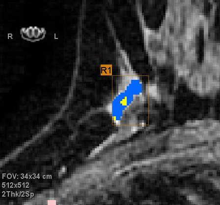
10 Sequences Axial T1 & T2 DWI Axial 3D dynamic VIBRANT T1 FSPGR fat sat 2mm thick; FOV 34 X 34 1 test: coverage, shimming for fat sat 1 pre 5 post contrast Automatic Subtraction processing Research diffusion, spectroscopy, other dynamic acquisition sequences, 3T
11 Problems with fat sat Contralateral implants Recent biopsy (?metal) Portacaths (NB ruin DWI) Coils/clips ok if Titanium small local bloom


")

12 Post processing Subtraction MIP Multiplanar reconstruction Curve analysis (Functool) Spectroscopy Diffusion CAD (Cadstream)
13 Reporting: BI-RADS The ACR BI-Rads Lexicon widely used 5 th Edition published 2013 Standardised: Imaging findings terminology Report organisation Assessment structure Classification of findings Some differences in Europe Eg no category 0, 4 abc in UK no category 0,3,4ab in Germany Cambridge Breast Unit
14 BIRADS BIRADS 0 (Needs additional imaging evaluation) BIRADS 1 (Normal) BIRADS 2 (Benign finding) BIRADS 3 (Probably benign finding - short interval follow-up) AVOID IF POSSIBLE BIRADS 4 Suspicious finding; further assessment and biopsy should be considered BIRADS 5 (Highly suggestive of malignancy; biopsy mandatory) BI-RADS 6 Known biopsy proven malignancy Cambridge Breast Unit

15 Interpretation: BI-RADS descriptors Density (amount of FG tissue) Background enhancement none/minimal: <25% mild: 25-50% moderate: 50-75% marked : >75%
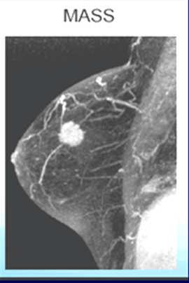
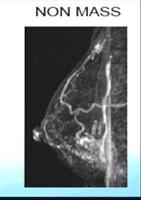
16 Reporting:First step Mass or non-mass?
17 BI-RADS descriptors: Morphology Mass 3 dimensional space occupying lesion <5mm Convex margin separate from surrounding FGT Shape: round, oval, (Inc lobulated,) irregular Margins: smooth, irregular, spiculated Internal EH characteristics: homogenous, heterogenous, rim, dark or enhancement, internal septations, central (target) Focus < 5 mm: To small to characterize margins, etc Heywang-Köbrunner et al. Eur Rad 2001
18 BI-RADS descriptors:enhancement Kinetics washout, plateau, persistent (caveat: papillomas and lymph nodes washout) ~ 70% of invasive cancers wash out ~ 9% of DCIS washes out Linear Enhancement, Rare, beware movement artifact Non Mass Enhancement Symmetric or assymetric? focal, linear ductal, linear clumped, segmental patchy/clumped, regional, diffuse stippled, punctate,


19 Interpretation: you do these! Invasive Ductal Cancer Irregular or spiculate mass Heterogenous enhancement Fast uptake and washout DCIS: Non-masslike Linear clumped enhancement Segmental, or ductal asymmetric pattern
20 MRI features and PPV Mass spiculated mass : 80 % irregular shape : 32 % < 5 mm mass : 3 % Non mass Linear (rare) segmental : 67 % clumped ductal : 31 %
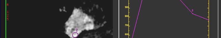
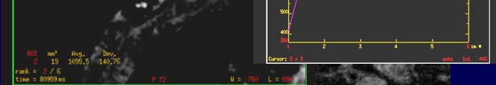
21 MRI features and PPV Enhancement Lack of enhancement has high negative predictive value (NPV) for malignancy (88% 96%) Type I curve: progressive enhancement pattern considered benign ~9% malignant Type II curve: plateau pattern concerning for malignancy 34% malignant Type III curve: washout pattern rapid uptake and washout strongly suggestive of malignancy 57%


22 Scar tissue: No indication for MRI routinely for scar tissue. It should NOT replace conventional assessment and core biopsy. Mass or non-masslike Irregular lesion No enhancement Low T2 signal
23 Indications UK and Europe: Mammographically occult lesions in dense breasts Problem solving: Discrepancy in size or position (mammography/ultrasound/clinical) Suspected multifocal disease Lobular carcinomas Axillary lymphadenopathy, primary unknown Evaluation of response to primary chemotherapy Assessment of breast implant integrity


24 Occult lesion, Dense breast Cambridge Breast Unit


25 Occult lesion, Dense breast Cambridge Breast Unit
 seemed to be a bit too lateral for true correlation.")
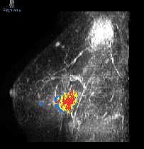
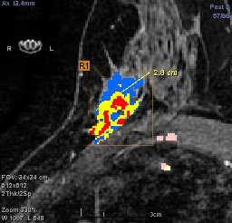
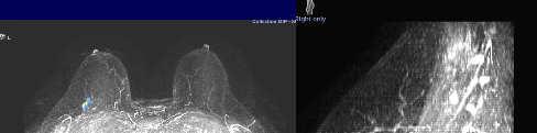
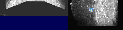
26 Problem Solving MRI to assess screen detected abnormality right breast original mammographic lesion appeared to be upper inner quadrant. Ultrasound lesion (B2) seemed to be a bit too lateral for true correlation. Cambridge Breast Unit
27 Problem solving MRI RIGHT shows a unifocal irregular area with rapid enhancement measuring 12 mm x 12 mm x 8 mm situated in the lower inner quadrant. On review of mammograms there is faint change in this area on the MLO film LEFT: normal needs second look u/sound Cambridge Breast Unit


28 Lobular carcinoma Mass or non-masslike enhancement Heterogenous enhancement Variable curve type T2 isointense to fibroglandular tissue


29 Lobular carcinoma 22mm grade2 invasive lobular ca MR detected multifocality 2 nd lesion inferiorly lobular ca on US biopsy


30 Unknown primary 47 year old presented with axillary lymphadenopathy Cambridge Breast Unit


31 Unknown primary Cambridge Breast Unit
32 Neoadjuvant baseline Cambridge Breast Unit
33 Neoadjuvant Mid Rx Cambridge Breast Unit


34 Comparison response Cambridge Breast Unit

35 Implants MRI is indicated for problem solving focal abnormalities where triple assessment has failed to resolve the diagnosis. It is considered by many as the gold standard for assessment of implant rupture.
36 Implants:EUSOMA Guidance MRI not recommended for screening for implant rupture in asymptomatic women. Symptoms suggestive of implant rupture, etc.after conventional imaging, non-contrast MRI is recommended to confirm or exclude rupture. Signs/symptoms of parenchymal disease (e.g. breast lump), when conventional imaging is not diagnostic, noncontrast MRI and DCE MRI is indicated In symptomatic patients that have undergone breast augmentation with direct polyacrylamide gel injection, non-contrast MRI and dynamic contrast-enhanced MRI are indicated. (Sardanelli 2010):
37 Sequences Implants Single breast (less chemical shift) Axial STIR Axial stir water sat (silicone only) Sagittal STIR Axial VIBRANT single phase


38 Implant failure Linguini Noose Keyhole Silicone leak Salad oil
39 Pre-operative MRI MRI improves diagnosis of breast cancer, but does it reduce rates of reoperation and recurrence? COMICE and MONET trials suggested not Preoperative Breast MRI in Clinical Practice: Multicenter International Prospective Meta- Analysis (MIPA) of Individual Woman Data An EIBIR-EuroAIM/EUSOBI Study (due 2018) (Sardinelli)
40 Take home messages Mass: Shape, margins etc do not override enhancement kinetics Lobular cancers may not show typical malignant type enhancement NME: distribution, symmetry key MRI is not needed to assess a tumour before surgery for biopsy-proven invasive breast cancer or DCIS except in specific clinical situations. Carrying out an unnecessary preoperative MRI scan may cause additional stress without any benefit and waste healthcare resources Do not substitute for conventional imaging and biopsy unless specific indications
41 Thank you
Armed Forces Institute of Pathology.
 Armed Forces Institute of Pathology www.radpath.com Armed Forces Institute of Pathology Breast Disease www.radpath.org Armed Forces Institute of Pathology Interpretation of Breast MRI Leonard M. Glassman
BI-RADS and Breast MRI. Kathy Borovicka, M.D. Thursday February 15, 2018
 BI-RADS and Breast MRI Kathy Borovicka, M.D. Thursday February 15, 2018 Learning Objectives Be familiar with the Breast Imaging Reporting and Data System (BI-RADS) Understand the components of a breast
BI-RADS Update. Martha B. Mainiero, MD, FACR, FSBI Brown University Rhode Island Hospital
 BI-RADS Update Martha B. Mainiero, MD, FACR, FSBI Brown University Rhode Island Hospital No Disclosures BI-RADS History 1980s Quality Issues ACR Accreditation BI-RADS 1994 2003 4 th Edition MRI, US January
Leonard M. Glassman MD
 BI-RADS The New BI-RADS Leonard M. Glassman MD FACR Former Chief of Breast Imaging American Institute for Radiologic Pathology Washington Radiology Associates, PC Breast Imaging Reporting and Data System
Contrast-enhanced Breast MRI RSSA 2013
 Contrast-enhanced Breast MRI RSSA 2013 Prof. dr. Maurice van den Bosch University Medical Center Utrecht, the Netherlands Index 1) Breast cancer 2) Why MRI of the breast 3) Technique 4) Interpretation
Pitfalls and Limitations of Breast MRI. Susan Orel Roth, MD Professor of Radiology University of Pennsylvania
 Pitfalls and Limitations of Breast MRI Susan Orel Roth, MD Professor of Radiology University of Pennsylvania Objectives Review the etiologies of false negative breast MRI examinations Discuss the limitations
BREAST MRI. VASILIKI FILIPPI RADIOLOGIST CT MRI & PET/CT Departments Hygeia Hospital, Athens, Greece
 BREAST MRI VASILIKI FILIPPI RADIOLOGIST CT MRI & PET/CT Departments Hygeia Hospital, Athens, Greece Breast ΜR Imaging (MRM) Breast MR imaging is an extremely powerful diagnostic tool, that when used in
Successful Breast MRI Program : The ingredients
 Successful Breast MRI Program : The ingredients Dr. Smriti Hari Associate Professor Deptt. Of Radiology All India Institute of Medical Sciences New Delhi How to perform Breast MRI Breast MRI descriptors
Standard Breast Imaging Modalities. Lilian Wang, M.D. Breast Imaging Section Department of Radiology Northwestern Medicine
 Standard Breast Imaging Modalities Lilian Wang, M.D. Breast Imaging Section Department of Radiology Northwestern Medicine Overview Standard breast imaging modalities Mammography Ultrasound MRI Imaging
Imaging in breast cancer. Mammography and Ultrasound Donya Farrokh.MD Radiologist Mashhad University of Medical Since
 Imaging in breast cancer Mammography and Ultrasound Donya Farrokh.MD Radiologist Mashhad University of Medical Since A mammogram report is a key component of the breast cancer diagnostic process. A mammogram
MRI BI-RADS: How to make it out?
 MRI BI-RADS: How to make it out? Poster No.: C-1850 Congress: ECR 2016 Type: Educational Exhibit Authors: M. Ben Ammar, A. Ben Miled, O. Ghdes, S. Harguem, A. Gaja, N. Mnif; Tunis/TN Keywords: Breast,
AB MR Interpretation Overview
 AB MR Interpretation Overview Goal of AB MR interpretation is to maintain high sensitivity and specificity In order to minimize false positives and short term follow ups, it is fundamental to focus only
WHICH INDICATION FOR BREAST MRI?
 WHICH INDICATION FOR BREAST MRI? Dr. P. De Visschere, Prof. Dr. G. Villeirs Genitourinary Radiology and Mammography University Hospital Gent Symposium Belgian Menopause Society 13/03/2010 Which Indication
Value of the BI-RADS classification in MR-Mammography for diagnosis of benign and malignant breast tumors
 Eur Radiol (2011) 21:2475 2483 DOI 10.1007/s00330-011-2210-7 BREAST Value of the BI-RADS classification in MR-Mammography for diagnosis of benign and malignant breast tumors Christian Sohns & Martin Scherrer
Index. C Calcifications fat necrosis 1, 61 fat necrosis 4, 69 nipple/peri-areolar involvement 1, 165
 A ADH. See Atypical ductal hyperplasia (ADH) American College of Radiology (ACR), BI-RADS background parenchymal enhancement, 8, 9, 81, 82 fibroglandular tissue guidelines, 6 American Joint Committee on
Mammographic imaging of nonpalpable breast lesions. Malai Muttarak, MD Department of Radiology Chiang Mai University Chiang Mai, Thailand
 Mammographic imaging of nonpalpable breast lesions Malai Muttarak, MD Department of Radiology Chiang Mai University Chiang Mai, Thailand Introduction Contents Mammographic signs of nonpalpable breast cancer
PLACE LABEL HERE. ACRIN 6657 MRI Form: Pre-Treatment (MRI-1)
") M3 ACRIN 6657 MRI Form: Pre-Treatment (MRI-1) If this is a revised or corrected form,indicate by checking box. ACRIN Study 6657 Case # Instructions: In accordance with the protocol, four MRI exams are
M3 ACRIN 6657 MRI Form: Pre-Treatment (MRI-1) If this is a revised or corrected form,indicate by checking box. ACRIN Study 6657 Case # Instructions: In accordance with the protocol, four MRI exams are
BREAST MRI. Elizabeth A. Rafferty, M.D. Avon Comprehensive Breast Center Massachusetts General Hospital Harvard Medical School
 BREAST MRI Elizabeth A. Rafferty, M.D. Avon Comprehensive Breast Center Massachusetts General Hospital Harvard Medical School BREAST MRI Any assessment of the breast parenchyma requires the administration
Non-mass Enhancement on Breast MRI. Aditi A. Desai, MD Margaret Ann Mays, MD
 Non-mass Enhancement on Breast MRI Aditi A. Desai, MD Margaret Ann Mays, MD Breast MRI Important screening and diagnostic tool, given its high sensitivity for breast cancer detection Breast MRI - Indications
MR sin plass i brystkreftdiagnostikk, dagens anbefalinger og fremtidsperspektiver
 MR sin plass i brystkreftdiagnostikk, dagens anbefalinger og fremtidsperspektiver Kathinka Kurz, MD, PhD, seksjonsoverlege SUS, kathinka.dehli.kurz@sus.no Technique - Subtraction Without contrast agent
ROLE OF MRI IN SCREENING, DIAGNOSIS AND MANAGEMENT OF BREAST CANCER. B.Zandi Professor of Radiology
 ROLE OF MRI IN SCREENING, DIAGNOSIS AND MANAGEMENT OF BREAST CANCER B.Zandi Professor of Radiology Introduction In the USA, Breast Cancer is : The Most Common Non-Skin Cancer The Second Leading cause of
Here are examples of bilateral analog mammograms from the same patient including CC and MLO projections.
 Good afternoon. It s my pleasure to be discussing Diagnostic Breast Imaging over the next half hour. I m Wei Yang, Professor of Diagnostic Radiology and Chief, the Section of Breast Imaging as well as
Breast MRI Update. Jeffrey C. Weinreb, MD, FACR Yale University School of Medicine
 Breast MRI Update Jeffrey C. Weinreb, MD, FACR jeffrey.weinreb@yale.edu Yale University School of Medicine I disclose the following financial relationships with relevant commercial interests: Bracco Bayer
PLACE LABEL HERE BASELINE / PRE-TREATMENT. ACRIN 6657 Extension MRI Form: Baseline / Pre-Treatment MRI 1. o Unknown
 T1 ACRIN 6657 Extension MRI Form: Baseline / Pre-Treatment MRI 1 If this is a revised or corrected form, please box. ACRIN Study 6657 No. Instructions: In accordance with the protocol, four MRI exams are
Breast Imaging Lexicon
 9//201 200 BI RADS th Edition 201 BI RADS th Edition Breast Imaging Lexicon Mammographic Pathology and Assessment Categories Deborah Thames, R.T.(R)(M)(QM) The Advanced Health Education Center Nonmember:
Lesion Imaging Characteristics Mass, Favoring Benign Circumscribed Margins Intramammary Lymph Node
 Lesion Imaging Characteristics Mass, Favoring Benign Circumscribed Margins Intramammary Lymph Node Oil Cyst Mass, Intermediate Concern Microlobulated Margins Obscured Margins Mass, Favoring Malignant Indistinct
Radiologic and pathologic correlation of non-mass like breast lesions on US and MRI: Benign, high risk, versus malignant
 Radiologic and pathologic correlation of non-mass like breast lesions on US and MRI: Benign, high risk, versus malignant Poster No.: C-1161 Congress: ECR 2013 Type: Educational Exhibit Authors: J. Kwak,
S. Murgo, MD. Chr St-Joseph, Mons Erasme Hospital, Brussels
 S. Murgo, MD Chr St-Joseph, Mons Erasme Hospital, Brussels? Introduction Mammography reports are sometimes ambiguous and indecisive. ACR has developped the BIRADS. BIRADS consists of a lexicon in order
National Diagnostic Imaging Symposium 2013 SAM - Breast MRI 1
 National Diagnostic Imaging Symposium 2013 December 8-12, 2013 Disney s Yacht Club Resort Lake Buena Vista, Florida Self Assessment Module Questions, Answers and References Day SAM Title - Each SAM title
CDIS: what's beyond microcalcifications? - Pictorial essay
 CDIS: what's beyond microcalcifications? - Pictorial essay Poster No.: C-1096 Congress: ECR 2014 Type: Educational Exhibit Authors: R. N. Lucas, C. A. S. Ruano, I. Oliveira, J. M. G. Lourenco, Z. 1 1 1
Breast MRI: Friend or Foe?
 Breast MRI: Friend or Foe? UCSF Postgraduate Course May 18, 2013 Cheryl Ewing, MD Clinical Professor of Surgery UCSF Department of Surgery APPLEGATE HAS DOUBLE MASTECTOMY IN CANCER SCARE DIAGNOSED WITH
BI-RADS MRI: A Primer
 Erguvan- ogan et al. I- RS MRI Women s Imaging Pictorial Essay WOMEN S IMGING asak Erguvan-ogan 1 Gary J. Whitman 1 nne. Kushwaha 1,2 Michael J. Phelps 1,3 Peter J. empsey 1 Erguvan-ogan, Whitman GJ, Kushwaha,
Breast Cancer Imaging
 Breast Cancer Imaging I. Policy University Health Alliance (UHA) will cover breast imaging when such services meet the medical criteria guidelines (subject to limitations and exclusions) indicated below.
Anyone can get breast cancer BREAST MRI BREAST CANCER. The incidence of getting breast cancer is 1:19 in Malaysia
 Anyone can get breast cancer BREAST MRI KATE Datin Dr Fatimah Moosa Sunway Medical Centre DATIN SERI ENDON KYLIE SIZE DOES NOT MAKE A DIFFERENCE BREAST CANCER The incidence of getting breast cancer is
Positive Predictive Value of
 Note: This copy is for your personal non-commercial use only. To order presentation-ready copies for distribution to your colleagues or clients, contact us at www.rsna.org/rsnarights. Mary C. Mahoney,
AMSER Case of the Month: November 2018
 AMSER Case of the Month: November 2018 52 year old female with an abnormal screening mammogram Areeg Rehman, MS 4 Nova Southeastern University Rebecca T. Sivarajah, MD Penn State University College of
Emerging Techniques in Breast Imaging: Contrast-Enhanced Mammography and Fast MRI
 Emerging Techniques in Breast Imaging: Contrast-Enhanced Mammography and Fast MRI Lilian Wang, M.D. Breast Imaging Section Department of Radiology Northwestern Medicine Overview Rationale for new imaging
Criteria of Malignancy. Evaluation Score
 30 5 Diagnostic Criteria Criteria of Malignancy Table 5.2 lists criteria in contrast-enhancing MR mammography that strongly indicate the presence of malignancy or are unspecific. Unifactorial evaluation
Amammography report is a key component of the breast
 Review Article Writing a Mammography Report Amammography report is a key component of the breast cancer diagnostic process. Although mammographic findings were not clearly differentiated between benign
8/31/2016 HIDING IN PLAIN SITE, ARCHITECTURAL DISTORTIONS AND BREAST ASYMMETRIES ARCHITECTURAL DISTORTIONS ARCHITECTURAL DISTORTIONS
 HIDING IN PLAIN SITE, ARCHITECTURAL DISTORTIONS AND BREAST ASYMMETRIES DEBORAH THAMES R.T. (R)(M)(QM) ARCHITECTURAL DISTORTIONS Definition is disruption of the natural flow of breast pattern towards the
Clinical Practice Guideline for the Indications for Use of Breast Magnetic Resonance Imaging (MRI)
") CIHRT Exhibit P-2595 Page 1 Question: Clinical Practice Guideline for the Indications for Use of Breast Magnetic Resonance Imaging (MRI) Eastern Health Breast Disease Site Group What are the current indications
CIHRT Exhibit P-2595 Page 1 Question: Clinical Practice Guideline for the Indications for Use of Breast Magnetic Resonance Imaging (MRI) Eastern Health Breast Disease Site Group What are the current indications
Triple-negative breast cancer: which typical features can we identify on conventional and MRI imaging?
 Triple-negative breast cancer: which typical features can we identify on conventional and MRI imaging? Poster No.: C-1862 Congress: ECR 2013 Type: Educational Exhibit Authors: V. Bertani 1, A. Gualano
Ultrasonography. Methods. Brief Description. Indications. Device-related Prerequisites. Technical Requirements. Evaluation Criteria
 1 Ultrasonography Brief Description Imaging modality using sound waves Tissue-specific wave reflection. Indications Evaluation of palpable breast nodules Evaluation of clinically occult mammographic findings
BREAST IMAGING and NEW IMAGING MODALITIES- A Surgeons view
 BREAST IMAGING and NEW IMAGING MODALITIES- A Surgeons view DR CHANTEL THORNTON SPECIALIST BREAST CANCER SURGEON BMSc (hons) MBBS (hons) FRACS Epworth Hospital, Richmond- Agora Centre for Women s Health
MRI features of Triple-negative breast cancer: our experience.
 MRI features of Triple-negative breast cancer: our experience. Poster No.: C-1852 Congress: ECR 2013 Type: Scientific Exhibit Authors: V. Bertani, A. Gualano, V. Londero, A. Dal Col, M. Marcon, P. 1 2
Breast MR biopsy. I Thomassin-Naggara, A.Jalaguier-Coudray, J Chopier
 Breast MR biopsy I Thomassin-Naggara, A.Jalaguier-Coudray, J Chopier Background EUSOBI When a radiologist perform a MR breast imaging he has to be able to realize or to be apart of a network who is able
Ductal carcinoma in situ: ultrasound, mammography and MRI features with pathologic correlation
 Ductal carcinoma in situ: ultrasound, mammography and MRI features with pathologic correlation Poster No.: C-2252 Congress: ECR 2013 Type: Educational Exhibit Authors: L. Fernandes, H. A. M. R. Tinto,
We are IntechOpen, the world s leading publisher of Open Access books Built by scientists, for scientists. International authors and editors
 We are IntechOpen, the world s leading publisher of Open Access books Built by scientists, for scientists 4,000 116,000 120M Open access books available International authors and editors Downloads Our
Evaluation of BI-RADS 3 lesions in women with a high risk of hereditary breast cancer.
 Evaluation of BI-RADS 3 lesions in women with a high risk of hereditary breast cancer. Poster No.: C-0346 Congress: ECR 2014 Type: Scientific Exhibit Authors: A. Thomas 1, R. Dominguez Oronoz 1, S. Roche
UW Radiology Review Course Breast Calcifications. BI-RADS 5 th Edition
 UW Radiology Review Course Breast Calcifications Grace Kalish, MD Vantage Radiology BI-RADS 5 th Edition Benign Skin Vascular Large rod like Coarse popcorn Suspicious Amorphous Coarse heterogenous Fine
Using lesion washout volume fraction as a biomarker to improve suspicious breast lesion characterization
 JOURNAL OF APPLIED CLINICAL MEDICAL PHYSICS, VOLUME 16, NUMBER 5, 2015 Using lesion washout volume fraction as a biomarker to improve suspicious breast lesion characterization Jie Huang, a Sarah M. Schafer,
Categorical Classification of Spiculated Mass on Breast MRI
 Categorical Classification of Spiculated Mass on Breast MRI Poster No.: C-1974 Congress: ECR 2013 Type: Authors: Scientific Exhibit Y. Kanda 1, S. Kanao 2, M. Kataoka 2, K. Togashi 2 ; 1 Kyoto City/JP,
Tips and Tricks to performing Magnetic Resonance Imaging Guided Breast Interventional Procedures Habib Rahbar, MD, FSBI October 23, 2018, 7:00pm ET
 Tips and Tricks to performing Magnetic Resonance Imaging Guided Breast Interventional Procedures Habib Rahbar, MD, FSBI October 23, 2018, 7:00pm ET SAM Questions/Answers/Rationales/References 1. Below
MP Magnetic Resonance Imaging for Detection and Diagnosis of Breast Cancer
 Medical Policy MP 6.01.29 BCBSA Ref. Policy: 6.01.29 Last Review: 09/19/2018 Effective Date: 09/19/2018 Section: Radiology Related Policies 6.01.45 Computer-Aided Evaluation of Malignancy With Magnetic
Ana Sofia Preto 19/06/2013
 Ana Sofia Preto 19/06/2013 Understanding the underlying pathophysiologic processes leading to the various types of calcifications Description and illustration of the several types of calcifications, according
Digital breast tomosynthesis (DBT) occult breast cancers: clinical, radiological and histopathological features.
 occult breast cancers: clinical, radiological and histopathological features.") Digital breast tomosynthesis (DBT) occult breast cancers: clinical, radiological and histopathological features. Poster No.: C-1707 Congress: ECR 2015 Type: Scientific Exhibit Authors: V. Vinci 1, A. Iqbal
Digital breast tomosynthesis (DBT) occult breast cancers: clinical, radiological and histopathological features. Poster No.: C-1707 Congress: ECR 2015 Type: Scientific Exhibit Authors: V. Vinci 1, A. Iqbal
AMSER Case of the Month: September 2018
 AMSER Case of the Month: September 2018 60-year-old woman with a left breast mass noted on screening mammography. Catherine McNulty, MS4 Tulane University School of Medicine Dr. Robin Sobolewski Breast
New Imaging Modalities for better Screening and Diagnosis
 New Imaging Modalities for better Screening and Diagnosis Miri Sklair-Levy, MD Department of Diagnostic Imaging Sheba Medical Center, Sackler School of Medicine, Tel Aviv University Department of Diagnostic
ORIGINAL ARTICLE EVALUATION OF BREAST LESIONS USING X-RAY MAMMOGRAM WITH HISTOPATHOLOGICAL CORRELATION
 Available online at www.journalijmrr.com INTERNATIONAL JOURNAL OF MODERN RESEARCH AND REVIEWS IJMRR ISSN: 2347-8314 Int. J. Modn. Res. Revs. Volume 3, Issue 10, pp 807-814, October, 2015 ORIGINAL ARTICLE
Throughout this policy, bracketed numbers link topics across multiple sections according to the indication numbers in the following list.
 Subject: Magnetic Resonance Imaging of the Breast Page: 1 of 33 Last Review Status/Date: September 2015 Magnetic Resonance Imaging of the Breast Description Magnetic resonance imaging (MRI) of the breast
Breast Imaging Update: Old Dog New Tricks
 Breast Imaging Update: Old Dog New Tricks Claire McKay, DO M&S Imaging Assoc. San Antonio, TX cmckayhart@juno.com Goals Describe modalities available, old and new Provide understanding of pros and cons
Diagnostic Dilemmas of Breast Imaging
 Diagnostic Dilemmas of Breast Imaging Common Causes of Error in Breast Cancer Detection By: Jason Cord, M.D. Mammography: Initial Imaging The standard for detection of breast cancer Screening mammography
The role of MRI in assessment of asymmetrical breast densities
 The Egyptian Journal of Radiology and Nuclear Medicine (2010) 41, 501 508 Egyptian Society of Radiology and Nuclear Medicine The Egyptian Journal of Radiology and Nuclear Medicine www.elsevier.com/locate/ejrnm
Case Scenario 1 History and Physical 3/15/13 Imaging Pathology
 Case Scenario 1 History and Physical 3/15/13 The patient is an 84 year old white female who presented with an abnormal mammogram. The patient has a five year history of refractory anemia with ringed sideroblasts
Role of positron emission mammography (PEM) for assessment of axillary lymph node status in patients with breast cancer
 for assessment of axillary lymph node status in patients with breast cancer") Role of positron emission mammography (PEM) for assessment of axillary lymph node status in patients with breast cancer Poster No.: C-1260 Congress: ECR 2011 Type: Scientific Paper Authors: K. M. Kulkarni,
Role of positron emission mammography (PEM) for assessment of axillary lymph node status in patients with breast cancer Poster No.: C-1260 Congress: ECR 2011 Type: Scientific Paper Authors: K. M. Kulkarni,
Medical Policy An independent licensee of the Blue Cross Blue Shield Association
 CAE of Malignancy with MRI of the Breast Page 1 of 9 Medical Policy An independent licensee of the Blue Cross Blue Shield Association Title: See also: Computer-Aided Evaluation of Malignancy with Magnetic
Contrast-Enhanced Spectral Mammography
 Contrast-Enhanced Spectral Mammography Illuminating Breast Cancer Detection SenoBright HD TM gehealthcare.com/senobright Mammography is the most reliable imaging technique for breasts, but limitations
We are IntechOpen, the world s leading publisher of Open Access books Built by scientists, for scientists. International authors and editors
 We are IntechOpen, the world s leading publisher of Open Access books Built by scientists, for scientists 3,800 116,000 120M Open access books available International authors and editors Downloads Our
BI-RADS Categorization As a Predictor of Malignancy 1
 Susan G. Orel, MD Nicole Kay, BA Carol Reynolds, MD Daniel C. Sullivan, MD BI-RADS Categorization As a Predictor of Malignancy 1 Index terms: Breast, biopsy, 00.1261 Breast neoplasms, localization, 00.125,
Indications for Breast MRI: Case-Based Review
 AJR Integrative Imaging LIFELONG LEARNING FOR RADIOLOGY Indications for Breast MRI: Case-Based Review Amy Argus 1, Mary C. Mahoney Objective The educational objectives of this continuing medical education
Detailed Program of the second BREAST IMAGING AND INTERVENTIONS PROGRAM am am : Clinician s requirements from breast imaging
 Detailed Program of the second BREAST IMAGING AND INTERVENTIONS PROGRAM 2012 Day one, 2 nd November BREAST IMAGING AND INTERVENTIONS PROGRAM 2012 9.00 AM 9.10 am Introduction 9.10 am - 9.30 am : Clinician
Essentials of Clinical MR, 2 nd edition. 73. Urinary Bladder and Male Pelvis
 73. Urinary Bladder and Male Pelvis Urinary bladder carcinoma is best locally staged with MRI. It is important however to note that a thickened wall (> 5 mm) is a non-specific finding seen in an underfilled
Pitfalls of Dynamic Contrast Enhanced MR Mammography (DCE-MRM) in Evaluation of Post-Biopsy Suspicious Breast Lesions
 in Evaluation of Post-Biopsy Suspicious Breast Lesions") Med. J. Cairo Univ., Vol. 86, No. 3, June: 1513-1522, 2018 www.medicaljournalofcairouniversity.net Pitfalls of Dynamic Contrast Enhanced MR Mammography (DCE-MRM) in Evaluation of Post-Biopsy Suspicious
Med. J. Cairo Univ., Vol. 86, No. 3, June: 1513-1522, 2018 www.medicaljournalofcairouniversity.net Pitfalls of Dynamic Contrast Enhanced MR Mammography (DCE-MRM) in Evaluation of Post-Biopsy Suspicious
Rate of Malignancy in MRI-Detected Probably Benign (BI-RADS 3) Lesions
 Lesions") Women s Imaging Original Research Spick et al. Malignancy in MRI BI-RADS 3 Lesions Women s Imaging Original Research Claudio Spick 1,2 Dieter H. M. Szolar 1 Pascal A. Baltzer 2 Manfred Tillich 1 Pia Reittner
Women s Imaging Original Research Spick et al. Malignancy in MRI BI-RADS 3 Lesions Women s Imaging Original Research Claudio Spick 1,2 Dieter H. M. Szolar 1 Pascal A. Baltzer 2 Manfred Tillich 1 Pia Reittner
Breast Cancer. Most common cancer among women in the US. 2nd leading cause of death in women. Mortality rates though have declined
 Breast Cancer Most common cancer among women in the US 2nd leading cause of death in women Mortality rates though have declined 1 in 8 women will develop breast cancer Breast Cancer Breast cancer increases
Intracystic papillary carcinoma of the breast
 Intracystic papillary carcinoma of the breast Poster No.: C-1932 Congress: ECR 2011 Type: Educational Exhibit Authors: V. Dimarelos, F. TZIKOS, N. Kotziamani, G. Rodokalakis, 1 2 3 1 1 1 2 T. MALKOTSI
COPE Library Sample
 Breast Anatomy LOBULE LOBE ACINI (MILK PRODUCING UNITS) NIPPLE AREOLA COMPLEX ENLARGEMENT OF DUCT AND LOBE LOBULE SUPRACLAVICULAR NODES INFRACLAVICULAR NODES DUCT DUCT ACINI (MILK PRODUCING UNITS) 8420
Inflammatory Breast Carcinoma: Mammographic, Ultrasonographic, MRI and Pathologic Findings
 Inflammatory Breast Carcinoma: Mammographic, Ultrasonographic, MRI and Pathologic Findings Poster No.: C-2248 Congress: ECR 2013 Type: Educational Exhibit Authors: L. Fernandes, J. Lopes Dias, H. A. M.
Breast Cancer. Saima Saeed MD
 Breast Cancer Saima Saeed MD Breast Cancer Most common cancer among women in the US 2nd leading cause of death in women 1 in 8 women will develop breast cancer Incidence/mortality rates have declined Breast
Large health system benefits from multiple scanners for breast MRI
 Publication for the Philips MRI Community Issue 42 DECEMBER 2010 Large health system benefits from multiple scanners for breast MRI Breast MR and MR-guided biopsy are daily practice at WellStar Health
Breast Cancer Diagnosis, Treatment and Follow-up
 Breast Cancer Diagnosis, Treatment and Follow-up What is breast cancer? Each of the body s organs, including the breast, is made up of many types of cells. Normally, healthy cells grow and divide to produce
3D Automated Breast Ultrasound (ABUS): The dense breast screening tool and its potential role for preoperative staging
: The dense breast screening tool and its potential role for preoperative staging") 3D Automated Breast Ultrasound (ABUS): The dense breast screening tool and its potential role for preoperative staging Introduction Breast cancer is by far the most common cancer amongst women across Europe,
3D Automated Breast Ultrasound (ABUS): The dense breast screening tool and its potential role for preoperative staging Introduction Breast cancer is by far the most common cancer amongst women across Europe,
ACRIN 6666 IM Additional Evaluation: Additional Views/Targeted US
 Additional Evaluation: Additional Views/Targeted US For revised or corrected form check box and fax to 215-717-0936. Instructions: The form is completed based on recommendations (from ID form) for additional
MRI Occult Invasive Breast Cancer
 MRI Occult Invasive Breast Cancer Poster No.: C-1573 Congress: ECR 2015 Type: Educational Exhibit Authors: R. Patel, N. Chhaya, K. Stafford, B. Holloway, D. Tsukagoshi, A. Malhotra; London/ Keywords: Cancer,
Disclosures. Breast Cancer. Breast Imaging Modalities. Breast Cancer Screening. Breast Cancer 6/4/2014
 : Information for the Primary Care Physician Disclosures No financial relationships with commercial entities producing health care products/services. Roxsann Roberts, MD Section Chief, MRI Erlanger/EmCare
Case Scenario 1: This case has been slightly modified from the case presented during the live session to add clarity.
 Case Scenario 1: This case has been slightly modified from the case presented during the live session to add clarity. Background: 46 year old married premenopausal female with dense breasts has noticed
Effectivity of combined diffusion-weighted imaging and contrast-enhanced MRI in malignant and benign breast lesions
 OI: https://doi.org/1.5114/pjr.218.74363 Received: 24.4.217 ccepted: 25.5.217 Published: 15.2.218 http://www.polradiol.com Original article Effectivity of combined diffusion-weighted imaging and contrast-enhanced
Case study 1. Rie Horii, M.D., Ph.D. Division of Pathology Cancer Institute Hospital, Japanese Foundation for Cancer Research
 NCCN/JCCNB Seminar in Japan April 15, 2012 Case study 1 Rie Horii, M.D., Ph.D. Division of Pathology Cancer Institute Hospital, Japanese Foundation for Cancer Research Present illness: A 50y.o.premenopausal
DCIS of the Breast--MRI findings with mammographic correlation.
 DCIS of the Breast--MRI findings with mammographic correlation. Poster No.: C-1560 Congress: ECR 2013 Type: Educational Exhibit Authors: N. B. Ibrahim, P. Morris, S. ANANDAN; Burlington, MA/US Keywords:
The latest developments - Automated Breast Volume Scanning. Dr. med. M. Golatta
 The latest developments - Automated Breast Volume Scanning Dr. med. M. Golatta Automated Breast Volume US: Why? o Mammography is limited in dense breasts: high false negative rate o Many of these tumors
Aims and objectives. Page 2 of 10
 Diagnostic performance of automated breast volume scanner (ABVS) versus hand-held ultrasound (HHUS) as second look for breast lesions detected only on magnetic resonance imaging. Poster No.: C-1701 Congress:
Patient Outcomes in Canceled MRI-Guided Breast Biopsies
 Women s Imaging Original Research Outcomes After Canceled MRI-Guided Breast Biopsies Women s Imaging Original Research Bethany L. Niell 1 Janie M. Lee 1, 2 Christopher Johansen 3 Elkan F. Halpern 4 Elizabeth
Scientific Exhibit Authors: A. Shimauchi, H. Abe, N. Mori, H. Ota, K. Takase, S.
 MRI- Detected Breast Masses: Are CAD-Measured Kinetic Diversity Analyses Useful for Differential Diagnosis? A New Approach with Whole Lesion Curve Distribution Analysis. Poster No.: C-1901 Congress: ECR
RADIOLOGIC EVALUATION OF BREAST CANCER
 RADIOLOGIC EVALUATION OF BREAST CANCER Orsolya Farkas, Gabriella Bodrogi and Gábor Szalai Department of Radiology, Pécs University Orsifarkas@yahoo.com Complex evaluation of the breast Patient history
Ge elastography cpt codes
 Ge elastography cpt codes Aetna considers digital mammography a medically necessary acceptable alternative to film mammography. Currently, there are no guideline recommendations from leading medical professional
The Radiology Aspects
 REQUIREMENTS FOR INTERNATIONAL ACCREDITATION OF BREAST CENTERS/UNITS The Radiology Aspects Miri Sklair-Levy, Israel RADIOLOGY GUIDELINES FOR QUALITY ASSURANCE IN BREAST CANCER SCREENING AND DIAGNOSIS Radiologists
Radiology Review Course Hotel del Coronado Coronado, California
 37 th Annual Radiology Review Course Hotel del Coronado Coronado, California Friday, April 21, 2017 - PM TABLE OF CONTENTS Friday, April 21, 2017 - PM SAM Session - Breast Imaging Update 12:45 PM 1:30
Invasive lobular carcinoma of the breast; spectrum of imaging findings.
 Invasive lobular carcinoma of the breast; spectrum of imaging findings. Poster No.: C-0847 Congress: ECR 2014 Type: Educational Exhibit Authors: D. Mandich, T. Diaz de Bustamante, L. Koren, M. Arroyo,