HoxA9 is frequently overexpressed in acute myeloid leukemia, myelodysplastic
|
|
|
- Linette Stephens
- 6 years ago
- Views:
Transcription
1 Abstract ENFORCED EXPRESSION OF HOXA9 IN B-LINEAGE ALL PROMOTES SURVIVAL AND PROLIFERATION OF LEUKEMIC CELLS by Jarrett Thomas Whelan March, 2009 Director: Fred E. Bertrand DEPARTMENT OF MICROBIOLOGY AND IMMUNOLOGY HoxA9 is frequently overexpressed in acute myeloid leukemia, myelodysplastic syndrome and a subset of acute lymphoblastic leukemia. In mouse models, HoxA9 has been shown to promote leukemogenesis. In spite of a central role in initiating leukemia, and in acting as a cofactor to promote leukemic cell growth and survival, the mechanistic pathways altered by HoxA9 overexpression that promote the growth and survival of leukemia, are not well defined. We have developed a stromal cell dependent model of human B-lineage ALL with conditional HoxA9 activity. The HoxA9-negative pre-b cell ALL cell line, BLIN-2, was stably transduced with a retrovirus bearing HoxA9 fused to the hormone binding domain of the human estrogen receptor. HoxA9 activity is stimulated by the addition of 4-hydroxytamoxifen to the growth medium. BLIN-2 cells have an absolute dependence on stromal cell contact for growth and survival, which permits the testing of the HoxA9 mediated effects in the context of the tumor microenvironment. Induction of HoxA9 activity in BLIN-2 resulted in increased proliferation in the absence of stromal cell support and induction of surface expression of IGF-1R. Through the use of specific IGF-1R inhibitors we demonstrated that the proliferative response upon HoxA9 stimulation was the result of signaling through the induced IGF-1R. In addition to promoting stromal cell independent proliferation, enforced induction of HoxA9 activity promoted apoptotic resistance to stromal
2 cell/growth factor withdrawal. Inhibition of IGF-1R signaling did not abrogate the anti-apoptotic effects of HoxA9, but did result in decreased proliferation. Models of IGF-1R typically attribute anti-apoptotic effects of IGF-1R signaling to Akt signaling, whereas the proliferative effects are mediated via the ERK pathway. No changes in Akt phosphorylation were observed in BLIN-2 cells with activated HoxA9, but levels of phospho-erk were increased. These results are consistent with a role for HoxA9 induction of IGF-1R promoting proliferation, but not apoptotic resistance. Overall, these data indicate that enforced expression of HoxA9 in leukemia promotes proliferation via an IGF-1R dependent pathway and that HoxA9 promotes apoptotic resistance to stromal cell/growth factor withdrawal through a pathway that is independent of IGF-1R signaling.
3 ENFORCED EXPRESSION OF HOXA9 IN B-LINEAGE ALL PROMOTES SURVIVAL AND PROLIFERATION OF LEUKEMIC CELLS A Dissertation Presented To The Faculty of the Department of Microbiology and Immunology Brody School of Medicine at East Carolina University In Partial Fulfillment of the Requirements for the Degree Doctor of Philosophy in Microbiology and Immunology by Jarrett Thomas Whelan March, 2009
4 ENFORCED EXPRESSION OF HOXA9 IN B-LINEAGE ALL PROMOTES SURVIVAL AND PROLIFERATION OF LEUKEMIC CELLS by Jarrett Thomas Whelan APPROVED BY: DIRECTOR OF DISSERTATION: Fred E. Bertrand, Ph.D. COMMITTEE MEMBER: James A. McCubrey, Ph.D. COMMITTEE MEMBER: Shaw M. Akula, Ph.D. COMMITTEE MEMBER: Everett C. Pesci, Ph.D. COMMITTEE MEMBER: Brett D. Keiper, Ph.D. CHAIR OF THE DEPARTMENT OF MICROBIOLOGY AND IMMUNOLOGY: DEAN OF THE GRADUATE SCHOOL: C. Jeffrey Smith, Ph.D. Paul Gemperline, Ph.D.
5 TABLE OF CONTENTS LIST OF TABLES... x LIST OF FIGURES... xi LIST OF ABBREVIATIONS... xiii CHAPTER 1: INTRODUCTION Hematopoiesis: A Brief Overview B-Cell Development: An Overview The Bone Marrow Microenvironment B-Cell Disorders Leukemia: An Overview Causes and Risk Factors Classifications Childhood Leukemias Treatment Options Molecular Genetics of Leukemia Human HOX Gene Family Regulation of HOX Genes HOX in Normal and Malignant Hematopoiesis HOXA9 in Hematopoiesis and Leukemogenesis Insulin-Like Growth Factor-1 Receptor (IGF-1R) IGF-1R and Cancer Experimental Objectives Overview... 39
6 1.6.2 System CHAPTER 2: MATERIALS AND METHODS Cell Culture and Cell Lines Cell Growth and Proliferation Retroviral Vectors Retroviral Infections Flow Cytometry/Cell Sorting Western Blot Immunoprecipitation Electrophoretic Mobility Shift Assays RT-PCR Southern Blot Analysis Transfection with sirna ELISA Assays Propidium Iodide Cell Cycle Analysis CHAPTER 3: HOXA9 INDUCES IGF-1R EXPRESSION IN B-CELL ALL Introduction Results Expression of the HoxA9:ER fusion protein HoxA9:ER binds to a HoxA9 DNA consensus binding sequence BLIN-2/HoxA9:ER cells exhibit increased proliferation in the absence of stromal cell support HoxA9 activity alters protein levels of c-myb... 64
7 3.2.5 HoxA9 activation induces surface expression of IGF-1R Inhibition of IGF-1R blocks proliferation of BLIN-2/HoxA9:ER Monoclonal antibody inhibition of the IGF-1R receptor Blocking IGF-1R signaling in RS4;11 cells inhibits proliferation Loss of endogenous HoxA9 expression reduces IGF-1R expression in MLL/AF4 positive leukemia Expression of IGF Phosphorylation of IGF-1R Conclusions CHAPTER 4: HOXA9 PROTECTS B-LINEAGE ALL CELLS FROM APOPTOSIS INDUCED BY STROMAL CELL WITHDRAWAL Introduction Results HoxA9 protects BLIN-2 cells for apoptosis HoxA9 mediated protection from apoptosis is independent of IGF-1R Pim-1 is a HoxA9 target in BLIN-2 cells BAD phosphorylation Conclusions CHAPTER 5: DISCUSSION Summary Model
8 5.3 Significance REFERENCES
9 x LIST OF TABLES Table 1: Common chemotherapeutic drugs used to treat leukemia... 20
10 xi LIST OF FIGURES Figure 1: An Overview of Hematopoiesis... 3 Figure 2: Early stages of B-cell development Figure 3: The Human HOX Genes Figure 4: The Insulin-like growth factor-1 receptor (IGF-1R) Figure 5: Retroviral transduction of BLIN-2 cells to generate stably transfected inducible BLIN-2/HoxA9:ER cells Figure 6: Model for induction of the HoxA9:ER fusion protein Figure 7: Figure 8: Figure 9: 4HT-mediated induction of HoxA9 in stably transduced BLIN-2/HoxA9:ER cells HoxA9:ER binds a consensus HoxA9 DNA binding sequence in BLIN-2/HoxA9:ER cells HoxA9 activity promotes BLIN-2/HoxA9:ER proliferation in the absence of stromal cells Figure 10: c-myb is increased in the presence of HoxA9 activity Figure 11: HoxA9 induces expression of IGF-1R Figure 12: Inhibition of IGF-1R reduces proliferation Figure 13: Loss of endogenous HoxA9 expression abrogates IGF-1R expression Figure 14: IGF-1 Gene Expression Figure 15: A12 inhibits IGF-1R phosphorylation in BLIN-2/HoxA9:ER cells Figure 16: HoxA9 protects BLIN-2 from stromal withdrawal induced apoptosis Figure 17: Propidium iodide cell cycle analysis Figure 18: Protection from PARP cleavage is independent of IGF-1R expression... 93
11 xii Figure 19: Figure 20: Pim-1 expression and BAD phosphorylation are increased in the presence of HoxA9 activity Model for HoxA9-mediated Proliferation and Protection from Apoptosis Figure 21: 5'-Flanking sequence of the IGF-1R gene
12 xiii LIST OF ABBREVIATIONS 4HT 4- Hydroxytamoxifen A Adenine AF10 ALL1 Fused gene from chromosome 10 AF4 ALL1 Fused gene from chromosome 4 AF6 ALL1 Fused gene from chromosome 6 AF9 ALL1 Fused gene from chromosome 9 Akt AKR mouse thymoma ALL Acute Lymphocytic/Lymphoblastic Leukemia ALL1 Acute Lymphoblastic Leukemia-1 AML Acute Myelogenous Leukemia AML1 Acute Myeloid Leukemia-1 AP Acute Phase APC Antigen Presenting Cell APL Acute Promyelocytic Leukemia ATO Arsenic Trioxide ATRA All-Trans Retinoic Acid BAD Bcl-2 Antagonist of cell Death B-cells Bursa derived cells Bcl-2 B-cell lymphoma-2 Bcl-xl B-cell lymphoma-xl BCR B-cell Receptor Bcr Break point cluster region BLIN-2 B-Lineage-2 BLIN-3 B-Lineage-3 BM Bone Marrow bp base pairs BP Blast Phase BSA Bovine Serum Albumin BTK Bruton s tyrosine kinase C Cytosine c-abl cellular-abelson CD Cluster of Differentiation CD40LG CD40 Ligand CDA 2-Chlorodeoxyadenosine cdna Complimentary DNA c-kit Cytokine stem cell factor receptor CLL Chronic Lymphocytic Leukemia CLP Common Lymphoid Precursor CML Chronic Myelogenous Leukemia c-myb cellular-myeloblastosis gene CP Chronic Phase CR Complete Response C-region Constant-region DH Heavy chain Diversity region
13 xiv dic Dicentric DIG Digoxigenin DL Light chain Diversity region DNA Deoxyribonucleic Acid E2A Transcription factor 3(E2A Ig enhancer binding factors E12/E47) EBV Epstein-Barr Virus ELISA Enzyme Linked Immunosorbent Assay ELL Eleven-Nineteen Lysine-Rich Leukemia gene EMSA Electrophoretic Mobility Shift Assay ENL Eleven-Nineteen Leukemia gene ER Estrogen Receptor ERK Extracellular signal Regulated Kinase ETS E26 Transformation-Specific FAB French, American, British FACS Fluorescence Activated Cell Sorting FDC Follicular Dendritic Cell G Guanine GAPDH Glyceraldehyde 3-phosphate dehydrogenase GC Germinal Center GFP Green Fluorescent Protein gp91phox gp91 Phagocyte NADPH oxidase Grb2 Growth factor receptor bound protein-2 HCL Hairy Cell Leukemia HD Hodgkin s Disease HEK-293 Human Endothelial Kidney-293 HIV Human Immunodeficiency Virus HnRNP70 Human small nuclear Ribonucleoprotein 70kDa HOX Homeobox containing gene HRX Human Trithorax HSC Hematopoietic Stem Cell HTLV-1 Human T-cell Leukemia Virus-1 Ig Immunoglobulin IGF-1 Insulin-like Growth Factor-1 IGF-1R Insulin-like Growth Factor-1 Receptor IGF-2 Insulin-like Growth Factor-2 Igα Immunoglobulin-alpha Igβ Immunoglobulin-beta IL-3 Interleukin-3 IL-7 Interleukin-7 INF-γ Interferon-gamma IRES Internal Ribosomal Entry Site IRS 1 4 Insulin Receptor Substrates 1 4 JAK Janus protein tyrosine Kinases JH Heavy chain Joining region JL Light chain Joining region LTR Long Terminal Repeat
14 xv mab Monoclonal Antibody MAPK Mitogen-Activated Protein Kinase mb-1 Immunoglobulin-alpha Meis1 Myeloid ecotropic viral integration site 1 MHC Major Histocompatibility Complex MigR1 Murine stem cell virus-ires-gfp-r1 MLL Mixed Lineage Leukemia MM Multiple Myeloma MSC Myeloid Stem Cell mtor Mammalian Target of Rampamycin MTT 3-(4,5-Dimethylthiazol-2-yl)-2,5-diphenyltetrazolium bromide NHL Non-Hodgkin s Lymphoma NK Natural Killer p Short arm PAGE Polyacrylamide Gel Electrophoresis PAMPs Pathogen-Associated Molecular Patterns PARP poly (ADP-ribose) polymerase Pax5 Paired Box 5 Pbx1a Pre-B-cell leukemia homeobox 1a Pc-G Polycomb-Gene PCR Polymerase Chain Reaction PDGF Platelet Derived Growth Factor Ph+ Philadelphia chromosome positive PI Propidium Iodide PI3K Phosphotidylinositol-3-Kinase Pim-1 Proviral integration site for Moloney murine leukemia virus-1 PML Promyelocytic Leukemia pre-bcr Pre-B Cell Receptor Pro-B cell Progenitor B-cell PRRs Pattern Recognition Receptors PVDF Polyvinylidene Fluoride q Long arm RAG-1 Recombinase Activating Gene -1 RAG-2 Recombinase Activating Gene -2 RARα Retinoic Acid Receptor-alpha Ras Rat sarcoma RT-PCR Reverse Transcription-PCR SEER Surveillance, Epidemiology and End Results Shc Sarc homology collagen adaptor protein SHM Somatic Hypermutation sirna Small interfering Ribonucleic Acid SOS Son of Sevenless STAT Signal Transducers and Activators of Transcription T Thymidine t-aml Therapy induced Acute Myeloid Leukemia TBST Tris-Buffered Saline with Tween-20
15 xvi T-cells Thymus derived cells TCR T-cell Receptor TdT Terminal deoxynucleotidyl Transferase TEL ETS variant 6 TH1 T-helper 1 TH2 T-helper 2 TKI Tyrosine Kinase Inhibitor Trx-G Trithorax-Gene VH Heavy chain variable region VL Light chain variable region VpreB Ig light chain variable region WHO World Health Organization Wnt Wingless/Int XLA X-linked agammaglobulinemia αir3 anti-igf-i Receptor-3 antibody λ5 Lambda-5 ΨLC Pseudo-Light Chain
16 CHAPTER 1: Introduction 1.1 Hematopoiesis: A Brief Overview Hematopoiesis is the formation of the blood s cellular components. Unlike embryogenesis, which occurs only once at the beginning of life, hematopoiesis is a self renewing process that occurs indefinitely throughout the life span of a healthy individual. All of the blood cells of an organism are derived from pluripotent hematopoietic stem cells (HSCs) residing in the bone marrow. HSCs give rise to all cells of the myeloid and lymphoid lineages (Fig. 1) (1). HSCs are categorized by a common protein expression profile. Most notably, HSCs express the cell adhesion molecule CD34 and lack expression of the cell surface molecule CD38 (1). CD34 is the ligand for L-selectin and CD38 is an NAD glycohydrolase that augments cell proliferation. As HSCs become committed to different blood cell lineages they lose expression of CD34 and gain expression of CD38 (1). HSCs undergo two types of cellular division in the stem cell niche; symmetrical and asymmetrical. Symmetrical division gives rise to two identical daughter cells, both remaining in the niche as stem cells. Asymmetrical division gives rise to one HSC and one of two committed progenitor cells, the myeloid stem cell (MSC) or the common lymphoid progenitor (CLP) (1). The HSC daughterremainsinthe niche as a stem cell and the committed progenitor cell leaves the niche to produce alarge number of progeny. The MSC differentiates through the myeloid lineage specific programs to give rise to all of the cells of the myeloid lineage, while the CLP gives rise to all of the cells of the lymphoid lineage.
17 2 Figure 1: An Overview of Hematopoiesis. Hematopoietic stem cells residing in the bone marrow differentiate into a myeloid stem cell or a common lymphoid progenitor. The myeloid stem cell gives rise to all the mature cells of the myeloid lineages, including: platelets (megakaryocytes), neutrophils, eosinophils, basophils, macrophages, and erythrocytes (not listed). The common lymphoid progenitor gives rise to cells of the lymphoid lineages, including: natural killer cells (NK), plasma cells, memory B-cells, helper T-cells, and cytotoxic T-cells. Dendritic cells (not shown) are derived from both the myeloid and lymphoid lineages. Leukemias can occur at any stage during hematopoiesis, leading to developmental arrest and uncontrolled proliferation. Acute and chronic myeloid leukemia (AML/CML) occur in cells of the myeloid lineages, and acute and chronic lymphocytic leukemia (ALL/CLL) occur in cells of the lymphoid lineages.

18 Platelets Neutrophil Eosinophil Basophil Hematopoietic Stem cell Myeloid stem cell Common lymphoid precursor Committed Progenitors Mø precursor NK precursor B cell precursor T Cell precursor Monocyte NK cell B cell T Cell Mature Cells Macrophage Activated NK cell Plasma cell Memory cell Helper T cell Cytotoxic T cell AML/CML ALL/CLL 3
19 4 Myeloid lineages. The myeloid lineage consists of both red and white blood cells. Red blood cells, or erythrocytes, are involved in the transport of O 2 and CO 2, bound to hemoglobin, to and from the tissues of the body, respectively. The white blood cells, or leukocytes, are comprised of a diverse group of cell types that function primarily to combat infections and assaults to the immune system. Leukocytes can be classified into three main categories: granulocytes, monocytes, and lymphocytes. There are three classes of granulocytes: neutrophils, basophils, and eosinophils. These cells are characterized by the presence of numerous granules within their cytoplasm which consist of lysosomes and secretory vesicles. Granulocytes are relatively short lived cells whose numbers increase during immune responses. Neutrophils are the most numerous of the granulocytes and play an important role in the innate immune response. The innate immune response defends the host from infection from organisms in a non-specific manner. Neutrophils eliminate many foreign pathogens by phagocytosis, a process involving internalization and degradation of foreign material by enzymes contained within the lysosomes. The function of eosinophils is not entirely understood. They are thought to play a major role in host defense against parasite infestation, although this role remains debatable (2). Basophils are the least abundant of the granulocytes. They act as the chief effector cells of allergic responses and as purveyors of various allergy-associated mediators such as histamine (3). Along with neutrophils, macrophages are one of the three phagocytic cells of the immune system. Macrophages, or mononuclear phagocytes, are mature monocytes whose main function is to ingest and process senescent erythrocytes. This function is important for heme and bilirubin metabolism. They are also involved in the removal of cellular
20 5 debris generated during tissue remodeling and clearance of cells that have undergone apoptosis (4). Furthermore, macrophages are important immune effector cells. As part of the innate immune response, macrophages scavenge the tissues of the body for invading pathogens to ingest and kill. They also play important regulatory and effector roles in adaptive immune responses (5). During adaptive immune responses, peptides derived from microorganisms that have been engulfed and degraded by macrophages are presented by major histocompatibility complex (MHC) class II molecules to T-cells (5). Macrophages stimulate the clonal expansion of T-cells through antigen presentation. Megakaryocytes give rise to circulating platelets involved in the formation of blood clots. Megakaryocytes are times larger than a typical red blood cell, hence their name (6). Dendritic cells (not shown in Fig. 1), the last of the phagocytic cells of the immune system, are actually derived from both the myeloid and lymphoid lineages and are the most potent stimulators of the adaptive immune response (7). As with macrophages, once an immature dendritic cell recognizes a pathogen the dendritic cell phagocytoses the pathogen and degrades it into smaller fragments that can be presented to B and T-cells in the spleen and lymph nodes. Innate immunity is the first defense against invading pathogens and depends on germ-line encoded pattern recognition receptors (PRRs) on immune cells that recognize highly conserved pathogen-associated molecular patterns (PAMPs) found within classes of microbes (8). The adaptive or specific immune response is mediated primarily by B and T-lymphocytes. B and T-lymphocytes have a diverse repertoire of antigen receptors due to immunoglobulin (Ig) gene rearrangements. Ig gene rearrangements give rise to a limitless number of receptors capable of recognizing the antigenic component of any
21 6 potential pathogen or toxin (8). Activation of B and T-cells via antigen-mediated binding to Ig receptors initiates cell-mediated (T-cell) and humoral (B-cell) immune responses, directed by secreted antibodies (8). Lymphoid lineages. The lymphoid lineages are comprised of B (bursal-derived), T (thymus-derived), and natural killer (NK) cells. Unlike B and T-cells, NK cells form part of the innate immune response. NK cells are large granular cells that kill a target cell naturally in a spontaneous fashion that does not require priming and is not restricted by the target cell s expression of MHC molecules (9). NK cells recognize virally infected or transformed cells, due to a release of cytokines, and kill them via perforin/granzyme release or death receptor-related pathways (9). NK cells also secrete cytokines and chemokines that influence the host s immune response. Interferon-gamma (INF-γ) release by NK cells activates antigen presenting cells (APC) (i.e. dendritic cells, macrophages, and B-cells) to upregulate expression of MHC class I molecules, induces macrophage killing of intracellular pathogens, inhibits proliferation of virus infected cells, and shapes the cellular (i.e. T H 1; see below) immune response (9). Thus, NK cells are at the interface between innate and adaptive immunity. T-cells develop from CLPs in the bone marrow (BM) and migrate to the thymus where they mature. Once positive selection of non-self-reactive T-cells has been completed in the thymus, the mature T-cells enter the bloodstream and recirculate between the blood and peripheral lymphoid tissue as naïve T-cells. When a naïve T-cell encounters its specific antigen, it is induced to proliferate and differentiate into a primed effector T-cell capable of contributing to the removal of antigen. Becoming a primed effector T-cell requires T-cell receptor (TCR)-mediated recognition of foreign antigen
22 7 bound to self MHC class I or II molecules with simultaneous co-stimulatory signals from professional APCs. A primed effector T-cell will differentiate into a cytotoxic T-cell, that kills infected target cells, or a helper T-cell. There are two types of helper T-cell, called T H 1 and T H 2. T H 1 cells activate macrophages to kill invading pathogens and induce cell-mediated immunity and cytotoxic T-cell activity. T H 2 cells initiate the humoral immune response by activating naïve antigen-specific B-cells to produce IgM antibodies that cause the destruction of extracellular microorganisms and prevent the spread of intracellular infections. B-cells, like T-cells, are derived from CLPs in the BM. However, B-cell development (see B-cell development) occurs in the BM. As B-lineage cells mature they migrate out of the BM into lymphoid follicles of the spleen and lymph nodes, where they encounter and respond to T-cell dependent foreign antigens bound to follicular dendritic cells (FDC). Like T-cells, B-cells have specific cell surface receptors encoded by the Ig superfamily of genes known as the B-cell receptor (BCR). Antigen-mediated engagement of the BCR results in the transmission of signals directly to the interior of the cell, leading to rapid proliferation and differentiation of mature B-cells into antibody secreting plasma cells or memory B-cells. Additionally, binding of antigen to the BCR results in the internalization and degradation of antigen which is then presented on the surface of the cell as peptides bound to MHC class II molecules. Antigen induced B-cell differentiation and proliferation result in a process known as the germinal center (GC) reaction. GCs are areas in follicular regions of secondary lymphoid organs where antigen primed B-cells proliferate extensively. Antigen specific B-cells must have bound, processed, and presented antigenic peptide-mhc class II
23 8 complexes to initiate the T H -cell-dependent GC reaction (10). The main purpose of the GC reaction is development of B-cell memory. During GC reactions BCR diversification occurs via the processes of somatic hypermutation (SHM) and affinity maturation. Somatic hypermutation occurs due to individual point mutations in the variable regions of the Ig genes (11). This process results in the generation of variant Igs, some of which bind to antigen with greater affinity, allowing for increased affinity of the antibody response. Another phenomenon of the GC reaction, known as affinity maturation, leads to the development of antibodies with greater antigen affinity. Briefly, at the early stages of an immune response, B-cells with relatively low affinity for antigen become activated to produce antibodies and proliferate. The low affinity antibodies will bind to and clear antigen, leaving only those B-cells with receptors of the highest affinity to capture antigen. Consequently, the higher affinity B-cells out compete low affinity B-cells for antigen and are clonally selected for to dominate a secondary immune response. This process is repeated leading to the production of antibodies with even greater affinity in a tertiary response. Another modification of the Ig genes that occurs during a humoral immune response is known as isotype or class switching. Isotype switching does not change the specificity of an antibody, but does change the effector function of the antibody. There are five distinct antibody isotypes: IgM, IgG, IgD, IgA, and IgE. The different isotypes result from recombination of DNA encoding constant regions (C-region) of Ig genes. IgM is the first class of antibody to appear during an immune response, IgG is the most abundant antibody in the serum and lymphatic system. IgA is involved in mediating
24 9 mucosal immunity, IgE is involved in allergic responses. The function of IgD in shaping a humoral immune response is still not completely understood (12) B-Cell Development: An Overview B-cell development is characterized by the expression of B-lineage restricted genes, the presence of cell surface markers and the ordered rearrangements of the heavy and light Ig genes to produce functional antigen receptors. The earliest recognizable B-lineage cell is the pro-b cell, or progenitor B-cell, derived from the multipotent CLP (Fig. 2). Maturation and lineage commitment of a CLP into a pro-b cell is dependent on upregulation of E2A and de novo expression of Pax5 transcription factors (13). Pro-B cells are characterized by the expression of the earliest B-lineage surface markers, CD19 and CD45R. The expression of CD19 and CD45R are maintained throughout B-cell development with the exception of mature plasma cells (14). Rearrangements of the Ig heavy chain variable (V H ), diversity (D H ), and joining (J H ) regions occur at the pro-b cell stage of development. D H to J H joining occurs in early Pro-B cells followed by V H to DJ H joining in late pro-b cells. Rearrangements of the heavy chain loci are mediated by recombinase activating gene 1 (RAG-1) and RAG-2 enzymes (15). Pro-B cells also express terminal deoxynucleotidyl transferase (TdT), a polymerase that adds nucleotides to the rearranging heavy chain segments. The addition of extra nucleotides generates additional diversity of the B-cell antigen receptor repertoire. At the pro-b cell stage of development, rearrangement of the heavy chain is completed. In addition, during the pro-b cell stage, cells express Igα and Igβ chain components of the antigen receptor, as well as the components of the surrogate light chain (ΨLC), encoded by the VpreB and λ5 (mouse) or 14.1 (human) genes (16,17). On-
25 10 Figure 2: Early stages of B-cell development. B-cells are derived from multipotent common lymphoid progenitors (CLP) residing in the BM. During the transition from the CLP to the progenitor B-cell (pro-b cell) stage, cells are characterized by the expression of CD34, recombinase activating genes 1 and 2 (RAG-1, -2) and are actively undergoing D to J heavy chain rearrangement, mediated by RAG-1 and 2. At the pro-b cell stage cells have completed D to J H rearrangement and are actively rearranging the V H to DJ H gene segments. Pro-B cells also express terminal deoxynucleotidyl transferase (TdT) and two of the first identifiable B-cell markers, CD19 and CD45R (B220 in mice). CD19 and CD45R expression are maintained throughout B-cell development. Once VDJ H rearrangement has been successfully completed the cells are pre-b cells. pre-b cells are actively undergoing V-J light chain rearrangements, and are characterized by expression of cytoplasmic mu (µ) heavy chain and surface expression of the pre-b cell receptor (Pre-BCR), which consists of µ heavy chain and the surrogate light chain (λ5 and VpreB). Once successful light chain rearrangement has occurred the cells have become immature B-cells and express surface IgM.
26 CLP Pro-B Pre-B B cell TdT µ µ µ Pre- BCR IgM D J H V DJ H VDJ H λ5, VpreB VDJ H VJ L V J L CD19 CD34 CD45R RAG-1 RAG-2 11
27 12 ce productive VDJ H rearrangement has occurred, cells begin to express cytoplasmic µ heavy chains, most of which are retained in the endoplasmic reticulum (ER). However, some µ heavy chains associate with the ΨLC, Igα and Igβ to form a stable complex known as the pre-b cell receptor (pre-bcr). At this point the pro-b cell has transitioned from the pro-b cell stage to the pre-b cell stage of B-cell development (18). Surface expression of the pre-bcr is required for allelic exclusion of heavy chain gene expression (i.e. shutting down of rearrangement on the second chromosome) (19), and is absolutely required for pre-b cell progression (18). Ligand-independent oligomerization of the pre-bcr is the likely mechanism of proliferative stimulus for the expansion of pre-b cells with functional heavy chain rearrangements (11). However, some potential pre-bcr ligands that promote proliferation have been described (20,21). During the pre-b cell stage of development, cells are also actively undergoing rearrangements of the V and J L light chain loci, beginning with one of the two kappa light chain genes. If the rearrangement does not produce a functional kappa light chain, the cell will rearrange the second kappa light chain gene. If the kappa light chain rearrangement is successful the B-cell will make an Ig with a kappa light chain. If the kappa chain rearrangements are unsuccessful, the cell will attempt to rearrange the lambda light chain genes in succession until a functional rearrangement has occurred. As with heavy chain rearrangements, light chain rearrangements are also mediated by RAG-1 and RAG-2. Cells that fail to complete functional rearrangements of either the heavy or light chain loci will undergo apoptosis. Once a successful VJ L rearrangement has occurred, a complete IgM molecule is expressed on the cell surface, and the cell is then defined as an immature B-cell.
28 13 Immature B-cells that have successfully expressed IgM on their surface are subject to both negative and positive selection via signals received through the antigen receptor. B-cells that express receptors that recognize auto-antigen, or self-reactive B-cells, undergo negative selection and have four possible fates: production of a new BCR by receptor editing, ignorance, anergy, or deletion (apoptosis) (13). If an immature B-cell expresses a receptor that recognizes self-antigen the cell is stimulated to commit apoptosis (deletion) or re-express RAG-1 and RAG-2 to undergo another round of receptor rearrangement (receptor editing) (22). Immunological ignorance of B-cells is the coexistence of self-reactive B-cells and their auto-antigen without any immunological response. This process results from 1) very low concentrations of self-antigen or 2) B-cells with low affinity to highly abundant self-antigen (23). Anergy occurs when an autoreactive B-cell binds to self-antigen in the periphery, this results in a reduction in surface IgM expression and cellular inactivation (23). The differential induction of ignorance, anergy and deletion, in that order, are directly proportional to the degree of receptor/self-antigen activation (23). These mechanisms prevent the development of autoimmunity. Non-self-reactive B-cells migrate into the periphery to secondary lymphoid organs where they undergo antigen specific positive selection (see GC reaction) The Bone Marrow Microenvironment An important aspect of B-cell development is the site where it takes place. During embryogenesis, hematopoiesis occurs in the fetal liver and BM, but as we age, hematopoiesis becomes restricted to the BM microenvironment (24). The ability of HSC to self-renew and differentiate is critical for tissue homeostasis. If differentiation were to
29 14 overwhelm self-renewal then the HSC compartment would become depleted. Conversely, if HSC self-renewal occurred excessively at the expense of the mature compartments then leukemia would likely develop. This balance between self-renewal and differentiation is tightly controlled by the stem cell niche within the BM microenvironment (25). An important function of the stem cell niche, therefore, is to regulate the balance between asymmetric/symmetric stem cell division. The BM microenvironment is a spongy extracellular matrix containing fibronectin, collagen, laminin, and other proteoglycans that provide a scaffolding where stromal cells, such as osteoblasts, fibroblasts, endothelial cells, reticular cells, dendritic cells, or macrophages interact with different developing blood cells (25). Besides physical interactions, stromal cells also provide essential cytokines and growth factors required for proper development of B lineage cells. These factors are also absolutely required for the proper development and maintenance of the B-cell pool (25). For example, in mice, the cytokine IL-7 is required for B-cell development; without it cells become arrested at the Pro-B cell stage of development (26). In humans, the cytokine(s) that promote B-cell development are not quite as well understood (11). However, a common characteristic of leukemic disease is that transformed blood cells often lose stromal cell/growth factor dependency, leading to uncontrolled proliferation. This effect results in overcrowding of the BM microenvironment and disruption of normal blood cell development B-Cell Disorders B-cell disorders are divided into two categories, 1) defects of Ig production (immunodeficiencies) and 2) excessive/uncontrolled proliferation (leukemias/lymphomas) (27). Rare defects in Ig production include X-linked agammaglobulinemia (XLA),
30 15 resulting from a mutation of the Bruton s tyrosine kinase (BTK) gene, which leads to developmental arrest at the pro-b cell stage (28) and hyper-igm syndrome, which results from a mutation in the CD40 ligand (CD40LG) gene leading to a loss of class switching and overproduction of IgM (29). Both of these diseases are inherited on the X chromosome and, therefore, only affect males. However, acquired disorders in Ig production are far more common than inherited genetic mutations. Leukemias corresponding to all stages of B-cell development have been found in humans, from the earliest stages to the terminally differentiated plasma cells. Acute B-cell leukemias occur at the earliest stages of B-cell development and involve the developmental arrest and uncontrolled proliferation of pro- and pre-b cells. Chronic B-cell leukemias involve immature, mature, or activated B-cells; multiple myelomas represent malignant outgrowths of plasma cells. Approximately 80% of all acute lymphocytic leukemias (ALL) are of the B-cell lineage (30), discussed in greater detail below. 1.2 Leukemia: An Overview Leukemia, which literally means white blood in Greek, is an acute or chronic disease of the blood and blood-forming organs that originates in the BM and is characterized by an abnormal increase in the number of white blood cells in the tissues of the body. Abnormal increases in the number of white blood cells in the BM prevent normal blood cell development resulting in symptoms of anemia, fatigue, excess bleeding and infections. Each type of leukemia is categorized based on the white blood cell type that is predominately involved. In 2009 approximately 140,000 individuals in the United States will be diagnosed with leukemia or the leukemia-related diseases, lymphoma or
31 16 multiple myeloma (MM), and it is estimated that these diseases combined will account for approximately 53,000 deaths in the United States this year (Source: Surveillance, Epidemiology and End Results (SEER) Program , National Cancer Institute, 2008) Causes and Risk Factors Leukemias result from alterations in DNA resulting in the activation of oncogenes, the repression of tumor suppressor genes, or the generation of novel proteins with unique functions. The exact cause of these mutations is not well understood, but there is strong evidence supporting four likely causes. These include exposure to moderate to high doses of ionizing radiation, chemical exposure, viral infection, and genetic predisposition. Ionizing radiation. Humans are constantly exposed to relatively low amounts of ionizing radiation (without harm) from naturally occurring radionuclides in the ground, in building materials, and in foods. However, exposure to moderate to high levels of ionizing radiation can cause cancer. Leukemias are especially sensitive to induction from exposure to moderate to high levels of radiation. The first evidence of this property came from studies of the Japanese survivors of the atomic bombings of Hiroshima and Nagasaki (31). Indeed, there was a one hundred-fold increase in the incidence of leukemia five years following exposure to atomic radiation compared with the unexposed population (32). Most leukemias were in excess and were especially pronounced at young ages following exposure, with the exception of chronic lymphoid leukemia (CLL), which is rare in the Japanese population (33). Chemicals exposure. Exposure to certain chemicals as the result of indoor/outdoor air pollution, or occupational exposure, is also associated with an
32 17 increased risk of leukemia. Use of some insecticides and pesticides on interior and exterior plants, in particular frequent prenatal use, is associated with increased risk of leukemia (34). Furthermore, there is an increased risk of leukemia and lymphoma caused by exposure to volatile organic compounds such as benzene and 1,3-butadiene (35). Leukemias can also occur as the result of radiation therapy or chemotherapy for the treatment of primary tumors. These leukemias are referred to as secondary leukemias. Chemotherapeutic agents associated with secondary leukemias include: alkylating agents, topoisomerase inhibitors, or immunosuppressive agents such as azathioprine (36). Viruses. Certain viruses can also cause leukemia. For example, Epstein-Barr virus (EBV) has been linked to the development of both Hodgkin s disease (HD) and Non-Hodgkin s lymphoma (NHL) (35). Human T-cell leukemia virus type 1 (HTLV-1) is associated with adult T-cell leukemia, and the human immunodeficiency virus (HIV) with B-cell lymphomas (35). Genetic predisposition. Genetic predisposition plays a role in the risk for developing leukemia. Although only accounting for a small number of all leukemias, certain inherited diseases are responsible for an increased risk of developing leukemia. The diseases Fanconi anemia, Bloom syndrome, ataxia telangiectasia, Down syndrome, Shwachman syndrome, and neurofibromatosis are all associated with an increased risk of leukemia (37). These diseases are characterized by defects in DNA repair mechanisms, aneuploidy and chromosomal abnormalities. Other studies suggest that there is a increased risk of leukemia in children with siblings who develop leukemia (38,39). Finally, leukemia occurs more frequently in white individuals of European descent than
33 18 any other race or ethnicity, suggesting a genetic component to the disease in this population (37) Classifications Leukemias are grouped into four broad categories: acute lymphocytic (ALL), chronic lymphocytic (CLL), acute myelogenous (AML), and chronic myelogenous (CML). Acute leukemias are characterized by a rapid onset and occur primarily in children and young adults. Subtyping of the various forms of acute leukemia are done according to the French-American-British (FAB) or the World Health Organization (WHO) classification systems. Leukemias are classified under either system based on the type of cell from which the leukemia developed, cellular cytogenetics and phenotyping to determine the degree of cellular maturity. Symptoms of acute leukemia will often appear suddenly and, based on the nature of the disease, aggressive treatments are generally required. Treatment strategies are tailored to the specific type of white blood cell involved. Chronic leukemias are not at all common in children, occurring chiefly in older individuals. Chronic leukemias are characterized by a much slower progression, which can vary greatly from patient to patient. CML is divided into three phases; chronic phase (CP), accelerated phase (AP), and blastic phase (BP) (40). If left untreated CP lasts 2 5 years; AP can last up to a year; and BP lasts 3 6 months and is fatal (40). CLL is staged clinically based on the schemes proposed by Rai et al. (41), and Binet et al.(42); low-risk, intermediate-risk, and high-risk or stages A, B, and C, respectively (41-43). For both schemes, anemia and platelet counts < /L are considered end stage CLL. The
34 19 treatments for both CLL and CML are dependent on the clinical stage and progression of disease Childhood Leukemias In vertebrates, hematopoiesis is extremely active during fetal development as well as in the first few years of life; therefore, it is not surprising that leukemia is the most common childhood malignancy. In fact, although adults are diagnosed with leukemia about 10 times more frequently than children, leukemia is the number one disease of children (0 14 years) and accounts for 32.6% of all childhood cancers (Source: Cancer Facts and Figures 2007, American Cancer Society). Additionally, exceeded only by accidents, cancer is the second leading cause of death in children. Approximately one third of all childhood deaths result from leukemia (Source: Cancer Facts and Figures 2007, American Cancer Society). In the United States approximately 2,000 children are diagnosed with ALL each year, approximately 500 children are diagnosed with AML, and less than 100 are diagnosed with CML (44). Although CLL is the most common of leukemias diagnosed in adults, it is rarely diagnosed in children (44) Treatment Options There are numerous chemotherapeutic agents with differing modes of action available for the treatment of cancer. There are compounds that interfere with DNA replication and gene expression, such as anti-metabolites and alkylating agents, others that inhibit cellular signaling pathways, such as tyrosine kinase inhibitors (TKI), and tumor cell specific monoclonal antibodies (mab). Leukemia treatment regimens are tailored to the type of leukemia that a patient has, as certain treatments may be highly effective for one type of leukemia and not at all effective for another (Table 1). The over-
35 20 Table 1. Common chemotherapeutic drugs used to treat leukemia Drug Type Mechanism Disease Vincristine Mitotic Inhibitor Disrupts microtubules, arrests mitosis in metaphase ND, NHL, ALL Anthracyclines (Daunorubicin Doxorubicin) Alkylating Agents Intercalates into DNA, inhibits replication ALL, AML, CLL Cyclophosphamide Alkylating Agent Forms DNA intrastrand crosslinks, inhibits replication AML, CLL, Childhood-ALL, HD, MM ATRA (all-trans retinoic acid) Retinoid Induces cellular differentiation APML Imatinib Tyrosine Kinase Inhibitor Inhibits pro-survival cellular signaling pathways Cladribine Anti-metabolite Purine analog, interferes with DNA processing Fludarabine Anti-metabolite Purine analog, interferes with DNA synthesis Methotrexate Anti-metabolite Inhibits folate metabolism, blocks nucleoside synthesis Ph+ CML, Ph+ALL Hairy Cell Leukemia CLL ALL, lymphomas Mercaptopurine Anti-metabolite Inhibits purine synthesis ALL Cytarabine Anti-metabolite Cytosine analog, prevents DNA synthesis Asparaginase Catalytic enzyme Deprives leukemic cells of circulating asparagine Rituximab Monoclonal Antibody Binds to CD20, induces apoptosis of CD20+ B cells ALL, AML, CML ALL CLL, NHL
36 21 all goal of treatment for leukemia is complete remission, and significant progress has been made over the past several decades to achieve this aim. However, conventional chemotherapeutics are still not always 100% effective at eradicating disease. ALL. The greatest success story for treatment of leukemia is that for treatment of childhood ALL. Treatments for childhood ALL include: vincristine, steroids, anthracyclines, cyclophosphamide, asparaginase, methotrexate, mercaptopurine. The fine tuning of treatment schedules, dose-intensities, and delivery have increased the cure rate for pediatric ALL from approximately 30%, forty years, ago to 80% today (45). Using the same principle for the treatment of adult ALL, complete response (CR) rates or the disappearance of all signs of cancer are 80% to 90% today and cure rates are up to 40% (45). AML. The most effective strategy for treating AML for the last 30 years is known as the regimen and consists of intravenous daunorubicin for 3 days followed by 7 days of continuous infusion with intravenous cytarabine (45). Presently, for patients with AML, the CR rate to daunorubicin and cytarabine is between 60% and 70% with a long term survival rate of 25% to 35% compared to 40 years ago when CR rates were between 20% and 30%, and long term survival was rare (45). CML. Up to 95% of CML cases result from a reciprocal chromosomal translocation that fuses the long arm of chromosome 9 to the long arm of chromosome 22, known as the Philadelphia chromosome (Ph+) because it was first discovered and described by two scientists in Philadelphia, PA (46,47). This translocation results in the fusion of the break point cluster region (Bcr) gene with the Abelson (c-abl) gene, which encodes a non-receptor tyrosine kinase involved in cellular signaling. As a result, the Bcr-Abl chimeric protein has constitutive tyrosine kinase activity resulting in the
37 22 activation of down stream signaling pathways that promote leukemic cell survival and proliferation (46). CML used to be considered a poor prognosis leukemia until the discovery of imatinib mesylate (48,49). Imatinib mesylate (Gleevec /STI571/ Novartis compound CGP 57148) is a tyrosine kinase inhibitor that has high affinity for the Abl kinase domain, while being essentially inactive against most other tyrosine kinases with the exception of the platelet-derived growth factor (PDGF) receptor and c-kit (50). Since the introduction of imatinib for treatment of CML, the CML-specific annual mortality rate has dropped from between 10% and 20% to 1% today (45). Resistance to imatinib occurs in approximately 3% to 4% of CML patients and 50% are the result of mutations in the Abl kinase domain (45). The Bcr-Abl fusion is also the most frequent genetic aberration in adult ALL and is found in 20% 30% of patients. Imatinib is also highly effective for the treatment of non-resistant, Ph +, adult ALL, and the prognosis for these patients is excellent.(51) CLL. The treatment of choice today for CLL in fludarabine. Fludarabine is an adenosine nucleoside analog that inhibits DNA polymerase (52). Before fludarabine, patients were treated with vincristine, steroids, cyclophosphamide, and anthracyclines. However, fludarabine has been demonstrated to be superior to these earlier treatments for CLL (53,54). Treatment with both fludarabine and cyclophosphamide has been shown to increase CR rates for CLL patients greater than fludarabine alone (55). More recently, in a study at the M. D. Anderson Cancer Center at the University of Texas, Houston, CLL patients were treated with a combination of fludarabine, cyclophosphamide and rituximab. Rituximab is a monoclonal antibody that recognizes CD20, a B-cell specific antigen that is expressed throughout B cell development beginning at the early pre-b cell stage (56).
38 23 Of the 300 patients in this study, 72 % had CR with an estimated 5 year survival of 79% (57). Other chemoimmunotherapy treatments still under investigation include fludarabine plus alemtuzumab, a CD52 specific mab; or lumiliximide, a CD23 specific mab (45). Leukemia subsets. There are specific subsets of leukemia for which individual, highly effective treatments have been discovered. Acute promyelocytic leukemia (APL) is a subtype of AML characterized by a unique chromosomal translocation resulting in the fusion of the retinoic acid receptor alpha (RARα) encoding gene and the promyelocytic leukemia (PML) encoding gene (58). APL may be the most severe form of acute leukemia, with a fatal course of only a few weeks. Initially, chemotherapy for APL consisted of anthracyclines and cytarabine with CR rates of approximately 80% (58). However, the duration of remission was only 11 to 25 months with cures rates between 35% to 45% (58). The discovery of the anti-apl activity of all-trans retinoic acid (ATRA) and arsenic trioxide (ATO) was a major breakthrough for the treatment of APL. The mechanism of ATO-induced APL leukemic cell death is not entirely understood. However, ATRA, which was first used clinically in 1985 (59), strongly induces promyelocytes to terminally differentiate (58). Today, combination therapy of ATRA and ATO have improved CR rates to 90% and cure rates to 70% to 85% (45). Hairy cell leukemia (HCL) is an uncommon form of CLL occurring primarily in older males. The name comes from the appearance of the cells under the microscope; cells appear to be covered with tiny hairs. The prognosis for HCL prior to the discovery of effective therapies was unfavorable. The mean survival for patients with HCL was 5 years (45). Since the advent of Cladribine (2-Chlorodeoxyadenosine; CDA), the 5 to 10 year survival of patients with HCL has increased to 90% (60). Today, HCL is associated
39 24 with excellent long-term outcome after 1 or 2, 5-day courses of a relatively nontoxic chemotherapy with Cladrabine (45). 1.3 Molecular Genetics of Leukemia Unlike solid tumors, which are vastly heterogeneous in nature, leukemias are generally regarded as clonal malignancies characterized by the accumulation of somatic mutations in cellular DNA (61). These mutations lead to an arrest in differentiation and confer a proliferative and/or survival advantage for the cells affected. With the exception of CLL, chromosomal translocations account for the vast majority of leukemias (61-63). In fact, nonrandom chromosomal translocations account for approximately 70% of all acute leukemias (63). Chromosomal translocations that have been observed in leukemia can be grouped into several categories, including: translocations that activate tyrosine kinases, those that affect the function of transcription factors, and those that regulate the cells transcriptional machinery (61). The most commonly occurring translocation leading to the activation of a tyrosine kinase is the Bcr-Abl fusion. As mentioned, approximately 95% of CML cases result from the reciprocal chromosomal translocation that fuses chromosome 9 to chromosome 22, the Philadelphia chromosome (46,47). This translocation results in expression of a Bcr-Abl chimeric protein that has constitutive tyrosine kinase activity which alters signaling pathways that control the proliferation, survival, and self renewal of hematopoietic cells (46). In addition, 20% 30% of adult ALL result from this same translocation (30). The most frequent targets of chromosomal translocations in acute leukemia are genes encoding transcription factors. There are two main mechanisms by which
40 25 chromosomal translocations lead to the activation of transcription factors. The first mechanism involves a transcription factor-encoding gene translocated to the vicinity of a constitutively active promoter or enhancer element, such as those of the TCR or Ig encoding genes. The second mechanism involves two genes encoding transcription factors being incorporated into a single fusion gene generating a chimeric protein with altered function (63). Chimeric transcription factors activate transcriptional cascades that modify the normal pattern of gene expression within the cell. Genes involved in chromosomal translocations in acute leukemia are highly conserved evolutionarily and are often directly or indirectly involved in controlling the earliest stages of embryonic development (63,64). For example, the chromosomal translocation t(12;21) fuses the 5 portion of the TEL gene, a member of the ETS family of transcription factors, with nearly the entire coding region of the AML1 gene, a runt-related transcription factor and regulator of HSC commitment (65). This fusion results in the transcriptional silencing, instead of activation, of AML1 target genes due to transcriptional repression domains retained in the TEL protein (66). This results in the altered self renewal and differentiation capacity of HSCs (30,65). Furthermore, members of the HOX gene family (described below) may lie downstream of AML1 (67). HOX genes encode a family of transcription factors that play a critical role in embryogenesis and hematopoiesis. The gene most frequently involved in chromosomal translocations in acute leukemia is the mixed lineage leukemia (MLL; a.k.a. HRX or ALL1) gene (63). To date, greater than 51 chromosomal translocation partners for MLL have been identified in acute leukemia (68). In decreasing frequency, the most frequent MLL fusion partners are AF4,
41 26 AF9, ENL, AF6, ELL, and AF10 (69). Approximately 80% of infant pre-b cell ALL are associated with MLL translocations (70,71). Leukemias bearing MLL translocations are associated with a poor prognosis. Furthermore, MLL translocations are found in therapy induced AML (t-aml) in infants, children and adults (68). In leukemic cells downstream targets of MLL fusion proteins have not been entirely identified (72). However, leukemias bearing MLL translocations are profoundly associated with overexpression of specific members of the HOX gene family, including HoxA7 and HoxA9 (68). 1.4 Human HOX Gene Family Hematopoiesis is a intricate process regulated by the expression of multiple transcription factors that are either activated or inhibited as hematopoiesis proceeds. Deregulation of transcription factor expression and activity is believed to be required for malignant transformation (63). A specific set of evolutionarily conserved homeodomain containing genes, HOX genes, are responsible for encoding a family of transcription factors involved in early development that control pattern formation during embryogenesis. The HOX gene family contains conserved sequences of 183 nucleotides that encode a 61 amino-acid, helix-turn-helix, DNA-binding motif. The core homeodomain DNA-binding motif recognizes DNA sequences ranging from TGAT to TAAT to TTAT and TTAC (73). In mammals, there are 39 different class I HOX genes organized into four individual clusters (HOXA, B, C, and D), covering distances of approximately kb, located on four separate chromosomes (Fig. 3). These clusters may be further grouped into subclasses based on their primary sequence into 13 paralog groups within each individual cluster. Each HOX gene has a polyadenylation site, promoter, and a high-
42 27 Figure 3: The Human HOX Genes. There are 39 human HOX genes organized into four clusters (A D) located on four separate chromosomes. The clusters are further divided into subclasses based on there primary sequence homology into 13 paralog groups. During embryogenesis the 3 HOX genes are expressed early in the anterior region of the embryo and the 5 HOX genes are expressed late in the posterior region of the developing embryo. The colored fields represent the anterior most limits of expression during embryogenesis i.e. HoxA1 is only expressed at the most anterior region of the developing embryo. Moving from the posterior to the anterior, expression levels fade and the boundaries of each expression domain overlap. Despite a role in embryogenesis and hematopoiesis, few HOX downstream targets have been identified.
43 Human Hox Genes Target genes? HOX HoxA HoxB HoxC HoxD Paralog groups Early Late 5 Anterior Posterior 28
44 29 ly conserved order within the clusters (74). A critical role of the HOX transcription factors is the tight control and maintenance of HSC differentiation and proliferation Regulation of HOX Genes Several HOX genes from the A, B, and C clusters are expressed in distinct patterns during hematopoiesis and embryogenesis (75). HOX genes exhibit a temporal spatial pattern of expression during both hematopoiesis and embryogenesis. In the embryo, the 3 HOX genes are expressed early in the anterior region of the developing embryo and the 5 HOX genes are expressed in the posterior region of the developing embryo late during embryogenesis. HOX genes are determinants of cell fate. The differentiation and stage-specific expression of each HOX gene programs cells to the appropriate developmental stage before advancing to the next stage of differentiation. During hematopoiesis, the 5 HOX genes are highly expressed in primitive stem/progenitor cells, and down-regulated as cells become committed to different lineage specific programs (76). Several mechanisms have been identified for the regulation of HOX gene expression. The identification of a TAAT retinoic acid response element sequence (RARE) in upstream promoter regions, common amongst HOX genes, allows for the binding of HOX proteins and subsequent activation through positive feedback (77). The genes involved in the maintenance of HOX gene expression in Drosophila melanogaster include the polycomb (Pc-G) gene, which encodes a repressor of HOX gene expression, and trithorax (Trx-G) gene, which encodes a protein responsible for the maintenance of HOX gene expression. Following the discovery of these genes in Drosophila, they were also identified in mammals. In humans, the MLL gene represents the human homologue of the Trx-G gene (77). Chromosomal translocations involving the
45 30 MLL gene almost invariably result in deregulation of HOX gene expression, and have been described in a number of human leukemias (75) HOX in Normal and Malignant Hematopoiesis Commitment and differentiation of HSCs is under precise genetic control, and there is evidence that HOX genes play a significant role in this regulatory process (75). Genes of the HOXA and HOXB clusters are preferentially expressed in CD34 + human BM cells. Additional analysis of CD34 + BM cells revealed two distinctive patterns of HOX gene expression in different sub-populations of CD34 + cells. HOXA and HOXB genes located at the 3 ends of the clusters are expressed invariantly in all CD34 + cells such as CLPs or pro-b cells. However, HOXA and HOXB genes located at the 5 ends of the clusters are expressed at high levels only in the most primitive sub-populations of CD34 + cells (76). Several experiments have been performed to illustrate the importance of HOX gene expression and regulation during hematopoiesis. For example, HoxA5 overexpression in human BM leads to an increased number of myeloid cells and lower numbers of erythroid progenitors (78). Conversely, limited expression of HoxA5 in BM cells results in higher numbers of erythroid progenitors and lower numbers of myelomonocytic cells (79). Overexpression of HoxA10 profoundly perturbs differentiation of CD34 + hematopoietic cells into megakaryocytes, inhibits B-cell development, enhances proliferation of hematopoietic progenitors, and ultimately may induce AML (80). Disruption of HoxB6 expression leads to expansion of erythroid progenitors but a lack of erythroid differentiation (75). These examples illustrate how
46 31 deregulation of HOX gene expression can have severe effects on normal hematopoiesis, and underscore the importance of maintaining proper HOX gene expression. Not surprisingly, deregulation of HOX gene expression, as a result of genetic mutation, is most highly associated with leukemia. The involvement of abnormal HOX gene expression in the development of leukemia was first identified in the WEHI-3 myeloid leukemic cell line. A block in the differentiation of myeloid leukemic cells results from a constitutively active HoxB8 gene due to a proviral insertion (81). Overexpression of IL-3 in this cell line led to the generation of IL-3 dependent myelomonocytic, megakaryocytes, and mast cells in vitro, and leukemia in vivo. The synergistic effects of the expression of both genes is highly transforming (81). More evidence demonstrating the involvement of aberrant HOX gene expression in leukemia came from the work of Nakamura and colleagues (82). Retroviral activation of HoxA7 and HoxA9 along with a retrovirally activated Meis1 gene in BXH-2 mice resulted in the development of myeloid leukemia. Coexpression of HoxA9, HoxA7 and Meis1 has been observed in human AML (83). Furthermore, overexpression of HoxA9 alone in murine BM results in AML, and coexpression of HoxA9 and Meis1b in mice, leads to the accelerated onset of leukemia compared to mice overexpressing HoxA9 alone, suggesting that cofactors of HOX may assist in the acceleration of the onset of leukemic transformation (83,84). Murine bone marrow transduction experiments using retroviral vectors provide a useful model for the study of human leukemia, and although it has not been shown for all HOX genes, overexpression of specific HOX genes has been shown to perturb normal hematopoiesis HOXA9 in Hematopoiesis and Leukemogenesis
47 32 Like other 5 members of the HOXA gene cluster, HoxA9 is strongly expressed in the most primitive CD34 + population of hematopoietic precursors and is subsequently downregulated as cells differentiate into the various sub-populations of blood cell types. To determine the role of HoxA9 in hematopoiesis, Lawrence et al. used HoxA9 knockout mice to examine the physiological effects that this mutation would have on hematopoietic cell development (85). Mice bearing the HoxA9 mutation were physically similar to their wild-type littermates, with respect to health and weight, and were not predisposed to infection or leukemia following a year of observation (85). However, HoxA9 -/- mice exhibited the most severe hematopoietic defects with a 33% reduction in the numbers of granulocytes, a 40% reduction in the numbers of B220 + B-cells, and a 35% reduction in the number of CD3 + T-cells. The homozygous HoxA9 knockout mice showed a percent distribution of myeloid/erythroid cell types similar to wild type mice; however, there was a global two-fold reduction in the total number of cell types in the mutant due to a decrease in the number of progenitor cells in the BM (85). As a result of the overall decrease in the number of progenitor cells, the homozygous HoxA9 mutant mice had smaller spleens and thymuses compared to wild type animals (85). These results demonstrate that HoxA9 plays a key role during the early stages of normal hematopoiesis. The transforming effects of HoxA9 are enhanced by other oncogenic factors, including overexpression of Meis1a and expression of the E2A-Pbx1a fusion protein. The Sauvageau group showed that primary BM cells engineered to overexpress HoxA9 invariably led to the development AML within 3 to 10 months when transplanted into syngeneic mice (74,84,86). Moreover, when HoxA9 was overexpressed in primary BM
48 33 cells along with the HOX cofactor Meis1a or the E2A-Pbx1a fusion oncoprotein, they develop into AML when transplanted into syngeneic mice with a significantly shorter latency period than cells overexpressing HoxA9 or Meis1a alone (84,87). This same group demonstrated that overexpression of HoxA9 in more mature lymphoid cells partially blocked B lymphopoiesis at the pre-b-cell stage but had no detectable effect on T lymphoid development (86). Based on the seemingly central role that HoxA9 plays in normal hematopoiesis and leukemogenesis, it is not surprising that HoxA9 is the most frequently deregulated HOX gene in acute leukemia (88), and overexpression of HoxA9 is the single most highly correlated factor with poor prognosis for patients with AML (89). Despite overwhelming evidence for the critical role that HoxA9 plays in normal and malignant hematopoiesis, very few direct downstream targets of HoxA9 have been empirically defined. These include: Pim-1 (90,91), c-myb (92), gp91phox (93), and some members of the Wnt gene family (94). Furthermore, an association between HoxA9 activity and growth receptor signaling has not been previously described. 1.5 Insulin-Like Growth Factor-1 Receptor The insulin-like growth factor-1 receptor (IGF-1R) is a ubiquitous transmembrane receptor tyrosine kinase that is essential for the development and growth of normal tissues (Fig. 4). Signaling through IGF-1R stimulates cell proliferation and differentiation, changes in cell size, and can protect cells from apoptosis. IGF-1R is primarily the receptor for IGF-1, but can also bind IGF-2 and insulin, although the affinity for insulin is three orders of magnitude lower than that for IGF-1 or IGF-2 (95). IGF-1 is a growth factor produced primarily by liver and stromal cells of the BM microenvironment. IGF-1
49 34 Figure 4: The Insulin-like growth factor-1 receptor (IGF-1R). IGF-1R is a heterotetramer receptor tyrosine kinase consisting of two extracellular alpha chains, containing the ligand binding domain, and two intracellular beta chains, containing the catalytic domains. The half-life of the ligand, IGF-1, is prolonged in the plasma by IGF-1 binding protein 1. Binding of IGF-1 results in autophosphorylation of the beta subunits of the receptor. This results in the recruitment and subsequent activation of the auxiliary signaling proteins, insulin receptor substrates 1 4 (IRS-1 4) and Shc. Phosphorylation of IRS-1 results in activation of the PI3K/Akt pathway leading to signals promoting cell survival and protection from apoptosis. IRS-1 along with Shc can also induce signaling through the MAPK/ERK pathway through activation of Ras, leading to increased cellular proliferation. Both pathways converge to promote tumor growth.
50 IGFBP1 IGF-1/2 IGF-1/2 IGF-1R Ras GTP GDP Raf MEK P Shc Sos Grb2 P Sos Grb2 P IRS-1-4 P P PI3K Cytoplasm MAPK Akt Proliferation Cell Survival/Apoptotic Resistance Tumor Progression 35
51 36 is a potent mitogenic and anti-apoptotic molecule involved in the regulation of cell proliferation in renewing epithelial cell populations of organs including: breast, prostate, colon, and lung (95). IGF-1 is a 6 8 kda protein found in serum between 10 and 100 nmol/l and functions to stimulate a variety of metabolic processes (89). Endocrine expression of IGF-1 from the liver and BM is regulated by growth hormone (GH). Recent evidence also suggests that other tissue types are also capable of expressing IGF-1 (89). Thus, IGF-1 ligand is subjected to endocrine and paracrine regulation, as well as autocrine regulation in the case of many types of tumor cells (96). The IGF-1 receptor is a pre-formed hetero-tetramer containing two alpha and two beta chains covalently linked by disulfide bonds. The receptor subunits are synthesized as part of a single polypeptide chain of 180 kd, which is proteolytically processed into alpha (130 kd) and beta (95 kd) subunits (97). The alpha chain is the extracellular domain and contains the site for ligand binding. The beta chain possesses the transmembrane domain, and the tyrosine kinase catalytic domain. Upon binding of IGF-1, the IGF-1R undergoes autophosphorylation at conserved tyrosine residues within the catalytic domain of the beta chain. Subsequent phosphorylation of additional tyrosine residues within the beta chain provides docking sites for molecules critical to the intracellular signaling, including the insulin receptor substrates (IRS) 1 4 and Shc. These substrates initiate phosphorylation cascades that transmit the IGF-1R signal (98). The principle pathways for transduction of the IGF-1 signal are mitogen-activated protein kinase/extra cellular receptor kinase (MAPK/ERK) and phosphatidylinositol 3-kinase/Akt (PI3K/Akt) pathways (99). Phosphorylated IRS-1 activates PI3K, leading to
52 37 activation of Akt. Phosphorylated Akt enhances protein synthesis through mtor and promotes cell survival. PI3K can also recruit Grb2/SOS by phosphorylated IRS-1 or Shc, which leads to the recruitment of Ras, and activation of the MAPK/ERK pathway. The MAPK/ERK pathway is primarily responsible for the mitogenic signal elicited following stimulation by IGF-1, and PI3K/Akt is responsible for the IGF-1R dependent induction of anti-apoptotic or survival processes (96). Some studies suggest that signaling through the PI3K/Akt pathway can enhance and/or synergize with MAPK/ERK signaling, providing a more robust pro-survival signal (100). Thus, signaling through IGF-1R can promote both cellular proliferation and apoptotic resistance through two different cellular signaling pathways. The caspase family of cysteine proteases plays a pivotal role in the execution of apoptosis (101). Stress-induced mitochondrial membrane instability results in the release of cytochrome C, which causes activation of the caspase cascade through caspase cleavage. This effect leads to collapse of the cellular infrastructure (101). The PARP protein is the best characterized substrate of caspase cleavage and PARP cleavage is often used as an indicator of apoptosis (102). IGF-1R signaling through PI3K/Akt can exert its anti-apoptotic effects through phosphorylation of the pro-apoptotic protein BAD (103,104). BAD influences the integrity of the mitochondrial membrane, and the release of cytochrome C, by associating with Bcl-2 and Bcl-xl, inhibiting their anti-apoptotic function (105). Phosphorylation of BAD by Akt blocks the pro-apoptotic activity of BAD (106). However, BAD is also phosphorylated, and inhibited, by the anti-apoptotic kinase Pim-1, a direct transcriptional target of HoxA9 (91) IGF-1R and Cancer
53 38 Overexpression of growth factors and/or their receptors is a common event in many cancers, and it is therefore not surprising that many studies have implicated IGF-1R and its ligands in the development, maintenance, and progression of cancer (95). Early studies of the IGF-1R gene noted sequence homology between IGF-1R and tyrosine kinase oncogenes (97). Early studies also demonstrated that IGF-1 is mitogenic for breast cancer cells in vitro (107). Later it was demonstrated that IGF-1R expression was present on many human cancers (108), and that IGF-1 can stimulate the growth of multiple human neoplasms (98,109). Overexpression of IGF-1, IGF-2, IGF-1R, or combinations thereof have been reported in glioblastomas, neuroblastomas, meningiomas (110), medulloblastomas (111), pancreatic carcinomas (112), and ovarian cancer (113). IGF-1R activity is also implicated in several solid tumors, such as those of the breast, prostate and colon, where aberrant HOX-gene expression has also been reported ( ). Furthermore, multiple studies have demonstrated an increased risk of cancer associated with high levels of circulating serum IGF-1 ( ). In fact, high serum levels of IGF-1 have been shown to correlate with childhood leukemia; an association between high infant birth weight and infant ALL that correlates with high levels of IGF-1, the so-called big baby hypothesis (124,125). In leukemia, studies have shown that autocrine signaling through IGF-1R promotes the growth of AML cells and inhibition of IGF-1R signaling in these cells results in the apoptosis (126). It was also reported by this group that chemoresistance of AML cells correlated with IGF-1 expression (126). In other studies signaling through IGF-1R has been reported to relieve leukemic cells of IL-3 dependency, and blocking IGF-1R signaling in these cells inhibited proliferation and induced apoptosis (127,128).
54 39 Additionally, one study demonstrated that proliferation of the pre-b ALL cell line, REH, could be inhibited in a dose-dependent manner by monoclonal antibodies against IGF-I and IGF-1R (129). The central role that the IGF-1R plays in promoting tumor survival and proliferation makes it an attractive target for cancer therapeutics. In 1989, the first evidence that an IGF-1R specific antibody (αir3) could reduce tumor growth rate in animal models was demonstrated (130). Since then various strategies have been employed to inhibit IGF-1R expression and signaling both in vitro and in vivo. Among these strategies are: antisense oligonucleotides (131), antisense RNA (132,133), sirna (134), triple helix-forming oligodeoxynucleotides (131), single chain antibodies (135), fully humanized monoclonal antibodies ( ), and small molecule kinase inhibitors (139,140). Inhibitors of IGF-1R signaling are currently being used in clinical trials to treat solid tumors in humans. So far, the results have been promising and suggest that targeting IGF-1R in vivo may inhibit tumor growth and induce tumor cell death. Furthermore, the use of IGF-1R inhibitors along with other conventional therapies may increase the efficacy of treatment (141). 1.6 Experimental Objectives Overview The goal of this proposed research project was to elucidate the molecular mechanisms that contribute to survival and proliferation of pre-b ALL cells as a result of deregulated HoxA9 expression. Deregulation of HoxA9 expression is highly associated with acute leukemia; however, the role that HoxA9 overexpression plays in promoting leukemic cell growth and survival is not well understood. The proposed experiments
55 40 were designed to test the hypothesis that HoxA9 overexpression promotes the growth and survival of acute leukemic cells through activation of specific downstream signaling pathways. Although tremendous improvements have been made in the treatment of ALL over the past few decades (45), conventional chemotherapy is not 100% effective in the treatment of leukemia. In fact, leukemias bearing MLL translocations are associated with a poor prognosis. Moreover, approximately 80% of infant pre-b cell ALL are associated with MLL translocations (70,71). These leukemias represent a major subgroup of ALL with a particularly poor prognosis and require innovative treatment strategies (70,71). Leukemias bearing translocations of the MLL gene are profoundly associated with overexpression of HOX genes, particularly HoxA9 (68). Furthermore, HoxA9 overexpression also represents the single most highly correlated factor with treatment failure and poor prognosis for patients with AML. Therefore, identifying pathways altered by HoxA9 overexpression, which promote the survival and proliferation of acute leukemic cells, might provide valuable therapeutic targets for treatment of acute leukemia System To determine how HoxA9 expression may contribute to the growth and survival of B-lineage acute leukemia, in vitro cell line models of pre-b cell ALL were utilized. Chiefly, the cell line that was used was the stromal cell-dependent pre-b ALL (CD10 +, CD19 +, pre-bcr + ) cell line, BLIN-2 (B-Lineage-2). BLIN-2 cells were derived from cyropreserved BM cells from a pediatric patient with newly diagnosed pre-b cell ALL (142). The karyotype of this cell line is 46,XX,18,dic(9;20)(p11;q11.1) and it shares clonal identity with the patient s original BM leukemic cells (142). BLIN-2 cells lack
56 41 HoxA9 expression and are absolutely dependent on stromal cell contact for survival and proliferation. As leukemic cells become more malignant they often lose dependency on growth factor/stromal cell support for survival and proliferation. Therefore, the BLIN-2 model allows us to test effects of HoxA9 overexpression on promoting growth factor/stromal cell independent growth of leukemic pre-b cells in the context of the tumor microenvironment.
57 42 CHAPTER 2: Materials and Methods 2.1 Cell Culture and Cell Lines BLIN-2 and BLIN-3 cells have been described previously ( ). BLIN-2/MigR1 or BLIN-2/HoxA9:ER cells were generated by retroviral transduction, followed by fluorescence activated cell sorting (FACS) for stable retroviral clones. BLIN-2 and BLIN-3 cells were maintained on foreskin fibroblast stromal cell layers as previously described ( ). BLIN-3 cultures were supplemented with 10 ng/ml IL-7 (Peprotech, Rock Hill, NJ). BLIN-2 cells were maintained in serum free XVIVO-10 (Lonza BioScience, Walkersville, MD) without phenol red. RS4;11 (145) and SEMK2 (146) cells were cultured in RPMI 1640 (Invitrogen, Carlsbad, CA) supplemented with 10% heat inactivated fetal calf serum, 100 U penicillin/ml, and 100 µg streptomycin/ml Cell Growth and Proliferation Cell growth was evaluated via the MTT assay using CellTiter 96 reagent (Promega, Madison, WI). Briefly, cells were washed 3 times in phenol red free XVIVO-10 medium and were seeded in triplicate into 96-well plates in phenol red free XVIVO-10 in the presence or absence of stromal cells. In some experiments cells were treated with 4HT (1 µm) (Sigma, St. Louis, MO), human recombinant IGF-1 (50 ng/ml) (Peprotech, Rocky Hill, NJ), AG1024 (1 µg/ml) (Calbiochem, La Jolla, CA) and/or the A12 mab (15 ug/ml), at the indicated times, 20 µl of CellTiter 96 labeling reagent [3-(4,5-dimethylthiazol-2-yl)-5-(3-carboxymethoxyphenyl)-2-(4sulfophenyl-2H-tetrazoli um, and phenazine ethosulfate] was added to each well and incubated at 37 C for 3 hr. The absorbance was measured at 492 nm and is expressed as relative proliferation. Unless indicated otherwise, all experiments were performed in triplicate.
58 Retroviral Vectors The full length murine HoxA9 cdna (87) (kind gift of G. Sauvageau, Institute for Research in Immunology and Cancer, Montreal, Canada) was fused in-frame with the hormone binding domain of the human estrogen receptor, and tagged with the 14 amino acid V5 epitope. The HoxA9:ER:V5 sequence was then cloned into the EcoRI and XhoI sites of the MigR1/IRES/GFP retroviral vector (147) to generate the HoxA9:ER construct Retroviral Infection T75 flasks were coated with 0.1% gelatin for 1 hr at room temperature (RT) HEK-293 cells were plated in 10 ml of Dulbecco's Modified Eagle's Medium (DMEM) supplemented with 10% heat inactivated fetal calf serum, 100 U penicillin/ml, and 100 µg streptomycin/ml. Cells were cultured overnight at 37 C. 20 pmol of the expression vector and 20 µg of pcl-ampho (Imgenex, San Diego, CA) were added to 1.5 ml of 0.25 M CaCl 2. The mixture was added to 1.5 ml of 2 HBS (50 mm HEPES, 280 mm NaCl, and 1.5 mm Na 2 HPO 4 ), ph 7.17 and incubated at RT for 30 min. The mixture was added to HEK-293 cell containing flasks and incubated at 37 C for 6 hr. Cells were then glycerol shocked with a 15% glycerol/phosphate buffered saline (PBS) solution, fresh DMEM was added and cells were incubated for 48 hr at 37 C prior harvesting of viral supernatant. Viral supernatant was filtered through a cellulose-acetate 0.45 µm filter (Millipore, Billerica, MA) and used immediately for retroviral transduction. For retroviral transduction, BLIN-2 cells were plated at a density of cells/well in 6 well plates without stromal cell feeder layers in 1 ml of XVIVO-10 media, 4 µl of polybrene, and 1 ml of filtered viral supernatant per well. Following 6 hr incubation at
59 44 37 C, 3 ml of XVIVO-10 media was added to each well. Following 24 hr incubation at 37 C, cells were transferred to flasks with stromal cell feeder layers and cultured for 48 hr. Successfully transfected cells were FACS purified based on GFP expression. HoxA9 expression was confirmed by western blot analysis. Transfected cells were maintained in phenol red free XVIVO-10 media in the presence of stromal cell layers Flow Cytometry/Cell Sorting Following retroviral infection, cells that were successfully transduced were purified on the basis of GFP expression by fluorescence activated cell sorting (FACS) using a FACS Vantage instrument (Becton-Dickenson, Moutainview, CA). For analysis of GFP expression, cells were collected and resuspended in 300 µl of FACS buffer (1 PBS, 2.5% fetal calf serum, and 0.02 NaN 3, ph 7.4) and analyzed using a FACSCalibur flow cytometer and CellQuest-Pro software. 2.3 Western Blot Leukemic cells were lysed in ice cold RIPA buffer (10 mm Tris-HCl ph 8.0, 150 mm NaCl, 1 mm EDTA, 1 % NP-40, 0.1% SDS, 1 mm phenylmethylsulfonyl fluoride, 1 mm Na 3 VO 4, 1 mm EGTA, 10 mm NaF, 1 mm sodium pyrophosphate, and 0.1 mm β-glycerophosphate) for 30 min at 4 C. The lysates were centrifuged for 30 min at 20,000 g at 4 C. The supernatant was removed and the protein was quantitated using Coomassie Plus Protein Assay Reagent (Pierce, Rockford, IL). Twenty to 40 µg of protein per sample was electrophoresed on a 10 % SDS-polyacrylamide gel electrophoresis (PAGE) gel and then transferred onto a polyvinylidene fluoride (PVDF) membrane (Millipore, Billerica, MA). The membranes were blocked with 5 % nonfat milk/tbst (50mM Tris, ph 8.0, 150 mm NaCl, 0.1% Tween-20) or 5 % bovine serum
60 45 albumin (BSA) /TBST. For some experiments, cytoplasmic and nuclear fractions were separated using the Pierce NE-PER Kit as per manufacture s instruction. Membranes were incubated with anti-hoxa9 (Upstate Biotechnology, Charlottesville, VA); anti-c-myb; anti-parp, anti-caspase 7, anti-cleaved caspase-7, anti-igf-1r, anti-erk, anti-phospho ERK, anti-phospho Akt (ser473), anti-bad, anti-phospho BAD (ser112), and anti-pim-1 antibodies (Cell Signaling Technology, Beverly, MA); anti-pax5 (BD Transduction Laboratories, San Diego, CA); anti-actin (Sigma, St. Louis, MO); anti-phosphotyrosine antibody (clone 4G10) (Upstate Biotechnology, Charlottesville, VA); anti-hnrnp70 (kind gift from Dr. Phillip Pekala, East Carolina University) or anti-β-tubulin primary antibodies, suspended in 5% nonfat milk/tbst or 5% BSA/TBST at the appropriate dilution for 2 hr to overnight. Membranes were washed in TBST 3 for 10 min each then the membranes were incubated with the appropriate secondary antibody (1 : 5,000 dilution) suspended in 5% nonfat milk/tbst or 5% BSA/TBST, conjugated to horseradish peroxidase, for 1 hr. Membranes were washed 3 for 10 min in TBST and visualized SuperSignal West Pico chemiluminescent substrate according to manufacturer s instructions Immunoprecipitation Cells were washed in phenol red free XVIVO-10 medium and then cultured in the presence or absence of A12 mab (15 ug/ml) and 4HT (1 µm). Cells were treated with recombinant human IGF-1 (100 ng/ml) for 5 minutes followed by the addition of 1 mm EGTA, 10 mm NaF, 1 mm sodium pyrophosphate, and 0.1 mm β-glycerophosphate. Cells were washed in ice cold PBS and lysed in buffer containing 50 mm Tris-HCl (ph 7.4), 150 mm NaCl, 1% Triton X-100, 1 mm EDTA, 1 mm phenylmethylsulfonyl
61 46 fluoride, 1 mm Na 3 VO 4, 1 mm EGTA, 10 mm NaF, 1 mm sodium pyrophosphate, and 0.1 mm β-glycerophosphate. 250 µg of cellular protein in 300 µl of lysis buffer was precleared for 2 hr at 4 C with 20 µl of protein A/G plus-agarose beads (Santa Cruz Biotechnology, Santa Cruz, CA). Immunoprecipitation was performed overnight at 4 C with 2 µg of anti-igf-1rα antibody (clone 24-31) (Thermo Fischer Scientific, Fremont, CA). Twenty microliters of beads were added to samples and were incubated at 4 C for 2 hr. Beads were collected by centrifugation at 1000 g for 5 min, washed twice in ice cold lysis buffer, and western blotting was performed as described using an anti-phosphotyrosine antibody (clone 4G10) (Upstate Biotechnology, Charlottesville, VA), per manufacturer s instructions, to detect phosphorylation of the co-immunoprecipitated IGF-1Rβ chain. IGF-1Rβ chain identity was confirmed by probing the blot with anti-igf-1rβ (Santa Cruz Biotechnology, Santa Cruz, CA). 2.4 Electrophoretic Mobility Shift Assays Complementary oligonucleotides (upper strand shown) containing a consensus binding site for HoxA9 (ctgcgatgatttacgaccgc) (148) were synthesized (Invitrogen, Carlsbad, CA). EMSA assays were performed using the DIG Gel Shift Kit, 2 nd Generation (Roche, Indianapolis, IN). Briefly, double-stranded, digoxegenin-labeled DNA and unlabeled probe were incubated with 10 µg of BLIN-2/HoxA9:ER or BLIN-2/MigR1 protein either in the presence or absence of 1 µg of anti-hoxa9 polyclonal antibody (Upstate Biotech) in labeling buffer [1 µg of d(i-c), 1 µg d(a-t) plus 2 µg BSA, in a final reaction volume of 20 µl]. Prior to the addition of DNA for EMSA, antibody and proteins were incubated together for 45 min at RT, prior to the addition of DNA. Labeled DNA targets were then incubated with each protein mixture for 30 min at
62 47 RT. Mixtures were run on a 6% non-denaturing polyacrylamide gel. Gel electrophoresis was performed in 0.25 TBE (Tris-Borate-EDTA) buffer. Gels were transferred to PVDF membranes and signals were revealed using the DIG Nucleic Acid Detection Kit (Roche, Indianapolis, IN) per the manufacturer s instruction. 2.5 RT-PCR Total RNA was extracted from cells using Tri-Reagent (Molecular Research Center, Cincinnati, OH, USA). Oligo-dT primed cdna was synthesized as follows: 10 µl of RNA was incubated with 1.6 µl of 5 RT buffer, 0.5 µl of DNAse I, and 0.5 µl of RNase inhibitor (Invitrogen, Carlsbad, CA) at 37 C for 1 hr, followed by heat inactivation at 75 C. To this reaction was added 2.4 µl of 5 RT buffer, 2 µl 0.1 M DTT, 1 µl of Oligo-dT, 1 µl of 10 mm dntps, and 1 µl of Reverse Transcriptase (Invitrogen, Carlsbad, CA). Following 1 hr incubation at 42 C and 10 min heat inactivation at 75 C, 1 µl of this preparation was used for each PCR reaction. Each PCR reaction mixture contained 39 µl of H 2 O, 5 µl of 10 Buffer (Invitrogen, Carlsbad, CA), 1.5 µl of 50 mm MgCl 2, 1 µl of 10 mm dntps, 1 µl of each primer at 20 µm, 0.5 µl Taq polymerase (Invitrogen, Carlsbad, CA), and 1 µl of cdna. For PCR reactions the primers and annealing temperatures were as follows: HoxA9 forward 5 - TGT GGT TCT CCT CCA GTT GAT AGA G - 3, reverse 5 - TCG GTG AGG TTG AAC AGT CGA G - 3 at 50 C; IGF-1R forward 5 - ACC ATT GAT TCT GTT ACT TC - 3, reverse 5 - ATA CTC TGT GAC ATT CTT AA - 3 at 51 C; c-myb forward 5 - GTT TTC AGA ACA GTT CAA GT - 3, reverse 5 - TAC ACT TAG AGT AAT GCT TT - 3 at 55 C; mb-1 forward 5 - GCT CCC CTA GAG GCA GCG ATT AAG GGC TCA - 3, reverse 5 - AGG GTA ACC TCA CTC TTC TCC AGG CCA
63 48 GGC C; MLL/AF4 forward 5 - CGT TCC TTG CTG AGA ATT TG - 3, reverse 5 - AAG CCC GTC GAG GAA AAG - 3 at 55 C; and GAPDH forward 5 - TCC ATG CCA TCA GTG CCA CC - 3, reverse 5 - ATG AGC TTG ACA AAG TGG TC -3 at 60 C Southern Blot Analysis Ten microliters of each PCR product was separated on a 1.5 % agarose gel and transferred to a nylon membrane (Hybond-N, GE Healthcare, Piscataway, NJ). DNA was cross-linked to membranes by UV exposure followed by pre-hybridization with 25 ml of southern blot buffer (200 µm Tris-HCL, ph 7.5, 1 M NaCl), 0.1 % SDS (w/v) and 1 ml of boiled salmon sperm DNA (10 mg/ml) at 42 C for 3 hr in a hybridization oven. Following pre-hybridization the blots were hybridized at 42 C overnight with oligonucleotide probes internal to the PCR primers. Probes were labeled with digoxigenin (DIG) using the DIG Oligonucleotide 3 -End Labeling Kit, 2 nd Generation (Roche, Indianapolis, IN) per the manufacturer s instructions. The following probes were used: HoxA9, 5 - TCA TGC GCG CTC CAC TCG GA - 3 ; IGF-1R, 5 - CTG CTC CTC TCC TAG GAT GA - 3 ; c-myb, 5 - GAA TTG TAG CCA GTT GTT AAT - 3 ; mb-1, 5 - GCT CCC CTA GAG GCA GCG ATT AAG GGC TCA - 3 ; MLL/AF4, 5 - TGC AGG GCT AGC CCG CCT CAG CCA CCT ACT - 3 ; and GAPDH 5 - TTC GTT GTC ATA CCA GGA - 3. Following hybridization, membranes were washed at 42 C in pre-warmed 1 SSC buffer (150 mm NaCl, 15 mm Na-Citrate, ph 7.0) and 0.1 % SDS 3 for 1 hr each. Hybridization signals were revealed using the DIG Nucleic Acid Detection Kit (Roche, Indianapolis, IN) per the manufacturer s instruction. 2.6 Transfection with sirna
64 49 HoxA9 sirna targeted to the sequence 5 -AAT CAA CAA AGA CCG AGC AAA-3 was synthesized by Ambion (Austin, TX). Cells were transfected using an AMAXA Nucleofector per manufacturer s instructions and analyzed 24 hr later by RT-PCR for HoxA9 and IGF-1R expression. 2.7 ELISA Assays Phosphorylated IGF-1R was detected using the STAR phospo-igf-1r (TYR1135/1136) ELISA kit from Millipore (Billerica, MA). The kit was used as per manufacture s protocol, with 40 µg of whole cell lysates per well. Plates were read of 450 nm using a Thermo Electron Corporation Multiskan EX plate reader. 2.8 Propidium Iodide Cell Cycle Analysis Cells were collected and resuspended in 1 ml of ice cold 1 PBS and 2 ml of ice cold methanol. Cells were incubated on ice for 30 min then collected and resuspended in 500 µl of 1 PBS and 0.2 µg/ml RNase A. The mixture was incubated at 37 for 30 min. 200 µl of propidium iodide staining solution (0.25 mg/ ml propidium iodide, 0.25 mm EDTA, and 0.25% vol/vol Triton X-100) was added and incubated at room temperature for 30 min in the dark. Following incubation, flow cytometry was performed using a FACSCalibur flow cytometer and analyzed using CellQuest-Pro software and and ModFit LT 3.0, as described previously (143).
65 CHAPTER 3: HoxA9 Induces IGF-1R Expression In B-Cell ALL 3.1 Introduction The homeobox (HOX) gene family encodes a group of transcription factors that are preferentially expressed during embryonic development and hematopoiesis and are commonly deregulated in acute lymphoblastic leukemia (ALL) (88). In hematopoiesis, the role of these transcription factors is to tightly control and maintain stem cell differentiation and proliferation (77). HOX gene family members contain conserved sequences of 183 nucleotides encoding a 61 amino-acid, helix-turn-helix, DNA binding motif. The core homeodomain DNA binding motif recognizes the DNA sequence (T/G)NA(T/C) (148). In mammals there are 39 different class I HOX genes organized into four clusters (A, B, C, and D), located on four separate chromosomes. The clusters may be further grouped into subclasses based on their primary sequence into 13 paralog groups within each individual cluster. The mixed lineage leukemia (MLL) gene encodes a protein responsible for maintenance of HOX gene expression, and translocations of the MLL gene are present at a high frequency in acute leukemia (73). Deregulation of HoxA9 is associated with leukemia of both the myeloid and lymphoid lineages (74,149,150). Gene expression signatures of MLL-dependent T and B cell ALL show upregulated expression of HoxA9 (88). In normal hematopoiesis, HoxA9 is strongly expressed in the CD34 + population of hematopoietic precursors and subsequently downregulated as cells differentiate into the various sub-populations of blood cell types (73). Overexpression of HoxA9 has been shown to perturb normal hematopoiesis in mice resulting in the development of leukemia (87).
66 51 Pim-1 (90,91), gp91phox (93) and members of the Wnt gene family (94) have been reported to be targets of HoxA9 activity. However, a connection between HoxA9 and growth receptor signaling has not been previously described. In the present study, we have made use of a novel system to evaluate whether HoxA9 expression alters stromal cell dependent growth. The pre-b cell line, BLIN-2, requires direct contact with viable bone marrow stromal cells for optimal survival and proliferation (142) and lacks expression of endogenous HoxA9. Parental BLIN-2 cells were stably transduced with an inducible HoxA9 retrovirus. Induction of HoxA9 increased the survival and proliferative capacity of BLIN-2 cells in the absence of stromal cell support, and resulted in surface expression of IGF-1R. The HoxA9-mediated proliferative effects in BLIN-2 were abrogated with specific inhibitors of IGF-1R. Furthermore, sirna knock-down of endogenous HoxA9 expression in cells bearing the MLL/AF4 chromosomal translocation resulted in loss of IGF-1R expression. These results are the first to functionally demonstrate a link between HoxA9 overexpression, induction of IGF-1R expression and proliferation of leukemic cells. 3.2 Results Expression of the HoxA9:ER fusion protein To determine how HoxA9 expression may contribute to the growth and survival of B-lineage acute leukemia, the stromal cell dependent pre-b ALL cell line, BLIN-2, was stably transduced with a 4-hydroxytamoxifen (4HT)-inducible HoxA9 retrovirus. This construct fuses the murine HoxA9 in frame with the hormone binding region of the human estrogen receptor and a 14 amino acid encoding V5 epitope tag (HoxA9:ER). GFP is expressed as a separately translated protein, under the control of an internal ribosomal
67 52 Figure 5: Retroviral transduction of BLIN-2 cells to generate stably transfected inducible BLIN-2/HoxA9:ER cells. A) Retroviral vector constructs. B) Post-sort FACS analysis of BLIN-2/MigR1 and BLIN-2/HoxA9:ER cells.
68 A MigR1 LTR IRES GFP LTR HoxA9:ER LTR HoxA9/ER/V5 IRES GFP LTR B Cell Number BLIN-2 BLIN-2/MigR1 BLIN-2/HoxA9:ER 0.08% 97% 92% GFP 53
69 54 entry site (IRES), in order to facilitate the identification of transduced cells (Fig. 5A). Cells were also transduced with the retroviral vector lacking the HoxA9 fusion gene (MigR1). Following retroviral transduction, BLIN-2/MigR1 and BLIN-2/HoxA9:ER cells were FACS purified to 97% and 92%, respectively (Fig. 5B). The HoxA9:ER fusion gene encodes a chimeric hormone-inducible protein, that is regulated by addition of the estrogen analog 4HT to the growth media (Fig. 6). In the absence of the hormone inducer, the fusion protein is largely retained in the cytoplasm and is rapidly targeted for degradation via the proteasome pathway (151). Similar inducible systems have been successfully used by others to study the activity of transcription factors and signaling molecules (152,153). The HoxA9:ER fusion protein is stabilized in the presence of 4HT, which permits HoxA9 to localize in the nucleus. To test regulation of HoxA9:ER activity in BLIN-2 cells, BLIN-2/MigR1 and BLIN-2/HoxA9:ER cells were grown in the presence or absence of 4HT and analyzed for HoxA9:ER protein expression in nuclear and cytoplasmic protein fractions. Little HoxA9:ER was observed in BLIN-2/HoxA9:ER cells cultured in the absence of 4HT. However, when cultured in the presence of 4HT, HoxA9:ER was readily detected in the nuclear protein fraction (Fig. 7). Pax5, a B-cell specific transcription factor, was included as a control for a nuclear specific protein (154). Actin was found almost entirely in the cytoplasm (155). This experiment confirms the stabilization and nuclear localization of the HoxA9:ER fusion protein in BLIN-2/HoxA9:ER cells treated with the estrogen analog 4HT.
70 55 Figure 6: Model for induction of the HoxA9:ER fusion protein. In the absence of estrogen, or the estrogen analog 4HT, the HoxA9:ER fusion protein is bound up by heat shock proteins (HSP) in the cytoplasm and rapidly targeted for proteolytic degradation. However, upon the addition of 4HT, the hormone analog out competes HSP for the binding domain of the ER because of a higher affinity for the binding site. This results in stabilization of the fusion protein which can subsequently translocates into the nucleus to activate the transcription of target genes.


71 No 4HT - HoxA9 Off Fusion Protein HoxA9:ER On/Off Switch HSP Degradation + 4HT - HoxA9 On Stabilized 4HT Transcriptional Activation 56
72 57 Figure 7: 4HT-mediated induction of HoxA9 in stably transduced BLIN-2/HoxA9:ER cells. Whole cell lysate (W) was prepared from BLIN-2/MigR1 cells (lanes 1 and 2), and cytoplasmic (C) and nuclear (N) proteins were prepared from BLIN-2/HoxA9:ER cells (lanes 3 6). 20 µg of protein per lane was separated on 8% SDS-PAGE gels followed by transfer to nitrocellulose membranes. Membranes were probed with the indicated antibodies. Cells were cultured in the presence or absence of 1µM 4HT for 24 hr.
73 58 HoxA9:ER HoxA9:ER MigR1 HoxA9 W W C N C N Actin Pax HT + + +
74 HoxA9:ER binds to a HoxA9 DNA consensus binding sequence EMSA analysis was performed to verify that HoxA9:ER was capable of binding to the HoxA9 DNA-binding consensus sequence. Protein lysates from BLIN-2/MigR1 and BLIN-2/HoxA9:ER cells were incubated with a digoxegenin labeled probe (ctgcgatgatttacgaccgc) and resolved through a non-denaturing polyacrylamide gel (Fig. 8). A shifted complex was observed when BLIN-2/HoxA9:ER lysate was incubated with the labeled probe (lane 6). This was competed away with excess unlabeled probe (lane 7). Pre-incubation of the lysate with anti-hoxa9 antibody resulted in a reduction in the intensity of the shifted band (lane 8), indicating that HoxA9 is part of this complex. No mobility shift was detected in lanes 2 5 using lysate from BLIN-2/MigR1, which lacks expression of endogenous HoxA9. These data indicate that the HoxA9:ER fusion protein is capable of binding the HoxA9 DNA binding sequence BLIN-2/HoxA9:ER cells exhibit increased proliferation in the absence of stromal cell support To examine the effects of HoxA9:ER on proliferation and survival, BLIN-2/MigR1 and BLIN-2/HoxA9:ER cells were cultured in the absence of stromal cell support with or without 1 µm 4HT for 5 days. Proliferation was determined by the MTT-assay, which measures mitochondrial respiration as a surrogate for cell number (Fig. 9). BLIN-2/MigR1 cells showed little change in proliferation over the course of the experiment in the presence or absence of 4HT. However, BLIN-2/HoxA9:ER cells, both in the presence and absence of 4HT, showed increased proliferation up to day three. After day three, only BLIN-2/HoxA9:ER cells cultured with 4HT continued to proliferate. Proliferation of BLIN-2/HoxA9:ER cells in the absence of 4HT may be due to leaky acti-
75 60 Figure 8: HoxA9:ER binds a consensus HoxA9 DNA binding sequence in BLIN-2/HoxA9:ER cells. EMSA was used to assess DNA binding of HoxA9:ER. 10 µg of total protein from BLIN-2/MigR1 or BLIN-2/HoxA9:ER cells was incubated with 4 ng of a labeled consensus binding sequence for HoxA9 (lanes 2-9) in the presence or absence of 250X excess unlabeled probe (Competitor) (lanes 3, 5, 7, and 9) and l µg of anti-hoxa9 antibody (lanes 4, 5, 8, and 9). The top arrow marks the migration of the HoxA9:ER/DNA complex (lane 6).
76 61 B MigR1 HoxA9:ER Lysate Competitor Anti-HoxA HoxA9:ER
77 62 Figure 9: HoxA9 activity promotes BLIN-2/HoxA9:ER proliferation in the absence of stromal cells. Proliferation of BLIN-2/MigR1 and BLIN-2/HoxA9:ER cells was evaluated by a quantitative colorimetric assay used to detect cell survival and proliferation, the MTT assay. Cells were cultured for five days in media alone (squares) or media containing 1µM 4HT (triangles). At the indicated time points cells were assayed for relative proliferation (expressed as absorbance at 492nm). Results are compiled from five independent experiments.
78 BLIN-2/MigR1 BLIN-2/HoxA9:ER Proliferation Control 4HT Proliferation Control 4HT * Days 0.2 p= Days 63
79 64 vity of the HoxA9:ER fusion protein; however, 4HT is required for maintained proliferation. Thus, HoxA9:ER activity can promote stromal cell-independent proliferation of B-ALL cells HoxA9 activity alters protein levels of c-myb c-myb has been reported to be a potential target of HoxA9 in leukemias expressing the MLL/ENL oncoprotein (92). c-myb protein expression was evaluated in whole cell protein lysates prepared from parental BLIN-2 and BLIN-2/HoxA9:ER cells cultured with 4HT. c-myb expression was increased in BLIN-2/HoxA9:ER cells as compared with parental BLIN-2 (Fig. 10A). To further verify increased c-myb protein expression upon induction of HoxA9 activity, cytoplasmic and nuclear protein fractions were prepared from parental and BLIN-2/HoxA9:ER cells stimulated with 4HT and analyzed for c-myb expression (Fig. 10B). c-myb nuclear protein expression was increased in 4HT treated BLIN-2/HoxA9:ER cells compared with parental BLIN-2 cells, indicating that HoxA9 activity resulted in increased c-myb protein levels HoxA9 activation induces surface expression of IGF-1R As c-myb has been reported to regulate IGF-1R expression ( ), BLIN-2/HoxA9:ER cells were examined by RT-PCR for expression of IGF-1R. IGF-1R expression was detected in BLIN-2/HoxA9:ER cells treated with 4HT. No detectable IGF-1R expression was detected in untreated BLIN-2/HoxA9:ER cells or BLIN-2/MigR1 empty vector control cells (Fig. 11A). The RS4;11 cell line bears the MLL/AF4 translocation and has been reported to overexpress endogenous HoxA9 (159). IGF-1R gene expression was also observed in these cells.
80 65 Figure 10: c-myb is increased in the presence of HoxA9 activity. A) Total cellular protein was prepared from parental BLIN-2 and BLIN-2/HoxA9:ER cells grown in the presence 1 µm 4HT for 24 hr. 20 µg of total cellular protein was resolved on 8% SDS-PAGE gels and transferred to PVDF membranes. Membranes were sequentially probed with antibodies against HoxA9, c-myb and β-tubulin. B) Cytoplasmic (C) and nuclear (N) proteins were prepared from BLIN-2 and BLIN-2/HoxA9:ER cells cultured with 1 µm 4HT, followed by western blot analysis. Actin is found predominantly in the cytoplasm. Pax5 and p70 are nuclear proteins.
81 A c-myb HoxA9 BLIN-2 HoxA9:ER B c-myb HoxA9 Actin C N BLIN-2 HoxA9:ER C N β-tubulin + 4HT Pax5 p70 + 4HT 66
82 67 Figure 11: HoxA9 induces expression of IGF-1R A) RT-PCR amplification of IGF-1R in BLIN-2/MigR1 and BLIN-2/HoxA9:ER cells cultured in the presence of 1 µm 4HT. RS4;11 is a t(4;11) cell line that expresses endogenous HoxA9. -RT = no reverse transcriptase. H2O = no cdna template. B) Western analysis of parental BLIN-2 and BLIN-2/HoxA9:ER cells cultured with 1 µm 4HT for 24 hr. C) FACS analysis of BLIN-2 and BLIN-2/HoxA9:ER cells cultured with 1 µm 4HT for 24hr. BLIN-2/HoxA9:ER cells exhibited low, but detectable levels of IGF-1R. Surface IGF-1R expression was not observed in parental BLIN-2 cells. Dotted lines are isotype matched negative controls; solid lines are anti-igf-1r stained cells. D) IGF-1R expression in RS4;11 and SEMK2. Whole cell lysates from BLIN-3, RS4;11 and SEMK2 were analyzed for IGF-1R protein expression via western blotting. IGF-1R expression was only observed in the RS4;11 and SEMK2 cells lines known to express HoxA9. BLIN-3, that does not express HoxA9, did not express IGF-1R.
83 HoxA9:ER BLIN-2 68 A B C BLIN-2 BLIN-2/HoxA9:ER Cell Number IGF-1R IGF-1R HoxA9 β-tubulin + 4HT SEMK2 RS4;11 BLIN-3 MigR1 MigR1 + 4HT HoxA9:ER HoxA9:ER+4HT RS4;11 H2 O IGF-1R HoxA9 GAPDH -RT D IGF-1R HoxA9 β-tubulin
84 69 IGF-1R protein expression was detected by western blot analysis in 4HT treated BLIN-2/HoxA9:ER cells, while no IGF-1R protein was detected in parental control cells (Fig. 11B). Surface expression of IGF-1R was next examined by flow cytometry (Fig. 11C). IGF-1R surface expression was detected on BLIN-2/HoxA9:ER cells but not on the parental BLIN-2 cells. Detection of IGF-1R expression in RS4;11 cells raised the possibility that IGF-1R expression may correlate with HoxA9 expression in cells bearing the MLL/AF4 translocation. To test this hypothesis, three MLL/AF4 cell lines were examined by western analysis for IGF-1R expression. The SEMK2 and BLIN-3 cell lines both express the MLL/AF4 fusion protein. However, BLIN-3 cells lack expression of endogenous HoxA9 (143,144), have retained an absolute requirement for growth factor/stromal cell contact for optimal growth and proliferation, and are IL-7 dependent (143). In contrast, RS4;11 and SEMK2 cells do not require stromal cell contact or exogenous IL-7 for survival and proliferation. IGF-1R protein expression was observed in the growth factor-independent cell lines SEMK2 and RS4;11 (Fig. 11D). Interestingly, IGF-1R expression was not observed in the stromal cell dependent cell line, BLIN-3. Collectively, these results suggest that HoxA9 induces expression of IGF-1R, which in turn, promotes growth factor/stromal cell independent growth in leukemic cells Inhibition of IGF-1R blocks proliferation of BLIN-2/HoxA9:ER IGF-1R has been reported to relieve leukemic cells of growth factor dependency (127,128). To determine whether IGF-1R signaling promotes enhanced proliferation of pre-b cell ALL, BLIN-2/HoxA9:ER and BLIN-2/MigR1 cells were cultured on stromal cells with 4HT in the presence or absence of the IGF-1R tyrosine kinase inhibitor
85 70 AG1024 (1 µg/ml) (Fig. 12A, and B). BLIN-2/HoxA9:ER cells cultured in the presence of 4HT showed about an 8-fold increase in proliferation after 10 days of culture (Fig. 12A). BLIN-2/HoxA9:ER proliferation was decreased in the presence of AG1024. At day 5 there was nearly a 1.5-fold decrease in the proliferation of BLIN-2/HoxA9:ER cells treated with AG1024, and by day 10 these cells showed a 2-fold reduction in proliferation, as compared to cells treated with 4HT alone. BLIN-2/MigR1 cells cultured with 4HT exhibited nearly 2-fold less proliferation than the BLIN-2/HoxA:ER cells (Fig. 12B). These cells exhibited little decreased proliferation when treated with AG Monoclonal antibody inhibition of the IGF-1R receptor In a series of separate and independent experiments, BLIN-2/HoxA9:ER cells were treated with the anti-igf-1r monoclonal antibody, A12. A12 is an IGF-1R specific monoclonal antibody that possesses high affinity for the IGF-1R receptor and blocks binding of IGF-1 (136). Binding of A12 to the IGF-1R receptor results in the rapid internalization and degradation of the receptor, thus inhibiting IGF-1R signaling and reducing cell surface receptor levels (136). A12 has been reported to inhibit growth of various cancer cell lines, including multiple myelomas, and has shown strong antitumor activity in nude mouse models (136,160). 4HT-treated BLIN-2 and BLIN-2/HoxA9:ER cells were cultured in the presence of 15 µg of A12 mab (Fig. 12C, and D). A12 significantly inhibited proliferation of BLIN-2/HoxA9:ER cells cultured with 4HT (Fig. 12C). A12 treatment had no effect on proliferation of parental BLIN-2 cells (Fig. 12D). These results suggest that HoxA9-mediated expression of IGF-1R is responsible for the increased proliferative capacity of BLIN-2/HoxA9:ER cells.
86 71 Figure 12: Inhibition of IGF-1R reduces proliferation. A B) Inhibition of IGF-1R signaling reduces BLIN-2/HoxA9:ER proliferation in the presence of stromal cell support. BLIN-2/HoxA9:ER cells (A) cultured with stromal cells for 10 days in the presence of 1 µm 4HT ( ) or in the presence of 1 µm 4HT plus 1 µg/ml of the IGF-1R inhibitor, AG1024 ( ). BLIN-2/HoxA9:ER cells (B) were cultured as described, in the presence of 1 µm 4HT alone ( ) or with 1 µm 4HT plus 1 µg/ml of AG1024 ( ). Proliferation was determined by colorimetric assay and is presented as absorbance at 492 nm. Data are from 5 independent experiments. * denotes statistical significance (p ). C D) Treatment with anti IGF-1R monoclonal antibody inhibits proliferation of BLIN-2/HoxA9:ER cells. BLIN-2/HoxA9:ER cells (C) were cultured on stromal cells with 1 µm 4HT to induce activity of HoxA9:ER, in the presence ( ) or absence ( ) of the anti-igf-1r antibody, A12 (15 µg). Parental BLIN-2 cells (D) cultured with 1 µm 4HT with ( ) or without A12 mab ( ). Proliferation was determined via the MTT assay. * denotes statistical significance (p < 0.02). Data are from 5 independent experiments. E F) Inhibition of IGF-1R signaling reduces proliferation of RS4;11 cells. RS4;11 cells were cultured in the presence or absence of 15 µg A12 monoclonal antibody (E: untreated, ; A12 only, ) and 50 ng of IGF-1 (F: IGF-1 only, ; IGF-1 and A12, ). Proliferation was determined via the MTT assay. Data are from 3 independent experiments. * denotes statistical significance (p < 0.05).
87 A BLIN-2/HoxA9:ER BLIN-2/HoxA9:ER 0.8 B BLIN-2/MigR1 BLIN-2/MigR1 Proliferation Proliferation Proliferation C DAYS Days * 4HT 4HT + AG1024 BLIN-2/HoxA9:ER 4HT 4HT + A E DAYS Days RS4;11 RS4;11 * * Control A12 Proliferation Proliferation Proliferation HT + AG D DAYS Days BLIN-2 4HT 4HT 4HT + A Days F DAYS RS4;11 RS4;11 IGF-1 IGF-1 + A12 DAYS Days DAYS Days
88 Blocking IGF-1R signaling in RS4;11 cells inhibits proliferation To test whether blocking IGF-1R signaling would be effective at inhibiting the proliferation of an MLL-positive leukemia expressing endogenous HoxA9, RS4;11 cells were cultured in the presence or absence of A12 mab. A12 treatment resulted in significant growth inhibition of RS4;11 cells (Fig. 12E). A12 growth inhibitory effects were observed as early as day 2. A12 treatment was not sufficient to completely inhibit the proliferation of RS4;11 cells thus there are likely other factors involved that contribute to the proliferation of these cells. To determine whether the observed RS4;11 growth effects were specific for IGF-1R inhibition, RS4;11 cells were cultured in the presence of IGF-1 or IGF-1 plus A12 (Fig. 12F). Treatment with IGF-1 abrogated the effects of the A12 antibody resulting in increased proliferation of the A12 treated cells. These results indicate that inhibition of IGF-1R has an anti-proliferative effect on cells overexpressing HoxA Loss of endogenous HoxA9 expression reduces IGF-1R expression in MLL/AF4 positive leukemia To test whether loss of endogenous HoxA9 affects IGF-1R expression in pre-b cell ALL, RS4;11 cells were transfected with increasing concentrations of HoxA9 sirna and RT-PCR was used to evaluate HoxA9 mrna levels 24 hr post-transfection (Fig. 13). Abrogation of HoxA9 gene expression with sirna resulted in a loss of IGF-1R and c-myb expression. Expression of the B-lineage specific gene mb-1 (Igα) and the MLL/AF4 fusion gene was not affected by the sirna. This result provides strong evidence that endogenous HoxA9 is required for maintenance or induction of IGF-1R expression.
89 74 Figure 13: Loss of endogenous HoxA9 expression abrogates IGF-1R expression. RS4;11 cells were mock transfected or transfected with the indicated amounts of HoxA9 sirna (µg), and RT-PCR was used to analyze mrna levels from cells after 24 hr culture. Shown is the negative image of an ethidium bromide gel. RT = no reverse transcriptase. Mock = mock transfected. H 2 O = no cdna template.
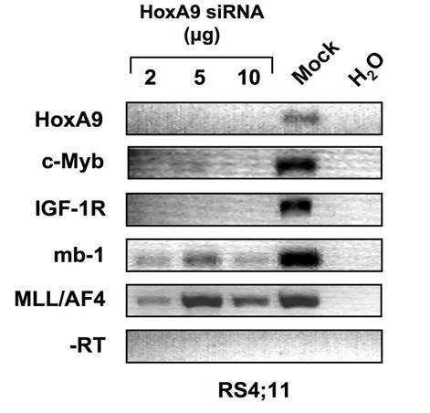
90 75
The Development of Lymphocytes: B Cell Development in the Bone Marrow & Peripheral Lymphoid Tissue Deborah A. Lebman, Ph.D.
 The Development of Lymphocytes: B Cell Development in the Bone Marrow & Peripheral Lymphoid Tissue Deborah A. Lebman, Ph.D. OBJECTIVES 1. To understand how ordered Ig gene rearrangements lead to the development
Introduction. Introduction. Lymphocyte development (maturation)
") Introduction Abbas Chapter 8: Lymphocyte Development and the Rearrangement and Expression of Antigen Receptor Genes Christina Ciaccio, MD Children s Mercy Hospital January 5, 2009 Lymphocyte development
Introduction Abbas Chapter 8: Lymphocyte Development and the Rearrangement and Expression of Antigen Receptor Genes Christina Ciaccio, MD Children s Mercy Hospital January 5, 2009 Lymphocyte development
Development of B and T lymphocytes
 Development of B and T lymphocytes What will we discuss today? B-cell development T-cell development B- cell development overview Stem cell In periphery Pro-B cell Pre-B cell Immature B cell Mature B cell
Principles of Adaptive Immunity
 Principles of Adaptive Immunity Chapter 3 Parham Hans de Haard 17 th of May 2010 Agenda Recognition molecules of adaptive immune system Features adaptive immune system Immunoglobulins and T-cell receptors
all of the above the ability to impart long term memory adaptive immunity all of the above bone marrow none of the above
 1. (3 points) Immediately after a pathogen enters the body, it faces the cells and soluble proteins of the innate immune system. Which of the following are characteristics of innate immunity? a. inflammation
CELL BIOLOGY - CLUTCH CH THE IMMUNE SYSTEM.
 !! www.clutchprep.com CONCEPT: OVERVIEW OF HOST DEFENSES The human body contains three lines of against infectious agents (pathogens) 1. Mechanical and chemical boundaries (part of the innate immune system)
Chapter 11. B cell generation, Activation, and Differentiation. Pro-B cells. - B cells mature in the bone marrow.
 Chapter B cell generation, Activation, and Differentiation - B cells mature in the bone marrow. - B cells proceed through a number of distinct maturational stages: ) Pro-B cell ) Pre-B cell ) Immature
The Adaptive Immune Response. B-cells
 The Adaptive Immune Response B-cells The innate immune system provides immediate protection. The adaptive response takes time to develop and is antigen specific. Activation of B and T lymphocytes Naive
Leukemias and Lymphomas Come From Normal Blood Cells
 Leukemias and Lymphomas Come From Normal Blood Cells by Steve Anderson, Ph.D. Steve Anderson has a Ph.D. in Immunology with 25 years experience in biomedical research. His scientific expertise includes
The Immune System. These are classified as the Innate and Adaptive Immune Responses. Innate Immunity
 The Immune System Biological mechanisms that defend an organism must be 1. triggered by a stimulus upon injury or pathogen attack 2. able to counteract the injury or invasion 3. able to recognise foreign
Andrea s SI Session PCB Practice Test Test 3
 Practice Test Test 3 READ BEFORE STARTING PRACTICE TEST: Remember to please use this practice test as a tool to measure your knowledge, and DO NOT use it as your only tool to study for the test, since
Adaptive Immunity: Humoral Immune Responses
 MICR2209 Adaptive Immunity: Humoral Immune Responses Dr Allison Imrie 1 Synopsis: In this lecture we will review the different mechanisms which constitute the humoral immune response, and examine the antibody
Hematopoiesis. Hematopoiesis. Hematopoiesis
 Chapter. Cells and Organs of the Immune System Hematopoiesis Hematopoiesis- formation and development of WBC and RBC bone marrow. Hematopoietic stem cell- give rise to any blood cells (constant number,
The Adaptive Immune Responses
 The Adaptive Immune Responses The two arms of the immune responses are; 1) the cell mediated, and 2) the humoral responses. In this chapter we will discuss the two responses in detail and we will start
Immunology - Lecture 2 Adaptive Immune System 1
 Immunology - Lecture 2 Adaptive Immune System 1 Book chapters: Molecules of the Adaptive Immunity 6 Adaptive Cells and Organs 7 Generation of Immune Diversity Lymphocyte Antigen Receptors - 8 CD markers
Ig light chain rearrangement: Rescue pathway
 B Cell Development Ig light chain rearrangement: Rescue pathway There is only a 1:3 chance of the join between the V and J region being in frame Vk Jk Ck Non-productive Rearrangement Light chain has a
Chapter 1. Chapter 1 Concepts. MCMP422 Immunology and Biologics Immunology is important personally and professionally!
 MCMP422 Immunology and Biologics Immunology is important personally and professionally! Learn the language - use the glossary and index RNR - Reading, Note taking, Reviewing All materials in Chapters 1-3
Defensive mechanisms include :
 Acquired Immunity Defensive mechanisms include : 1) Innate immunity (Natural or Non specific) 2) Acquired immunity (Adaptive or Specific) Cell-mediated immunity Humoral immunity Two mechanisms 1) Humoral
Adaptive immune responses: T cell-mediated immunity
 MICR2209 Adaptive immune responses: T cell-mediated immunity Dr Allison Imrie allison.imrie@uwa.edu.au 1 Synopsis: In this lecture we will discuss the T-cell mediated immune response, how it is activated,
Medical Virology Immunology. Dr. Sameer Naji, MB, BCh, PhD (UK) Head of Basic Medical Sciences Dept. Faculty of Medicine The Hashemite University
 Head of Basic Medical Sciences Dept. Faculty of Medicine The Hashemite University") Medical Virology Immunology Dr. Sameer Naji, MB, BCh, PhD (UK) Head of Basic Medical Sciences Dept. Faculty of Medicine The Hashemite University Human blood cells Phases of immune responses Microbe Naïve
Medical Virology Immunology Dr. Sameer Naji, MB, BCh, PhD (UK) Head of Basic Medical Sciences Dept. Faculty of Medicine The Hashemite University Human blood cells Phases of immune responses Microbe Naïve
Immune System AP SBI4UP
 Immune System AP SBI4UP TYPES OF IMMUNITY INNATE IMMUNITY ACQUIRED IMMUNITY EXTERNAL DEFENCES INTERNAL DEFENCES HUMORAL RESPONSE Skin Phagocytic Cells CELL- MEDIATED RESPONSE Mucus layer Antimicrobial
WHY IS THIS IMPORTANT?
 CHAPTER 16 THE ADAPTIVE IMMUNE RESPONSE WHY IS THIS IMPORTANT? The adaptive immune system protects us from many infections The adaptive immune system has memory so we are not infected by the same pathogen
Adaptive Immune System
 Short Course on Immunology Adaptive Immune System Bhargavi Duvvuri Ph.D IIIrd Year (Immunology) bhargavi@yorku.ca Supervisor Dr.Gillian E Wu Professor, School of Kinesiology and Health Sciences York University,
LESSON 2: THE ADAPTIVE IMMUNITY
 Introduction to immunology. LESSON 2: THE ADAPTIVE IMMUNITY Today we will get to know: The adaptive immunity T- and B-cells Antigens and their recognition How T-cells work 1 The adaptive immunity Unlike
The T cell receptor for MHC-associated peptide antigens
 1 The T cell receptor for MHC-associated peptide antigens T lymphocytes have a dual specificity: they recognize polymporphic residues of self MHC molecules, and they also recognize residues of peptide
Antigen-Independent B-Cell Development Bone Marrow
 Antigen-Independent B-Cell Development Bone Marrow 1. DNA rearrangements establish the primary repertoire, creating diversity 2. Allelic exclusion ensures that each clone expresses a single antibody on
COURSE: Medical Microbiology, MBIM 650/720 - Fall TOPIC: Antigen Processing, MHC Restriction, & Role of Thymus Lecture 12
 COURSE: Medical Microbiology, MBIM 650/720 - Fall 2008 TOPIC: Antigen Processing, MHC Restriction, & Role of Thymus Lecture 12 FACULTY: Dr. Mayer Office: Bldg. #1, Rm B32 Phone: 733-3281 Email: MAYER@MED.SC.EDU
Question 1. Kupffer cells, microglial cells and osteoclasts are all examples of what type of immune system cell?
 Abbas Chapter 2: Sarah Spriet February 8, 2015 Question 1. Kupffer cells, microglial cells and osteoclasts are all examples of what type of immune system cell? a. Dendritic cells b. Macrophages c. Monocytes
1. Overview of Adaptive Immunity
 Chapter 17A: Adaptive Immunity Part I 1. Overview of Adaptive Immunity 2. T and B Cell Production 3. Antigens & Antigen Presentation 4. Helper T cells 1. Overview of Adaptive Immunity The Nature of Adaptive
White Blood Cells (WBCs)
") YOUR ACTIVE IMMUNE DEFENSES 1 ADAPTIVE IMMUNE RESPONSE 2! Innate Immunity - invariant (generalized) - early, limited specificity - the first line of defense 1. Barriers - skin, tears 2. Phagocytes - neutrophils,
YOUR ACTIVE IMMUNE DEFENSES 1 ADAPTIVE IMMUNE RESPONSE 2! Innate Immunity - invariant (generalized) - early, limited specificity - the first line of defense 1. Barriers - skin, tears 2. Phagocytes - neutrophils,
ACTIVATION OF T LYMPHOCYTES AND CELL MEDIATED IMMUNITY
 ACTIVATION OF T LYMPHOCYTES AND CELL MEDIATED IMMUNITY The recognition of specific antigen by naïve T cell induces its own activation and effector phases. T helper cells recognize peptide antigens through
TARGETED THERAPY FOR CHILDHOOD CANCERS
 TARGETED THERAPY FOR CHILDHOOD CANCERS AZIZA SHAD, MD AMEY DISTINGUISHED PROFESSOR OF PEDIATRIC HEMATOLOGY ONCOLOGY, BLOOD AND MARROW TRANSPLANTATION LOMBARDI CANCER CENTER GEORGETOWN UNIVERSITY HOSPITAL
Immunology Lecture 4. Clinical Relevance of the Immune System
 Immunology Lecture 4 The Well Patient: How innate and adaptive immune responses maintain health - 13, pg 169-181, 191-195. Immune Deficiency - 15 Autoimmunity - 16 Transplantation - 17, pg 260-270 Tumor
Helminth worm, Schistosomiasis Trypanosomes, sleeping sickness Pneumocystis carinii. Ringworm fungus HIV Influenza
 Helminth worm, Schistosomiasis Trypanosomes, sleeping sickness Pneumocystis carinii Ringworm fungus HIV Influenza Candida Staph aureus Mycobacterium tuberculosis Listeria Salmonella Streptococcus Levels
Follicular Lymphoma. ced3 APOPTOSIS. *In the nematode Caenorhabditis elegans 131 of the organism's 1031 cells die during development.
 Harvard-MIT Division of Health Sciences and Technology HST.176: Cellular and Molecular Immunology Course Director: Dr. Shiv Pillai Follicular Lymphoma 1. Characterized by t(14:18) translocation 2. Ig heavy
Chapter 22: The Lymphatic System and Immunity
 Bio40C schedule Lecture Immune system Lab Quiz 2 this week; bring a scantron! Study guide on my website (see lab assignments) Extra credit Critical thinking questions at end of chapters 5 pts/chapter Due
ACTIVATION AND EFFECTOR FUNCTIONS OF CELL-MEDIATED IMMUNITY AND NK CELLS. Choompone Sakonwasun, MD (Hons), FRCPT
, FRCPT") ACTIVATION AND EFFECTOR FUNCTIONS OF CELL-MEDIATED IMMUNITY AND NK CELLS Choompone Sakonwasun, MD (Hons), FRCPT Types of Adaptive Immunity Types of T Cell-mediated Immune Reactions CTLs = cytotoxic T lymphocytes
ACTIVATION AND EFFECTOR FUNCTIONS OF CELL-MEDIATED IMMUNITY AND NK CELLS Choompone Sakonwasun, MD (Hons), FRCPT Types of Adaptive Immunity Types of T Cell-mediated Immune Reactions CTLs = cytotoxic T lymphocytes
M.Sc. III Semester Biotechnology End Semester Examination, 2013 Model Answer LBTM: 302 Advanced Immunology
 Code : AS-2246 M.Sc. III Semester Biotechnology End Semester Examination, 2013 Model Answer LBTM: 302 Advanced Immunology A. Select one correct option for each of the following questions:- 2X10=10 1. (b)
WBCs Disorders 1. Dr. Nabila Hamdi MD, PhD
 WBCs Disorders 1 Dr. Nabila Hamdi MD, PhD ILOs Compare and contrast ALL, AML, CLL, CML in terms of age distribution, cytogenetics, morphology, immunophenotyping, laboratory diagnosis clinical features
Molecular Hematopathology Leukemias I. January 14, 2005
 Molecular Hematopathology Leukemias I January 14, 2005 Chronic Myelogenous Leukemia Diagnosis requires presence of Philadelphia chromosome t(9;22)(q34;q11) translocation BCR-ABL is the result BCR on chr
Immunobiology 7. The Humoral Immune Response
 Janeway Murphy Travers Walport Immunobiology 7 Chapter 9 The Humoral Immune Response Copyright Garland Science 2008 Tim Worbs Institute of Immunology Hannover Medical School 1 The course of a typical antibody
B Lymphocyte Development and Activation
 Harvard-MIT Division of Health Sciences and Technology HST.176: Cellular and Molecular Immunology Course Director: Dr. Shiv Pillai 09/26/05; 9 AM Shiv Pillai B Lymphocyte Development and Activation Recommended
Structure and Function of Antigen Recognition Molecules
 MICR2209 Structure and Function of Antigen Recognition Molecules Dr Allison Imrie allison.imrie@uwa.edu.au 1 Synopsis: In this lecture we will examine the major receptors used by cells of the innate and
TCR, MHC and coreceptors
 Cooperation In Immune Responses Antigen processing how peptides get into MHC Antigen processing involves the intracellular proteolytic generation of MHC binding proteins Protein antigens may be processed
General Overview of Immunology. Kimberly S. Schluns, Ph.D. Associate Professor Department of Immunology UT MD Anderson Cancer Center
 General Overview of Immunology Kimberly S. Schluns, Ph.D. Associate Professor Department of Immunology UT MD Anderson Cancer Center Objectives Describe differences between innate and adaptive immune responses
Chapter 24 The Immune System
 Chapter 24 The Immune System The Immune System Layered defense system The skin and chemical barriers The innate and adaptive immune systems Immunity The body s ability to recognize and destroy specific
The Adaptive Immune Response. T-cells
 The Adaptive Immune Response T-cells T Lymphocytes T lymphocytes develop from precursors in the thymus. Mature T cells are found in the blood, where they constitute 60% to 70% of lymphocytes, and in T-cell
Antigen Presentation and T Lymphocyte Activation. Abul K. Abbas UCSF. FOCiS
 1 Antigen Presentation and T Lymphocyte Activation Abul K. Abbas UCSF FOCiS 2 Lecture outline Dendritic cells and antigen presentation The role of the MHC T cell activation Costimulation, the B7:CD28 family
Andrea s Final Exam Review PCB 3233 Spring Practice Final Exam
 NOTE: Practice Final Exam Although I am posting the answer key for this practice exam, I want you to use this practice to gauge your knowledge, and try to figure out the right answer by yourself before
membrane form secreted form 13 aa 26 aa K K V V K K 3aa
 Harvard-MIT Division of Health Sciences and Technology HST.176: Cellular and Molecular Immunology Course Director: Dr. Shiv Pillai secreted form membrane form 13 aa 26 aa K K V V K K 3aa Hapten monosaccharide
κ λ Antigen-Independent B-Cell Development Bone Marrow Ordered Rearrangement of Ig Genes During B-Cell Development in the Bone Marrow
 Antigen-Independent B-Cell Development Bone Marrow 1. DNA rearrangements establish the primary repertoire, creating diversity 2. Allelic exclusion ensures that each clone expresses a single antibody on
All animals have innate immunity, a defense active immediately upon infection Vertebrates also have adaptive immunity
 1 2 3 4 5 6 7 8 9 The Immune System All animals have innate immunity, a defense active immediately upon infection Vertebrates also have adaptive immunity Figure 43.2 In innate immunity, recognition and
Immunology for the Rheumatologist
 Immunology for the Rheumatologist Rheumatologists frequently deal with the immune system gone awry, rarely studying normal immunology. This program is an overview and discussion of the function of the
B cell development in the bone marrow.
 development in the bone. s develop from multipotent haematopoietic stem s in the bone that produce lymphoid progenitors which in turn generate s. s with a unique antibody specificity develop by initial
Myeloproliferative Disorders - D Savage - 9 Jan 2002
 Disease Usual phenotype acute leukemia precursor chronic leukemia low grade lymphoma myeloma differentiated Total WBC > 60 leukemoid reaction acute leukemia Blast Pro Myel Meta Band Seg Lymph 0 0 0 2
T-cell activation T cells migrate to secondary lymphoid tissues where they interact with antigen, antigen-presenting cells, and other lymphocytes:
 Interactions between innate immunity & adaptive immunity What happens to T cells after they leave the thymus? Naïve T cells exit the thymus and enter the bloodstream. If they remain in the bloodstream,
Effector T Cells and
 1 Effector T Cells and Cytokines Andrew Lichtman, MD PhD Brigham and Women's Hospital Harvard Medical School 2 Lecture outline Cytokines Subsets of CD4+ T cells: definitions, functions, development New
SINGLE CHOICE. 5. The gamma invariant chain binds to this molecule during its intracytoplasmic transport. A TCR B BCR C MHC II D MHC I E FcγR
 A Name: Group: SINGLE CHOICE 1. Which is the most important ligand of TLR5? A endospore B flagellin C polysaccharide capsule D DNA E pilus 2. The antibody-binding site is formed primarily by... A the constant
The development of T cells in the thymus
 T cells rearrange their receptors in the thymus whereas B cells do so in the bone marrow. The development of T cells in the thymus The lobular/cellular organization of the thymus Immature cells are called
Properties & Overview of IRs Dr. Nasser M. Kaplan JUST, Jordan. 10-Jul-16 NM Kaplan 1
 Properties & Overview of IRs Dr. Nasser M. Kaplan JUST, Jordan 10-Jul-16 NM Kaplan 1 Major components of IS & their properties Definitions IS = cells & molecules responsible for: 1- Physiologic; protective
Adaptive Immunity: Specific Defenses of the Host
 17 Adaptive Immunity: Specific Defenses of the Host SLOs Differentiate between innate and adaptive immunity, and humoral and cellular immunity. Define antigen, epitope, and hapten. Explain the function
Immune response. This overview figure summarizes simply how our body responds to foreign molecules that enter to it.
 Immune response This overview figure summarizes simply how our body responds to foreign molecules that enter to it. It s highly recommended to watch Dr Najeeb s lecture that s titled T Helper cells and
Introduction to Immunology Lectures 1-3 by Bellur S. Prabhakar. March 13-14, 2007
 Introduction to Immunology Lectures 1-3 by Bellur S. Prabhakar. March 13-14, 2007 TheComponents Of The Immune System and Innate Immunity: Ref: Immunobiology-5 th edition. Janeway et al. Chapters-1 & 2.
Effector mechanisms of cell-mediated immunity: Properties of effector, memory and regulatory T cells
 ICI Basic Immunology course Effector mechanisms of cell-mediated immunity: Properties of effector, memory and regulatory T cells Abul K. Abbas, MD UCSF Stages in the development of T cell responses: induction
5/1/13. The proportion of thymus that produces T cells decreases with age. The cellular organization of the thymus
 T cell precursors migrate from the bone marrow via the blood to the thymus to mature 1 2 The cellular organization of the thymus The proportion of thymus that produces T cells decreases with age 3 4 1
Overview of the Lymphoid System
 Overview of the Lymphoid System The Lymphoid System Protects us against disease Lymphoid system cells respond to Environmental pathogens Toxins Abnormal body cells, such as cancers Overview of the Lymphoid
Test Bank for Basic Immunology Functions and Disorders of the Immune System 4th Edition by Abbas
 Test Bank for Basic Immunology Functions and Disorders of the Immune System 4th Edition by Abbas Chapter 04: Antigen Recognition in the Adaptive Immune System Test Bank MULTIPLE CHOICE 1. Most T lymphocytes
Introduction to Immunology Part 2 September 30, Dan Stetson
 Introduction to Immunology Part 2 September 30, 2016 Dan Stetson stetson@uw.edu 441 Lecture #2 Slide 1 of 26 CLASS ANNOUNCEMENT PLEASE NO TREE NUTS IN CLASS!!! (Peanuts, walnuts, almonds, cashews, etc)
Immunopathology. 2-Patterned hemodynamic responses, cell surface associated and soluble mediator systems (e.g., complement and coagulation systems).
.") Immunopathology The chief role of the immune system is to protect the host from invasion by foreign agents. Immune responses can be elicited by a wide range of agents including toxins, drugs, chemicals,
Immunopathology The chief role of the immune system is to protect the host from invasion by foreign agents. Immune responses can be elicited by a wide range of agents including toxins, drugs, chemicals,
SPECIFIC IMMUNITY = ACQUIRED IMMUNITY = ADAPTIVE IMMUNITY SPECIFIC IMMUNITY - BASIC CHARACTERISTIC
 SPECIFIC IMMUNITY - BASIC CHARACTERISTIC SPECIFIC IMMUNITY = ACQUIRED IMMUNITY = ADAPTIVE IMMUNITY BASIC TERMINOLOGY SPECIFIC IMMUNITY humoral mediated with antibodies cellular mediated with T lymphocytes
MICROBIO320 EXAM 1-Spring 2011 Name True/False (1 point each) T 2. T cell receptors are composed of constant and variable regions.
 T 2. T cell receptors are composed of constant and variable regions.") True/False (1 point each) T 1. Mature T cells (lymphocytes) bind only to processed antigen. T 2. T cell receptors are composed of constant and variable regions. F 3. Natural Killer cells do not proliferate
True/False (1 point each) T 1. Mature T cells (lymphocytes) bind only to processed antigen. T 2. T cell receptors are composed of constant and variable regions. F 3. Natural Killer cells do not proliferate
Innate immunity (rapid response) Dendritic cell. Macrophage. Natural killer cell. Complement protein. Neutrophil
 Dendritic cell. Macrophage. Natural killer cell. Complement protein. Neutrophil") 1 The immune system The immune response The immune system comprises two arms functioning cooperatively to provide a comprehensive protective response: the innate and the adaptive immune system. The innate
1 The immune system The immune response The immune system comprises two arms functioning cooperatively to provide a comprehensive protective response: the innate and the adaptive immune system. The innate
CYTOKINE RECEPTORS AND SIGNAL TRANSDUCTION
 CYTOKINE RECEPTORS AND SIGNAL TRANSDUCTION What is Cytokine? Secreted popypeptide (protein) involved in cell-to-cell signaling. Acts in paracrine or autocrine fashion through specific cellular receptors.
Third line of Defense. Topic 8 Specific Immunity (adaptive) (18) 3 rd Line = Prophylaxis via Immunization!
 (18) 3 rd Line = Prophylaxis via Immunization!") Topic 8 Specific Immunity (adaptive) (18) Topics - 3 rd Line of Defense - B cells - T cells - Specific Immunities 1 3 rd Line = Prophylaxis via Immunization! (a) A painting of Edward Jenner depicts a cow
Topic 8 Specific Immunity (adaptive) (18) Topics - 3 rd Line of Defense - B cells - T cells - Specific Immunities 1 3 rd Line = Prophylaxis via Immunization! (a) A painting of Edward Jenner depicts a cow
MACROPHAGE "MONOCYTES" SURFACE RECEPTORS
 LECTURE: 13 Title: MACROPHAGE "MONOCYTES" SURFACE RECEPTORS LEARNING OBJECTIVES: The student should be able to: Describe the blood monocytes (size, and shape of nucleus). Enumerate some of the monocytes
Blood and Immune system Acquired Immunity
 Blood and Immune system Acquired Immunity Immunity Acquired (Adaptive) Immunity Defensive mechanisms include : 1) Innate immunity (Natural or Non specific) 2) Acquired immunity (Adaptive or Specific) Cell-mediated
Chapter 17B: Adaptive Immunity Part II
 Chapter 17B: Adaptive Immunity Part II 1. Cell-Mediated Immune Response 2. Humoral Immune Response 3. Antibodies 1. The Cell-Mediated Immune Response Basic Steps of Cell-Mediated IR 1 2a CD4 + MHC cl.
Allergy and Immunology Review Corner: Chapter 1 of Immunology IV: Clinical Applications in Health and Disease, by Joseph A. Bellanti.
 Allergy and Immunology Review Corner: Chapter 1 of Immunology IV: Clinical Applications in Health and Disease, by Joseph A. Bellanti. Chapter 1: Overview of Immunology Prepared by David Scott, MD, Scripps
The Immune System: Innate and Adaptive Body Defenses Outline PART 1: INNATE DEFENSES 21.1 Surface barriers act as the first line of defense to keep
 The Immune System: Innate and Adaptive Body Defenses Outline PART 1: INNATE DEFENSES 21.1 Surface barriers act as the first line of defense to keep invaders out of the body (pp. 772 773; Fig. 21.1; Table
Immunity. Chapter 38
 Immunity Chapter 38 Impacts, Issues Frankie s Last Wish Infection with a common, sexually transmitted virus (HPV) causes most cervical cancers including the one that killed Frankie McCullogh 38.1 Integrated
Third line of Defense
 Chapter 15 Specific Immunity and Immunization Topics -3 rd of Defense - B cells - T cells - Specific Immunities Third line of Defense Specific immunity is a complex interaction of immune cells (leukocytes)
Prepared by: Dr.Mansour Al-Yazji
 C L L CLL Prepared by: Abd El-Hakeem Abd El-Rahman Abu Naser Ahmed Khamis Abu Warda Ahmed Mohammed Abu Ghaben Bassel Ziad Abu Warda Nedal Mostafa El-Nahhal Dr.Mansour Al-Yazji LEUKEMIA Leukemia is a form
HEMATOLOGIC MALIGNANCIES BIOLOGY
 HEMATOLOGIC MALIGNANCIES BIOLOGY Failure of terminal differentiation Failure of differentiated cells to undergo apoptosis Failure to control growth Neoplastic stem cell FAILURE OF TERMINAL DIFFERENTIATION
chapter 17: specific/adaptable defenses of the host: the immune response
 chapter 17: specific/adaptable defenses of the host: the immune response defense against infection & illness body defenses innate/ non-specific adaptable/ specific epithelium, fever, inflammation, complement,
RAS Genes. The ras superfamily of genes encodes small GTP binding proteins that are responsible for the regulation of many cellular processes.
 ۱ RAS Genes The ras superfamily of genes encodes small GTP binding proteins that are responsible for the regulation of many cellular processes. Oncogenic ras genes in human cells include H ras, N ras,
Immunology Lesson plan for international medical students
 Immunology Lesson plan for international medical students Kermanshah University of medical sciences (KUMS) international affairs School of medicine Department of Immunology Lecturers: Dr. Ali Gorgin Karaji
SPECIFIC AIMS. II year (1st semester)
") II year (1st semester) Scientific Field IMMUNOLOGY AND IMMUNOPATHOLOGY TUTOR ECTS MALISAN F. COORDINATOR MED/04 Immunology and Immunopathology Malisan Florence 5 MED/04 Immunology and Immunopathology Testi
II year (1st semester) Scientific Field IMMUNOLOGY AND IMMUNOPATHOLOGY TUTOR ECTS MALISAN F. COORDINATOR MED/04 Immunology and Immunopathology Malisan Florence 5 MED/04 Immunology and Immunopathology Testi
CHAPTER:4 LEUKEMIA. BY Mrs. K.SHAILAJA., M. PHARM., LECTURER DEPT OF PHARMACY PRACTICE, SRM COLLEGE OF PHARMACY 8/12/2009
 LEUKEMIA CHAPTER:4 1 BY Mrs. K.SHAILAJA., M. PHARM., LECTURER DEPT OF PHARMACY PRACTICE, SRM COLLEGE OF PHARMACY Leukemia A group of malignant disorders affecting the blood and blood-forming tissues of
T cell Receptor. Chapter 9. Comparison of TCR αβ T cells
 Chapter 9 The αβ TCR is similar in size and structure to an antibody Fab fragment T cell Receptor Kuby Figure 9-3 The αβ T cell receptor - Two chains - α and β - Two domains per chain - constant (C) domain
The Innate Immune Response
 The Innate Immune Response FUNCTIONS OF THE IMMUNE SYSTEM: Recognize, destroy and clear a diversity of pathogens. Initiate tissue and wound healing processes. Recognize and clear damaged self components.
General Biology. A summary of innate and acquired immunity. 11. The Immune System. Repetition. The Lymphatic System. Course No: BNG2003 Credits: 3.
 A summary of innate and acquired immunity General iology INNATE IMMUNITY Rapid responses to a broad range of microbes Course No: NG00 Credits:.00 External defenses Invading microbes (pathogens). The Immune
Campbell's Biology: Concepts and Connections, 7e (Reece et al.) Chapter 24 The Immune System Multiple-Choice Questions
 Chapter 24 The Immune System Multiple-Choice Questions") Campbell's Biology: Concepts and Connections, 7e (Reece et al.) Chapter 24 The Immune System 24.1 Multiple-Choice Questions 1) The body's innate defenses against infection include A) several nonspecific
Campbell's Biology: Concepts and Connections, 7e (Reece et al.) Chapter 24 The Immune System 24.1 Multiple-Choice Questions 1) The body's innate defenses against infection include A) several nonspecific
Chapter 10 (pages ): Differentiation and Functions of CD4+ Effector T Cells Prepared by Kristen Dazy, MD, Scripps Clinic Medical Group
: Differentiation and Functions of CD4+ Effector T Cells Prepared by Kristen Dazy, MD, Scripps Clinic Medical Group") FIT Board Review Corner September 2015 Welcome to the FIT Board Review Corner, prepared by Andrew Nickels, MD, and Sarah Spriet, DO, senior and junior representatives of ACAAI's Fellows-In-Training (FITs)
FIT Board Review Corner September 2015 Welcome to the FIT Board Review Corner, prepared by Andrew Nickels, MD, and Sarah Spriet, DO, senior and junior representatives of ACAAI's Fellows-In-Training (FITs)
Chapter 23 Immunity Exam Study Questions
 Chapter 23 Immunity Exam Study Questions 1. Define 1) Immunity 2) Neutrophils 3) Macrophage 4) Epitopes 5) Interferon 6) Complement system 7) Histamine 8) Mast cells 9) Antigen 10) Antigens receptors 11)
Mon, Wed, Fri 11:00 AM-12:00 PM. Owen, Judy, Jenni Punt, and Sharon Stranford Kuby-Immunology, 7th. Edition. W.H. Freeman and Co., New York.
 Course Title: Course Number: Immunology Biol-341/541 Semester: Fall 2013 Location: HS 268 Time: Instructor: 8:00-9:30 AM Tue/Thur Dr. Colleen M. McDermott Office: Nursing Ed 101 (424-1217) E-mail*: mcdermot@uwosh.edu
April 01, Immune system.notebook
 I. First Line of Defense: Skin and Mucus Membranes Non Specific A. Skin Surface 1. dry, dead, thick, secretions 2. sweat and sebaceous glands: antibiotics, lactic acid, RNase B. Mucus (moist and sometimes
Done By : WESSEN ADNAN BUTHAINAH AL-MASAEED
 Done By : WESSEN ADNAN BUTHAINAH AL-MASAEED Acute Myeloid Leukemia Firstly we ll start with this introduction then enter the title of the lecture, so be ready and let s begin by the name of Allah : We
MCAT Biology - Problem Drill 16: The Lymphatic and Immune Systems
 MCAT Biology - Problem Drill 16: The Lymphatic and Immune Systems Question No. 1 of 10 1. Which of the following statements about pathogens is true? Question #01 (A) Both viruses and bacteria need to infect