BI-RADS and Breast MRI. Kathy Borovicka, M.D. Thursday February 15, 2018
|
|
|
- Doreen Richards
- 6 years ago
- Views:
Transcription
1 BI-RADS and Breast MRI Kathy Borovicka, M.D. Thursday February 15, 2018
2 Learning Objectives Be familiar with the Breast Imaging Reporting and Data System (BI-RADS) Understand the components of a breast imaging report, including the description of breast density, description of important findings, assessment and management. Be familiar with the indications for breast magnetic resonance imaging (MRI) Understand the terminology used in reporting the findings for breast MRI including mass and non mass enhancement
3 BI-RADS Breast Imaging Reporting and Data System American College of Radiology Most recent: 5th Edition (2013) Communication among physicians using common terminology with clear, concise, and standardized reporting Mammmography, US, MRI Follow up and outcome monitoring
4 BI-RADS 0: Incomplete assessment 1: Negative. 2: Benign 3: Probably benign 4: Suspicious 5: Highly suspicious 6: Known carcinoma
5 BI-RADS 0 Usually given for screening examinations with additional imaging recommended. Or possibly request for comparison with prior examination(s) within 30 days The radiologist describes the additional mammography views and ultrasound if needed on the call back form
6 BI-RADS 1 & 2 BI-RADS 1: Negative BI-RADS 2: Benign calcified fibroadenomas, skin calcifications, metallic foreign bodies (core biopsy and surgical clips), fat containing lesions (oil cysts, lipomas, galactoceles, and hamartomas) intramammary lymph nodes, vascular calcifications, architectural distortion from prior surgery, implants Recommend routine mammography screening Essentially 0% likelihood of malignancy
7 BI-RADS 3 Probably benign Short-interval (typically 6 month) follow up to demonstrate at least 2 year stability Greater than 0% to less or equal to 2% likelihood of malignancy Noncalcified circumscribed solid mass, focal asymmetry, and a solitary group of punctate calcifications
8 BI-RADS 4 Suspicious Can be further categorized into: 4A: Low suspicion of malignancy: > 2% to 10% likelihood of malignancy 4B: Moderate suspicion of malignancy: > 10% to 50% likelihood of malignancy 4C: > 50% to than 95% likelihood of malignancy
9 BI-RADS 5 Highly suggestive of malignancy Tissue diagnosis 95% likelihood of malignancy Identifies lesions for which any non-malignant percutaneous tissue diagnosis is automatically considered discordant, resulting in the recommendation for repeat (usually surgical) biopsy
10 BI-RADS 6 Known biopsy proven malignancy Reserved for examinations performed after biopsy proven malignancy in which there are no additional imaging abnormalities other than then known cancer Preoperative breast MRI Surgical excision when clinically appropriate
11 Breast Imaging Radiology Report 1. Indication for examination History: Personal or Family history of breast cancer Prior biopsies and pathologies including dates 2. Comparison to prior breast imaging 3. Description of overall breast composition 4. Clear description of important findings 5. Assessment 6. Management
12 Breast Magnetic Resonance Imaging (MRI) Breast MRI has superior sensitivity to detecting malignancy in comparison to mammography and ultrasound Overlap in benign and malignant diseases limits the specificity of breast MRI Higher cost Limited availability
13 *Indications for breast MRI American Cancer Society and American College of Radiology: Indicators for the performance of breast MRI 1. Preoperative evaluation: extent of disease. Multifocal: multiple foci of disease in one quadrant Multicentric: multiple disease in different quadrants Additional unilateral disease: 10-27% patients Contralateral disease: 3% patients Tumor size is more accurately depicted with breast MRI Helpful: dense breasts, invasive lobular carcinoma, extensive ductal carcinoma in situ, those who will be treated with neoadjuvant chemotherapy

14 JAOCR. Oct 2017

15 JAOCR. Oct 2017
16 Invasive lobular carcinoma Infiltrative growth pattern (single file growth along normal tissue planes) Mammography and ultrasound underestimate the tumor size of invasive lobular carcinoma Posteriorly: MRI helps with detection of invasion into the pectorals musculature (enhancement) as well as chest wall invasion
17 Indications for breast MRI 2. Post neoadjuvant chemotherapy: decrease the tumor size preoperatively to offer breast conservation axillary mets locally advanced disease Compare the per to post neoadjuvant chemotherapy breast imaging Importance of placement of biopsy markers in case tumor significantly shrinks and no longer seen with imaging
18 Indications for breast MRI 3. Positive surgical margins, post breast conservation identify the extent of residual disease which aids in surgical planning for re-exision granulation tissue in the tumor post may result enhancement in the early postoperative period the least false-positive results were found when MRI was performed between days following surgery (Frei et al.)
19 Indications for breast MRI 4. Metastatic axillary lymphadenopathy, unknown primary malignancy Usually <1% of all breast carcinoma cases MRI can detect 62-86% of occult primary breast cancer option: breast conservation therapy rather than mastectomy targeted hormonal and chemotherapeutic treatments based on the histology
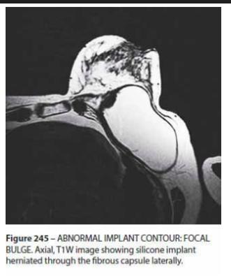
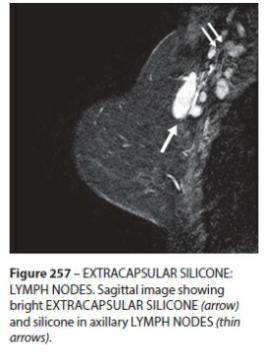
20 Indications for breast MRI 5. Silicone breast implant integrity Noncontrast Intracapsular silicone implant rupture: implant envelope degrades but the silicone is still contained by the surrounding fibrous capsule. MRI: subcapsular, linguine, keyhole/noose signs Extracapsular silicone implant rupture: free silicone outside the fibrous capsule within the breast and axilla
21 MRI: Implants 1. Implant material and lumen type saline silicone 2. Location: retroglandular, retropectoral 3. Abnormal implant contour: focal bulge 4. Implant rupture: intracapsular +/- extracapsular rupture 6. Peri-implant fluid (post-surgical, ialcl: implant-associated anaplastic large cell lymphoma)

22
23
24 Indications for breast MRI 6. Breast cancer screening: MRI is used as an adjunct to mammography in high-risk women Not appropriate for screening in the general average risk population High risk women: BRCA1 and BRCA2 gene mutations and their untested first-degree relatives Prior chest radiation between ages Certain syndromes: Lifetime risk for breast cancer >20% as determined by risk models
25 Insufficient evidence for women with intermediate risk: lifetime risk between 15-20% diagnosis of atypia or lobular carcinoma in situ (LCIS) dense breasts on mammography personal history of breast cancer Case-by-case basis
26 Indications for breast MRI 7. Equivocal mammography findings Used as a problem-solving tool 3 dimensional localization of a lesion seen on only 1 view: asymmetries, architectural distortions, and equivocal changes in the appearance of prior surgical or biopsy sites Recent study of 115 patients, MRI was 100% sensitive and 92% specific in such a setting Aid in recommendation for short term follow up versus biopsy
27 MRI Report Indication Technique Amount of intravenous contrast given (Gadavist- weight dependent) Breast density: fat, scattered, heterogeneous, extremely dense Background parenchymal enhancement (BPE): minimal, mild, moderate, marked BPE: symmetric or asymmetric (postradiaton or pathology)
28 MRI Sequences Localizer Precontrast axial and sagittal T1 T2: fluid sensitive sequence Postcontrast T1 axial fat sat (4) Subtracted images (4) Color images
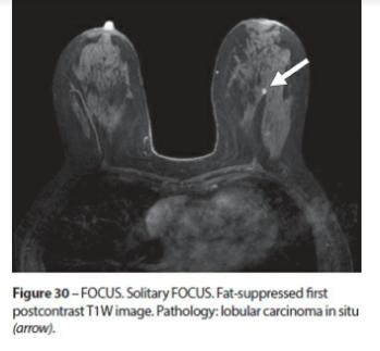
29 MRI: Focus Focus: punctate (dot) enhancement, <5mm, no borders, nonspecific, and too small to be characterized morphologically Multiple: background parenchymal enhancement (BPE) with foci separated widely in the breast by normal fibroglandular breast tissue and fat Suspicious: Unique and distinct from BPE, solitary focus, no fatty hilum, washout kinetics, new or increased from prior Benign: not unique when compared to BPE, bright on T2, fatty hilum, persistent kinetics, stable

30
31
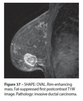
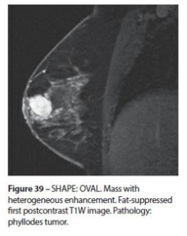
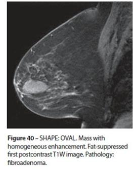
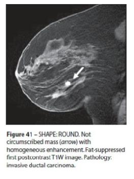
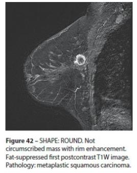
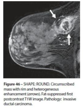
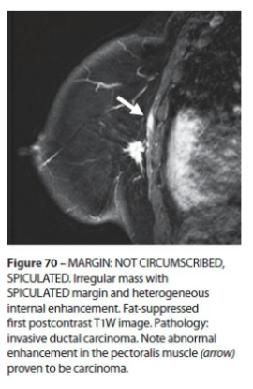
32 MRI: Mass 3D space-occupying structure, convex-outward contour Shape: oval (includes lobulated), round, or irregular Margin: circumscribed, irregular, spiculated Internal architecture Internal mass enhancement characteristics: homogenenous (confluent + uniform), heterogeneous (nonuniform), rim enhancement, dark internal separations (suggests a fibroadenoma)
33 MRI: Margin margin analysis depends greatly on the spatial resolution, such that a not circumscribed margin may appear relatively circumscribed on low spatial resolution scans mammography has extremely high spatial resolution that may not be matched with the current magnets in clinical use today margin and shape analysis should be performed on the first postcontrast image to avoid washout or progressive enhancement of the surrounding tissue
34
35


36
37
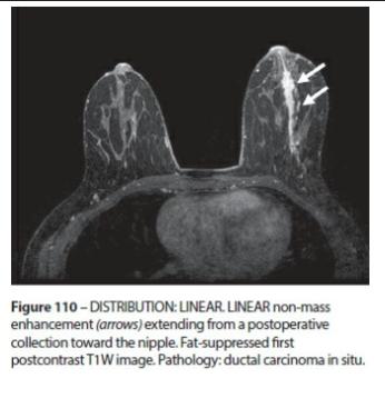
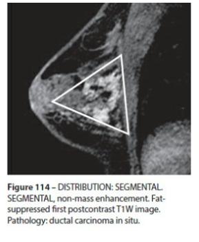
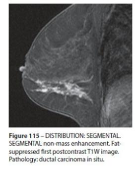
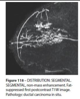
38 MRI: Non-mass enhancement (NME) Not a focus or a mass Distribution: focal area, linear, segmental, regional, multiple regions, or diffuse Focal area: confined area of the breast, less than a breast quadrant and within a single duct system Linear enhancement: line corresponding to a single duct. Enhancement can be within or surrounding a duct Segmental enhancement: triangular or cone shaped with the apex at the nipple and ducts and their branches extending posteriorly
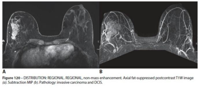
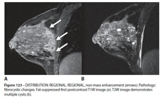
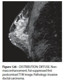
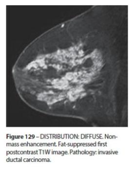
39 MRI: NME Regional enhancement: spans at least a quadrant, broader area than a single duct system, may be geographic, and lacks convex-outward contour Mutliple regions: enhancement over at least two broad areas, separated by normal tissue or fat Diffuse enhancement: widely scattered and evenly distributed, similar appearing enhancement throughout the breast fibroglandular tissue Regional, mutiple and diffuse: characteristic of more benign disease, such as proliferative changes, although multicentric carcinoma may have this appearance.
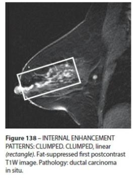
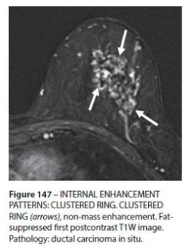
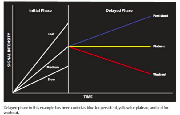
40 MRI: NME Nonmass enhancements can be further described: homogeneous, heterogeneous, clumped, clustered ring Clumped: aggregate of enhancing foci in a cobblestone pattern that my be occasionally confluent. Clustered ring: rim like enhancement around adjacent ducts. Can also see with fibrocystic changes.
41
42
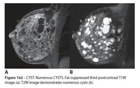
43
44
45
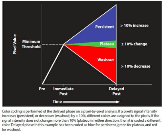
46 Kinetic Curve Assessment Abnormal enhancement: enhancement of higher signal intensity compared to surrounding normal BPE on a contrast enhanced scan Kinetic technique: dynamic measurement in which the uptake and washout contrast in tissues is monitored for a period of time following contrast injection depends on: perfusion, capillary permeability, blood volume, contrast media distribution volume Tumors: dense, highly permeable vasculature, rapid blood flow, microheterogeneity. Tumors therefore enhance more rapidly and more strongly than normal tissue
47 Time intensity curve (TIC) The kinetic information is typically expressed as a TIC CAD (Computer assisted device) systems: calculate kinetic information, generating both color maps and graphs Initial phase of enhancement: slow, medium, fast Threshold: % increase in signal intensity Delayed phase enhancement: persistent, plateau, washout Overall: morphology trumps kinetics
48
49
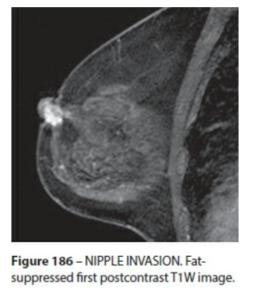
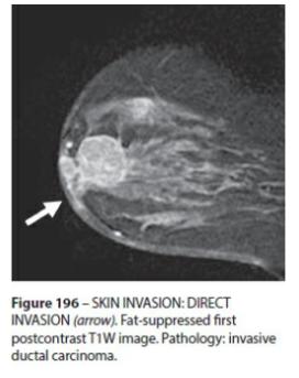
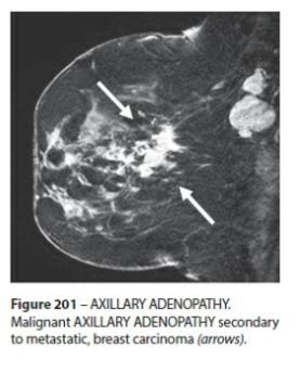
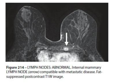
50 MRI Skin lesions Cysts Fat containing lesions: lymph nodes, fat necrosis, hamartoma, postoperative seroma/hematoma with fat Nonenhanging findings: ductal precontrast high signal on T1W, cyst, postoperative collections (hematoma/seroma), post-therapy skin and trabecular thickening, non-enhancing mass, architectural distortion, signal void from foreign bodies (biopsy markers, surgical clips) Associated feature: skin thickening, skin retraction, skin invasion (direct invasion, inflammatory cancer), nipple retraction, nipple invasion, axillary adenopathy
51
52

53
54
55 SUMMARY Be familiar with BI-RADS applying to all breast imaging modalities: mammography, ultrasound, and breast MRI Understand the components of a breast imaging report: Indication, Comparison (PRIORS), Technique (2D or 3D mammogram, US, MRI), Breast density, Important findings, Assessment and Management (BI- RADS). Indications for breast MRI: preoperative evaluation, post neoadjuvant chemotherapy, positive surgical margins post breast conservation therapy, metastatic axillary LAD with unknown primary breast malignancy, silicone breast implant integrity, breast cancer screening in high risk women, and equivocal mammography findings. MRI findings: focus, mass or nonmass enhancement.
56 References ACR BI-RADS ATLAS 2013 (text and images) Case Report: Breast Implant-Associated Anaplastic Large Cell Lymphoma: Report of 2 Cases and Review of the Literature. Aesthetic Surgery Journal: 2014, Vol 34 (6) Clinical indications for breast MRI Applied Radiology: October Utilization of Breast MRI for extent of disease in newly diagnosed malignancy. JAOCR: October 2017, Volume 6 (4)
Leonard M. Glassman MD
 BI-RADS The New BI-RADS Leonard M. Glassman MD FACR Former Chief of Breast Imaging American Institute for Radiologic Pathology Washington Radiology Associates, PC Breast Imaging Reporting and Data System
Armed Forces Institute of Pathology.
 Armed Forces Institute of Pathology www.radpath.com Armed Forces Institute of Pathology Breast Disease www.radpath.org Armed Forces Institute of Pathology Interpretation of Breast MRI Leonard M. Glassman
Imaging in breast cancer. Mammography and Ultrasound Donya Farrokh.MD Radiologist Mashhad University of Medical Since
 Imaging in breast cancer Mammography and Ultrasound Donya Farrokh.MD Radiologist Mashhad University of Medical Since A mammogram report is a key component of the breast cancer diagnostic process. A mammogram
MRI BI-RADS: How to make it out?
 MRI BI-RADS: How to make it out? Poster No.: C-1850 Congress: ECR 2016 Type: Educational Exhibit Authors: M. Ben Ammar, A. Ben Miled, O. Ghdes, S. Harguem, A. Gaja, N. Mnif; Tunis/TN Keywords: Breast,
Non-mass Enhancement on Breast MRI. Aditi A. Desai, MD Margaret Ann Mays, MD
 Non-mass Enhancement on Breast MRI Aditi A. Desai, MD Margaret Ann Mays, MD Breast MRI Important screening and diagnostic tool, given its high sensitivity for breast cancer detection Breast MRI - Indications
BI-RADS Update. Martha B. Mainiero, MD, FACR, FSBI Brown University Rhode Island Hospital
 BI-RADS Update Martha B. Mainiero, MD, FACR, FSBI Brown University Rhode Island Hospital No Disclosures BI-RADS History 1980s Quality Issues ACR Accreditation BI-RADS 1994 2003 4 th Edition MRI, US January
Breast Imaging Lexicon
 9//201 200 BI RADS th Edition 201 BI RADS th Edition Breast Imaging Lexicon Mammographic Pathology and Assessment Categories Deborah Thames, R.T.(R)(M)(QM) The Advanced Health Education Center Nonmember:
AB MR Interpretation Overview
 AB MR Interpretation Overview Goal of AB MR interpretation is to maintain high sensitivity and specificity In order to minimize false positives and short term follow ups, it is fundamental to focus only
MRI in breast cancer: diagnosis and intervention. Dr Sue Barter Addenbrookes Hospital, Cambridge UK
 MRI in breast cancer: diagnosis and intervention Dr Sue Barter Addenbrookes Hospital, Cambridge UK Intervention will be discussed in High Risk Screening! Indications UK and Europe: Breast MRI is well established
Lesion Imaging Characteristics Mass, Favoring Benign Circumscribed Margins Intramammary Lymph Node
 Lesion Imaging Characteristics Mass, Favoring Benign Circumscribed Margins Intramammary Lymph Node Oil Cyst Mass, Intermediate Concern Microlobulated Margins Obscured Margins Mass, Favoring Malignant Indistinct
Pitfalls and Limitations of Breast MRI. Susan Orel Roth, MD Professor of Radiology University of Pennsylvania
 Pitfalls and Limitations of Breast MRI Susan Orel Roth, MD Professor of Radiology University of Pennsylvania Objectives Review the etiologies of false negative breast MRI examinations Discuss the limitations
Amammography report is a key component of the breast
 Review Article Writing a Mammography Report Amammography report is a key component of the breast cancer diagnostic process. Although mammographic findings were not clearly differentiated between benign
Index. C Calcifications fat necrosis 1, 61 fat necrosis 4, 69 nipple/peri-areolar involvement 1, 165
 A ADH. See Atypical ductal hyperplasia (ADH) American College of Radiology (ACR), BI-RADS background parenchymal enhancement, 8, 9, 81, 82 fibroglandular tissue guidelines, 6 American Joint Committee on
National Diagnostic Imaging Symposium 2013 SAM - Breast MRI 1
 National Diagnostic Imaging Symposium 2013 December 8-12, 2013 Disney s Yacht Club Resort Lake Buena Vista, Florida Self Assessment Module Questions, Answers and References Day SAM Title - Each SAM title
Mammographic imaging of nonpalpable breast lesions. Malai Muttarak, MD Department of Radiology Chiang Mai University Chiang Mai, Thailand
 Mammographic imaging of nonpalpable breast lesions Malai Muttarak, MD Department of Radiology Chiang Mai University Chiang Mai, Thailand Introduction Contents Mammographic signs of nonpalpable breast cancer
ROLE OF MRI IN SCREENING, DIAGNOSIS AND MANAGEMENT OF BREAST CANCER. B.Zandi Professor of Radiology
 ROLE OF MRI IN SCREENING, DIAGNOSIS AND MANAGEMENT OF BREAST CANCER B.Zandi Professor of Radiology Introduction In the USA, Breast Cancer is : The Most Common Non-Skin Cancer The Second Leading cause of
S. Murgo, MD. Chr St-Joseph, Mons Erasme Hospital, Brussels
 S. Murgo, MD Chr St-Joseph, Mons Erasme Hospital, Brussels? Introduction Mammography reports are sometimes ambiguous and indecisive. ACR has developped the BIRADS. BIRADS consists of a lexicon in order
Breast Cancer Imaging
 Breast Cancer Imaging I. Policy University Health Alliance (UHA) will cover breast imaging when such services meet the medical criteria guidelines (subject to limitations and exclusions) indicated below.
Here are examples of bilateral analog mammograms from the same patient including CC and MLO projections.
 Good afternoon. It s my pleasure to be discussing Diagnostic Breast Imaging over the next half hour. I m Wei Yang, Professor of Diagnostic Radiology and Chief, the Section of Breast Imaging as well as
Breast MRI Update. Jeffrey C. Weinreb, MD, FACR Yale University School of Medicine
 Breast MRI Update Jeffrey C. Weinreb, MD, FACR jeffrey.weinreb@yale.edu Yale University School of Medicine I disclose the following financial relationships with relevant commercial interests: Bracco Bayer
Contrast-enhanced Breast MRI RSSA 2013
 Contrast-enhanced Breast MRI RSSA 2013 Prof. dr. Maurice van den Bosch University Medical Center Utrecht, the Netherlands Index 1) Breast cancer 2) Why MRI of the breast 3) Technique 4) Interpretation
Standard Breast Imaging Modalities. Lilian Wang, M.D. Breast Imaging Section Department of Radiology Northwestern Medicine
 Standard Breast Imaging Modalities Lilian Wang, M.D. Breast Imaging Section Department of Radiology Northwestern Medicine Overview Standard breast imaging modalities Mammography Ultrasound MRI Imaging
PLACE LABEL HERE. ACRIN 6657 MRI Form: Pre-Treatment (MRI-1)
") M3 ACRIN 6657 MRI Form: Pre-Treatment (MRI-1) If this is a revised or corrected form,indicate by checking box. ACRIN Study 6657 Case # Instructions: In accordance with the protocol, four MRI exams are
M3 ACRIN 6657 MRI Form: Pre-Treatment (MRI-1) If this is a revised or corrected form,indicate by checking box. ACRIN Study 6657 Case # Instructions: In accordance with the protocol, four MRI exams are
CDIS: what's beyond microcalcifications? - Pictorial essay
 CDIS: what's beyond microcalcifications? - Pictorial essay Poster No.: C-1096 Congress: ECR 2014 Type: Educational Exhibit Authors: R. N. Lucas, C. A. S. Ruano, I. Oliveira, J. M. G. Lourenco, Z. 1 1 1
Criteria of Malignancy. Evaluation Score
 30 5 Diagnostic Criteria Criteria of Malignancy Table 5.2 lists criteria in contrast-enhancing MR mammography that strongly indicate the presence of malignancy or are unspecific. Unifactorial evaluation
BREAST MRI. VASILIKI FILIPPI RADIOLOGIST CT MRI & PET/CT Departments Hygeia Hospital, Athens, Greece
 BREAST MRI VASILIKI FILIPPI RADIOLOGIST CT MRI & PET/CT Departments Hygeia Hospital, Athens, Greece Breast ΜR Imaging (MRM) Breast MR imaging is an extremely powerful diagnostic tool, that when used in
Ana Sofia Preto 19/06/2013
 Ana Sofia Preto 19/06/2013 Understanding the underlying pathophysiologic processes leading to the various types of calcifications Description and illustration of the several types of calcifications, according
Breast Imaging! Ravi Adhikary, MD!
 Breast Imaging! Ravi Adhikary, MD! ACS Estimated Cancers Statistics 2014! Breast! New Cases in Women! 232,670 (+67,570 in situ)! Deaths in Women! 40,000! Colon! 48,380! 24,040! Cervical! 12,360! 4,020!
Case Scenario 1 History and Physical 3/15/13 Imaging Pathology
 Case Scenario 1 History and Physical 3/15/13 The patient is an 84 year old white female who presented with an abnormal mammogram. The patient has a five year history of refractory anemia with ringed sideroblasts
MEDICAL IMAGING AND BREAST DISEASE HOW CAN WE HELP YOU?
 MEDICAL IMAGING AND BREAST DISEASE HOW CAN WE HELP YOU? Barbara M. Preston, M.D. SCREENING MAMMOGRAPHY AVERAGE RISK PATIENTS KAISER RECOMMENDATION: ALL WOMEN (INCLUDING TRANSGENDER FEMALES) Every 1-21
Ductal carcinoma in situ: ultrasound, mammography and MRI features with pathologic correlation
 Ductal carcinoma in situ: ultrasound, mammography and MRI features with pathologic correlation Poster No.: C-2252 Congress: ECR 2013 Type: Educational Exhibit Authors: L. Fernandes, H. A. M. R. Tinto,
WHICH INDICATION FOR BREAST MRI?
 WHICH INDICATION FOR BREAST MRI? Dr. P. De Visschere, Prof. Dr. G. Villeirs Genitourinary Radiology and Mammography University Hospital Gent Symposium Belgian Menopause Society 13/03/2010 Which Indication
RADIOLOGIC EVALUATION OF BREAST CANCER
 RADIOLOGIC EVALUATION OF BREAST CANCER Orsolya Farkas, Gabriella Bodrogi and Gábor Szalai Department of Radiology, Pécs University Orsifarkas@yahoo.com Complex evaluation of the breast Patient history
Triple-negative breast cancer: which typical features can we identify on conventional and MRI imaging?
 Triple-negative breast cancer: which typical features can we identify on conventional and MRI imaging? Poster No.: C-1862 Congress: ECR 2013 Type: Educational Exhibit Authors: V. Bertani 1, A. Gualano
Diagnostic Dilemmas of Breast Imaging
 Diagnostic Dilemmas of Breast Imaging Common Causes of Error in Breast Cancer Detection By: Jason Cord, M.D. Mammography: Initial Imaging The standard for detection of breast cancer Screening mammography
8/31/2016 HIDING IN PLAIN SITE, ARCHITECTURAL DISTORTIONS AND BREAST ASYMMETRIES ARCHITECTURAL DISTORTIONS ARCHITECTURAL DISTORTIONS
 HIDING IN PLAIN SITE, ARCHITECTURAL DISTORTIONS AND BREAST ASYMMETRIES DEBORAH THAMES R.T. (R)(M)(QM) ARCHITECTURAL DISTORTIONS Definition is disruption of the natural flow of breast pattern towards the
BI-RADS MRI: A Primer
 Erguvan- ogan et al. I- RS MRI Women s Imaging Pictorial Essay WOMEN S IMGING asak Erguvan-ogan 1 Gary J. Whitman 1 nne. Kushwaha 1,2 Michael J. Phelps 1,3 Peter J. empsey 1 Erguvan-ogan, Whitman GJ, Kushwaha,
PLACE LABEL HERE BASELINE / PRE-TREATMENT. ACRIN 6657 Extension MRI Form: Baseline / Pre-Treatment MRI 1. o Unknown
 T1 ACRIN 6657 Extension MRI Form: Baseline / Pre-Treatment MRI 1 If this is a revised or corrected form, please box. ACRIN Study 6657 No. Instructions: In accordance with the protocol, four MRI exams are
BREAST MRI. Elizabeth A. Rafferty, M.D. Avon Comprehensive Breast Center Massachusetts General Hospital Harvard Medical School
 BREAST MRI Elizabeth A. Rafferty, M.D. Avon Comprehensive Breast Center Massachusetts General Hospital Harvard Medical School BREAST MRI Any assessment of the breast parenchyma requires the administration
ACRIN 6666 IM Additional Evaluation: Additional Views/Targeted US
 Additional Evaluation: Additional Views/Targeted US For revised or corrected form check box and fax to 215-717-0936. Instructions: The form is completed based on recommendations (from ID form) for additional
RSNA, /radiol Appendix E1. Methods
 RSNA, 2016 10.1148/radiol.2016151097 Appendix E1 Methods US and Near-infrared Data Acquisition Four optical wavelengths (740 nm, 780 nm, 808 nm, and 830 nm) were used to sequentially deliver the light
Radiologic and pathologic correlation of non-mass like breast lesions on US and MRI: Benign, high risk, versus malignant
 Radiologic and pathologic correlation of non-mass like breast lesions on US and MRI: Benign, high risk, versus malignant Poster No.: C-1161 Congress: ECR 2013 Type: Educational Exhibit Authors: J. Kwak,
Ultrasonography. Methods. Brief Description. Indications. Device-related Prerequisites. Technical Requirements. Evaluation Criteria
 1 Ultrasonography Brief Description Imaging modality using sound waves Tissue-specific wave reflection. Indications Evaluation of palpable breast nodules Evaluation of clinically occult mammographic findings
Radiology Review Course Hotel del Coronado Coronado, California
 37 th Annual Radiology Review Course Hotel del Coronado Coronado, California Friday, April 21, 2017 - PM TABLE OF CONTENTS Friday, April 21, 2017 - PM SAM Session - Breast Imaging Update 12:45 PM 1:30
ORIGINAL ARTICLE EVALUATION OF BREAST LESIONS USING X-RAY MAMMOGRAM WITH HISTOPATHOLOGICAL CORRELATION
 Available online at www.journalijmrr.com INTERNATIONAL JOURNAL OF MODERN RESEARCH AND REVIEWS IJMRR ISSN: 2347-8314 Int. J. Modn. Res. Revs. Volume 3, Issue 10, pp 807-814, October, 2015 ORIGINAL ARTICLE
Benign, Reactive and Inflammatory Lesions of the Breast
 Benign, Reactive and Inflammatory Lesions of the Breast Marilin Rosa, MD Associate Member Section Head of Breast Pathology Department of Anatomic Pathology Program Director, Breast Pathology Fellowship
BREAST MRI. Elizabeth A. Rafferty, M.D. Avon Comprehensive Breast Center Massachusetts General Hospital Harvard Medical School
 BREAST MRI Elizabeth A. Rafferty, M.D. Avon Comprehensive Breast Center Massachusetts General Hospital Harvard Medical School BREAST MRI Any assessment of the breast parenchyma requires the administration
Inflammatory Breast Carcinoma: Mammographic, Ultrasonographic, MRI and Pathologic Findings
 Inflammatory Breast Carcinoma: Mammographic, Ultrasonographic, MRI and Pathologic Findings Poster No.: C-2248 Congress: ECR 2013 Type: Educational Exhibit Authors: L. Fernandes, J. Lopes Dias, H. A. M.
DCIS of the Breast--MRI findings with mammographic correlation.
 DCIS of the Breast--MRI findings with mammographic correlation. Poster No.: C-1560 Congress: ECR 2013 Type: Educational Exhibit Authors: N. B. Ibrahim, P. Morris, S. ANANDAN; Burlington, MA/US Keywords:
Indications for Breast MRI: Case-Based Review
 AJR Integrative Imaging LIFELONG LEARNING FOR RADIOLOGY Indications for Breast MRI: Case-Based Review Amy Argus 1, Mary C. Mahoney Objective The educational objectives of this continuing medical education
Detailed Program of the second BREAST IMAGING AND INTERVENTIONS PROGRAM am am : Clinician s requirements from breast imaging
 Detailed Program of the second BREAST IMAGING AND INTERVENTIONS PROGRAM 2012 Day one, 2 nd November BREAST IMAGING AND INTERVENTIONS PROGRAM 2012 9.00 AM 9.10 am Introduction 9.10 am - 9.30 am : Clinician
UW Radiology Review Course Breast Calcifications. BI-RADS 5 th Edition
 UW Radiology Review Course Breast Calcifications Grace Kalish, MD Vantage Radiology BI-RADS 5 th Edition Benign Skin Vascular Large rod like Coarse popcorn Suspicious Amorphous Coarse heterogenous Fine
Breast Cancer. Most common cancer among women in the US. 2nd leading cause of death in women. Mortality rates though have declined
 Breast Cancer Most common cancer among women in the US 2nd leading cause of death in women Mortality rates though have declined 1 in 8 women will develop breast cancer Breast Cancer Breast cancer increases
Breast Imaging: Multidisciplinary Approach. Madelene Lewis, MD Assistant Professor Associate Program Director Medical University of South Carolina
 Breast Imaging: Multidisciplinary Approach Madelene Lewis, MD Assistant Professor Associate Program Director Medical University of South Carolina No Disclosures Objectives Discuss a multidisciplinary breast
BREAST IMAGING and NEW IMAGING MODALITIES- A Surgeons view
 BREAST IMAGING and NEW IMAGING MODALITIES- A Surgeons view DR CHANTEL THORNTON SPECIALIST BREAST CANCER SURGEON BMSc (hons) MBBS (hons) FRACS Epworth Hospital, Richmond- Agora Centre for Women s Health
Breast Cancer. Saima Saeed MD
 Breast Cancer Saima Saeed MD Breast Cancer Most common cancer among women in the US 2nd leading cause of death in women 1 in 8 women will develop breast cancer Incidence/mortality rates have declined Breast
The radiologic workup of a palpable breast mass
 Imaging in Practice CME CREDIT EDUCTIONL OJECTIVE: The reader will consider which breast masses require further workup and which imaging study is most appropriate Lauren Stein, MD Imaging Institute, Cleveland
Anyone can get breast cancer BREAST MRI BREAST CANCER. The incidence of getting breast cancer is 1:19 in Malaysia
 Anyone can get breast cancer BREAST MRI KATE Datin Dr Fatimah Moosa Sunway Medical Centre DATIN SERI ENDON KYLIE SIZE DOES NOT MAKE A DIFFERENCE BREAST CANCER The incidence of getting breast cancer is
Intracystic papillary carcinoma of the breast
 Intracystic papillary carcinoma of the breast Poster No.: C-1932 Congress: ECR 2011 Type: Educational Exhibit Authors: V. Dimarelos, F. TZIKOS, N. Kotziamani, G. Rodokalakis, 1 2 3 1 1 1 2 T. MALKOTSI
Benign breast lesions frequently encountered on MR
 Benign breast lesions frequently encountered on MR Poster No.: C-1385 Congress: ECR 2012 Type: Educational Exhibit Authors: J. R. Almeida 1, J. C. Marques 2 ; 1 Lisbon/PT, 2 lisbon/pt Keywords: Hemangioma,
Imaging the Symptomatic Patient. Avice M.O Connell MD,FACR,FSBI Professor of Imaging Sciences Director, Women s Imaging University of Rochester
 Imaging the Symptomatic Patient Avice M.O Connell MD,FACR,FSBI Professor of Imaging Sciences Director, Women s Imaging University of Rochester The four most common symptoms Mass Pain Discharge Infection
Presented by: Lillian Erdahl, MD
 Presented by: Lillian Erdahl, MD Learning Objectives What is Breast Cancer Types of Breast Cancer Risk Factors Warning Signs Diagnosis Treatment Options Prognosis What is Breast Cancer? A disease that
CLINICAL SIGNIFICANCE OF BENIGN EPITHELIAL CHANGES
 Papillomas. Papillomas are composed of multiple branching fibrovascular cores, each having a connective tissue axis lined by luminal and myoepithelial cells ( Fig. 23-11 ). Growth occurs within a dilated
AMSER Case of the Month: November 2018
 AMSER Case of the Month: November 2018 52 year old female with an abnormal screening mammogram Areeg Rehman, MS 4 Nova Southeastern University Rebecca T. Sivarajah, MD Penn State University College of
BI-RADS Categorization As a Predictor of Malignancy 1
 Susan G. Orel, MD Nicole Kay, BA Carol Reynolds, MD Daniel C. Sullivan, MD BI-RADS Categorization As a Predictor of Malignancy 1 Index terms: Breast, biopsy, 00.1261 Breast neoplasms, localization, 00.125,
Breast MRI: Friend or Foe?
 Breast MRI: Friend or Foe? UCSF Postgraduate Course May 18, 2013 Cheryl Ewing, MD Clinical Professor of Surgery UCSF Department of Surgery APPLEGATE HAS DOUBLE MASTECTOMY IN CANCER SCARE DIAGNOSED WITH
CPC 4 Breast Cancer. Rochelle Harwood, a 35 year old sales assistant, presents to her GP because she has noticed a painless lump in her left breast.
 CPC 4 Breast Cancer Rochelle Harwood, a 35 year old sales assistant, presents to her GP because she has noticed a painless lump in her left breast. 1. What are the most likely diagnoses of this lump? Fibroadenoma
MR sin plass i brystkreftdiagnostikk, dagens anbefalinger og fremtidsperspektiver
 MR sin plass i brystkreftdiagnostikk, dagens anbefalinger og fremtidsperspektiver Kathinka Kurz, MD, PhD, seksjonsoverlege SUS, kathinka.dehli.kurz@sus.no Technique - Subtraction Without contrast agent
Breast Cancer Diagnosis, Treatment and Follow-up
 Breast Cancer Diagnosis, Treatment and Follow-up What is breast cancer? Each of the body s organs, including the breast, is made up of many types of cells. Normally, healthy cells grow and divide to produce
Throughout this policy, bracketed numbers link topics across multiple sections according to the indication numbers in the following list.
 Subject: Magnetic Resonance Imaging of the Breast Page: 1 of 33 Last Review Status/Date: September 2015 Magnetic Resonance Imaging of the Breast Description Magnetic resonance imaging (MRI) of the breast
Pitfalls of Dynamic Contrast Enhanced MR Mammography (DCE-MRM) in Evaluation of Post-Biopsy Suspicious Breast Lesions
 in Evaluation of Post-Biopsy Suspicious Breast Lesions") Med. J. Cairo Univ., Vol. 86, No. 3, June: 1513-1522, 2018 www.medicaljournalofcairouniversity.net Pitfalls of Dynamic Contrast Enhanced MR Mammography (DCE-MRM) in Evaluation of Post-Biopsy Suspicious
Med. J. Cairo Univ., Vol. 86, No. 3, June: 1513-1522, 2018 www.medicaljournalofcairouniversity.net Pitfalls of Dynamic Contrast Enhanced MR Mammography (DCE-MRM) in Evaluation of Post-Biopsy Suspicious
Breast Cancer Screening with Mammography
 Progress in Public Health Breast Cancer Screening with Mammography JMAJ 44(7): 318 324, 2001 Tokiko ENDO Director, Department of Radiology, National Nagoya Hospital Abstract: Breast cancer has been increasing
Non-Discrimination Statement and Multi-Language Interpreter Services information are located at the end of this document.
 MRI OF THE BREAST Non-Discrimination Statement and Multi-Language Interpreter Services information are located at the end of this document. Coverage for services, procedures, medical devices and drugs
Breast calcification: Management and Pictorial Review
 Breast calcification: Management and Pictorial Review Poster No.: C-0692 Congress: ECR 2014 Type: Educational Exhibit Authors: V. de Lara Bendahan, M. F. Ramos Solis, A. Amador Gil, C. 1 2 3 2 4 4 Gómez
MRI features of Triple-negative breast cancer: our experience.
 MRI features of Triple-negative breast cancer: our experience. Poster No.: C-1852 Congress: ECR 2013 Type: Scientific Exhibit Authors: V. Bertani, A. Gualano, V. Londero, A. Dal Col, M. Marcon, P. 1 2
Melissa Hartman, DO Women s Health Orlando VA Medical Center
 Melissa Hartman, DO Women s Health Orlando VA Medical Center Most common non-skin cancer and Second deadliest cancer in women Majority are diagnosed by abnormal screening study An approach to breast cancer
Breast pathology. 2nd Department of Pathology Semmelweis University
 Breast pathology 2nd Department of Pathology Semmelweis University Breast pathology - Summary - Benign lesions - Acute mastitis - Plasma cell mastitis / duct ectasia - Fat necrosis - Fibrocystic change/
Diseases of the breast (2 of 2) Breast cancer
 Breast cancer") Diseases of the breast (2 of 2) Breast cancer Epidemiology & etiology The most common type of cancer & the 2 nd most common cause of cancer death in women 1 of 8 women in USA Affects 7% of women Peak at
Diseases of the breast (2 of 2) Breast cancer Epidemiology & etiology The most common type of cancer & the 2 nd most common cause of cancer death in women 1 of 8 women in USA Affects 7% of women Peak at
Mammographic evaluation of palpable breast masses with pathological correlation: a tertiary care centre study in Nepal
 Original article 21 Mammographic evaluation of palpable breast masses with pathological correlation: a tertiary care centre study in Nepal G. Gurung, R. K. Ghimire, B. Lohani Department of Radiology and
Leonard M. Glassman MD Analysis of Breast Calcifications
 Importance of Calcification Leonard M. Glassman MD FACR American Institute for Radiologic Pathology Washington Radiology Associates, PC Washington DC 45% of all breast cancers present as calcification
ANNEX 1 OBJECTIVES. At the completion of the training period, the fellow should be able to:
 1 ANNEX 1 OBJECTIVES At the completion of the training period, the fellow should be able to: 1. Breast Surgery Evaluate and manage common benign and malignant breast conditions. Assess the indications
EARLY DETECTION: MAMMOGRAPHY AND SONOGRAPHY
 EARLY DETECTION: MAMMOGRAPHY AND SONOGRAPHY Elizabeth A. Rafferty, M.D. Avon Comprehensive Breast Center Massachusetts General Hospital Harvard Medical School Breast Cancer Screening Early detection of
Using T2-Weighted Sequences to More Accurately Characterize Breast Masses Seen on MRI
 Residents Section Pattern of the Month Westra et al. MRI of reast Masses Residents Section Pattern of the Month Downloaded from www.ajronline.org by 46.3.195.58 on 12/28/17 from IP address 46.3.195.58.
Alena Levit MD Avice O Connell MD University of Rochester, Rochester, NY
 Alena Levit MD Avice O Connell MD University of Rochester, Rochester, NY Purpose Review imaging spectrum of both common benign and malignant breast lesions Describe and demonstrate CT features with mammogram,
Breast Health. Learning Objectives. Breast Anatomy. Poll Question. Breast Anatomy
 Learning Objectives Describe breast anatomy to a patient Breast Health Answer questions about causes of breast pain and masses Explain breast cancer screening/diagnostic modalities Appropriately triage
MP Magnetic Resonance Imaging for Detection and Diagnosis of Breast Cancer
 Medical Policy MP 6.01.29 BCBSA Ref. Policy: 6.01.29 Last Review: 09/19/2018 Effective Date: 09/19/2018 Section: Radiology Related Policies 6.01.45 Computer-Aided Evaluation of Malignancy With Magnetic
Interpretation of Positron Emission Mammography: Feature Analysis and Rates of Malignancy
 Women s Imaging Original Research Women s Imaging Original Research Deepa Narayanan 1,2 Kathleen S. Madsen 3 Judith E. Kalinyak 1 Wendie. erg 4,5 Narayanan D, Madsen KS, Kalinyak JE, erg W 1 Naviscan,
Invasive lobular carcinoma of the breast; spectrum of imaging findings.
 Invasive lobular carcinoma of the breast; spectrum of imaging findings. Poster No.: C-0847 Congress: ECR 2014 Type: Educational Exhibit Authors: D. Mandich, T. Diaz de Bustamante, L. Koren, M. Arroyo,
Clinical Practice Guideline for the Indications for Use of Breast Magnetic Resonance Imaging (MRI)
") CIHRT Exhibit P-2595 Page 1 Question: Clinical Practice Guideline for the Indications for Use of Breast Magnetic Resonance Imaging (MRI) Eastern Health Breast Disease Site Group What are the current indications
CIHRT Exhibit P-2595 Page 1 Question: Clinical Practice Guideline for the Indications for Use of Breast Magnetic Resonance Imaging (MRI) Eastern Health Breast Disease Site Group What are the current indications
Breast Health and Imaging Glossary
 Contact: Lorna Vaughan HerSpace Breast Imaging & Biopsy Associates 300 State Route 35 South W. Long Branch, NJ 07764 732-571-9100, ext. 104 lorna@breast-imaging.com Breast Health and Imaging Glossary Women
Advanced Course on Multimodality Detection and Diagnosis of Breast Diseases
 BREAST SEMINAR Advanced Course on Multimodality Detection and 3D image of the breast tissue Invited speaker LÁSZLÓ TABÁR, MD,FACR (Hon) Falun, Sweden Nov 22-23, 2014 ATHENS, Greece Royal Olympic Hotel
AMSER Case of the Month: September 2018
 AMSER Case of the Month: September 2018 60-year-old woman with a left breast mass noted on screening mammography. Catherine McNulty, MS4 Tulane University School of Medicine Dr. Robin Sobolewski Breast
November 23, Dear Maryland Breast and Cervical Cancer Program Provider:
 STATE OF MARYLAND DHMH Maryland Department of Health and Mental Hygiene 201 W. Preston Street Baltimore, Maryland 21201 Martin O Malley, Governor Anthony G. Brown, Lt. Governor John M. Colmers, Secretary
Evolution of diagnostic ultrasound systems Current achievements in breast ultrasound
 Evolution of diagnostic ultrasound systems Current achievements in breast ultrasound Dr. Ayumi Izumori, M. D. Department of Breast Surgery, Takamatsu Heiwa Hospital Tokushima Breast Care Clinic, Japan
We are IntechOpen, the world s leading publisher of Open Access books Built by scientists, for scientists. International authors and editors
 We are IntechOpen, the world s leading publisher of Open Access books Built by scientists, for scientists 4,000 116,000 120M Open access books available International authors and editors Downloads Our
Breast Cancer Screening
 Scan for mobile link. Breast Cancer Screening What is breast cancer screening? Screening examinations are tests performed to find disease before symptoms begin. The goal of screening is to detect disease
BREAST SURGERY PROGRESS TEST Name:
 General Surgery Residency Program Excellent surgeons BREAST SURGERY PROGRESS TEST Name: Choose the BEST answer for the following questions. 1. All of the following factors are associated with an increased
Case study 1. Rie Horii, M.D., Ph.D. Division of Pathology Cancer Institute Hospital, Japanese Foundation for Cancer Research
 NCCN/JCCNB Seminar in Japan April 15, 2012 Case study 1 Rie Horii, M.D., Ph.D. Division of Pathology Cancer Institute Hospital, Japanese Foundation for Cancer Research Present illness: A 50y.o.premenopausal
COPE Library Sample
 Breast Anatomy LOBULE LOBE ACINI (MILK PRODUCING UNITS) NIPPLE AREOLA COMPLEX ENLARGEMENT OF DUCT AND LOBE LOBULE SUPRACLAVICULAR NODES INFRACLAVICULAR NODES DUCT DUCT ACINI (MILK PRODUCING UNITS) 8420