Texas Medical Center Library. Yan Yang
|
|
|
- Clare McCormick
- 6 years ago
- Views:
Transcription


1 Texas Medical Center Library UT GSBS Dissertations and Theses (Open Access) Graduate School of Biomedical Sciences CHARACTERIZING THE EFFECT OF DASATINIB ON ANTI-TUMOR IMMUNE RESPONSE AND EXPLORING THE POTENTIAL OF COMBINING TARGETED THERAPY AND IMMUNOTHERAPY IN A C- KIT MUTANT MASTOCYTOMA MODEL Yan Yang Follow this and additional works at: Part of the Immunology and Infectious Disease Commons, and the Medicine and Health Sciences Commons Recommended Citation Yang, Yan, "CHARACTERIZING THE EFFECT OF DASATINIB ON ANTI-TUMOR IMMUNE RESPONSE AND EXPLORING THE POTENTIAL OF COMBINING TARGETED THERAPY AND IMMUNOTHERAPY IN A C-KIT MUTANT MASTOCYTOMA MODEL" (2013). UT GSBS Dissertations and Theses (Open Access) This Dissertation (PhD) is brought to you for free and open access by the Graduate School of Biomedical Sciences at DigitalCommons@TMC. It has been accepted for inclusion in UT GSBS Dissertations and Theses (Open Access) by an authorized administrator of DigitalCommons@TMC. For more information, please contact laurel.sanders@library.tmc.edu.
2 CHARACTERIZING THE EFFECT OF DASATINIB ON ANTI-TUMOR IMMUNE RESPONSE AND EXPLORING THE POTENTIAL OF COMBINING TARGETED THERAPY AND IMMUNOTHERAPY IN A C-KIT MUTANT MASTOCYTOMA MODEL by Yan Yang, BMed APPROVED: Patrick Hwu, MD (Supervisory Professor) Gregory Lizee, PhD Shao-Cong Sun, PhD Stephen Ullrich, PhD Scott Woodman, MD PhD APPROVED: Dean, The University of Texas Graduate School of Biomedical Sciences at Houston
3 CHARACTERIZING THE EFFECT OF DASATINIB ON ANTI-TUMOR IMMUNE RESPONSE AND EXPLORING THE POTENTIAL OF COMBINING TARGETED THERAPY AND IMMUNOTHERAPY IN A C-KIT MUTANT MASTOCYTOMA MODEL A DISSERTATION Presented to the Faculty of The University of Texas Health Science Center at Houston and The University of Texas M. D. Anderson Cancer Center Graduate School of Biomedical Sciences in Partial Fulfillment of the Requirements for the Degree of DOCTOR OF PHILOSOPHY By Yan Yang, BMed Houston, Texas December, 2013 ii
4 Dedication To my parents Meiyuan Shi and Fang Yang, and my husband Ran Xu, for all their support throughout my life and for inspiring me to achieve my goals. iii
5 Acknowledgements First and foremost, my deepest appreciation goes to my advisor Dr. Patrick Hwu. Thank you for his supervision, leadership and patience throughout my PhD study. His guidance all these years has been instrumental for my career development. I would also like to thank all members of my committees, Drs. Gregory Lizee, Scott Woodman, Shao-cong Sun, Stephen Ullrich, Willem Overwijk, Michael Davies and Jonathan Trent for their support, ideas and contributions they provided me throughout this dissertation work. My sincere appreciation also goes to all the present and past members of the laboratory. In particular, to Chengwen Liu, Weiyi Peng, Xiaoxing Yu, Mingyin Zhang, Chunyu Xu, Rina Mbofung, Shruti Malu, Jahan Khalili, Zhimin Dai, Qing Sun for their support and helpful scientific discussions. Last but not least, I would like to express my appreciation to my family, friends, and loved ones for their continuous support and encouragement. iv
6 Abstract Targeted therapy and immunotherapy eradicate malignant cells through different mechanisms, and the strengths and weaknesses of these two approaches are potentially complementary. Therefore, the combination of molecular targeted drugs with immunebased therapies is considered an attractive approach for improving the therapeutic efficacy of cancer treatment. The mutation and amplification of the c-kit protooncogene are associated with multiple different cancer types, and multiple c-kit inhibitors have been tested clinically. In spite of some encouraging results using these agents, most patients with c-kit mutant cancers still relapse due to drug resistance. This also demonstrates the inherent limitation of molecular targeted drugs as mono-therapies, and highlights the need for combinatorial therapeutic approaches. In this study, we show in the c-kit mutant mastocytoma P815 mouse model that the therapeutic effect of the c-kit inhibitor dasatinib is substantially dependent on the antitumor T cell response. Even though the direct effect of dasatinib on antigen-driven T cell expansion is inhibitory, 3-day dasatinib treatment significantly increased the proportion of tumor-specific T cells in the circulation of the treated mice. We also found that dasatinib augmented the priming of T cells through inhibition of immunosuppressive components, including down-regulation of Treg cells and IL-10. When dasatinib was combined with anti-ox40, an agonistic antibody that provides T cells with a potent costimulatory signal, we found that the two agents together gave a much better therapeutic effect compared with either therapy alone, leading to complete tumor eradication in most of the treated mice. Anti-OX40 alone enhanced the infiltration of v
7 CD8+ T cells within the tumor microenvironment. Furthermore, the combination of dasatinib and anti-ox40 significantly up-regulated the expression of IFN-γ-induced Th1 chemokines CXCL 9, 10 and 11, which was consistent with the enrichment of tumorspecific T cells at the tumor sites and better therapeutic effect. This study demonstrates that combinations of molecular targeted agents with immunotherapeutic approaches may result in superior therapeutic effects, suggesting that employing a similar combination strategy may provide clinical benefits to patients with c-kit mutant cancers. vi
8 Table of contents Dedication... iii Acknowledgements... iv Abstract... v Table of contents... vii List of figures... x List of tables... xii Abbreviations... xiii Chapter 1. Introduction Targeted therapy of cancer C-KIT Dasatinib P815 mouse model Anti-tumor T cell response Immunotherapy of cancer Costimulatory molecules Anti-OX Mechanisms of cancer immunosuppression Rationale for combining targeted therapy and immunotherapy vii
9 Chapter 2. Dasatinib elicits a strong tumor-specific T cell response which contributes substantially to the therapeutic effect of dasatinib T cell mediated immunity contributes to the anti-tumor effects of dasatinib Short-term dasatinib treatment can increase levels of circulating tumor antigenspecific T cells rather than long-term dasatinib treatment Dasatinib down-regulates certain immunosuppressive components in P815 tumorbearing mice, which mediates dasatinib s effect of enhancing anti-tumor T cell response APCs from dasatinib-treated mice don t present more tumor antigen than the APCs from control group Dasatinib treatment can decrease the proportion of regulatory T cells and enhance vaccine-mediated T cell priming, but doesn t decrease the proportion of myeloid-derived suppressor cells (MDSC) Down-regulation of IL-10 production by tumor cells is another factor contributing to dasatinib s effect of enhancing anti-tumor T cell response Chapter 3. The therapeutic effect of dasatinib on P815 and underlying anti-tumor responses are potentiated by anti-ox Addition of anti-ox40 antibody improves the anti-tumor efficacy of dasatinib treatment The combination regimen increases the infiltration of tumor-specific CD8+ T cells in the tumor microenvironment viii
10 3.3 The combination of dasatinib and anti-ox40 leads to up-regulation of IFN-γ and IFN-γ induced chemokines in the tumor microenvironment Combining dasatinib with anti-ox40 leads to a dual inhibition of Treg and increases the CD8+ T cell / Treg ratio in the tumor microenvironment The therapeutic effect of 3-day dasatinib combined with anti-ox40 is better than 8- day dasatinib combined with anti-ox Chapter 4. Discussion and future directions Summary and discussion Future directions Chapter 5. Methods and materials Bibliography Vita ix
11 List of figures Figure 1 C-KIT structure and signaling schematic diagram Figure 2 Major costimulatory molecules in lymphocytes Figure 3 In vitro and in vivo anti-tumor effect of dasatinib on P815 mastocytoma Figure 4 The in vivo anti-tumor effect of dasatinib is dependent on T cells Figure 5 Effect of dasatinib treatment on tumor antigen-specific T cell response Figure 6 Effect of dasatinib on T cell proliferation and cytolytic activity in vitro Figure 7 Therapeutic effects of short-term dasatinib treatment versus long-term dasatinib treatment Figure 8 IFN-γ staining of P1A-specific T cells stimulated with dendritic cells from lymph nodes of the vehicle or dasatinib-treated mice Figure 9 Effect of 3-day dasatinib treatment on levels of regulatory T cells Figure 10 Effect of 3-day dasatinib pre-treatment on vaccine-mediated T cell priming.. 53 Figure 11 Effect of 3-day dasatinib treatment on levels of MDSC Figure 12 Gene expression profiles of P815 tumors treated with vehicle or dasatinib Figure 13 Cytokine profiles in the tumor microenvironment of P815 tumors treated with vehicle or dasatinib Figure 14 Effect of IL-10 blockade on tumor growth, survival and T cell response Figure 15 SiRNA-mediated c-kit knockdown in P815 cells Figure 16 Gene expression profiles of P815 tumor cells Figure 17 Therapeutic effect of combining dasatinib with anti-ox40/anti-pd-1/anti- CTLA-4 antibody Figure 18 Therapeutic effect of combining dasatinib and anti-ox x
12 Figure 19 Effect of dasatinib and anti-ox40 treatment on the level of the P1A-specific T cells in the peripheral blood Figure 20 Influence of dasatinib and anti-ox40 treatment on the tumor infiltrating lymphocytes Figure 21 Levels of CXCL9, 10 and 11 in tumors from 4 different groups Figure 22 Effect of dasatinib and anti-ox40 treatment on the level and function of regulatory T cells Figure 23 Therapeutic effects of short-term dasatinib combined with anti-ox40 treatment versus long-term dasatinib treatment combined with anti-ox Figure 24 Comparative dynamics of T cell response and dasatinib administration Figure 25 Mechanism underlying the anti-tumor effect of the combination of dasatinib and anti-ox xi
13 List of tables Table 1 C-KIT genetic aberrations associated with human malignancies... 8 Table 2 Summary of different types of cancer immunotherapy Table 3 Immunosuppressive mechanisms employed by cancer Table 4 List of the chemokine primers for realtime PCR xii
14 Abbreviations ACT: Adoptive Cell Therapy Akt: Protein Kinase B APC: Antigen Presenting Cell CD: Cluster of Differentiation CFSE: Carboxyfluorescein Succinimidyl Ester CTL: Cytotoxic T lymphocyte CTLA-4: Cytotoxic T Lymphocyte Antigen 4 DC: Dendritic Cell ELISA: Enzyme-Linked Immunosorbent Assay ERK: Extracellular Signal Regulated Kinases FACS: Fluorescence-Activated Cell Sorting FDA: Food and Drug Administration GIST: Gastrointestinal Stromal Tumor HLA: Human Leukocyte Antigen IFN-γ: Interferon-gamma IgG: Immunoglobulin G MAPK: Mitogen-Activated Protein Kinase MDSC: Myeloid Derived Suppressor Cell MHC: Major Histocompatibility Complex NK: Natural Killer cells PBMC: Peripheral Blood Mononuclear Cells PD-1: Program Cell Death Protein 1 xiii
15 PI3K: Phosphadityl-Inositol-3 Kinase Realtime-PCR: Quantitative Realtime Polymerase Chain Reaction TCR: T cell Receptor TIL: Tumor Infiltrating Lymphocyte TNFR: Tumor Necrosis Factor Receptor Treg: Regulatory T cells 7-AAD: 7-aminoactinomycin D xiv
16 Chapter 1. Introduction 1.1 Targeted therapy of cancer Discoveries of specific proteins and pathways that contribute to the carcinogenesis and malignant phenotype of cancer cells have led to the generation of therapeutic agents which can specifically target cancer cells. Unlike chemotherapy and radiation therapy, which commonly impair rapidly dividing cells, targeted therapy interferes with the specific pathways necessary for cancer progression and therefore it is considered more effective and less toxic than conventional treatments. Targeted therapy drugs include antibodies and small molecule inhibitors. Monoclonal antibodies target specific antigens either are extracellular growth factors or receptors on cell surface. Binding of the antibody with the ligand or the receptor could effectively block the activity of that specific oncogenic pathway. Monoclonal antibodies which have achieved significant success in the clinic include anti-cd20(rituximab), anti-vegf(bevacizumab), anti- EGFR( Cetuximab) and anti-her2/neu(trastuzumab). Those monoclonal antibodies have been used for the treatment of various types of cancers, including breast cancer, lymphoma, colon cancer and lung cancer(1-5). Small molecule targeted therapy drugs are another class of agents discovered or structurally designed for oncogenic proteins. Compared with monoclonal antibodies, this class of drug is less specific because they normally affect a subset of kinases with similar structure on the targeted domain rather than block the activity of one target. But the advantage of small molecule inhibitors is their ability of interfering with intracellular molecules which monoclonal antibodies cannot(6). Multiple small molecule inhibitors have been approved by FDA for cancer 1
17 treatment, for example, erlotinib, gefenitib and vemurafenib. And more drugs are being tested in clinical and preclinical studies(7, 8). Advent of molecularly targeted agents is considered as a milestone in the history of cancer therapy. However, the instability and complexity of cancer genome limit the success of targeted therapy. The responses to targeted therapy are usually of limited durability. A general phenomenon in patients is the development of drug resistance (9-11). Multiple mechanisms are involved in the occurrence of drug resistance in patients: mutation of the target protein could reduce the binding affinity between the drug and the target; the expression of the target can be up-regulated at DNA, mrna and protein levels; enhanced activities of other oncoproteins could compensate for the inhibition of the target protein; overexpression of the drug transporters could reduce the intracellular drug concentration; drug-resistant cancer cell clones could outgrow the drug-sensitive clones(12). Therefore, it is unlikely that targeted therapy alone can lead to cure. 1.2 C-KIT C-KIT (CD117) is a tyrosine kinase receptor, which is encoded by the proto-oncogene KIT located on human chromosome 4 and mouse chromosome 5. Under physiological condition, this receptor is expressed by hematopoietic stem cells, mast cells, melanocytes and interstitial cells of Cajal. Its ligand is stem cell factor (SCF) which is secreted by endothelial cells and fibroblasts(13). The extracellular part of c-kit is composed of five immunoglobulin-like domains, this is the ligand binding domain and is encoded by exon 2-9. The juxtamembrane domain which is believed to have auto-inhibitory function is 2
18 encoded by exon 11. The intracellular part contains 2 tyrosine kinase domains, one ATPbinding pocket encoded by exon 13 and 14 and one kinase activation loop encoded by exon17 (Figure 1)(14, 15). Upon the binding of SCF, 2 c-kit receptors form a dimer and phosphorylate each other on certain tyrosine residues. The phosphorylated tyrosine residues serve as the docking sites for multiple effector molecules, then activate a wide array of downstream signal transduction pathways, including MAPK pathway, PI3K/Akt and JAK/STAT pathway, which play critical roles in multiple aspects of cellular functionality(16-18). The amplification and mutation of this gene are associated with several types of cancer, including gastrointestinal stromal tumors(gist), mast cell leukemia, melanoma, testicular seminoma and acute myeloid leukemia. Mutation of c-kit could cause the activation of this receptor without the binding of SCF, and the deregulated activation of downstream oncogenic pathways leads to the uncontrolled growth and survival of cells(19, 20). Therefore, c-kit became an attractive target for the treatment of those cancers. And multiple c-kit inhibitors have been developed and applied in clinical practice. GIST is derived from c-kit expressing interstitial cells of Cajal (ICC). Activating mutation of c-kit has been found in more than 80% of GIST patients. The most common genetic aberrations occurring in GIST are duplication of codon on exon 9 and V560G mutation on exon 11(13, 21, 22). The tumor is refractory to conventional chemotherapy and irradiation therapy. However, introduction of c-kit inhibitors into the 3
19 clinic has significantly improved the prognosis of the patients and change the natural history of this disease. It is one of the most successful examples showing the effectiveness of molecular targeted therapy. Currently, imatinib is applied in the clinic for treating patients with unresectable tumors and patients after surgical removal for prevention purpose. Another c-kit inhibitor sunitinib has been shown to be effective in treating GIST patients who have developed resistance to imatinib(23). Mutation of c-kit in the overall melanoma population is at a relatively low frequency, resulting in the disappointing outcome of early clinical trials using imatinib(24-26). In 2010, Curtin and his colleagues reported that in the rare subsets of acral, mucosal and chronic sun damaged (CSD) melanoma, incidence rates of c-kit genetic aberration are 36%, 39% and 28% respectively, which are quite high. The most common mutations are L576P on exon 11 and K642E on exon 13.While in the subset of melanoma arising from skin without chronic sun damage, the mutation rate is zero (27). There are multiple clinical case reports showing that patients with c-kit mutations respond well to c-kit inhibitors such as dasatinib and imatinib(28-31). More clinical trials determining the therapeutic efficacy of c-kit inhibitors in melanoma are currently ongoing. Mastocytosis is not a malignant disease per se, but when it progresses to an advanced stage, it could evolve into mast cell leukemia. C-KIT mutation drives approximately 95% of mastocytosis and D816V mutation is identified in 80% of the patient samples(32-34). In mastocytosis cell lines, studies have demonstrated that imatinib can t efficiently inhibit the activity of c-kit with D816V mutation, while dasatinib could significantly block its 4
20 activity and lead to the apoptosis of the cells(35). Therefore, dasatinib might be an effective targeted drug for this disease. C-KIT mutations have also been found in germ cell tumor and acute myeloid leukemia (core binding factor subtype). The summary of the various cancers associated with c-kit mutations as illustrated below(table 1) (13, 36-38). 5
21 Figure 1 C-KIT structure and signaling schematic diagram. The extracellular region is encoded by exons 2 through 9, and contains 5 immunoglobulin-like domains. The Juxtamembrane domain is encoded by exon 11. There are 2 intracellular tyrosine kinase domains: the ATP-binding pocket encoded by exons 13 and 14, and the kinase activation loop encoded by exon 17. Binding of SCF causes the dimerization of c-kit and autophosphorylation of this receptor, consequently triggering the activation of downstream Ras/MEK/MAPK, PI3K/Akt and JAK/STAT pathways. 6
22 7

23 Table 1 C-KIT genetic aberrations associated with human malignancies 8
24 1.3 Dasatinib Dasatinib, a small molecule tyrosine kinase inhibitor drug, was originally identified as a potent Src/Abl inhibitor by researchers at Bristol-Myers Squibb about ten years ago. Due to its potent anti-proliferative activity against multiple hematological and solid tumor cell lines, it has been approved by FDA for the treatment of Philadelphia chromosomepositive acute lymphoblastic leukemia and chronic myelogenous leukemia. The targeted kinase spectrum of dasatinib is quite broad, including Src family members, Bcr/abl, c- KIT, PDGFR and Ephrin A. The mechanism dasatinib s action involves its competitively binding with the ATP-binding site of the kinase to inhibit the phosphorylation of tyrosine involved in the signal transduction. The plasma half-life of dasatinib is only 5-6 hours, quite short compared with other tyrosine kinase inhibitors(39). This drug is currently being tested in clinical trials for treating multiple solid tumors such as melanoma, GIST, pancreatic cancer and breast cancer. Even though imatinib is the most commonly used c-kit inhibitor in the clinic, dasatinib has been shown to be a potential stronger inhibitor for certain forms of mutant c-kit. For example, it has been shown that L576P, the most common c-kit mutation in melanoma, is sensitive to dasatinib rather than imatinib. One study suggests that the different binding affinities of imatinib and dasatinib might be caused by the different structural changes of the mutant kinases(31). P815, a tumor cell line with D814Y mutation we used in this model, is also not sensitive to the inhibition of imatinib, but it is sensitive to killing mediated by dasatinib. That is why we used dasatinib as the targeted therapy drug in this study(35, 40). 9
25 1.4 P815 mouse model P815 is a mastocytoma line induced by a highly carcinogenic compound methylcholanthrene(mta) in a DBA/2 mouse. Tumor antigens, from this immunogenic tumor, have been identified and cloned. P1A, the first tumor antigen identified, is a nonmutated gene normally expressed in the placenta and testis of the DBA/2 mouse. A transgenic mouse strain with TCR specific for H-2L d : P1A complex has been generated and named P1CTL mice. People could utilize these transgenic mice to do adoptive T cell transfer experiments studying the anti-tumor CTL response in vivo. Another antigen identified later on is P1E. Therefore, this model allows people to monitor immune responses against two MHC I-restricted epitopes at the same time. This animal model has long been considered as a good tool for analyzing the relationship between tumor and anti-tumor T cell immunity. Studying the combination of targeted therapy and immunotherapy is quite challenging due to the low availability of the suitable animal models. We need an immunogenic tumor, whose growth is driven by a single oncogenic event and can be implanted in immunocompetent mice. We found that the P815 tumor model fits these criteria. Growth of P815 is controlled by a mutant c-kit receptor (D814Y mutation) and disrupting c-kit signaling could significantly inhibit the proliferation and promote the apoptosis of this tumor line. In addition, P815 is a very immunogenic tumor allowing us to study the therapeutic effect of immunotherapy. 10
26 1.5 Anti-tumor T cell response The theory of cancer immune surveillance was developed by Burnet and Thomas in 1950s(41) and it is currently well-accepted that immune system can recognize and eliminate malignant cells. Both innate components and adaptive components from the immune system play a role in controlling the growth of cancer cells. NK cells, NKT cells and macrophages which can directly eliminate the transformed cells are the major players of innate anti-tumor immune response(42). T cells are the major effectors of the adaptive anti-tumor immunity. The anti-tumor T cell response is a multi-step process. First, tumor antigens need to be taken up by dendritic cells, then APCs process the proteins into peptides and load them to MHC molecules. Activated dendritic cells then migrate into tumor-draining lymph nodes. When naïve T cells with the specific TCR encounter the dendritic cells presenting the corresponding peptide, they get activated and undergo clonal expansion. During this priming phase, number of the T cell precursors specific to an antigen could increase more than 1000 fold and these cells form a pool of effector cells. CD8+ effector T cells are normally considered as the killer cells with cytolytic activity. They migrate into tumor microenvironment following chemokine gradients. Once encountering tumor cells, CTLs release vesicles containing perforin and granzymes. Perforin is a cytolytic protein forming pores on the cell membrane of the target cells. Granzymes are serine proteases which can cleave caspase-3 and activate the downstream apoptotic pathway in the target cells. Surface expression of FasL is another mechanism CTLs employ to eliminate tumor cells. Engagement of FasL on CTLs and Fas receptor on tumor cells activates caspase-8 and triggers the extrinsic apoptotic cascade. Those molecules are the major mediators of T cell cytolytic activity. CD4+ effector T cells are 11
27 generally considered as the T helper cells which coordinate many elements of the immune response. They can help activate APCs through CD40-CD40L axis and production of cytokines, and they also participate in priming CD8+ T cell as well as activating macrophages(43). Recent studies suggest that a subset of CD4+ T cells might also possess cytolytic activity and can directly kill tumor cells(44-46). The majority of the effector T cells die after the effector phase, leaving a small pool of memory T cells which can elicit more rapid response upon secondary antigen stimulation. Because of the potent capability of T cells to specifically recognize and eliminate cancer cells, T cellsbased therapies show great promise in developing immune treatment for cancers. 1.6 Immunotherapy of cancer Cancer immunotherapy is based on the idea of boosting the immune reaction to eradicate cancer cells. It has been successfully implemented in the clinical treatment of a number of malignancies, such as lymphoma, renal carcinoma and melanoma(47-53). Immunotherapy for cancer can be divided into several categories: adoptive cell therapy, cytokine based therapy, cancer vaccine and antibodies targeting costimulatory molecules, etc (Table 2). Adoptive cell therapy (ACT) transfers cytolytic lymphocytes which have been stimulated or genetically modified ex vivo back into patients. The most effective adoptive cell therapy approach for cancer so far is adoptive T cell therapy with tumor infiltrating T lymphocytes (TIL) which involves isolation of TILs from patients resected tumor masses, ex vivo expansion of isolated TILs to large numbers and re-infusion of those 12
28 tumor-specific TILs. Clinical responses to ACT in early years were not quite promising, but adding preparative lymphodepletion regimen significantly improved the response rate of this therapy(54, 55). The potential mechanisms involve increased availability of homeostatic cytokines and down-regulation of immune suppressive cells(56). According to a recent clinical study conducted at the NCI, the overall response rate for patients receiving chemotherapy-based preparative regimen and ACT was 49%, when adding 12Gy total body irradiation to the standard treatment approach, the overall response rate was improved to 72%(57). Compared with other therapeutic regimens, ACT turned out to be the most efficient one for late-stage melanoma patients. Multiple clinical trials are currently underway to determine the therapeutic efficacy of this immunotherapy approach in other types of cancers, including renal carcinoma and ovarian cancer (58). One advantage of this approach is enabling people to directly manipulate the transferred T cells genetically, which might offer a therapeutic advantage. Genetic modification can be used to re-direct the antigen specificity, enhance the survival and improve the migration ability of transferred T cells(59-63). Cytokine-based therapies have also been successfully implemented in clinical cancer treatment. The two most successful examples are IFN-α and IL-2. IFN-α is produced by leukocytes and it is one of the first cytokines that has been found to exhibit anti-tumor effect. It does not only induce apoptosis in cancer cells but also stimulates the innate and adaptive immune responses against malignant cells. Therefore, IFN- α has been approved by the FDA for the treatment of a number of tumors(64). IL-2 is the first interleukin that has been identified and cloned. It is mainly produced by activated T cells and plays a 13
29 critical role in regulating T cell proliferation, differentiation and survival. In addition to its function in T cells, IL-2 also exerts pleiotropic effect on other subsets of immune cells: it could promote the immunoglobulin production by B cells, boost the proliferation and activation of NK cells and also affect the function of macrophages and dendritic cells(65). Due to its biological function of expanding and activating cytolytic T cells and NK cells, IL-2 has been used in the clinic for cancer treatment for a long time. High dose IL-2 therapy could induce response in 15% of the patients and leads to cure in 5% of the patients with late-stage melanoma(66). Therefore, it is considered as one of the first-line treatment options for metastatic melanoma. It is also being used for treating renal carcinoma and combined with adoptive T cell therapy and cancer vaccines(67). There are several other cytokines showing promising results in cancer-related studies, such as GM- CSF, IL-15 and IL-21. Clinical trials of validating the therapeutic efficacy of those cytokines are underway. Vaccines have been successfully used to induce adaptive immune responses against infections in the clinic. Identification of multiple cancer antigens has paved the way for the development of cancer vaccine. The concept of cancer vaccine was proposed long time ago and people have conducted extensive studies to develop all sorts of cancer vaccine for several decades. Common forms of cancer vaccine include whole cell-based vaccine, protein-based vaccine, peptide-based vaccine, DNA-based vaccine, recombinant virus vaccine and dendritic cell vaccine. However, the success of vaccines in cancer treatment is very limited so far. Taking melanoma as an example, a study published 2 years ago showing that gp100 peptide vaccine combined with IL-2 improved the 14
30 prognosis of late-stage melanoma patients as compared with IL-2 alone is one of the first clinical studies showing that vaccine could provide certain therapeutic effect in late-stage cancer patients(68). For most of the cancer vaccine-related clinical trials, the results turned out to be disappointing. Further studies are needed to improve the therapeutic efficacy of cancer vaccine. 15

31 Table 2 Summary of different types of cancer immunotherapy 16
32 1.7 Costimulatory molecules T cell activation against tumor antigens is a critical step of an effective anti-tumor immune response. After undergoing the positive selection and negative selection in thymus, naïve T cells mature and migrate to the peripheral lymph nodes in which they might encounter the APC presenting the cognate peptide/ MHC complex. This is the socalled first-signal for the activation of naïve T cells. And the binding between CD28 on T cells and CD80/86 on APC is also critical for T cell activation. CD28 activation could induce the phosphorylation of its tyrosine residues, which recruits proteins with SH-2 domain, such as PI3K and Grb2. Signaling through CD28 enhances the TCR signaling and activates the expression of certain anti-apoptosis genes, such as bcl-xl. This provides the second signal for T cell priming. However, the fate of T cells is finally determined by the integration of signals from multiple co-existing molecules in the T cell-apc focal synapses. In addition to CD28, costimulatory molecules including 4-1BB, OX40, LAG-3, CD27, BTLA, CTLA-4, PD-1 and several other ones simultaneously play important roles in this process. Some of them provide positive signal for T cell activation and some of them could negatively interfere with T cell function(figure 2). When naïve T cells encounter antigen presented in the context of proper costimulation, they could undergo rapid expansion, differentiation and then become cytotoxic effector cells. However, negative signals coming from the costimulatory pathways might result in T cell anergy or exhaustion. pathways Progresses in the understanding of the costimulatory molecules-related has led to the development of several antibodies specific targeting those molecules. The most successful examples are anti-ctla-4 and anti-pd-1. 17
33 CTLA-4 is an inhibitory checkpoint molecule expressed by T cells right upon T cell activation. CTLA-4 competes with CD80/CD86 for receptor binding with CD28. CTLA- 4 ligation inhibits the activation and expansion of T cells thereby effectively dampens the magnitude of an immune response including anti-tumor T cell response(69-71). CTLA-4 blockade could remove this brake on T cells activation and has been shown to effectively enhance the anti-tumor immunity in multiple animal models(72, 73). Ipilimumab, the antagonistic antibody against CTLA-4, demonstrated superior therapeutic effect when combined with dacarbazine or gp100 vaccine in melanoma clinical trials(74, 75), thus was approved for the treatment of late-stage melanoma in Currently, this drug is undergoing clinical trials for treating lung cancer and prostate cancer patients. PD-1 is another key negative regulator expressed by T cells. Binding of PD-1 ligands with PD-1 receptor could render T cells undergo exhaustion and become unresponsive. This pathway has been well characterized as negatively controlling the T cell response to chronic viral infection and cancer (76, 77). PD-L1 expression has also been identified in various types of tumors and currently is considered one of most important immune evasion mechanisms employed by cancer cells(77-81). Increased knowledge about the function of PD-1/PD-L1 axis in immune regulation has led to the development of antagonistic PD-1 and PD-L1 antibodies. The phase I clinical trials have ensured the safety and tolerability of PD-1 and PD-L1 antibodies, and also showed durable therapeutic response in some patients. (82-86). Larger clinical trials of treating late-stage cancer patients with these antibodies are underway. 18
34 The initial success of anti-pd-1 and anti-ctla-4 has raised hope that costimulatory antibodies would revolutionize cancer immunotherapy. A number of antibodies against other costimulatory molecules have been shown to be effective in preclinical studies, for examples, anti-ox40(87, 88), anti-4-1bb(89, 90) and anti-cd40(91, 92). And studies trying to develop new humanized antibodies against those T cell regulatory molecules represent an active area of research. 19
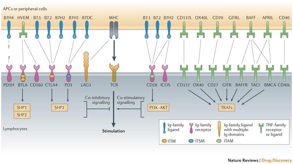
35 Figure 2 Major costimulatory molecules in lymphocytes. Costimulatory molecules play an important role in regulating the function of lymphocytes. They can be classified into two types: negative and positive costimulatory molecules. Important negative costimulatory molecules include programmed cell death protein 1 (PD-1), B and T lymphocyte attenuator (BTLA), CD160, cytotoxic T lymphocyte antigen 4 (CTL-4), and lymphocyte activation gene 3 (LAG3). Important positive costimulatory molecules include CD28, inducible co-stimulator (ICOS), CD137(4-1BB), OX40, CD27, glucocorticoid-induced TNFR-related protein (GITR), B cell activation factor receptor (BAFFR), transmembrane activator and CAML interactor (TACI), B cell maturation antigen (BCMA) and CD40L. Most of those costimulatory molecules belong to immunoglobulin(ig) superfamily or tumor necrosis factor receptor (TNFR) superfamily. The ligands for those molecules are mainly expressed by APCs and other peripheral cells. Costimulatory pathways are important targets for cancer immunotherapy. Reprinted by permission from Macmillan Publishers Ltd: Nature Review Drug Discovery 12(2): , 2013 Macmillan Publishers Ltd 20
36 21
37 1.8 Anti-OX40 OX40(CD134) is a costimulatory molecule which belongs to the tumor necrosis family receptor (TNFR) superfamily. It is known to play an important role in the proliferation, differentiation, survival and migration of T cells. OX40 is expressed on T cell after its activation, with variable dynamics in various conditions. Both TCR signaling and CD28 ligation have regulatory role on the expression of OX40, and certain cytokines, such as IL-2 and TNF, might also prolong or enhance the expression. But generally, the expression of OX40 on CD8+ T cells is more transient than CD4+ T cells(93). OX40L(CD252) is expressed as a trimer on the cell surface of a number subsets of immune cells, such as dendritic cells, B cells, and endothelial cells(94). OX40 crosslinking results in OX40 binding with several intracellular adaptors, including TRAF2, TRAF3 and TRAF5, which leads to the activation of NF-κB pathway and PI3K/Akt pathway. This prevents T cell apoptosis by enhancing the expression of bcl-2, bcl-xl and survivin, and also promotes the division, cytokine production and memory response formation through some unknown signaling mechanisms(93). OX40 and OX40L knockout mice were generated more than a decade ago. OX40 deficient mice infected with LCMV and influenza virus demonstrated defective CD4+ T cell response(95). OX40L deficient mice had reduced dendritic cell mediated-t cell priming and contact hypersensitivity response(96). These early animal studies suggest OX40 plays an important role for T cell costimulation. Studies have shown that OX40 activation could help maintain the survival of effector T cells beyond the initial several rounds of cell division after their encounter with antigen. And OX40 agonist can enhance 22
38 the clonal expansion and formation of memory response for both CD4+ and CD8+ T cells in vivo(97, 98). In addition, OX40 ligation on Tregs could effectively block the suppressive function of those cells. Studies have found that OX40 stimulation suppresses the expression of Foxp3 gene, which is the master regulator of Treg cells, and also prevents the induction of Treg cells from CD4 + conventional T cells(99-101). Therefore, the advantage of utilizing OX40 ligation to boost T cell response is affecting both the effector side and the regulatory side. OX40 has also been shown to be expressed by other subsets of immune cells, such as NK, NKT and dendritic cells, and affects function of these cells ( ). The OX40-OX40L axis has also been shown to be involved in the pathogenesis of multiple autoimmune diseases by animal studies. For example, in an induced experimental autoimmune encephalomyelitis (EAE) model, OX40L knockout mice exhibited much less severe clinical manifestations than the wild type mice(105). Later on, animal experiments demonstrated that expression of OX40L on tumor cells could enhance the tumor immunogenicity and mediate the tumor rejection(106), suggesting OX40 signaling also plays important role in tumor immunity. Then, agonistic anti-ox40 antibody alone was shown to potentiate the anti-tumor immune response in multiple tumor models through multiple mechanisms, including promoting effector and memory T cell response, suppressing Treg function and enhancing the migration of CTL into tumor microenvironment(88, 107, 108). In addition, the combination of anti-ox40 with chemotherapy, irradiation therapy and other immunotherapy regimens have also been tried and showed promising therapeutic effect( ). Those studies have paved the 23
39 way for translating OX40 ligation therapy into the clinic. A humanized agonistic anti- OX40 antibody has recently been generated and is being tested in clinical trials. 1.9 Mechanisms of cancer immunosuppression A wide variety of mechanisms are employed by cancer cells to evade or subvert an immune attack. It is well known that cancer cells can down-regulate their antigen presentation machinery to avoid being killed by immune effectors. Mutations in genes encoding MHC molecules, TAP and LMPs are commonly seen in cancer cells(113, 114). Besides evading the immune attack by impairing the antigen presentation pathway, cancer cells could also directly exert inhibitory effect on immune effectors by multiple immunosuppression mechanisms, including producing inhibitory cytokines and enzymes, recruiting suppressive cells and expressing inhibitory surface molecules(115, 116), which are listed below(table 3). Cancer cells and the stromal cells in the tumor microenvironment could secrete large amounts of inhibitory cytokines such as IL-10(117), TGF-β (118) and VEGF(119). These cytokines can directly inhibit the antigen presentation capability of dendritic cells, the proliferation and cytolytic function of T cells and cytokine production by immune effectors. Enzymes with immunosuppression properties also profoundly exist in the tumor microenvironment. For example, indoleamine 2,3- dioxygenase (IDO)(120), arginase(121) and inducible nitric oxide synthases (inos)(116) could catabolize some amino acids which are essential for the proliferation and function of T cells. Tumor cells can produce chemokines, such as CCL22, CCL2 and CXCL2 to attract regulatory T cell 24
40 (Treg), myeloid-derived suppressor cell (MDSC) and M2 tumor-associated macrophage (TAM) into tumor microenvironment(116, 122, 123). Profound existence of cells with immune-inhibitory properties substantially hinders the function of anti-tumor immune cells. Last but not least, cancer cells can express multiple immune-inhibitory molecules, such as PD-L1 and FasL( ). Engagement of those molecules with their corresponding ligands could lead to T cell exhaustion, anergy or depletion in the tumor microenvironment. These mechanisms may act in concert to restrain anti-tumor immunity. Therefore, cancer-induced immune suppression is a major obstacle for the success of immunotherapy and overcoming it might help eradicate cancer cells successfully. 25

41 Table 3 Immunosuppressive mechanisms employed by cancer 26
42 1.10 Rationale for combining targeted therapy and immunotherapy Advances in the development of multiple targeted therapy agents and immunotherapy approaches in the recent years avail a great opportunity for combinatorial therapies. There are several rationales for combining these two(127). Firstly, even though some targeted therapy drugs can induce dramatic tumor shrinkage in some patients, the responses are usually of limited durability. And although certain targeted therapy drugs are known to be able to induce a high response rate in the subset of the patients who have the corresponding mutation, they barely lead to a cure. However, immunotherapy is quite different. Although the overall response rates for most of the immunotherapy regimens are relatively low, durable and complete responses do happen in some patients. In fact, immunotherapy might be the only treatment able to elicit a memory response against tumor and provide long-term protection against recurrence. Besides, targeted therapy and immunotherapy eradicate cancer cells by completely different mechanisms: one is blocking the oncogenic pathway which supports the growth of tumor cells, another is boosting the cytotoxic immune cells to eliminate tumor cells. The targeted therapy-resistant cells could still be sensitive to immune-mediated killing, and tumor cells successfully evade the immune surveillance could still be eliminated by blocking their growth signaling. Therefore, those two types of treatments have complementary strengths and weaknesses, and combinatorial therapy might provide better therapeutic outcomes. 27
43 Secondly, cancer has been proved to utilize multiple mechanisms to suppress the immune system and evade the immune surveillance. These mechanisms are the major contributors of cancer immune evasion and cancer-induced immune tolerance. Overcoming hese barriers is one of the keys to the success of immunotherapy. Recent studies have shown that certain oncogenic signals could directly up-regulate the immunosuppressive mechanisms to advance the tumor growth (128, 129). Therefore, the effectiveness of immunotherapy could be enhanced upon the down-regulation of oncogenic signalmediated immunosuppression. In addition, some non-specific kinase inhibitors have already been shown to exert suppressive effect on immunosuppressive cells. For example, sunitinib treatment inhibiting MDSC and Tregs is considered as an additional advantage of using this drug to treat renal cancer patients(130). Therefore, if targeted therapy can be employed to down-regulate immunosuppressive components, it could be successfully integrated into immunotherapy protocol in cancer treatment settings. Thirdly, the anti-tumor immune response could be induced upon the treatment of a number of cytotoxic regimens, because the massive death of cancer cells results in release of tumor antigens. In fact, anti-tumor immune response is one of the parameters determining the therapeutic outcomes of certain chemotherapies, radiotherapies and targeted therapies(131). For example, the polymorphism of TLR4 gene could serve as an independent prognostic factor of the response to anthracycline and local irradiation in breast cancer patients(132). And a correlation of clinical response with CD8 and IFN-γ expression levels was also observed in breast cancer patient receiving chemotherapy(133). These are all evidences indicating that the anti-tumor immune response might 28
44 substantially contribute to the therapeutic effect of cytotoxic regimens. In addition, it is well-known that some mutant oncogenic proteins function as tumor antigens eliciting tumor specific immune response, for example, mutant β-catenin and p53 (134, 135). Besides, deep sequencing analysis of cancer genomes also shows that multiple mutations accumulated in tumor cells could potentially be presented by MHC molecules and function as target antigens(136). These lines of evidences suggest that tumor debris generated in situ after targeted therapy treatment could potentially be a great sources of tumor antigen which might initiate strong anti-tumor immune response with appropriate boosting strategy. Therefore, the combinatorial approaches may provide therapeutic synergy and result in stronger and more durable clinical response. Studies involving combinatorial therapy are currently ongoing. For example, clinical trial testing the therapeutic efficacy of BRAF inhibitor vemurafenib and anti-ctla-4 antibody ipilimumab in melanoma patients is being conducted at multiple cancer centers. However, successful implementation of combinatorial regimen in the clinic requires understanding of the interaction of the targeted agent and immune response. In this study, we explored the effect of one c-kit inhibitor dasatinib on anti-tumor T cell response in this specific model - P815 mastocytoma mouse model. And we operated under the hypothesis that the therapeutic efficacy of the targeted therapy could be enhanced by boosting the endogenous anti-tumor immune response with an immune-stimulating approach. In doing so, we found that the underlying anti-tumor T cell response is an 29
45 important contributory factor of dasatinib s therapeutic effect in this model and the combination of dasatinib plus anti-ox40 synergizes to induce a potent anti-tumor T cell response and provides superior therapeutic effect. 30
46 Chapter 2. Dasatinib elicits a strong tumor-specific T cell response which contributes substantially to the therapeutic effect of dasatinib 2.1 T cell mediated immunity contributes to the anti-tumor effects of dasatinib It has been shown that dasatinib can suppress the c-kit kinase activity effectively and inhibit the growth of P815 tumor cells in vitro (35, 40). In our in vitro experiment, we also showed that dasatinib could induce apoptosis of P815 cells in a dose-dependent manner by Annexin V+7-AAD staining. Dasatinib could induce the apoptosis of P815 starting at a concentration as low as 10nM (Figure 3A), which indicates that P815 is very sensitive to dasatinib. To test whether dasatinib could kill P815 in vivo, we inoculate mice subcutaneously with tumor cells on their left abdominal walls and treated the tumor-bearing mice with daily gavage of vehicle or different doses of dasatinib (5mg/kg, 10mg/kg, 50mg/kg and 150mg/kg) on days 8, 9, and 10 following tumor inoculation. We found that at the dose of 150mg/kg, dasatinib significantly inhibited the growth of P815 and prolonged mouse survival (Figure 3B and 3C). H&E staining right after dasatinib treatment revealed extensive vacuolar degeneration in dasatinib-treated tumors, morphologic changes consistent with significant tumor cell death (Figure 3D). This result suggested that dasatinib could directly induce the cell death of P815 in vivo. To assess a potential role for T cell-mediated immunity in the therapeutic efficacy of dasatinib, we depleted CD4+ and CD8+ T cells in the mice to see how it affected the therapeutic effect of dasatinib. We gave the tumor-bearing mice monoclonal antibodies against CD4 or CD8, intraperitoneally, starting from day 8 once a week. Interestingly, 31
47 both CD4+ T cell-depletion and CD8+ T cell-depletion reduced anti-tumor responses in dasatinib-treated mice almost to the same level as in vehicle-treated mice, effectively abrogating the survival benefit provided by dasatinib. (Figure 4A and 4B). This result revealed that an underlying immune response contributes substantially to the therapeutic anti-tumor effect of dasatinib in this model. 32
48 Figure 3 In vitro and in vivo anti-tumor effect of dasatinib on P815 mastocytoma. For in vitro experiment, P815 cells were treated with DMSO or dasatinib for 24 hours then subjected to flow cytometric analysis. For in vivo experiment, DBA/2 mice were subcutaneously inoculated with P815 tumor cells on day 0. Tumor-bearing mice were treated with vehicle or dasatinib (150 mg/kg) on day 8, 9 and 10 by gavage. (A) Dot plots for Annexin V + 7-AAD staining of treated P815 tumor cells. (B) Tumor sizes in mice treated with vehicle or dasatinib (*P < 0.05). (C) Kaplan-Meier survival curves of the tumor-bearing mice for 2 different groups (*P < 0.05). (D) Representative H&E -stained slides of control tumor and dasatinib-treated tumor. This research was originally published in Blood. Yang Y, Liu C, Peng W, Lizée G, Overwijk WW, Liu Y, Woodman SE, Hwu P. Antitumor T-cell responses contribute to the effects of dasatinib on c-kit mutant murine mastocytoma and are potentiated by anti- OX40. Blood. 2012; 120(23): the American Society of Hematology 33
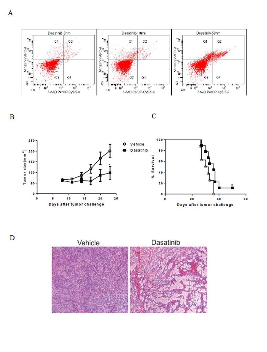
49 34
50 Figure 4 The in vivo anti-tumor effect of dasatinib is dependent on T cells. Mice were injected i.p. with 200μg Rat IgG or CD4 or CD8 depletion antibodies starting from day 8 once a week. (A) Tumor sizes of CD4+/CD8+ T cell depletion experiment (Vehicle+IgG versus Dasatinib +IgG: *P < 0.05). (B) Kaplan-Meier survival curves of CD4+/CD8+ T cell depletion experiment (Vehicle+IgG versus Dasatinib +IgG: *P = 0.05). This research was originally published in Blood. Yang Y, Liu C, Peng W, Lizée G, Overwijk WW, Liu Y, Woodman SE, Hwu P. Antitumor T-cell responses contribute to the effects of dasatinib on c-kit mutant murine mastocytoma and are potentiated by anti- OX40. Blood. 2012; 120(23): the American Society of Hematology 35
51 36
52 2.2 Short-term dasatinib treatment can increase levels of circulating tumor antigenspecific T cells rather than long-term dasatinib treatment We next sought to determine the effect of dasatinib on the tumor antigen-specific T cell response. P815 is an immunogenic tumor which can elicit spontaneous T cell priming against tumor antigens. To see how dasatinib affected the anti-tumor T cell immunity, we collected the blood of the mice on day 18. Using a P1A-specific tetramer and flow cytometry, we found that 3 days of dasatinib treatment significantly increased the tumor antigen-specific T cell levels in the peripheral blood of tumor-bearing mice (Figure 5A and 4B). Peripheral blood T cells were also examined for antigen-specific IFN-γ production using intracellular staining following stimulation with or without P1A, P1E peptides for 6 hours. We found that while CD8+ T cells from vehicle-treated mice were relatively unresponsive to peptide stimulation, a significant proportion of CD8+ T cells from dasatinib-treated mice showed IFN-γ positivity (Figure 5C and 5D). IFN-γ ELISA showed results consistent with the intracellular IFN-γ staining, which indicated that 3 days of dasatinib treatment significantly augmented the anti-tumor T cell response (Figure. 5E). Moreover, CD4+ T cell depletion in vivo significantly compromised the enhanced tumor antigen-specific CD8+ T cell response mediated by dasatinib (Figure 5F), suggesting that the CD8+ T cell response against P815 is T helper cell-dependent. This result also partially explains why in the T cell-depletion experiment, CD4+ T celldepletion abrogated the therapeutic benefit provided by dasatinib as well as CD8+ T celldepletion did. 37
53 However, dasatinib is a multiple tyrosine kinase inhibitor which inhibits the activities of c-kit, EphA, Src family members, Brc/abl, PDGFR and some other tyrosine kinases (39). It has been reported that dasatinib inhibits TCR signal transduction, cellular proliferation and cytokine production by T cells upon antigen stimulation probably due to the inhibitory effect of dasatinib on Src family member Lck (137, 138). So to test whether dasatinib has the same effect on the P1A-specific T cells, we first isolated the splenocytes from P1CTL transgenic mice, whose TCRs are specific for P1A peptide/h2l d complexes. Then we cultured the CFSE-labeled splenocytes in the presence of P1A peptide and different concentrations of dasatinib for 48 hours. Flowcytometry showed that dasatinib inhibited the antigen-driven T cell expansion at a dose dependent manner (Figure 6A). We also cocultured pre-activated P1CTL T cells with P815 tumor cells or P1A peptide in the presence of different concentrations of dasatinib for 24 hours, and the harvested supernatants were subject to IFN-γ ELISA. ELISA result showed that dasatinib significantly inhibited the cytolytic activity of P1A-specific T cells against P815, starting at a concentration as low as 10nm(Figure 6B). These results are consistent with the previous study showing that dasatinib, like other multiple tyrosine kinase inhibitors, directly inhibits T cell proliferation and activity. Since we observed enhanced T cell response using a 3-day dasatinib regimen, which was in contrast with the direct effect of dasatinib on T cell response in vitro, next we investigated whether increasing the length of dasatinib treatment is harmful or helpful to its therapeutic effect on P815. We found that prolonged dasatinib treatment didn t provide any enhanced anti-tumor effects or survival benefit compared with short-term 38
54 dasatinib treatment (Figure 7A and 7B). In fact, P1A tetramer staining showed that longterm dasatinib treatment actually compromised the positive effect of dasatinib on tumor antigen-specific CD8+ T cell priming (Figure 7C), the P1A tetramer+ T cell level in 8- day treatment group was significantly lower than that in 3-day treatment group. Our data together with results from other groups suggest that antigen-specific T cell responses can be impaired with the persistent presence of dasatinib, but this drug is still capable of enhancing anti-tumor immune responses if the dosing and schedule are optimized. 39
55 Figure 5 Effect of dasatinib treatment on tumor antigen-specific T cell response. Peripheral blood of the treated mice was collected and subjected to flow cytometric analysis on day 18. Cells were also stimulated with P1A and P1E peptides for IFN- γ ELISA. (A) Representative dot plots for P1A tetramer staining in the peripheral blood of vehicle or dasatinib-treated mice. (B) Percentage of P1A tetramer+ T cells in total CD8+ T cells (*P < 0.05). (C) Representative dot plots for intracellular IFN-γ staining. (D) Percentage of IFN-γ+ T cells in total CD8+ T cells (P1A: *P < 0.05, P1E: *P < 0.05). (E) IFN-γ secretion by peripheral blood T cells in response to the stimulation of P1A or P1E peptides detected by ELISA. (P1A: *P < 0.05, P1E: **P < 0.01). (F) P1A tetramer staining of CD4+ T cell depletion experiment. This research was originally published in Blood. Yang Y, Liu C, Peng W, Lizée G, Overwijk WW, Liu Y, Woodman SE, Hwu P. Antitumor T-cell responses contribute to the effects of dasatinib on c-kit mutant murine mastocytoma and are potentiated by anti- OX40. Blood. 2012; 120(23): the American Society of Hematology 40
56 41
57 Figure 6 Effect of dasatinib on T cell proliferation and cytolytic activity in vitro. For proliferation assay, splenocytes from P1CTL transgenic mice were labeled with CFSE and cultured with P1A peptide and 0nm, 1nm, 5nm, 10nm, 25nm or 50nm dasatinib. For IFN-γ release assay, P1A-specific T cells were cultured alone or with P815 cells or with P1A peptide for 24 hours. (A) Histograms of the CFSE (FITC channel) gated on CD8+ cells 48 hours after labeling. (B) IFN-γ secretion by P1A-specific T cells detected by ELISA. 42
58 43
59 Figure 7 Therapeutic effects of short-term dasatinib treatment versus long-term dasatinib treatment. (A) Tumor sizes in mice treated with vehicle or 3-day dasatinib or 8-day dasatinib. (B) Kaplan-Meier survival curves of mice treated with vehicle or 3-day dasatinib or 8-day dasatinib. (C) Percentage of P1A tetramer+ T cells in total CD8+ T cells for these 3 groups (Vehicle versus 3-day dasatinib: **P < 0.01, 3-day dasatinib versus 8-day dasatinib: *P < 0.05). This research was originally published in Blood. Yang Y, Liu C, Peng W, Lizée G, Overwijk WW, Liu Y, Woodman SE, Hwu P. Antitumor T-cell responses contribute to the effects of dasatinib on c-kit mutant murine mastocytoma and are potentiated by anti- OX40. Blood. 2012; 120(23): the American Society of Hematology 44
60 45
61 2.3 Dasatinib down-regulates certain immunosuppressive components in P815 tumorbearing mice, which mediates dasatinib s effect of enhancing anti-tumor T cell response APCs from dasatinib-treated mice don t present more tumor antigen than the APCs from control group. One of the most possible mechanisms to explain why there are higher levels of P1Aspecific T cells in dasatinib treated-mice is: drug-induced apoptosis of tumor cells could generate tumor debris which might be taken up by APCs, and the tumor antigens presented by the APCs might mediate the priming of tumor-specific T cells. To test whether this hypothesis is valid here, we set out to assess the amount of tumor antigen P1A presented by the APCs in the tumor-draining lymph nodes with or without dasatinib treatment. After we treated the mice with vehicle or dasatinib, we isolated the tumordraining lymph nodes and sort out CD11b+ CD11c+ dendritic cells and coculture them with pre-activated P1A-specific T cells overnight in the presence of Golgi-Plug, after which IFN-γ intracellular staining was done to assess the reactivation of those T cells. We found that levels of antigen presented by the dendritic cells between these two groups were not significantly different. The IFN-γ production of the P1A-specific T cells after stimulation was comparable in these two groups, which indicated that there was not more tumor antigen presented by APCs to T cells after dasatinib treatment (Figure 8). 46
62 Figure 8 IFN-γ staining of P1A-specific T cells stimulated with dendritic cells from lymph nodes of the vehicle or dasatinib-treated mice. Splenocytes from P1CTL transgenic mice were collected and culture with IL-2 and anti- CD3 for 7 days. CD11b+CD11c+ cells from the lymph nodes of the treated mice were sorted out by FACS. P1A-specific T cells and dendritic cells were co-cultured for 8 hours and then subjected to flow cytometric analysis. Dot plots show the IFN-γ intracellular staining of P1A-specific T cells. 47
63 48
64 2.3.2 Dasatinib treatment can decrease the proportion of regulatory T cells and enhance vaccine-mediated T cell priming, but doesn t decrease the proportion of myeloid-derived suppressor cells (MDSC) Dasatinib has been reported to suppress the proliferation and function of Treg cells in vitro(139), but the in vivo effect of dasatinib treatment on murine Treg cells has not been reported. Flow cytometric analysis showed that dasatinib alone significantly decreased the percentage of Foxp3+CD25+ Treg cells in total CD4+ T cells in the peripheral blood of tumor-bearing mice (Figure 9A and 9B). The ratio of Tregs to CD8+ effector T cells was also markedly decreased by dasatinib (Figure 9C). We also examined the Treg levels within the tumor microenvironment immediately following treatment and found a similar decrease in dasatinib group (Figure 9D). However, this decrease was transient, as Treg cell numbers went back to normal 3 days after drug withdrawal (9E). Because fast-growing tumors are usually accompanied by expansion of Treg cells, to identify whether this dasatinib-induced decrease in the Treg proportion was due to decreased tumor volume or a direct inhibitory effect of dasatinib on Treg, we treated tumor-free mice with dasatinib for 3 days. We observed a similar decrease in Treg cells in the dasatinib-treated tumor-free mice, suggesting that dasatinib has a direct inhibitory effect on Treg cell levels (Figure 10A). Since the drop in Tregs was significant, we next investigated whether the short Treg-depleted window caused by dasatinib can render T cells more amenable to vaccine-induced antigen-specific expansion in a tumor-free system. We i.v. injected the mice with splenocytes from P1CTL transgenic mice. After 3 49
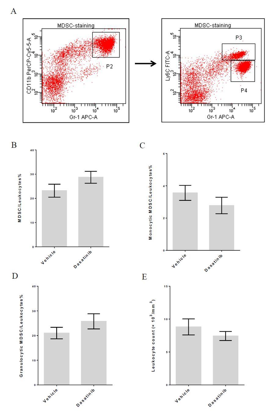
65 days of dasatinib treatment, P1A peptide-pulsed dendritic cells were transferred into the recipients. Flow cytometric analysis of peripheral blood on day 7 after vaccination showed that magnitude of P1A-specific T cell expansion was significantly higher in dasatinib-treated group(figure 10B and 10C). Therefore, this result supports the idea that dasatinib preferentially decreases Tregs in vivo and makes the host immune environment more favorable for antigen-driven effector T cell proliferation. We also checked myeloid-derived suppressor cell levels in those mice. Peripheral blood samples of the tumor-bearing mice after 3-day dasatinib treatment were collected and subjected to flow cytometric analysis. In mice, cells expressing Gr1 and CD11b are considered as MDSCs(140). To further distinguish between subset of MDSCs, we also stained for another marker Ly6C. Ly6C high population is monocytic MDSC and Ly6C low population is granulocytic MDSC(141, 142). We found that dasatinib treatment didn t significantly change the proportion of MDSCs in leukocyte population. And the percentages of granulocytic and monocytic MDSCs were not affected by drug treatment either. We also did complete blood count test (CBC) for peripheral blood samples, and data showed that 3-day dasatinib treatment didn t significantly change the leukocyte counts in treated mice(figure 11). It has been shown that other c-kit inhibitors such as imatinib and sunitinib can down-regulate MDSC level in animal models and cancer patients(130, 143). However, in our study, we didn t observe significant drop of MSDCs after dasatinib treatment. Therefore, it is unlikely that the augmented T cell response in dasatinib-treated mice is attributed to the change in the MDSC population. 50
66 Figure 9 Effect of 3-day dasatinib treatment on levels of regulatory T cells. Peripheral blood of the treated mice was collected and subjected to flow cytometric analysis on day11 and day14. (A) Representative dot plots for Treg staining in the peripheral blood of tumor-bearing mice on day11. (B) Percentage of Treg cells in CD4+ T cells in the peripheral blood of tumor-bearing mice on day11(***p < 0.001). (C) Treg/CD8+ T cell ratio in the peripheral blood of tumor-bearing mice on day11(***p < 0.001). (D) Percentage of Treg cells in CD4+ T cells in tumors on day11 (*P < 0.05). (E) Percentage of Treg cells in CD4+ T cells in the peripheral blood of tumor-bearing mice on day14. This research was originally published in Blood. Yang Y, Liu C, Peng W, Lizée G, Overwijk WW, Liu Y, Woodman SE, Hwu P. Antitumor T-cell responses contribute to the effects of dasatinib on c-kit mutant murine mastocytoma and are potentiated by anti- OX40. Blood. 2012; 120(23): the American Society of Hematology 51
67 52
68 Figure 10 Effect of 3-day dasatinib pre-treatment on vaccine-mediated T cell priming. (A) Percentage of Treg cells in CD4+ T cells in the peripheral blood of tumor-free mice (**P < 0.01) after 3-day vehicle or dasatinib treatment (B) Treg/CD8+ T cell ratio in the peripheral blood of tumor-free mice after 3-day vehicle or dasatinib treatment (*P < 0.05) (C) Representative dot plots for P1A tetramer staining in the peripheral blood on day 7 after vaccination. (D) Percentage of P1A tetramer+ T cells in total CD8+ T cells on day 7 after vaccination( ***P < 0.001). This research was originally published in Blood. Yang Y, Liu C, Peng W, Lizée G, Overwijk WW, Liu Y, Woodman SE, Hwu P. Antitumor T-cell responses contribute to the effects of dasatinib on c-kit mutant murine mastocytoma and are potentiated by anti- OX40. Blood. 2012; 120(23): the American Society of Hematology 53
69 54
70 Figure 11 Effect of 3-day dasatinib treatment on levels of MDSC. Peripheral blood of the treated mice was collected and subjected to flow cytometric analysis and CBC test on day 11. (A) Representative dot plots for MDSC staining in the peripheral blood of tumor-bearing mice. (B) Percentage of MDSC in leukocytes in the peripheral blood of tumor-bearing mice. (C) Percentage of monocytic MDSC in leukocytes in the peripheral blood of tumor-bearing mice. (D) Percentage of granulocytic MDSC in leukocytes in the peripheral blood of tumor-bearing mice. (E) Leukocyte count in the peripheral blood of tumor-bearing mice. 55
71 56
72 2.3.3 Down-regulation of IL-10 production by tumor cells is another factor contributing to dasatinib s effect of enhancing anti-tumor T cell response. Profound immunosuppression is commonly found within the tumor microenvironments of various types of cancer. And it is one of the most critical mechanisms of tumor immune evasion. Therefore, it is possible that there are some immune modulatory components existing in the tumor microenvironment have contributed to the enhanced anti-tumor T cell response after dasatinib treatment. To investigate whether any changes in immunomodulatory gene expression occurred in the tumor microenvironment after 3 day dasatinib treatment, tumor samples were harvested and analyzed for changes in transcription profiles using gene expression microarray. In P815 tumor microenvironment with or without dasatinib treatment, expression levels of certain immunomodulatory genes were quite different. One of the most significant changes observed among the immune-related genes was that of IL-10 transcription. Dasatinib treatment significantly down-regulated the level of IL-10 mrna in the tumor microenvironment (Figure 12). Considering that IL-10 is a well-known immunosuppressive cytokine, this might suggest that IL-10 down-regulation is one of the mechanisms contributing to the enhanced antitumor immune response in dasatinib-treated mice. Next, we wanted to determine the protein levels of immunomodulators in tumor microenvironment by Luminex analysis. The results also showed a significant decrease in IL-10 production after dasatinib treatment (Figure 13). To determine whether the down regulation of IL-10 in the tumor microenvironment was relevant for the improved anti-tumor T cell response induced by dasatinib, we gave mice 57
73 anti-il-10 blocking antibody after starting dasatinib treatment and monitored the tumor growth and anti-tumor T cell response. We found that IL-10 blockade significantly suppressed the tumor growth and improved the survival of the mice (Figure 14A and 14B). Tetramer staining showed that compared with control group, blocking IL-10 also helped T cell priming (Figure 14C). This result suggested that the down-regulation of this tumor-derived immunosuppressive cytokine after targeted therapy treatment is another factor contributing to the anti-tumor T cell response induced by dasatinib. The notion that constitutive activation of oncogenic pathways could up-regulate a number of immunosuppressive mechanisms has been supported by several recent studies (129, 144). For example, one study has shown that c-kit signaling regulates the expression levels of IDO, an enzyme which can convert tryptophan into immunosuppressive metabolites in GIST. Furthermore, imatinib treatment suppresses the activation of the c- KIT pathway, thereby inhibiting the production of IDO by tumor cells and reactivating the T cell-mediated anti-tumor immune response (128). This study revealed a link between c-kit and an immunosuppressive molecule. Therefore, it is possible that a link between c-kit signaling and other immunoregulatory cytokines, such as IL-10, also exists. Besides, ERK and p38 activation have been shown to be involved in IL-10 expression (145, 146), and both are downstream molecules in the c-kit activation pathway. Therefore, we next set out to determine the effect of c-kit signaling on the transcription of immune-related molecules, especially IL-10. We knocked down c-kit by sirna (Figure 15A and 15B) and used microarray analysis to assess how the expression of immunomodulatory gene products was affected. We also treated P815 tumor cells with 58
74 DMSO or 25nm dasatinib in vitro for 24 hours and analyzed those samples by microarray. We found that certain immune-related gene expression levels were altered by c-kit knockdown: for example, IL-4, IL-6 and IL-13;; however, these changes were moderate and not significant. In contrast to what we found in the microarray analysis of in vivo dasatinib treatment, IL-10 mrna levels in c-kit knockdown P815 tumor cells and in vitro dasatinib-treated P815 tumor cells were higher than the control cells (Figure 16), indicating that the decreased level of IL-10 in the tumor microenvironment after targeted drug treatment is not dictated by the tune-down of the oncogenic c-kit signaling in this tumor model, but may have been caused by the decreased viability of IL- 10-producing tumor cells directly. Still, our study suggests that the decreased production of immunosuppressive cytokines caused by targeted agent-mediated tumor cell death might augment T cell priming, contributing to the anti-tumor immune response and preventing the tumor immune escape. 59
75 Figure 12 Gene expression profiles of P815 tumors treated with vehicle or dasatinib. 24 hours after vehicle or dasatinib treatment, P815 tumors were harvested and subjected to microarray analysis. 20 most differentially expressed immune-related genes were selected based on the fold changes of the means of the 2 samples in each group. Heatmap shows the color-coded expression levels of the selected immune-related genes for tumor samples treated with vehicle or dasatinib. 60
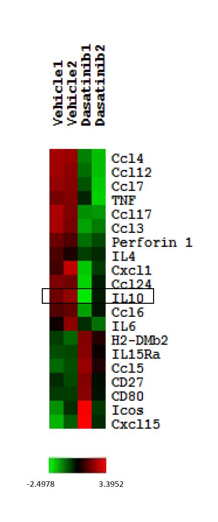
76 61
77 Figure 13 Cytokine profiles in the tumor microenvironment of P815 tumors treated with vehicle or dasatinib. After 3-day vehicle or dasatinib treatment, tumors were harvested and subjected to Luminex assay. Bar graphs show cytokine profiles in the lysates of vehicle or dasatinibtreated P815 tumors (For IL-10, Vehicle versus Dasatinib: *P< 0.05). 62
78 63
79 Figure 14 Effect of IL-10 blockade on tumor growth, survival and T cell response. Mice were injected i.p. with 200μg control IgG antibody or IL-10 blocking antibody starting from day 8 once a week and also treated with vehicle or dasatinib on day 8, 9 and 10. Peripheral blood of the treated mice was collected and subjected to flow cytometric analysis on day 18. (A)Tumor sizes for 3 different groups (Vehicle+IgG versus Vehicle+anti-IL-10 : *P < 0.05). (B) Kaplan-Meier survival curves for 3 different groups (Vehicle+IgG versus Vehicle+anti-IL-10: *P < 0.05). (C) Percentage of P1A tetramer+ T cells in total CD8+ T cells (Vehicle+IgG versus Vehicle+anti-IL-10 : *P < 0.05). 64
80 65
81 Figure 15 SiRNA-mediated c-kit knockdown in P815 cells P815 cells were transfected with non-targeting sirna or sirna against c-kit. 24 hours after transfection, P815 cells were harvested and subjected to realtime PCR and flow cytometric analysis. (A)Taqman realtime PCR shows c-kit mrna levels in P815 cells transfected with non-targeting sirna (NT RNA) or sirna against c-kit or no sirna.(*p < 0.5). (B) Histogram shows the c-kit protein levels in P815 cells transfected with non-targeting sirna (NT RNA) or sirna against c-kit or no sirna. 66
82 67
83 Figure 16 Gene expression profiles of P815 tumor cells. P815 cells were transfected wit c-kit sirna pool or non-targeting pool. 24 hours or 36 hours after transfection, P815 cells were harvested and subjected to microarray analysis. P815 cells were also treated with DMSO or dasatinib in culture for 24 hours and then subjected to microarray analysis. Heatmap shows the color-coded expression levels of the selected immune-related genes for P815 cells. 68

84
85 Chapter 3. The therapeutic effect of dasatinib on P815 and underlying anti-tumor responses are potentiated by anti-ox Addition of anti-ox40 antibody improves the anti-tumor efficacy of dasatinib treatment. Since we found that the anti-tumor effects of dasatinib were largely dependent on CD8+ T cells, we hypothesized that providing further co-stimulation of these T cells with agonistic antibodies could further boost the therapeutic response. Since anti-ctla-4 and anti-pd-1 are costimulatory antibodies already available in the clinic, we decided to try these 2 antibodies. Anti-OX40 is a highly promising agonistic antibody currently undergoing preclinical and clinical investigation, so we also include this antibody in our initial screening. We treated 8-day tumor-bearing mice with dasatinib or vehicle for 3 days and then administered 2 doses of these 3 antibodies on days 10 and 13 (Figure 17). As indicated by the survival curve of the tumor-bearing mice, combination of dasatinib and anti-ox40 gave the best anti-tumor effect. Survival curve indicated that neither the combination of dasatinib with anti-ctla-4 nor combination of dasatinib with anti-pd-1 provided synergistic therapeutic effect. While combining dasatinib with anti-ox40 led to significantly enhanced tumor regression and complete cure in 75% of the mice (Figure 18A-C). The therapeutic effect of the combination treatment was striking considering the large tumor burden (60mm 2-80mm 2 ) at the time of treatment. To determine whether T cells are the major mediator of this therapeutic effect, mice were injected with CD4 or CD8 depletion antibodies starting from day 8 once a week. Depletion of CD8+, and to a lesser extent CD4+, T cells abolished the therapeutic effect of dasatinib combined with 70
86 anti-ox40 (Figure 18D), supporting the notion that T cells are the major immune effector cells responsible for the therapeutic effect of this combination regimen. 71
87 Figure 17 Therapeutic effect of combining dasatinib with anti-ox40/anti-pd-1/anti- CTLA-4 antibody. Mice received daily gavage of vehicle or dasatinib (150mg/kg) on day 8-10 and i.p. injection of IgG control antibody or 200µg anti-ox40 or 200µg anti-pd-1 or 100ug anti- CTLA-4 on day 10 and 13. (A) Schema of treatment. (B) Kaplan-Meier survival curves of the tumor-bearing mice for 8 different groups. 72
88 73
89 Figure 18 Therapeutic effect of combining dasatinib and anti-ox40. (A)Tumor sizes for 4 different groups (Vehicle+IgG versus Dasatinib+anti-OX40: ***P < 0.001, Dasatinib+IgG versus Dasatinib+anti-OX40: **P < 0.01, Vehicle+anti-OX40 versus Dasatinib+anti-OX40: *P < 0.05). (B) Kaplan-Meier survival curves of the tumorbearing mice for 3 different groups (Vehicle+IgG versus Dasatinib+anti-OX40: ***P < 0.001, Dasatinib+IgG versus Dasatinib+anti-OX40: **P < 0.01, Vehicle+anti-OX40 versus Dasatinib+anti-OX40: *P < 0.05). (C) Plots represent the tumor size of individual mice for each group. (D) Kaplan-Meier survival curves of CD4+/CD8+ T cell depletion experiment. This research was originally published in Blood. Yang Y, Liu C, Peng W, Lizée G, Overwijk WW, Liu Y, Woodman SE, Hwu P. Antitumor T-cell responses contribute to the effects of dasatinib on c-kit mutant murine mastocytoma and are potentiated by anti- OX40. Blood. 2012; 120(23): the American Society of Hematology 74
90 75
91 3.2 The combination regimen increases the infiltration of tumor-specific CD8+ T cells in the tumor microenvironment. Although it has been reported that OX40 agonist therapy can enhance the expansion and survival of CTL(111), we found that addition of anti-ox40 to the dasatinib regimen did not further enhance or prolong the tumor antigen-specific T cell response in our tumor model: the percentages of P1A tetramer+ CD8+ T cells were similar between the dasatinib-treated group and the combination-treated group (Figure 19A-D). Considering that the combination treatment yielded a much better therapeutic effect than dasatinib alone, we next sought to assess the effect of this combination regimen on T cell infiltration of the tumor site. Thus, tumor infiltrating lymphocytes were isolated from P815 tumors on day 16 and subjected to flow cytometric analysis. We found that anti- OX40 alone increased the overall infiltration of CD8+ effector T cells, as previously reported. And the combination of dasatinib and anti-ox40 led to enhanced infiltration of P1A-specific T cells into the tumors, to levels greater than that observed with either anti- OX40 or dasatinib alone (Figure. 20A and 20B). IFN- γ staining of TILs also showed that mice receiving the combination demonstrated the highest levels of IFN-γ production in response to P1A antigen stimulation (Figure 20C). This result is consistent with a model whereby dasatinib enhances the level of circulating tumor-specific T cells and anti-ox40 helps those CTL migrate into tumor site. 76
92 Figure 19 Effect of dasatinib and anti-ox40 treatment on the level of the P1Aspecific T cells in the peripheral blood. Peripheral blood of the treated mice was collected and subjected to flow cytometric analysis on (A) day 14, (B) day 18, (C) day 21 and (D) day 26. Graphs show the percentages of P1A tetramer+ cells in CD8+ T cells. This research was originally published in Blood. Yang Y, Liu C, Peng W, Lizée G, Overwijk WW, Liu Y, Woodman SE, Hwu P. Antitumor T-cell responses contribute to the effects of dasatinib on c-kit mutant murine mastocytoma and are potentiated by anti- OX40. Blood. 2012; 120(23): the American Society of Hematology 77
93 78
94 Figure 20 Influence of dasatinib and anti-ox40 treatment on the tumor infiltrating lymphocytes. (A) Total number of CD8+ T cells and the ratio of CD8+ T cells number/tumor weight (For CD8+ T cells total number, Vehicle+IgG versus Vehicle+anti-OX40: ***P < 0.001, Dasatinib+IgG versus Dasatinib+anti-OX40: *P < 0.05, Vehicle+IgG versus Dasatinib+anti-OX40: *P < 0.05; For CD8+ T cells number/gram, Vehicle+IgG versus Vehicle+anti-OX40: ***P < 0.001, Dasatinib+IgG versus Dasatinib+anti-OX40: *P < 0.05, Vehicle+IgG versus Dasatinib+anti-OX40: **P < 0.01). (B) Total number of P1A tetramer+ CD8+T cells and the ratio of P1A tetramer+ CD8+ T cells number/ tumor weight (For P1A tetramer+ T cells total number, Vehicle+anti-OX40 versus Dasatinib+anti-OX40: *P < 0.05, Dasatinib+IgG versus Dasatinib+anti-OX40: **P < 0.01, Vehicle+IgG versus Dasatinib+anti-OX40: **P < 0.01; For P1A tetramer+ T cells number/gram, Vehicle+anti-OX40 versus Dasatinib+anti-OX40: *P < 0.05, Dasatinib+IgG versus Dasatinib+anti-OX40: *P = 0.05, Vehicle+IgG versus Dasatinib+anti-OX40: *P < 0.05). (C) Total number of IFN-γ+ CD8+T cells and the ratio of IFN-γ+ CD8+T cells number/tumor weight (For IFN-γ+ T cells total number, Vehicle+anti-OX40 versus Dasatinib+anti-OX40: ***P < 0.001, Dasatinib+IgG versus Dasatinib+anti-OX40: ***P < 0.001, Vehicle+IgG versus Dasatinib+anti-OX40: ***P < 0.001; For IFN-γ+ T cells number/gram, Vehicle+anti-OX40 versus Dasatinib+anti- OX40: *P < 0.05, Dasatinib+IgG versus Dasatinib+anti-OX40: *P = 0.05, Vehicle+IgG versus Dasatinib+anti-OX40: *P <0.05 ) 79
95 This research was originally published in Blood. Yang Y, Liu C, Peng W, Lizée G, Overwijk WW, Liu Y, Woodman SE, Hwu P. Antitumor T-cell responses contribute to the effects of dasatinib on c-kit mutant murine mastocytoma and are potentiated by anti- OX40. Blood. 2012; 120(23): the American Society of Hematology 80
96 81
97 3.3 The combination of dasatinib and anti-ox40 leads to up-regulation of IFN-γ and IFN-γ induced chemokines in the tumor microenvironment. Since the repertoire of chemokines at the tumor site is an important determinant for whether sufficient CTL can be recruited to mediate a successful immune response, we next sought to determine whether the combination regimen impacted the chemokine profile within the tumor microenvironment. Quantitative RT-PCR was used to assess the transcript expression of a panel of chemokines. The results showed that the combination of dasatinib and anti-ox40 significantly increased the expression of the IFN-γ induced chemokines CXCL9, 10 and 11 in the tumor microenvironment compared with other groups (Figure 21). The high levels of CXCL 9, 10 and 11 in the combination treatment group correlated with an increased proportion of tumor-infiltrating antigen-specific T cells, and improved therapeutic efficacy. This result suggests that induction of IFN-γ associated chemokines may induce the migration of effector T cells into tumors to induce strong anti-tumor immunity following dasatinib plus anti-ox40 treatment. 82
98 Figure 21 Levels of CXCL9, 10 and 11 in tumors from 4 different groups. Tumors were harvested on day 16 after tumor inoculation. Real-time PCR was performed to detect the chemokine levels in the tumor microenvironment. GAPDH was used as the normalization reference gene. Graph shows the normalized fold expression of CXCL 9, 10 and 11 (CXCL9: ***P <0.001, CXCL10: *P < 0.05, CXCL11: *P < 0.05). This research was originally published in Blood. Yang Y, Liu C, Peng W, Lizée G, Overwijk WW, Liu Y, Woodman SE, Hwu P. Antitumor T-cell responses contribute to the effects of dasatinib on c-kit mutant murine mastocytoma and are potentiated by anti- OX40. Blood. 2012; 120(23): the American Society of Hematology 83
99 84
100 3.4 Combining dasatinib with anti-ox40 leads to a dual inhibition of Treg and increases the CD8+ T cell / Treg ratio in the tumor microenvironment. Next, we investigated the effect of this combination on Treg levels and function. Flow cytometric analysis of peripheral blood on day 11 showed that the addition of anti-ox40 to dasatinib didn t further down-regulate Treg levels, but Treg levels in the combination group remained much lower than that in the control group(figure 22A and 22B). Since both dasatinib and anti-ox40 have been shown to exert inhibitory effects on Treg function(100, 101, 139), we performed a Treg suppression assay to characterize Treg function in these mice. CD4+CD25 high cells were isolated from the spleens of drugtreated mice and cultured with CFSE-labeled T effector cells, and the proliferation of the T effector cells was analyzed by flow cytometry 3 days later. We found that the combination of dasatinib and anti-ox40 resulted in Tregs with less suppressive function compared with the other three groups, suggesting that inhibition of Treg function provides an additional benefit of combining these two agents together(figure 22C). We also examined the effect of this combination on Treg levels in the tumor microenvironment on day 16. The numbers of the Treg cells infiltrating tumors were highly variable, with no statistically significant differences between the groups(figure 22D). However, we did find that the combination regimen significantly increased the ratio of CD8+ effector T cells to Treg cells within the tumor microenvironment (Figure 22E), which is known to be favorable for T cell-mediated tumor rejection. 85
101 Figure 22 Effect of dasatinib and anti-ox40 treatment on the level and function of regulatory T cells. (A) Percentage of Treg cells in CD4+ T cells in the peripheral blood on day 11 (Vehicle+IgG versus Dasatinib +IgG: ***P <0.001, Vehicle+anti-OX40 versus Dasatinib+anti-OX40: **P < 0.01, Vehicle+IgG versus Dasatinib+anti-OX40: *P <0.05). (B) Treg/CD8+ T cell ratio in the peripheral blood on day11 (Vehicle+IgG versus Dasatinib +IgG: ***P <0.001, Vehicle+anti-OX40 versus Dasatinib+anti-OX40: **P < 0.01, Vehicle+IgG versus Dasatinib+anti-OX40: *P <0.05) (C) Influence of dasatinib and anti-ox40 on the function of Tregs isolated from 4 different groups (Vehicle+IgG versus Dasatinib+IgG: *P < 0.05 Vehicle+IgG versus Vehicle+anti-OX40: *P < 0.05, Dasatinib+IgG versus Dasatinib+anti-OX40: **P < 0.01, Vehicle+anti-OX40 versus Dasatinib+anti-OX40: *P < 0.05, Vehicle+IgG versus Dasatinib+anti-OX40: ***P < ). (D) Total number of Tregs in tumor infiltrating lymphocytes on day 16. (E) Ratio of CD8+ T cell/treg in tumor infiltrating lymphocytes on day 16 (Vehicle+anti-OX40 versus Dasatinib+anti-OX40: **P < 0.01, Dasatinib+IgG versus Dasatinib+anti-OX40: *P <0.05, Vehicle+IgG versus Dasatinib+anti-OX40: ***P < 0.001). This research was originally published in Blood. Yang Y, Liu C, Peng W, Lizée G, Overwijk WW, Liu Y, Woodman SE, Hwu P. Antitumor T-cell responses contribute to the effects of dasatinib on c-kit mutant murine mastocytoma and are potentiated by anti- OX40. Blood. 2012; 120(23): the American Society of Hematology 86
102 87
103 3.5 The therapeutic effect of 3-day dasatinib combined with anti-ox40 is better than 8- day dasatinib combined with anti-ox40 Although 3-day dasatinib and 8-day dasatinib achieved similar overall therapeutic effect in P815 tumor-bearing mice, we showed these 2 regimens were quite different in terms of inducing anti-tumor immunity. 3-day dasatinib treatment enhanced the tumor-specific T cell priming while 8-day dasatinib treatment suppressed this T cell response. Next, we explored when combined with anti-ox40, which regimen could achieve better therapeutic effect. The survival curve clearly indicated that the combination of 3-day dasatinib with anti-ox40 provided better therapeutic effect than the combination of 8- day dasatinib with anti-ox40(figure 23A and 23B). This result is consistent with our expectation: with short-term treatment, tumor antigen-specific T cells prime and largely exist in the circulation which later on are attracted into tumor microenvironment with the help of anti-ox40; however, in mice treated with long-term dasatinib, the level of tumorspecific T cells in the circulation is comparatively low, therefore, CTLs attracted into tumor microenvironment are not many. 88
104 Figure 23 Therapeutic effects of short-term dasatinib combined with anti-ox40 treatment versus long-term dasatinib treatment combined with anti-ox40. (A) Tumor sizes in mice for 3 different groups. (B) Kaplan-Meier survival curves of mice for 3 different groups. (3-day dasatinib + anti-ox40 versus 8-day dasatinib + anti-ox40: *P = 0.05). 89
105 90
106 Chapter 4. Discussion and future directions 4.1 Summary and discussion Starting with the observation that the anti-tumor effect of dasatinib in P815 mastocytoma model largely relied on the presence of T cells, we subsequently found that short-term dasatinib treatment could induce a potent tumor specific-t cell response. We next identified the mechanism by which dasatinib promoted the priming of tumor-specific T cells, which was through the attenuation of certain immunosuppressive factors, including Treg cells and IL-10. The addition of anti-ox40 further potentiated this anti-tumor immune response by increasing CTL migration into the tumor microenvironment. The combination of dasatinib and anti-ox40 turned out to be a potent therapy which resulted in tumor clearance in most of the treated mice. This study demonstrates a strategy by which targeted drug and immune-based therapy can be combined to achieve better antitumor responses and provides experimental evidence to support moving this combinatorial regimen into clinical trials. The therapeutic effect of molecular targeted drugs has long been believed to be primarily due to direct tumoricidal effects from oncogenic pathway blockade. However, in our study we show that, in addition to the direct cytotoxic effects of targeted therapy drugs, an underlying immune response also contributed significantly to the tumor eradication in our model. We observed that 3-day dasatinib treatment triggered a potent anti-tumor T cell response, and that depletion of CD4+ or CD8+ T cells resulted in a significant blunting of the anti-tumor effect of dasatinib. This result is consistent with the recent 91
107 emerging notion that preserving patients immune function during the administration of cancer therapy regimens might help achieving optimal therapeutic effects. A major area of concern relates to deleterious off-target effects of small molecule tyrosine kinase inhibitors, including the potentially suppressive effect of these drugs on immune cells. In particular, dasatinib has been shown to inhibit antigen-dependent T cell proliferation both in vitro and in vivo (137, 138, 147), and this negative effect has been attributed to dasatinib s inhibitory effect on the activity of the Src family member Lck, which plays a critical role in the TCR signal transduction pathway (138). Other studies have shown that dasatinib treatment in vitro decreased ERK and Akt activity in T cells upon TCR stimulation (137). However, evidence from clinical practice suggests that this drug may not be entirely immunosuppressive. A clinical study showed that in CML and Ph+ ALL patients treated with dasatinib, a significant proportion of the patients developed marked lymphocytosis which was attributed to the clonal expansion of NK cells and cytotoxic T cells(148). In addition, one of the most common side effects of dasatinib is pleural effusion, a potentially immune-mediated reaction wherein high numbers of lymphocytes are found in the pleural fluid of patients (149). The apparent discrepancy between the clinical experience and studies in animal models may be due to differences in the treatment schedule and dose. Our study addresses this paradoxical issue. We found that 3 day-dasatinib treatment stimulated a potent tumor antigen-specific T cell response; however, 8 day-dasatinib treatment suppressed the development of the T cell response. When combined with an immunomodulatory antibody anti-ox40, the therapeutic effect of long-term dasatinib treatment was even worse than short-term 92
108 treatment. This suggested that prolonged inhibition of T cells caused by persistent administration of the tyrosine kinase inhibitor might be undesirable, considering that preserving patients anti-tumor immunity is beneficial. Under normal circumstances, T cell response peaks around day 6-8 days after antigen encounter. When we gave mice dasatinib for 3 days, we created a short-term window of low Treg/ T effector ratio and low IL-10 level, which provided T cells with a better opportunity to mount an effective response. However, when we treated mice with dasatinib for 8 days, we didn t release T cells from dasatinib inhibition within the requisite timeframe. Therefore, the T cell response was suppressed by the prolonged administration of dasatinib. The comparative dynamics of T cell response and drug administration are demonstrated in the below graph (Figure 24). In the combination experiment, we found that the therapeutic effect of 8-day dasatinib plus anti-ox40 treatment was actually worse than 3-day dasatinib plus anti- OX40 treatment. This result is consistent with our anticipation that short-term treatment elicits strong endogenous T cell response, which can be potentiated by the addition of anti-ox40. However, long-term treatment suppresses the development of the T cell response, which doesn t work as well as short-term treatment when combined with anti- OX40. These data suggest that optimization of dosing and schedule is a critical factor in order to achieve maximal therapeutic effect from combining targeted therapy with immunotherapy. It has been previously believed that sustained target inhibition is a driving principle for targeted therapy. However, whether continuous administration of a targeted therapy drug is necessary has been recently questioned. Studies suggest that intermittent or high-dose 93
109 pulse treatment may provide certain therapeutic advantages. One study showed that, in a xenograft model of melanoma positive for the BRAF V600E mutation, persistent administration of vermurafenib conferred the development of drug resistance, but intermittent treatment prevented the emergence of drug resistance(150). It is speculated that selective pressure exerted by prolonged drug treatment, is one of the prerequisites for tumor cells acquiring resistance. Therefore, the continuous treatment actually favors the development of drug resistance by providing this kind of selective pressure. However, intermittent administration of a targeted therapy drug may avoid this effect. Another study showed that, in BCR-ABL mutant CML cell lines, 20 minute high-dose pulse dasatinib treatment and 48 hour low-dose continuous treatment provided equivalent cytotoxicity in vitro. This suggested that once the threshold of target inhibition is exceeded, the oncogenic shock can initiate an irreversible cell death program to commit cancer cells to death; and this threshold can be exceeded by either extending the treatment time or increasing the inhibition magnitude(151). These results support potential clinical utility of intermittent administration of a targeted therapy drug. In addition, our study showed that short-term dasatinib treatment can elicit strong anti-tumor T cell response, while long-term treatment, which exerts prolonged inhibitory effect on immune cells, actually compromised the development of anti-tumor immunity. Therefore, in the combination treatment setting, a high-dose pulse or intermittent treatment may be superior to continuous administration. Next we further explored the mechanism of how dasatinib induced a strong anti-tumor T cell response. In the P815 model, we found that dendritic cells from dasatinib-treated 94
110 mice didn t present more tumor antigen than their counterparts from the untreated control group, suggesting that the difference in the magnitude of the T cell response was not caused by the first step of T cell priming - which was in contrast with our prior hypothesis. Release of a plethora of proteins from dying tumor cells is normally considered a good resource of tumor antigens for potential T cell priming. Furthermore, several studies have shown that cytotoxic drug regimens could elicit underlying antitumor immune responses (131). However, tumor cells can die in multiple ways, including apoptosis, necrosis and autophagy, and there is no consensus about which form of cell death is the most capable of stimulating potent anti-tumor immune responses(152). Emerging evidence demonstrates that immunogenicity of the tumor death might depend on the integration of multiple co-existing signals in a localized context. Zitvogel and colleagues have conducted a series of extensive studies about cell death as induced by chemotherapy and irradiation therapy. This has led to a concept known as immunogenic cell death (ICD) which indicates that the cells undergoing apoptosis deliver a series of immunogenic signals which stimulate APCs to efficiently take up the tumor antigens, process and present them to T cells, thereby eliciting an immune response against the tumor (153). Several immunogenic factors delivered by cells undergoing ICD have been identified, including exposure of an ER chaperone protein Calreticulin (CRT) on the tumor cell surface and the release of ATP and HMGB1, a well-known damage-associated molecular pattern (DAMP) protein ( ). In light of these results, it is possible that dasatinib-induced P815 tumor cell death is not inducing ICD. Another possibility is, the antigen-presenting capability of DCs is compromised by dasatinib. The effect of dasatinib on the DC function of taking up, processing, and presenting antigen has not yet been 95
111 reported. But there are studies showing that c-kit activation in DC enhanced the production of IL-6 which in turn promoted Th2/ Th17 responses; blocking c-kit signaling helped skew the response toward Th1 direction, suggesting that c-kit signaling might play a critical role in regulating DC function (157). It also has been shown that imatinib can inhibit DC differentiation and priming of CTL responses in vitro, but it is unclear whether this could be attributed to the inhibition of c-kit (158, 159). Considering dasatinib and imatinib have partially overlapping kinase target spectrums, it is possible that dasatinib also suppresses DC function, and the increased tumor antigen released after dasatinib treatment was offset by the inhibitory effect of dasatinib on DC function. The exact effect of dasatinib on the immunogenicity of P815 tumor death and DC function needs to be further studied. However, in this model the enhanced T cell priming did not appear to be caused by increased tumor antigen presentation. Next, we examined Treg levels in these mice, since dasatinib has been shown to inhibit Treg proliferation and suppressive function in vitro (139). Tregs can suppress immune effector cells and are critical mediators of cancer immune tolerance. Tregs were first identified by the Sakaguchi group as a CD4+ T cell subset expressing Foxp3 and high levels of CD25 ( ). They comprise about 5-10% of the peripheral CD4+ T cell population. Regulatory T cells can be further classified as naturally occurring Treg (ntreg) which are derived from the thymus and induced Treg (itreg) which develop from conventional CD4+ T cells. Tregs can strongly inhibit the activation, expansion, cytolytic activity and cytokine production of effector T cells. Mechanisms for the suppressive function of Tregs include expression of IL-10 and TGF-β, releasing granzymes and 96
112 perforin to induce the apoptosis of effector cells, and producing camp and adenosine to disrupt the normal metabolism of effector cells. In addition, Tregs can also modulate the activation status of dendritic cells to indirectly suppress effector T cell priming ( ). Many studies have shown that cancer cells can subvert immune-mediated control by promoting and recruiting Tregs, and Treg levels in circulation or tumor microenvironment are inversely correlated with the prognosis of patients with various types of cancer(166). Therefore, down-regulation of Treg levels or function could directly influence the magnitude of an anti-tumor immune response and the outcome of anti-tumor therapy ( ). In our study, we found that Treg cells were more sensitive than effector T cells to dasatinib inhibition, and 3-day dasatinib treatment significantly decreased the Treg/CD4+ and Treg/CD8+ ratios in the blood of the mice. The intratumoral Treg levels were also down-regulated following dasatinib treatment. When we pre-treated mice with vehicle or dasatinib for 3 days then gave P1A peptide-pulsed DC vaccine, the antigen-mediated CD8+ T cell response was significantly higher in dasatinib-pre-treated mice. This experiment suggests that suppression of Tregs might be one mechanism mediating the enhanced anti-tumor T cell response after dasatinib treatment. The homeostatic proliferation, survival and function of Treg cells depends on multiple external signals, including TCR, IL-2R, CD28, CTLA-4 and other less-characterized pathways (174). Lck, a tyrosine kinase that plays a critical role in the TCR signaling pathway, is also known to be important for Treg homeostasis and function. Mice with a conditional null allele of Lck in the Treg compartment have significantly reduced number of Treg cells, and their 97
113 proliferation capability and immunosuppressive function are also impaired (175). Since Lck activity is strongly inhibited by dasatinib in effector T cells (137), it is possible that dasatinib also inhibits Lck activity in Treg cells which leads to the reduced number and function of those cells. It is also possible that other unidentified tyrosine kinases involved in Treg proliferation, survival and function are inhibited by dasatinib, which will require further experimental exploration to determine. Activation of the OX40 signal has been shown to attenuate the suppressive function of Treg cells (99-101); Consistent with this, our experiment showed that dasatinib combined with anti-ox40 exerted a dual inhibition effect on the function of Treg cells. Therefore, an additional benefit of combining these two agents together may lie in the down-regulation of Treg levels and function. One may also speculate that other tyrosine kinase inhibitors may also have a similar effect of enhancing underlying anti-tumor immune response when being administered properly, since many of them have been shown to exert inhibitory effects on immunosuppressive cells. For example, imatinib, sunitinib and sorafeinib have all been shown to inhibit Treg proliferation or suppressive function(130, 176, 177). However, they have also been reported to negatively interfere with T effector cells(178). Considering these drugs have partially overlapping target spectrum with dasatinib, given that they all inhibit Src family members, the underlying molecular mechanisms mediating the immunomodulatory effect may be similar. Additionally, sunitinib is known to potently suppress MDSC function and accumulation in cancer patients.(130). Whether these drugs can also be combined with immunotherapy to achieve synergistic therapeutic 98
114 effect requires further study. We speculate that one advantage of dasatinib, compared to other tyrosine kinase inhibitors, is its short-half life. The overall mean terminal half-lives of other tyrosine kinase inhibitors are typically 2 to 3 days, while dasatinib has a half-life of 5-6 hours.(39). This may prove to be therapeutically advantageous when combined with immunotherapy. Because prolonged half-life may lead to prolonged immunosuppression, short half-life may lead to transient immunosuppression. Feasible combinations of long half-life tyrosine kinase inhibitors with immunotherapy need further experimental exploration. Again, the dosing and schedule is an essential factor to consider in combining long half-life tyrosine kinase inhibitors with immunotherapy. We also explored how dasatinib affected the immune microenvironment of P815 tumors. From microarray and luminex assays, we found that IL-10 levels were significantly down-regulated at the tumor site. IL-10 has been shown to exert pleiotropic effects on both adaptive and innate immune responses, and it is generally considered a negative regulator of anti-tumor immune responses. Many different types of cancer cells directly produce IL-10, and systemic IL-10 level is negatively correlated with prognosis in multiple cancer types ( ). IL-10 is known to impair the antigen presenting capability of DCs by suppressing the surface expression of HLA class II molecules and certain positive costimulatory molecules (for example, CD80 and CD86), and upregulating certain negative costimulatory molecules (for example, PD-L1)(184, 185). It has also been shown that IL-10-treated dendritic cells can induce T cell anergy after antigen presentation (186). IL-10 can also down-regulate HLA class I and class II in certain types of cancer, which reduces tumor recognition by cytolytic T cells (187, 188). 99
115 IL-10 can suppress T cell proliferation by reducing the production of IL-2 and IFN-γ (189). Furthermore, IL-10 is not only the major mediator of Treg suppressive function, but also involved in the proliferation and differentiation of certain subsets of Tregs( ). In addition, IL-10 can enhance HLA-G surface expression on tumor cells, which prevents tumor lysis by NK cells (193). Therefore, tumor-derived IL-10 has long been considered as an important factor in cancer immune escape. Our in vivo experiments also showed that blocking IL-10 could ameliorate immunosuppression and enhance the endogenous anti-tumor T cell response. Since P815 itself produces significant amounts of IL-10, eradication of tumor cells would directly lead to a decrease of this immunosuppressive cytokine within the tumor microenvironment. This could be an important contributing factor to the tumor-specific T cell response induced upon dasatinib treatment. The ability to relieve immune suppression could be considered as another beneficial mechanism provided by targeted therapy drugs. Recently emerging evidence suggests links between oncogenic signaling and immunosuppression in cancer. For example, MAPK signaling pathway in BRAF mutant melanoma cell lines has been shown to promote the production of IL-10, VEGF and IL-6, which are immunosuppressive soluble factors, and overexpression of PD-L1 in tumor microenvironment(129, 144). Therefore, MAPK signaling is considered as a critical mechanism through which melanoma immune evasion is mediated. PTEN loss and hyperactivity of PI3K/Akt pathway have been shown to increase PD-L1 expression in colon cancer cell lines and melanoma cell lines(194, 195). STAT3 activation has been shown to be associated with the expansion and function of MDSC in the tumor 100
116 microenvironment(196). The MAPK pathway, PI3K/Akt pathway and JAK/STAT pathway are all frequently activated in cancers and c-kit signaling has been shown to activate these pathways in various types of cancer cells.(17). Therefore, it is possible that the down-regulation of c-kit could directly suppress the activation of these pathways, thereby alleviating immunosuppression in the tumor microenvironment. Although this has not been observed in our tumor model, exploration of potential links between c-kit oncogenic signaling and cancer immunosuppression in other cancer models is still desirable. These studies can lead to a better understanding of the signaling pathways driving immunosuppression, and could pave the way for utilizing c-kit inhibitors to subvert cancer immunosuppression in combination treatment settings. Effective T cell priming alone is not sufficient to induce potent anti-tumor immune responses, because adequate migration of CTLs into the tumor microenvironment is also essential for tumor cell eradication. For example, cancer vaccines can successfully induce the generation of large numbers of tumor-specific T cells in the circulation, but still usually fail to provide significant therapeutic benefit (197). In addition, extensive infiltration of tumor-reactive immune cells is often correlated with better prognosis in patients. For example, in melanoma, ovarian and colon cancers, CD8+ T cell infiltration can serve as a strong positive prognostic marker (198, 199). These lines of evidence demonstrate that presence of CTL within the tumor microenvironment is crucially important for an effective anti-tumor response. 101
117 Two recent studies have shown that administration of agonistic anti-ox40 antibody potentiated CD8+ T cell infiltration and suppressed Treg infiltration in murine tumors (88, 104). In our study, anti-ox40 treatment did not significantly affect the clonal expansion of tumor-specific T cells, but we did observe increased percentages and absolute numbers of CD8+ T cells at tumor sites in mice from the anti-ox40 treatment group, which is consistent with the enhancing-migration function suggested by the above-mentioned studies. In addition, the combination of dasatinib and anti-ox40 increased the percentages and absolute numbers of P1A-specific T cells, and decreased the Treg/CD8+ T cell ratio within the tumor microenvironment. Therefore, the major effect of anti- OX40 in this mouse model seems to be potentiating anti-tumor T cell responses by enhancing the recruitment of CTL into tumor microenvironment. Considering that impaired CTL trafficking has been shown to be a rate-limiting step for cancer immunotherapy, anti-ox40 might be a promising therapeutic agent to boost the clinical efficacy of this approach. T cell migration is a complicated multi-step process which involves a number of molecules. The repertoire of chemokines is a key factor determining the migration and infiltration of T cells into tumor sites. In particular, the CXCR3 - CXCL9/10/11 axis is known to play an important role in CD8+ T cell chemotaxis and homing to tumors. CXCL9, 10 and 11 are T cell chemoattractants induced by IFN-γ (200), and CXCR3 is a chemokine receptor preferentially expressed by Th1 cells. Ligation of CXCR3 with those chemokines induces integrin activation and cellular cytoskeletal changes which promote the chemotactic migration of T cells (201). In our study, we demonstrated that this 102
118 combinatorial regimen significantly up-regulated the expression of CXCL9, 10 and 11 at tumor sites. Furthermore, high levels of those cytokines correlated well with the high levels of tumor-specific T cells in tumors treated with the combinatorial regimen, which is consistent with our prediction that the tumor microenvironment is made more permissive due to the induction of these cytokines. At this time, the exact mechanism by which the combination of dasatinib and anti-ox40 enhances the level of Th1 cytokines is not clear. But there is one report showing that activating the OX40 signaling pathway can directly induce the expression of CXCL9 by vascular endothelial cells at the tumor site, suggesting that one possible mechanism of anti-ox40 enhancing CTL trafficking is through augmenting Th1 chemokine signals. Our results support a model that dasatinib, OX40, CTL, IFN-γ and CXCL9, 10 and 11 compose a positive feedback loop in which dasatinib leads to increased tumor-specific T cells in the circulation; the OX40 signal enhances the migration of CTL into the tumor sites by inducing the expression of CXCL9; large amounts of IFN-γ are produced after CTLs encounter antigen on tumor cells; IFN-γ further up-regulates CXCL9, 10 and 11, which bring more CTLs into the tumor microenvironment and produces more IFN-γ and IFN-γ-induced chemokines (Figure 25). Therefore, treatment with dasatinib plus anti-ox40 promotes T cell responses at both the priming and trafficking stages, explaining the superior therapeutic effect of this combination. 103
119 Figure 24 Comparative dynamics of T cell response and dasatinib administration. 104
120 Figure 25 Mechanism underlying the anti-tumor effect of the combination of dasatinib and anti-ox40. Tumor antigens are phagocytosed by mdcs which present them to T cells and drive the activation and expansion of tumor-specific T cells. Dasatinib suppresses Treg cells and down-regulates IL-10 levels in the tumor microenvironment, which helps T cell prime better. Anti-OX40 also exerts inhibitory effect on Treg cells. Anti-OX40 provides a costimulatory signal, which augments the migration of circulating tumor-specific cytotoxic T cells into the tumor microenvironment. CTL release IFN-γ after encounter tumor cells and IFN-γ up-regulates the expression levels of CXCL9,10 and 11 which further bring more CTL into tumor microenvironment. Tumor cells are eradicated by the tumor antigen-specific T cells in the tumor microenvironment. This is a modified version of a figure originally published in Oncoimmunology. Yang Y, Lizée G, Hwu P. Strong emerging rationale for combining oncogene-targeted agents with immunotherapy. Oncoimmunology Feb 1;2(2):e Landes Bioscience 105
121 106
122 4.2 Future directions The results presented in this thesis together with other related studies give rise to several other questions calling for further validation. Studies analyzing the interaction between targeted therapy drugs and the anti-tumor immune response will provide guidance to clinical practice in the future. Treg cells are one of the most important immunosuppressive components for tumorinduced tolerance. Numerous studies have demonstrated the benefit of eliminating Treg cells for improving the efficacy of cancer immunotherapy. In this study, we show that dasatinib has strong anti-treg function, which might make this drug a good candidate for integration into cancer immunotherapy strategies. However, the molecular mechanism of dasatinib-mediated Treg inhibition is poorly understood. It is not known whether Lck is the molecular mediator of this inhibitory effect. Since Tregs are more sensitive to dasatinib inhibition than effector T cells, do Treg cells have higher Lck dependence than T effector cells? Are there any other tyrosine kinases involved? Further work characterizing the proteomic and transcriptional changes after dasatinib treatment in Treg cells will help to answer those questions. It also would be valuable to monitor the level and function of T effector cells and Treg cells from patients treated with dasatinib. This will ascertain if the similar low Treg/ T effector ratio window exists in the bodies of human beings to facilitate development of immune response against tumor. Phenotypic characterization of those cells will also help 107
123 to determine the potential clinical value of combining this drug with immunotherapy. More pre-clinical and clinical studies are needed to determine the optimal doses and schedules of dasatinib administration before applying the combination in patient treatment. Since there are other tyrosine kinase inhibitors - for example imatinib, sorafenib and sunitinib - that show suppressive effect on Tregs, these drugs are also potentially able to reverse immunosuppression in the combination therapy setting, although this awaits further experimental validation. With the advent of the animal model in which mice spontaneously develop GIST owing to the mutant c-kit gene, scientists have been able to study the interaction between imatinib and the host anti-gist immune response (128). It would be interesting to try the combination of imatinib/dasatinib and anti-ox40 to observe the therapeutic effect and see whether the a similar positive feedback loop composed of targeted agent, CTL, IFN-γ and Th1 cytokines can be generated in this model. Since a humanized anti-ox40 antibody is now available, these studies will pave the way for translating the combination of c-kit inhibitor and anti-ox40 into GIST clinical trials. Since melanoma is the most responsive cancer to immunotherapy, further development of mouse c-kit mutant melanoma models is desirable, which will offer opportunities of conducting preclinical studies testing the feasibility of using this combination to treat melanoma patients. Our work and that of others have shown that agonistic OX40 antibody can boost the migration of CTL into the tumor microenvironment. However, the exact mechanism underlying this phenomenon is largely unknown. It would be interesting to characterize 108
124 OX40 expression by the various subsets of cells in both human and murine tumor microenvironments and study the phenotypic changes to these cells before and after OX40 activation both in vitro and in vivo. OX40 or OX40L conditional knockout mice could be utilized to identify the cellular mediators for the enhanced migration of CTL to the tumor site induced by OX40 activation. Those studies would help to evaluate the potential clinical utility of applying agonistic anti-ox40 antibody for cancer treatment. Several recent publications have reported that combinations of targeted therapy and immunotherapy provide a synergistic therapeutic effect in various tumor models. BRAF inhibitor PLX4032 was shown to synergize with adoptive T cell therapy in a melanoma mouse model (202). Another c-kit inhibitor imatinib combined with anti-ctla-4 provided superior anti-tumor effect in a GIST mouse model (128). The result of a GIST clinical trial combining imatinib and IFN-α also appears very promising (203). However, more studies are needed to understand the interaction between various targeted therapy drugs and host immune responses. Most importantly, optimization of the dosing and scheduling for these treatments is needed to achieve maximal therapeutic effect. More speculatively, future animal studies and clinical trials can be conducted to combine cytokine-based therapy, cancer vaccines, adoptive T cell therapy and antibodies against other co-stimulatory or checkpoint molecules with molecular targeted drugs. 109
125 Chapter 5. Methods and materials Cell lines, reagents, and animals P815 murine mastocytoma cells were obtained from ATCC (Manassas, VA). All tumor cell lines were maintained in MEM (Life Technologies, Grand Island, NY) with 10% FBS (Atlanta Biologicals, Lawrenceville, GA) and 100μg/ml Normocin (Invivogen, San Diego, CA). DBA/2 mice, 6-10 weeks old, were purchased from National Cancer Institute (Bethesda, MD) and maintained in a pathogen-free animal facility at M. D. Anderson Cancer Center. P1CTL transgenic mice were provided by Dr. Yang Liu (University of Michigan Health System, Ann Arbor, MI). Mice were handled in accordance with protocols approved by the Institutional Animal Care and Use Committee. Dasatinib was purchased from LC Laboratories (Woburn, MA) and dissolved in vehicle (50% Polyethylene glycol, 50%H 2 O) before gavage. Anti-OX40 antibody was purchased from Harlan (Indianapolis, IN). CD8 depletion antibody (clone HB129/ ), CD4 depletion antibody (clone GK1.5), anti-il-10 blocking antibody (clone JES5-2A5), anti- CTLA-4 antibody (clone 9H10), anti-pd-1 antibody (clone J43) and Rat IgG 2b isotype control antibody were purchased from Bioxcell (West Lebanon, NH). Reagents for flow cytometric analysis Flow cytometric analysis was performed using the following antibodies: FITCconjugated αcd3, APC-conjugated αcd8, PE-conjugated αifn-γ, FITC-conjugated αcd11c, APC-conjugated αcd11b, Percp-Cy5.5-conjugated αcd11b, APC-conjugated αannexin V and Annexin V binding buffer were purchased from BD Bioscience (San 110
126 Jose, CA); APC-conjugated αfoxp3, PerCP-Cy5.5-conjugated αcd25, PE-conjugated αcd4, FITC-conjugated αcd8, Pacific Blue-conjugated αcd8, APC-conjugated αcd117, APC-conjugated αgr-1, FITC-conjugated αly6c and 7-AAD viability staining solution were purchased from ebioscience (San Diego, CA); PE-conjugated P1A tetramer was synthesized at the MHC Tetramer Production Facility of Baylor College of Medicine (Houston, TX). IFN-γ and Treg staining was performed according to the protocol provided by the manufacturers (BD Bioscience and ebioscience). Cells were analyzed using the BD Caton II flow cytometer, with data analyzed by using BD Diva software. Tumor inoculation and immunotherapy DBA/2 mice were subcutaneously inoculated with P815 tumor cells on the left abdominal wall on day 0. Mice with moderate tumor size were selected and divided into different groups on day 8 (6-10 mice in each group). Tumor-bearing mice were treated with vehicle or dasatinib (150mg/kg) on day 8, 9 and 10 by gavage. In the combination treatment experiments, mice were also injected intraperitoneally with anti-ox40 antibody (200μg/mice), anti-pd-1 antibody (200μg/mice), anti-ctla-4 antibody (100μg/mice) on day 10 and 13. Tumor growth was monitored by measuring the perpendicular diameters of tumors. Mice were sacrificed when the tumor diameters exceeded 20 mm. For CD4+/ CD8+T cell-depletion experiments, tumor-bearing mice were injected intraperitoneally with 200μg of monoclonal antibodies starting from day 8 once a week. Rat IgG was used as control antibody. Depletion of CD4+ and CD8+T cells was confirmed by analyzing the peripheral blood of the mice by flow cytometry. For IL-10 blocking experiment, 111
127 tumor-bearing mice were injected intraperitoneally with 200μg of IL-10 blocking antibody starting from day 8 twice a week. Monitoring tumor antigen-specific T cell response On day 14, 18, 21 and 26, 50μl of peripheral blood was collected by using tail-clip method and then treated with ACK lysis buffer (Cambrex, Walkersville, MD) for 5 minutes to deplete the red blood cell. Cells were stained with αcd3, αcd8 and P1A tetramer antibodies. For IFN-γ intracellular staining, cells were stimulated with or without 10μg/ml P1A or P1E peptide in the presence of 1:1000 Golgiplug (BD Bioscience) for 6 hours. Cells were then collected and stained with αcd3, αcd8 and αifn-γ antibodies. The percentage of antigen-specific T cells was analyzed by using flow cytometry. In another experiment, collected peripheral blood cells were stimulated with or without P1A or P1E peptide (Peptides International, Louisville, KY) for 24 hours. The supernatants were harvested and the concentration of IFN-γ was detected by ELISA. The IFN-γ ELISA kit was purchased from Biolegend (San Diego, CA) and the experiments were conducted according to manufacturer s instruction. T cell CFSE proliferation assay and IFN-γ release assay Splenocytes from P1CTL transgenic mice were harvested and labeled with 1µM CFSE (Invitrogen, Carlsbad, CA) for 5 minutes at 37 ºC. After being washed with PBS, splenocytes were cultured with 10μg/ml P1A peptide in the presence of 0nm, 1nm, 5nm, 10nm, 25nm or 50nm dasatinib. Two days later, CFSE dilution was analyzed by BD Caton II flow cytometer. Splenocytes were also harvested and cultured in the presence of 112
128 250 units/ml human interleukin-2 Proleukin (Chiron, Emeryville, California) and 0.3 μg/ml anti-mouse CD3 (BD Bioscience, San Jose, CA) for 7 days. More than 95% of the cultured cells were CD8+ P1A-specific T cells after 7 days culture. Then the effector T cells were cultured alone or with P815 tumor cells as 1:1 ratio or in the presence of 10 μg/ml P1A peptide for 24 hours in 96-well round bottom plates. Supernatants were harvested and concentration of IFN-γ was detected by ELISA. Dendritic cell antigen presenting assay Mice were treated with vehicle or dasatinib for 2 days. On day3, left inguinal lymph nodes which are the tumor draining lymph nodes and right inguinal lymph nodes which are the non-draining lymph nodes were harvested and stained with αcd11c and αcd11b. Double positive cells were sorted out by FACS and cocultured in 96-well round bottom plate together with P1A-specific T cells which had been cultured with IL-2 and anti-cd3 for 7 days. 1:1000 Golgiplug was added in the culture. Eight hours later, cells were collected and stained with αcd8 and αifn-γ antibodies and analyzed by flow cytometry. Dendritic cell vaccination Murine bone marrow cells were grown in complete medium with 100ng/ml GM-CSF for 8 days then stimulated with 1μg/ml LPS for 1 day. Non-adherent cells were collected and resuspended in Opti-MEM Medium (Life Technologies, Grand Island, NY) at a concentration of cells/ml, and pulsed with 10 μg/ml P1A peptide for 4 hours at 37 C. On day-3, 3 million splenocytes from P1CTL transgenic mice were transferred into DBA/2 mice via intravenous injection. Then mice were treated with vehicle or 113
129 dasatinib for 3 days. On day 0, 1 million P1A peptide-pulsed DCs were transferred into DBA/2 mice. Peripheral blood was collected on day 7 for flow cytometric analysis. C-KIT knockdown C-KIT was knocked down by ON-TARGETplus SMARTpool (Thermo Scientific, Pittsburgh, PA). P815 cells were transfected with the Cell Line Nucleofector Kit T (Lonza, Allendale, NJ) by using program C-013 according to manufacturer s instruction. Concentrations of the mouse c-kit sirna pool and non-targeting pool were 2µM. And 2μg of pmaxgfp vector in the kit was also used for transfection to check the transfection efficiency. Flow cytometric analysis 24 hours after electroporation indicated that transfection efficiency was higher than 80%. 24 hours later, the cells were stained with αcd117 (c-kit) antibody and analyzed by flow cytometry. Taqman Realtime PCR for C-KIT Cells were harvested 24 hours after sirna transfections. RNA was isolated by using RNAeasy mini Kit (Qiagen, Valencia, CA) according to manufacturer s instruction. CDNA synthesis was done by using Cloned AMV first-strand cdna synthesis kit (Invitrogen, Rockville, MD ). Taqman Realtime PCR was carried out using 7500 Fast Realtime PCR machine (Applied Biosystems, Foster city, CA). Primer for c-kit and GAPDH were purchased from life technologies(grand Island, NY). Data analysis was performed using 7500 Fast system software. Microarray analysis and Luminex assay 114
130 P815 tumors were harvested 24 hours after vehicle or dasatinib treatment. There were 2 tumors in each group. Cultured P815 cells were harvested 24 hours and 36 hours after being transfected with sirna. P815 tumor cells were also treated with DMSO or 25nm dasatinib in vitro for 24 hours and then collected. Samples were snap-frozen in liquid nitrogen and sent to Expression Analysis (Durham, NC) for microarray analysis by using the Mouse WG6 arrays (Illumina, San Diego, CA). In P815 tumor samples, to identify differentially expressed genes between tumors treated with vehicle and tumors treated with dasatinib, we calculated the mean of the QS-normalized, background-subtracted, log2 transformed signals of the 2 samples. Heatmaps were made by Cluster 3.0 and viewed by TreeView, displaying the 20 most differentially expressed immune-related genes selected based on the fold change of the mean. For Luminex assay, tumors were harvested after 3-day dasatinib treatment. Tumor masses were homogenized and then sonicated in lysis buffer which contained protease inhibitors. After centrifuge, the tumor lysates were used for the analysis of multiple cytokines by multiplex Luminex assay (Aushon Biosystems, Woburn, MA).The experiment was conducted according to manufacturer s instruction. Immunohistochemistry Tumors were harvested after 3-day vehicle or dasatinib treatment. Samples were embedded in OCT and snap-frozen in liquid nitrogen. Immunohistochemistry was done by Research Histopathology Facility at M. D. Anderson. Harvest of tumor infiltrating lymphocytes 115
131 On day 16, tumors were harvested and dissected into fragments by cutting, and then digested in tumor digestion buffer for 2 hours at 37ºC. The tumor digestion buffer was made by dissolving 1mg/ml collagenase, 100µg/ml hyaluronidase and 20mg/ml DNase (Sigma-Aldrich, St. Louis, MO) in RPMI medium. Then tumor digests were filtered through 70µm nylon mesh. Cell suspensions were layered over the Lympholyte-M density separation medium (Cedarlane, Hornby, Ontario, Canada) and centrifuged at 1000g for 20 minutes. Lymphocyte layers at the interface were isolated and analyzed by flow cytometry. SYBR Green Realtime PCR for chemokines Total RNA was isolated from tumors on day 16 after tumor inoculation by using RNAeasy mini Kit (Qiagen, Valencia, CA) according to the protocol provided by the manufacturer. Cloned AMV first-strand cdna synthesis kit (Invitrogen, Rockville, MD ) was used for cdna synthesis. SYBR Green Realtime PCR was done by using Bio-Rad C1000 Thermal cycler (Bio-rad, Hercules, CA ). And the panel of chemokine primers is shown below (Table 4). The chemokines on the Realtime PCR panel include CCL1-13, CCL17, CCL19-22, CCL24-25, CCL27-28, CXCL1-2, CXCL4-5, CXCL7, CXCL9-16, XCL1 and CX3CL1. GAPDH was used as the normalization reference gene. Data analysis was performed using Bio-Rad CFX manager software. 116

132 Table 4 List of the chemokine primers for realtime PCR This table was originally published in Cancer Research. Peng W, Liu C, Xu C, Lou Y, Chen J, Yang Y, Yagita H, Overwijk WW, Lizée G, Radvanyi L, Hwu P. PD-1 blockade enhances T-cell migration to tumors by elevating IFN-γ inducible chemokines. Cancer Research. 2012; 72(20): the American Association for Cancer Research. 117
T-cell activation T cells migrate to secondary lymphoid tissues where they interact with antigen, antigen-presenting cells, and other lymphocytes:
 Interactions between innate immunity & adaptive immunity What happens to T cells after they leave the thymus? Naïve T cells exit the thymus and enter the bloodstream. If they remain in the bloodstream,
Immune Checkpoints. PD Dr med. Alessandra Curioni-Fontecedro Department of Hematology and Oncology Cancer Center Zurich University Hospital Zurich
 Immune Checkpoints PD Dr med. Alessandra Curioni-Fontecedro Department of Hematology and Oncology Cancer Center Zurich University Hospital Zurich Activation of T cells requires co-stimulation Science 3
Cancer immunity and immunotherapy. General principles
 1 Cancer immunity and immunotherapy Abul K. Abbas UCSF General principles 2 The immune system recognizes and reacts against cancers The immune response against tumors is often dominated by regulation or
ACTIVATION AND EFFECTOR FUNCTIONS OF CELL-MEDIATED IMMUNITY AND NK CELLS. Choompone Sakonwasun, MD (Hons), FRCPT
, FRCPT") ACTIVATION AND EFFECTOR FUNCTIONS OF CELL-MEDIATED IMMUNITY AND NK CELLS Choompone Sakonwasun, MD (Hons), FRCPT Types of Adaptive Immunity Types of T Cell-mediated Immune Reactions CTLs = cytotoxic T lymphocytes
ACTIVATION AND EFFECTOR FUNCTIONS OF CELL-MEDIATED IMMUNITY AND NK CELLS Choompone Sakonwasun, MD (Hons), FRCPT Types of Adaptive Immunity Types of T Cell-mediated Immune Reactions CTLs = cytotoxic T lymphocytes
Darwinian selection and Newtonian physics wrapped up in systems biology
 Darwinian selection and Newtonian physics wrapped up in systems biology Concept published in 1957* by Macfarland Burnet (1960 Nobel Laureate for the theory of induced immune tolerance, leading to solid
Tumors arise from accumulated genetic mutations. Tumor Immunology (Cancer)
") Tumor Immunology (Cancer) Tumors arise from accumulated genetic mutations Robert Beatty MCB150 Mutations Usually have >6 mutations in both activation/growth factors and tumor suppressor genes. Types of
Tumor Immunology (Cancer) Tumors arise from accumulated genetic mutations Robert Beatty MCB150 Mutations Usually have >6 mutations in both activation/growth factors and tumor suppressor genes. Types of
Determinants of Immunogenicity and Tolerance. Abul K. Abbas, MD Department of Pathology University of California San Francisco
 Determinants of Immunogenicity and Tolerance Abul K. Abbas, MD Department of Pathology University of California San Francisco EIP Symposium Feb 2016 Why do some people respond to therapeutic proteins?
T cell maturation. T-cell Maturation. What allows T cell maturation?
 T-cell Maturation What allows T cell maturation? Direct contact with thymic epithelial cells Influence of thymic hormones Growth factors (cytokines, CSF) T cell maturation T cell progenitor DN DP SP 2ry
Bihong Zhao, M.D, Ph.D Department of Pathology
 Bihong Zhao, M.D, Ph.D Department of Pathology 04-28-2009 Is tumor self or non-self? How are tumor antigens generated? What are they? How does immune system respond? Introduction Tumor Antigens/Categories
General Overview of Immunology. Kimberly S. Schluns, Ph.D. Associate Professor Department of Immunology UT MD Anderson Cancer Center
 General Overview of Immunology Kimberly S. Schluns, Ph.D. Associate Professor Department of Immunology UT MD Anderson Cancer Center Objectives Describe differences between innate and adaptive immune responses
Therapeutic efficacy of MUC1- specific CTL and CD137 costimulation. mammary cancer model. Pinku Mukherjee & Sandra Gendler
 Therapeutic efficacy of MUC1- specific CTL and CD137 costimulation in a spontaneous mammary cancer model Pinku Mukherjee & Sandra Gendler Goal of Immunotherapy Boosting the low level anti-tumor immune
Immune Checkpoint Inhibitors: The New Breakout Stars in Cancer Treatment
 Immune Checkpoint Inhibitors: The New Breakout Stars in Cancer Treatment 1 Introductions Peter Langecker, MD, PhD Executive Medical Director, Global Oncology Clinipace Worldwide Mark Shapiro Vice President
IMMUNOTHERAPY FOR CANCER A NEW HORIZON. Ekaterini Boleti MD, PhD, FRCP Consultant in Medical Oncology Royal Free London NHS Foundation Trust
 IMMUNOTHERAPY FOR CANCER A NEW HORIZON Ekaterini Boleti MD, PhD, FRCP Consultant in Medical Oncology Royal Free London NHS Foundation Trust ASCO Names Advance of the Year: Cancer Immunotherapy No recent
Scott Abrams, Ph.D. Professor of Oncology, x4375 Kuby Immunology SEVENTH EDITION
 Scott Abrams, Ph.D. Professor of Oncology, x4375 scott.abrams@roswellpark.org Kuby Immunology SEVENTH EDITION CHAPTER 11 T-Cell Activation, Differentiation, and Memory Copyright 2013 by W. H. Freeman and
ACTIVATION OF T LYMPHOCYTES AND CELL MEDIATED IMMUNITY
 ACTIVATION OF T LYMPHOCYTES AND CELL MEDIATED IMMUNITY The recognition of specific antigen by naïve T cell induces its own activation and effector phases. T helper cells recognize peptide antigens through
T Lymphocyte Activation and Costimulation. FOCiS. Lecture outline
 1 T Lymphocyte Activation and Costimulation Abul K. Abbas, MD UCSF FOCiS 2 Lecture outline T cell activation Costimulation, the B7:CD28 family Inhibitory receptors of T cells Targeting costimulators for
Tumor Immunity and Immunotherapy. Andrew Lichtman M.D., Ph.D. Brigham and Women s Hospital Harvard Medical School
 Tumor Immunity and Immunotherapy Andrew Lichtman M.D., Ph.D. Brigham and Women s Hospital Harvard Medical School Lecture Outline Evidence for tumor immunity Types of tumor antigens Generation of anti-tumor
COURSE: Medical Microbiology, PAMB 650/720 - Fall 2008 Lecture 16
 COURSE: Medical Microbiology, PAMB 650/720 - Fall 2008 Lecture 16 Tumor Immunology M. Nagarkatti Teaching Objectives: Introduction to Cancer Immunology Know the antigens expressed by cancer cells Understand
Tumor Immunology: A Primer
 Transcript Details This is a transcript of a continuing medical education (CME) activity accessible on the ReachMD network. Additional media formats for the activity and full activity details (including
Allergy and Immunology Review Corner: Chapter 19 of Immunology IV: Clinical Applications in Health and Disease, by Joseph A. Bellanti, MD.
 Allergy and Immunology Review Corner: Chapter 19 of Immunology IV: Clinical Applications in Health and Disease, by Joseph A. Bellanti, MD. Chapter 19: Tolerance, Autoimmunity, and Autoinflammation Prepared
Effector mechanisms of cell-mediated immunity: Properties of effector, memory and regulatory T cells
 ICI Basic Immunology course Effector mechanisms of cell-mediated immunity: Properties of effector, memory and regulatory T cells Abul K. Abbas, MD UCSF Stages in the development of T cell responses: induction
Central tolerance. Mechanisms of Immune Tolerance. Regulation of the T cell response
 Immunoregulation: A balance between activation and suppression that achieves an efficient immune response without damaging the host. Mechanisms of Immune Tolerance ACTIVATION (immunity) SUPPRESSION (tolerance)
Mechanisms of Immune Tolerance
 Immunoregulation: A balance between activation and suppression that achieves an efficient immune response without damaging the host. ACTIVATION (immunity) SUPPRESSION (tolerance) Autoimmunity Immunodeficiency
Basic Principles of Tumor Immunotherapy and Mechanisms of Tumor Immune Suppression. Bryon Johnson, PhD
 Basic Principles of Tumor Immunotherapy and Mechanisms of Tumor Immune Suppression Bryon Johnson, PhD Disclosures There will be discussion about the use of products for non-fda indications in this presentation.
Medical Virology Immunology. Dr. Sameer Naji, MB, BCh, PhD (UK) Head of Basic Medical Sciences Dept. Faculty of Medicine The Hashemite University
 Head of Basic Medical Sciences Dept. Faculty of Medicine The Hashemite University") Medical Virology Immunology Dr. Sameer Naji, MB, BCh, PhD (UK) Head of Basic Medical Sciences Dept. Faculty of Medicine The Hashemite University Human blood cells Phases of immune responses Microbe Naïve
Medical Virology Immunology Dr. Sameer Naji, MB, BCh, PhD (UK) Head of Basic Medical Sciences Dept. Faculty of Medicine The Hashemite University Human blood cells Phases of immune responses Microbe Naïve
The Immune System. Innate. Adaptive. - skin, mucosal barriers - complement - neutrophils, NK cells, mast cells, basophils, eosinophils
 Objectives - explain the rationale behind cellular adoptive immunotherapy - describe methods of improving cellular adoptive immunotherapy - identify mechanisms of tumor escape from cellular adoptive immunotherapy
5% of patients with genetic immunodeficiency develop a cancer during their lifetime (200x)
") Immune surveillance 5% of patients with genetic immunodeficiency develop a cancer during their lifetime (200x) Transplanted patients following an immunosuppressor therapy are 80 times more likely to develop
Immune surveillance 5% of patients with genetic immunodeficiency develop a cancer during their lifetime (200x) Transplanted patients following an immunosuppressor therapy are 80 times more likely to develop
Posters and Presentations
 Posters and Presentations June 2017: American Society of Clinical Oncology (ASCO) Annual - Preliminary Correlative Analysis of PD-L1 expression from the SUNRISE Study. View April 2017: American Association
Lecture outline. Immunological tolerance and immune regulation. Central and peripheral tolerance. Inhibitory receptors of T cells. Regulatory T cells
 1 Immunological tolerance and immune regulation Abul K. Abbas UCSF 2 Lecture outline Central and peripheral tolerance Inhibitory receptors of T cells Regulatory T cells 1 The immunological equilibrium:
T Cell Activation, Costimulation and Regulation
 1 T Cell Activation, Costimulation and Regulation Abul K. Abbas, MD University of California San Francisco 2 Lecture outline T cell antigen recognition and activation Costimulation, the B7:CD28 family
Adaptive immune responses: T cell-mediated immunity
 MICR2209 Adaptive immune responses: T cell-mediated immunity Dr Allison Imrie allison.imrie@uwa.edu.au 1 Synopsis: In this lecture we will discuss the T-cell mediated immune response, how it is activated,
International Society of Breast Pathology. Immune Targeting in Breast Cancer. USCAP 2017 Annual Meeting
 International Society of Breast Pathology USCAP 2017 Annual Meeting Immune Targeting in Breast Cancer Ashley Cimino-Mathews, MD Assistant Professor of Pathology and Oncology The Johns Hopkins Hospital
The T cell receptor for MHC-associated peptide antigens
 1 The T cell receptor for MHC-associated peptide antigens T lymphocytes have a dual specificity: they recognize polymporphic residues of self MHC molecules, and they also recognize residues of peptide
The Adaptive Immune Response: T lymphocytes and Their Functional Types *
 OpenStax-CNX module: m46560 1 The Adaptive Immune Response: T lymphocytes and Their Functional Types * OpenStax This work is produced by OpenStax-CNX and licensed under the Creative Commons Attribution
FOCiS. Lecture outline. The immunological equilibrium: balancing lymphocyte activation and control. Immunological tolerance and immune regulation -- 1
 1 Immunological tolerance and immune regulation -- 1 Abul K. Abbas UCSF FOCiS 2 Lecture outline Principles of immune regulation Self-tolerance; mechanisms of central and peripheral tolerance Inhibitory
2/16/2018. The Immune System and Cancer. Fatal Melanoma Transferred in a Donated Kidney 16 years after Melanoma Surgery
 C007: Immunology of Melanoma: Mechanisms of Immune Therapies Delphine J. Lee, MD, PhD Chief and Program Director, Dermatology, Harbor UCLA Medical Center Principal Investigator, Los Angeles Biomedical
Supplemental Table I.
 Supplemental Table I Male / Mean ± SEM n Mean ± SEM n Body weight, g 29.2±0.4 17 29.7±0.5 17 Total cholesterol, mg/dl 534.0±30.8 17 561.6±26.1 17 HDL-cholesterol, mg/dl 9.6±0.8 17 10.1±0.7 17 Triglycerides,
5/1/13. The proportion of thymus that produces T cells decreases with age. The cellular organization of the thymus
 T cell precursors migrate from the bone marrow via the blood to the thymus to mature 1 2 The cellular organization of the thymus The proportion of thymus that produces T cells decreases with age 3 4 1
Regulation of anti-tumor immunity through migration of immune cell subsets within the tumor microenvironment Thomas F. Gajewski, M.D., Ph.D.
 Regulation of anti-tumor immunity through migration of immune cell subsets within the tumor microenvironment Thomas F. Gajewski, M.D., Ph.D. Professor, Departments of Pathology and Medicine Program Leader,
Nobel Prize in Physiology or Medicine 2018
 Nobel Prize in Physiology or Medicine 2018 Arunika Mukhopadhaya The Nobel Prize in Physiology or Medicine for the year 2018 was awarded to James P Allison of the United States and Tasuku Honjo of Japan
Immunological Tolerance
 Immunological Tolerance Introduction Definition: Unresponsiveness to an antigen that is induced by exposure to that antigen Tolerogen = tolerogenic antigen = antigen that induces tolerance Important for
Antigen Presentation and T Lymphocyte Activation. Abul K. Abbas UCSF. FOCiS
 1 Antigen Presentation and T Lymphocyte Activation Abul K. Abbas UCSF FOCiS 2 Lecture outline Dendritic cells and antigen presentation The role of the MHC T cell activation Costimulation, the B7:CD28 family
Immunology Lecture 4. Clinical Relevance of the Immune System
 Immunology Lecture 4 The Well Patient: How innate and adaptive immune responses maintain health - 13, pg 169-181, 191-195. Immune Deficiency - 15 Autoimmunity - 16 Transplantation - 17, pg 260-270 Tumor
Tumor Microenvironment and Immune Suppression
 Tumor Microenvironment and Immune Suppression Hassane M. Zarour,, MD Department of Medicine, Division of Hematology-Oncology, University of Pittsburgh Cancer Institute Hallmarks of Cancer: The Next Generation
The development of T cells in the thymus
 T cells rearrange their receptors in the thymus whereas B cells do so in the bone marrow. The development of T cells in the thymus The lobular/cellular organization of the thymus Immature cells are called
The Adaptive Immune Responses
 The Adaptive Immune Responses The two arms of the immune responses are; 1) the cell mediated, and 2) the humoral responses. In this chapter we will discuss the two responses in detail and we will start
CYTOKINE RECEPTORS AND SIGNAL TRANSDUCTION
 CYTOKINE RECEPTORS AND SIGNAL TRANSDUCTION What is Cytokine? Secreted popypeptide (protein) involved in cell-to-cell signaling. Acts in paracrine or autocrine fashion through specific cellular receptors.
T cell and Cell-mediated immunity
 T cell and Cell-mediated immunity ( 第十章 第十二章第十二章 ) Lu Linrong ( 鲁林荣 ) PhD Laboratory of Immune Regulation Institute of Immunology Zhejiang University, School of Medicine Medical Research Building B815-819
VISTA, a novel immune checkpoint protein ligand that suppresses anti-tumor tumor T cell responses. Li Wang. Dartmouth Medical School
 VISTA, a novel immune checkpoint protein ligand that suppresses anti-tumor tumor T cell responses Li Wang Dartmouth Medical School The B7 Immunoglobulin Super-Family immune regulators APC T cell Co-stimulatory:
Discover the PD-1 pathway and its role in cancer 105/15 -ONCO- 07/15
 Discover the PD-1 pathway and its role in cancer 105/15 -ONCO- 07/15 Immunology in cancer The role of immunology in cancer Evolving knowledge of the immune system has provided a better understanding of
Immune Regulation and Tolerance
 Immune Regulation and Tolerance Immunoregulation: A balance between activation and suppression of effector cells to achieve an efficient immune response without damaging the host. Activation (immunity)
TCR, MHC and coreceptors
 Cooperation In Immune Responses Antigen processing how peptides get into MHC Antigen processing involves the intracellular proteolytic generation of MHC binding proteins Protein antigens may be processed
Effector T Cells and
 1 Effector T Cells and Cytokines Andrew Lichtman, MD PhD Brigham and Women's Hospital Harvard Medical School 2 Lecture outline Cytokines Subsets of CD4+ T cells: definitions, functions, development New
T cell and Cell-mediated immunity
 T cell and Cell-mediated immunity Lu Linrong ( 鲁林荣 ) PhD Laboratory of Immune Regulation Institute of Immunology Zhejiang University, it School of Medicine i Medical Research Building B815-819 Email: Lu.Linrong@gmail.com
Scott Abrams, Ph.D. Professor of Oncology, x4375 Kuby Immunology SEVENTH EDITION
 Scott Abrams, Ph.D. Professor of Oncology, x4375 scott.abrams@roswellpark.org Kuby Immunology SEVENTH EDITION CHAPTER 13 Effector Responses: Cell- and Antibody-Mediated Immunity Copyright 2013 by W. H.
Immunology for the Rheumatologist
 Immunology for the Rheumatologist Rheumatologists frequently deal with the immune system gone awry, rarely studying normal immunology. This program is an overview and discussion of the function of the
1. Overview of Adaptive Immunity
 Chapter 17A: Adaptive Immunity Part I 1. Overview of Adaptive Immunity 2. T and B Cell Production 3. Antigens & Antigen Presentation 4. Helper T cells 1. Overview of Adaptive Immunity The Nature of Adaptive
T Cell Activation. Patricia Fitzgerald-Bocarsly March 18, 2009
 T Cell Activation Patricia Fitzgerald-Bocarsly March 18, 2009 Phases of Adaptive Immune Responses Phases of T cell responses IL-2 acts as an autocrine growth factor Fig. 11-11 Clonal Expansion of T cells
Improving cancer immunotherapy by targeting tumorinduced immune suppression
 Improving cancer immunotherapy by targeting tumorinduced immune suppression Author J. Stewart, Trina, J. Smyth, Mark Published 2011 Journal Title Cancer and Metastasis Reviews DOI https://doi.org/10.1007/s10555-011-9280-5
Chapter 13: Cytokines
 Chapter 13: Cytokines Definition: secreted, low-molecular-weight proteins that regulate the nature, intensity and duration of the immune response by exerting a variety of effects on lymphocytes and/or
New insights into CD8+ T cell function and regulation. Pam Ohashi Princess Margaret Cancer Centre
 New insights into CD8+ T cell function and regulation Pam Ohashi Princess Margaret Cancer Centre New insights into CD8+ T cell function and regulation Pam Ohashi Princess Margaret Cancer Centre No Disclosures
CELLULAR AND MOLECULAR REQUIREMENTS FOR REJECTION OF B16 MELANOMA IN THE SETTING OF REGULATORY T CELL DEPLETION AND HOMEOSTATIC PROLIFERATION
 CELLULAR AND MOLECULAR REQUIREMENTS FOR REJECTION OF B16 MELANOMA IN THE SETTING OF REGULATORY T CELL DEPLETION AND HOMEOSTATIC PROLIFERATION Justin Kline 1, Long Zhang 1, and Thomas F. Gajewski 1,2 Departments
CELL BIOLOGY - CLUTCH CH THE IMMUNE SYSTEM.
 !! www.clutchprep.com CONCEPT: OVERVIEW OF HOST DEFENSES The human body contains three lines of against infectious agents (pathogens) 1. Mechanical and chemical boundaries (part of the innate immune system)
Radiation Therapy as an Immunomodulator
 Radiation Therapy as an Immunomodulator Yvonne Mowery, MD, PhD February 20, 2017 Tumor/Immune System Balance Kalbasi, JCI 2013 UNC-Duke-NC State-Wake Forest Spring 2017 2 RT Can Shift Balance Toward Elimination
Chapter 10 (pages ): Differentiation and Functions of CD4+ Effector T Cells Prepared by Kristen Dazy, MD, Scripps Clinic Medical Group
: Differentiation and Functions of CD4+ Effector T Cells Prepared by Kristen Dazy, MD, Scripps Clinic Medical Group") FIT Board Review Corner September 2015 Welcome to the FIT Board Review Corner, prepared by Andrew Nickels, MD, and Sarah Spriet, DO, senior and junior representatives of ACAAI's Fellows-In-Training (FITs)
FIT Board Review Corner September 2015 Welcome to the FIT Board Review Corner, prepared by Andrew Nickels, MD, and Sarah Spriet, DO, senior and junior representatives of ACAAI's Fellows-In-Training (FITs)
Immunology Basics Relevant to Cancer Immunotherapy: T Cell Activation, Costimulation, and Effector T Cells
 Immunology Basics Relevant to Cancer Immunotherapy: T Cell Activation, Costimulation, and Effector T Cells Andrew H. Lichtman, M.D. Ph.D. Department of Pathology Brigham and Women s Hospital and Harvard
LESSON 2: THE ADAPTIVE IMMUNITY
 Introduction to immunology. LESSON 2: THE ADAPTIVE IMMUNITY Today we will get to know: The adaptive immunity T- and B-cells Antigens and their recognition How T-cells work 1 The adaptive immunity Unlike
Understanding Checkpoint Inhibitors: Approved Agents, Drugs in Development and Combination Strategies. Michael A. Curran, Ph.D.
 Understanding Checkpoint Inhibitors: Approved Agents, Drugs in Development and Combination Strategies Michael A. Curran, Ph.D. MD Anderson Cancer Center Department of Immunology Disclosures I have research
Adaptive Immunity. Jeffrey K. Actor, Ph.D. MSB 2.214,
 Adaptive Immunity Jeffrey K. Actor, Ph.D. MSB 2.214, 500-5344 Lecture Objectives: Understand role of various molecules including cytokines, chemokines, costimulatory and adhesion molecules in the development
Molecular mechanisms of the T cellinflamed tumor microenvironment: Implications for cancer immunotherapy
 Molecular mechanisms of the T cellinflamed tumor microenvironment: Implications for cancer immunotherapy Thomas F. Gajewski, M.D., Ph.D. Professor, Departments of Pathology and Medicine Program Leader,
Micro 204. Cytotoxic T Lymphocytes (CTL) Lewis Lanier
 Lewis Lanier") Micro 204 Cytotoxic T Lymphocytes (CTL) Lewis Lanier Lewis.Lanier@ucsf.edu Lymphocyte-mediated Cytotoxicity CD8 + αβ-tcr + T cells CD4 + αβ-tcr + T cells γδ-tcr + T cells Natural Killer cells CD8 + αβ-tcr
Micro 204 Cytotoxic T Lymphocytes (CTL) Lewis Lanier Lewis.Lanier@ucsf.edu Lymphocyte-mediated Cytotoxicity CD8 + αβ-tcr + T cells CD4 + αβ-tcr + T cells γδ-tcr + T cells Natural Killer cells CD8 + αβ-tcr
Cytokines modulate the functional activities of individual cells and tissues both under normal and pathologic conditions Interleukins,
 Cytokines http://highered.mcgraw-hill.com/sites/0072507470/student_view0/chapter22/animation the_immune_response.html Cytokines modulate the functional activities of individual cells and tissues both under
The g c Family of Cytokines Prof. Warren J. Leonard M.D.
 The Family of Cytokines Chief, Laboratory of Molecular Immunology Director, Immunology Center National Heart, Lung, and Blood Institute National Institutes of Health Department of Health and Human Services
Test Bank for Basic Immunology Functions and Disorders of the Immune System 4th Edition by Abbas
 Test Bank for Basic Immunology Functions and Disorders of the Immune System 4th Edition by Abbas Chapter 04: Antigen Recognition in the Adaptive Immune System Test Bank MULTIPLE CHOICE 1. Most T lymphocytes
Engineered Immune Cells for Cancer Therapy : Current Status and Prospects
 When Engineering Meets Immunology Engineered Immune Cells for Cancer Therapy : Current Status and Prospects Yong Taik Lim, Ph.D. Nanomedical Systems Laboratory (http://www.nanomedicalsystems.org) SKKU
SEVENTH EDITION CHAPTER
 Judy Owen Jenni Punt Sharon Stranford Kuby Immunology SEVENTH EDITION CHAPTER 16 Tolerance, Autoimmunity, and Transplantation Copyright 2013 by W. H. Freeman and Company Immune tolerance: history * Some
T Cell Receptor & T Cell Development
 T Cell Receptor & T Cell Development Questions for the next 2 lectures: How do you generate a diverse T cell population with functional TCR rearrangements? How do you generate a T cell population that
NKTR-255: Accessing The Immunotherapeutic Potential Of IL-15 for NK Cell Therapies
 NKTR-255: Accessing The Immunotherapeutic Potential Of IL-15 for NK Cell Therapies Saul Kivimäe Senior Scientist, Research Biology Nektar Therapeutics NK Cell-Based Cancer Immunotherapy, September 26-27,
NKTR-255: Accessing IL-15 Therapeutic Potential through Robust and Sustained Engagement of Innate and Adaptive Immunity
 NKTR-255: Accessing IL-15 Therapeutic Potential through Robust and Sustained Engagement of Innate and Adaptive Immunity Peiwen Kuo Scientist, Research Biology Nektar Therapeutics August 31 st, 2018 Emerging
Immune Tolerance. Kyeong Cheon Jung. Department of Pathology Seoul National University College of Medicine
 Immune Tolerance Kyeong Cheon Jung Department of Pathology Seoul National University College of Medicine Immune tolerance Unresponsiveness to an antigen that is induced by previous exposure to that antigen
Novel RCC Targets from Immuno-Oncology and Antibody-Drug Conjugates
 Novel RCC Targets from Immuno-Oncology and Antibody-Drug Conjugates Christopher Turner, MD Vice President, Clinical Science 04 November 2016 Uveal Melanoma Celldex Pipeline CANDIDATE INDICATION Preclinical
DNA vaccine, peripheral T-cell tolerance modulation 185
 Subject Index Airway hyperresponsiveness (AHR) animal models 41 43 asthma inhibition 45 overview 41 mast cell modulation of T-cells 62 64 respiratory tolerance 40, 41 Tregs inhibition role 44 respiratory
Co-operative Radio-Immune-Stimulating Cancer Therapy by Bertil R.R. Persson PhD, MDh.c Professor emeritus of medical radiation physics at Lund
 Co-operative Radio-Immune-Stimulating Cancer Therapy by Bertil R.R. Persson PhD, MDh.c Professor emeritus of medical radiation physics at Lund University, 221 85 LUND Sweden. E-mail: bertil_r.persson@med.lu.se
Developing Novel Immunotherapeutic Cancer Treatments for Clinical Use
 Developing Novel Immunotherapeutic Cancer Treatments for Clinical Use Oncology for Scientists March 8 th, 2016 Jason Muhitch, PhD Assistant Professor Department of Urology Email: jason.muhitch@roswellpark.org
Immune surveillance hypothesis (Macfarlane Burnet, 1950s)
") TUMOR-IMMUNITÄT A.K. Abbas, A.H. Lichtman, S. Pillai (6th edition, 2007) Cellular and Molecular Immunology Saunders Elsevier Chapter 17, immunity to tumors Immune surveillance hypothesis (Macfarlane Burnet,
TUMOR-IMMUNITÄT A.K. Abbas, A.H. Lichtman, S. Pillai (6th edition, 2007) Cellular and Molecular Immunology Saunders Elsevier Chapter 17, immunity to tumors Immune surveillance hypothesis (Macfarlane Burnet,
Tolerance 2. Regulatory T cells; why tolerance fails. FOCiS. Lecture outline. Regulatory T cells. Regulatory T cells: functions and clinical relevance
 1 Tolerance 2. Regulatory T cells; why tolerance fails Abul K. Abbas UCSF FOCiS 2 Lecture outline Regulatory T cells: functions and clinical relevance Pathogenesis of autoimmunity: why selftolerance fails
Primer on Tumor Immunology. International Society for Biological Therapy of Cancer. C. H. June, M.D. November 10, 2005
 Primer on Tumor Immunology International Society for Biological Therapy of Cancer C. H. June, M.D. November 10, 2005 Outline: Primer on Tumor Immunology T Cell Receptors T Cell Biology Tumor immunology
Third line of Defense
 Chapter 15 Specific Immunity and Immunization Topics -3 rd of Defense - B cells - T cells - Specific Immunities Third line of Defense Specific immunity is a complex interaction of immune cells (leukocytes)
Immunotherapy on the Horizon: Adoptive Cell Therapy
 Immunotherapy on the Horizon: Adoptive Cell Therapy Joseph I. Clark, MD, FACP Professor of Medicine Loyola University Chicago Stritch School of Medicine Maywood, IL June 23, 2016 Conflicts of Interest
Immune response. This overview figure summarizes simply how our body responds to foreign molecules that enter to it.
 Immune response This overview figure summarizes simply how our body responds to foreign molecules that enter to it. It s highly recommended to watch Dr Najeeb s lecture that s titled T Helper cells and
Autoimmunity. Autoimmunity arises because of defects in central or peripheral tolerance of lymphocytes to selfantigens
 Autoimmunity Autoimmunity arises because of defects in central or peripheral tolerance of lymphocytes to selfantigens Autoimmune disease can be caused to primary defects in B cells, T cells and possibly
Tumor Immunology. Wirsma Arif Harahap Surgical Oncology Consultant
 Tumor Immunology Wirsma Arif Harahap Surgical Oncology Consultant 1) Immune responses that develop to cancer cells 2) Escape of cancer cells 3) Therapies: clinical and experimental Cancer cells can be
Oncolytic Immunotherapy: A Local and Systemic Antitumor Approach
 Oncolytic Immunotherapy: A Local and Systemic Antitumor Approach Oncolytic immunotherapy Oncolytic immunotherapy the use of a genetically modified virus to attack tumors and induce a systemic immune response
Tolerance 2. Regulatory T cells; why tolerance fails. Abul K. Abbas UCSF. FOCiS
 1 Tolerance 2. Regulatory T cells; why tolerance fails Abul K. Abbas UCSF FOCiS 2 Lecture outline Regulatory T cells: functions and clinical relevance Pathogenesis of autoimmunity: why selftolerance fails
Enhancing the Clinical Activity of HER2/neu Specific T Cells. William Gwin, MD Internal Medicine, Resident University of Washington
 Enhancing the Clinical Activity of HER2/neu Specific T Cells William Gwin, MD Internal Medicine, Resident University of Washington Immunotherapy and Cancer Cancer vaccines were originally used in melanoma
Immunology. T-Lymphocytes. 16. Oktober 2014, Ruhr-Universität Bochum Karin Peters,
 Immunology T-Lymphocytes 16. Oktober 2014, Ruhr-Universität Bochum Karin Peters, karin.peters@rub.de The role of T-effector cells in the immune response against microbes cellular immunity humoral immunity
Tolerance, autoimmunity and the pathogenesis of immunemediated inflammatory diseases. Abul K. Abbas UCSF
 Tolerance, autoimmunity and the pathogenesis of immunemediated inflammatory diseases Abul K. Abbas UCSF Balancing lymphocyte activation and control Activation Effector T cells Tolerance Regulatory T cells
Immunotherapy: The Newest Treatment Route
 Immunotherapy: The Newest Treatment Route IWMF Patient Forum, Phoenix, AZ Madhav Dhodapkar, MD Professor of Medicine and Immunobiology Chief, Section of Hematology Yale University or the Oldest William
Prof. Ibtesam Kamel Afifi Professor of Medical Microbiology & Immunology
 By Prof. Ibtesam Kamel Afifi Professor of Medical Microbiology & Immunology Lecture objectives: At the end of the lecture you should be able to: Enumerate features that characterize acquired immune response