Non-mass Enhancement on Breast MRI. Aditi A. Desai, MD Margaret Ann Mays, MD
|
|
|
- Dina Fitzgerald
- 6 years ago
- Views:
Transcription
1 Non-mass Enhancement on Breast MRI Aditi A. Desai, MD Margaret Ann Mays, MD
2 Breast MRI Important screening and diagnostic tool, given its high sensitivity for breast cancer detection
3 Breast MRI - Indications Screening High risk screening Screening of contralateral breast in new breast cancer diagnosis Implant evaluation Extent of disease New invasive cancer/dcis diagnosis Post-lumpectomy with positive margins Neoadjuvant chemotherapy response Additional Evaluation Recurrent breast cancer Axillary/metastatic breast cancer with MG/US occult disease One-view MG distortion without sonographic correlate ACR Practice Parameter for the Performance of Contrast-Enhanced Magnetic Resonance Imaging (MRI) of the Breast. Available at American College of Radiology. Accessed February 3, 2017.
4 Breast MRI High Risk Screening Carriers of BRCA1 or BRCA2 gene mutations Lifetime breast cancer risk >20% calculated by statistical models Tyrer Cuzick, Gail, Claus History of mantle radiation therapy between yo High risk syndromes Li-Fraumeni syndrome, Cowden disease, Bannayan-Riley- Ruvalcaba syndrome
5 Breast MRI - Use Rapid increase in volume JAMA Intern Med Jan;174(1):114-21
6 Breast MRI Non-mass Enhancement BI-RADS definition: (NME) an area of enhancement distinct from the surrounding parenchyma not a space-occupying mass or focus (<5 mm area of enhancement)
7 NME Dilemmas Substantial overlap between benign, high risk, and malignant processes that can demonstrate NME
8 Talk Outline Background Parenchymal Enhancement Pictoral Review of NME Differential Diagnosis Predictive Value of Lexicon Management Takeaways

9 Background Parenchymal Enhancement (BPE) Hormonally Mediated Best performed on days 7-15 of menstrual cycle (proliferative phase)
10 BPE Age More pronounced in younger and pre-menopausal patients (35-50y), who are constituting a greater percentage of screening patients JAMA Intern Med Jan;174(1):114-21


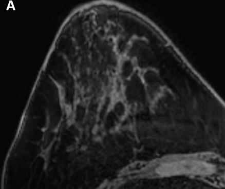
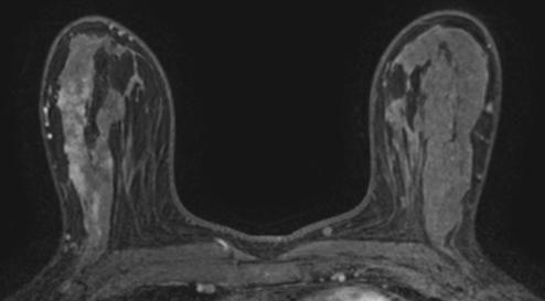
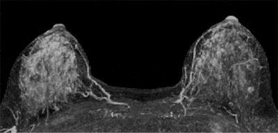
11 BPE Appearance Typically symmetric and diffuse
12 BPE Picture Framing Peripheral to central enhancement of breast tissue secondary to arterial supply Radiographics Jan-Feb;34(1):

13 BPE Picture Framing

14 BPE Difficulties in Interpretation Higher false-positive rate in patients with moderate or severe background enhancement Higher rates of BI-RADS 3 categorization AJR Am J Roentgenol Jan;196(1):218-24
15 BPE Problems Can be asymmetric, focal, or regional Frequently described as patchy, focal, or nodular Can be difficult to differentiate from NME

16 BPE - Nodular

17 BPE - Focal

18 BPE - Focal MRI MRI-guided Biopsy
19 Non-mass Enhancement (NME)
20 Non-mass Enhancement (NME)
21 NME Distribution Focal Enhancement in a confined area, <25% of a quadrant
22 NME Distribution Focal
23 NME Distribution Linear/Ductal Linear: Enhancement in a line that may not conform to a duct Ductal: Enhancement in a line that may have branching, conforming to a duct Ductal distribution eliminated from 2013 BI-RADS (5 th edition) due to underuse
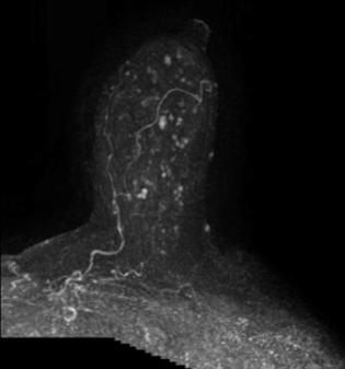
24 NME Distribution Linear
25 NME Distribution Segmental Triangular region of enhancement, apex pointing to the nipple, suggesting a duct or its branches
26 NME Distribution - Segmental
27 NME Distribution Regional Enhancement in a large volume of tissue not conforming to a ductal distribution, georaphic
28 NME Distribution Regional
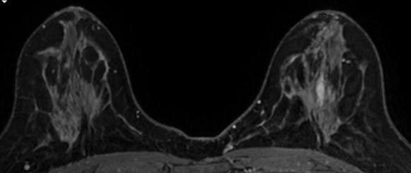
29 NME Distribution Multiregional Enhancement in at least two large volumes of tissue not conforming to a ductal distribution, multiple geographic area Typically due to BPE

30 NME Distribution Multiregional
31 NME Distribution Diffuse Enhancement distributed uniformly throughout the breast Typically due to BPE
32 NME Distribution Diffuse

33 Non-mass Enhancement (NME)

34 NME Internal Enhacement Homogenous Confluent, uniform enhancement

35 NME Internal Enhancement Heterogenous Nonuniform enhancement in a random pattern

36 NME Internal Enhancement Clumped Cobblestone-like enhancement, with occasional confluent areas

37 NME Internal Enhancement Clustered Ring Minute ring enhancements which are clustered Added in 2013 BI-RADS (5 th edition)
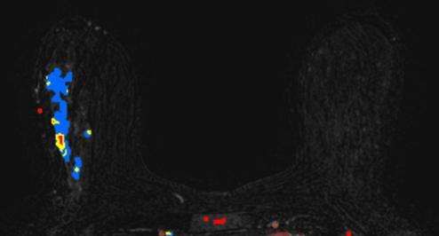
38 NME Kinetics
39 NME Differential Diagnosis Benign High Risk Malignant Fibrocystic changes Focal adenosis Apocrine metaplasia Pseudoangiomatous stromal hyperplasia Radiation effect Radial scar/ complex sclerosing lesion Intraductal papilloma Flat epithelial atypia Atypical ductal hyperplasia Ductal carcinoma in situ Invasive ductal carcinoma Invasive lobular carcinoma AJR Am J Roentgenol Jan;204(1):219-27
40 NME DCIS Distribution Most common: linear, segmental Less common: regional, focal Internal Enhancement Pattern Clustered ring Clumped Heterogeneous AJR Am J Roentgenol Sep;191(3):689-99
41 Predictive Value of BI-RADS Lexicon Lexicon for masses can be highly predictive for malignancy Predictive value of non mass enhancement lexicon mixed in literature

42 Retrospective Highest PPV: Segmental Clumped Linear/Ductal
43 Retrospective Most benign descriptors: Linear, homogenous enhancement Most frequent malignant descriptors: Segmental, clustered ring enhancement (PPV 100%) Segmental, clumped enhancement (PPV 88%)

44 Small sample size


45 Prospective Ductal distribution and clumped internal enhancement had highest PPV of malignancy

46 NME Kinetics Predictive Value Kinetics not predictive of malignancy Mahoney, et al (Radiology Jul; 264(1): 51 58
47 NME Predictive Value Interobserver variability in MRI interpretation likely resulting in varied PPVs of NME lexicon in the literature
48 Case 1 Linear NME Patient A Patient B Biopsy-proven fibroadenoma


49 Case 1 Linear NME Patient A Patient B Biopsy-proven fibroadenoma
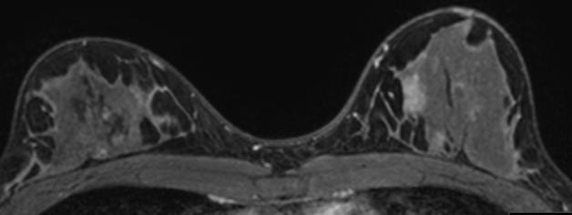
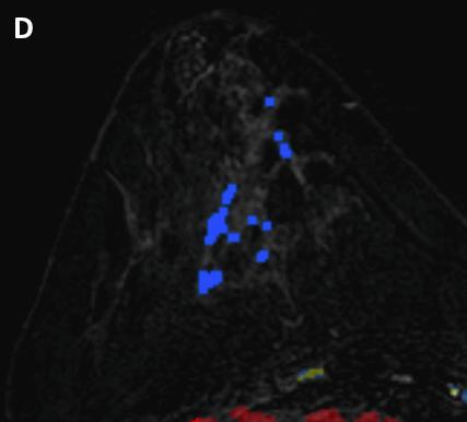
50 Case 2 Segmental NME Patient A Patient B

51 Case 2 Segmental NME Patient A Patient B
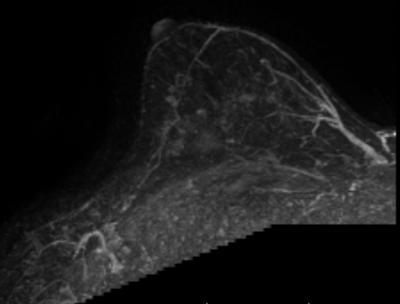
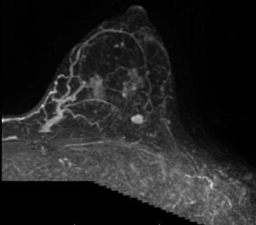
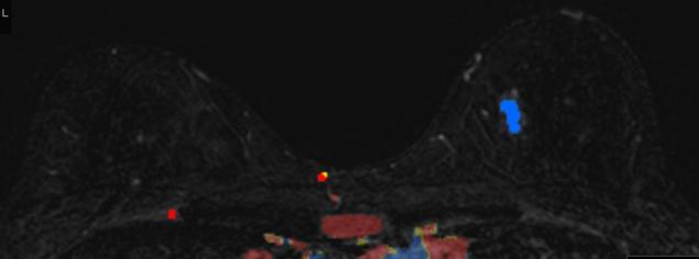
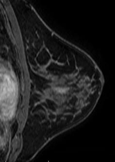
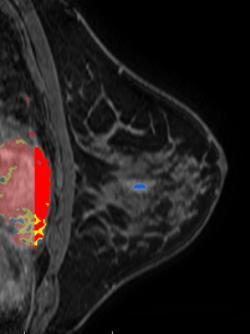
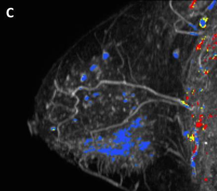
52 Case 3 Regional NME Patient A Patient B

53 Case 3 Regional NME Patient A Patient B
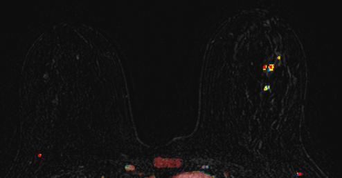
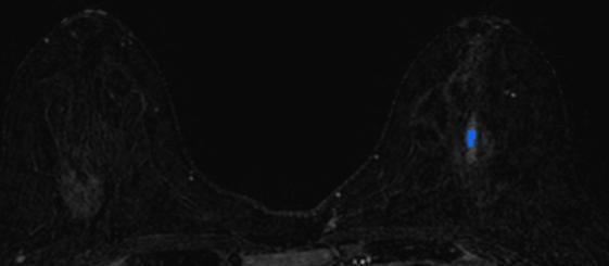
54 Case 4 Focal NME Patient A Patient B


55 Case 4 Focal NME Patient A Patient B
56 NME Management Biopsy (BI-RADS 4) often pursued, given substantial overlap in imaging appearances of benign and malignant causes of NME Surveillance (BI-RADS 3) an option, particularly if multiple or bilateral findings of low suspicion
57 NME Rad-Path Concordance Does the pathology explain the findings? Also correlate with features on other modalities (MG, US)
58 NME Rad-Path Concordance Benign High Risk Malignant Fibrocystic changes Focal adenosis Apocrine metaplasia Pseudoangiomatous stromal hyperplasia Radiation effect Radial scar/ complex sclerosing lesion Intraductal papilloma Flat epithelial atypia Atypical ductal hyperplasia Ductal carcinoma in situ Invasive ductal carcinoma Invasive lobular carcinoma AJR Am J Roentgenol Jan;204(1):219-27

59 Rad-Path Concordance Benign Fibrocystic changes/apocrine metaplasia: focal or regional distribution Coarse calcs on MG, lobulated mass on US AJR Am J Roentgenol Jan;204(1):219-27

60 Rad-Path Concordance Benign Focal adenosis: focal distribution, variable internal enhancement Simple, sclerosing, apocrine, tubular, microglandular Sclerosing adenosis often associated with MG calcs AJR Am J Roentgenol Jan;204(1):219-27
:219-27")
61 Rad-Path Concordance Benign PASH: focal or segmental distribution, clumped internal enhancement Often associated with T2 hyperintense cystic spaces AJR Am J Roentgenol Jan;204(1):219-27

62 Rad-Path Concordance Benign Radiation effect: focal or diffuse, within 18 months of treatment completion Persistent focal or diffuse enhancement >18 months after treatment completion raises concern for recurrence AJR Am J Roentgenol Jan;204(1):219-27

63 Rad-Path Concordance High Risk Radial scar/complex sclerosing lesion: linear or clumped NME Often associated with architectural distortion on MG AJR Am J Roentgenol Jan;204(1):219-27

64 Rad-Path Concordance High Risk Intraductal papilloma: mass, focus, or linear NME within 3 cm of nipple Clinically associated with spontaneous, unilateral, bloody nipple discharge AJR Am J Roentgenol Jan;204(1):219-27
:219-27")
65 Rad-Path Concordance High Risk Flat epithelial atypia/atypical ductal hyperplasia: variable appearance ranging from mass to non mass enhancement Often associated with calcifications on MG AJR Am J Roentgenol Jan;204(1):219-27

66 Rad-Path Concordance Malignant Ductal carcinoma in situ: segmental or linear distribution, clumped or heterogeneous internal enhancement Coarse, heterogeneous or pleomorphic calcs on MG Extent can be overestimated on MR due to periductal and stromal fibrosis AJR Am J Roentgenol Jan;204(1):219-27

67 Rad-Path Concordance Malignant Invasive ductal carcinoma: mass or NME, variable distribution, clumped or heterogeneous internal enhacement Spiculated mass, architectural distortion, associated microcalcifications on MG AJR Am J Roentgenol Jan;204(1):219-27

68 Rad-Path Concordance Malignant Invasive lobular carcinoma: focal or regional NME Asymmetry or distortion on MG, subtle shadowing on US Often occult on MG or US MRI useful in determining extent of disease, which is often underestimated on other modalities AJR Am J Roentgenol Jan;204(1):219-27
69 NME Pearls Diffuse or multiregional NME typically due to BPE Segmental, clumped enhancement and segmental, clustered ring enhancement most closely associated with malignancy Kinetics not predictive of malignancy PPV of NME lexicon limited, and given significant overlap between benign and malignant processes, biopsy frequently pursued Rad-Path concordance important in ensuring biopsy results explain multimodality imaging findings
70 References ACR Practice Parameter for the Performance of Contrast-Enhanced Magnetic Resonance Imaging (MRI) of the Breast. Available at American College of Radiology. Accessed February 3, JAMA Intern Med Jan;174(1): Radiographics Jan-Feb;34(1): AJR Am J Roentgenol Jan;196(1): AJR Am J Roentgenol Jan;204(1): AJR Am J Roentgenol Sep;191(3): AJR Am J Roentgenol Jul;179(1): AJR Am J Roentgenol Aug;187(2): Radiology Jul; 264(1):
BI-RADS and Breast MRI. Kathy Borovicka, M.D. Thursday February 15, 2018
 BI-RADS and Breast MRI Kathy Borovicka, M.D. Thursday February 15, 2018 Learning Objectives Be familiar with the Breast Imaging Reporting and Data System (BI-RADS) Understand the components of a breast
Imaging in breast cancer. Mammography and Ultrasound Donya Farrokh.MD Radiologist Mashhad University of Medical Since
 Imaging in breast cancer Mammography and Ultrasound Donya Farrokh.MD Radiologist Mashhad University of Medical Since A mammogram report is a key component of the breast cancer diagnostic process. A mammogram
Lesion Imaging Characteristics Mass, Favoring Benign Circumscribed Margins Intramammary Lymph Node
 Lesion Imaging Characteristics Mass, Favoring Benign Circumscribed Margins Intramammary Lymph Node Oil Cyst Mass, Intermediate Concern Microlobulated Margins Obscured Margins Mass, Favoring Malignant Indistinct
Armed Forces Institute of Pathology.
 Armed Forces Institute of Pathology www.radpath.com Armed Forces Institute of Pathology Breast Disease www.radpath.org Armed Forces Institute of Pathology Interpretation of Breast MRI Leonard M. Glassman
Treatment options for the precancerous Atypical Breast lesions. Prof. YOUNG-JIN SUH The Catholic University of Korea
 Treatment options for the precancerous Atypical Breast lesions Prof. YOUNG-JIN SUH The Catholic University of Korea Not so benign lesions? Imaging abnormalities(10% recall) lead to diagnostic evaluation,
CLINICAL SIGNIFICANCE OF BENIGN EPITHELIAL CHANGES
 Papillomas. Papillomas are composed of multiple branching fibrovascular cores, each having a connective tissue axis lined by luminal and myoepithelial cells ( Fig. 23-11 ). Growth occurs within a dilated
Radiologic and pathologic correlation of non-mass like breast lesions on US and MRI: Benign, high risk, versus malignant
 Radiologic and pathologic correlation of non-mass like breast lesions on US and MRI: Benign, high risk, versus malignant Poster No.: C-1161 Congress: ECR 2013 Type: Educational Exhibit Authors: J. Kwak,
Leonard M. Glassman MD
 BI-RADS The New BI-RADS Leonard M. Glassman MD FACR Former Chief of Breast Imaging American Institute for Radiologic Pathology Washington Radiology Associates, PC Breast Imaging Reporting and Data System
BREAST MRI. VASILIKI FILIPPI RADIOLOGIST CT MRI & PET/CT Departments Hygeia Hospital, Athens, Greece
 BREAST MRI VASILIKI FILIPPI RADIOLOGIST CT MRI & PET/CT Departments Hygeia Hospital, Athens, Greece Breast ΜR Imaging (MRM) Breast MR imaging is an extremely powerful diagnostic tool, that when used in
BI-RADS Update. Martha B. Mainiero, MD, FACR, FSBI Brown University Rhode Island Hospital
 BI-RADS Update Martha B. Mainiero, MD, FACR, FSBI Brown University Rhode Island Hospital No Disclosures BI-RADS History 1980s Quality Issues ACR Accreditation BI-RADS 1994 2003 4 th Edition MRI, US January
Pitfalls and Limitations of Breast MRI. Susan Orel Roth, MD Professor of Radiology University of Pennsylvania
 Pitfalls and Limitations of Breast MRI Susan Orel Roth, MD Professor of Radiology University of Pennsylvania Objectives Review the etiologies of false negative breast MRI examinations Discuss the limitations
Breast calcification: Management and Pictorial Review
 Breast calcification: Management and Pictorial Review Poster No.: C-0692 Congress: ECR 2014 Type: Educational Exhibit Authors: V. de Lara Bendahan, M. F. Ramos Solis, A. Amador Gil, C. 1 2 3 2 4 4 Gómez
Mammographic imaging of nonpalpable breast lesions. Malai Muttarak, MD Department of Radiology Chiang Mai University Chiang Mai, Thailand
 Mammographic imaging of nonpalpable breast lesions Malai Muttarak, MD Department of Radiology Chiang Mai University Chiang Mai, Thailand Introduction Contents Mammographic signs of nonpalpable breast cancer
Breast Cancer Imaging
 Breast Cancer Imaging I. Policy University Health Alliance (UHA) will cover breast imaging when such services meet the medical criteria guidelines (subject to limitations and exclusions) indicated below.
04/10/2018 HIGH RISK BREAST LESIONS. Pathology Perspectives of High Risk Breast Lesions ELEVATED RISK OF BREAST CANCER HISTORICAL PERSPECTIVES
 Pathology Perspectives of High Risk Breast Lesions Savitri Krishnamurthy MD Professor of Pathology Deputy Division Head Director of Clinical Trials, Research and Development The University of Texas MD
Breast pathology. 2nd Department of Pathology Semmelweis University
 Breast pathology 2nd Department of Pathology Semmelweis University Breast pathology - Summary - Benign lesions - Acute mastitis - Plasma cell mastitis / duct ectasia - Fat necrosis - Fibrocystic change/
Ana Sofia Preto 19/06/2013
 Ana Sofia Preto 19/06/2013 Understanding the underlying pathophysiologic processes leading to the various types of calcifications Description and illustration of the several types of calcifications, according
National Diagnostic Imaging Symposium 2013 SAM - Breast MRI 1
 National Diagnostic Imaging Symposium 2013 December 8-12, 2013 Disney s Yacht Club Resort Lake Buena Vista, Florida Self Assessment Module Questions, Answers and References Day SAM Title - Each SAM title
Breast Disease: What PCPs Need to Know. Eunice Cho MD FACS
 Breast Disease: What PCPs Need to Know Eunice Cho MD FACS New Breast Cancer Screening Guideline for women with average risk Every other year AGE 40 AGE 45 AGE 55 AGE 55 + Talk with your doctor about when
CDIS: what's beyond microcalcifications? - Pictorial essay
 CDIS: what's beyond microcalcifications? - Pictorial essay Poster No.: C-1096 Congress: ECR 2014 Type: Educational Exhibit Authors: R. N. Lucas, C. A. S. Ruano, I. Oliveira, J. M. G. Lourenco, Z. 1 1 1
RSNA, /radiol Appendix E1. Methods
 RSNA, 2016 10.1148/radiol.2016151097 Appendix E1 Methods US and Near-infrared Data Acquisition Four optical wavelengths (740 nm, 780 nm, 808 nm, and 830 nm) were used to sequentially deliver the light
Breast MRI Update. Jeffrey C. Weinreb, MD, FACR Yale University School of Medicine
 Breast MRI Update Jeffrey C. Weinreb, MD, FACR jeffrey.weinreb@yale.edu Yale University School of Medicine I disclose the following financial relationships with relevant commercial interests: Bracco Bayer
Imaging the Symptomatic Patient. Avice M.O Connell MD,FACR,FSBI Professor of Imaging Sciences Director, Women s Imaging University of Rochester
 Imaging the Symptomatic Patient Avice M.O Connell MD,FACR,FSBI Professor of Imaging Sciences Director, Women s Imaging University of Rochester The four most common symptoms Mass Pain Discharge Infection
UW Radiology Review Course Breast Calcifications. BI-RADS 5 th Edition
 UW Radiology Review Course Breast Calcifications Grace Kalish, MD Vantage Radiology BI-RADS 5 th Edition Benign Skin Vascular Large rod like Coarse popcorn Suspicious Amorphous Coarse heterogenous Fine
Index. C Calcifications fat necrosis 1, 61 fat necrosis 4, 69 nipple/peri-areolar involvement 1, 165
 A ADH. See Atypical ductal hyperplasia (ADH) American College of Radiology (ACR), BI-RADS background parenchymal enhancement, 8, 9, 81, 82 fibroglandular tissue guidelines, 6 American Joint Committee on
Diseases of the breast (1 of 2)
") Diseases of the breast (1 of 2) Introduction A histology introduction Normal ducts and lobules of the breast are lined by two layers of cells a layer of luminal cells overlying a second layer of myoepithelial
Diseases of the breast (1 of 2) Introduction A histology introduction Normal ducts and lobules of the breast are lined by two layers of cells a layer of luminal cells overlying a second layer of myoepithelial
BI-RADS CATEGORIZATION AND BREAST BIOPSY categorization in the selection of appropriate breast biopsy technique is also discussed. Patients and method
 Original Article Positive Predictive Value of BI-RADS Categorization in an Asian Population Yah-Yuen Tan, Siew-Bock Wee, Mona P.C. Tan and Bee-Kiang Chong, 1 Departments of General Surgery and 1Diagnostic
Melissa Hartman, DO Women s Health Orlando VA Medical Center
 Melissa Hartman, DO Women s Health Orlando VA Medical Center Most common non-skin cancer and Second deadliest cancer in women Majority are diagnosed by abnormal screening study An approach to breast cancer
Cytyc Corporation - Case Presentation Archive - March 2002
 FirstCyte Ductal Lavage History: 68 Year Old Female Gail Index: Unknown Clinical History: Negative Mammogram in 1995 6 yrs. later presents with bloody nipple discharge Subsequent suspicious mammogram Suspicious
MEDICAL POLICY SUBJECT: MAGNETIC RESONANCE IMAGING (MRI) OF THE BREAST. POLICY NUMBER: CATEGORY: Technology Assessment
 OF THE BREAST. POLICY NUMBER: CATEGORY: Technology Assessment") MEDICAL POLICY SUBJECT: MAGNETIC RESONANCE IMAGING (MRI) OF THE BREAST PAGE: 1 OF: 9 If the member's subscriber contract excludes coverage for a specific service it is not covered under that contract.
MEDICAL POLICY SUBJECT: MAGNETIC RESONANCE IMAGING (MRI) OF THE BREAST PAGE: 1 OF: 9 If the member's subscriber contract excludes coverage for a specific service it is not covered under that contract.
Breast Pathology. Breast Development
 Breast Pathology Lecturer: Hanina Hibshoosh, M.D. Reading: Kumar, Cotran, Robbins, Basic Pathology, 6th Edition, pages 623-635 Breast Development 5th week - thickening of the epidermis - milk line 5th
Breast Evaluation & Management Guidelines
 Breast Evaluation & Management Guidelines Pamela L. Kurtzhals, M.D. F.A.C.S. Head, Dept. of General Surgery Scripps Clinic, La Jolla Objective Review screening & diagnostic guidelines Focused patient complaints
LYMPHATIC DRAINAGE AXILLARY (MOSTLY) INTERNAL MAMMARY SUPRACLAVICULAR
 INTERNAL MAMMARY SUPRACLAVICULAR") BREAST LYMPHATIC DRAINAGE AXILLARY (MOSTLY) INTERNAL MAMMARY SUPRACLAVICULAR HISTOLOGY LOBE: (10 in whole breast) LOBULE: (many per lobe) ACINUS/I, aka ALVEOLUS/I: (many per lobule) DUCT(S): INTRA- or
BREAST LYMPHATIC DRAINAGE AXILLARY (MOSTLY) INTERNAL MAMMARY SUPRACLAVICULAR HISTOLOGY LOBE: (10 in whole breast) LOBULE: (many per lobe) ACINUS/I, aka ALVEOLUS/I: (many per lobule) DUCT(S): INTRA- or
Amammography report is a key component of the breast
 Review Article Writing a Mammography Report Amammography report is a key component of the breast cancer diagnostic process. Although mammographic findings were not clearly differentiated between benign
MRI BI-RADS: How to make it out?
 MRI BI-RADS: How to make it out? Poster No.: C-1850 Congress: ECR 2016 Type: Educational Exhibit Authors: M. Ben Ammar, A. Ben Miled, O. Ghdes, S. Harguem, A. Gaja, N. Mnif; Tunis/TN Keywords: Breast,
IBCM 2, April 2009, Sarajevo, Bosnia and Herzegovina
 Preoperative diagnosis and treatment planning in breast cancer The pathologist s perspective L. Mazzucchelli Istituto Cantonale di Patologia Locarno, Switzerland IBCM 2, 23-25 April 2009, Sarajevo, Bosnia
Sclerosing Adenosis of the Breast: Report of Two Cases and Review of the Literature
 Signature: Pol J Radiol, 2015; 80: 122-127 DOI: 10.12659/PJR.892706 CASE REPORT Received: 2014.10.10 Accepted: 2014.11.18 Published: 2015.03.07 Authors Contribution: A Study Design B Data Collection C
BREAST IMAGING and NEW IMAGING MODALITIES- A Surgeons view
 BREAST IMAGING and NEW IMAGING MODALITIES- A Surgeons view DR CHANTEL THORNTON SPECIALIST BREAST CANCER SURGEON BMSc (hons) MBBS (hons) FRACS Epworth Hospital, Richmond- Agora Centre for Women s Health
AMSER Case of the Month: November 2018
 AMSER Case of the Month: November 2018 52 year old female with an abnormal screening mammogram Areeg Rehman, MS 4 Nova Southeastern University Rebecca T. Sivarajah, MD Penn State University College of
Flat Epithelial Atypia
 Flat Epithelial Atypia Richard Owings, M.D. University of Arkansas for Medical Sciences Department of Pathology Flat epithelial atypia can be a difficult lesion May be a subtle diagnosis Lots of changes
BREAST PATHOLOGY. Fibrocystic Changes
 BREAST PATHOLOGY Lesions of the breast are very common, and they present as palpable, sometimes painful, nodules or masses. Most of these lesions are benign. Breast cancer is the 2 nd most common cause
EARLY DETECTION: MAMMOGRAPHY AND SONOGRAPHY
 EARLY DETECTION: MAMMOGRAPHY AND SONOGRAPHY Elizabeth A. Rafferty, M.D. Avon Comprehensive Breast Center Massachusetts General Hospital Harvard Medical School Breast Cancer Screening Early detection of
Immunohistochemical studies (ER & Ki-67) in Proliferative breast lesions adjacent to malignancy
 in Proliferative breast lesions adjacent to malignancy") IOSR Journal of Dental and Medical Sciences (IOSR-JDMS) e-issn: 2279-0853, p-issn: 2279-0861.Volume 13, Issue 3 Ver. IV. (Mar. 2014), PP 84-89 Immunohistochemical studies (ER & Ki-67) in Proliferative
IOSR Journal of Dental and Medical Sciences (IOSR-JDMS) e-issn: 2279-0853, p-issn: 2279-0861.Volume 13, Issue 3 Ver. IV. (Mar. 2014), PP 84-89 Immunohistochemical studies (ER & Ki-67) in Proliferative
ROLE OF MRI IN SCREENING, DIAGNOSIS AND MANAGEMENT OF BREAST CANCER. B.Zandi Professor of Radiology
 ROLE OF MRI IN SCREENING, DIAGNOSIS AND MANAGEMENT OF BREAST CANCER B.Zandi Professor of Radiology Introduction In the USA, Breast Cancer is : The Most Common Non-Skin Cancer The Second Leading cause of
04/10/2018. Intraductal Papillary Neoplasms Of Breast INTRADUCTAL PAPILLOMA
 Intraductal Papillary Neoplasms Of Breast Savitri Krishnamurthy MD Professor of Pathology Deputy Division Head The University of Texas MD Anderson Cancer Center 25 th Annual Seminar in Pathology Pittsburgh,
Breast Cancer Screening and Diagnosis
 Breast Cancer Screening and Diagnosis Priya Thomas, MD Assistant Professor Clinical Cancer Prevention and Breast Medical Oncology University of Texas MD Anderson Cancer Center Disclosures Dr. Thomas has
Criteria of Malignancy. Evaluation Score
 30 5 Diagnostic Criteria Criteria of Malignancy Table 5.2 lists criteria in contrast-enhancing MR mammography that strongly indicate the presence of malignancy or are unspecific. Unifactorial evaluation
Spectrum of findings of sclerosing adenosis at breast MRI.
 Spectrum of findings of sclerosing adenosis at breast MRI. Poster No.: C-0738 Congress: ECR 2012 Type: Scientific Exhibit Authors: F. Vasselli 1, F. Pediconi 2, M. Telesca 2, M. Luciani 2, V. Casali 2,
Radiology Review Course Hotel del Coronado Coronado, California
 37 th Annual Radiology Review Course Hotel del Coronado Coronado, California Friday, April 21, 2017 - PM TABLE OF CONTENTS Friday, April 21, 2017 - PM SAM Session - Breast Imaging Update 12:45 PM 1:30
Proliferative Breast Disease: implications of core biopsy diagnosis. Proliferative Breast Disease
 Proliferative Breast Disease: implications of core biopsy diagnosis Jean F. Simpson, M.D. Breast Pathology Consultants, Inc. Nashville, TN Proliferative Breast Disease Must be interpreted in clinical and
University of Washington Radiology Review Course: Strange and Specific Diagnoses. Case #1
 University of Washington Radiology Review Course: Strange and Specific Diagnoses Katherine E. Dee, MD Seattle Breast Center Via Radiology 2014 Case #1 37 year old presents with bilateral palpable lumps.
Imaging Guidelines for Breast Cancer Screening
 Imaging Guidelines for Breast Cancer Screening Sarah Colwick, MD Dr. Sarah Colwick was born and raised in Sikeston, MO. She attended college and medical school at the University of Missouri-Kansas City
Disclosures. Breast Cancer. Breast Imaging Modalities. Breast Cancer Screening. Breast Cancer 6/4/2014
 : Information for the Primary Care Physician Disclosures No financial relationships with commercial entities producing health care products/services. Roxsann Roberts, MD Section Chief, MRI Erlanger/EmCare
EARLY DETECTION: MAMMOGRAPHY AND SONOGRAPHY
 EARLY DETECTION: MAMMOGRAPHY AND SONOGRAPHY Elizabeth A. Rafferty, M.D. Avon Comprehensive Breast Center Massachusetts General Hospital Harvard Medical School Breast Cancer Screening Early detection of
Tips and Tricks to performing Magnetic Resonance Imaging Guided Breast Interventional Procedures Habib Rahbar, MD, FSBI October 23, 2018, 7:00pm ET
 Tips and Tricks to performing Magnetic Resonance Imaging Guided Breast Interventional Procedures Habib Rahbar, MD, FSBI October 23, 2018, 7:00pm ET SAM Questions/Answers/Rationales/References 1. Below
Benign, Reactive and Inflammatory Lesions of the Breast
 Benign, Reactive and Inflammatory Lesions of the Breast Marilin Rosa, MD Associate Member Section Head of Breast Pathology Department of Anatomic Pathology Program Director, Breast Pathology Fellowship
Papillary Lesions of the Breast A Practical Approach to Diagnosis. (Arch Pathol Lab Med. 2016;140: ; doi: /arpa.
 Papillary Lesions of the Breast A Practical Approach to Diagnosis (Arch Pathol Lab Med. 2016;140:1052 1059; doi: 10.5858/arpa.2016-0219-RA) Papillary lesions of the breast Span the spectrum of benign,
MRI in breast cancer: diagnosis and intervention. Dr Sue Barter Addenbrookes Hospital, Cambridge UK
 MRI in breast cancer: diagnosis and intervention Dr Sue Barter Addenbrookes Hospital, Cambridge UK Intervention will be discussed in High Risk Screening! Indications UK and Europe: Breast MRI is well established
AB MR Interpretation Overview
 AB MR Interpretation Overview Goal of AB MR interpretation is to maintain high sensitivity and specificity In order to minimize false positives and short term follow ups, it is fundamental to focus only
BI-RADS Categorization As a Predictor of Malignancy 1
 Susan G. Orel, MD Nicole Kay, BA Carol Reynolds, MD Daniel C. Sullivan, MD BI-RADS Categorization As a Predictor of Malignancy 1 Index terms: Breast, biopsy, 00.1261 Breast neoplasms, localization, 00.125,
Segmental Breast Calcifications
 Residents Section Pattern of the Month Chen et al. Segmental reast Calcifications Residents Section Pattern of the Month Residents inradiology Po-Hao Chen 1 Erica T. Ghosh 1,2 Priscilla J. Slanetz 1,2
Throughout this policy, bracketed numbers link topics across multiple sections according to the indication numbers in the following list.
 Subject: Magnetic Resonance Imaging of the Breast Page: 1 of 33 Last Review Status/Date: September 2015 Magnetic Resonance Imaging of the Breast Description Magnetic resonance imaging (MRI) of the breast
Vesalius SCALpel : Benign breast disease (see also: breast folios)
") Vesalius SCALpel : Benign breast disease (see also: breast folios) Breast cancer risk Imaging Pain non-proliferative: only fibroadenoma may be associated with a slight risk of cancer proliferative: moderate
Vesalius SCALpel : Benign breast disease (see also: breast folios) Breast cancer risk Imaging Pain non-proliferative: only fibroadenoma may be associated with a slight risk of cancer proliferative: moderate
1 NORMAL HISTOLOGY AND METAPLASIAS
 1 NORMAL HISTOLOGY AND METAPLASIAS, MD Anatomy and Histology 1 Metaplasias 2 ANATOMY AND HISTOLOGY The female breast is composed of a branching duct system, which begins at the nipple with the major lactiferous
Diagnostic Breast Evaluation: When to Refer and What the Results Really Mean. Julie Dreadin RN, MS, WHNP-BC. Peggy Mancuso PhD, CNM, RN
 Diagnostic Breast Evaluation 1 Running head: DIAGNOSTIC BREAST EVALUATION Diagnostic Breast Evaluation: When to Refer and What the Results Really Mean Julie Dreadin RN, MS, WHNP-BC Peggy Mancuso PhD, CNM,
Promise of a beautiful day
 Promise of a beautiful day Ductal carcinoma in Situ Lobular Carcinoma in Situ Natural History Manosmed Tartous Oct 2009 Gérard ABADJIAN MD Pathology Department Hôtel-Dieu de France. Associate Professor
Imaging-Guided Core Needle Biopsy of Papillary Lesions of the Breast
 Eric L. Rosen 1 Rex C. Bentley 2 Jay A. Baker 1 Mary Scott Soo 1 Received January 30, 2002; accepted after revision April 12, 2002. 1 Department of Radiology, Breast Imaging Division, Duke University Medical
Ductal Carcinoma in Situ. Laura C. Collins, M.D. Department of Pathology Beth Israel Deaconess Medical Center and Harvard Medical School Boston, MA
 Ductal Carcinoma in Situ Laura C. Collins, M.D. Department of Pathology Beth Israel Deaconess Medical Center and Harvard Medical School Boston, MA Definition of DCIS WHO 2012 A neoplastic proliferation
Benign breast lesions frequently encountered on MR
 Benign breast lesions frequently encountered on MR Poster No.: C-1385 Congress: ECR 2012 Type: Educational Exhibit Authors: J. R. Almeida 1, J. C. Marques 2 ; 1 Lisbon/PT, 2 lisbon/pt Keywords: Hemangioma,
Improving the Identification of Underserved Women at High Risk for Breast Cancer and Increasing the use of Breast MRI Screening in this Population
 Improving the Identification of Underserved Women at High Risk for Breast Cancer and Increasing the use of Breast MRI Screening in this Population Greenwood HI, Truong L, Price ER. UCSF Department or Radiology
Pitfalls of Dynamic Contrast Enhanced MR Mammography (DCE-MRM) in Evaluation of Post-Biopsy Suspicious Breast Lesions
 in Evaluation of Post-Biopsy Suspicious Breast Lesions") Med. J. Cairo Univ., Vol. 86, No. 3, June: 1513-1522, 2018 www.medicaljournalofcairouniversity.net Pitfalls of Dynamic Contrast Enhanced MR Mammography (DCE-MRM) in Evaluation of Post-Biopsy Suspicious
Med. J. Cairo Univ., Vol. 86, No. 3, June: 1513-1522, 2018 www.medicaljournalofcairouniversity.net Pitfalls of Dynamic Contrast Enhanced MR Mammography (DCE-MRM) in Evaluation of Post-Biopsy Suspicious
WHICH INDICATION FOR BREAST MRI?
 WHICH INDICATION FOR BREAST MRI? Dr. P. De Visschere, Prof. Dr. G. Villeirs Genitourinary Radiology and Mammography University Hospital Gent Symposium Belgian Menopause Society 13/03/2010 Which Indication
Breast Update Therese Cusick MS MD FACS
 Breast Update 2017 Therese Cusick MS MD FACS Conflict of Interest Disclosure Nothing to disclose Sources Adapted from SESAP- Surgical Education and Self-Assessment program American College of Surgeons
BREAST MRI. Elizabeth A. Rafferty, M.D. Avon Comprehensive Breast Center Massachusetts General Hospital Harvard Medical School
 BREAST MRI Elizabeth A. Rafferty, M.D. Avon Comprehensive Breast Center Massachusetts General Hospital Harvard Medical School BREAST MRI Any assessment of the breast parenchyma requires the administration
BI-RADS MRI: A Primer
 Erguvan- ogan et al. I- RS MRI Women s Imaging Pictorial Essay WOMEN S IMGING asak Erguvan-ogan 1 Gary J. Whitman 1 nne. Kushwaha 1,2 Michael J. Phelps 1,3 Peter J. empsey 1 Erguvan-ogan, Whitman GJ, Kushwaha,
High risk lesions of the breast : Review of the current diagnostic and management strategies
 High risk lesions of the breast : Review of the current diagnostic and management strategies Poster No.: C-1204 Congress: ECR 2016 Type: Educational Exhibit Authors: P. Jagmohan, F. J. Pool, P. G. Pillay,
Breast Imaging Lexicon
 9//201 200 BI RADS th Edition 201 BI RADS th Edition Breast Imaging Lexicon Mammographic Pathology and Assessment Categories Deborah Thames, R.T.(R)(M)(QM) The Advanced Health Education Center Nonmember:
CURRICULUM FOR THE BREAST PATHOLOGY ROTATION UNIVERSITY OF FLORIDA DEPARTMENT OF PATHOLOGY
 CURRICULUM FOR THE BREAST PATHOLOGY ROTATION UNIVERSITY OF FLORIDA DEPARTMENT OF PATHOLOGY JULY, 2003 The following is a conceptual curriculum and set of guidelines for Pathology Residents on the Breast
Proliferative Epithelial lesions of the Breast. Sami Shousha, MD, FRCPath Charing Cross Hospital & Imperial College, London
 Proliferative Epithelial lesions of the Breast Sami Shousha, MD, FRCPath Charing Cross Hospital & Imperial College, London Amman, November2013 Proliferative Epithelial Lesions of the Breast Usual type
Breast Cancer. Most common cancer among women in the US. 2nd leading cause of death in women. Mortality rates though have declined
 Breast Cancer Most common cancer among women in the US 2nd leading cause of death in women Mortality rates though have declined 1 in 8 women will develop breast cancer Breast Cancer Breast cancer increases
Standard Breast Imaging Modalities. Lilian Wang, M.D. Breast Imaging Section Department of Radiology Northwestern Medicine
 Standard Breast Imaging Modalities Lilian Wang, M.D. Breast Imaging Section Department of Radiology Northwestern Medicine Overview Standard breast imaging modalities Mammography Ultrasound MRI Imaging
Atypical Ductal Hyperplasia and Papillomas: A Comparison of Ultrasound Guided Breast Biopsy and Stereotactic Guided Breast Biopsy
 Atypical Ductal Hyperplasia and Papillomas: A Comparison of Ultrasound Guided Breast Biopsy and Stereotactic Guided Breast Biopsy Breast Cancer is the most common cancer diagnosed in women in the United
Value of the BI-RADS classification in MR-Mammography for diagnosis of benign and malignant breast tumors
 Eur Radiol (2011) 21:2475 2483 DOI 10.1007/s00330-011-2210-7 BREAST Value of the BI-RADS classification in MR-Mammography for diagnosis of benign and malignant breast tumors Christian Sohns & Martin Scherrer
Image guided core biopsies:
 Recommendations on the Surgical, Radiologic and Pathologic Approaches to Breast Disease: Using best practices based on multidisciplinary methodologies developed through the Allina Breast Committee. Image
Atypical proliferative lesions diagnosed on core biopsy - 6 year review
 Atypical proliferative lesions diagnosed on core biopsy - 6 year review Dr Angela Harris, Dr Julie Weigner & Dr Ricardo Vilain NSW Health Pathology Pathology North, Hunter Anatomical Pathology & Cytology
MEDICAL IMAGING AND BREAST DISEASE HOW CAN WE HELP YOU?
 MEDICAL IMAGING AND BREAST DISEASE HOW CAN WE HELP YOU? Barbara M. Preston, M.D. SCREENING MAMMOGRAPHY AVERAGE RISK PATIENTS KAISER RECOMMENDATION: ALL WOMEN (INCLUDING TRANSGENDER FEMALES) Every 1-21
Breast Cancer. Saima Saeed MD
 Breast Cancer Saima Saeed MD Breast Cancer Most common cancer among women in the US 2nd leading cause of death in women 1 in 8 women will develop breast cancer Incidence/mortality rates have declined Breast
M Wani, M Khan, N Ul Gani, S Sangeen, B Singh, M Shafi, A Bilal, S Umer
 ISPUB.COM The Internet Journal of Surgery Volume 12 Number 1 Juvenile Fibroadenoma With Fibroadenomatoid Hyperplasia M Wani, M Khan, N Ul Gani, S Sangeen, B Singh, M Shafi, A Bilal, S Umer Citation M Wani,
Can magnetic resonance imaging obviate the need for biopsy for microcalcifications?
 Original Article Can magnetic resonance imaging obviate the need for biopsy for microcalcifications? Shinya Yamamoto, Takashi Chishima Department of Breast Surgery, Yokohama Rosai Hospital, Yokohama 222-0036,
Diagnostic Dilemmas of Breast Imaging
 Diagnostic Dilemmas of Breast Imaging Common Causes of Error in Breast Cancer Detection By: Jason Cord, M.D. Mammography: Initial Imaging The standard for detection of breast cancer Screening mammography
ACRIN 6666 IM Additional Evaluation: Additional Views/Targeted US
 Additional Evaluation: Additional Views/Targeted US For revised or corrected form check box and fax to 215-717-0936. Instructions: The form is completed based on recommendations (from ID form) for additional
A712(19)- Test slide, Breast cancer tissues with corresponding normal tissues
- Test slide, Breast cancer tissues with corresponding normal tissues") A712(19)- Test slide, Breast cancer tissues with corresponding normal tissues (formalin fixed) For research use only Specifications: No. of cases: 12 Tissue type: Breast cancer tissues with corresponding
A712(19)- Test slide, Breast cancer tissues with corresponding normal tissues (formalin fixed) For research use only Specifications: No. of cases: 12 Tissue type: Breast cancer tissues with corresponding
INDEX. in this web service Cambridge University Press
 abscess. See also subareolar abscess acute mastitis, 44 lactational/puerperal mastitis, 55 mammary tuberculosis, 42 tuberculous, 43 adeno gastric, 198, 200 invasive, 157 lung, 197, 200 prostatic, 199 200
BREAST MRI. Elizabeth A. Rafferty, M.D. Avon Comprehensive Breast Center Massachusetts General Hospital Harvard Medical School
 BREAST MRI Elizabeth A. Rafferty, M.D. Avon Comprehensive Breast Center Massachusetts General Hospital Harvard Medical School BREAST MRI Any assessment of the breast parenchyma requires the administration
COMMON BENIGN DISORDERS AND DISEASES OF THE BREAST
 COMMON BENIGN DISORDERS AND DISEASES OF THE BREAST Aberrations of Normal Development and Involution (ANDI). The basic principles underlying the aberrations of normal development and involution (ANDI) classification
Pathologic outcomes of coarse heterogeneous calcifications detected on mammography
 Pathologic outcomes of coarse heterogeneous calcifications detected on mammography Poster No.: C-1957 Congress: ECR 2011 Type: Scientific Paper Authors: H. J. Lim, K. R. Cho, K. W. Hwang, B. K. Seo, O.
What are the risk factors for breast cancer?
 What are the risk factors for breast cancer? A risk factor is anything that affects your chance of getting a disease, such as cancer. Different cancers have different risk factors. For example, exposing
Excerpts from the American College of Surgeons Educational Courses about Breast Disease:
 1 Excerpts from the American College of Surgeons Educational Courses about Breast Disease: Gynecomastia Gynecomastia is a benign enlargement of the male breast that can be unilateral or bilateral. It typically