Effusion Cytology: Diagnostic Challenges
|
|
|
- Aubrie Johnson
- 6 years ago
- Views:
Transcription
1 Effusion Cytology: Diagnostic Challenges Tarik M. Elsheikh, MD Professor and Medical Director, Anatomic Pathology Cleveland Clinic
2 Outside Consult Case 45 year old woman, presented with nausea, dyspnea, emesis and productive cough Chest X-ray: left pleural effusion, LLL consolidation and atelectasis CT: Mediastinal lymphadenopathy Never smoker, no significant PMH


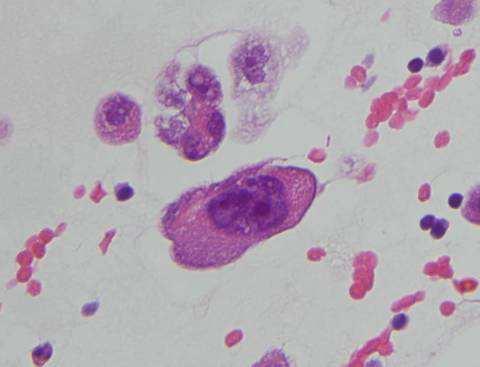
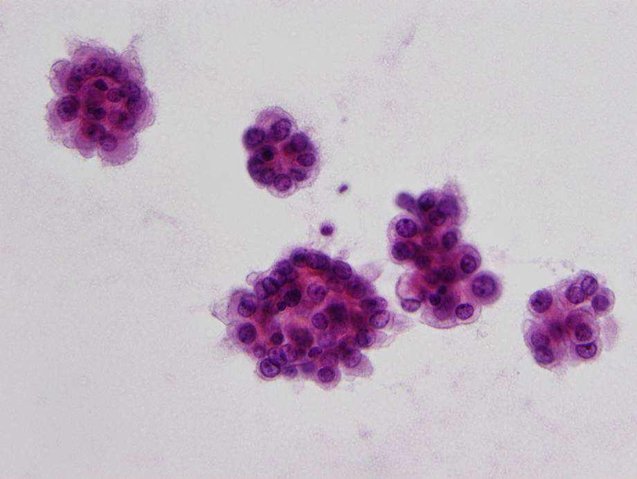
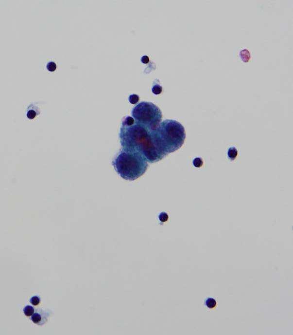
3 Left pleural fluid

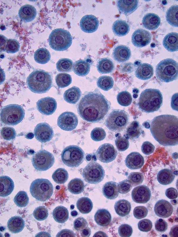
4


5 CK5/6 Calretinin Additional IHC P63 -, WT1 -, D240 - TTF1 Mammaglobin - CEA + Outside Dx: Atypical, Favor reactive mesothelial cells

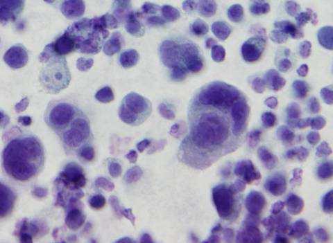
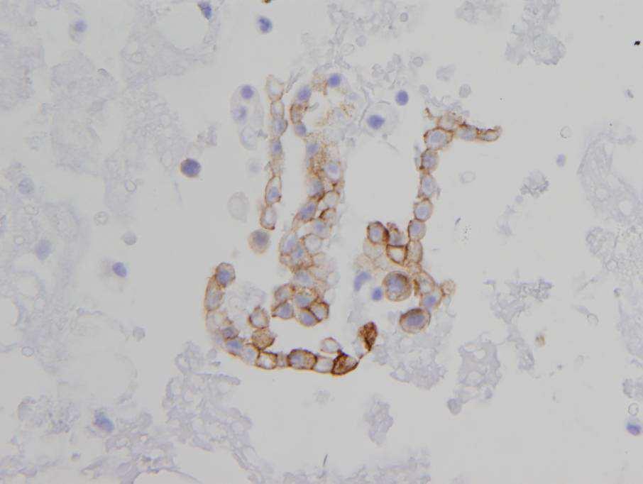
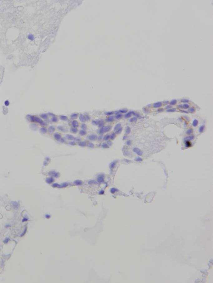
6 Pleural fluid-outside Consult


7 Moc 31 BerEp4 GCFP GATA3 WT1 -, D240 -, calretinin +, CK5/6 + P63 -, P40 - Moc 31+, BerEp4+, CEA+, B Napsin A -, TTF1 - ER/PR - PAX8 +, WT1 - Mamaglobin -, GATA3 +, GCFP + PAX8 Dx: Met adenocarcinoma, breast or mullerian origin
8 CAP Inter-laboratory Comparison Program in Body Cavity Fluids Data bank of 10,396 lab responses, Assessed characteristics of specimens that performed well and performed poor Poor performers in hypocellular and hypercellular malignant specimens Moriarty et al 2004
9 CAP inter-laboratory Comparison Program 2 Hypocellular: Location errors Rare malignant cells, had diagnostic features but were overlooked. Adeno ca, squamous ca, small cell ca, melanoma and lymphoma This emphasizes importance of careful methodical screening Moriarty et al 2004

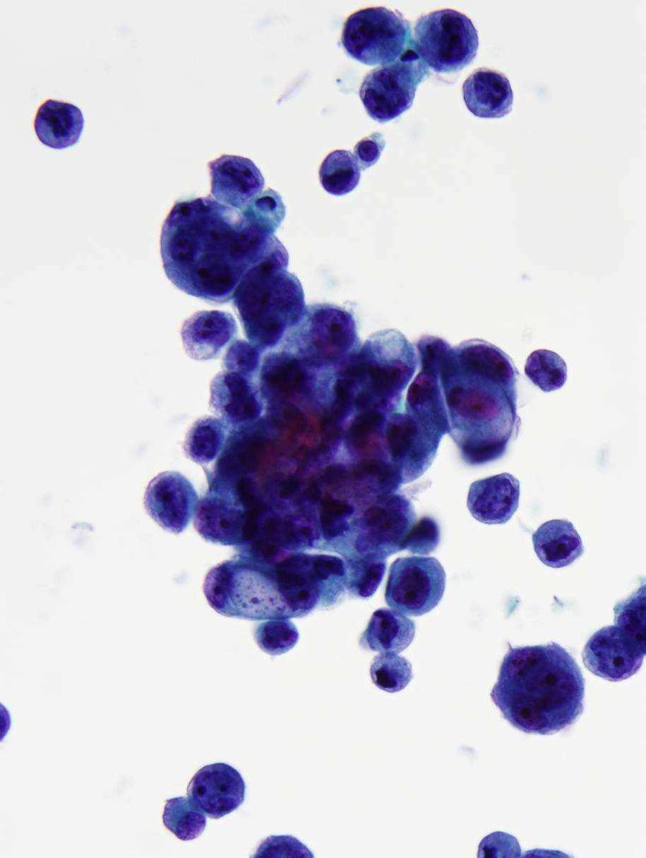
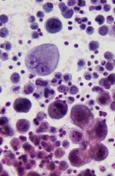
10 Carcinoid tumor in peritoneal fluid- false negative
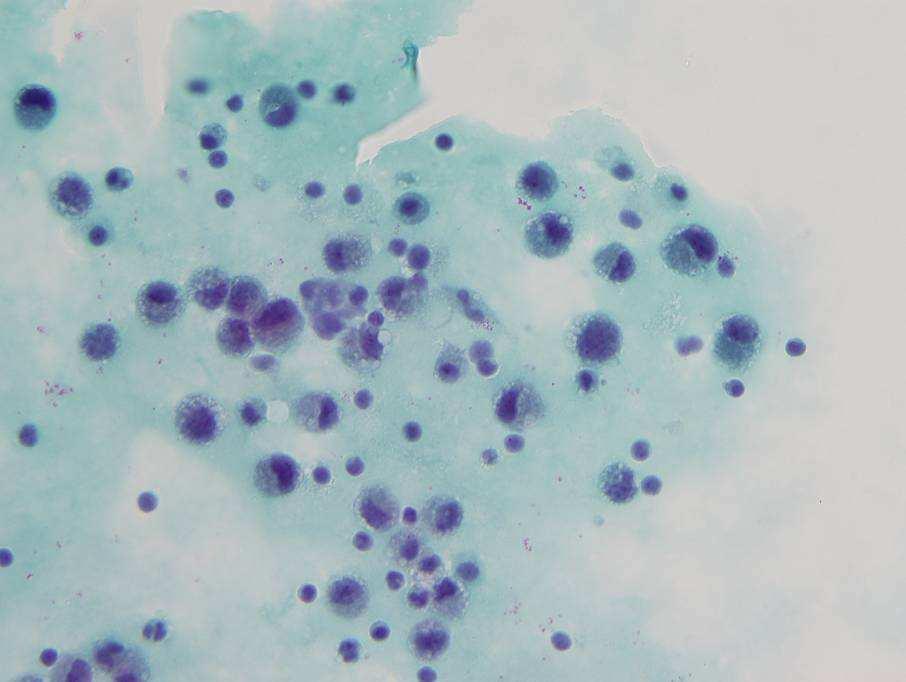
11 CAP inter-laboratory Comparison Program 3 Hypercellular: Interpretation errors Cellularity not a factor Poor carcinoma performers Single cells 3-D clusters Poor mesothelioma and lymphoma performers Moriarty et al 2004
12 Common Challenges in Fluids Atypical cells, suspicious but not diagnostic of malignancy Obviously malignant cells, but uncertain of classification or site of origin
13 Outline Benign effusions Work up of malignant effusions Cytomorphologic features Specific histologic types Immunohistochemistry Diagnostic challenges and potential pitfalls
14 Benign Effusions 70-80% of all effusions Mesothelial cells, histiocytes, inflammatory cells Common etiologies: Infectious: viral, bacterial, TB, fungal, parasitic Congestive heart failure Pulmonary embolism Myocardial infarction Cirrhosis Nephritic syndrome Collagen vascular disease Pancreatitits Trauma


15 Benign Effusions Variable cellularity, mostly single cells Occasional small clusters, < cells Exception: washings Windows


16 Spectrum of change Bi-nucleation and multi-nucleation common Dense cytoplasm, clear outer rim (lacy skirt) Oval-round nuclei, pale chromatin Small nucleoli, may be prominent


17 Atypia in Reactive Mesothelial Cells Nuclear hyperchromasia High N/C ratio Prominent nucleoli
18 Malignant effusions Rarely present as an occult malignancy (7-14%) Most patients have established history of malignancy Poor prognostic sign Lung, breast, ovary, GI tract- most common
19 Work-up of Malignant Effusions Morphology Low power architectural pattern High power cytologic detail Specific histologic types Ancillary studies IHC Flow cytometry
 population")

20 General Features of Malignancy Second (foreign) population Distinctly different from mesothelial cells Cohesive clusters, > cells


21 General Features of Malignancy 2 Malignant Nuclear Features High N/C ratio Nuclear hyperchromasia Nuclear irregularity Irregular distribution of chromatin Macro nucleoli
22 Architectural Patterns in Fluids Cell balls/cannonballs Papillary Acini Single cells (large or small) Linear/Indian file Signet ring Multinucleation Bizarre giant cells Clear cells Psammoma bodies
23 Cannonballs Tightly cohesive spherical cell clusters with an almost perfectly round contour and community border
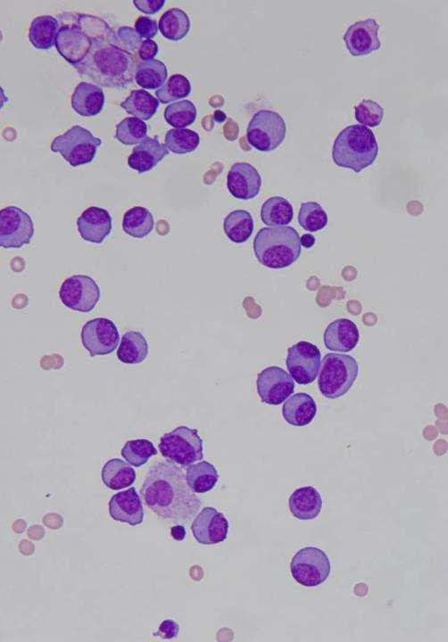
")

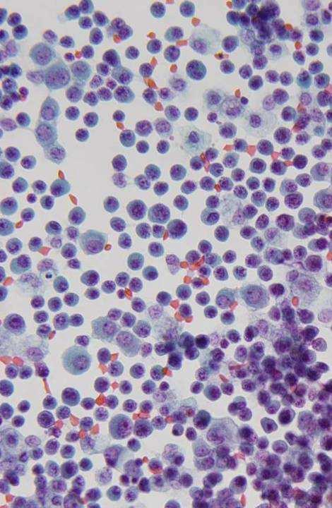
24 Cannonballs Mesothelial hyperplasia Breast Uniform cells suggest breast (more common) Pleomorphic cells suggest ovary, lung Other sources include GI tract, mesothelial hyperplasia and mesothelioma

25 Papillary 3-D clusters that are longer than wider Ascites ovary, uterus Pleural effusions lung, breast

26 Primary peritoneal CA Others: GU, pancreas, primary peritoneal, mesothelial hyperplasia, mesothelioma Thyroid PC is rarely associated with effusions

27 Acinar groups Breast 3D groups with lumens Characteristic of adeno ca, but not specific Lung, colon, ovary, stomach, breast, etc. Mesothelial hyperplasia, mesothelioma Small round cell tumors

28 Lung Better appreciated on cell blocks


29 Indian/single files Small cell Breast Small cell ca Carcinoid tumor pancreas gastric mesothelioma PD carcinoma


30 Single small cells Lymphoma/leukemia Small cell carcinoma Carcinoid Breast Stomach Small round cell tumor Lymphoma Carcinoid
31 Single large cells Squamous carcinoma Melanoma Poorly differentiated adenocarcinoma Renal, adrenal, hepatocellular ca, germ cell tumors Sarcoma Lymphoma Reactive mesothelial cells, mesothelioma


32 Ber-EP4+ Lung non-small cell CA
33 Plasmacytoma, pleural
34 Bizarre giant cells Undifferentiated carcinoma lung or pancreas Squamous ca Melanoma Mesothelioma Sarcoma R/O rheumatoid effusion, as it may show bizarre or giant cells

35 Lung Pancreas Mesothelioma

36 Cytoplasmic Vacuoles Vacuoles without indentation Non-specific, often degenerative Benign: degenerative Malignant: nonspecificovary, lung, pancreas, etc.


37 Signet Ring Cells Vacuoles indent the nucleus Lobular breast carcinoma Gastric carcinoma Colon


38 Diff DX: Degenerative Changes Benign CEA Degenerated mesothelial cells and histiocytes can have large vacuoles imparting a signet ring appearance
39 Multinucleated cells Reactive mesothelial cells, mesothelioma Giant cell carcinoma Hepatocellular carcinoma Melanoma Sarcoma Renal cell carcinoma Anaplastic large cell lymphoma Hodgkin disease Germ cell tumors

40 Esophagus Breast


41 Clear cells Kidney Ovary/GYN Germ cell tumor Endometrium


42 Clear cell ca cervix, peritoneal fluid Clear cells in fluids may have an unexpectedly dense cytoplasm

43 Psammoma bodies Psammoma bodies have no diagnostic significance Ovary Thyroid Mesothelioma Mesothelial hyperplasia Endosalpingiosis Mesothelial hyperplasia
44 Specific Histologic Types Cells tend to round up in fluids a certain degree of uniformity among various cell types Exaggerated in ThinPrep (personal experience) Mets in fluids, therefore, may not necessarily exactly resemble the primary tumor Familiar with variable presentations of specific histologic types
45 Histologic Types in Effusions Adeno CA: most common cancer Less common: squamous ca, small cell ca, lymphoma, melanoma Children: hematopoietic and SBCT

46 Adenocarcinoma Large clusters or isolated cells Foreign population Cannonballs: more common in breast cancer Signet ring: stomach and breast

47 Columnar cells Mucinous tumors, colon, ovary, endometriosis Colon


48 Gastric and colo-rectal cancers: may lose tall columnar configuration in fluids
49 Pitfalls- Adeno CA A second population of benign mesothelial cells may be absent Tumor cells resemble histiocytes

50 86 yo woman, peritoneal fluid


51 B72.3 Dx: Adenocarcinoma Tumor cells resemble histiocytes Follow-up: Signet ring CA of stomach

52 Potential Pitfalls Breast Relatively common: predominance of intermediate cells with bland features, hidden among mesothelial cells potential false negative diagnosis


53 Lung Tumor cells may resemble mesothelial cells Lobular CA, lung, ovary, melanoma IHC helpful in these situations MOC31


54 Mesothelioma High cellularity, large clusters cells Clusters have irregular knobby borders
 Single cells may")
55 Mesothelial appearance of cells Spectrum: benign atypical malignant (No second foreign population) Single cells may predominate

56 Pleural fluid, 66 yo man
57 Dx: Mesothelioma


58 Challenges in DX of Mesothelioma Sparsely cellular specimensaccount for most of false Neg. cases


59 Variable atypia: Moderate Severe


60 Carcinoma vs. Mesothelioma Calretinin Severe atypia- difficult to distinguish MM from CA

61 Mesothelioma Subtle atypia- indistinguishable from benign reactive cells Mesothelioma should be considered when numerous clusters with > 15 cells, even if no significant atypia
62 Groups Cells Mesothelioma Large clusters, many Knobby borders Papillary clusters Cell in cell and chains Large, more variable Multinucleation RMC Small clusters, few flat sheets Rare Rare Less variable Rare Cell borders Lacy skirts and blebs Less prominent Nucleus -Severe irregularity -Irregular chromatin distribution -Macro nucleoli -Mild -Finely granular chromatin -Small nucleoli

63 Pitfalls- adeno Degenerated reactive mesothelial cells and histiocytes can mimic malignancy Do not issue a definitive DX additional specimens
64 Diagnosis of Mesothelioma 2 Adequate cellularity: Dx established in 2/3 cases Uncertain cases Atypical mesothelial proliferation, recommend biopsy Pleural Bx alone 40-60% Combined cytology and Bx 80% P16 deletion (FISH)-homozygous deletion of 9p21 in malignant mesothelioma 60% sensitivity, 100% specificity in fluids Definitive DX: histology to document invasion (AFIP fascicle 2006) Monaco 2011


65 Squamous cell CA < 1% of effusions contain squamous ca Primary tumor is known in most cases Most commonly from lung, larynx and GYN Single cells or clusters, dense cytoplasm Keratinization is rarely seen (10%)


66 FNA lung Pleural fluid Squamous Cells tend to round up in fluids and may form balls Maybe difficult to distinguish squamous from adeno ca


67 Squamous CA frequently vacuolated Vaginal Cytoplasmic vacuoles are nonspecific in malignancy


68 Pleural fluid from a 73 yo man TTF1 P63 Cell block: adeno CA may resemble squamous CA IHC and Pap cytology are more reliable


69 Small Cell Carcinoma Isolated cells, clusters and chains Round or angular nuclei, molding May show elongation and spindling

70 Vertebral Column arrangement Nuclear molding + Indian file Differential diagnosis: Small cell CA Lymphoma Breast, GI tract Pediatric tumors


71 Melanoma Diagnosis can be very subtle in effusions


72 Resemble mesothelial cells isolated round cells prominent nucleoli clusters are uncommon Rarely pigment or intra-nuclear inclusions ICC for S100, HMB 45, Melan A - May show single bizarre cells

73 Lymphoma Specific classification seldom needed Most patients have Hx of lymphoma May subclassify based on size of cells and nuclear irregularity FCM and/or IHC essential for DX


74 Nuclear knobs suggest lymphoma, if single cells

75 Karyorrhexis and apoptosis suggest lymphoma, when extensive Mesothelial cells are conspicuously absent
76 CAP inter-laboratory Comparison Program Lymphoma Good performers with History of malignancy Monomorphic hypercellular population Chronic inflammation (negative/reactive) Performed well when polymorphic: lymphs, plasma cells, neutrophils, reactive mesothelial cells Poor performer, if lymphocytes predominated mistaken for lymphoma Moriarty et al 2004


77 CD 3 CD 20
78 Immunohistochemistry PD malignancy specific cell lineage Determine primary site Differential cytokeratins and non-keratins Organ specific markers Adeno CA vs. reactive mesothelial cells or mesothelioma
79 Ber-EP4, MOC-31, B72.3 Calretinin,WT1, CK5/6, D2-40 Practical IHC Panel Adeno CA Mesothelial TTF-1, Napsin A (lung) + - mcea + - Leu-M1 (CD 15)* + - EMA** + +/- * CD15: good marker in tissue, BUT in fluids, also stains inflammatory cells interpretation compromised

 Antibody")
80 RMC IHC- issues EMA EMA + in most adenoca and mesotheliomas Thick membrane + in mesothelioma Diffuse cyto + in adeno ca Neg in reactive mesothelial cells (+ in 4% of cases) Antibody dependent and Lab dependent
 has best S&S for mesothelioma")

81 EMA: Antibody Dependent (Creaney 2008) Clone E 29 (Dako) has best S&S for mesothelioma Clone Mc5 is not reliable to differentiate MM from RMC Saad 2005, Leong 2006, Creaney 2008

82 Case Study: Pleural fluid from 52 yo man


83 Diff DX: Reactive mesothelial cells vs. Adenocarcinoma

84 B72.3 MOC 31 Ber Ep4


85 CK 5/6 Calretinin WT1 DX: Reactive mesothelial cells Benign Follow-up


86 Mesothelioma Ber-EP4 Many neoplasms show overlapping immunoreactivity Mesothelial Epithelial Calretinin % adenoca D % serous ca MOC % mesothelioma Ber-EP % mesothelioma
87 Squamous CA vs. Mesothelioma Overlapping Reactivity in IHC Mesothelioma Squamous CA Calretinin + 100% + 30 % D > 90% + 50 % CK 5/6 + > 90% % WT1* (+) > 90% (-) 0 % P63* (-) 0 % (+) 100 % MOC31,CEA,B72.3 (-) [+ < 10%, focal] (+) %
88 IHC issues Calretinin is sensitive for mesothelioma, but not specific Epithelial markers are useful in distinguishing mesothelioma from carcinoma, but not squamous ca from adeno ca IHC panel should include positive and negative markers for each possible DX Optimum 2 positive and 2 negative markers
89 Summary Careful methodical screening to eliminate location errors Appreciation of low power architectural patterns in addition to high power cytologic details Awareness of diagnostic challenges and potential pitfalls IHC is a powerful tool, but need to be familiar with overlapping reactivity patterns
90 Thank You!
Ascitic Fluid and Use of Immunocytochemistry. Mercè Jordà, University of Miami
 Ascitic Fluid and Use of Immunocytochemistry Mercè Jordà, University of Miami Is It Malignant? Yes? No Ascitic Fluid Cytomorphologic Useful Findings Tight clusters with smooth borders Cellular and nuclear
SELECTED DILEMMAS IN RESPIRATORY CYTOPATHOLOGY (2 CASES)
") SELECTED DILEMMAS IN RESPIRATORY CYTOPATHOLOGY (2 CASES) Dr. Mariamma Joseph Professor of Pathology Division Head Cytopathology Department of Pathology and Laboratory Medicine LHSC and Western University
SELECTED DILEMMAS IN RESPIRATORY CYTOPATHOLOGY (2 CASES) Dr. Mariamma Joseph Professor of Pathology Division Head Cytopathology Department of Pathology and Laboratory Medicine LHSC and Western University
Presentation material is for education purposes only. All rights reserved URMC Radiology Page 1 of 98
 Presentation material is for education purposes only. All rights reserved. 2011 URMC Radiology Page 1 of 98 Radiology / Pathology Conference February 2011 Brooke Koltz, Cytopathology Resident Presentation
Serous effusion Objectives. Cytology of Serous Effusions From basics to challenges
 Cytology of Serous Effusions From basics to challenges Cytology of Serous Effusions From basics to challenges Pınar Fırat, MD, MIAC Department of Pathology, İstanbul University, İstanbul Faculty of Medicine,
Introduction. 23 rd Annual Seminar in Pathology. FLUIDS, Part 1. Pittsburgh, PA Gladwyn Leiman UVMMC, VT
 23 rd Annual Seminar in Pathology Pittsburgh, PA Gladwyn Leiman UVMMC, VT FLUIDS, Part 1 "Blue walls", Claudia Hansen, 2009 Introduction o Challenging to everyone o Almost any benign or malignant process
ACCME/Disclosures. Diagnosing Mesothelioma in Limited Tissue Samples. Papanicolaou Society of Cytopathology Companion Meeting March 12 th, 2016
 Diagnosing Mesothelioma in Limited Tissue Samples Papanicolaou Society of Cytopathology Companion Meeting March 12 th, 2016 Sanja Dacic, MD, PhD University of Pittsburgh ACCME/Disclosures GENERAL RULES
Serous Effusions. Spasenija Savic Prince, MD Pathology, University Hospital Basel, Switzerland
 Serous Effusions Spasenija Savic Prince, MD Pathology, University Hospital Basel, Switzerland Serous membrane Body cavities: Pleural Pericardial Peritoneal Effusion = Excess of fluid 80% Benign 20% Malignant
Case 1. Slide 1 History: 65 year old male presents with bilateral pleural effusions, a 40 pack year smoking history and peripheral and hilar lung
 Case 1. Slide 1 History: 65 year old male presents with bilateral pleural effusions, a 40 pack year smoking history and peripheral and hilar lung masses. Specimen shown is from a tap of the pleural effusion.
Salivary Gland Cytology
 Salivary Gland Cytology Diagnostic challenges and potential pitfalls Tarik M. Elsheikh, MD Professor and Medical Director Anatomic Pathology Cleveland Clinic FNA Salivary Gland Lesions Indications Distinguish
Case 3 - GYN. History: 66 year old, routine Pap test. Dr. Stelow
 Case 3 - GYN History: 66 year old, routine Pap test Dr. Stelow Case 3 66 year year old woman Routine Pap Test Cytologic Features 3 dimensional clusters of cells with small to moderate amount of
BOSNIAN-TURKISH CYTOPATHOLOGY SCHOOL June 18-19, 2016 Sarajevo. Case Discussions. 60 year old woman Routine gynecologic control LBC
 BOSNIAN-TURKISH CYTOPATHOLOGY SCHOOL June 18-19, 2016 Sarajevo Case Discussions Prof Dr Sıtkı Tuzlalı Tuzlalı Pathology Laboratory 60 year old woman Routine gynecologic control LBC 1 2 Endometrial thickening
Mody. AIS vs. Invasive Adenocarcinoma of the Cervix
 Common Problems in Gynecologic Pathology Michael T. Deavers, M.D. Houston Methodist Hospital, Houston, Texas Common Problems in Gynecologic Pathology Adenocarcinoma in-situ (AIS) of the Cervix vs. Invasive
Prepared By Jocelyn Palao and Layla Faqih
 Prepared By Jocelyn Palao and Layla Faqih The structure of the suspected atypical cell should always be compared to the structure of other similar, benign, cells which are present in the smears. The diagnosis
Gynecologic Cytopathology: Glandular lesions
 Gynecologic Cytopathology: Glandular lesions Lin Wai Fung (MSc, MPH, CMIAC) 17/4/2014 Glandular lesions of the uterus Endocervix Endometrium Normal endocervical cells Sheets, strips well-preserved architecture:
How to Recognize Gynecologic Cancer Cells from Pelvic Washing and Ascetic Specimens
 How to Recognize Gynecologic Cancer Cells from Pelvic Washing and Ascetic Specimens Wenxin Zheng, M.D. Professor of Pathology and Gynecology University of Arizona zhengw@email.arizona.edu http://www.zheng.gynpath.medicine.arizona.edu/index.html
FNA of Thyroid. Toward a Uniform Terminology With Management Guidelines. NCI NCI Thyroid FNA State of the Science Conference
 FNA of Thyroid NCI NCI Thyroid FNA State of the Science Conference Toward a Uniform Terminology With Management Guidelines Thyroid Thyroid FNA Cytomorphology NCI Thyroid FNA State of the Science Conference
Lung Cytology: Lessons Learned from Errors in Practice
 Lung Cytology: Lessons Learned from Errors in Practice Stephen S. Raab, M.D. Department of Laboratory Medicine Eastern Health and Memorial University of Newfoundland, St. John s, NL and University of Washington,
Lung Tumor Cases: Common Problems and Helpful Hints
 Lung Tumor Cases: Common Problems and Helpful Hints Brandon T. Larsen, MD, PhD Senior Associate Consultant Department of Laboratory Medicine and Pathology Mayo Clinic Arizona Arizona Society of Pathologists
Respiratory Tract Cytology
 Respiratory Tract Cytology 40 th European Congress of Cytology Liverpool, UK Momin T. Siddiqui M.D. Professor of Pathology and Laboratory Medicine Director of Cytopathology Emory University Hospital, Atlanta,
Mesothelioma: diagnostic challenges from a pathological perspective. Naseema Vorajee August 2016
 Mesothelioma: diagnostic challenges from a pathological perspective Naseema Vorajee August 2016 Naseema.vorajee@nhls.ac.za Pleural diseases (whether neoplastic, reactive or infective) may have similar
Thyroid master class. Thyroid Fine needle aspiration cytology and liquid-based techniques: Hologic and Becton Dickinson
 Thyroid master class Thyroid Fine needle aspiration cytology and liquid-based techniques: Hologic and Becton Dickinson Principle of LBC Collection of cells in liquid medium Immediate fixation Processor-prepared
ACCME/Disclosures. Case 4 USCAP Pulmonary Panel Case 4 History
 Case 4 USCAP Pulmonary Panel 2016 Andrew Churg, MD Department of Pathology Vancouver General Hospital & University of British Columbia Vancouver, BC achurg@mail.ubc.ca. ACCME/Disclosures The USCAP requires
Cytological Sub-classification of Lung Cancer: Morphologic and Molecular Characteristics. Mercè Jordà, University of Miami
 Cytological Sub-classification of Lung Cancer: Morphologic and Molecular Characteristics Mercè Jordà, University of Miami Mortality Lung cancer is the most frequent cause of cancer incidence and mortality
Oncocytic-Appearing Salivary Gland Tumors. Oncocytic, Cystic, Mucinous, and High Grade Salivary Gland Tumors SALIVARY GLAND FNA: PART II
 William C. Faquin, MD, PhD Professor of Pathology Harvard Medical School Director of Head and Neck Pathology Massachusetts Eye and Ear Massachusetts General Hospital SALIVARY GLAND FNA: PART II Oncocytic,
Problem 1: Differential of Neuroendocrine Carcinoma 3/23/2017. Disclosure of Relevant Financial Relationships
 Differential of Neuroendocrine Carcinoma Alain C. Borczuk,MD Weill Cornell Medicine Disclosure of Relevant Financial Relationships USCAP requires that all faculty in a position to influence or control
Cytomorphologic Thresholds for Classifying Thyroid FNAs as Suspicious and Positive for PTC
 Cytomorphologic Thresholds for Classifying Thyroid FNAs as Suspicious and Positive for PTC Tarik M. Elsheikh, MD Professor and Medical Director Anatomic Pathology Cleveland Clinic Laboratories Case Study
Urinary Bladder: WHO Classification and AJCC Staging Update 2017
 Urinary Bladder: WHO Classification and AJCC Staging Update 2017 Houston Society of Clinical Pathologists 58 th Annual Spring Symposium Houston, TX April 8, 2017 Jesse K. McKenney, MD Classification
LGM International, Inc.
 Liqui-PREP TM Cytology Atlas Preface The following pictures are examples with descriptions of cytology slides processed with the Liqui-PREP TM System.. The descriptions are reviewed by Pathologists. It
The 24 th Congress Of The IAP-Arab Division KHARTOUM - SUDAN Arab School of Pathology Cytology workshop December 5-6, 2012
 International Academy of Pathology - Arab Division The 24 th Congress Of The IAP-Arab Division KHARTOUM - SUDAN Arab School of Pathology Cytology workshop December 5-6, 2012 Body Fluid Cytology Mousa Al-Abbadi,
QUALITY ASSURANCE PROGRAM CYTOLOGY CYCLE 01/2018 (TRIAL)
") [Pick the Date] FINAL REPORT QUALITY ASSURANCE PROGRAM CYTOLOGY CYCLE 01/2018 (TRIAL) NOTES FROM THE COORDINATOR 1. For this cycle 01/2018, a total of 32 pen drives had been circulated. Twenty-eight institutions
[Pick the Date] FINAL REPORT QUALITY ASSURANCE PROGRAM CYTOLOGY CYCLE 01/2018 (TRIAL) NOTES FROM THE COORDINATOR 1. For this cycle 01/2018, a total of 32 pen drives had been circulated. Twenty-eight institutions
From Morphology to Molecular Pathology: A Practical Approach for Cytopathologists Part 1-Cytomorphology. Songlin Zhang, MD, PhD LSUHSC-Shreveport
 From Morphology to Molecular Pathology: A Practical Approach for Cytopathologists Part 1-Cytomorphology Songlin Zhang, MD, PhD LSUHSC-Shreveport I have no Conflict of Interest. FNA on Lymphoproliferative
3/22/2017. Disclosure of Relevant Financial Relationships. Cytomorphologic Thresholds for Classifying Thyroid FNAs as Suspicious and Positive for PTC
 Cytomorphologic Thresholds for Classifying Thyroid FNAs as Suspicious and Positive for PTC Tarik M. Elsheikh, MD Professor and Medical Director Anatomic Pathology Cleveland Clinic Laboratories Disclosure
Pancreatitis: A Potential Pitfall in Endoscopic Ultrasound Guided Pancreatic FNA
 Pancreatitis: A Potential Pitfall in Endoscopic Ultrasound Guided Pancreatic FNA Jack Yang, MD Department of Pathology, Medical University of South Carolina Objectives Understand the indication of EUS
A 53 year-old woman with a lung mass, right hilar mass and mediastinal adenopathy.
 November 2015 Case of the Month A 53 year-old woman with a lung mass, right hilar mass and mediastinal adenopathy. Contributed by: Rasha Salama, M.D., IU Department of Pathology and Laboratory Medicine
4/12/2018. MUSC Pathology Symposium Kiawah Island April 18, Jesse K. McKenney, MD
 MUSC Pathology Symposium Kiawah Island April 18, 2018 Jesse K. McKenney, MD 1 Urothelial Carcinoma with Alternative Differentiation 2 Urothelial Carcinoma with Alternative Differentiation Recognition as
Objectives. Atypical Glandular Cells. Atypical Endocervical Cells. Reactive Endocervical Cells
 2013 California Society of Pathologists 66 th Annual Meeting San Francisco, CA Atypical Glandular Cells to Early Invasive Adenocarcinoma: Cervical Cytology and Histology Christina S. Kong, MD Associate
Diagnostic Cytology of Cancer Cases
 Diagnostic Cytology of Cancer Cases Somporn Techangamsuwan Companion Animal Cancer Research Unit (CAC-RU) Department of Pathology, Faculty of Veterinary Science, Chulalongkorn University 1 Tumor or Non-tumor
Case year female. Routine Pap smear
 Case 1 57 year female Routine Pap smear Diagnosis? 1. Atypical glandular cells of unknown significance (AGUS) 2. Endocervical AIS 3. Endocervical adenocarcinoma 4. Endometrial adenocarcinoma 5. Adenocarcinoma
Cutaneous metastases. Thaddeus Mully. University of California, San Francisco Professor, Departments of Pathology and Dermatology
 Cutaneous metastases Thaddeus Mully University of California, San Francisco Professor, Departments of Pathology and Dermatology DISCLOSURE OF RELATIONSHIPS WITH INDUSTRY Thaddeus Mully Course C005 Essential
Impact of immunostaining of pulmonary and mediastinal cytology
 Impact of immunostaining of pulmonary and mediastinal cytology Harman Sekhon MD, PhD Director of Cytopathology Head of Ottawa-site Ontario Tumour Bank June 20, 2014 Disclaimer Pfizer: Honorarium-Advisory
Cytyc Corporation - Case Presentation Archive - October 2001
 ThinPrep Pap Test History: 82 Year Old Female Specimen Type: Peritoneal Washings Case provided by Dr. Berle Stratton, Southwest Washington Medical Center, Vancouver, Washington. *The images, analysis and
Outline 11/2/2017. Pancreatic EUS-FNA general aspects. Cytomorphologic features of solid neoplasms/lesions of the pancreas
 ENDOSCOPIC ULTRASOUND GUIDED-FINE NEEDLE ASPIRATION CYTOLOGY OF PANCREAS Khalid Amin M.D. Assistant Professor Department of Laboratory Medicine and Pathology University of Minnesota Outline Pancreatic
Hyperchromatic Crowded Groups: What is Your Diagnosis? Session 3000
 Hyperchromatic Crowded Groups: What is Your Diagnosis? Session 3000 Thomas A. Bonfiglio, M.D. Professor Emeritus, Pathology and Laboratory Medicine University of Rochester Disclosures In the past 12 months,
The Panel Approach to Diagnostics. Lauren Hopson International Product Specialist Cell Marque Corporation
 The Panel Approach to Diagnostics Lauren Hopson International Product Specialist Cell Marque Corporation Cell Marque Rocklin, California About Cell Marque: IVD primary antibody manufacturer Distributors
Histopathological diagnosis of CUP
 Histopathological diagnosis of CUP Dr Karin Oien karin.oien@glasgow.ac.uk Disclosure slide Dr Karin Oien has no financial interests in any company mentioned in this presentation. Dr Karin Oien is conducting
Almost any suspected tumor can be aspirated easily and safely. Some masses are more risky to aspirate including:
 DOES THIS PATIENT HAVE CANCER? USING IN-HOUSE CYTOLOGY TO HELP YOU MAKE THIS DIAGNOSIS. Joyce Obradovich, DVM, Diplomate, ACVIM (Oncology) Animal Cancer & Imaging Center, Canton, Michigan Almost every
The role of immunohistochemistry in surgical pathology of the uterine corpus and cervix
 The role of immunohistochemistry in surgical pathology of the uterine corpus and cervix Prof. Ben Davidson, MD PhD Department of Pathology, Norwegian Radium Hospital, Oslo University Hospital, Oslo, Norway
Salivary Glands 3/7/2017
 Salivary Glands 3/7/2017 Goals and objectives Focus on the entities unique to H&N Common board type facts Information for your future practice Salivary Glands Salivary Glands Major gland. Paratid. Submandibular.
CINtec p16 INK4a Staining Atlas
 CINtec p16 INK4a Staining Atlas Rating Rating Positive The rating positive will be assigned if the p16 INK4a -stained slide shows a continuous staining of cells of the basal and parabasal cell layers of
Update on Thyroid FNA The Bethesda System. Shikha Bose M.D. Associate Professor Cedars Sinai Medical Center
 Update on Thyroid FNA The Bethesda System Shikha Bose M.D. Associate Professor Cedars Sinai Medical Center Thyroid Nodules Frequent occurrence Palpable: 4-7% of adults Ultrasound: 10-31% Majority benign
Cytyc Corporation - Case Presentation Archive - March 2002
 FirstCyte Ductal Lavage History: 68 Year Old Female Gail Index: Unknown Clinical History: Negative Mammogram in 1995 6 yrs. later presents with bloody nipple discharge Subsequent suspicious mammogram Suspicious
INTRODUCTION TO PATHOLOGICAL TECHNIQUES. 1. Types of routine biopsy procedures 2. Special exams (IHC, FISH)
") INTRODUCTION TO PATHOLOGICAL TECHNIQUES 1. Types of routine biopsy procedures 2. Special exams (IHC, FISH) Biopsy-Indications Diffuse/multifocal lesions (neoplastic, inflammatory, etc) Etiology of the
INTRODUCTION TO PATHOLOGICAL TECHNIQUES 1. Types of routine biopsy procedures 2. Special exams (IHC, FISH) Biopsy-Indications Diffuse/multifocal lesions (neoplastic, inflammatory, etc) Etiology of the
Immunohistochemistry on Fluid Specimens: Technical Considerations
 Immunohistochemistry on Fluid Specimens: Technical Considerations Blake Gilks Dept of Pathology University of British Columbia, Vancouver, BC, Canada Disclosures None Learning Objectives At the end of
Synonyms. Nephrogenic metaplasia Mesonephric adenoma
 Nephrogenic Adenoma Synonyms Nephrogenic metaplasia Mesonephric adenoma Definition Benign epithelial lesion of urinary tract with tubular, glandular, papillary growth pattern Most frequently in the urinary
SQUAMOUS CELLS: Atypical squamous cells (ASC) - of undetermined significance (ASC-US) - cannot exclude HSIL (ASC-H)
 - of undetermined significance (ASC-US) - cannot exclude HSIL (ASC-H)") SQUAMOUS CELLS: Atypical squamous cells (ASC) - of undetermined significance (ASC-US) - cannot exclude HSIL (ASC-H) ASC refers to cytologic changes suggestive of SIL, which are qualitativley or quantitatively
SQUAMOUS CELLS: Atypical squamous cells (ASC) - of undetermined significance (ASC-US) - cannot exclude HSIL (ASC-H) ASC refers to cytologic changes suggestive of SIL, which are qualitativley or quantitatively
Papillary Lesions of the Breast A Practical Approach to Diagnosis. (Arch Pathol Lab Med. 2016;140: ; doi: /arpa.
 Papillary Lesions of the Breast A Practical Approach to Diagnosis (Arch Pathol Lab Med. 2016;140:1052 1059; doi: 10.5858/arpa.2016-0219-RA) Papillary lesions of the breast Span the spectrum of benign,
Applications of IHC. Determination of the primary site in metastatic tumors of unknown origin
 Applications of IHC Determination of the primary site in metastatic tumors of unknown origin Classification of tumors that appear 'undifferentiated' by standard light microscopy Precise classification
Case of the month. Dr Charles Bénière, Institut universitaire de pathologie, Lausanne
 Case of the month Dr Charles Bénière, Institut universitaire de pathologie, Lausanne Clinical history 39 years old male, smoker (19 pack-year) without any prior medical record nor professional exposure.
Objectives. Salivary Gland FNA: The Milan System. Role of Salivary Gland FNA 04/26/2018
 Salivary Gland FNA: The Milan System Dr. Jennifer Brainard Section Head Cytopathology Cleveland Clinic Objectives Introduce the Milan System for reporting salivary gland cytopathology Define cytologic
Differential diagnosis of HCC
 Hepatocellular Carcinoma Quest for an Ideal Immunohistochemical Panel Sanjay Kakar, MD UCSF Differential diagnosis of HCC Hepatocellular lesions Adenoma, FNH, HG dysplasia Adenocarcinoma CholangioCA, metastasis
Proceeding of the SEVC Southern European Veterinary Conference
 www.ivis.org Proceeding of the SEVC Southern European Veterinary Conference Oct. 17-19, 2008 Barcelona, Spain http://www.sevc.info Reprinted in the IVIS website with the permission of the SEVC www.ivis.org
Follow up of the Guidelines for Cytopathologic Diagnosis of Malignant Mesothelioma
 Follow up of the Guidelines for Cytopathologic Diagnosis of Malignant Mesothelioma Assoc. Prof. Katalin Dobra, Senior Lecturer in Molecular Pathology Karolinska University Hospital Stockholm, Sweden Disclosure
FNA Cytology of Metastatic Malignancies of Unknown Primary Site
 FNA Cytology of Metastatic Malignancies of Unknown Primary Site Tarik M. Elsheikh Jan F. Silverman Pathologic Diagnosis of Metastasis Smaller specimens, less invasive techniques FNA cytology is highly
FNA OF SALIVARY GLANDS: A PRACTICAL APPROACH
 FNA OF SALIVARY GLANDS: A PRACTICAL APPROACH FNA of Salivary Glands: Challenges Wide range of neoplastic and non-neoplastic lesions Cytological overlap between the different benign and malignant tumors
performed to help sway the clinician in what the appropriate diagnosis is, which can substantially alter the treatment of management.
 Hello, I am Maura Polansky at the University of Texas MD Anderson Cancer Center. I am a Physician Assistant in the Department of Gastrointestinal Medical Oncology and the Program Director for Physician
Immunohistochemical classification of lung carcinomas and mesotheliomas. Prof. Mogens Vyberg NordiQC Institute of Pathology Aalborg, Denmark
 Immunohistochemical classification of lung carcinomas and mesotheliomas Prof. Mogens Vyberg NordiQC Institute of Pathology Aalborg, Denmark Endobronchial ultrasound guided transbronchial needle biopsy
Biliary Tract Neoplasia: A Cyto-histologic Review. Michelle Reid, MD, MSc Professor of Pathology Director of Cytopathology Emory University Hospital
 Biliary Tract Neoplasia: A Cyto-histologic Review Michelle Reid, MD, MSc Professor of Pathology Director of Cytopathology Emory University Hospital Bile Duct Brushings (BDB) BDBs are the initial diagnostic
Cancers of unknown primary : Knowing the unknown. Prof. Ahmed Hossain Professor of Medicine SSMC
 Cancers of unknown primary : Knowing the unknown Prof. Ahmed Hossain Professor of Medicine SSMC Definition Cancers of unknown primary site (CUPs) Represent a heterogeneous group of metastatic tumours,
Award Top Quizzes For Residents
 Award Top Quizzes For Residents Giovanni Negri, MD, Bolzano (Italy) Eva M. Wojcik MD, Department of Pathology, Loyola University, Chicago (USA) Esther D. Rossi MD, PhD, MIAC, Division of Anatomic Pathology
Value of antimesothelioma HBME 1 in the diagnosis of inflammatory and malignant pleural effusions
 Romanian Journal of Morphology and Embryology 2006, 47(4):351 355 ORIGINAL PAPER Value of antimesothelioma HBME 1 in the diagnosis of inflammatory and malignant pleural effusions LILIANA MOCANU 1), ANCA
Applications of Flow Cytometry in Diagnostic Cytology of Body Cavity Fluids
 Applications of Flow Cytometry in Diagnostic Cytology of Body Cavity Fluids Awtar Krishan, PhD. Professor, Department of Pathology University of Miami School of Medicine akrishan@med.miami.edu Beckman
Case # year old man with a 2 cm right kidney mass
 Case # 4. 52 year old man with a 2 cm right kidney mass Figure 1 Figure 2 Figure 3 Figure 4 Diagnosis: Negative/Non-diagnostic Normal kidney tissue Fine needle aspiration (FNA) of the kidney is performed
5/21/2018. Difficulty in Underdiagnosing Prostate Cancer. Diagnosis of Prostate Cancer. Evaluation of Prostate Cancer and Atypical on Needle Biopsy
 Evaluation of Prostate Cancer and Atypical on Needle Biopsy Jonathan I. Epstein Difficulty in Underdiagnosing Prostate Cancer Limited tissue on needle biopsy (1 cm. x
Desmoplastic Melanoma R/O BCC. Clinical Information. 74 y.o. man with lesion on left side of neck r/o BCC
 R/O BCC Sabine Kohler, M.D. Professor of Pathology and Dermatology Dermatopathology Service Stanford University School of Medicine Clinical Information 74 y.o. man with lesion on left side of neck r/o
Gross appearance of nodular hyperplasia in material obtained from suprapubic prostatectomy. Note the multinodular appearance and the admixture of
 Tiền liệt tuyến Tiền liệt tuyến Gross appearance of nodular hyperplasia in material obtained from suprapubic prostatectomy. Note the multinodular appearance and the admixture of solid and microcystic areas.
Normal endometrium: A, proliferative. B, secretory.
 Normal endometrium: A, proliferative. B, secretory. Nội mạc tử cung Nội mạc tử cung Cyclic changes in endometrium.. Approximate relationship of useful microscopic changes. Arias-Stella reaction in endometrial
Thyroid follicular neoplasms in cytology. Ulrika Klopčič Institute of Oncology, Department of Cytopathology, Ljubljana, Slovenia
 Thyroid follicular neoplasms in cytology Ulrika Klopčič Institute of Oncology, Department of Cytopathology, Ljubljana, Slovenia Lecture overview importance of FNAB in assessing thyroid lesions follicular
Neuroendocrine neoplasms of the lung
 Neuroendocrine neoplasms of the lung M Papotti, L Righi, & M Volante University of Turin at San Luigi Hospital TORINO NETs OF THE LUNG Menu - Spectrum of NE lung tumors - CARCINOID TUMORS - SCLC /LCNEC
Diagnosis of lung cancer. Diagnosis Subtyping. Morphologic features Approach to small samples
 Lung Cytology Lung Cytology Pınar Fırat,MD,MIAC Diagnosis of lung cancer Detection of infections Evaluation of interstitial diseases Morphologic features Approach to small samples Lung Cancer Second most
Prostate Pathology: Prostate Carcinoma, variants and Gleason Grading (Part 1)
") Prostate Pathology: Prostate Carcinoma, variants and Gleason Grading (Part 1) Jae Y. Ro, MD, PhD June 7, 2012 Ten Leading Cancer Types for the Estimated New Cancer Cases and Deaths By Sex, United States,
Prostate Pathology: Prostate Carcinoma, variants and Gleason Grading (Part 1) Jae Y. Ro, MD, PhD June 7, 2012 Ten Leading Cancer Types for the Estimated New Cancer Cases and Deaths By Sex, United States,
Differential diagnosis of hematolymphoid tumors composed of medium-sized cells. Brian Skinnider B.C. Cancer Agency, Vancouver General Hospital
 Differential diagnosis of hematolymphoid tumors composed of medium-sized cells Brian Skinnider B.C. Cancer Agency, Vancouver General Hospital Lymphoma classification Lymphoma diagnosis starts with morphologic
DETERMINATION OF A LYMPHOID PROCESS
 Chapter 2 Applications of Touch Preparation Cytology to Intraoperative Consultations: Lymph Nodes and Extranodal Tissues for Evaluation of Hematolymphoid Disorders INTRODUCTION As discussed in Chap. 1,
SCOPE OF PRACTICE PGY-5
 Recognize normal cytomorphology of cells derived from the respiratory, gastrointestinal, and genitourinary tracts, and body fluid (Cerebrospinal fluid, pleural and peritoneal fluid) Recognize normal cytomorphology
Update on 2015 WHO Classification of Lung Adenocarcinoma 1/3/ Mayo Foundation for Medical Education and Research. All rights reserved.
 1 Our speaker for this program is Dr. Anja Roden, an associate professor of Laboratory Medicine and Pathology at Mayo Clinic as well as consultant in the Anatomic Pathology Laboratory and co-director of
Neoplasia 2018 Lecture 2. Dr Heyam Awad MD, FRCPath
 Neoplasia 2018 Lecture 2 Dr Heyam Awad MD, FRCPath ILOS 1. List the differences between benign and malignant tumors. 2. Recognize the histological features of malignancy. 3. Define dysplasia and understand
57th Annual HSCP Spring Symposium 4/16/2016
 An Unusual Malignant Spindle Cell Lesion to Involve the Breast Erinn Downs-Kelly, D.O. Associate Professor of Pathology University of Utah & ARUP Laboratories No disclosures Case 39 y/o female with no
Case #1 FNA of nodule in left lobe of thyroid in 67 y.o. woman
 Challenging Cases Manon Auger M.D., F.R.C.P. (C) Professor, Department of Pathology McGill University Director, Cytopathology Laboratory McGill University it Health Center Case #1 FNA of nodule in left
Cytology Report Format
 Squamous Precursor Lesions and Malignancies In Pap Test Dina R. Mody, MD, FCAP Director of Cytology The Methodist Hospital, Houston, TX Professor of Pathology and Laboratory Medicine Weill Medical College
ACCURACY OF IMMUNOHISTOCHEMISTRY IN EVALUATION
 POL J PATHOL 2011; 2: 95-100 ACCURACY OF IMMUNOHISTOCHEMISTRY IN EVALUATION OF MALIGNANT PLEURAL AND PERITONEAL EFFUSIONS FERESHTEH ENSANI, FARNAZ NEMATIZADEH, GITI IRVANLOU Department of Cytology, Cancer
05/07/2018. Types of challenges. Challenging cases in uterine pathology. Case 1 ` 65 year old female Post menopausal bleeding Uterine Polyp
 Types of challenges Challenging cases in uterine pathology Nafisa Wilkinson Gynaecological Pathologist UCLH London Lack of complete history often, NO clinical history at all! Cases from other centres often
Normal Morphology. Anatomic Considerations. Normal Urothelial Histology and Cytology
 1 Normal Morphology Anatomic Considerations The urinary tract can be divided into three regions: the kidney; the calyces, pelves and ureters (upper collecting system or upper tract); and the bladder and
Potential Pitfalls for False Suspicion of Papillary Thyroid Carcinoma:
 SUPPLEMENT 1 SPECIAL ISSUE: CYTOPATHOLOGY OF THE THYROID GLAND Guest Editor: Zubair Baloch Potential Pitfalls for False Suspicion of Papillary Thyroid Carcinoma: A Cytohistologic Review of 22 Cases Xin
Diagnostic IHC in lung and pleura pathology
 Diagnostic IHC in lung and pleura pathology Mogens Vyberg Professor of Clinical Pathology Director of NordiQC Aalborg University Hospital, Aalborg, Denmark WHO 2004 and Web Malignant mesothelioma Epithelioid
Anaplastic Large Cell Lymphoma (of T cell lineage)
") Anaplastic Large Cell Lymphoma (of T cell lineage) Definition T-cell lymphoma comprised of large cells with abundant cytoplasm and pleomorphic, often horseshoe-shaped nuclei CD30+ Most express cytotoxic
Anaplastic Large Cell Lymphoma (of T cell lineage) Definition T-cell lymphoma comprised of large cells with abundant cytoplasm and pleomorphic, often horseshoe-shaped nuclei CD30+ Most express cytotoxic
Neuroendocrine Lung Tumors Myers
 Diagnosis and Classification of Neuroendocrine Lung Tumors Jeffrey L. Myers, M.D. A. James French Professor Director, Anatomic Pathology & MLabs University of Michigan, Ann Arbor, MI myerjeff@umich.edu
40th European Congress of Cytology Liverpool, UK, 2-5 th October 2016
 40th European Congress of Cytology Liverpool, UK, 2-5 th October 2016 EUS FNA of abdominal organs: An approach to reporting and triage for ancillary testing Date and time: Sunday 2 nd October 2016 15.00-16.30
The clinically challenging entity of liver metastasis from tumors of unknown primary
 The clinically challenging entity of liver metastasis from tumors of unknown primary Xuchen Zhang, MD, PhD Associate Professor of Pathology Department of Pathology Yale University School of Medicine Liver
Exfoliative cytology of diffuse mesothelioma
 Exfoliative cytology of diffuse mesothelioma G. HEFIN ROBERTS AND G. M. CAMPBELL From the Pathology Department, Southern General Hospital, Glasgow J. clin. Path., 1972, 25, 577-582 SYNOPSIS The exfoliative
DIAGNOSTIC SLIDE SEMINAR: PART 1 RENAL TUMOUR BIOPSY CASES
 DIAGNOSTIC SLIDE SEMINAR: PART 1 RENAL TUMOUR BIOPSY CASES Dr. Andrew J. Evans MD, PhD, FACP, FRCPC Consultant in Genitourinary Pathology University Health Network, Toronto, ON Case 1 43 year-old female,
Proliferative Epithelial lesions of the Breast. Sami Shousha, MD, FRCPath Charing Cross Hospital & Imperial College, London
 Proliferative Epithelial lesions of the Breast Sami Shousha, MD, FRCPath Charing Cross Hospital & Imperial College, London Amman, November2013 Proliferative Epithelial Lesions of the Breast Usual type