Scope Differences in brain activation patterns Differences observed in chemo-exposed vs. chemo-naive brains at rest Mechanistic Considerations
|
|
|
- Brooke Doyle
- 6 years ago
- Views:
Transcription

1 Overview of Neuroimaging Studies in Evaluating the Post-Chemo Brain International Cognition & Cancer Task Force Conference March 2012 Dan Silverman, MD, PhD Ahmanson Translational Imaging Division Dept. Molecular and Medical Pharmacology University of California, Los Angeles
2 Studies of the Post-Chemo Brain Scope Differences in brain activation patterns Differences observed in chemo-exposed vs. chemo-naive brains at rest Mechanistic Considerations Potential Implications for Clinical Decisions
3 Studies of the Post-Chemo Brain: Scope Imaging studies of regional cerebral function after chemotherapy by structural and functional neuroimaging modalities, with focus upon: * human brain, * data published in peer-reviewed reviewed literature
4 Studies of the Post-Chemo Brain Scope Differences in brain activation patterns Differences observed in chemo-exposed vs. chemo-naive brains at rest Mechanistic Considerations Potential Implications for Clinical Decisions
5 Inject 15 O-water 2 min scan Baseline control task (read, repeat) PET Scan Protocol Inject 15 O-water 2 min scan 12 min. 12 min. Short-term term memory recall task Inject 15 O-water 2 min scan Long-term memory recall task 12 min. Inject 15 O-water 2 min scan Baseline control task (read, repeat) 12 min. Inject 15 O-water 2 min scan Short-term term memory recall task 12 min. Inject 15 O-water 2 min scan Long-term memory recall task 30 min scan Inject 18 FDG 45 min. uptake Resting metabolism
, was highly significant (p<0.0005 after correction for multiple comparisons, Z=5.")
6 Cortical Activation in Chemotherapy Treated (left) and Untreated (right) Subjects During Short Term Memory Task Color scale corresponds to voxels with significant activation (p<0.01). Peak activation occurring in the inferior frontal gyrus (bright yellow area in left image), was highly significant (p< after correction for multiple comparisons, Z=5.95) in treated patients, but not in untreated patients, who showed more significant activation in the parietal cortex (bright yellow area in right image). See text for details.
7 fmri published studies Ferguson et al., 2007 Kesler et al., 2009 Cherrier et al Cimprich et al., 2010 De Ruiter et al., 2011 Kesler et al., 2011 Scherling et al., 2011 Scherling et al., 2012 Reviewed in ICCTF Neuroimaging Workshop yesterday by Dr. Michiel de Ruiter

8 Monozygotic Twins Study
9 Monozygotic Twins Study Twin A: 60 y.o. woman who underwent adjuvant chemo for stage II breast cancer (doxorubicin, docetaxel, cyclophosphamide) 22 months previously, + ongoing tamoxifen Twin B: 60 y.o. woman without cancer from Ferguson et al., J Clin Oncol, 2007

10 Twin A: More frontal and parietal activation during working memory task by fmri from Ferguson et al., J Clin Oncol, 2007
11 Monozygotic Twins Study: limitations to keep in mind Structural and functional brain differences between twins were observed, but n n = 1 per group Twins A and B differ in chemo and cancer Twin A still undergoing tamoxifen and cognitive-behavioral therapy from Ferguson et al., J Clin Oncol, 2007
12
13 Hypoactivation (fmri) during Memory Encoding of Visual Paired Associates 10 Yrs after High Dose Chemo + Tamoxifen Hypoactivation identified in parahippocampal gyrus (PHG) and posterior parietal cortex (PPC) De Ruiter et al. (2011) Human Brain Mapping
14 Posterior Hypo activation (fmri) during Memory Encoding of Visual Paired Associates 10 Yrs after High Dose Chemo + Tamoxifen De Ruiter et al. (2011) Vs. Chemo > Control Anterior Hyper activation ([O 15]Water) during Retrieval of Verbal Paired Associates 5 10 Yrs after Standard Dose Chemo Silverman et al. (2007)
) during Retrieval of Verbal Paired Associates 5 10 Yrs after Standard Dose Chemo Silverman et al.")
15 Posterior Hypoactivation (fmri) during Memory Encoding of Visual Paired Associates 10 Yrs after High Dose Chemo + Tamoxifen Ruiter et al. (2010) Vs. Chemo > Control Anterior Hyperactivation ([O 15]Water)) during Retrieval of Verbal Paired Associates 5 10 Yrs after Standard Dose Chemo Silverman et al. (2007)
16 Studies of the Post-Chemo Brain Scope Differences in brain activation patterns Differences observed in chemo-exposed vs. chemo-naive brains at rest Mechanistic Considerations Potential Implications for Clinical Decisions


17 FDG PET Scans in Chemotherapy treated and Untreated Subjects at Rest Chemotherapy No Chemotherapy Breast Ca No Breast Ca 55 y.o. female 55 y.o. female 51 y.o. female Red arrows indicate location of superior frontal gyrus. Yellow arrows indicate location of Broca s area and contralateral counterpart. (ROI analysis was performed by research personnel blinded to subjects therapy and disease status.)
18 Short term term Visual Memory Test in Which Chemotherapy treated treated Patients Were Impaired: Rey Osterrieth Complex Figure Delayed Recall Visuoconstruction: Subject is required to reproduce this complex figure as accurately as possible. Visual memory: subject must reproduce, uncued, the design from memory.

19 Correlation of Short Term Recall Performance with Resting Metabolism in Chemotherapy Treated Subjects Sagittal ( left ) and transaxial ( right ) views of statistical parametric maps identifying areas where regional brain metabolism correlated with ROCF performance across chemotherapy-treated subjects. Voxels with correlative significance of p<0.01 are depicted in yellow, and superimposed upon an average MR T1-weighted image for anatomical reference. Red cursor lines intersect at the voxel of peak significance, located in the left inferior frontal cortex.
20 Lentiform Nucleus Resting Metabolic Activity Decreases in Patients Treated with Chemotherapy + Tamoxifen Averaged l/r LN activity during FDG scan * * 0.9 Chemo + Tamoxifen Chemo only Breast Cancer, No Chemo Reference Controls Level of metabolism in lentiform nuclei measured in subjects undergoing chemotherapy+tamoxifen therapy tended to be lower (by 7-8%, p<0.01) than the level seen in all other control groups, including those subjects who received chemotherapy without tamoxifen, as well as those who received no chemotherapy for their breast cancer, and a reference group without chemotherapy or breast cancer. n=31 (11, 5, 5, 10)
21 Structural MRI published studies Breast Cancer Survivors Saykin et al., 2003 (reduced GM and WM >5yrs) Eberling et al., 2004 Yoshikawa et al., 2005 Inagaki et al., 2007 de Ruiter et al., 2011 Koppelmans et al., 2012 Breast Cancer Prospective McDonald et al., 2010 Reviewed in ICCTF Neuroimaging Workshop yesterday by Dr. Brenna McDonald
 >5 years post-diagnosis, various")
22 Breast Cancer Survivors Saykin et al., 2003 Breast cancer and leukemia survivors treated with chemotherapy and healthy controls (Ns=12 per group) >5 years post-diagnosis, various chemotherapy regimens VBM of gray and white matter Chemotherapy-treated patients showed reduced GM and WM
23
24 Gray Matter Density (VBM) Decreases during First Month after Chemotherapy (within group analysis) McDonald et al. (2010) Breast Cancer Res Treat.
 Chemo Treated < Healthy Controls at 1 Year McDonald et al.")
25 Areas of Relatively Decreased Gray Matter Density (VBM) in Subjects with Breast Ca a) Chemo Treated < Healthy Controls at 1 Month b) Non Chemo Chemo Treated < Healthy Controls at 1 Month c) Chemo Treated < Healthy Controls at 1 Year McDonald et al. (2010) Breast Cancer Res Treat.
26 Demography and Therapy Effects of chemo vs. effects of more advanced cancer stage? ( not specific to this study general problem of non randomization in human studies) McDonald et al. (2010) Breast Cancer Res Treat.
27 DTI published studies Abraham, 2008 (reduced WM integrity in CC genu) Deprez, 2011 Deruiter, 2011 Deprez, 2012 Reviewed in ICCTF Neuroimaging Workshop yesterday by Dr. Sabine Deprez

28 Abraham et al Adjuvant chemotherapy for breast cancer: effects on cerebral white matter seen in diffusion tensor imaging - Clinical Breast Cancer Methods: FA ROI analysis in genu and splenum of CC Correlation with Processing speed
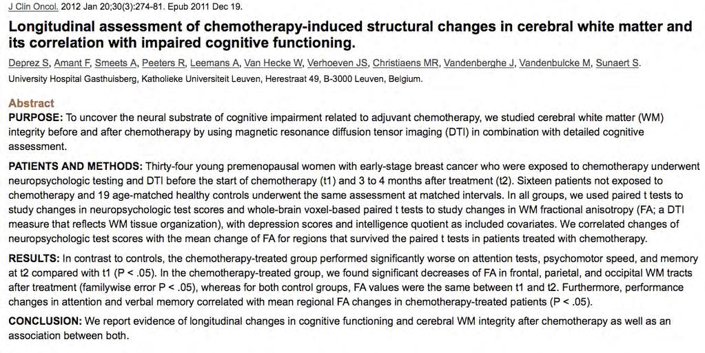
29 Deprez et al., 2012 Longitudinal assessment of chemotherapy-induced structural changes in cerebral white matter and its correlation with impaired cognitive functioning. - J Clin Oncol SPM Voxel-based whole brain analysis of FA Correlation FA and neuropsychological tests
30
31 Comparing Results Across Modalities and Activation vs. Cognitive Rest Designs
 Chemo Control Silverman et al.")
32 Gray Matter Density (MRI) Decreases vs. Cerebral Blood Flow Increases (PET) Chemo Control Silverman et al. (2007) Breast Cancer Res Treat. McDonald et al. (2010) Breast Cancer Res Treat.
33 Studies of the Post-Chemo Brain Scope Differences in brain activation patterns Differences observed in chemo-exposed vs. chemo-naive brains at rest Mechanistic Considerations Potential Implications for Clinical Decisions


34 Proposed Candidate Mechanisms from Ahles and Saykin, Nat Rev Cancer, 2007
35 Candidate Mechanisms: Cytokines chemotherapy agents can boost cytokines some cross the blood-brain barrier some cause release of central cytokines through peripheral-to-central neuronal communication can impair cognitive function direct evidence for role in post-chemo brain remains to be established
36 Implications for Novel Treatment Options I Etanercept (TNF receptor antagonists) Infliximab (Mab to TNF a) NSAIDS and Thalidomide Cytokine synthesis inhibitors Soluble Cytokine Receptors (competitors) Cytokine Receptor Antagonists Estrogen DHEA Fish Oils (inhibitors of TNF a, IL 1b) Exercise Wilson, Finch, Cohen: Cytokines and Cognition JAGS 50: Illman, Corringham et al: Are Inflammatory Cytokines the Common Link B/T Cancer- Associated Cachexia and Depression? J Suppport Oncol 2005;3:37-50
37 Implications for Novel Treatment Options II IL 10 (suppresses production of PICs) Being studied in RA, IBD, ARDS, HIV, Psoriasis An MS trial was halted by manufacturer (??) IL 4 (anti allergic, anti inflammatory, anti tumor) Used in Psoriasis, promising in leukemia Negative MS trial (Bayer)
38 Correlation Between Baseline Cytokine Levels and Baseline FDG Metabolism


39 Baseline Inflammatory Cytokine Positive Correlations with Regional Brain Metabolism 1 year after Chemo IL1RA CRP IL6 TNF All color voxels are p<0.01.
 r=-0.52 p=0.03 All color voxels are p<0.05. At peak voxel in lgfi, p<0.0005. Slice view at 32,20, 2.")
40 Baseline CRP Levels Negatively Correlate with Baseline Metabolism of Left Inferior Frontal Gyrus in Chemotherapy Subjects svoi SPM IL1RA negatively correlated with baseline left GFi T=4.08, p< Part of Largest Cluster (333 voxels) r=-0.52 p=0.03 All color voxels are p<0.05. At peak voxel in lgfi, p< Slice view at 32,20, 2.
41 Studies of the Post-Chemo Brain Scope Differences in brain activation patterns Differences observed in chemo-exposed vs. chemo-naive brains at rest Mechanistic Considerations Potential Implications for Clinical Decisions
42 Potential Implications of Brain PET Imaging in Clinical Decision-Making Search for baseline brain metabolic indicators of future vulnerability and image-guided guided preventive/therapeutic strategies (e.g. e.g.,, Does a patient having lower inferior frontal metabolism suggest being a potential candidate for cytokine-targeted targeted manipulations?) Monitor cerebral response to potentially neurotoxic therapies -- analogously to using MUGA studies to monitor cardiac response to doxorubicin (Adriamycin) -- taking advantage of the typical lead time (2-10 years) of metabolic changes preceding neurologic symptoms. (This could be accomplished as simple add-on view to whole-body PET studies performed for tumor assessments.)
43
44 Candidate Mechanisms Direct chemotherapy toxicity possible Most chemotherapy agents are thought to cross the blood-brain barrier in only low concentrations, however (some exceptions include 5-FU and methotrexate)
45 J Label Compd Radiopharm 2005; 48:
46 Comparing cyclophosphamide and fluorocyclophosphamide toxic behaviors against breast cancer cells

47 Small animal PET and CT imaging with [F-18] fluorocyclophosphamide
48 PET with [F 18]fluorocylophosphamide performed before first course of chemotherapy predicts tumor volume changes measured 3 weeks later. JNM, 2007; 48:
49 Comparing [F 18]fluorocyclophosphamide biodistribution measured by small animal PET and by harvesting organs postmortem


50 PET versions of the following drugs 18F - Paclitaxe l 5-Fluorouraci l 18F - Cyclophosphamid 18F - Inhibits cellular proliferation through stabilization of tubulin Antimetabolite, analog of pyrimidine, impairs pyrimidine synthesis Alkylating agent, cross-links DNA
51
52 Chemotherapy Group (n=20): Baseline Activation during Short Term Memory Tasks
 All color voxels are p<0.01.")
53 Chemotherapy Group at Baseline: Activation in Inferior Frontal Gyrus during Short-Term Memory Task SPM Largest cluster: Left Inferior Frontal Gyrus (2581 contiguous voxels at p<0.01; peak voxel t = 5.72, p<0.0005) All color voxels are p<0.01. svoi Left Inferior Frontal Gyrus Metabolism (Average SEM) 1,11 1,105 1,1 1,095 1,09 Control STM 0,014 0,012 0,01 0,008 0,006 0,004 0,002 0 STM - Control Region that was most significantly activated during short-term memory task (t=2.90, p=0.01)
54 Chemotherapy Group at Baseline: Deactivation in Hindbrain during Short-Term Memory Task SPM Most significant cluster: R. ant. Cerebellum/post. Pons t = 7.03, p< (p FWE-corr corr=0.001) All color voxels are p<0.01. svoi Pons Metabolism (Average SEM) 0,746 0,744 0,742 0,74 0,738 0,736 0,734 Control STM 0 0,002 0,004 0,006 0,008 0,01 STM - Control Region that was most significantly deactivated during short-term memory task (t=-2.15, p=0.05)
55 Comparison of Different Cytokine Batches Previous analyses have used batch 1 cytokine values. We have stopped using that batch and now use batches 2 and 3 instead for our analyses. The cytokine values are comparable, so the analysis results should not be substantially affected.
Opening up the Window into Chemobrain : A Neuroimaging Review
 Sensors 2013, 13, 3169-3203; doi:10.3390/s130303169 Review OPEN ACCESS sensors ISSN 1424-8220 www.mdpi.com/journal/sensors Opening up the Window into Chemobrain : A Neuroimaging Review Carole S. Scherling
Who is at risk? What should we do in the clinic?
 Cognitive Changes after Cancer Treatment Patricia A. Ganz, M.D. Professor, UCLA Schools of Medicine & Public Health Director, UCLA-LIVESTRONG Survivorship Center of Excellence Jonsson Comprehensive Cancer
9/29/2017. Disclosures. Objectives. Cancer-Related Cognitive Dysfunction (CRCD)
") Cancer Related Cognitive Dysfunction (CRCD): Diagnosis, Pathophysiology and Management Disclosures No conflict of interest disclosures M. Beatriz Currier, MD John M Cassel Memorial Breast Cancer Symposium
Cancer Related Cognitive Dysfunction (CRCD): Diagnosis, Pathophysiology and Management Disclosures No conflict of interest disclosures M. Beatriz Currier, MD John M Cassel Memorial Breast Cancer Symposium
Cognitive Changes Associated with Cancer and Its Treatments: Current Knowledge and Challenges
 Cognitive Changes Associated with Cancer and Its Treatments: Current Knowledge and Challenges Tim A. Ahles, Ph.D. Department of Psychiatry and Behavioral Sciences Neurocognitive Research Lab Memorial Sloan-Kettering
Biomarkers Workshop In Clinical Trials Imaging for Schizophrenia Trials
 Biomarkers Workshop In Clinical Trials Imaging for Schizophrenia Trials Research focused on the following areas Brain pathology in schizophrenia and its modification Effect of drug treatment on brain structure
Brain Structure and Function in Nephropathic Cystinosis
 Brain Structure and Function in Nephropathic Cystinosis Doris A. Trauner M.D. Professor, Depts. of Neurosciences and Pediatrics University of California San Diego School of Medicine La Jolla, CA USA Cystinosis
Stuttering Research. Vincent Gracco, PhD Haskins Laboratories
 Stuttering Research Vincent Gracco, PhD Haskins Laboratories Stuttering Developmental disorder occurs in 5% of children Spontaneous remission in approximately 70% of cases Approximately 1% of adults with
Diffusion Tensor Imaging 12/06/2013
 12/06/2013 Beate Diehl, MD PhD FRCP University College London National Hospital for Neurology and Neurosurgery Queen Square London, UK American Epilepsy Society Annual Meeting Disclosure None Learning
Gross Organization I The Brain. Reading: BCP Chapter 7
 Gross Organization I The Brain Reading: BCP Chapter 7 Layout of the Nervous System Central Nervous System (CNS) Located inside of bone Includes the brain (in the skull) and the spinal cord (in the backbone)
Cerebral Hyporesponsiveness and Cognitive Impairment 10 Years After Chemotherapy for Breast Cancer
 r Human Brain Mapping 32:1206 1219 (2011) r Cerebral Hyporesponsiveness and Cognitive Impairment 10 Years After Chemotherapy for Breast Cancer Michiel B. de Ruiter, 1,2 * Liesbeth Reneman, 2 Willem Boogerd,
Biomedical Technology Research Center 2011 Workshop San Francisco, CA
 Diffusion Tensor Imaging: Parkinson s Disease and Atypical Parkinsonism David E. Vaillancourt court1@uic.edu Associate Professor at UIC Departments t of Kinesiology i and Nutrition, Bioengineering, and
Summary of findings from the previous meta-analyses of DTI studies in MDD patients. SDM (39) 221 Left superior longitudinal
 221 Left superior longitudinal") Supplemental Data Table S1 Summary of findings from the previous meta-analyses of DTI studies in MDD patients Study Analysis Method Included studies, n MDD (medicated) HC Results (MDDHC)
Supplemental Data Table S1 Summary of findings from the previous meta-analyses of DTI studies in MDD patients Study Analysis Method Included studies, n MDD (medicated) HC Results (MDDHC)
Parallel Session 9.1: Late effects
 Parallel Session 9.1: Late effects Sanne Schagen: Effects of chemotherapy on cognition and brain structure in pts with non-cns disease Olga Husson: Health-related quality of life and disease specific symptoms
Supplementary Material. Functional connectivity in multiple cortical networks is associated with performance. across cognitive domains in older adults
 Supplementary Material Functional connectivity in multiple cortical networks is associated with performance across cognitive domains in older adults Emily E. Shaw 1,2, Aaron P. Schultz 1,2,3, Reisa A.
Resistance to forgetting associated with hippocampus-mediated. reactivation during new learning
 Resistance to Forgetting 1 Resistance to forgetting associated with hippocampus-mediated reactivation during new learning Brice A. Kuhl, Arpeet T. Shah, Sarah DuBrow, & Anthony D. Wagner Resistance to
Procedia - Social and Behavioral Sciences 159 ( 2014 ) WCPCG 2014
 WCPCG 2014") Available online at www.sciencedirect.com ScienceDirect Procedia - Social and Behavioral Sciences 159 ( 2014 ) 743 748 WCPCG 2014 Differences in Visuospatial Cognition Performance and Regional Brain Activation
Available online at www.sciencedirect.com ScienceDirect Procedia - Social and Behavioral Sciences 159 ( 2014 ) 743 748 WCPCG 2014 Differences in Visuospatial Cognition Performance and Regional Brain Activation
Supplementary Material S3 Further Seed Regions
 Supplementary Material S3 Further Seed Regions Figure I. Changes in connectivity with the right anterior insular cortex. (A) wake > mild sedation, showing a reduction in connectivity between the anterior
Neuroimaging data help to clarify the nosological status of schizoaffective disorder (SAD)?
?") Neuroimaging data help to clarify the nosological status of schizoaffective disorder (SAD)? Mercè Madre Rull mmadrer@gmail.com FIDMAG Research Foundation Hermanas Hospitalarias Barcelona, Spain Overview
Neuroimaging data help to clarify the nosological status of schizoaffective disorder (SAD)? Mercè Madre Rull mmadrer@gmail.com FIDMAG Research Foundation Hermanas Hospitalarias Barcelona, Spain Overview
Telencephalon (Cerebral Hemisphere)
") Telencephalon (Cerebral Hemisphere) OUTLINE The Cortex - Lobes, Sulci & Gyri - Functional Subdivisions - Limbic Lobe & Limbic System The Subcortex - Basal Ganglia - White Matter (Internal Capsule) - Relations
Telencephalon (Cerebral Hemisphere) OUTLINE The Cortex - Lobes, Sulci & Gyri - Functional Subdivisions - Limbic Lobe & Limbic System The Subcortex - Basal Ganglia - White Matter (Internal Capsule) - Relations
Quantitative Neuroimaging- Gray and white matter Alteration in Multiple Sclerosis. Lior Or-Bach Instructors: Prof. Anat Achiron Dr.
 Quantitative Neuroimaging- Gray and white matter Alteration in Multiple Sclerosis Lior Or-Bach Instructors: Prof. Anat Achiron Dr. Shmulik Miron INTRODUCTION Multiple Sclerosis general background Gray
CHEMOBRAIN Cancer free but losing my mind
 REVIEW ARTICLE CHEMOBRAIN Cancer free but losing my mind Edel D.M. Masembe (B.Pharm) 1 1 Department of Pharmacy, Makerere University College of Health Sciences ABSTRACT Background: Chemotherapy, the mainstay
VIII. 10. Right Temporal-Lobe Contribution to the Retrieval of Family Relationships in Person Identification
 CYRIC Annual Report 2009 VIII. 10. Right Temporal-Lobe Contribution to the Retrieval of Family Relationships in Person Identification Abe N. 1, Fujii T. 1, Ueno A. 1, Shigemune Y. 1, Suzuki M. 2, Tashiro
Differential contributions of subregions of medial temporal lobe to memory system in. amnestic mild cognitive impairment: insights from fmri study
 Differential contributions of subregions of medial temporal lobe to memory system in amnestic mild cognitive impairment: insights from fmri study Jiu Chen 1, *, Xujun Duan 2, *, Hao Shu 1, Zan Wang 1,
Supplementary Online Content
 Supplementary Online Content Devenney E, Bartley L, Hoon C, et al. Progression in behavioral variant frontotemporal dementia: a longitudinal study. JAMA Neurol. Published online October 26, 2015. doi:10.1001/jamaneurol.2015.2061.
Functional aspects of anatomical imaging techniques
 Functional aspects of anatomical imaging techniques Nilendu Purandare Associate Professor & Consultant Radiologist Tata Memorial Centre Functional/metabolic/molecular imaging (radioisotope scanning) PET
BREAST CANCER (BC) IS ONE OF
 IS ONE OF") ORIGINAL CONTRIBUTION Prefrontal Cortex and Executive Function Impairments in Primary Breast Cancer Shelli R. Kesler, PhD; Jamie S. Kent, MA; Ruth O Hara, PhD Objectives: To examine differences in prefrontalexecutive
ORIGINAL CONTRIBUTION Prefrontal Cortex and Executive Function Impairments in Primary Breast Cancer Shelli R. Kesler, PhD; Jamie S. Kent, MA; Ruth O Hara, PhD Objectives: To examine differences in prefrontalexecutive
Cerebral Cortex 1. Sarah Heilbronner
 Cerebral Cortex 1 Sarah Heilbronner heilb028@umn.edu Want to meet? Coffee hour 10-11am Tuesday 11/27 Surdyk s Overview and organization of the cerebral cortex What is the cerebral cortex? Where is each
Diffusion Tensor Imaging in Psychiatry
 2003 KHBM DTI in Psychiatry Diffusion Tensor Imaging in Psychiatry KHBM 2003. 11. 21. 서울대학교 의과대학 정신과학교실 권준수 Neuropsychiatric conditions DTI has been studied in Alzheimer s disease Schizophrenia Alcoholism
Identification of Neuroimaging Biomarkers
 Identification of Neuroimaging Biomarkers Dan Goodwin, Tom Bleymaier, Shipra Bhal Advisor: Dr. Amit Etkin M.D./PhD, Stanford Psychiatry Department Abstract We present a supervised learning approach to
Is DTI Increasing the Connectivity Between the Magnet Suite and the Clinic?
 Current Literature In Clinical Science Is DTI Increasing the Connectivity Between the Magnet Suite and the Clinic? Spatial Patterns of Water Diffusion Along White Matter Tracts in Temporal Lobe Epilepsy.
Use of Multimodal Neuroimaging Techniques to Examine Age, Sex, and Alcohol-Related Changes in Brain Structure Through Adolescence and Young Adulthood
 American Psychiatric Association San Diego, CA 24 May 2017 Use of Multimodal Neuroimaging Techniques to Examine Age, Sex, and Alcohol-Related Changes in Brain Structure Through Adolescence and Young Adulthood
Chemo Fog: What it is and what can we do about it
 Chemo Fog: What it is and what can we do about it Lori Bernstein, Ph.D., C.Psych. Dept of Psychosocial Oncology & Palliative Care & Cancer Survivorship Program Health, Wellness, and Cancer Survivorship
DWI assessment of ischemic changes in the fetal brain
 DWI assessment of ischemic changes in the fetal brain Dafi Bergman, 4 th year Medical student in the 4-year program, Sackler school of medicine B.Sc Life and Medical Sciences, Tel Aviv University Supervised
Supplemental Information. Direct Electrical Stimulation in the Human Brain. Disrupts Melody Processing
 Current Biology, Volume 27 Supplemental Information Direct Electrical Stimulation in the Human Brain Disrupts Melody Processing Frank E. Garcea, Benjamin L. Chernoff, Bram Diamond, Wesley Lewis, Maxwell
Title:Atypical language organization in temporal lobe epilepsy revealed by a passive semantic paradigm
 Author's response to reviews Title:Atypical language organization in temporal lobe epilepsy revealed by a passive semantic paradigm Authors: Julia Miro (juliamirollado@gmail.com) Pablo Ripollès (pablo.ripolles.vidal@gmail.com)
Imaging in Pediatric `neurohiv Dr Jackie Hoare Head of Liaison Psychiatry Groote Schuur Hospital, UCT
 Imaging in Pediatric `neurohiv Dr Jackie Hoare Head of Liaison Psychiatry Groote Schuur Hospital, UCT ? Spectrum of Neurocognitive disorders The adult literature on HIV related CNS damage supports a spectrum
Breast Cancer Basics. Clinical Oncology for Public Health Professionals. Ben Ho Park, MD, PhD
 This work is licensed under a Creative Commons Attribution-NonCommercial-ShareAlike License. Your use of this material constitutes acceptance of that license and the conditions of use of materials on this
Introduction to the Course and the Techniques. Jeffry R. Alger, PhD Ahmanson-Lovelace Brain Mapping Center Department of Neurology
 Introduction to the Course and the Techniques Jeffry R. Alger, PhD Ahmanson-Lovelace Brain Mapping Center Department of Neurology (jralger@ucla.edu) CTSI Neuroimaging April 2014 Rationale for the Course
A possible mechanism for impaired joint attention in autism
 A possible mechanism for impaired joint attention in autism Justin H G Williams Morven McWhirr Gordon D Waiter Cambridge Sept 10 th 2010 Joint attention in autism Declarative and receptive aspects initiating
Hippocampal brain-network coordination during volitionally controlled exploratory behavior enhances learning
 Online supplementary information for: Hippocampal brain-network coordination during volitionally controlled exploratory behavior enhances learning Joel L. Voss, Brian D. Gonsalves, Kara D. Federmeier,
NEURORADIOLOGY DIL part 4
 NEURORADIOLOGY DIL part 4 Strokes and infarcts K. Agyem MD, G. Hall MD, D. Palathinkal MD, Alexandre Menard March/April 2015 OVERVIEW Introduction to Neuroimaging - DIL part 1 Basic Brain Anatomy - DIL
Theory of mind skills are related to gray matter volume in the ventromedial prefrontal cortex in schizophrenia
 Theory of mind skills are related to gray matter volume in the ventromedial prefrontal cortex in schizophrenia Supplemental Information Table of Contents 2 Behavioral Data 2 Table S1. Participant demographics
Early Diagnosis of Alzheimer s Disease and MCI via Imaging and Pattern Analysis Methods. Christos Davatzikos, Ph.D.
 Early Diagnosis of Alzheimer s Disease and MCI via Imaging and Pattern Analysis Methods Christos Davatzikos, Ph.D. Director, Section of Biomedical Image Analysis Professor of Radiology http://www.rad.upenn.edu/sbia
The Adverse Effect of Chemotherapy on the Developing Brain. Ellen van der Plas, PhD Research Fellow at SickKids
 The Adverse Effect of Chemotherapy on the Developing Brain Ellen van der Plas, PhD Research Fellow at SickKids Overview of today s talk Why study cancer survivors? Quick introduction on acute lymphoblastic
Supplementary Results: Age Differences in Participants Matched on Performance
 Supplementary Results: Age Differences in Participants Matched on Performance 1 We selected 14 participants for each age group which exhibited comparable behavioral performance (ps >.41; Hit rates (M ±
Neuroimaging and Psychiatry
 Neuroimaging and Psychiatry Chris Gale Otago Regional Psychiatry Training Programme Feb 12, 2011 In psychosis. Decrease volume frontal, parietal, hippocampus, corpus collosum. Increase CSF volume Functional
P. Hitchcock, Ph.D. Department of Cell and Developmental Biology Kellogg Eye Center. Wednesday, 16 March 2009, 1:00p.m. 2:00p.m.
 Normal CNS, Special Senses, Head and Neck TOPIC: CEREBRAL HEMISPHERES FACULTY: LECTURE: READING: P. Hitchcock, Ph.D. Department of Cell and Developmental Biology Kellogg Eye Center Wednesday, 16 March
European Prevention of Alzheimer s Dementia (EPAD)
") European Prevention of Alzheimer s Dementia (EPAD) Ron Marcus, MD ISCTM Adaptive Design Workshop February 20, 2018 1 EPAD Goal The European Prevention of Alzheimer's Dementia (EPAD) project aims to develop
European Prevention of Alzheimer s Dementia (EPAD) Ron Marcus, MD ISCTM Adaptive Design Workshop February 20, 2018 1 EPAD Goal The European Prevention of Alzheimer's Dementia (EPAD) project aims to develop
Supplementary Information
 Supplementary Information The neural correlates of subjective value during intertemporal choice Joseph W. Kable and Paul W. Glimcher a 10 0 b 10 0 10 1 10 1 Discount rate k 10 2 Discount rate k 10 2 10
Adjuvant Systemic Therapy in Early Stage Breast Cancer
 Adjuvant Systemic Therapy in Early Stage Breast Cancer Julie R. Gralow, M.D. Director, Breast Medical Oncology Jill Bennett Endowed Professor of Breast Cancer Professor, Global Health University of Washington
Supplementary Information Methods Subjects The study was comprised of 84 chronic pain patients with either chronic back pain (CBP) or osteoarthritis
 or osteoarthritis") Supplementary Information Methods Subjects The study was comprised of 84 chronic pain patients with either chronic back pain (CBP) or osteoarthritis (OA). All subjects provided informed consent to procedures
Supplementary Information Methods Subjects The study was comprised of 84 chronic pain patients with either chronic back pain (CBP) or osteoarthritis (OA). All subjects provided informed consent to procedures
Advances in Clinical Neuroimaging
 Advances in Clinical Neuroimaging Joseph I. Tracy 1, PhD, ABPP/CN; Gaelle Doucet 2, PhD; Xaiosong He 2, PhD; Dorian Pustina 2, PhD; Karol Osipowicz 2, PhD 1 Department of Radiology, Thomas Jefferson University,
Nature Neuroscience: doi: /nn Supplementary Figure 1. Task timeline for Solo and Info trials.
 Supplementary Figure 1 Task timeline for Solo and Info trials. Each trial started with a New Round screen. Participants made a series of choices between two gambles, one of which was objectively riskier
Modeling of early-infant brain growth using longitudinal data from diffusion tensor imaging.
 Modeling of early-infant brain growth using longitudinal data from diffusion tensor imaging. Guido Gerig, Neda Sadeghi, PhD, Marcel Prastawa, Tom Fletcher, Clement Vachet Scientific Computing and Imaging
NIH Public Access Author Manuscript Brain Imaging Behav. Author manuscript; available in PMC 2014 December 01.
 NIH Public Access Author Manuscript Published in final edited form as: Brain Imaging Behav. 2013 December ; 7(4): 363 373. doi:10.1007/s11682-013-9283-7. Neuroimaging Biomarkers and Cognitive Function
Pediatric MS MRI Study Methodology
 General Pediatric MS MRI Study Methodology SCAN PREPARATION axial T2-weighted scans and/or axial FLAIR scans were obtained for all subjects when available, both T2 and FLAIR scans were scored. In order
How do individuals with congenital blindness form a conscious representation of a world they have never seen? brain. deprived of sight?
 How do individuals with congenital blindness form a conscious representation of a world they have never seen? What happens to visual-devoted brain structure in individuals who are born deprived of sight?
Brain diffusion tensor imaging changes in cerebrotendinous xanthomatosis reversed with
 Brain diffusion tensor imaging changes in cerebrotendinous xanthomatosis reversed with treatment Claudia B. Catarino, MD, PhD, 1*, Christian Vollmar, MD, PhD, 2,3* Clemens Küpper, MD, 1,4 Klaus Seelos,
Text to brain: predicting the spatial distribution of neuroimaging observations from text reports (submitted to MICCAI 2018)
") 1 / 22 Text to brain: predicting the spatial distribution of neuroimaging observations from text reports (submitted to MICCAI 2018) Jérôme Dockès, ussel Poldrack, Demian Wassermann, Fabian Suchanek, Bertrand
1 / 22 Text to brain: predicting the spatial distribution of neuroimaging observations from text reports (submitted to MICCAI 2018) Jérôme Dockès, ussel Poldrack, Demian Wassermann, Fabian Suchanek, Bertrand
Imaging of Alzheimer s Disease: State of the Art
 July 2015 Imaging of Alzheimer s Disease: State of the Art Neir Eshel, Harvard Medical School Year IV Outline Our patient Definition of dementia Alzheimer s disease Epidemiology Diagnosis Stages of progression
Supporting Online Material for
 www.sciencemag.org/cgi/content/full/324/5927/646/dc1 Supporting Online Material for Self-Control in Decision-Making Involves Modulation of the vmpfc Valuation System Todd A. Hare,* Colin F. Camerer, Antonio
Neuroimaging Findings in Young Drinkers: Does Teenage Drinking Harm the Brain? Susan F. Tapert, Ph.D. University of California, San Diego
 Neuroimaging Findings in Young Drinkers: Does Teenage Drinking Harm the Brain? Susan F. Tapert, Ph.D. University of California, San Diego 2 Overview What is normal adolescence? How do binge drinkers differ?
Treating New Learning and Memory Deficits in Rehabilitation Populations: the modified Story Memory Technique (msmt)
") Treating New Learning and Memory Deficits in Rehabilitation Populations: the modified Story Memory Technique (msmt) Nancy D. Chiaravalloti, Ph.D. Nancy Moore, MA Objectives Understand techniques for memory
Treating New Learning and Memory Deficits in Rehabilitation Populations: the modified Story Memory Technique (msmt) Nancy D. Chiaravalloti, Ph.D. Nancy Moore, MA Objectives Understand techniques for memory
SPAMALIZE s Cerebellum Segmentation routine.
 SPAMALIZE s Cerebellum Segmentation routine. Outline: - Introduction - Data Inputs - Algorithm Steps - Display Notes - Example with menu selections Introduction: This program attempts to segment the cerebellum
Title of file for HTML: Peer Review File Description:
 Title of file for HTML: Supplementary Information Description: Supplementary Figure, Supplementary Tables, Supplementary Methods and Supplementary References Title of file for HTML: Peer Review File Description:
Cognitive Impairment in AYA Cancer Survivors
 Managing Challenging Issues in Adolescent and Young Adult (AYA) Cancer Survivors: Cognitive Impairment in AYA Cancer Survivors Alex Chan, PharmD, MPH, FCCP, FISOPP, BCPS, BCOP Associate Professor and Specialist
Early identification of neurobiological markers of remission. Michael Bodnar, PhD Ashok K. Malla, MD Martin Lepage, PhD
 Early identification of neurobiological markers of remission Michael Bodnar, PhD Ashok K. Malla, MD Martin Lepage, PhD Outline Why study remission? Defining remission Data collection Results neurocognition
Metabolic and microstructural alterations in the SLE brain correlate with cognitive impairment
 Metabolic and microstructural alterations in the SLE brain correlate with cognitive impairment Meggan Mackay,, Betty Diamond, David Eidelberg JCI Insight. 2019;4(1):e124002.. Research Article Neuroscience
DOWNLOAD PDF THE EFFECT OF AEROBIC EXERCISE ON INFORMATION PROCESSING IN OLDER ADULTS
 Chapter 1 : Exercise - Wikipedia Aerobic exercise (two RCTs), strength exercise alone (one RCT) or combined with balance and exercise (one RCT) or a combination of aerobic, strength. balance and flexibility
Bio 3411 Midterm Review:
 Midterm Review: Structure/Development/Systems/ Plastics/Talents/Diseases/Genes Structure General Overview Wednesday 1( 2( THE BRAIN ATLAS 3 rd ed, p. 8! THE BRAIN ATLAS 3 rd ed, p. 9! Mid-line (sagittal)
Post Stroke Brain Plasticity
 Post Stroke Brain Plasticity François CHOLLET MD, PhD Neurology Department: Stroke Unit Toulouse University Hospital (CHU) Neurosciences Institute of Toulouse CNRS, INSERM, University, CHU Versailles le
Neuroanatomy lecture (1)
") Neuroanatomy lecture (1) Introduction: Neuroanatomy has two parts: the central and peripheral nervous system. The central nervous system is composed of brain and spinal cord. The brain has the following
Neuroanatomy lecture (1) Introduction: Neuroanatomy has two parts: the central and peripheral nervous system. The central nervous system is composed of brain and spinal cord. The brain has the following
Supplemental Information. Triangulating the Neural, Psychological, and Economic Bases of Guilt Aversion
 Neuron, Volume 70 Supplemental Information Triangulating the Neural, Psychological, and Economic Bases of Guilt Aversion Luke J. Chang, Alec Smith, Martin Dufwenberg, and Alan G. Sanfey Supplemental Information
By Lauren Stowe, PhD, CCC-SLP & Gina Rotondo, MS, CCC-SLP The Speech Therapy Group
 By Lauren Stowe, PhD, CCC-SLP & Gina Rotondo, MS, CCC-SLP The Speech Therapy Group http://www.acquiredbraininjury.com/interactive brain/interactivebrain.swf 1. Hormones make the science messy 2. Difference
Supporting Information
 Supporting Information Carhart-Harris et al. 10.1073/pnas.1119598109 Fig. S1. Slices for arterial spin labeling (ASL) result. Lightbox display of slices showing regions where there were significant decreases
Medical Neuroscience Tutorial Notes
 Medical Neuroscience Tutorial Notes Blood Supply to the Brain MAP TO NEUROSCIENCE CORE CONCEPTS 1 NCC1. The brain is the body's most complex organ. LEARNING OBJECTIVES After study of the assigned learning
Effects Of Attention And Perceptual Uncertainty On Cerebellar Activity During Visual Motion Perception
 Effects Of Attention And Perceptual Uncertainty On Cerebellar Activity During Visual Motion Perception Oliver Baumann & Jason Mattingley Queensland Brain Institute The University of Queensland The Queensland
Speed, Comfort and Quality with NeuroDrive
 Speed, Comfort and Quality with NeuroDrive Echelon Oval provides a broad range of capabilities supporting fast, accurate diagnosis of brain conditions and injuries. From anatomical depiction to vascular
Methods to examine brain activity associated with emotional states and traits
 Methods to examine brain activity associated with emotional states and traits Brain electrical activity methods description and explanation of method state effects trait effects Positron emission tomography
Cognitive Neuroscience of Memory
 Cognitive Neuroscience of Memory Types and Structure of Memory Types of Memory Type of Memory Time Course Capacity Conscious Awareness Mechanism of Loss Sensory Short-Term and Working Long-Term Nondeclarative
Neuroimaging in Clinical Practice
 Neuroimaging in Clinical Practice John Gabrieli Department of Brain and Cognitive Sciences & Martinos Imaging Center at the McGovern Institute for Brain Research, MIT Disclosures Neither I nor my spouse/partner
Results. NeuRA fmri March 2017
 Introduction With cognitive, sensory or motor stimulation, specific brain regions are activated, requiring higher energy use and higher levels of blood flow. Functional magnetic resonance imaging (fmri)
CEREBRUM. Dr. Jamila EL Medany
 CEREBRUM Dr. Jamila EL Medany Objectives At the end of the lecture, the student should be able to: List the parts of the cerebral hemisphere (cortex, medulla, basal nuclei, lateral ventricle). Describe
Funding: NIDCF UL1 DE019583, NIA RL1 AG032119, NINDS RL1 NS062412, NIDA TL1 DA
 The Effect of Cognitive Functioning, Age, and Molecular Variables on Brain Structure Among Carriers of the Fragile X Premutation: Deformation Based Morphometry Study Naomi J. Goodrich-Hunsaker*, Ling M.
Common disease 175,000 new cases/year 44,000 deaths/year Less than 10% with newly diagnosed at presentation have stage IV disease Chronic disease,
 Chemotherapy for Metastatic Breast Cancer: Recent Results HARMESH R. NAIK, MD. Karmanos Cancer Institute and St. Mary Hospital Metastatic breast cancer (MBC) Common disease 175,000 new cases/year 44,000
Structural lesion analysis: applications
 Structural lesion analysis: applications Dagmar Timmann Department of Neurology University of Duisburg-Essen, Germany Human cerebellar lesion conditions 1. Focal cerebellar lesions a. Stroke Pro s: - acute
Visual Rating Scale Reference Material. Lorna Harper Dementia Research Centre University College London
 Visual Rating Scale Reference Material Lorna Harper Dementia Research Centre University College London Background The reference materials included in this document were compiled and used in relation to
CEREBRUM Dr. Jamila Elmedany Dr. Essam Eldin Salama
 CEREBRUM Dr. Jamila Elmedany Dr. Essam Eldin Salama Objectives At the end of the lecture, the student should be able to: List the parts of the cerebral hemisphere (cortex, medulla, basal nuclei, lateral
BIOL Dissection of the Sheep and Human Brain
 BIOL 2401 Dissection of the Sheep and Human Brain Laboratory Objectives After completing this lab, you should be able to: Identify the main structures in the sheep brain and to compare them with those
MRI and CT of the CNS
 MRI and CT of the CNS Dr.Maha ELBeltagy Assistant Professor of Anatomy Faculty of Medicine The University of Jordan 2018 Computed Tomography CT is used for the detection of intracranial lesions. CT relies
Supplementary appendix
 Supplementary appendix This appendix formed part of the original submission and has been peer reviewed. We post it as supplied by the authors. Supplement to: Schiller R, IJsselstijn H, Hoskote A, et al.
Diffusion Tensor Imaging in Dementia. Howard Rosen UCSF Department of Neurology Memory and Aging Center
 Diffusion Tensor Imaging in Dementia Howard Rosen UCSF Department of Neurology Memory and Aging Center www.memory.ucsf.edu Overview Examples of DTI findings in Alzheimer s disease And other dementias Explore
Table 1. Summary of PET and fmri Methods. What is imaged PET fmri BOLD (T2*) Regional brain activation. Blood flow ( 15 O) Arterial spin tagging (AST)
 Regional brain activation. Blood flow ( 15 O) Arterial spin tagging (AST)") Table 1 Summary of PET and fmri Methods What is imaged PET fmri Brain structure Regional brain activation Anatomical connectivity Receptor binding and regional chemical distribution Blood flow ( 15 O)
Table 1 Summary of PET and fmri Methods What is imaged PET fmri Brain structure Regional brain activation Anatomical connectivity Receptor binding and regional chemical distribution Blood flow ( 15 O)
Functional MRI and Diffusion Tensor Imaging
 Functional MRI and Diffusion Tensor Imaging Andrew Steven March 23, 2018 Ochsner Neuroscience Symposium None Disclosure 1 Objectives Review basic principles of BOLD fmri and DTI. Discuss indications and
Homework Week 2. PreLab 2 HW #2 Synapses (Page 1 in the HW Section)
") Homework Week 2 Due in Lab PreLab 2 HW #2 Synapses (Page 1 in the HW Section) Reminders No class next Monday Quiz 1 is @ 5:30pm on Tuesday, 1/22/13 Study guide posted under Study Aids section of website
Homework Week 2 Due in Lab PreLab 2 HW #2 Synapses (Page 1 in the HW Section) Reminders No class next Monday Quiz 1 is @ 5:30pm on Tuesday, 1/22/13 Study guide posted under Study Aids section of website
Dentate imaging: methods and applications
 Dentate imaging: methods and applications Dagmar Timmann Department of Neurology University of Duisburg-Essen, Germany Cerebellar output via deep nuclei Folie 2 Brodal 2010 Cerebellar nuclei Fastigial
Rhythm and Rate: Perception and Physiology HST November Jennifer Melcher
 Rhythm and Rate: Perception and Physiology HST 722 - November 27 Jennifer Melcher Forward suppression of unit activity in auditory cortex Brosch and Schreiner (1997) J Neurophysiol 77: 923-943. Forward
Supplementary Materials for
 Supplementary Materials for Folk Explanations of Behavior: A Specialized Use of a Domain-General Mechanism Robert P. Spunt & Ralph Adolphs California Institute of Technology Correspondence may be addressed
Author's response to reviews
 Author's response to reviews Title: MRI-negative PET-positive Temporal Lobe Epilepsy (TLE) and Mesial TLE differ with Quantitative MRI and PET: a case control study Authors: Ross P Carne (carnero@svhm.org.au)
HST.583 Functional Magnetic Resonance Imaging: Data Acquisition and Analysis Fall 2008
 MIT OpenCourseWare http://ocw.mit.edu HST.583 Functional Magnetic Resonance Imaging: Data Acquisition and Analysis Fall 2008 For information about citing these materials or our Terms of Use, visit: http://ocw.mit.edu/terms.