Ch 3: Observing Microorganisms Through a Microscope
|
|
|
- Dominick York
- 6 years ago
- Views:
Transcription

1 Ch 3: Observing Microorganisms Through a Microscope
2 SLOs Review the metric units of measurement Define total magnification and resolution Explain how electron and light microscopy differ Differentiate between acidic and basic dyes Compare simple, differential, negative, and special stains List the steps in preparing a Gram stain. Describe the appearance of Gram-positive and Gram-negative cells after each step Compare and contrast Gram stain and acid-fast stain Explain why endospore and capsule stains are used
3 SLOs cont.: Check Your Understanding If a microbe measures 10 μm in length, how long is it in nanometers? What does it mean when a microscope has a resolution of 0.2 nm? Why do electron microscopes have greater resolution than light microscopes? Why doesn t a negative stain color a cell? Why is the Gram stain so useful? Which stain would be used to identify microbes in the genera Mycobacterium and Nocardia? How do unstained endospores appear? Stained endospores?

4 Units of Measurement Review Table µm = m = mm 1 nm = m = mm 1000 nm = µm µm = nm

5 Figure 3.2 Microscopes and Magnification. Unaided eye 200 m Light microscope 200 nm 10 mm Scanning electron microscope 10 nm 1 mm Tick Actual size Red blood cells Transmission electron microscope 10 pm 100 m E. coli bacteria T-even bacteriophages (viruses) Atomic force microscope 0.1 nm 10nm DNA double helix Foundation Fig 3.2
6 Sizes Among Microorganisms Protozoa: 100 µm Yeasts: 8 µm Cells Alive How big is a...? Bacteria: 1-5 µm (some much longer than wide) Rickettsia: 0.4 µm = nm Chlamydia and Mycoplasma: 0.25 µm Viruses: nm

7 Principles of the Compound Light Microscope Magnification: Ocular and objective lenses of compound microscope (total mag.?) Resolution: Ability of lens to... Maximum resolving power depends on... For light microscope: m Contrast: Stains change refractive index contrast between bacteria and surrounding medium Fig 3.1


8 Refractive Index Measures light-bending ability of a medium Light may bend in air so much that it misses the small high-magnification lens. Immersion oil is used to keep light from bending. Fig 3.3

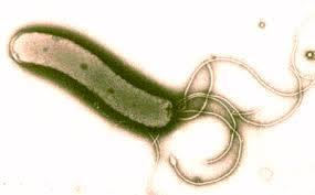
9 Microscopy: The Instruments Brightfield Microscopy Simplest of all the optical microscopy illumination. techniques Dark objects are visible against a bright background. Darkfield Microscopy Light objects visible against dark background. used to enhance the contrast in unstained samples. Instrument of choice for spirochetes Fig 3.4

10 Spirochetes (Treponema pallidum) viewed with darkfield microscope


11 Fluorescence Microscopy Uses UV light. Fluorescent substances absorb UV light and emit visible light. Cells may be stained with fluorescent chemicals (fluorochromes). Immunofluorescence Fig 3.6; T. pallidum
12 Figure 3.6a Fig 3.6 Principle of Immunofluorescence
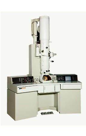
13 Electron Microscopy: Detailed Images of Cell Parts Uses electrons, electromagnetic lenses, and fluorescent screens Electron wavelength ~ 100,000 x smaller than visible light wavelength Specimens may be stained with heavy metal salts Two types of EMs:?


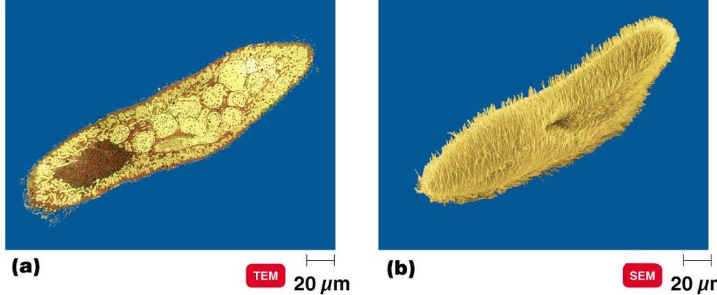
14 SEM or TEM? Bacterial division Compare to Fig 3.10 Leaf surface
15 ? 10, ,000 ; resolution 2.5 nm.
16 Preparation of Specimens for Light Microscopy Staining Techniques Provide Contrast Smear air-dry heat-fix Basic dyes: cationic chromophore Acidic dyes: anionic chromophore negative staining (good for capsules) Three types of staining techniques: Simple, differential, and special
17 Simple Stains Use a single basic dye. A mordant may be used to hold the stain or coat the specimen to enlarge it. Differential Stains React differently with different bacteria Gram stain Acid fast stain
 Application of iodine (mordant) Alcohol wash")
18 Figure 3.12 Gram staining. Gram-positive Gram-negative Application of crystal violet (purple dye) Application of iodine (mordant) Alcohol wash (decolorization) Application of safranin (counterstain) Rod (gram-negative) Cocci (gram-positive) Fig 3.12


19 Gram Stain Compare to Fig 3.12 crystal violet safranin
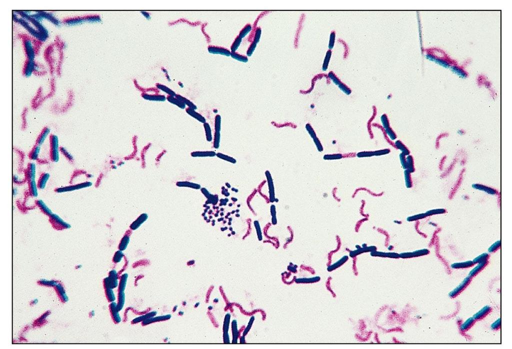
20

21 Gram Stains using Compound Light Microscope Streptococcus mutans Bacillus anthracis


22 Negative Stain Observe cell shape and size Fig 3.14 Used for bacteria with capsules


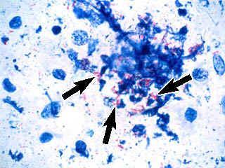
23 Acid Fast Stain Cells that retain a basic stain in the presence of acid-alcohol are called acid-fast. Non acid-fast cells lose the primary stain when rinsed with acid-alcohol, and are counterstained with a different color basic stain Fig 3.13


24 Special Stains Compare to Fig 3.14 Endospore stain: Heat is required to drive a stain into the endospore. Flagella staining: requires a mordant to make the flagella wide enough to see. Capsule stain uses basic stain and negative stain


25 Clinical Case: Microscopic Mayhem
MULTIPLE CHOICE. Choose the one alternative that best completes the statement or answers the question.
 Exam Name MULTIPLE CHOICE. Choose the one alternative that best completes the statement or answers the question. 1) A nanometer would be a suitable unit of measurement for which of the following? 1) A)
chapter one: the history of microbiology
 chapter one: the history of microbiology Revised 8/29/2016 microbes microscopic (small) organisms, viruses, prions prefix sci. notation frac. equivalent dec. equivalent kilo- (k) 1 10 3 1000/1 = 1000 1000
Staining Technology and Bright- Field Microscope Use
 Staining Technology and Bright- Field Microscope Use 2 Abstract We will introduce bright-field microscope use, practice Gram staining with foodborne pathogens, and practice endospore staining with Bacillus
IDENTIFICATION OF MICROORGANISMS YUSRON SUGIARTO
 IDENTIFICATION OF MICROORGANISMS YUSRON SUGIARTO BACKGROUND One of the most fundamental tasks that someone working with micro-organisms must perform is to identify microorganisms. There are a number of
Bacterial Structure and Function
 Bacterial Structure and Function Charles Okolie, PhD. Room 311 (on level 4), First College Building, Landmark University okolie.charles@lmu.edu.ng Tel: Ext: Mobile: 08060241166 Structure of Bacteria The
CHAPTER 4 CELL STRUCTURE/FUNCTION. 2. The uses the visible light to illuminate cell. 3. How is the magnification of a compound microscope calculated?
 CHAPTER 4 CELL STRUCTURE/FUNCTION 1. Define magnification and the term resolution. 2. The uses the visible light to illuminate cell. 3. How is the magnification of a compound microscope calculated? 4.
Medical Microbiology. Microscopic Techniques :
 ! Lecture 2 Dr. Ismail I. Daood Medical Microbiology Microscopic Techniques : Several types of microscopes are used in study of microbiology one of the most important tools for studying microorganisms
Thursday, October 16 th
 Thursday, October 16 th Good morning. Those of you needing to take the Enzymes and Energy Quiz will start very soon. Students who took the quiz Wednesday: Please QUIETLY work on the chapter 6 reading guide.
Ch 4. Functional Anatomy of Prokaryotic and Eukaryotic Cells
 Ch 4 Functional Anatomy of Prokaryotic and Eukaryotic Cells Objectives Compare and contrast the overall cell structure of prokaryotes and eukaryotes. Identify the three basic shapes of bacteria. Describe
Microscopy Text book Chapter 3
 Lab 2 Goals and Objectives: Microscopy lecture: Text Chapter 3 (supplement pg 39) Exercise 1: Brightfield Microscopy Learn how to use the microscope (guide in supplement pg 41) Diopter and interocular
Microbiology: A Systems Approach
 Microbiology: A Systems Approach First Edition Cowan & Talaro Chapter 4 Prokaryotic Profiles: the Bacteria and the Archaea Chapter 4 Fig. 4.1 3 3 parts flagella filament long, thin, helical structure composed
Basic Microscopy Laboratory Exercises
 Basic Microscopy Laboratory Exercises Laboratory Exercise After you have completed the Basic Microscopy elearning course, it is strongly recommended that you complete the following laboratory exercises
Prokaryotic Cell Structure
 Prokaryotic Cell Structure Chapter 3 Prokaryotes vs Eukaryotes DNA Prokaryotes Eukaryotes Organelles Size & Organization Kingdoms 1 Where do viruses fit in? Acellular microorganisms Cannot reproduce outside
Prokaryotic Cell Structure
 Prokaryotic Cell Structure Chapter 3 Prokaryotes vs Eukaryotes DNA Prokaryotes Eukaryotes Organelles Size & Organization Kingdoms Where do viruses fit in? Acellular microorganisms Cannot reproduce outside
O.k., Now Starts the Good Stuff (Part I) Prokaryotic Cell Structure and Function
 Prokaryotic Cell Structure and Function") O.k., Now Starts the Good Stuff (Part I) Prokaryotic Cell Structure and Function Prokaryotic Characteristics DNA not enclosed in membrane. No histone proteins associated with DNA. Lack membrane-bound organelles
O.k., Now Starts the Good Stuff (Part I) Prokaryotic Cell Structure and Function Prokaryotic Characteristics DNA not enclosed in membrane. No histone proteins associated with DNA. Lack membrane-bound organelles
BIOLOGY. A Tour of the Cell CAMPBELL. Robert Hooke (1665) Antoni van Leeuwenhoek (1674) Reece Urry Cain Wasserman Minorsky Jackson
 Antoni van Leeuwenhoek (1674) Reece Urry Cain Wasserman Minorsky Jackson") 6 A Tour of the Cell CAMPBELL BIOLOGY TENTH EDITION Reece Urry Cain Wasserman Minorsky Jackson Lecture Presentation by Nicole Tunbridge and Kathleen Fitzpatrick Robert Hooke (1665) Figure 1.1 The structure
6 A Tour of the Cell CAMPBELL BIOLOGY TENTH EDITION Reece Urry Cain Wasserman Minorsky Jackson Lecture Presentation by Nicole Tunbridge and Kathleen Fitzpatrick Robert Hooke (1665) Figure 1.1 The structure
Classification of Infectious Agents. Dr W. D. Colby
 Classification of Infectious Agents Dr W. D. Colby Nonliving Infectious Agents PRIONS: abnormally configured self-replicating protein templates VIRUSES: nucleic acid (DNA or RNA) genes packaged in protein
2018 Science Olympiad: Microbe Mission - Sample Tournament Div C
 2018 Science Olympiad: Microbe Mission - Sample Tournament Div C Section A: Types of cells and their parts 1. Please state if the cell is prokaryotic or eukaryotic. Then label the following molecular components
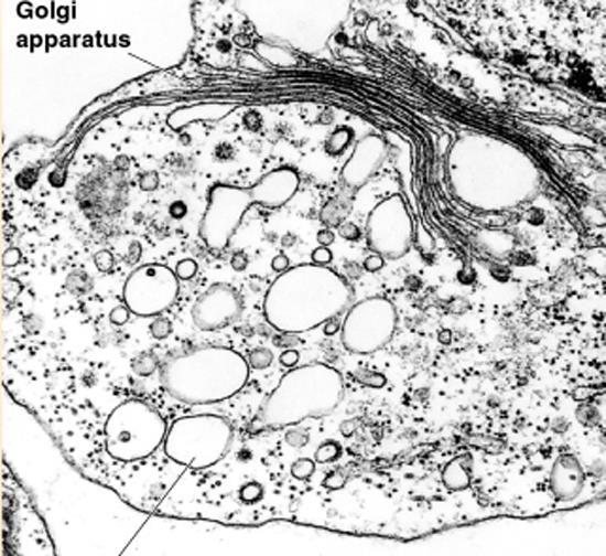
Fig. LPS in Gram negative bacteria
 Structure of bacterial cell Dentistry college - first class Medical biology- Lec.3 Lecturer D. Hanan S A- Cell wall ***Chemical composition of the cell wall Bacteria are divided into two separated groups
Mt. San Antonio College Microbiology 22 Lab Schedule for Spring 2018 Tues/Thurs. Split Lab Sections ONLY
 Mt. San Antonio College Microbiology 22 Lab Schedule for Spring 2018 Tues/ Split Lab Sections ONLY Wk 1 Feb. 27 Orientation with Introductions & Safety Rules/Regulations March 1 Orientation with Pathogen
Mt. San Antonio College Microbiology 22 Lab Schedule for Spring 2018 Mon/Weds. Split Lab Sections ONLY
 Mt. San Antonio College Microbiology 22 Lab Schedule for Spring 2018 Mon/ Split Lab Sections ONLY Wk 1 Feb. 26 Orientation with Introductions & Safety Rules/Regulations Feb. 28 Orientation with Pathogen
Mt. San Antonio College Microbiology 22 Lab Schedule for Fall 2017 Tues/Thurs. Split Lab Sections ONLY
 Mt. San Antonio College Microbiology 22 Lab Schedule for Fall 2017 Tues/ Split Lab Sections ONLY Wk 1 Aug. 29 Orientation with Introductions & Safety Rules/Regulations Aug. 31 Orientation with Pathogen
Weds. Date. Aug. 26. Sept. 2
 Mt.SanAntonioCollege Microbiology 22 Lab Schedule for Fall 2015 Mon./ Split Lab Sections ONLY Wk. Mon. 1 Aug. 24 Orientation with Introductions & Safety Rules/Regulations 2 Aug. 31 Exercise #1: The Microscope
Chapter 6: A Tour of the Cell. 1. Studying Cells 2. Intracellular Structures 3. The Cytoskeleton 4. Extracellular Structures
 Chapter 6: A Tour of the Cell 1. Studying Cells 2. Intracellular Structures 3. The Cytoskeleton 4. Extracellular Structures 1. Studying Cells Concepts of Microscopy MAGNIFICATION factor by which the image
1. Studying Cells. Concepts of Microscopy 11/7/2016. Chapter 6: A Tour of the Cell
 Electron microscope Light microscope Unaided eye 11/7/2016 Chapter 6: A Tour of the Cell 1. Studying Cells 2. Intracellular Structures 3. The Cytoskeleton 4. Extracellular Structures 1. Studying Cells
Medical School Histology Basics Introduction to Microscopy. VIBS 289 lab
 Medical School Histology Basics Introduction to Microscopy VIBS 289 lab Larry Johnson Texas A&M University Objectives Learn the difference in magnification and resolution Learn about different types of
number Done by Corrected by Doctor Dr. Hamed Al Zoubi
 number Done by Corrected by 46 2017/9/20 Doctor Dr. Hamed Al Zoubi 66 /8486535 مركز الرائد للخدمات الطالبية 66 /8486535 مركز الرائد للخدمات الطالبية 2 nd year Medical Students JU Bacterial Structure and
All living creatures share two basic purposes 1. survival 2. reproduction
 Infectious Diseases All living creatures share two basic purposes 1. survival 2. reproduction *Organisms must take nutrients essential for growth and proliferation from the environment. *In many conditions
SSSS Division C Microbe Mission Test
 SSSS Division C Microbe Mission Test Created by IvySpear Do not turn the page until instructed Time Allowed: 50 minutes You are allowed an 8.5 by 11 inch cheat sheet and non-programmable calculators Any
Calcium carbonate producing yeast from soil enhance chemical resistance on cement concrete specimen
 International Journal of ChemTech Research CODEN (USA): IJCRGG ISSN: 0974-4290 Vol.7, No.01, pp 435-439, 2014-2015 Calcium carbonate producing yeast from soil enhance chemical resistance on cement concrete
Chapter 4 Prokaryotic Profiles
 Chapter 4 Prokaryotic Profiles Topics: External Structures Cell Envelope Internal Structures Cell Shapes, Arrangement, and Sizes Prokaryotes are unicellular organisms Prokaryotes include two small groups
Bacterial Cell Structures. Stijn van der Veen
 Bacterial Cell Structures Stijn van der Veen How do I know what bacterium makes my patient ill? Bacterial species can be differentiated by: Morphology (shape) Composition (cell envelope and other structures)
Bacteria. Bacteria and Archaea are both: unicelluar (single-celled) prokaryotes (lacking a nucleus and membrane bound organelles)
 prokaryotes (lacking a nucleus and membrane bound organelles)") Bacteria Bacteria and Archaea are both: unicelluar (single-celled) prokaryotes (lacking a nucleus and membrane bound organelles) 1 Grouped by their need for oxygen obligate anaerobes are poisoned by oxygen
Bacteria Bacteria and Archaea are both: unicelluar (single-celled) prokaryotes (lacking a nucleus and membrane bound organelles) 1 Grouped by their need for oxygen obligate anaerobes are poisoned by oxygen
Levels of Organization. Anatomical Position
 Levels of Organization Anatomical Position A stance in which a person stands erect with the feet flat on the floor, arms at the sides, and the palms, face, and eyes facing forward. The body standing erect,
General Biology. The Fundamental Unit of Life The Cell. All organisms are made of cells The cell is the simplest collection of matter that can live
 General Biology Course No: BNG2003 Credits: 3.00 3. A Tour of the Cell Prof. Dr. Klaus Heese The Fundamental Unit of Life The Cell All organisms are made of cells The cell is the simplest collection of
Bacteriology. Spirochetes. Three important genera: 1. Treponema 2. Borrelia 3. Leptospira. Treponema pallidum. Causes syphilis.
 Bacteriology Spirochetes Three important genera: 1. Treponema 2. Borrelia 3. Leptospira Treponema pallidum Causes syphilis Organism: - Spirochetes with 6-14 regularly spaced spirals - Its length is the
BIOSC 041. v Today s lecture. v Today s lab. v Note- Monday is a holiday good time to do some reading!
 BIOSC 041 v Today s lecture Review questions Chapter 6, Cells More review questions v Today s lab Quick review of lab safety The Scientific Method start thinking about which environments you might want
LECTURER: WILLIAM GARIBA AKANWARIWIAK Senior Lecturer, Biological Sciences
 BIOL 151 CELL STRUCTURE LECTURER: WILLIAM GARIBA AKANWARIWIAK Senior Lecturer, Biological Sciences 7/1/2017 1 ATTENDANCE AT LECTURES Attendance at lectures is an integral part of the requirements of assessment
HELMINTHS IMAGE DISEASE STAGE SOURCE SYMPTOMS FOUND LEN TAENIA SAGINATA (BEEF) TAENIA SOLIUM (PORK) TAENIASIS (TAPEWORM)
 TAENIA SOLIUM (PORK) TAENIASIS (TAPEWORM)") HELMINTHS IMAGE DISEASE STAGE SOURCE SYMPTOMS FOUND LEN TAENIA SAGINATA (BEEF) TAENIA SOLIUM (PORK) TAENIASIS (TAPEWORM) HOOKS /AND /OR/SUCKERS SCOLEX (ADULT) INGESTION OF CONTAMINATED PORK OR BEEF DIARRHEA
HELMINTHS IMAGE DISEASE STAGE SOURCE SYMPTOMS FOUND LEN TAENIA SAGINATA (BEEF) TAENIA SOLIUM (PORK) TAENIASIS (TAPEWORM) HOOKS /AND /OR/SUCKERS SCOLEX (ADULT) INGESTION OF CONTAMINATED PORK OR BEEF DIARRHEA
Nucleic acids. Nucleic acids are information-rich polymers of nucleotides
 Nucleic acids Nucleic acids are information-rich polymers of nucleotides DNA and RNA Serve as the blueprints for proteins and thus control the life of a cell RNA and DNA are made up of very similar nucleotides.
Small living organism Not visible to the naked eye Must be viewed under a microscope Found everywhere in the environment, including on and in the
 Small living organism Not visible to the naked eye Must be viewed under a microscope Found everywhere in the environment, including on and in the human body Many Microorganisms are part of normal flora
A Tour of the Cell. Chapter 6. Biology. Edited by Shawn Lester. Inner Life of Cell. Eighth Edition Neil Campbell and Jane Reece
 Chapter 6 A Tour of the Cell Inner Life of Cell Edited by Shawn Lester PowerPoint Lecture Presentations for Biology Eighth Edition Neil Campbell and Jane Reece Lectures by Chris Romero, updated by Erin
Identification & Control of Filamentous Bacteria
 Identification & Control of Filamentous Bacteria Toni Glymph, Wastewater Microbiologist ToniGlymph@msn.com ARWA-Lonoke Filamentous Bacteria Sludge Bulking Filamentous Bacteria Sludge Foaming FILAMENTOUS
Level 3 Cambridge Technical in Laboratory Skills 05847/05848/05849/05874/05879
 Oxford Cambridge and RSA Level 3 Cambridge Technical in Laboratory Skills 05847/05848/05849/05874/05879 Unit 2: Laboratory techniques Friday 12 January 2018 Morning Time allowed: 2 hours You must have:
The objectives of this chapter are to:
 The objectives of this chapter are to: Introduce microscopy and the microscopes and methods used to examine microorganisms. Discuss the shapes and sizes of Bacteria and Archaea. Provide evidence of the
S-LAYER ;- Protoplasts, Spheroplasts, and L Forms The Mycoplasmas 1
 S-LAYER ;- A paracrystalline protein or glycolprotein layer has been demonstrated in some bacteria (both G+ and G- bacteria as well as archae bacteria). This layer can be shown by electron microscopy.
2) In what two ways do Archaea differ from true bacteria?
 In what two ways do Archaea differ from true bacteria?") STATION 1: 1) Explain the benefit of saprotrophic bacteria/fungi. 2) In what two ways do Archaea differ from true bacteria? 3) When transitioning from a low power to a higher power on a microscope, describe
STATION 1: 1) Explain the benefit of saprotrophic bacteria/fungi. 2) In what two ways do Archaea differ from true bacteria? 3) When transitioning from a low power to a higher power on a microscope, describe
Phases of the bacterial growth:
 L3: Physiology of Bacteria: Bacterial growth Growth is the orderly increase in the sum of all the components of an organism. Cell multiplication is a consequence of growth, in unicellular organism, growth
Topic 03 Prokaryotes (3.3)
") Topic 03 Prokaryotes (3.3) Topics Characteristics (comparison) External Structures Cell Envelope Internal Structures Cell Shapes, Arrangement, and Sizes Classification 1 Relative size of bacterial cell
Topic 03 Prokaryotes (3.3) Topics Characteristics (comparison) External Structures Cell Envelope Internal Structures Cell Shapes, Arrangement, and Sizes Classification 1 Relative size of bacterial cell
Unit II Problem 2 Microbiology Lab: Pneumonia
 Unit II Problem 2 Microbiology Lab: Pneumonia - What are the steps needed to obtain a proper sputum specimen? You need the following: A wide-mouth labeled container. Gloves. Water. Mouth wash + tissues.
Starch grains - excess sugars
 (a) Membrane system - site of light reactions (photosynthesis) - chlorpophyll pigments - enzymes - electron carriers - flattened, fluid-filled sacs (called thylakoids which are stacked to form grana) -
Bacterial Structures. Capsule or Glycocalyx TYPES OF FLAGELLA FLAGELLA. Average size: µm 2-8 µm Basic shapes:
 PROKARYOTIC One circular chromosome, not in a membrane No histones No organelles Peptidoglycan cell walls Binary fission EUKARYOTIC Paired chromosomes, in nuclear membrane Histones Organelles Polysaccharide
LESSON 2.6 WORKBOOK Diagnosing infections, and, what s up your nose?
 Staphylococcus aureus Morphology: The physical form or structure of a microbe.. LESSON 2.6 WORKBOOK Diagnosing infections, and, what s up your nose? Now we have discussed the different requirements that
Early scientists who observed cells made detailed sketches of what they saw.
 Early scientists who observed cells made detailed sketches of what they saw. Early scientists who observed cells made detailed sketches of what they saw. CORK Early scientists who observed cells made detailed
PHARMACEUTICAL MICROBIOLOGY JIGAR SHAH INSTITUTE OF PHARMACY NIRMA UNIVERSITY
 PHARMACEUTICAL MICROBIOLOGY JIGAR SHAH INSTITUTE OF PHARMACY NIRMA UNIVERSITY VIRUS - HISTORY In 1886, the Dutch Chemist Adolf Mayer showed TMD In 1892, the Russian Bactriologist Dimtri Iwanowski isolate
Structure of Prokaryotic & Eukaryotic Cells
 Structure of Prokaryotic & Eukaryotic Cells Review of Prokaryotic & Eukaryotic Cells Nucleus vs nucleoid DNA : circular vs linear, presence of histones Membranous organelles Cell wall-peptidoglycan Cell
Chlamydia group: That is because:
 Chlamydia group: We will start by saying that it is the causative agent of respiratory tract infections, especially lower respiratory tract infections. It is a special group of microorganisms because this
Bacterial Mechanisms of Pathogenicity. 2 nd Lecture
 Bacterial Mechanisms of Pathogenicity 2 nd Lecture Preferred Portal of Entry Just because a pathogen enters your body it does not mean it s going to cause disease. pathogens - preferred portal of entry
Biology Multiple Choice, 2 pt each.
 Biology 3340 Spring 2007 Name Exam 1, Version A Write your name on both the exam booklet and the mark sense sheet. On the upper left corner of the mark sense sheet in the Key ID box, mark the version letter
Chapter 4. A Tour of the Cell. Lectures by Chris C. Romero, updated by Edward J. Zalisko
 Chapter 4 A Tour of the Cell Lectures by Chris C. Romero, updated by Edward J. Zalisko PowerPoint Lectures for Campbell Essential Biology, Fourth Edition Eric Simon, Jane Reece, and Jean Dickey Campbell
In the space provided, write the letter of the term or phrase that best completes each statement or best answers each question.
 CHAPTER 3 TEST Cell Structure Circle T if the statement is true or F if it is false. T F 1. Small cells can transport materials and information more quickly than larger cells can. T F 2. Newly made proteins
The Microscopic World of Cells. The Microscopic World of Cells. The Microscopic World of Cells 9/21/2012
 Organisms are either: Single-celled, such as most prokaryotes and protists or Multicelled, such as plants, animals, and most fungi How do we study cells? Light microscopes can be used to explore the structures
Imaging of Chromosomes at Nanometer-Scale Resolution, Using Soft X-Ray Microscope
 Imaging of Chromosomes at Nanometer-Scale Resolution, Using Soft X-Ray Microscope K. Takemoto, A. Yamamoto 1, I. Komura 2, K. Nakanishi 3, H. Namba 2 and H. Kihara Abstract In order to clarify the process
CH 7 CELL STRUCTURE AND FUNCTION
 1 Review What is a cell Explain What three statements make up the cell theory Infer How did the invention of the microscope help the development of the cell theory 2 Review How do microscopes work Apply
OCR (A) Biology GCSE. Topic 1: Cell Level Systems
 Biology GCSE. Topic 1: Cell Level Systems") OCR (A) Biology GCSE Topic 1: Cell Level Systems Notes (Content in bold is for higher tier only) Cell structures Microscopes (1.1a and c) Light (optical) microscopes The specimen is placed onto a slide,
OCR (A) Biology GCSE Topic 1: Cell Level Systems Notes (Content in bold is for higher tier only) Cell structures Microscopes (1.1a and c) Light (optical) microscopes The specimen is placed onto a slide,
Microbiology. Microbiology
 Microbiology Microbiology What are GERMS? What are GERMS? Microorganisms that make you sick (pathogens) There are many different types of microorganisms: Bacteria (strep throat, food poisoning like E.
A Tour of the Cell. reference: Chapter 6. Reference: Chapter 2
 A Tour of the Cell reference: Chapter 6 Reference: Chapter 2 Monkey Fibroblast Cells stained with fluorescent dyes to show the nucleus (blue) and cytoskeleton (yellow and red fibers), image courtesy of
Mahon: Textbook of Diagnostic Microbiology, 4 th Edition
 Mahon: Textbook of Diagnostic Microbiology, 4 th Edition Chapter 01: Bacterial Cell Structure, Physiology, Metabolism, and Genetics Test Bank MULTIPLE CHOICE 1. To survive, microbial inhabitants have learned
3. Which cell has the greater ratio of surface area to volume?
 Chapter 4 Worksheet A Tour of the Cell Exercise 1 Metric System Review/Size and Scale of Our World (4.1) Use the information in the two modules and the chart in Module 4.2 to complete the following table
Chapter 4. A Tour of the Cell. Lectures by Edward J. Zalisko
 Chapter 4 A Tour of the Cell PowerPoint Lectures for Campbell Essential Biology, Fifth Edition, and Campbell Essential Biology with Physiology, Fourth Edition Eric J. Simon, Jean L. Dickey, and Jane B.
Lab 2: Investigating Variation Across Spatial Scales
 Lab 2: Investigating Variation Across Spatial Scales What are scales and variation in a biological context? The world around us displays incredible diversity across many scales. Today s lab investigates
Medical Bacteriology- Lecture 13 Spirochaetales 1- Spirochaetaceae Treponema Borrelia 2- Leptospiraceae Leptospira
 Medical Bacteriology- Lecture 13 Spirochaetales 1- Spirochaetaceae Treponema Borrelia 2- Leptospiraceae Leptospira OS = outer sheath AF = axial fibrils AF Leptospira interrogans Characteristics: Spirochaetaceae
Examination of the light microscopic slide of renal biopsy specimens by utilizing Low-vacuum scanning electron microscope
 SCIENTIFIC INSTRUMENT NEWS 2017 Vol. 9 SEPTEMBER Technical magazine of Electron Microscope and Analytical Instruments. Article Examination of the light microscopic slide of renal biopsy specimens by utilizing
An Automated Membrane Filtration System for Direct Gram Staining
 1507 An Automated Membrane Filtration System for Direct Gram Staining G. Tsabary 1, D. Gohman 1, D. Shimonov 1, Y. Gluckman-Yavo 1, A. Shinderman 1, G. Ingber 1 and M. Pezzlo 2 1 POCARED Diagnostics, Ltd.,
MICROBIOLOGY ROBERT W. BAUMAN. Chapter 14. Pathogenicity
 MICROBIOLOGY ROBERT W. BAUMAN Chapter 14 Pathogenicity Microbial Mechanisms of Pathogenicity Pathogenicity -The ability to cause disease Virulence - The extent of pathogenicity Virulence Factors Adhesion
NOTES: Chapter 6 STRUCTURE OF THE CELL
 Biology 120 Section 10 J. Greg Doheny NOTES: Chapter 6 STRUCTURE OF THE CELL NOTES Relative sizes of cells: If a Eukaryotic cell was the size of Columbia College, a prokaryotic cell (like a bacterium)
Immune System. Name: Class: Date: Multiple Choice Identify the choice that best completes the statement or answers the question.
 Class: Date: Immune System Multiple Choice Identify the choice that best completes the statement or answers the question. 1. Which of the bacteria is the cause of pneumonia? a. staphylococci c. Treponema
Rescue of mutant rhodopsin traffic by metformin-induced AMPK activation accelerates photoreceptor degeneration Athanasiou et al
 Supplementary Material Rescue of mutant rhodopsin traffic by metformin-induced AMPK activation accelerates photoreceptor degeneration Athanasiou et al Supplementary Figure 1. AICAR improves P23H rod opsin
Medical Bacteriology Lecture 11
 Medical Bacteriology Lecture 11 Spirochaetaceae Treponema Borrelia 1 Spirochaetaceae Characteristics - Gran negative rods - spiral single cells, or cork-screw-shaped, extremely thin and can be very long
Supporting Information. Evolution of atomically precise silver clusters to superlattices
 Copyright WILEY-VCH Verlag GmbH & Co. KGaA, 69469 Weinheim, Germany, 2012. Supporting Information for Part. Part. Sys. Charact., DOI: 10.1002/ppsc.((please add manuscript number)) Evolution of atomically
Explain how the genetic information in the nucleus is used to direct the production of proteins in the cytoplasm.
 Cells: The building blocks of life Study Guide Compare the following pairs of terms, noting the most significant differences: prokaryotic cells versus eukaryotic cells, plant cells versus animal cells.
Characteristics of Mycobacterium
 Mycobacterium Characteristics of Mycobacterium Very thin, rod shape. Culture: Aerobic, need high levels of oxygen to grow. Very slow in grow compared to other bacteria (colonies may be visible in up to
Engineering the Growth of TiO 2 Nanotube Arrays on Flexible Carbon Fibre Sheets
 Engineering the Growth of TiO 2 Nanotube Arrays on Flexible Carbon Fibre Sheets Peng Chen, a Li Gu, b Xiudong Xue, a Mingjuan Li a and Xuebo Cao* a a Key Lab of Organic Synthesis of Jiangsu Province and
one Organelle Function
 5 (a) Complete the table by giving one function of each organelle. 8 Areas outside the box will not be scanned for marking Organelle Function Mitochondrion Rough endoplasmic Smooth endoplasmic Golgi body
LESSON 1.4 WORKBOOK. Viral sizes and structures. Workbook Lesson 1.4
 Eukaryotes organisms that contain a membrane bound nucleus and organelles. Prokaryotes organisms that lack a nucleus or other membrane-bound organelles. Viruses small, non-cellular (lacking a cell), infectious
SOUTH EASTERN KENYA UNIVERSITY UNIVERSITY EXAMINATION 2015/2016 FIRST SEMESTER EXAMINATION FOR THE DEGREE OF BACHELOR OF PUBLIC HEALTH
 SOUTH EASTERN KENYA UNIVERSITY UNIVERSITY EXAMINATION 2015/2016 FIRST SEMESTER EXAMINATION FOR THE DEGREE OF BACHELOR OF PUBLIC HEALTH HPH 102: MEDICAL MICROBIOLOGY DATE: 7/12/2015 TIME:10:30-12:30PM INSTRUCTIONS
Chapter 21: Prokaryotes & Viruses
 Chapter 21: Prokaryotes & Viruses Microorganisms Single-celled organisms that are too small to be seen without a microscope Bacteria are the smallest living organisms Viruses are smaller but are not alive
Module No. # 01 Lecture No. # 02 Glimpses of Microbial World-Bacteria. Good morning students. (Refer Slide Time: 00:29)
") Biochemical Engineering Prof. Dr. Rintu Banerjee Department of Agricultural and Food Engineering Assistant Prof. Dr. Saikat Chakraborty Department of Chemical Engineering Indian Institute of Technology,
Biochemical Engineering Prof. Dr. Rintu Banerjee Department of Agricultural and Food Engineering Assistant Prof. Dr. Saikat Chakraborty Department of Chemical Engineering Indian Institute of Technology,
The Chain of Infection
 The Chain of Infection As healthcare professionals, it is important to understand two facts about infection: 1.The various ways infection can be transmitted. 2. The ways the infection chain can be broken.
Made by :aseel al-waked corrected by : sarah awaisheh
 Microbiology sheet (2) Made by :aseel al-waked corrected by : sarah awaisheh DATE :25-9-2016 "Anatomy" and Function of Prokaryotes: Last lecture we talked about the shape of the bacteria. Now the shape
On Different Wavelengths: The Spectrum of Retinal Imaging. On Different Wavelengths: The Spectrum of Retinal Imaging. Wavelength Specific Imaging
 On Different Wavelengths: The Spectrum of Retinal Imaging Timothy J. Bennett, CRA, FOPS, OCT-C Penn State Hershey Eye Center Hershey, PA On Different Wavelengths: The Spectrum of Retinal Imaging Wavelengths
Laser Effects on Skin Melanin
 Modern Applied Science January, 2009 Laser Effects on Skin Melanin Khalid M. Omar Universiti Sains Malaysia, School of Physics 11800 Penang, Malaysia E-mail: khalhadithi@yahoo.com Khaled A. Al-Khaza leh,
BACTERIOLOGY PROGRAMME AND PLAN OF TEACHING 3 rd Semester (academic year )
") BACTERIOLOGY PROGRAMME AND PLAN OF TEACHING 3 rd Semester (academic year 2012-2013) 19. 10. 2012. Introduction in microbiology, bacterial taxonomy, general bacterial prop Bacterial structures, biosynthesis
BACTERIOLOGY PROGRAMME AND PLAN OF TEACHING 3 rd Semester (academic year 2012-2013) 19. 10. 2012. Introduction in microbiology, bacterial taxonomy, general bacterial prop Bacterial structures, biosynthesis
EFFECT OF ZNO NANO PARTICLES AGAINST STRAINS OF ESCHERICHIA COLI
 Vol 7, Issue 5, 2014 ISSN - 0974-2441 Research Article EFFECT OF ZNO NANO PARTICLES AGAINST STRAINS OF ESCHERICHIA COLI ANJU THANGAM*, PRITAM, SAKTHI RAMLAKSHMI Department of Biotechnology, SRM University,
Lecture 3. Microbial Physiology
 Micro-Biology For 3 rd Sem. Students of ISM-IUK, Bishkek Lecture 3 Microbial Physiology LECTURE OBJECTIVES 1. Bacterial Growth 2. Growth Requirements 3. Nutritional types of microorganisms 4. Enzymes,
3D Imaging of Biological Specimens by Electron Microscopy
 3D Imaging of Biological Specimens by Electron Microscopy Focus on Microscopy Hannover, May 2010 Elke Spiess, Wim Voorhout and Wim Busing FEI Company The world of decreasing dimension From van Leeuwenhoek
Supporting Information For
 Supporting Information For MicroRNA-Catalyzed Cancer Therapeutics Based on DNA-Programmed Nanoparticle Complex Xucheng Luo, 1 Zhi Li, 1 Ganglin Wang, 1 Xuewen He, 2,3 Xiaoqin Shen, 1 Quanhong Sun, 1 Li
PRESENTER: DENNIS NYACHAE MOSE KENYATTA UNIVERSITY
 18/8/2016 SOURCES OF MICROBIAL CONTAMINANTS IN BIOSAFETY LABORATORIES IN KENYA PRESENTER: DENNIS NYACHAE MOSE KENYATTA UNIVERSITY 1 INTRODUCTION Contamination occurs through avoidable procedural errors
J. Cell Sci. 129: doi: /jcs : Supplementary information
 Movie 1. AgLDL is contained in small sub-regions of the lysosomal synapse that are acidic. J774 cells were incubated with agldl dual labeled with a ph sensitive and a ph insensitive fluorophore for 1 hr.