Detection of Circulating Tumor Cells Harboring a Unique ALK-Rearrangement in ALK- Positive Non-Small-Cell Lung Cancer.
|
|
|
- Bonnie Riley
- 6 years ago
- Views:
Transcription
1 Detection of Circulating Tumor Cells Harboring a Unique ALK-Rearrangement in ALK- Positive Non-Small-Cell Lung Cancer Pailler, et al
2 Data Supplement Table S1. Numbers and Percentages of ALK-Rearranged Cells in Tumor and in CTCs of ALK-Negative Patients. Tumor Patients Sex Age Smoking Status (No. of pack-year) Tumor histology Molecular status a Biopsy Origin b PN1 F 42 0 Adenocarcinoma EGFR (exon 20 inframe duplication) % of Rearranged Cells c Rearranged CTCs (/1 ml) CTCs CTCs Characterized by ISET % of Rearranged CTCs e Total CTCs Identified f (/ml) CTCs Counts by CellSearch CTCs (/7.5 ml) Lung (PT) 0% 0 0% 45 1 PN2 M Adenocarcinoma ALK amplification Lymph node (MS) 0% 0 0% 26 1 PN3 M Adenocarcinoma KRAS (G12A) Pleura (MS) 0% 1 0% 4 11 PN4 F Adenocarcinoma KRAS (G12C) Suprarenal (MS) NA 1 2% 42 3 PN5 M Adenocarcinoma KRAS (G12C) Lymph node (MS) NA 0 0% 13 3 PN6 F Adenocarcinoma KRAS (G13C) Brain (MS) IHC - d 1 5% 20 4 PN7 F Adenocarcinoma KRAS (S19E) Lung (PT) NA 1 3% 33 1 PN8 M Adenocarcinoma KRAS (G12C) Bone (MS) NA 0 0% 37 1 PN9 M PN10 M PN11 F PN12 M PN13 M PN14 M Squamous cell carcinoma Squamous cell carcinoma Squamous cell carcinoma Squamous cell carcinoma Squamous cell carcinoma Squamous cell carcinoma No mutation Lung (PT) 0% 1 5% 20 0 FGFR1 amplification Brain (MS) 0% 1 5% 19 1 No mutation Lung (PT) 0% 1 10% 10 1 STK11 (missense mutation) Lung (PT) 0% 1 4% 23 0 No mutation Lung (PT) 0% 1 8% 12 1 No mutation Lung (PT) 0% 1 6% 16 0
3 Abbreviations: A, alamine; ALK, anaplastic lymphoma kinase; C, cysteine; CTC, circulating tumor cell; E, glutamic acid; EGFR, epidermal growth factor receptor; F, female; FGFR1, fibroblast growth factor receptor 1; G, guanine; IHC, immunohistochemistry; ISET, isolation by size of epithelial tumor cells; KRAS, v-ki-ras2 Kirsten rat sarcoma viral oncogene homolog; M, male; MS, metastatic site; N/A, not available; PT, primary tumor; S, serine; STK11, serine/threonine kinase 11. a Molecular status of genes frequently mutated or amplified in NSCLC. b Tumor biopsy was obtained from either primary tumor (PT) or metastasis (MS). c Percentage of rearranged tumor cells determined by FISH in tumor samples. d FISH failed in the biopsy sample. The ALK protein was found negative by immunohistochemistry. e Proportion of ALK-rearranged CTCs determined by FA-FISH (filter-adapted FISH) among total numbers of CTCs determined in independent experiments by combining four-color immunofluorescent staining with cytomorphologic examination. f Total numbers of CTCs per milliliter were calculated as the mean of CTCs identified by combining four-color immunofluorescent staining with cytomorphologic examination in 3 x 1 ml of blood.
4 Table S2. Descriptive Statistic of Numbers of ALK-Rearranged CTCs in ALK-Positive Patients and ALK-Negative Patients. Status No. Label Mean Median Positive 18 Standard Deviation Minimum Maximum Rearranged CTCs Total CTCs % Rearranged CTCs 63% 57% 24% 28% 100% Negative 14 Rearranged CTCs Total CTCs % Rearranged CTCs 4% 4% 3% 0% 10% Abbreviations: ALK, Anaplastic Lymphoma Kinase; CTC, Circulating Tumor Cell
5 Table S3. Phenotypic Heterogeneity of CTC Subpopulations According to Cytokeratins and Vimentin Marker Expression in ALK-Positive Patients Phenotypic characterization b Patients Total CTCs (/ml) a CTCs in clusters (/ml) DAPI+ Vimentin+ (/ml) DAPI+ Cytokeratins+ (/ml) DAPI+ Vimentin+ Cytokeratins+ (/ml) DAPI+ 16 μm (/ml) c P P P P P P P P P P P P P P P P P P
6 Abbreviations: ALK, Anaplastic Lymphoma Kinase; CTC, Circulating Tumor Cell; DAPI, 4,6-diamidino-2-phenylindole. a Total numbers of CTCs per milliliter were calculated as the mean of CTCs identified by combining four-color immunofluorescent staining with cytomorphologic examination in 3 x 1 ml of blood. b CTC subpopulations were characterized by combining a four-color immunofluorescent staining with cytomorphological as previously reported. 22 Data are presented as the mean of 3 x 1 ml of blood. c Large cells ( 16 μm) negative for cytokeratin and vimentin markers and only positive for DAPI were consistently detected in all patients.
7 Supplementary Methods Immunohistochemistry on Tumor Tissue Immunohistochemistry was performed using a Ventana Benchmark autostainer (Roche Diagnostics, Basel, Switzerland) and standard procedures. Primary antibodies included anti-cytokeratins (clones AE1-AE3; 1:75; Diagnostics BioSystems, Pleasanton, CA, USA), anti-vimentin (clone V9; 1:200; Dako, Glostrup, Denmark), anti-ecadherin (clone 4A2C7; 1:30; Invitrogen, Carlsbad, CA, USA), anti-n-cadherin (clone 6G11; 1:30; Dako) and anti-alk (clone 5A4; 1:50; Novocastra, Newcastle Upon Tyne, UK). Epithelial and mesenchymal protein expressions were scored semi-quantitatively based on the percentage and intensity of stained tumor cells (-: no staining, +/-: weak staining in less than 50% of cells, +: weak staining in more than 50% of cells, ++: moderate staining, +++: strong staining). ALK staining was considered positive when a diffuse cytoplasmic staining was observed in tumor cells, the intensity being variable between cases. Fluorescent In Situ Hybridization Assay on Tumor Tissue For each biopsy, 5 µm paraffin-embedded tissue sections were deparaffinized and stained with Hematoxylin-Eosin-Safran for tumor tissue examination. An adjacent biopsy section was then submitted to dual-color FISH assay using Dako Pre-treatment Kit (Dako) and Vysis ALK Break Apart Rearrangement Probe Kit (Abott Molecular Inc., Des Plaines, IL, USA) according to previously described protocols. 34,35 Signals were enumerated in at least 100 tumor nuclei using an epi-fluorescence microscope Eclipse Ti Nikon (Nikon Instrument Europe B.V., Surrey, England) with single interference filters for green (FITC), red (Texas red) and blue (4,6-diamidino-2-phenylindole [DAPI]) and a dual (red/green) band-pass filter. The commercial break apart probes consist of two probes that flank the highly conserved inversion breakpoint within ALK. In cells with a native status of ALK, the overlapping of the probes adjacent to the 3 (red) and 5 (green) ends of the gene results in a fused (3 5, yellow) signal. The ALK-rearrangement with a breakpoint at the 2p23 ALK region results in two characteristic split patterns: - one pattern is the split of the 3 (red) and 5 (green) probes (a distance greater than two signal diameters is considered as a split), - the second pattern consists in an isolated, unique or amplified red (3 ) signal. FISH-positive cases were defined as those with more than 15% split or isolated signals. 9,23 Filter Adapted - Fluorescent In Situ Hybridization Assay of Enriched CTC on Filters Three experimental approaches were used to characterize CTCs on filters: (i) the first consisted in performing FA-FISH directly; (ii) the second in performing four-color immunofluorescent staining combined to cytomorphological examination in order to determine CTC counts (iii) and the third in evaluating EMT markers in ALK-rearranged CTCs by combining a four-color immunofluorescent staining approach with FA-FISH. ISET filters are composed of 10 spots. Each spot (corresponding to filtration of 1 ml of blood) was precisely cut out for independent analysis. Filters were thawed and individual spots were immobilized on glass slides using adhesive ribbon. Filters were incubated for 2 hours at room temperature in a solution of methanol: acetic acid (9:1) for optimized fixation and then digested with pepsin solution. Pepsin digestion conditions on filters were specifically
8 established for the Vysis LSI Dual Color ALK Break Apart Rearrangement Probe Kit (37 C, pepsin (Sigma-Aldrich Corp., Saint Louis, MO, USA) at 10% in an HCL 0.01N). After washing with PBS 1X, filters were fixed at room temperature in a fixative solution containing formaldehyde (Sigma-Aldrich Corp.) and dehydrated using successive baths containing increasing concentrations of ethanol. Hybridization on filters was optimized for the the Vysis LSI Dual Color ALK Break Apart Rearrangement Probe Kit (Abbott Molecular Inc.). Filters were co-denaturated then hybridized in a dark humid chamber overnight at 44 C. After incubation, filters were washed in Stringent Wash Buffer 1X (Dako) and then with Wash Buffer 1X (Dako) and dehydrated again in successive ethanol solutions. Filters were finally mounted using DAPI Vectashield (Dako). FA-FISH signals were analysed either manually using an epi-fluorescence microscope Eclipse Ti Nikon, or an automated scanning Ariol system with a Leica DM6000 microscope (Leica Microsystems, Mannheim, Germany). Cells were detected with a X100 magnification for a manual scan or with a X63 magnification for an automatic scan. Single interference filter sets for blue (DAPI), green (FITC), red (Texas red), and dual (red/green) band-pass filters were used. Images were analysed with the NIS- Elements software (Nikon Instrument Europe B.V, version 3.2) or with the 3.5 Ariol software (Leica Microsystems). Filters were analysed by trained experimenters (EP, AB, MO) and validated by an experienced cytogenetician (NA). FA-FISH method was established using serial dilutions of ALK-rearranged H2228 cell line spiked into peripheral blood samples of healthy individuals (Data Supplement S5). Immunofluorescent Staining of Enriched CTC on Filters Immunofluorescent staining of filters was performed as previously reported. 22 Monoclonal antibodies included Alexa Fluor 488 conjugated anti-vimentin (Santa Cruz Biotechnology, Heidelberg, Germany), Alexa Fluor 488 conjugated anti-e-cadherin (BD Biosciences, Mississauga, ON, Canada), anti-pancytokeratins (Dako), anti-n-cadherin (BD Biosciences) and allophycocyanin-conjugated anti-cd45 (BD Biosciences). The antipancytokeratins (Dako) and the anti-n-cadherin antibodies were conjugated with Alexa Fluor 546 using Zenon Mouse IgG Labeling Kit (Invitrogen). The specificity of antibodies directed against vimentin, N-cadherin, cytokeratins and E-cadherin was carefully validated by FACS and fluorescent immunostaining on filters using a panel of cell lines with known mesenchymal or epithelial phenotype (Fig 3A; Data Supplement S6). Imaging was carried out either manually with the epi-fluorescence microscope Eclipse Ti Nikon with a X60 magnification or with the automatic Ariol system with X20 magnification. Pictures were treated with either the NIS-Elements software, the 3.5 Ariol software (Leica Microsystems) or the ImageJ software (NIH ImageJ, Version 1.45). For determining CTC counts, filters were stained with Mayer Hemalun at room temperature for 30 min after fluorescent imaging with X20 magnification. Then, filters were analysed by the automated Ariol system in order to relocate images obtained after cytological coloration within different phenotypical subpopulations. The determination of the total numbers of CTCs present on filters was carried out by combining immunofluorescence staining and cytomorphological examination in three spots per patient sample. For characterizing ALK-rearranged CTCs with EMT markers, a two-step method combining the four-color immunofluorescent staining and FA- FISH on filters was established. After four-color immunofluorescent staining, filters were
9 treated for FA-FISH as previously detailed. Finally, FISH signals were precisely relocated in phenotypically characterized CTCs with either the Nikon microscope with a X60 magnification or the automated ARIOL system with a X63 magnification. Cell Lines The H2228 cell line was used for the development of FA-FISH (Data Supplement S6). A panel of epithelial and mesenchymal cell lines including MCF7 (breast carcinoma), Hela (cervix carcinoma) and A549 (lung carcinoma) was used to test the specificity of EMT markers (Fig 3A; Data Supplement S6). The H2228 and A549 cell lines were cultured in 1640 RPMI medium (Life Technologies, Cergy Pontoise, France), SK-BR-3, MDA-MB435S, Hela in DMEM medium (Life Technologies Corp., Grand Island, NY, USA), MCF7 in EMEM medium (ATCC, Manassas, VA, USA) supplemented 10% fetal bovine and maintained in a humidified incubator in 5% CO 2 at 37 C. Cell lines were spiked at various dilutions into blood from healthy donors and filtered as done for patient samples. Statistical Analysis In order to differentiate between ALK-positive and ALK-negative patients, the optimal cutoff of ALK-rearranged CTC levels was evaluated using receiver operating characteristic (ROC) curve. Optimal cutoff levels of the number and the percentage of ALKrearranged CTCs were determined by maximizing the sum of sensitivity plus specificity in terms of ALK status prediction. Concordance between tumor status and levels of ALKrearranged CTCs (less or more than the cut-off) was determined by Cohen s kappa coefficient defined as, when is the observed percentage agreement and the expected percentage agreement.
10 Supplementary References 33. Kallergi G, Papadaki MA, Politaki E, et al: Epithelial to mesenchymal transition markers expressed in circulating tumour cells of early and metastatic breast cancer patients. Breast Cancer Res 13:R59, Hofman P, Ilie M, Hofman V, et al: Immunohistochemistry to identify EGFR mutations or ALK rearrangements in patients with lung adenocarcinoma. Ann Oncol, Shaw AT, Yeap BY, Mino-Kenudson M, et al: Clinical features and outcome of patients with non-small-cell lung cancer who harbor EML4-ALK. J Clin Oncol 27: , 2009
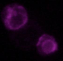
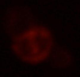
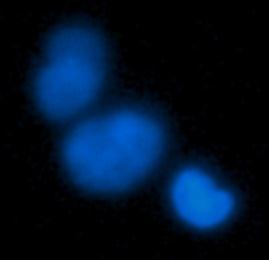
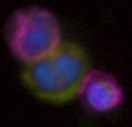
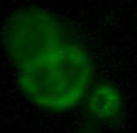
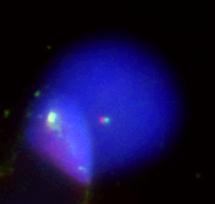
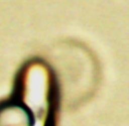
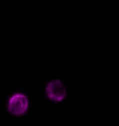
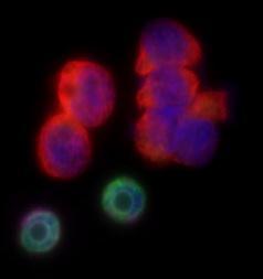
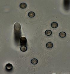
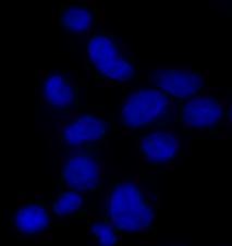
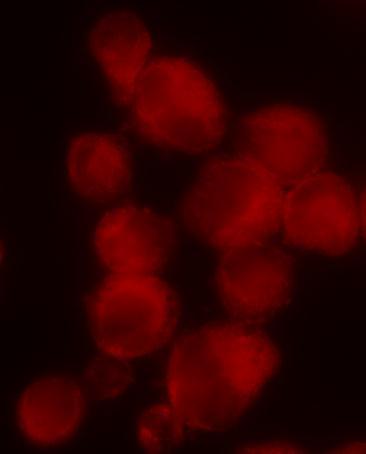
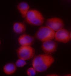
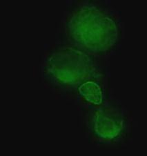
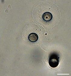
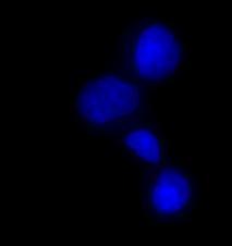
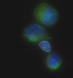
11 Legends for Supplementary Figures Figure S1. Representative example of a cluster carrying hematopoietic cells and a CTC. Two CD45 positive cells together with a CTC expressing both cytokeratins and vimentin markers are shown. Figure S2. Estimation of the percentages of CTC subsets according to anaplastic lymphoma kinase (ALK) ALK status in the 18 ALK-positive patients. Numbers of CTCs harboring abnormal ALK patterns (3 /5 > 2 or 3 /5 2, 3 or 5, 3 /5 2, ) were determined using FA-FISH. Total numbers of CTCs were determined in independent experiments by combining four-color immunofluorescent staining (cytokeratins/vimentin/cd45/dapi) and cytomorphologic analysis. The percentages of CTCs with a native status of ALK were estimated using the numbers of CTCs harboring abnormal ALK patterns and of total CTCs. Figure S3. Marker detection in circulating tumor cells (CTCs) harboring a native anaplastic lymphoma kinase (ALK) gene status. (A) Representative example of vimentin/cytokeratins/ CD45/DAPI immunofluorescent staining allowing to identify a CTC expressing both cytokeratins and vimentin and harboring a native ALK status in an ALK-positive patient. (B) Representative example of vimentin/cytokeratins/cd45/dapi immunofluorescent staining allowing to identify a CTC expressing only cytokeratins and harboring a native ALK status in an ALK-negative patient. Pores are indicated. Yellow arrows show fused 3 5 ALKnative signals. Figure S4. Levels of circulating tumor cells (CTCs) harboring different patterns of anaplastic lymphoma kinase (ALK) abnormalities detected before and during crizotinib treatment in five ALK-positive patients. Data are presented as the mean of values obtained from three spots (3 x 1 ml). Figure S5. Detection of anaplastic lymphoma kinase (ALK)-rearrangement in H2228 cell line on filters using filter-adapted fluorescent in situ hybridization (FA-FISH). H2228 cell line was spiked into peripheral blood sample from a healthy individual. Examples of ALKrearrangement patterns and of gain of ALK gene copies present in H2228 cell line. Scale: white bars correspond to 10 μm. Figure S6. Detection of epithelial mesenchymal transition (EMT) markers on filters of Hela and MCF7 cell lines spiked into peripheral blood of a healthy individual. Four-color immunofluorescent staining with monoclonal antibodies specific for vimentin, cytokeratins, CD45 and DAPI and for E-cadherin, N-cadherin, CD45 and DAPI experiments are shown. (A) Hela cell line expressing mesenchymal markers. 33 (B) MCF7 cell line expressing epithelial markers.

12 Fig S1. Hemalun hematopoietic cell Dapi CD45 Vimentin Cytokeratins Merge CTC hematopoietic cell in a pore
13 % of CTCs subsets Fig S2. 100% 50% 3 /5 x 2 3 /5 > 2 3 /5 2, 3 /5, 0% P1 P2 P3 P4 P5 P6 P7 P8 P9 P10 P11 P12 P13 P14 P15 P16 P17 P18


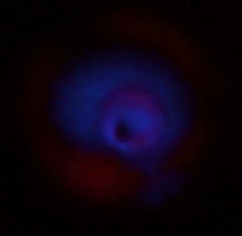
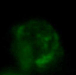
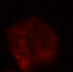
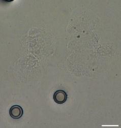
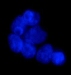
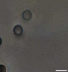
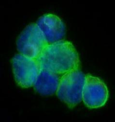
14 Fig S3. A Bright Field Dapi CD45 a pore Vimentin Cytokeratins Merge 2 x 3 /5 B Bright Field Dapi CD45 CTC in a pore Vimentin Cytokeratins Merge 2 x 3 /5
15 CTCs with abnormal ALK / ml CTCs with abnormal ALK / ml CTCs with abnormal ALK / ml CTCs with abnormal ALK / ml CTCs with abnormal ALK / ml Fig S4. 20 P5 Baseline Day P6 Baseline Day /5, 3 /5 2, 0 3 /5 > 2 3 /5, 3 /5 2, 3 /5 > 2 20 P8 Baseline Day P14 Baseline Day /5, 3 /5 2, 0 3 /5 > 2 3 /5, 3 /5 2, 3 /5 > 2 20 P18 Baseline Day /5, 3 /5 2, 3 /5 > 2

16 Fig S5. 3 /5, 3 /5, and 3 3 /5 2, and 3


17 Fig S6. A Bright Field Dapi CD45 Vimentin Cytokeratins Hela Merge hematopoietic cell Bright Field Dapi CD45 E-cadherin N-cadherin Merge Hela hematopoietic cell B Bright Field Dapi CD45 Vimentin Cytokeratins MCF7 Merge hematopoietic cell Bright Field Dapi CD45 E-cadherin N-cadherin Merge MCF7
Detection of Circulating Tumor Cells Harboring a Unique ALK Rearrangement in ALK-Positive Non Small-Cell Lung Cancer
 Published Ahead of Print on May 13, 2013 as 10.1200/JCO.2012.44.5932 The latest version is at http://jco.ascopubs.org/cgi/doi/10.1200/jco.2012.44.5932 JOURNAL OF CLINICAL ONCOLOGY O R I G I N A L R E P
HER2 FISH pharmdx TM Interpretation Guide - Breast Cancer
 P A T H O L O G Y HER2 FISH pharmdx TM Interpretation Guide - Breast Cancer For In Vitro Diagnostic Use FDA approved as an aid in the assessment of patients for whom Herceptin TM (trastuzumab) treatment
Dako IT S ABOUT TIME. Interpretation Guide. Agilent Pathology Solutions. ALK, ROS1 and RET IQFISH probes (Dako Omnis) MET IQFISH probe (Dako Omnis)
 MET IQFISH probe (Dako Omnis)") INTERPRETATION Dako Agilent Pathology Solutions IQFISH Interpretation Guide ALK, ROS1 and RET IQFISH probes (Dako Omnis) MET IQFISH probe (Dako Omnis) IT S ABOUT TIME For In Vitro Diagnostic Use ALK, ROS1,
INTERPRETATION Dako Agilent Pathology Solutions IQFISH Interpretation Guide ALK, ROS1 and RET IQFISH probes (Dako Omnis) MET IQFISH probe (Dako Omnis) IT S ABOUT TIME For In Vitro Diagnostic Use ALK, ROS1,
Quality in Control. ROS1 Analyte Control. Product Codes: HCL022, HCL023 and HCL024
 Quality in Control ROS1 Analyte Control Product Codes: HCL022, HCL023 and HCL024 Contents What is ROS1? 2 The Role of ROS1 in Cancer 3 ROS1 Assessment 3 ROS1 Analyte Control Product Details 4 ROS1 Analyte
HER2 CISH pharmdx TM Kit Interpretation Guide Breast Cancer
 P A T H O L O G Y HER2 CISH pharmdx TM Kit Interpretation Guide Breast Cancer FROM CERTAINTY COMES TRUST For in vitro diagnostic use HER2 CISH pharmdx Kit HER2 CISH pharmdx Kit is intended for dual-color
IT S ABOUT TIME. IQFISH pharmdx Interpretation Guide THREEHOURSTHIRTYMINUTES. HER2 IQFISH pharmdxtm. TOP2A IQFISH pharmdxtm
 I N T E R P R E TAT I O N IQFISH pharmdx Interpretation Guide TM HER2 IQFISH pharmdxtm TOP2A IQFISH pharmdxtm Breast carcinoma (FFPE) stained with HER2 IQFISH pharmdx Breast carcinoma (FFPE) stained with
Three Hours Thirty Minutes
 INTERPRETATION HER2 IQFISH pharmdx TM Interpretation Guide Three Hours Thirty Minutes it s about time Breast carcinoma (FFPE) stained with HER2 IQFISH pharmdx Gastric cancer (FFPE) stained with HER2 IQFISH
(A) PCR primers (arrows) designed to distinguish wild type (P1+P2), targeted (P1+P2) and excised (P1+P3)14-
 PCR primers (arrows) designed to distinguish wild type (P1+P2), targeted (P1+P2) and excised (P1+P3)14-") 1 Supplemental Figure Legends Figure S1. Mammary tumors of ErbB2 KI mice with 14-3-3σ ablation have elevated ErbB2 transcript levels and cell proliferation (A) PCR primers (arrows) designed to distinguish
1 Supplemental Figure Legends Figure S1. Mammary tumors of ErbB2 KI mice with 14-3-3σ ablation have elevated ErbB2 transcript levels and cell proliferation (A) PCR primers (arrows) designed to distinguish
Nature Methods: doi: /nmeth Supplementary Figure 1
 Supplementary Figure 1 Finite-element analysis of cell cluster dynamics in different cluster trap architectures. (a) Cluster-Chip (b) Filter (c) A structure identical to the Cluster-Chip except that one
Quality in Control ALK-Lung Analyte Control (EML4-ALK) ALK-Lymphoma Analyte Control (NPM-ALK)
 ALK-Lymphoma Analyte Control (NPM-ALK)") Quality in Control ALK-Lung Analyte Control (EML4-ALK) ALK-Lymphoma Analyte Control (NPM-ALK) 10 Product Codes: HCL007, HCL008 and HCL009 HCL010, HCL011 and HCL012 Page 1 of Contents 1. What is ALK?...
Quality in Control ALK-Lung Analyte Control (EML4-ALK) ALK-Lymphoma Analyte Control (NPM-ALK) 10 Product Codes: HCL007, HCL008 and HCL009 HCL010, HCL011 and HCL012 Page 1 of Contents 1. What is ALK?...
High Level of Chromosomal Instability in Circulating Tumor. Cells of ROS1-Rearranged Non-Small-Cell Lung Cancer
 Annals of Oncology Advance Access published April 6, 2015 1 High Level of Chromosomal Instability in Circulating Tumor Cells of ROS1-Rearranged Non-Small-Cell Lung Cancer E. Pailler 1,2, N. Auger 3, C.R.
A Novel CTC-Detecting Technique Using TelomeScan and Its Clinical Applications
 A Novel CTC-Detecting Technique Using TelomeScan and Its Clinical Applications Yasuo Urata CEO and President Oncolys BioPharma Inc. February 16, 2013 Telomere Length is a Limiting Factor for Cell Replication
Analysis of Histologic Features Suspecting Anaplastic Lymphoma Kinase (ALK)- Positive Pulmonary Adenocarcinoma. Joungho Han 1
- Positive Pulmonary Adenocarcinoma. Joungho Han 1") Analysis of Histologic Features Suspecting Anaplastic Lymphoma Kinase (ALK)- Positive Pulmonary Adenocarcinoma In Ho Choi Dong Won Kim Sang Yoon Ha 1 Yoon-La Choi 1 Hee Jeong Lee 2 Joungho Han 1 Department
Analysis of Histologic Features Suspecting Anaplastic Lymphoma Kinase (ALK)- Positive Pulmonary Adenocarcinoma In Ho Choi Dong Won Kim Sang Yoon Ha 1 Yoon-La Choi 1 Hee Jeong Lee 2 Joungho Han 1 Department
Rearrangement of the ALK gene occurs in approximately
 Original Article Combined Use of ALK Immunohistochemistry and FISH for Optimal Detection of ALK-Rearranged Lung Adenocarcinomas Lynette M. Sholl, MD,* Stanislawa Weremowicz, PhD, * Stacy W. Gray, MD, Kwok-Kin
Original Articles. Implications for Optimal Clinical Testing
 Original Articles Comparison of Reverse Transcription-Polymerase Chain Reaction, Immunohistochemistry, and Fluorescence In Situ Hybridization Methodologies for Detection of Echinoderm Microtubule-Associated
Product Introduction
 Product Introduction Product Codes: HCL026, HCL027 and HCL028 Contents Introduction to HER2 2 HER2 immunohistochemistry 3 Cell lines as controls 5 HER2 Analyte Control DR IHC 7 HER2 Analyte Control DR
Personalized Medicine: Lung Biopsy and Tumor
 Personalized Medicine: Lung Biopsy and Tumor Mutation Testing Elizabeth H. Moore, MD Personalized Medicine: Lung Biopsy and Tumor Mutation Testing Genomic testing has resulted in a paradigm shift in the
Identification of Novel Variant of EML4-ALK Fusion Gene in NSCLC: Potential Benefits of the RT-PCR Method
 International journal of Biomedical science ORIGINAL ARTICLE Identification of Novel Variant of EML4-ALK Fusion Gene in NSCLC: Potential Benefits of the RT-PCR Method Martin K. H. Maus 1, 2, Craig Stephens
Significance of Chromosome Changes in Hematological Disorders and Solid Tumors
 Significance of Chromosome Changes in Hematological Disorders and Solid Tumors Size of Components of Human Genome Size of haploid genome 3.3 X 10 9 DNA basepairs Estimated genetic constitution 30,000
Significance of Chromosome Changes in Hematological Disorders and Solid Tumors
 Significance of Chromosome Changes in Hematological Disorders and Solid Tumors Size of Components of Human Genome Size of haploid genome! Estimated genetic constitution! Size of average chromosome
Priti Lal, MD, 1 Paulo A. Salazar, 1 Clifford A. Hudis, MD, 2 Marc Ladanyi, MD, 1 and Beiyun Chen, MD, PhD 1. Abstract
 Anatomic Pathology / DUAL- VS SINGLE-COLOR SCORING IN IMMUNOHISTOCHEMICAL AND FISH HER-2 TESTING HER-2 Testing in Breast Cancer Using Immunohistochemical Analysis and Fluorescence In Situ Hybridization
(a) Significant biological processes (upper panel) and disease biomarkers (lower panel)
 Significant biological processes (upper panel) and disease biomarkers (lower panel)") Supplementary Figure 1. Functional enrichment analyses of secretomic proteins. (a) Significant biological processes (upper panel) and disease biomarkers (lower panel) 2 involved by hrab37-mediated secretory
Supplementary Figure 1. Functional enrichment analyses of secretomic proteins. (a) Significant biological processes (upper panel) and disease biomarkers (lower panel) 2 involved by hrab37-mediated secretory
Vysis ALK Break Apart FISH Probe Kit
 Vysis ALK Break Apart FISH Probe Kit 06N38 30-608521/R1 Key to Symbols Used Global Trade Item Number Manufacturer Reference Number Lot Number In Vitro Diagnostic Medical Device Contains sufficient for
Detection of Anaplastic Lymphoma Kinase (ALK) gene in Non-Small Cell lung Cancer (NSCLC) By CISH Technique
 gene in Non-Small Cell lung Cancer (NSCLC) By CISH Technique") Cancer and Clinical Oncology; Vol. 7, No. 1; 2018 ISSN 1927-4858 E-ISSN 1927-4866 Published by Canadian Center of Science and Education Detection of Anaplastic Lymphoma Kinase (ALK) gene in Non-Small Cell
Cancer and Clinical Oncology; Vol. 7, No. 1; 2018 ISSN 1927-4858 E-ISSN 1927-4866 Published by Canadian Center of Science and Education Detection of Anaplastic Lymphoma Kinase (ALK) gene in Non-Small Cell
VENTANA ALK (D5F3) Rabbit Monoclonal Primary Antibody. ALK IHC Biomarker Testing Aiding in patient diagnosis
 Rabbit Monoclonal Primary Antibody. ALK IHC Biomarker Testing Aiding in patient diagnosis") VENTANA (D5F3) Rabbit Monoclonal Primary Antibody IHC Biomarker Testing Aiding in patient diagnosis 2 IHC Biomarker Testing Lung cancer is the leading cause of death Lung cancer is the most prevalent form
VENTANA (D5F3) Rabbit Monoclonal Primary Antibody IHC Biomarker Testing Aiding in patient diagnosis 2 IHC Biomarker Testing Lung cancer is the leading cause of death Lung cancer is the most prevalent form
Anaplastic lymphoma kinase (ALK) gene rearrangement
 gene rearrangement") Original Article Heterogeneity of Anaplastic Lymphoma Kinase Gene Rearrangement in Non Small-Cell Lung Carcinomas A Comparative Study Between Small Biopsy and Excision Samples Hideyuki Abe, CT,* Akihiko
Original Article Heterogeneity of Anaplastic Lymphoma Kinase Gene Rearrangement in Non Small-Cell Lung Carcinomas A Comparative Study Between Small Biopsy and Excision Samples Hideyuki Abe, CT,* Akihiko
Interpretation Guide. Product Name: ALK Cell Line Analyte Control Product Codes: ALK2/CS and ALK2/CB. Page 1 of 9
 Interpretation Guide Product Name: ALK Cell Line Analyte Control Product Codes: ALK2/CS and ALK2/CB ALK2/CS/CB_IG_V_001 www.histocyte.com Page 1 of 9 Contents 1. What is ALK?... 2 2. Role of ALK in Cancer...
Supplementary Figure 1
 Supplementary Figure 1 Supplementary Fig. 1: Quality assessment of formalin-fixed paraffin-embedded (FFPE)-derived DNA and nuclei. (a) Multiplex PCR analysis of unrepaired and repaired bulk FFPE gdna from
Instant Quality FISH. The name says it all.
 COMPANION DIAGNOSTICS Instant Quality FISH Instant Quality FISH. The name says it all. IQ: Instant Quality every time. Breast carcinoma stained with : Triple filter showing Blue DAPI colors nuclei, FITC
Method for semi-automated microscopy of filtration-enriched circulating tumor cells
 Pailler et al. BMC Cancer (2016) 16:477 DOI 10.1186/s12885-016-2461-4 TECHNICAL ADVANCE Open Access Method for semi-automated microscopy of filtration-enriched circulating tumor cells Emma Pailler 1,2,3,
Assessment Run B HER2 IHC
 Assessment Run B26 208 HER2 IHC Material The slide to be stained for HER2 comprised the following 5 materials: IHC: HER2 Score* (0, +, 2+, 3+) FISH: HER2 gene/chr 7 ratio**. Breast carcinoma, no. 2+..3
Product Introduction. Product Codes: HCL029, HCL030 and HCL031. Issue
 Product Introduction Product Codes: HCL029, HCL030 and HCL031 Issue 1. 180510 Contents Introduction to Estrogen Receptor 2 ER immunohistochemistry 3 Quality control 5 Cell lines as controls 6 Estrogen
DM Seminar. ALK gene rearrangements & ALK targeted therapy in NSCLC Dr Sarat
 DM Seminar ALK gene rearrangements & ALK targeted therapy in NSCLC Dr Sarat Introduction Discovery of activating mutations in kinase domain of epidermal growth factor receptor (EGFR) opened a new era of
Molecular Testing in Lung Cancer
 Molecular Testing in Lung Cancer Pimpin Incharoen, M.D. Assistant Professor, Thoracic Pathology Department of Pathology, Ramathibodi Hospital Genetic alterations in lung cancer Source: Khono et al, Trans
Proteomic profiling of small-molecule inhibitors reveals dispensability of MTH1 for cancer cell survival
 Supplementary Information for Proteomic profiling of small-molecule inhibitors reveals dispensability of MTH1 for cancer cell survival Tatsuro Kawamura 1, Makoto Kawatani 1, Makoto Muroi, Yasumitsu Kondoh,
Instant Quality FISH. The name says it all.
 PRODUCT INFORMATION HER2 IQFISH pharmdx Instant Quality FISH Instant Quality FISH. The name says it all. HER2 IQFISH pharmdx IQ: Instant Quality every time. HER2 IQFISH pharmdx stains of a HER2 non-amplified
Rearrangement of the anaplastic lymphoma kinase (ALK)
") Brief Report Clinical Implications of Variant ALK FISH Rearrangement Patterns Xin Gao, MD,* Lynette M. Sholl, MD,* Mizuki Nishino, MD,* Jennifer C. Heng, BS, Pasi A. Jänne, MD, PhD,* and Geoffrey R. Oxnard,
Brief Report Clinical Implications of Variant ALK FISH Rearrangement Patterns Xin Gao, MD,* Lynette M. Sholl, MD,* Mizuki Nishino, MD,* Jennifer C. Heng, BS, Pasi A. Jänne, MD, PhD,* and Geoffrey R. Oxnard,
PD-L1 Analyte Control DR
 Quality in Control PD-L1 Analyte Control DR PD-L1_PI_v2 Product Codes: HCL019, HCL020 and HCL021 Contents PD-L1 Analyte Control DR 2 What is PD-L1? 3 The Role of PD-L1 in Cancer 3 PD-L1 Assessment 4 PD-L1
Identifying ALK+ NSCLC patients for targeted treatment
 VENTANA (D5F3) CDx Assay Identifying + NSCLC patients for targeted treatment VENTANA (D5F3) CDx Assay Identify + NSCLC patients eligible for treatment with XORI, ZYKADIA or ALECENSA NSCLC tissue samples
Electronic Supplementary Information
 Electronic Supplementary Material (ESI) for Journal of Materials Chemistry B. This journal is The Royal Society of Chemistry 2016 Electronic Supplementary Information Single mammalian cell encapsulation
SUPPLEMENTARY MATERIAL. Sample preparation for light microscopy
 SUPPLEMENTARY MATERIAL Sample preparation for light microscopy To characterize the granulocytes and melanomacrophage centers, cross sections were prepared for light microscopy, as described in Material
Oncology Genetics: Cytogenetics and FISH 17/09/2014
 Oncology Genetics: Cytogenetics and FISH 17/09/2014 Chris Wragg Head of Oncology Genomics, BGL BGL Bristol Genetics Laboratory (BGL) CPA accredited Genetics laboratory serving a core population of 4-5million
Breast cancer: Antibody selection, protocol optimzation controls and EQA
 Breast cancer: Antibody selection, protocol optimzation controls and EQA Workshop in Diagnostic Immunohistochemistry Oud St. Jan/ Old St. John Brugge (Bruges), Belgium June 13th 15nd 2018 Rasmus Røge,
Supporting Information
 Supporting Information Chan et al. 1.173/pnas.9654916 A Patient B Xenograft C * remaining feature of normal lymph node * * * D lymphocytes Infiltrating transitional carcinoma cells E Enlarged axillary
Assessment Run B HER2 IHC
 Assessment Run B24 2017 HER2 IHC Material The slide to be stained for HER2 comprised the following 5 materials: IHC: HER2 Score* (0, 1+, 2+, 3+) FISH: HER2 gene/chr 17 ratio** 1. Breast carcinoma, no.
CD14 + S100A9 + Monocytic Myeloid-Derived Suppressor Cells and Their Clinical Relevance in Non-Small Cell Lung Cancer
 CD14 + S1A9 + Monocytic Myeloid-Derived Suppressor Cells and Their Clinical Relevance in Non-Small Cell Lung Cancer Po-Hao, Feng M.D., Kang-Yun, Lee, M.D. Ph.D., Ya-Ling Chang, Yao-Fei Chan, Lu- Wei, Kuo,Ting-Yu
Breast Cancer Interpretation Guide
 Breast Cancer Interpretation Guide UCT D O R P NEW ERBB2/ C E P S ht e ZytoLig lor Prob o C l a u 2D D17S12 ng to the i d r o c c a ting for re-tes idelines 2013 ASCO Gu Breast Cancer Interpretation Guide
Epithelial interleukin-25 is a key mediator in Th2-high, corticosteroid-responsive
 Online Data Supplement: Epithelial interleukin-25 is a key mediator in Th2-high, corticosteroid-responsive asthma Dan Cheng, Zheng Xue, Lingling Yi, Huimin Shi, Kan Zhang, Xiaorong Huo, Luke R. Bonser,
LUNG CANCER. pathology & molecular biology. Izidor Kern University Clinic Golnik, Slovenia
 LUNG CANCER pathology & molecular biology Izidor Kern University Clinic Golnik, Slovenia 1 Pathology and epidemiology Small biopsy & cytology SCLC 14% NSCC NOS 4% 70% 60% 50% 63% 62% 61% 62% 59% 54% 51%
Virtual Journal Club: Front-Line Therapy and Beyond Recent Perspectives on ALK-Positive Non-Small Cell Lung Cancer.
 Virtual Journal Club: Front-Line Therapy and Beyond Recent Perspectives on ALK-Positive Non-Small Cell Lung Cancer Reference Slides ALK Rearrangement in NSCLC ALK (anaplastic lymphoma kinase) is a receptor
Assessment performed on Tuesday, July 29, 2014, at Lions Gate Hospital, North Vancouver
 Assessors report for ciqc Run 37: BRAF V600E (April 2014) Assessors: B Gilks, R Wolber, K Ung, P Tavassoli, J Garratt and J Won (recorder) Assessment performed on Tuesday, July 29, 2014, at Lions Gate
Updated Molecular Testing Guideline for the Selection of Lung Cancer Patients for Treatment with Targeted Tyrosine Kinase Inhibitors
 Q: How is the strength of recommendation determined in the new molecular testing guideline? A: The strength of recommendation is determined by the strength of the available data (evidence). Strong Recommendation:
VEGFR2-Mediated Vascular Dilation as a Mechanism of VEGF-Induced Anemia and Bone Marrow Cell Mobilization
 Cell Reports, Volume 9 Supplemental Information VEGFR2-Mediated Vascular Dilation as a Mechanism of VEGF-Induced Anemia and Bone Marrow Cell Mobilization Sharon Lim, Yin Zhang, Danfang Zhang, Fang Chen,
The fusion of anaplastic lymphoma kinase (ALK) gene with
 gene with") ORIGINAL ARTICLE Detection of ALK Gene Rearrangement in Non-small Cell Lung Cancer A Comparison of Fluorescence In Situ Hybridization and Chromogenic In Situ Hybridization with Correlation of ALK Protein
ORIGINAL ARTICLE Detection of ALK Gene Rearrangement in Non-small Cell Lung Cancer A Comparison of Fluorescence In Situ Hybridization and Chromogenic In Situ Hybridization with Correlation of ALK Protein
Advances in Pathology and molecular biology of lung cancer. Lukas Bubendorf Pathologie
 Advances in Pathology and molecular biology of lung cancer Lukas Bubendorf Pathologie Agenda The revolution of predictive markers Liquid biopsies PD-L1 Molecular subtypes (non-squamous NSCLC) Tsao AS et
Downregulation of angiotensin type 1 receptor and nuclear factor-κb. by sirtuin 1 contributes to renoprotection in unilateral ureteral
 Supplementary Information Downregulation of angiotensin type 1 receptor and nuclear factor-κb by sirtuin 1 contributes to renoprotection in unilateral ureteral obstruction Shao-Yu Yang 1,2, Shuei-Liong
GENETIC TESTING FOR TARGETED THERAPY FOR NON-SMALL CELL LUNG CANCER (NSCLC)
") CANCER (NSCLC) Non-Discrimination Statement and Multi-Language Interpreter Services information are located at the end of this document. Coverage for services, procedures, medical devices and drugs are
CANCER (NSCLC) Non-Discrimination Statement and Multi-Language Interpreter Services information are located at the end of this document. Coverage for services, procedures, medical devices and drugs are
Cells and viruses. Human isolates (A/Kawasaki/173/01 [H1N1], A/Yokohama/2057/03 [H3N2],
![Cells and viruses. Human isolates (A/Kawasaki/173/01 [H1N1], A/Yokohama/2057/03 [H3N2],](/thumbs/86/93801604.jpg "Cells and viruses. Human isolates (A/Kawasaki/173/01 [H1N1], A/Yokohama/2057/03 [H3N2],") Supplementary information Methods Cells and viruses. Human isolates (A/Kawasaki/173/01 [H1N1], A/Yokohama/2057/03 [H3N2], and A/Hong Kong/213/03 [H5N1]) were grown in Madin-Darby canine kidney (MDCK) cells
Supplementary information Methods Cells and viruses. Human isolates (A/Kawasaki/173/01 [H1N1], A/Yokohama/2057/03 [H3N2], and A/Hong Kong/213/03 [H5N1]) were grown in Madin-Darby canine kidney (MDCK) cells
HER-2/neu amplification detected by fluorescence in situ hybridization in fine needle aspirates from primary breast cancer
 Original article Annals of Oncology 13: 1398 1403, 2002 DOI: 10.1093/annonc/mdf217 HER-2/neu amplification detected by fluorescence in situ hybridization in fine needle aspirates from primary breast cancer
Layered-IHC (L-IHC): A novel and robust approach to multiplexed immunohistochemistry So many markers and so little tissue
: A novel and robust approach to multiplexed immunohistochemistry So many markers and so little tissue") Page 1 The need for multiplex detection of tissue biomarkers. There is a constant and growing demand for increased biomarker analysis in human tissue specimens. Analysis of tissue biomarkers is key to
Page 1 The need for multiplex detection of tissue biomarkers. There is a constant and growing demand for increased biomarker analysis in human tissue specimens. Analysis of tissue biomarkers is key to
Transform genomic data into real-life results
 CLINICAL SUMMARY Transform genomic data into real-life results Biomarker testing and targeted therapies can drive improved outcomes in clinical practice New FDA-Approved Broad Companion Diagnostic for
ALK positive Lung Cancer. Shirish M. Gadgeel, MD. Director of the Thoracic Oncology program University of Michigan
 ALK positive Lung Cancer Shirish M. Gadgeel, MD. Director of the Thoracic Oncology program University of Michigan Objectives What is ALK translocation? What drugs are used in what sequence? How many times
Supplementary Materials. for Garmy-Susini, et al, Integrin 4 1 signaling is required for lymphangiogenesis and tumor metastasis
 Supplementary Materials for Garmy-Susini, et al, Integrin 4 1 signaling is required for lymphangiogenesis and tumor metastasis 1 Supplementary Figure Legends Supplementary Figure 1: Integrin expression
Assessment Run B HER-2 IHC. HER-2/chr17 ratio**
 Assessment Run B2 20 HER-2 IHC Material The slide to be stained for HER-2 comprised the following 5 tissues: IHC HER-2 Score* (0, +, 2+,3+) FISH HER-2/chr7 ratio**. Breast ductal carcinoma 0..3 2. Breast
Supplementary Information POLO-LIKE KINASE 1 FACILITATES LOSS OF PTEN-INDUCED PROSTATE CANCER FORMATION
 Supplementary Information POLO-LIKE KINASE 1 FACILITATES LOSS OF PTEN-INDUCED PROSTATE CANCER FORMATION X. Shawn Liu 1, 3, Bing Song 2, 3, Bennett D. Elzey 3, 4, Timothy L. Ratliff 3, 4, Stephen F. Konieczny
Challenges for use of CTCs as a Diagnostic. Farideh Z. Bischoff, Ph.D. Interim CSO Sr. Director, Translational Clinical Development Biocept, Inc.
 Challenges for use of CTCs as a Diagnostic Farideh Z. ischoff, Ph.D. Interim CSO Sr. Director, Translational Clinical Development iocept, Inc. Current Technology for CTC Testing Existing CTC testing platform
SUPPLEMENTARY INFORMATION. Supplementary Figures S1-S9. Supplementary Methods
 SUPPLEMENTARY INFORMATION SUMO1 modification of PTEN regulates tumorigenesis by controlling its association with the plasma membrane Jian Huang 1,2#, Jie Yan 1,2#, Jian Zhang 3#, Shiguo Zhu 1, Yanli Wang
Dr. dr. Primariadewi R, SpPA(K)
") Curriculum Vitae Dr. dr. Primariadewi R, SpPA(K) Education : Medical Doctor from UKRIDA Doctoral Degree from Faculty of Medicine University of Indonesia Pathologist Specialist and Consultant from Faculty
Curriculum Vitae Dr. dr. Primariadewi R, SpPA(K) Education : Medical Doctor from UKRIDA Doctoral Degree from Faculty of Medicine University of Indonesia Pathologist Specialist and Consultant from Faculty
Islet viability assay and Glucose Stimulated Insulin Secretion assay RT-PCR and Western Blot
 Islet viability assay and Glucose Stimulated Insulin Secretion assay Islet cell viability was determined by colorimetric (3-(4,5-dimethylthiazol-2-yl)-2,5- diphenyltetrazolium bromide assay using CellTiter
Supplemental Information. Otic Mesenchyme Cells Regulate. Spiral Ganglion Axon Fasciculation. through a Pou3f4/EphA4 Signaling Pathway
 Neuron, Volume 73 Supplemental Information Otic Mesenchyme Cells Regulate Spiral Ganglion Axon Fasciculation through a Pou3f4/EphA4 Signaling Pathway Thomas M. Coate, Steven Raft, Xiumei Zhao, Aimee K.
High Sensitivity Immunomagnetic CTC Isolation as Compared to Alternative Isolation Methods
 High Sensitivity Immunomagnetic CTC Isolation as Compared to Alternative Isolation Methods 1. Introduction: An overview of CTC isolation methods 2. Challenges for direct comparisons of CTC recovery 3.
Applications of IHC. Determination of the primary site in metastatic tumors of unknown origin
 Applications of IHC Determination of the primary site in metastatic tumors of unknown origin Classification of tumors that appear 'undifferentiated' by standard light microscopy Precise classification
Supplemental figure 1. PDGFRα is expressed dominantly by stromal cells surrounding mammary ducts and alveoli. A) IHC staining of PDGFRα in
 IHC staining of PDGFRα in") Supplemental figure 1. PDGFRα is expressed dominantly by stromal cells surrounding mammary ducts and alveoli. A) IHC staining of PDGFRα in nulliparous (left panel) and InvD6 mouse mammary glands (right
Supplemental figure 1. PDGFRα is expressed dominantly by stromal cells surrounding mammary ducts and alveoli. A) IHC staining of PDGFRα in nulliparous (left panel) and InvD6 mouse mammary glands (right
INTRODUCTION TO PATHOLOGICAL TECHNIQUES. 1. Types of routine biopsy procedures 2. Special exams (IHC, FISH)
") INTRODUCTION TO PATHOLOGICAL TECHNIQUES 1. Types of routine biopsy procedures 2. Special exams (IHC, FISH) Biopsy-Indications Diffuse/multifocal lesions (neoplastic, inflammatory, etc) Etiology of the
INTRODUCTION TO PATHOLOGICAL TECHNIQUES 1. Types of routine biopsy procedures 2. Special exams (IHC, FISH) Biopsy-Indications Diffuse/multifocal lesions (neoplastic, inflammatory, etc) Etiology of the
Pepsin Solution ready-to-use
 SIE HABEN DIE VISION, WIR HABEN DIE SUBSTANZ. Pepsin Solution Single component Pepsin Solution: only one component refrigerator stable Pepsin is a commonly used digestive enzyme for immunohistochemical
Supplementary Appendix
 Supplementary Appendix This appendix has been provided by the authors to give readers additional information about their work. Supplement to: Yatsenko AN, Georgiadis AP, Röpke A, et al. X-linked TEX11
Evolution of Pathology
 1 Traditional pathology Molecular pathology 2 Evolution of Pathology Gross Pathology Cellular Pathology Morphologic Pathology Molecular/Predictive Pathology Antonio Benivieni (1443-1502): First autopsy
Rob Ross, MD. Infinity Pharmaceuticals March 9 th, 2011
 Heat Shock Protein 90 (Hsp90) Inhibition as a Potential Novel Approach to the Treatment of Patients with ALK Mutated Non-small Cell Lung Cancer (NSCLC) Rob Ross, MD Infinity Pharmaceuticals March 9 th,
Supplementary Information Supplementary Fig. 1. Elevated Usp9x in melanoma and NRAS mutant melanoma cells are dependent on NRAS for 3D growth.
 Supplementary Information Supplementary Fig. 1. Elevated Usp9x in melanoma and NRAS mutant melanoma cells are dependent on NRAS for 3D growth. a. Immunoblot for Usp9x protein in NRAS mutant melanoma cells
Interpretation Manual - Gastric or Gastroesophageal Junction Adenocarcinoma. PD-L1 IHC 22C3 pharmdx is FDA-approved for in vitro diagnostic use
 Interpretation Manual - Gastric or Gastroesophageal Junction Adenocarcinoma PD-L1 IHC 22C3 pharmdx is FDA-approved for in vitro diagnostic use For countries outside of the United States, see the local
Xalkori. Xalkori (crizotinib) Description
 Description") Federal Employee Program 1310 G Street, N.W. Washington, D.C. 20005 202.942.1000 Fax 202.942.1125 5.21.12 Subject: Xalkori Page: 1 of 6 Last Review Date: June 22, 2017 Xalkori Description Xalkori (crizotinib)
Federal Employee Program 1310 G Street, N.W. Washington, D.C. 20005 202.942.1000 Fax 202.942.1125 5.21.12 Subject: Xalkori Page: 1 of 6 Last Review Date: June 22, 2017 Xalkori Description Xalkori (crizotinib)
(A) RT-PCR for components of the Shh/Gli pathway in normal fetus cell (MRC-5) and a
 RT-PCR for components of the Shh/Gli pathway in normal fetus cell (MRC-5) and a") Supplementary figure legends Supplementary Figure 1. Expression of Shh signaling components in a panel of gastric cancer. (A) RT-PCR for components of the Shh/Gli pathway in normal fetus cell (MRC-5) and
Supplementary figure legends Supplementary Figure 1. Expression of Shh signaling components in a panel of gastric cancer. (A) RT-PCR for components of the Shh/Gli pathway in normal fetus cell (MRC-5) and
Disclosures Genomic testing in lung cancer
 Disclosures Genomic testing in lung cancer No disclosures Objectives Understand how FISH and NGS provide complementary data for the evaluation of lung cancer Recognize the challenges of performing testing
Molecular Diagnosis of Lung Cancer
 Molecular Diagnosis of Lung Cancer Lucian R. Chirieac, M.D. Assistant Professor of Pathology Harvard Medical School Staff Pathologist, Department of Pathology Brigham and Women's Hospital 75 Francis Street
HPV/p16 Analyte Control
 HPV/p16 Analyte Control Utility review and ring study results Colin Tristram, Director 2017 HPV/p16 Analyte Control Collaboration: Dr Max Robinson at Newcastle University a leading pathologist in head
Approximately one third of all cancer-related deaths in
 ORIGINAL ARTICLE Comparison Between Epidermal Growth Factor Receptor (EGFR) Gene Expression in Primary Non-small Cell Lung Cancer (NSCLC) and in Fine-Needle Aspirates from Distant Metastatic Sites Cecilia
Supplementary materials and methods.
 Supplementary materials and methods. Microarray printing and data analysis. The LNA-modified oligonucleotide probe set for all annotated mirnas from mouse (Mus musculus) and human (Homo sapiens) in the
T he HER2/neu type 1 tyrosine kinase growth factor
 710 ORIGINAL ARTICLE HER2 amplification status in breast cancer: a comparison between immunohistochemical staining and fluorescence in situ hybridisation using manual and automated quantitative image analysis
RNA extraction, RT-PCR and real-time PCR. Total RNA were extracted using
 Supplementary Information Materials and Methods RNA extraction, RT-PCR and real-time PCR. Total RNA were extracted using Trizol reagent (Invitrogen,Carlsbad, CA) according to the manufacturer's instructions.
Supplementary Online Content
 Supplementary Online Content Rimm DL, Han G, Taube JM, et al. A prospective, multi-institutional, pathologistbased assessment of 4 immunohistochemistry assays for PD-L1 expression in non small cell lung
Journal of Breast Cancer
 ORIGINAL ARTICLE Journal of Breast Cancer J Breast Cancer 2009 December; 12(4): 235-40 DOI: 10.4048/jbc.2009.12.4.235 Comparison of Silver-Enhanced in situ Hybridization and Fluorescence in situ Hybridization
Presentation material is for education purposes only. All rights reserved URMC Radiology Page 1 of 98
 Presentation material is for education purposes only. All rights reserved. 2011 URMC Radiology Page 1 of 98 Radiology / Pathology Conference February 2011 Brooke Koltz, Cytopathology Resident Presentation
Material and Methods. Flow Cytometry Analyses:
 Material and Methods Flow Cytometry Analyses: Immunostaining of breast cancer cells for HER2 was performed by incubating cells with anti- HER2/neu APC (Biosciences, Cat# 340554), anti-her2/neu PE (Biosciences,
HCC1937 is the HCC1937-pcDNA3 cell line, which was derived from a breast cancer with a mutation
 SUPPLEMENTARY INFORMATION Materials and Methods Human cell lines and culture conditions HCC1937 is the HCC1937-pcDNA3 cell line, which was derived from a breast cancer with a mutation in exon 20 of BRCA1
ONCO TEAM DIAGNOSTIC
 ONCO TEAM DIAGNOSTIC ONCO TEAM SPECIALISTS The team at ONCO TEAM has 23 specialists: o 11 general pathology seniors: - 8 PhD (1 PhD on lung tumors pathology) o 3 general pathology specialists; o 2 dermopathology
SOMAPLEX REVERSE PHASE PROTEIN MICROARRAY HUMAN KIDNEY TUMOR & NORMAL TISSUE
 SOMAPLEX REVERSE PHASE PROTEIN MICROARRAY HUMAN KIDNEY TUMOR & NORMAL TISSUE 45 CLINICAL CASES SERIAL DILUTION MULTIPLE PROTEIN CONCENTRATION QUANTITATIVE ASSAY PRODUCT NUMBER: PM1-001-N SOMAPLEX REVERSE
Respiratory Department of Peking Union Medical College Hospital, Chinese Academy of Medical Sciences and Peking Union Medical College, Peking, China
 Thoracic Cancer ISSN 1759-7706 ORIGINAL ARTICLE Efficacy of bronchoscopic biopsy for the detection of epidermal growth factor receptor mutations and anaplastic lymphoma kinase gene rearrangement in lung
Central Pathology Review and Tissue MicroArrays. Dr Lisa Storer Children s Brain Tumour Research Centre Nottingham
 Central Pathology Review and Tissue MicroArrays Dr Lisa Storer Children s Brain Tumour Research Centre Nottingham What is a tissue microarray? Tissue microarrays (TMAs) consist of paraffin blocks in which
Clinical Validation of Cytocell Pathology Probes
 Clinical Validation of Cytocell Pathology Probes Shivanand Richardson Specialized Technician Molecular Pathology Department of Pathology, University Medical Center Utrecht The Netherlands 5th annual Cytocell
Assessment Run C1 2017
 Assessment Run C1 2017 PD-L1 The first assessment in this new NordiQC Companion module C1 focused on the accuracy of the PD-L1 IHC assays performed by the participating laboratories to identify patients