Apocrine and eccrine adnexal tumors
|
|
|
- Ada Baker
- 6 years ago
- Views:
Transcription
1 Apocrine and eccrine adnexal tumors Timothy McCalmont, MD University of California San Francisco, CA
2 Past misguided notions Syringoma is eccrine Eccrine poroma rather than eccrine or apocrine poroma Spiradenoma is eccrine while its sibling cylindroma is apocrine Microcystic adnexal carcinoma shows mixed follicular and eccrine differentiation
3 Timothy H. McCalmont, M.D.
4 Factors crucial to the evaluation of adnexal lineage Embryology Combined and associated tumors Anatomic distribution Morphology & phenotype By light microscopy, EM, and immunostaining
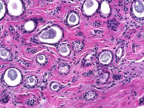
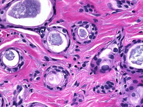
5 Embryology. Apocrine differs from eccrine
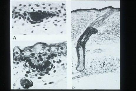
6
7 Folliculosebaceousapocrine unit
8 Does ontogeny reflect phylogeny? Alleged combinations of eccrine and follicular (or eccrine and sebaceous) are probably fictional
9
10

11
12
13
14
15
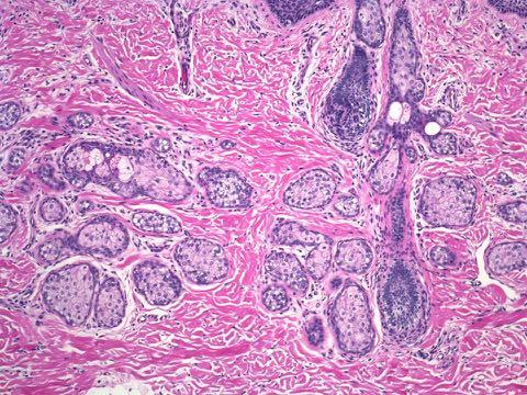
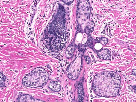
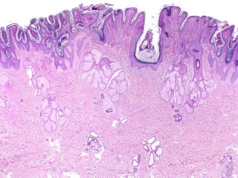
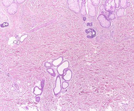
16
17
18
19
20
21
22
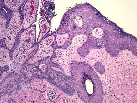
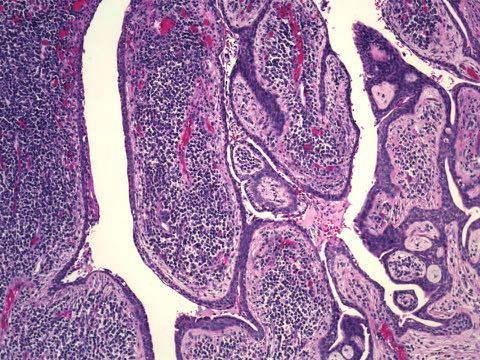
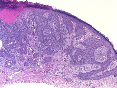
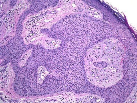
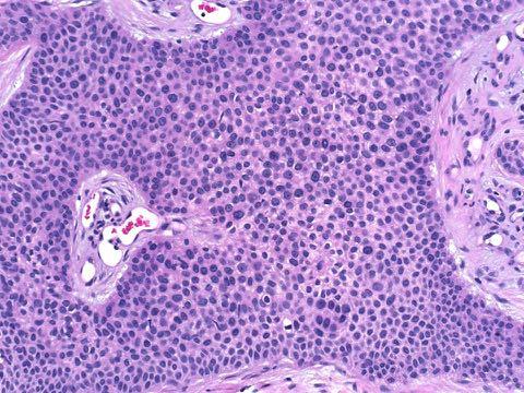
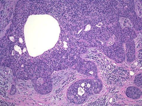
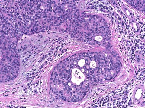
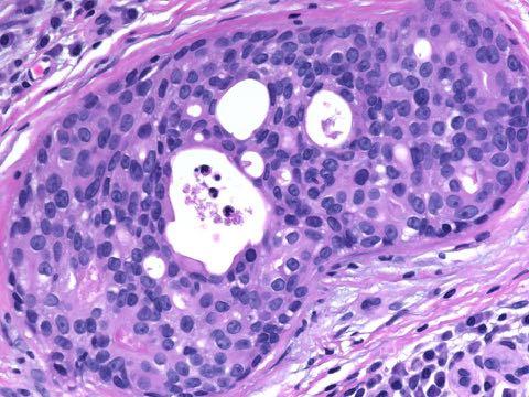
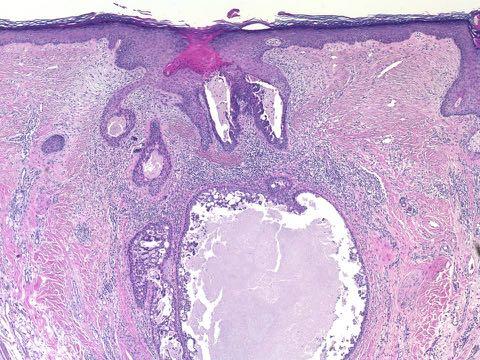
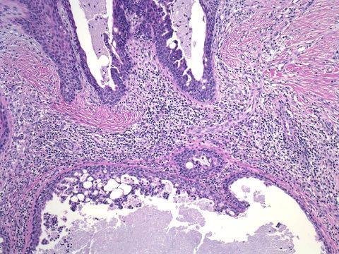
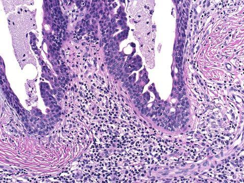
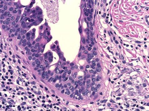
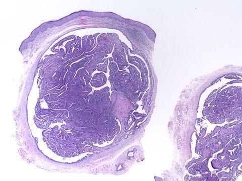
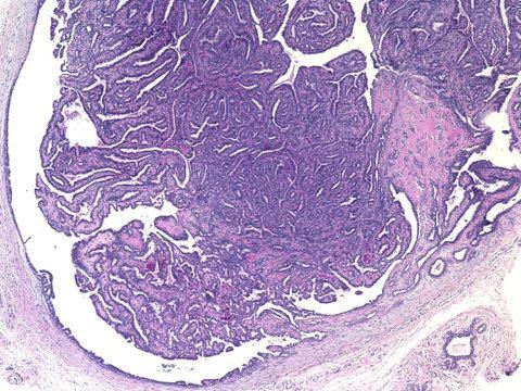
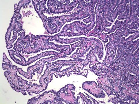
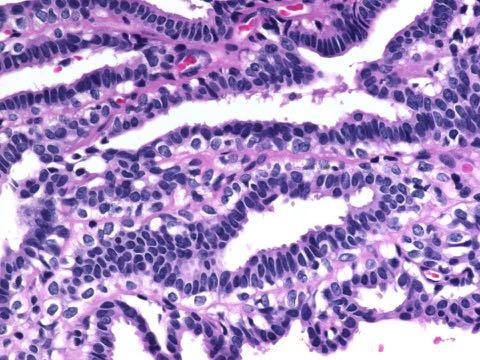
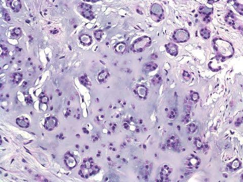
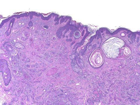
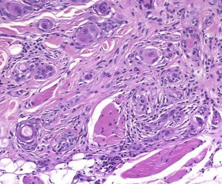
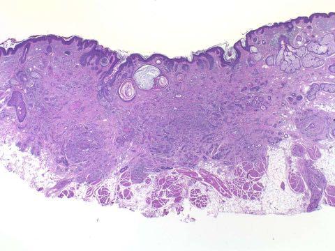
23 Syringocystadenoma papilliferum
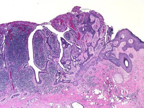
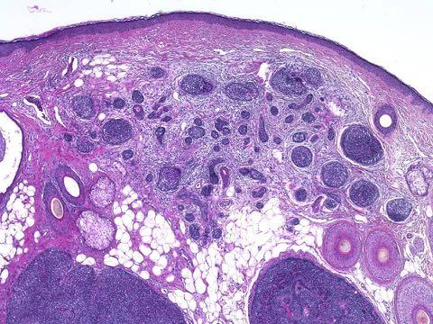
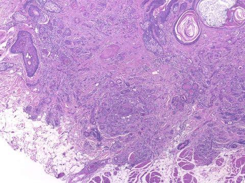
24 Nevus sebaceus and secondary neoplasms Syringocystadenoma (apocrine) Trichoblastoma (follicular) Trichilemmoma (follicular) Sebaceous adenoma (sebaceous) Tubular adenoma (apocrine) Poroma (apocrine, when occurring in nevus sebaceus)
25 Combined tumors.
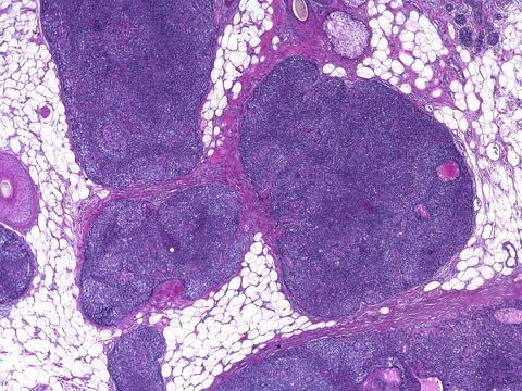
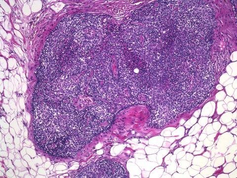
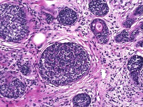
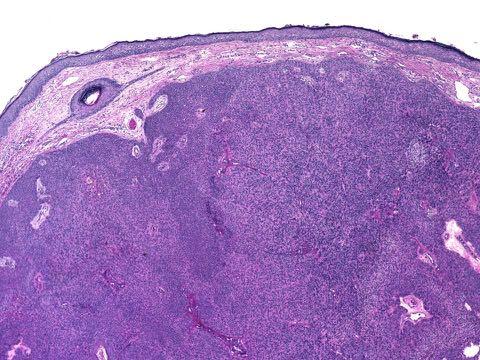
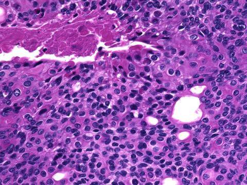
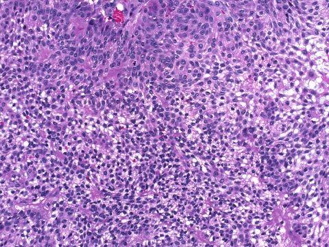
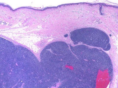
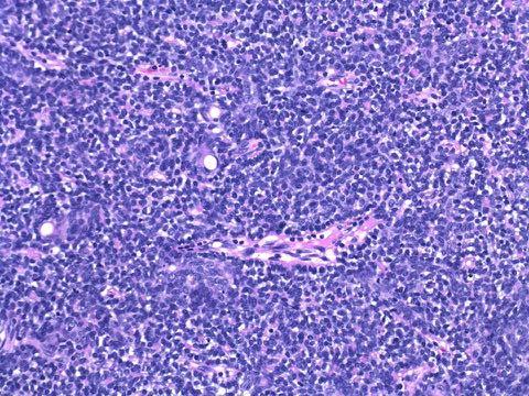
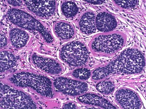
26 Combined tumors Elements that commonly occur conjointly, non-coincidentally, are of related lineage Example: glomangiomyoma
27
28
29 Combined adnexal tumors Spiradenoma and cylindroma Spiradenoma and trichoepithelioma Cylindroma and trichoepithelioma Tubular adenoma and sebaceous adenoma
30
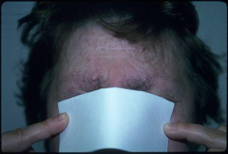
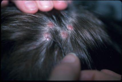
31
32
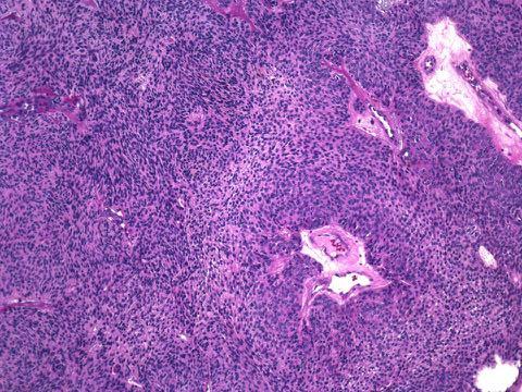
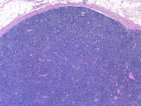
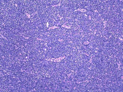
33 Spiradenoma
34
35
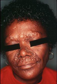
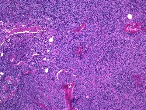
36 Cylindroma
37 Q.E.D. Trichoepithelioma, spiradenoma, and cylindroma are all folliculosebaceous-apocrine
38 Neoplastic associations (syndromic)
39
40
41
42
43 What s the lesson of Brooke-Spiegler? The combination of spiradenoma with cylindroma and trichoepithelioma in a genetic syndrome indicates folliculosebaceous-apocrine lineage
44 Anatomic distribution.
45 Distribution of adnexa The distribution of adnexal tumors parallels the structures toward which they differentiate
46 Trichoblastoma (trichoepithelioma)
47 Trichoblastoma Sex 72:28 (F>M) Age 3-99 years head/neck 96% arm/leg 4% hand/foot <1%
48
49 Syringoma Sex 79:21 (F>M) Age yrs head/neck 90% axilla/genital 5% arm/leg 5% hand/foot <1%
50 (Arch Dermatol 113: , 1977)
51
52 Spiradenoma Sex 53:47 (F>M) Age years head/neck/trunk 76% arm/leg 24% hand/foot <1%
53 Houston, we have a problem. If spiradenoma or syringoma were purely eccrine, we d see them as palmoplantar tumors
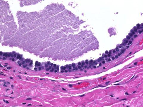
54 Morphology.
55 Morphologic attributes Light microscopic pattern Immunophenotype (immunohistochemistry) Enzyme expression (histochemistry) Ultrastructure (electron microscopy)

56
57 Follicular attributes Basaloid (germinative) cells with adjacent mesenchymal cells (resembling the bulb and papilla) Matrical ( shadow ) cells Pallid (outer sheath) cells with adjacent thickened membrane Trichohyaline granules
58
59 Sebaceous attributes Cells with coarsely vacuolated cytoplasm and scalloped nuclei

60
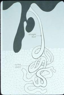
61 Apocrine attributes Decapitation? Cuticulated ducts? Pallid (clear) cells? Eccrine attributes Cuticulated ducts? Pallid (clear) cells?
62
63 Immunohistochemical reagents Carcinoembryonic antigen (CEA) S100 protein Gross cystic disease fluid protein (GCDFP-15) Epithelial membrane antigen (EMA) Anti-keratins
64
65
66 Morphologic conclusions We can precisely define follicular and sebaceous differentiation We cannot precisely identify apocrine and eccrine lineage
67 Morphologic conclusions The distinction and classification of apocrine and eccrine lesion requires integration of a variety of factors, including embryology, distribution, and microscopy
68 Eccrine Neoplasms Less common than apocrine, probably because of the monophasic nature of the eccrine apparatus of little proliferative potential (hyperproliferation precedes mutagenesis in oncogenesis)
69 Eccrine tumors Syringoma, sometimes Poroma, commonly Hidradenoma, sometimes Papillary adenoma, commonly
70 Poroma Of eccrine or apocrine lineage of palm or sole, manifest as a vascular nodule: eccrine poroma within nevus sebaceus or on axillary skin: apocrine poroma
71
72
73
74
75
76
77
78
79 Poroma Patterns Juxtaepidermal (epidermal and dermal poroma) Intraepidermal (hidroacanthoma simplex) Intradermal (dermal duct tumor)
80

81
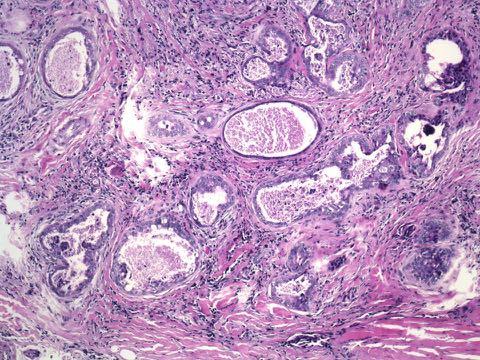
82
83
84
85
86
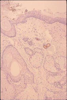
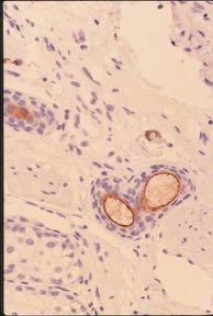
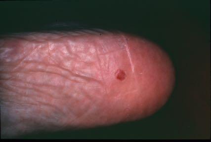
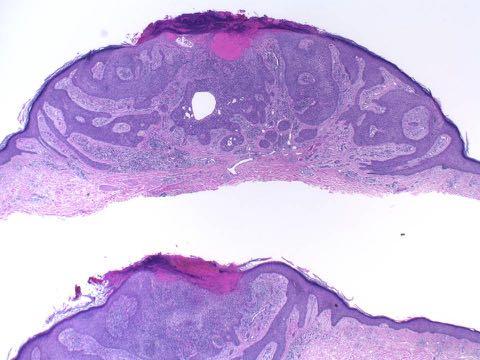
87
88 Poroma Hallmarks Small cuboidal ( poroid ) cells Prominent ductular differentiation Highly vascular stroma Necrosis en masse, sometimes
89 Papillary adenoma A proliferative adenoma, with tubules lined by a cellular multilayered epithelium from which papillary projections extend into the luminal space Eccrine or apocrine in lineage
90
91
92
93
94 Aggressive (digital) papillary adenoma
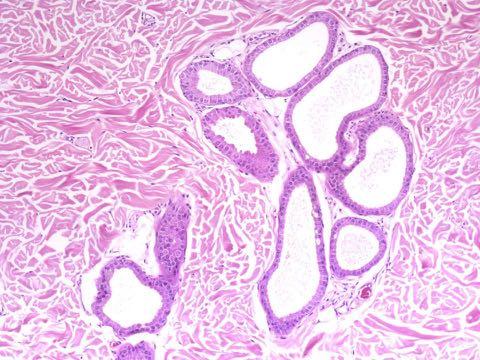
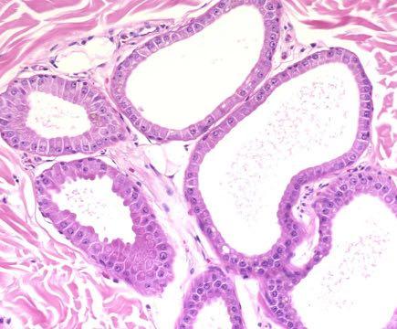
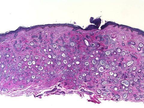
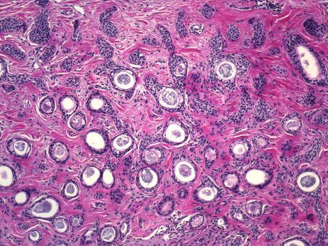
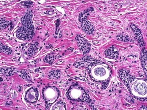
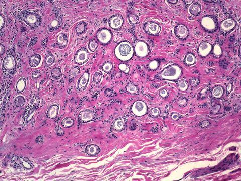
95
96
97
98 Aggressive papillary adenoma In retrospect, most examples represent malignancy, i.e. papillary adenocarcinoma Of course an adenoma may seem aggressive if it is truly an adenocarcinoma
99 Apocrine tumors Apocrine differentiation often occurs jointly with follicular and sebaceous attributes, thus diverse! Strictly apocrine lesions are often indistinguishable from strictly eccrine neoplasms via conventional microscopy
100 Strictly apocrine tumors Syringoma, commonly Poroma, commonly Hidradenoma, commonly Apocrine adenomas, including hidradenoma papilliferum and syringocystadenoma Spiradenoma and cylindroma
101 Tumors with apocrinefollicular (or folliculosebaceous-apocrine) differentiation Chondroid syringoma (mixed tumor) Microcystic adnexal carcinoma
102 Syringoma Occur at virtually any site; commonly periorbital Uncommon at acral or glabrous sites Apocrine or eccrine in lineage; most are probably apocrine Usually small, often characterized by considerable sclerosis
103
104
105
106
107
108
109
110 Syringoma Hallmarks Numerous small nests or ducts, often with a central cuticle Enveloping sclerotic stroma Tadpole or comma-shaped nests, sometimes
111 Hidradenoma Close relative of poroma Usually dermal, usually composed of cells with ample pale eosinophilic cytoplasm ( clear cell hid.) May be cystic ( solid-cystic hid.) Eccrine or apocrine in lineage; most are apocrine
112
113
114
115
116
117
118 Acrospiroma Bridging designation that encompasses poroma and hidradenoma Equivalent to the garbage can designation nodular hidradenoma
119 Hidradenoma Hallmarks Cuboidal cells, often with pale cytoplasm and generally larger than those of poroma Limited ductular differentiation Solid or cystic or both Stromal sclerosis, often
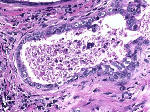
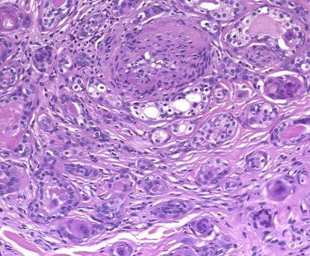
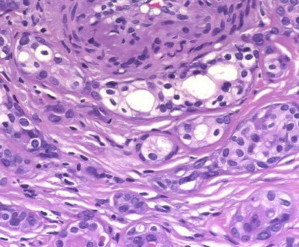
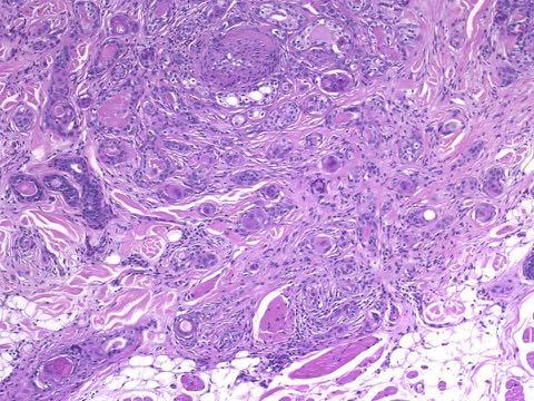
120 Apocrine adenomas Papillary adenoma Tubular adenoma Tubulopapillary adenoma Hidradenoma papilliferum Syringocystadenoma
121 Hidradenoma papilliferum A papillary adenoma of apocrine lineage with a circumscribed but frond-like pattern Occur on genital skin or in the axillary vault, commonly Unrelated to conventional (simple) hidradenoma
122
123
124
125
126 Apocrine adenoma spectrum Tubular adenoma Papillary adenoma Hidradenoma papilliferum Tubulopapillary adenoma Syringocystadenoma papilliferum Periocular apocrine adenoma
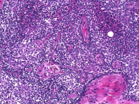
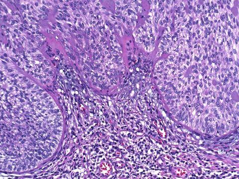
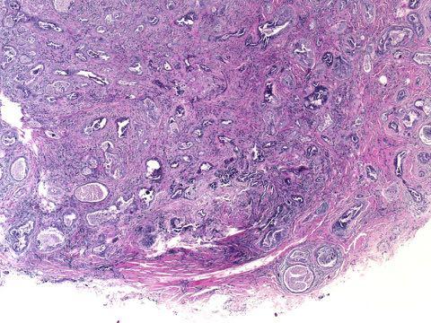
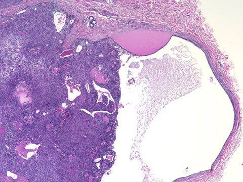
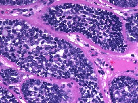
127 Spiradenoma A tumor with primitive glandular and little ductular differentiation Simply spiradenoma (not eccrine spiradenoma)
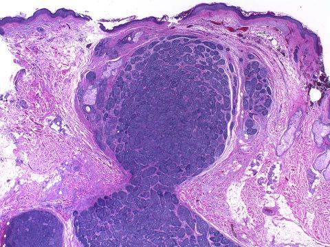
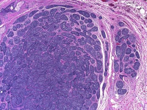
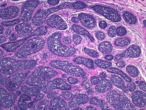
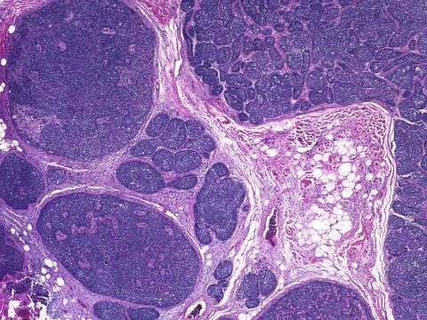
128 Apocrine spiradenoma! Non-volar Coexists with cylindroma Occurs in the breast Coexists with trichepithelioma in Brooke-Spiegler syndrome All suggest folliculosebaceousapocrine lineage
129
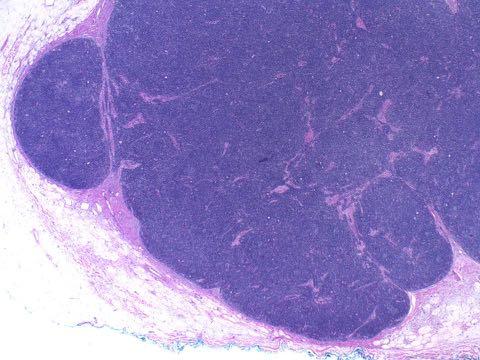
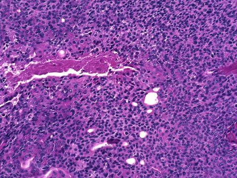
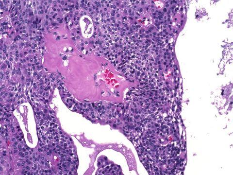
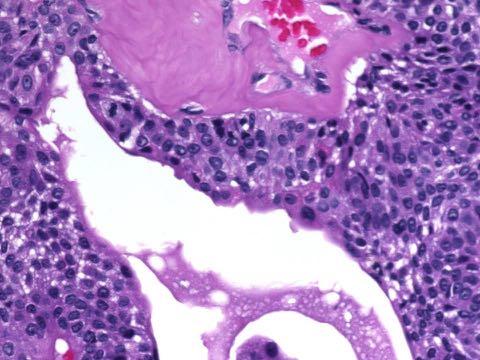
130
131
132
133
134 Spiradenoma Has a child s jigsaw puzzle pattern with only a few pieces arrayed in non-complex fashion A trabecular pattern within nodules, with dark peripheral cells and pale central cells Superimposed lymphocytes
135 Cylindroma Also neoplasm of apocrine lineage with primitive glandular differentiation
 (Weyers et")
136 Multinodular cylindroma ( turban tumor ) (Weyers et al)
137 Cylindroma Has an adult jigsaw puzzle pattern with 500 interlocking pieces arrayed in a complex pattern Often deposition of basement membrane material in small droplets within nests or circumferentially around nests
138
139
140
141
142
143
144
145 Mixed tumor (chondroid syringoma) A hamartoma-like proliferation with tubular and ductular epithelial structures enveloped by equal proportions of mesenchyme Stroma may be cartilaginous ( chrondroid ), myxoid, fibrocytic, adipocytic, or a mix
146 Mixed tumor Most (90%) display prominent apocrine differentiation May observe follicular germinative, outer sheath, isthmic or infundibular, or sebaceous diff. AKA: pleomorphic adenoma
147
148
149
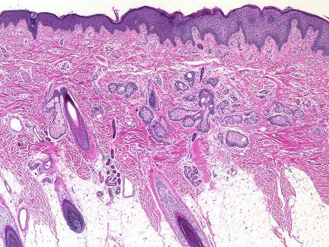
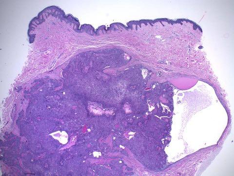
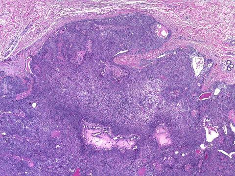
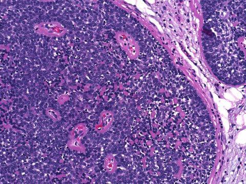
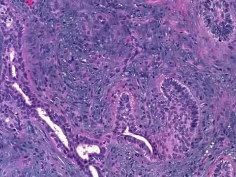
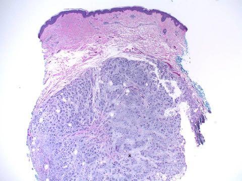
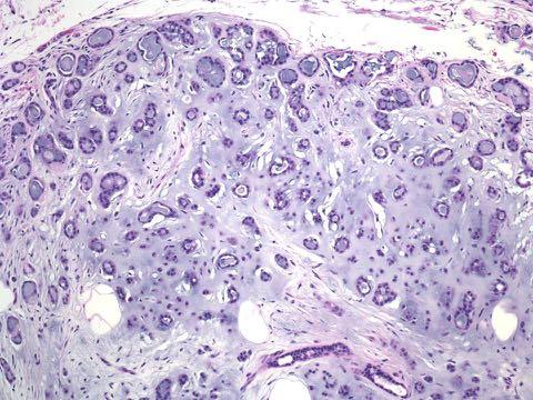
150
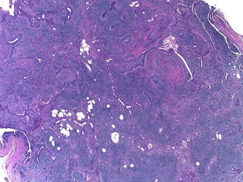
151
152
153 Am J Dermatopathol 1992; 14:186-94: Apocrine type of cutaneous mixed tumor with follicular & sebaceous differentiation. Requena L et al
154 Mixed tumor Mixed tumors may also show strictly ductular differentiation, uncommonly
155
156
157
158 Microcystic adnexal carcinoma (MAC) Rare, but among the most common adnexal carcinomas Presents on the head and neck, most commonly Slow growing, with onset often backdated to 20s or 30s
159 MAC Typified by combined follicular and glandular differentiation Cytologically bland but architecturally malignant and infiltrative Prone to misdiagnosis, especially in superficial biopsies
160
161
162
163
164
165
166
167
168
169
170
171
172
173
174
175 MAC Hallmarks Poor circumscription with deep infiltration, often of nerve or muscle Stromal desmoplasia Follicular differentiation, commonly microcystic (infundibular) Ductular differentiation, commonly syringomatous (Remember that syringomatous carcinoma is half a MAC)
Apocrine and eccrine adnexal tumors
 Apocrine and eccrine adnexal tumors Timothy McCalmont, MD University of California San Francisco, CA ECCRINE TUMORS Generally uncommon, because the sweat apparatus has little proliferative potential (hyperproliferation
Appendageal skin tumors
 Appendageal skin tumors Ibrahim Khalifeh, M.D. Associate Professor Department of Pathology American University of Beirut Medical Center Beirut, Lebanon Appendageal tumors Neoplasms whose differentiation
Simplified approach to cutaneous adnexal tumors
 Simplified approach to cutaneous adnexal tumors Chandra Smart, MD Associate Clinical Professor UCLA Department of Pathology Introduction: Cutaneous adnexal neoplasms (CANs) are a diverse group of tumors
Skin Adnexal Tumors - A Histopathological Spectrum at a Tertiary Care Hospital
 Original Article GCSMC J Med Sci Vol (VI) No (I) January-June 2017 Skin Adnexal Tumors - A Histopathological Spectrum at a Tertiary Care Hospital Neeraja Barve*, Hansa Goswami**, Urvi Parikh *** Abstract
BASAL CELL CARCINOMA WITH ECCRINE DIFFERENTIATION: A RARE ENTITY Divvya B 1, Rehana Tippoo 2, P. Viswanathan 3, B. Krishnaswamy 4, A.
 BASAL CELL CARCINOMA WITH ECCRINE DIFFERENTIATION: A RARE ENTITY Divvya B 1, Rehana Tippoo 2, P. Viswanathan 3, B. Krishnaswamy 4, A. Anvar Ali 5 HOW TO CITE THIS ARTICLE: Divvya B, Rehana Tippoo, P. Viswanathan,
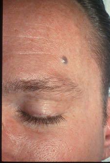
BSD 2015 Case 19. Female 21. Nodule on forehead. The best diagnosis is:
 BSD 2015 Case 19 Female 21. Nodule on forehead. The best diagnosis is: A. mixed tumour of skin B. porocarcinoma C. nodular hidradenoma D. metastatic adenocarcinoma BSD 2015 Case 19 Female 21 Nodule on
What is new on adnexal neoplasms. Omar P. Sangueza, MD. Professor and Director of Dermatopathology. Wake Forest University School of Medicine
 What is new on adnexal neoplasms Omar P. Sangueza, MD Professor and Director of Dermatopathology Wake Forest University School of Medicine Winston Salem, North Carolina Carney complex is an autosomal dominant
- Selected Tumors of the Skin Appendages - Primary vs. Metastasis
 - Selected Tumors of the Skin Appendages - Primary vs. Metastasis Napa Valley 2018 Victor G. Prieto, MD, PhD Chair of Pathology UT MD Anderson Cancer Center vprieto@mdanderson.org Napa Valley in May Introduction
International Journal of Health Sciences and Research ISSN:
 International Journal of Health Sciences and Research www.ijhsr.org ISSN: 2249-9571 Original Research Article A Clinicopathological Study of Adnexal Tumors of Skin in a Tertiary Care Research Hospital
American College of Mohs Surgery. Diagnostic Quality Control Program (Review of Answers)
") American College of Mohs Surgery Diagnostic Quality Control Program 2010 (Review of Answers) Question 1 A flesh-colored plaque on the right ala of an otherwise healthy 57 year-old male is referred for
American College of Mohs Surgery Diagnostic Quality Control Program 2010 (Review of Answers) Question 1 A flesh-colored plaque on the right ala of an otherwise healthy 57 year-old male is referred for
Cluster designation 5 staining of normal and non-lymphoid neoplastic skin*
 J Cutan Pathol 2005: 32: 50 54 Copyright # Blackwell Munksgaard 2005 Blackwell Munksgaard. Printed in Denmark Journal of Cutaneous Pathology Cluster designation 5 staining of normal and non-lymphoid neoplastic
Immunohistology and Molecular Studies of Sweat Gland Tumors
 Immunohistology and Molecular Studies of Sweat Gland Tumors 2 Ana M. Molina-Ruiz, Laura Fuertes, and Luis Requena Introduction Adnexal tumors are generally classified according to two principles: (1) benign
tumors (40 cases) accounting for 64% of the tumors.
 accounting for 64% of the tumors.") Nepal Medical Association Building Exhibition Road, Kathmandu Journal of Pathology of Nepal (2015) Vol. 5, 727-732 Association of Clinical Pathologist of Nepal-2010 Journal of PATHOLOGY of Nepal www.acpnepal.com
Nepal Medical Association Building Exhibition Road, Kathmandu Journal of Pathology of Nepal (2015) Vol. 5, 727-732 Association of Clinical Pathologist of Nepal-2010 Journal of PATHOLOGY of Nepal www.acpnepal.com
10/2/17. A 45-year-old female presents with a lesion on the cheek, rule out basal cell carcinoma. Overview
 10/2/17 Overview Desmoplastic trichoepithelioma vs. morpheaform BCC vs. microcystic adnexal carcinoma Trichilemmomas & Cowden s syndrome Cutaneous Adnexal Tumours with Clinical Impact Rajiv M. Patel, M.D.
Whitney A. High, MD, JD, MEng
 ADS Dermatopathology Meeting 2014 Selected Adnexal Tumors Whitney A. High, MD, JD, MEng Associate Professor, Dermatology & Pathology Director of Dermatopathology (Dermatology) University of Colorado School
3. Histopathology. 1. Introduction. 2. Case History. Volume 6 Issue 4, April Licensed Under Creative Commons Attribution CC BY
 Spiradenocylindroma with Trichoepithelioma A Collision Tumor with Multiple Differentiation R. Lavanya 1, S. K. Sridevi 2, P. Viswanathan 3, P. V. S.Prasad 4 1 II nd Year Post Graduate, Department of Pathology,
Keratinocyte tumors. Actinic Keratosis. Squamous cell carcinoma in situ. Squamous Cell Carcinoma. (aka Bowen s disease)
") Actinic Keratosis Keratinocyte tumors Prepared by Kurt Schaberg Precancerous, risk of malignancy ~8-20% per year (progresses to SCC); Due to chronic sun exposure Rough scaly plaque; typically due to sun
Actinic Keratosis Keratinocyte tumors Prepared by Kurt Schaberg Precancerous, risk of malignancy ~8-20% per year (progresses to SCC); Due to chronic sun exposure Rough scaly plaque; typically due to sun
A clinicopathologic study of skin appendageal tumors
 Net Study A clinicopathologic study of skin appendageal tumors Pradeep S. Nair Department of Dermatology and Venereology, Medical College Hospital, Trivandrum- 695 011, Kerala, India Address for correspondence:
ORIGINAL ARTICLE. Cutaneous Sebaceous Neoplasms With a Focal Glandular Pattern (Seboapocrine Lesions): A Clinicopathological Study of Three Cases
: A Clinicopathological Study of Three Cases") ORIGINAL ARTICLE Cutaneous Sebaceous Neoplasms With a Focal Glandular Pattern (Seboapocrine Lesions): A Clinicopathological Study of Three Cases Dmitry V. Kazakov, MD, PhD,* Eduardo Calonje, MD, Dip RCPath,
ORIGINAL ARTICLE Cutaneous Sebaceous Neoplasms With a Focal Glandular Pattern (Seboapocrine Lesions): A Clinicopathological Study of Three Cases Dmitry V. Kazakov, MD, PhD,* Eduardo Calonje, MD, Dip RCPath,
Microcystic Adnexal Carcinoma of the Nipple
 1, 2 3 1 1 3 2 Microcystic Adnexal Carcinoma of the Nipple Chien-Hui Hong 1, 2 Ming-Feng Hou 3 Gwo-Shing Chen 1 A 36-year-old woman came to our OPD with presentation of a solitary, indurated, nonpainful
A 5 Year Histopathological Study of Skin Adnexal Tumors at a Tertiary Care Hospital
 IOSR Journal of Dental and Medical Sciences (IOSR-JDMS) e-issn: 2279-0853, p-issn: 2279-0861.Volume 14, Issue 4 Ver. VII (Apr. 2015), PP 01-05 www.iosrjournals.org A 5 Year Histopathological Study of Skin
Salivary Glands 3/7/2017
 Salivary Glands 3/7/2017 Goals and objectives Focus on the entities unique to H&N Common board type facts Information for your future practice Salivary Glands Salivary Glands Major gland. Paratid. Submandibular.
The expression of CD23 in cutaneous non-lymphoid neoplasms
 J Cutan Pathol 2007: 34: 693 698 doi: 10.1111/j.1600-0560.2006.00685.x Blackwell Munksgaard. Printed in Singapore Copyright # Blackwell Munksgaard 2006 Journal of Cutaneous Pathology The expression of
Gross appearance of nodular hyperplasia in material obtained from suprapubic prostatectomy. Note the multinodular appearance and the admixture of
 Tiền liệt tuyến Tiền liệt tuyến Gross appearance of nodular hyperplasia in material obtained from suprapubic prostatectomy. Note the multinodular appearance and the admixture of solid and microcystic areas.
Histopathological Study of Skin Adnexal Tumors - A Ten Years Study
 Original Research Article Histopathological Study of Skin Adnexal Tumors - A Ten Years Study V. Srinivas Kumar 1, V. Geeta 1*, Nikhil Kumar Voruganti 2, O. Shravan Kumar 3, Tamilarasi 4 1 Associate Professor
Research Article Histopathological Study of Skin Adnexal Tumours Institutional Study in South India
 Skin Cancer, Article ID 543756, 4 pages http://dx.doi.org/10.1155/2014/543756 Research Article Histopathological Study of Skin Adnexal Tumours Institutional Study in South India Ankit Sharma, 1 Deepak
21/07/2017. Hobnail endothelial cells are not the same as epithelioid endothelial cells
 UPDATE IN CUTANEOUS VASCULAR S DERMATOPATHOLOGY SESSION BELFAST PATHOLOGY JUNE 21/2017 Dr E Calonje St John s Institute of Dermatology, London, United Kingdom THE FAMILY OF VASCULAR S WITH EPITHELIOID
Basal cell carcinoma 5/28/2011
 Goal of this Presentation A practical approach to the diagnosis of cutaneous carcinomas and their mimics Thaddeus Mully, MD University of California San Francisco To review common non-melanoma skin cancers
Original Article: A study of biopsy confirmed skin adnexal tumours: experience at a tertiary care teaching hospital
 Original Article: A study of biopsy confirmed skin adnexal tumours: experience at a tertiary care teaching hospital K. Radhika, B.V. Phaneendra, N. Rukmangadha, M.K. Reddy Department of Pathology, Sri
Chondroid Syringoma. Cytokeratin 20 Immunolocalization of Merkel Cells and Reappraisal of Apocrine Folliculo-Sebaceous Differentiation
 Chondroid Syringoma Cytokeratin 20 Immunolocalization of Merkel Cells and Reappraisal of Apocrine Folliculo-Sebaceous Differentiation Mohamed E. Salama, MD; Muhammad Azam, MD; Chan K. Ma, MD; Adrian Ormsby,
USCAP Neuropathology night panel CASE 2
 USCAP Neuropathology night panel CASE 2 B.K. Kleinschmidt-DeMasters MD University of Colorado at Denver and Health Sciences Center Denver, Colorado The Chinese Wall, Flat Tops Wilderness, Colorado Clinical
Enterprise Interest None
 Enterprise Interest None B3 lesions of the breast What are they at surgery? Case 4 Edi Brogi MD PhD Attending Pathologist - Director of Breast Pathology Memorial Sloan Kettering Cancer Center New York
Journal of International Academy of Forensic Science & Pathology (JIAFP)
") Journal of International Academy of Forensic Science & Pathology (JIAFP) ISSN 2395-0722 MICROCYSTIC ADNEXAL CARCINOMA-A CASE REPORT WITH REVIEW OF LITERATURE Case Report Sulakshana M S 1,Natarajan M 2
Journal of International Academy of Forensic Science & Pathology (JIAFP) ISSN 2395-0722 MICROCYSTIC ADNEXAL CARCINOMA-A CASE REPORT WITH REVIEW OF LITERATURE Case Report Sulakshana M S 1,Natarajan M 2
Histopathological Study of Tumours of Epidermis and Epidermal Appendages
 Original 460 Article Indian Journal of Pathology: Research and Practice Volume 6 Number 2, April - June 2017 (Part 2) DOI: http://dx.doi.org/10.21088/ijprp.2278.148x.6217.22 Histopathological Study of
Cutaneous Adnexal Tumors
 Cutaneous Adnexal Tumors Lesions with Predominant Follicular Differentiation Special Emphasis on Basal Cell Carcinoma 2014-04-01 Prof. Dr. med. Katharina Glatz Pathologie Cutaneous Adnexal Tumors Hair
Brooke-Spiegler syndrome
 CASE REPORT Osama Nour Eldin MD, Dhuha Al-Rqabah MD, ElShahat Farag Ahmed MD, Ahmed Al-Mutairi MD Department of Dermatology, Farwaniya Hospital, Kuwait ABSTRACT Brooke- Spiegler syndrome is an uncommon
Four Different Tumors Arising in a Nevus Sebaceous
 Published online: April 20, 2016 2016 The Author(s) Published by S. Karger AG, Basel 1662 6567/16/0081 0075$39.50/0 This article is licensed under the Creative Commons Attribution-NonCommercial 4.0 International
Diseases of the breast (1 of 2)
") Diseases of the breast (1 of 2) Introduction A histology introduction Normal ducts and lobules of the breast are lined by two layers of cells a layer of luminal cells overlying a second layer of myoepithelial
Diseases of the breast (1 of 2) Introduction A histology introduction Normal ducts and lobules of the breast are lined by two layers of cells a layer of luminal cells overlying a second layer of myoepithelial
Solid Cystic Hidradenoma: A Case Report
 IOSR Journal of Dental and Medical Sciences (IOSR-JDMS) e-issn: 2279-0853, p-issn: 2279-0861.Volume 14, Issue 8 Ver. VII (Aug. 2015), PP 32-36 www.iosrjournals.org Solid Cystic Hidradenoma: A Case Report
Microcystic Squamous Cell Carcinoma of the Lung A Clinicopathologic Study of Three Cases
 Anatomic Pathology / Microcystic SCC of the Lung Microcystic Squamous Cell Carcinoma of the Lung A Clinicopathologic Study of Three Cases Annikka Weissferdt, MD, and Cesar A. Moran, MD Key Words: Squamous
SEBACEOUS NEOPLASMS. Dr. Prachi Saraogi Clinical Fellow in Dermatology
 SEBACEOUS NEOPLASMS Dr. Prachi Saraogi Clinical Fellow in Dermatology Sebaceous neoplasms Sebaceous adenoma (Benign) Sebaceous carcinoma (Malignant) SEBACEOUS ADENOMA Benign tumours composed of incompletely
Papillary Lesions of the Breast A Practical Approach to Diagnosis. (Arch Pathol Lab Med. 2016;140: ; doi: /arpa.
 Papillary Lesions of the Breast A Practical Approach to Diagnosis (Arch Pathol Lab Med. 2016;140:1052 1059; doi: 10.5858/arpa.2016-0219-RA) Papillary lesions of the breast Span the spectrum of benign,
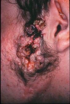
CASE REPORT SOLITARY SEBACEOUS NEVUS OF JADASSOHN COMPLICATED BY SQUAMOUS CELL CARCINOMA AND BASAL CELL CARCINOMA
 CASE REPORT Dennis H. Kraus, MD, Section Editor SOLITARY SEBACEOUS NEVUS OF JADASSOHN COMPLICATED BY SQUAMOUS CELL CARCINOMA AND BASAL CELL CARCINOMA Ahmad Ridzwan Arshad, FRCS, 1 Wan S. Azman, MS, 1 Ayadurai
confusing, especially in small, limited biopsies. One such case of digital papillary adnexal adenocarcinoma with unusual histologic features will be
 1 Cutaneous Adnexal Lesions: The Must-Know Stuff 2011 USCAP Annual Meeting Dermatopathology Companion Meeting Doina Ivan, M.D. University of Texas MD Anderson Medical Center, Houston, TX The diagnosis
Malignant tumors of melanocytes: Part 1. Deba P Sarma, MD., Omaha
 Malignant tumors of melanocytes: Part 1 Deba P Sarma, MD., Omaha The melanocytic tumor is one of the most difficult and confusing areas in Dematopathology. It is true that most (95%) of such lesions are
Enterprise Interest None
 Enterprise Interest None What are triple negative breast cancers? A synopsis of their histological patterns Ian Ellis Molecular Medical Sciences, University of Nottingham Department of Histopathology,
Clinicopathological Study of 1016 Consecutive Adnexal Skin Tumors
 ORIGINAL REPORT Clinicopathological Study of 1016 Consecutive Adnexal Skin Tumors Kambiz Kamyab-Hesari 1, Kamran Balighi 2, Nasim Afshar 2, Nessa Aghazadeh 2, Ziba Rahbar 2, Maryam Seraj 2, and Maede Rayati
1 NORMAL HISTOLOGY AND METAPLASIAS
 1 NORMAL HISTOLOGY AND METAPLASIAS, MD Anatomy and Histology 1 Metaplasias 2 ANATOMY AND HISTOLOGY The female breast is composed of a branching duct system, which begins at the nipple with the major lactiferous
Pitfalls in thyroid tumor pathology. Prof.Valdi Pešutić-Pisac MD, PhD
 Pitfalls in thyroid tumor pathology Prof.Valdi Pešutić-Pisac MD, PhD Too many or... Tumour herniation through a torn capsule simulating capsular invasion fibrous capsule with a sharp discontinuity, suggestive
International Journal of Health Sciences and Research ISSN:
 International Journal of Health Sciences and Research www.ijhsr.org ISSN: 2249-9571 Case Report Clear Cell Hidradenoma of the Ear Lobe and Its Management: A Rare Case Report K. Sampath Kumar Singh 1*,
Synonyms. Nephrogenic metaplasia Mesonephric adenoma
 Nephrogenic Adenoma Synonyms Nephrogenic metaplasia Mesonephric adenoma Definition Benign epithelial lesion of urinary tract with tubular, glandular, papillary growth pattern Most frequently in the urinary
Lesions Mimicking Adenoid Cystic Carcinoma. Diagnostic Problems in Salivary Gland Pathology An Update 5/29/2009
 Diagnostic Problems in Salivary Gland Pathology An Update Lesions Mimicking Adenoid Cystic Carcinoma Stacey E. Mills, M.D. W.S. Royster Professor of Pathology Director of Surgical and Cytopathology University
University Journal of Pre and Para Clinical Sciences
 ISSN 2455 2879 Volume 2 Issue 1 2016 Metaplastic carcinoma breast a rare case report Abstract : Metaplastic carcinoma of the breast is a rare malignancy with two distinct cell lines described as a breast
Spectrum of Preneoplastic and Neoplastic Cystic Lesions of the Kidney in Adult. by dr. Banan Burhan Mohammed Lecturer in Pathology Department
 Spectrum of Preneoplastic and Neoplastic Cystic Lesions of the Kidney in Adult by dr. Banan Burhan Mohammed Lecturer in Pathology Department Various hereditary, acquired, and neoplastic conditions can
DIAGNOSTIC SLIDE SEMINAR: PART 1 RENAL TUMOUR BIOPSY CASES
 DIAGNOSTIC SLIDE SEMINAR: PART 1 RENAL TUMOUR BIOPSY CASES Dr. Andrew J. Evans MD, PhD, FACP, FRCPC Consultant in Genitourinary Pathology University Health Network, Toronto, ON Case 1 43 year-old female,
Histopathology of Skin Adnexal Tumors - A Two Year Retrospective Study at a Tertiary Care Hospital
 Original Article Print ISSN: 2321-6379 Online ISSN: 2321-595X DOI: 10.17354/ijss/2016/616 Histopathology of Skin Adnexal Tumors - A Two Year Retrospective Study at a Tertiary Care Hospital Muktanjalee
Notice of Faculty Disclosure
 California Society of Pathology Diagnostic Problems in Surgical Pathology December 2015 Case 2 Laura C. Collins, M.D. Associate Professor of Pathology Associate Director of Anatomic Pathology Beth Israel
Original Article. Histomorphological Spectrum of Skin Adnexal Tumours : A Retrospective Study in a Tertiary Care Centre
 Original Article Histomorphological Spectrum of Skin Adnexal Tumours : A Retrospective Study in a Tertiary Care Centre G Jeyanthi. 1, Meenakumari Gopalakrishnan 2, N. Sharmila Thilagavathy. 3, S. Shifa
Eccrine Differentiation in Basal Cell Carcinoma
 295S Eccrine Differentiation in Basal Cell Carcinoma Peter J. Heenan and Matthew S. Bogle Eccrine differentiation according to histologic and immuno-histochemical criteria was demonstrated in 16 of 66
Desmoplastic Melanoma R/O BCC. Clinical Information. 74 y.o. man with lesion on left side of neck r/o BCC
 R/O BCC Sabine Kohler, M.D. Professor of Pathology and Dermatology Dermatopathology Service Stanford University School of Medicine Clinical Information 74 y.o. man with lesion on left side of neck r/o
Oncocytic-Appearing Salivary Gland Tumors. Oncocytic, Cystic, Mucinous, and High Grade Salivary Gland Tumors SALIVARY GLAND FNA: PART II
 William C. Faquin, MD, PhD Professor of Pathology Harvard Medical School Director of Head and Neck Pathology Massachusetts Eye and Ear Massachusetts General Hospital SALIVARY GLAND FNA: PART II Oncocytic,
Disclosure. Relevant Financial Relationship(s) None. Off Label Usage None MFMER slide-1
 None. Off Label Usage None MFMER slide-1") Disclosure Relevant Financial Relationship(s) None Off Label Usage None 2013 MFMER slide-1 Case Presentation A 43 year old male, with partial nephrectomy for a right kidney mass 2013 MFMER slide-2 2013
Disclosure Relevant Financial Relationship(s) None Off Label Usage None 2013 MFMER slide-1 Case Presentation A 43 year old male, with partial nephrectomy for a right kidney mass 2013 MFMER slide-2 2013
Introduction. Results. Discussion. Histopathologic and immunohistochemical findings. Results. conclusions,
 1/5 2/5 Carcinoma distinctive carcinoma. form erysipeloides (CE), metastasis. which clinically Itfrom has resembles been termed erysipelas, is an uncommon, but may extend It164 toclassically back, presents
Mammary analogue secretory carcinoma of salivary gland A case report of new entity
 Case Report Mammary analogue secretory carcinoma of salivary gland A case report of new entity Vaibhav Bhika Bari 1*, Sandhya Unmesh Bholay 2 1 Assistant Professor, 2 Associate Professor Rajiv Gandhi Medical
Skin Adnexal Tumors: A Histopathological Study of 60 Cases at a Tertiary Care Centre
 Original Article DOI: 10.21276/APALM.1787 Skin Adnexal Tumors: A Histopathological Study of 60 Cases at a Tertiary Care Centre Alaka Sahu 1, Dilip Kumar Sa 2 *, Salil Kumar Nayak 1 and Kailash Chandra
Development of Six Tumors in a Sebaceus Nevus of Jadassohn: Report of a Case
 The Korean Journal of Pathology 2013; 47: 569-574 CASE STUDY Development of Six Tumors in a Sebaceus Nevus of Jadassohn: Report of a Case Serap Gozel 1 Melahat Donmez 1 Noyan Can Akdur 1 Hulya Yikilkan
Tinh hoàn
 Tinh hoàn Tinh hoàn Tinh hoàn Tiền liệt tuyến Tiền liệt tuyến Mào tinh hoàn Mào tinh hoàn Túi tinh Túi tinh Túi tinh Túi tinh So-called cystadenoma of seminal vesicle. Gross appearance of granulomatous
Puja R. Kathrotiya, MD; Andrew T. Bridge, MD; Simon J. Warren, MBBS; Ha Do, MD; Alison S. Klenk, MD; Lisa Y. Xu, MD; Anubhav N.
 Primary Apocrine Adenocarcinoma of the Axilla Puja R. Kathrotiya, MD; Andrew T. Bridge, MD; Simon J. Warren, MBBS; Ha Do, MD; Alison S. Klenk, MD; Lisa Y. Xu, MD; Anubhav N. Mathur, MD, PhD Practice Points
South East England General Histopathology EQA Scheme
 South East England General Round a Final Case Analyses Cases 635 to 646 Circulated January February 2016 131 responses (89.73%) Prepared April 2016 Authorised by: Prof J Schofield Date: 19/4/2016 With
Apocrine Hidradenocarcinoma of the Scalp: A Classification Conundrum
 Head and Neck Pathol (2009) 3:42 46 DOI 10.1007/s12105-008-0096-8 CASE REPORT Apocrine Hidradenocarcinoma of the Scalp: A Classification Conundrum Marc Cohen Æ David S. Cassarino Æ Hubert B. Shih Æ Elliot
A Cutaneous Myoepithelial Carcinoma Arising in a Papillary Eccrine Adenoma
 The Korean Journal of Pathology 2011; 45: 644-649 http://dx.doi.org/10.4132/koreanjpathol.2011.45.6.644 A Cutaneous Myoepithelial Carcinoma Arising in a Papillary Eccrine Adenoma Ji-Han Jung Soyoung Im
Brief Report. Shivanand Gundalli 1, Smita Kadadavar 1, Somil Singhania 1, Rutuja Kolekar 2 INTRODUCTION. Melanocytic Nevus
 Our Dermatology Online Histopathological spectrum of benign melanocytic nevi our experience in a tertiary care centre Shivanand Gundalli 1, Smita Kadadavar 1, Somil Singhania 1, Rutuja Kolekar 2 1 Department
Diagnostic Cytology of Cancer Cases
 Diagnostic Cytology of Cancer Cases Somporn Techangamsuwan Companion Animal Cancer Research Unit (CAC-RU) Department of Pathology, Faculty of Veterinary Science, Chulalongkorn University 1 Tumor or Non-tumor
Oncocytic carcinoma: A rare malignancy of the parotid gland
 ISPUB.COM The Internet Journal of Pathology Volume 8 Number 2 Oncocytic carcinoma: A rare malignancy of the parotid gland K Mardi, J Sharma Citation K Mardi, J Sharma.. The Internet Journal of Pathology.
Basement membrane in lobule.
 Bahram Memar, MD Basement membrane in lobule. Normal lobule-luteal phase Normal lobule-follicular phase Lactating breast Greater than 95% are adenocarcinomas in situ carcinomas and invasive carcinomas.
CYSTIC TUMORS OF THE KIDNEY JOHN N. EBLE, M.D. CYSTIC NEPHROMA
 Page 1 CYSTIC TUMORS OF THE KIDNEY JOHN N. EBLE, M.D. Department of Pathology & Laboratory Medicine Phone (317) 274-4806 Medical Science A-128 FAX: (317) 278-2018 635 Barnhill Drive jeble @iupui.edu Indianapolis,
04/10/2018. Intraductal Papillary Neoplasms Of Breast INTRADUCTAL PAPILLOMA
 Intraductal Papillary Neoplasms Of Breast Savitri Krishnamurthy MD Professor of Pathology Deputy Division Head The University of Texas MD Anderson Cancer Center 25 th Annual Seminar in Pathology Pittsburgh,
Glycogen-rich adenocarcinoma in the lower lip: report of a case with particular emphasis on differential diagnosis
 Open Journal of Stomatology, 2011, 1, 109-113 doi:10.4236/ojst.2011.13017 Published Online September 2011 (http://www.scirp.org/journal//). Glycogen-rich adenocarcinoma in the lower lip: report of a case
MALIGNANT POROMA SYNONYM: POROCARCINOMA ECCRINE POROMA MALIGNANT Divvya B 1, M. Valluvan 2, Rehana Tippoo 3, P. Viswanathan 4, R.
 MALIGNANT POROMA SYNONYM: POROCARCINOMA ECCRINE POROMA MALIGNANT Divvya B 1, M. Valluvan 2, Rehana Tippoo 3, P. Viswanathan 4, R. Ramesh 5 HOW TO CITE THIS ARTICLE: Divvya B, M. Valluvan, Rehana Tippoo,
Malignant cylindroma in a patient with Brooke-Spiegler syndrome
 DERMATOLOGY PRACTICAL & CONCEPTUAL www.derm101.com Malignant cylindroma in a patient with Brooke-Spiegler syndrome Liliane Borik 1,2, Patricia Heller 2, Monica Shrivastava 3, Viktoryia Kazlouskaya 2 1
Salivary Gland Cytology
 Salivary Gland Cytology Diagnostic challenges and potential pitfalls Tarik M. Elsheikh, MD Professor and Medical Director Anatomic Pathology Cleveland Clinic FNA Salivary Gland Lesions Indications Distinguish
Case Report Endocrine Mucin-Producing Sweat Gland Carcinoma, a Histological Challenge
 Hindawi Volume 2017, Article ID 6343709, 4 pages https://doi.org/10.1155/2017/6343709 Case Report Endocrine Mucin-Producing Sweat Gland Carcinoma, a Histological Challenge Mary Anne Brett, Samih Salama,
Note: The cause of testicular neoplasms remains unknown
 - In the 15- to 34-year-old age group, they are the most common tumors of men. - Tumors of the testis are a heterogeneous group of neoplasms that include: I. Germ cell tumors : 95%; all are malignant.
Review of the AP Part II Practical Examination. Dr David Clift Co Chief Examiner
 Review of the AP Part II Practical Examination Dr David Clift Co Chief Examiner General Remarks The part II practical examination involved 15 cases which were presented with sufficient clinical data to
Salivary gland Workshop Trondheim 31th may 2012
 Salivary gland Workshop Trondheim 31th may 2012 Peter Jebsen cytopathologist Oslo University Hospital Rikshospitalet Anna Bofin ass. Professor St. Olavs Hospital, Trondheim Drying artifacts Lymfocytes
Salivary gland tumor cytologic and histologic correlation: Algorithmic and risk stratification based approaches
 Salivary gland tumor cytologic and histologic correlation: Algorithmic and risk stratification based approaches Christopher C. Griffith, MD, PhD Raja R. Seethala, MD 1. Salivary gland tumor cytology: A
SESSION 1: GENERAL (BASIC) PATHOLOGY CONCEPTS Thursday, October 16, :30am - 11:30am FACULTY COPY
 PATHOLOGY CONCEPTS Thursday, October 16, :30am - 11:30am FACULTY COPY") SESSION 1: GENERAL (BASIC) PATHOLOGY CONCEPTS Thursday, October 16, 2008 9:30am - 11:30am FACULTY COPY GOAL: Describe the basic morphologic (structural) changes which occur in various pathologic conditions.
SESSION 1: GENERAL (BASIC) PATHOLOGY CONCEPTS Thursday, October 16, 2008 9:30am - 11:30am FACULTY COPY GOAL: Describe the basic morphologic (structural) changes which occur in various pathologic conditions.
Pleomorphic adenoma of breast - a case report and distinction with metaplastic carcinoma D Gupta, S Agrawal, N Trivedi, A Tewari
 of breast - a case report and distinction with metaplastic carcinoma D Gupta, S Agrawal, N Trivedi, A Tewari Introduction, also known as mixed tumour, is a benign tumour which typically presents as a painless,
Diagnostically Challenging Cases in Gynecologic Pathology
 Diagnostically Challenging Cases in Gynecologic Pathology Eric C. Huang, M.D., Ph.D. Department of Pathology and Laboratory Medicine University of California, Davis Medical Center Case 1 Presentation 38
Slide seminar. Asist. Prof. Jože Pižem, MD, PhD Institute of Pathology Medical Faculty, University of Ljubljana
 Slide seminar Asist. Prof. Jože Pižem, MD, PhD Institute of Pathology Medical Faculty, University of Ljubljana Case 5 A 57-year-old man with a dermal/subcutaneous lesion on the scalp, which was interpreted
Disclosure of Relevant Financial Relationships
 Squamous entities of the thyroid: Reactive to Neoplastic Michelle D. Williams Associate Professor Dept of Pathology, Head & Neck Section University of Texas MD Anderson Cancer Center Disclosure of Relevant
FORELIMB SWEAT GLAND ADENOCARCINOMA IN A CAT
 I: 2047-2051 ISSN: 2277 4998 FORELIMB SWEAT GLAND ADENOCARCINOMA IN A CAT ABEDI G 1, HESARAKI S 2, ASGHARI A 1* 1: Department of Clinical Science, Science and Research branch, Islamic Azad University,
NODULAR CYSTIC HIDRADENOMA OVER THE GLUTEAL REGION: A RARE CYTOMORPHOLOGICAL DIAGNOSIS
 NODULAR CYSTIC HIDRADENOMA OVER THE GLUTEAL REGION: A RARE CYTOMORPHOLOGICAL DIAGNOSIS Abstract: The primary as well as metastatic tumours of the skin can be diagnosed by fine needle aspiration cytology
PLEOMORPHIC ADENOMA ( BENIGN MIXED TUMOR )
") ( BENIGN MIXED TUMOR ) Grossly, the tumor is freely movable, solid, sometimes lobulated and occasionally cystic. If recurrent, multinodular masses are common. Histologically, within a fibrous capsule,
( BENIGN MIXED TUMOR ) Grossly, the tumor is freely movable, solid, sometimes lobulated and occasionally cystic. If recurrent, multinodular masses are common. Histologically, within a fibrous capsule,
1/10/2018. Soft Tissue Tumors Showing Melanocytic Differentiation. Overview. Desmoplastic/ Spindle Cell Melanoma
 2016 MFMER slide-1 2016 MFMER slide-2 2016 MFMER slide-3 Soft Tissue Tumors Showing Melanocytic Differentiation Andrew L. Folpe, M.D. Professor of Laboratory Medicine and Pathology Mayo Clinic, Rochester,
SOFT TISSUE TUMOR PATHOLOGY: AN UPDATE
 SOFT TISSUE TUMOR PATHOLOGY: AN UPDATE Jason L. Hornick, MD, PhD July 18, 2013 Department of Pathology Brigham and Women s Hospital Harvard Medical School Boston, MA, USA I have no disclosures. New Soft
Normal thyroid tissue
 Thyroid Pathology Overview Normal thyroid tissue Normal thyroid tissue with follicles filled with colloid. Thyroid cells form follicles, spheres of epithelial cells (always single layered in health, usually
Macro- and microacinar proliferations of the prostate
 Macro- and microacinar proliferations of the prostate (with emphasis on cancer mimics) Rodolfo Montironi, MD (IT), FRCPath (UK), IFCAP (USA) Polytechnic University of Marche Region (Ancona) School of Medicine,
Brooke-Spiegler Syndrome
 Brooke-Spiegler Syndrome A Case Report Ching-Hao Chang Hsin-Chun Ho Hong-Shang Hong Brooke-Spiegler syndrome is a rare autosomal dominantly inherited disease characterized by the development of multiple
ARIZONA SOCIETY OF PATHOLOGISTS 13 TH APRIL 2013 HEAD AND NECK CYTOPATHOLOGY. F ZAHRA ALY, MD, PhD
 ARIZONA SOCIETY OF PATHOLOGISTS 13 TH APRIL 2013 HEAD AND NECK CYTOPATHOLOGY F ZAHRA ALY, MD, PhD The main areas sites amenable for cytopathology include lymph nodes, thyroid, major salivary glands especially