Table of Contents. Introduction 3. Background 4
|
|
|
- Bethanie Stewart
- 6 years ago
- Views:
Transcription

1 Training manual
2 Table of Contents Introduction 3 Background 4 What are X-rays? 4 How are X-rays Generated? 5 Primary and Scatter Radiation 6 Interactions with Matter 6 Biological Effects of Radiation 7 Linear No-Threshold Risk Model 8 Basic X-Ray Safety 9 Safety Rules to Minimize Radiation Dose 9 Worker Radiation Dose Limits 9 Medical Procedure Doses 10 MaxRay Safety 11 Backscatter Shield 11 Geometry of the Backscatter Zone 12 Dosimetry 13 Accidental Exposure Prevention 13 Exposure Time 15 Safe Storage 16
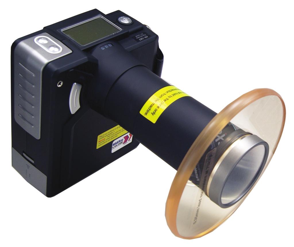
3 Introduction There are many beneficial uses of ionizing radiation; however, of equal importance we note that there are potential risks associated with its use. Radiation safety training is an important part of any radiation safety program. Receiving appropriate training ensures users are following proper safety practices to maximize the benefits of ionizing radiation while minimizing potential risks and maintaining a safe work environment. In this training manual, we discuss basic X-ray safety in addition to specific safety information about operating the MaxRay DX The MaxRay is a small, lightweight (4.4 lbs.) handheld X-ray system meant for dental radiology that is certified by the FDA and is completely safe when used as intended. All operators must read, and become familiar with, the User s Manual associated with the MaxRay system. The MaxRay DX-3000 mobile X-ray system. This handheld unit is to be operated only by authorized personnel. DO NOT operate the Max- Ray in any manner other than that specified herein, and in the User s Manual. And, DO NOT allow anyone other than trained and approved personnel to operate the MaxRay unit. 3

4 Background What are X-rays? X-rays are a form of ionizing radiation and are a part of the electromagnetic spectrum. X-rays are the same as the light from the sun, except that their energy is much higher. As X-rays travel through and interact with various materials, human tissue for instance, they transfer energy to the atoms of that material. This process of energy transfer can result in atomic ionization. X-rays can penetrate certain materials, but they can be blocked or shielded with high-density materials. The electromagnetic spectrum. When living systems are exposed to ionizing radiation there is a risk for biological damage to occur. Exposure to X-rays in the workplace, however, is highly regulated and current safety standards are very effective at keeping risks to a minimum. 4
5 How are X-rays Generated? X-rays are produced in a type of vacuum tube specifically designed for that function. As power is applied to the tube, X-rays are emitted in a prescribed fashion from a shielded housing. The MaxRay uses a Toshiba D-081B X-ray tube. Diagram of an X-ray tube. Generally, the three parameters that can be adjusted by the X-ray technician (tube potential (kvp), tube current (ma), and time (sec)) establish the characteristics of the X-ray beam emanating from the tube. The tube potential determines the energy range of X-rays and the tube current establishes the rate at which X-rays are emitted. In the tube, X-rays are produced by two means, Bremsstrahlung radiation and characteristic radiation. The two are described below. Bremsstrahlung Radiation: this is the main type of radiation produced. It occurs as the high energy electrons experience a sudden slowing down, or breaking, at the anode target. A spectrum of photon energies is produced. Bremsstrahlung is also known as breaking radiation. 5
6 Characteristic Radiation: this type of radiation is produced when an electron interacts with an inner shell electron of a target atom of the anode. As the inner shell electron is displaced, an electron from an outer shell drops to fill the vacancy. It is this process that releases characteristic X-rays. All X-ray tubes have some form of filtration, whether it be inherent to the design or added afterward to adjust the usefulness of the X-ray beam. The X-ray housing will have additional shielding to minimize leakage radiation that can cause unwanted exposure to the operator. Primary and Scatter Radiation Once X-rays leave the tube housing, they are categorized as primary or secondary radiation. Secondary radiation is further characterized into scatter radiation and leakage radiation. Primary radiation describes the useful beam of radiation that is produced in the tube and exits the filtration window as designed. This is the radiation which is fundamental in producing the radiograph. Continued exposure to the primary beam can result in a significant hazard. Scatter radiation refers to the radiation that is scattered after the primary beam interacts with the patient. The patient is therefore the major source of scatter radiation. Even though the primary beam is much more intense than scatter radiation, it is this scatter that is of primary concern when protecting the safety of the operator. As stated above, leakage radiation refers to radiation from the X-ray tube that penetrates the device housing. Leakage is usually quite small relative to the primary beam and scatter. Interactions with Matter The interaction of X-rays with matter is a random process. As tissue is exposed, the X-rays may interact with the atoms of the material through which they pass. A small percentage of the X-rays will pass through matter without interacting. Those X-rays that interact will do so by one of two methods, photoelectric absorption or Compton scatter. 6
7 Photoelectric absorption: In photoelectric absorption, the incident X-ray energy is completely absorbed in the interaction medium (e.g., tissue) and the X-ray is removed from the beam and does not have the ability to scatter. Compton Scatter: With Compton scatter, the incident X-ray scatters in the interaction medium and only a partial amount of original energy is absorbed. The remaining energy goes to the scattered X-ray. This scattered energy is therefore available to be absorbed elsewhere, for example, in the operator. During the process of photoelectric absorption or Compton scatter, energy is transferred to the interaction medium. We quantify the effect of this energy absorption using a parameter called absorbed dose, i.e., the amount of energy absorbed for a given mass of absorbing medium. Someone s risk from radiation exposure is directly proportional to the dose they receive. The regulatory agencies set limits on absorbed dose for workers and the general public to ensure that radiation risk is kept as low as practical. Biological Effects of Radiation While X-rays are a significant part of the diagnostic process, it is important to be aware that there is a potential for biological damage to occur when exposed to ionizing radiation. Efforts should be made to evaluate the benefit and potential risk in order to avoid unnecessary radiation exposure. The benefits of medical/dental evaluation using X-ray technology are obvious, but the risk of biological effects of ionizing radiation must be weighed against the benefits. These effects are commonly grouped into two categories: Non-stochastic Effects (deterministic effects) Non-stochastic effects are non-random and are directly related to the radiation dose received. For these effects to occur, a threshold dose must be met. Once the threshold has been exceeded, the severity of biological damage (e.g., skin burns, hair loss, reddening of the skin, cataracts) increases with the dose received. These effects are seen only after exposure to large doses of radiation (> 1,000 msv), much larger than doses received when undergoing dental imaging. Stochastic Effects (probabilistic effects) Stochastic effects are randomly occurring and the severity of biological damage (e.g., cancer, birth defects) is independent of the dose received. Since it is based on probability, the 7
8 chance of occurrence increases with radiation dose. Stochastic effects are of typical concern when speaking of exposure to diagnostic X-rays; radiation dose is very small, therefore the only real potential outcome is the very small chance of cancer. Linear No-Threshold Risk Model Because the random chance of cancer is so small, science must use a small set of existing data to predict cancer probability. Currently, the prediction is based on what s called a linear, no-threshold model and is intended to convey that cancer risk is thought to be proportional (linear) to dose, with zero dose resulting in zero risk (no-threshold). This model is conservative, and follows the philosophy that it is better that risk be overestimated rather than underestimated. Linear No-Threshold Model Biological Effects of Radiation Exposure Radiation Dose 8
9 Basic X-ray Safety Safety Rules to Minimize Radiation Dose ALARA. The safety principle of ALARA (As Low As Reasonably Achievable) ensures that radiation exposure levels are kept as low as practical, far below the exposure limits set by the regulatory agencies. It is a regulatory requirement and it is mandated that all radiation safety programs follow the ALARA principle. In order to maintain ALARA, it is important to remember and practice the radiation protection triad of time, distance, and shielding: Time: minimize exposure time; Distance: maximize distance (between you and the source); and, Shielding: use appropriate radiation shielding. The most effective shielding for X-rays is lead (e.g. high density). Patients should be shielded to protect their thyroid and reproductive organs, and the X-ray operator should wear a leaded apron. Some of the handheld X-ray systems come equipped with a leaded-plastic backscatter shield which is very effective. With this shield, leaded aprons may not be required by your regulator, but it s always a safe bet to wear the apron anyway. Pregnancy. Because the fetus is undergoing rapid cell reproduction, it is important to reduce radiation exposure during pregnancy. As the X-ray operator, if you are, or become, pregnant, you should notify your employer immediately. It is your responsibility to declare your pregnancy. For the safety of your patients, you should question the patient regarding the possibility of them being pregnant. If the patient is, or may be, pregnant, they should be advised by your radiation safety officer prior to exposure. Worker Radiation Dose Limits Occupational dose limits are set by regulatory agencies to limit cancer risk as well as the other potential biological effects of radiation. Annual occupational dose limits, as established in U.S. federal law (10 CFR 20) are provided below, however some locally established dose limits may be more protective. Check with your local regulator for dose limits that apply specifically to you. 9
10 Annual Occupation Dose Limits Type of Limit Occupational Dose Limit Total effective dose equivalent 50 msv Lens of the eye 150 msv Skin 500 msv Hands and feet 500 msv Embryo/fetus 5 msv (over the length of pregnancy) **From 10CFR and 10CFR Medical Procedure Doses Dental imaging procedures contribute to a much lower patient dose than other imaging studies. The table below presents typical patient doses associated with various medical imaging procedures. Procedure Dose (msv) X-ray (single exposure) Hand/Foot Dental Chest 0.10 Abdomen 0.60 Pelvis 0.70 Mammogram (2 views) 0.72 CT (multiple exposures) Head 2 Chest 7 Full Body 10 **Data source: NRC 10

11 MaxRay Safety Backscatter Shield The MaxRay DX-3000 has a circular, lead infused plastic disc (0.35 mm lead-equivalent) surrounding the X-ray beam emission port. The purpose of this backscatter shield is to attenuate radiation scattered from the patient. The backscatter shield should never be removed, as the shield is very effective at reducing radiation scatter in the direction of the operator. As seen by the figure, the shield, in relation to the patient s head, provides a safety zone in which the operator should remain during exposures. The backscatter shield provides a safety zone to the left of the green line. (Editor s Note: The photograph was taken in the studio with the Maxray DX-3000 s battery removed. In the clinical setting, the patient would be wearing a leaded cover and thyroid shield.) 11

12 Geometry of the Backscatter Zone When producing an X-ray image, the operator should stand directly behind the unit, holding it in the manner shown below. In order to maximize the backscatter zone, the emission port should be perpendicular to the area being radiographed, i.e., the backscatter shield should be parallel to the operator. Proper parallel orientation. If the backscatter shield is not parallel to the operator, the backscatter zone angel changes, limiting the zone coverage. Additionally, the emission port should be held close to the patient to maximize the effectiveness of the backscatter shield. When taking a difficult image, the operator should first attempt to move the patient s head, while maintaining proper alignment. This allows for the operator to stay within the backscatter zone and maintain operator safety. 12
, their annual dose to the whole body is less than 0.")
13 Dosimetry The MaxRay has been shown to be a very safe handheld X-ray system when used as intended. The occupational dose from leakage radiation at 1 cm from the case is less than 0.05 msv to the fingers for an entire work year. And, as long as the operator remains within the safety zone (provided by the backscatter shield), their annual dose to the whole body is less than 0.20 msv. These dose estimates assume that the operator makes 15,000 to 18,000 dental X-rays each year; the unit is very safe. For reference, the regulatory annual dose limits are 500 msv to the fingers and 50 msv to the whole body. Accidental Exposure Prevention Accidental exposures are easy to prevent if the operator remains aware of the direction in which the emission port is pointing and the on/off status of the MaxRay. As a general rule, whether on or off, the operator should NEVER point the MaxRay emission port at anyone, except the area of the patient about to be radiographed. Exposure occurs only when the activation button is pressed. The operator should remain vigilant and keep their finger off the activation button until ready for the intended exposure. The activation button. 13

14 As the operator, it is important to be aware of your surroundings in order to maintain ALARA. Always ensure that you are within the backscatter zone, and that all unnecessary persons are out of the room prior to initiating an exposure. When taking an image, the current status of the unit on the LCD display window will change from READY to EXPOSURE. The operator will hear a steady tone during the exposure; this sound will end when the selected time has passed. As a safety feature, the exposure will stop when the activation button is released, even if the selected time cycle is not complete. The LCD display window. 14

15 NEVER attempt to operate the MaxRay handheld X-ray system if any covers, shielding material, collimators, etc. have been removed. As the operator, NEVER place any part of your body in the primary beam. Exposure Time The area within the yellow lines represents the primary beam. There is only one variable of exposure on the MaxRay unit that can be changed for a given radiograph exposure time. The MaxRay has exposure factors of ma and kvp that are fixed and cannot be changed by the operator. The exposure time is changed based on patient age (adult or child), image receptor (film or digital), and the location of teeth being imaged. The operator makes selections of age, receptor, and postion on the MaxRay, and in turn the MaxRay provides a suggested range (3 options) of exposure time in increments of 0.05 seconds. For example, a selection of adult, digital, molar results in time options of 0.95, 1.00, and 1.05 seconds. Generally adults, film, and larger teeth require longer exposure times. See the User s Manual for more details on selecting the exposure time. There is a direct correlation between exposure time and dose. If exposure time is increased, 15

16 patient dose increases. There is certainly a trade-off between image quality and patient dose. It is important to practice ALARA by keeping dose as low as possible while maintaining adequate image quality for diagnosis. Exposure to the operator and the patient should be limited and having to repeat images should be avoided. Safe Storage Because the handheld X-ray system is portable, certain safety precautions must be implemented to ensure worker and patient safety. The MaxRay has a removable battery pack that, when removed, renders the unit totally safe in that it will NOT emit X-rays. For a safe work environment, when not in use, the MaxRay should be stored in a locked cabinet so that the device is accessible only to authorized personnel. If stored for an extended period of time (over night or over the weekend), remove the battery pack and store it in a locked drawer or cabinet, physically separated from the X-ray unit. Remove battery when storing for an extended period. 16
17
Code of Practice for Radiation Protection in Dentistry. Code of Practice For Radiation Protection in Dentistry
 Code of Practice for Radiation Protection in Dentistry Code of Practice For Radiation Protection in Dentistry 10 OCTOBER 2017 CONTENTS 1. INTRODUCTION... 3 1.0 CITATION... 3 1.1 BACKGROUND... 3 1.2 PURPOSE
Radiation Safety Manual
 King Abdulaziz University Faculty of Dentistry Radiation Safety Manual FOR X-RAY EQUIPMENT OPERATORS October 2009 Radioactivity and Radiation All matter in our environment is made of atoms. Most atoms
Basic radiation protection & radiobiology
 Basic radiation protection & radiobiology By Dr. Mohsen Dashti Patient care & management 202 Wednesday, October 13, 2010 Ionizing radiation. Discussion issues Protecting the patient. Protecting the radiographer.
Radiation Safety for New Medical Physics Graduate Students
 Radiation Safety for New Medical Physics Graduate Students John Vetter, PhD Medical Physics Department UW School of Medicine & Public Health Background and Purpose of This Training This is intended as
RADIATION SAFETY. Junior Radiology Course
 RADIATION SAFETY Junior Radiology Course Expectations for the Junior Radiology Course Medical School wants students to learn basic principles, factual knowledge, safety info, etc. Medical Students want
Utilize radiation safety principles to reduce the amount of radiation used to achieve desired clinical result.
 Minimizing Dose Understand the importance and methods of pre-procedure patient assessment including a review of previous radiologic exams, disease processes and anatomical considerations that may increase
UQ X-ray Safety Training Module
 UQ X-ray Safety Training Module 23 January 2018, v2 1 UQ X-ray Safety Training Module Course Overview: This training module has been developed for workers at the University of Queensland, and forms part
Multiple Choice Identify the letter of the choice that best completes the statement or answers the question.
 RA202 Rad protection class two True/False Indicate whether the sentence or statement is true or false. 1. Secondary radiation comes from scatter and leakage. 2. Grids are considered a protection device.
The x-rays produced penetrate the body which absorbs, refracts, or reflects the x-ray beam energy depending on the tissue. Bone
 Authors Sari Cohen, Poh Yan Lim, Merng Koon Wong, Siew Hong Lau, Donna Russell-Larson 1.6.2 Image intensifier Poh Yan Lim, Merng Koon Wong The discovery of x-rays had a profound impact on the diagnosis
Radiation Safety & Determining Need for Radiographs
 Radiation Safety & Determining Need for Radiographs Guidelines for Radiographic Examination All radiation is harmful! These guidelines have been established to protect the patient and operator from unnecessary
Radiation Safety For Anesthesiologists. R2 Pinyada Pisutchareonpong R2 Nawaporn Sateantantikul Supervised by Aj Chaowanan Khamtuicrua
 Radiation Safety For Anesthesiologists R2 Pinyada Pisutchareonpong R2 Nawaporn Sateantantikul Supervised by Aj Chaowanan Khamtuicrua Modern World Non Ionizing VS Ionizing Non Ionizing Harmless Ex. visible
Radiation Protection- Cath lab
 Radiation Protection- Cath lab Dr. Mawya A Khafaji Associate Prof. Medical Physics, Faculty of Medicine, KAU Head of Medical Physics Unit Dept. of Radiology -KAUH Head, Volunteer Office -KAUH Outline:
RELIANT HOLDINGS LTD AND ITS AFFILIATES Safety Management System. Preparation: Safety Mgr Authority: CEO Issuing Dept: Safety Page: Page 1 of 5
 Preparation: Safety Mgr Authority: CEO Issuing Dept: Safety Page: Page 1 of 5 Purpose The purpose of this program is to protect employees who may encounter ionizing radiation and its hazards while performing
in developing institutional policies, procedures, and /or protocols. The Canadian Society of
 1 TITLE: GUIDELINES FOR RADIATION SAFETY APPROVED; October 2015 REVISION DATE: February 2016 Disclaimer The Canadian Society of Gastroenterology Nurses and Associates present this guideline for use in
Radiation Safety Characteristics of the NOMAD Portable X-ray System
 Radiation Safety Characteristics of the NOMAD Portable X-ray System D. Clark Turner 1, Donald K. Kloos 1, Robert Morton 2 1 ARIBA X-Ray, Inc., 754 South 400 East, Orem, UT 84097 USA, www.aribaxray.com
Radiation Safety Bone Densitometer
 Radiation Safety Bone Densitometer Outline I. State Regulations II. Fundamentals of Radiation Safety III. IV. i. Characteristics of x-ray radiation ii. Units of radiation dose iii. Biological effects iv.
created by high-voltage devices Examples include medical and dental x-rays, light, microwaves and nuclear energy
 What is radiation? Radiation is energy emitted from a source, that travels through space and can penetrate matter. Listed below are two types that we are exposed to and contribute to our overall radiation
Radiation Safety Guide. Analytical X-Ray Equipment
 Radiation Safety Guide Analytical X-Ray Equipment Table of Content Page 1. Radiation 2 A. Radiation Quantities 2 B. Background Radiation 2 C. Biological Effect of Radiation 3 D. Radiation Injury To The
Introduction. Chapter 15 Radiation Protection. Advisory bodies. Regulatory bodies. Main Principles of Radiation Protection
 Introduction Chapter 15 Radiation Protection Radiation Dosimetry I Text: H.E Johns and J.R. Cunningham, The physics of radiology, 4 th ed. F.M. Khan, The Physics of Radiation Therapy, 4th ed., Chapter
Dental Extraoral X-ray Systems
 Dental Extraoral X-ray Systems PROPOSED REVISIONS TO 4732.XXXX, 1.0 4732.#### DENTAL EXTRAORAL X-RAY SYSTEMS; STATIONARY AND MOBILE. Subpart 1. Applicability. A registrant s x-ray system used for dental
Dental Intraoral X-ray Systems
 Dental Intraoral X-ray Systems PROPOSED REVISIONS TO 4732.XXXX, 2.0 4732.#### DENTAL INTRAORAL X-RAY SYSTEMS; STATIONARY AND MOBILE. Commented [JC(1]: Based on part 4732.0880. Subpart 1. Applicability.
Mapping the ASRT Objectives for Radiation Protection (47 Objectives) and Radiation Biology (21 Objectives) to this Text
 and Radiation Biology (21 Objectives) to this Text") Appendix B Mapping the ASRT Objectives for Radiation Protection (47 Objectives) and Radiation Biology (21 Objectives) to this Text 1. Identify and justify the need to minimize unnecessary radiation exposure
Appendix B Mapping the ASRT Objectives for Radiation Protection (47 Objectives) and Radiation Biology (21 Objectives) to this Text 1. Identify and justify the need to minimize unnecessary radiation exposure
Radiation Safety Characteristics of the NOMAD Portable X-ray System
 Radiation Safety Characteristics of the NOMAD Portable X-ray System D. Clark Turner1, Donald K. Kloos1, Robert Morton2 1Aribex, Inc., 754 South 400 East, Orem, UT 84097 USA, www.aribex.com 2Quality and
Radiation Units and Dosimetry 15 August Kalpana M. Kanal, Ph.D., DABR 1
 Introduction Radiation Units and Dosimetry Radiation dose quantities are used as indicators of the risk of biologic damage to patients from x-rays and thus a good knowledge of the different dose parameters
Radiation Dose Specification
 Chapter 9 Dose Limits for Exposure to Ionizing Radiation Dose Limits for exposure to Ionizing Radiation apply to: Occupational workers Nonoccupational workers Radiation Dose Specification Equivalent Dose
Radiation Safety in the Catheterization Lab
 SCAI FALL FELLOWS COURSE - 2015 Radiation Safety in the Catheterization Lab V. Vivian Dimas, MD, FSCAI Associate Professor Pediatrics, Cardiology UT Southwestern Medical Center Dallas TX None Disclosures
Why radiation protection matters?
 Why radiation protection matters? Elias Brountzos Professor of Radiology 2 nd Department of Radiology Medical School, University of Athens Athens, Greece A definition for radiation protection Radiation
Chem 481 Lecture Material 3/11/09
 Chem 481 Lecture Material 3/11/09 Health Physics NRC Dose Limits The NRC has established the following annual dose limits. Organ NRC Limit (mrem/year) Comments Whole Body 5000 (50 msv/yr) Lens of the Eye
Module Rhodes
 Module 6 10-526-197 Rhodes Health Physicist Concerned with providing occupation radiation protection and minimizing radiation dose to the public. Diagnostic Imaging has changed our world Live longer Work
PERSONNEL MONITORING AND DOSIMETRY POLICIES
 PERSONNEL MONITORING AND DOSIMETRY POLICIES All individuals who are required to have their exposure to ionizing radiation monitored must be trained prior to using the source(s) of radiation. The radioactive
2017 Course of the Nordic Association for Clinical Physics on occupational dosimetry in hospitals
 2017 Course of the Nordic Association for Clinical Physics on occupational dosimetry in hospitals 27 September 2017 Dr. Pedro Ortiz López Retired member of the ICRP and the IAEA 2 Pedro Ortiz López (Chair)
The College of Dental Surgeons of Saskatchewan Radiation and Imaging Standard
 The College of Dental Surgeons of Saskatchewan Radiation and Imaging Standard Legislation Radiation safety has long been a priority in Saskatchewan. This province, the first in Canada to have radiation
RADIATION SAFETY. for Surgical & Invasive Procedures
 RADIATION SAFETY for Surgical & Invasive Procedures TABLE OF CONTENTS Purpose 3 Learning Objectives 3 Instructions 3 Introduction 4 Radioactivity and Radiation 5 Types of Radiation. 6 Primary... 6 Secondary..
CT Radiation Risks and Dose Reduction
 CT Radiation Risks and Dose Reduction Walter L. Robinson, M.S. D.A.B.S.N.M., D.A.B.M.P., D.A.B.R. Consultant Certified Medical Radiation Health & Diagnostic Imaging Physicist Medical Radiation and Children
Krueger-Gilbert Health Physics, Inc.
 Krueger-Gilbert Health Physics, Inc. 1 Educational Objectives Radiation bioeffects Sources of radiation for the US population Typical radiation doses in diagnostic imaging Maryland, FDA and JCAHO guidelines
Radiation Safety Training Module: Diagnostic Radiology Radiation Protection in Diagnostic Radiology
 Radiation Safety Training Module: Diagnostic Radiology Radiation Protection in Diagnostic Radiology Radiological Safety Division Atomic Energy Regulatory Board Content Mission of AERB ICRP-Principle for
OPTION I TEST REVIEW
 IB PHYSICS 3 Name: Period: Date: DEVIL PHYSICS BADDEST CLASS ON CAMPUS OPTION I TEST REVIEW s2. This question is about defects of hearing. The graph below shows an audiogram for a person who has not been
Quiz True/False: Large amounts of radiation to insects will cause them to mutate!
 RADS, REMS & ROENTGENS Jack L. Barr, M.S., R.T.R., F.A.S.R.T. Quiz True/False: Large amounts of radiation to insects will cause them to mutate! LARGE AMOUNTS OF RADIATION WILL CAUSE VEGETABLES TO BECOME
Upon successful completion of the course, the student should be competent in the following tasks:
 COURSE INFORMATION Course Prefix/Number: RAD 201 Course Title: Radiation Biology Lab Hours/Week: 3.0 Credit Hours/Semester: 2.0 VA Statement/Distance Learning Attendance Textbook Information Student Code
Krueger Gilbert Health Physics, Inc.
 Krueger Gilbert Health Physics, Inc. 1 Educational Objectives Radiation bioeffects Sources of radiation for the US population Typical radiation doses in diagnostic imaging Maryland, FDA and JCAHO guidelines
NRMP Reporting Requirements
 NRMP ing s (s will be made to NAVENVIRHLTHCEN Medical (1) (i) The total dose delivered differs from the prescribed dose by 20 percent or more; (ii) The total dosage delivered differs from the prescribed
Radiation Safety for Dental Auxiliaries. Susan W. Grammer, RDH, M.Ed. Course Content. A. Radiation History and the Use of Radiographs
 Radiation Safety for Dental Auxiliaries Susan W. Grammer, RDH, M.Ed. Course Content A. Radiation History and the Use of Radiographs B. Introduction to Physics C. X-ray Machine and Production of X-Rays
Lab & Rad Safety Newsletter
 Ohio UNIVERSITY Fall 2018 Lab & Rad Safety Newsletter Alan Watts Radiation Safety Officer In This Issue: Instruction Concerning Risks From Occupational Radiation Exposure... pg.1-5 = Required = Optional
Ionizing Radiation. Michael J. Vala, CHP. Bristol-Myers Squibb
 Ionizing Radiation Michael J. Vala, CHP Bristol-Myers Squibb michael.vala@bms.com 732-227-5096 2013 American Industrial Hygiene Association, New Jersey Section, Inc. Course Objectives At the end of this
a. If dosimeters should be issued; b. What type(s) will be used and; c. The frequency that dosimeters will be exchanged
 will be used and; c. The frequency that dosimeters will be exchanged") Monitoring Criteria for External Radiation RMSO Standard Operating Procedure Risk Management & Safety Main Office, Merica Hall Room 323 Phone: (307) 766-3277 Fax: (307)766-6116 Regulated Materials Management
Monitoring Criteria for External Radiation RMSO Standard Operating Procedure Risk Management & Safety Main Office, Merica Hall Room 323 Phone: (307) 766-3277 Fax: (307)766-6116 Regulated Materials Management
PRINCIPLES AND METHODS OF RADIATION PROTECTION
 PRINCIPLES AND METHODS OF RADIATION PROTECTION Lesson Outcomes At the end of the lesson, student should be able to: Define what is radiation protection (RP) Describe basic principles of RP Explain methods
Radiation Safety - Things You Need to Know
 Radiation Safety - Things You Need to Know Michael Casey Ph.D. Phlebotomy Autumn Seminar 13 th October 2012 Radiation is a form of energy transport What is Radiation? It is caused by electrical disturbances
"Optimal Dose Techniques and Image Quality: Can We Have Both?" Lorusso, J. R., Fitzgeorge, L., Lorusso, D., & Lorusso, E. 2014
 "Optimal Dose Techniques and Image Quality: Can We Have Both?" Lorusso, J. R., Fitzgeorge, L., Lorusso, D., & Lorusso, E. 2014 Introduction Background Important to regularly investigate dose optimization
GUIDELINES ON IONISING RADIATION DOSE LIMITS AND ANNUAL LIMITS ON INTAKE OF RADIOACTIVE MATERIAL
 RADIATION PROTECTION AUTHORITY OF ZIMBABWE (RPAZ) RADIATION PROTECTION ACT [CHAPTER 15:15] GUIDELINES ON IONISING RADIATION DOSE LIMITS AND ANNUAL LIMITS ON INTAKE OF RADIOACTIVE MATERIAL Compiled by Radiation
PAGE 1 OF 5 HEALTH, SAFETY & ENVIROMENTAL MANUAL PROCEDURE: S560 Radiation Safety REV /14/2012
 PAGE 1 OF 5 RADIATION SAFETY PURPOSE: A wide usage of x-ray machines and isotopes for examination of steel plate fabricated and erected structures require a knowledge of the radiation hazard and the precautionary
Dental ConeBeam Computed Tomography (CBCT) X-ray Systems
 X-ray Systems") Dental ConeBeam Computed Tomography (CBCT) X-ray Systems PROPOSED REVISIONS TO 4732.XXXX, 1.0 4732.#### DENTAL CONEBEAM COMPUTED TOMOGRAPHY (CBCT) X-RAY SYSTEMS; STATIONARY AND MOBILE. Subpart 1. Applicability.
Dental ConeBeam Computed Tomography (CBCT) X-ray Systems PROPOSED REVISIONS TO 4732.XXXX, 1.0 4732.#### DENTAL CONEBEAM COMPUTED TOMOGRAPHY (CBCT) X-RAY SYSTEMS; STATIONARY AND MOBILE. Subpart 1. Applicability.
Radiation Safety General Awareness and ALARA Training
 Radiation Safety General Awareness and ALARA Training Authorized User The following materials should be used to provide training to laboratory personnel that do not use radioactive material. Have each
ALBERTA REGULATION 182/2003 RADIATION PROTECTION REGULATION
 Us (Consolidated up to 74/2004) Radiation Protection Act Table of Contents ALBERTA REGULATION 182/2003 RADIATION PROTECTION REGULATION 1 Interpretation Part 1 General Provisions 2 Prohibited radiation
DETERMINATION OF ENTRANCE SKIN DOSE FROM DIAGNOSTIC X-RAY OF HUMAN CHEST AT FEDERAL MEDICAL CENTRE KEFFI, NIGERIA
 DETERMINATION OF ENTRANCE SKIN DOSE FROM DIAGNOSTIC X-RAY OF HUMAN CHEST AT FEDERAL MEDICAL CENTRE KEFFI, NIGERIA Full Length Research Article 1 Ibrahim, U, 3 Daniel, I.H., 3 Ayaninola, O., 4 Ibrahim,
Quality Assurance and Radiation Protection Manual for Non-Human Use Radiation Generating Equipment
 Quality Assurance and Radiation Protection Manual for Non-Human Use Radiation Generating Equipment DRAFT DRAFT DRAFT RECORD OF REVISION PAGE Revision # Date of Revision Change Entered Original 9/15/1998
AWARNESS TOWARDS RADIATION PROTECTION MEASURES AMONG DENTAL PRACTITIONERS IN COORG DISTRICT: A QUESTIONNAIRE STUDY
 Original Article International Journal of Dental and Health Sciences Volume 02,Issue 06 AWARNESS TOWARDS RADIATION PROTECTION MEASURES AMONG DENTAL PRACTITIONERS IN COORG DISTRICT: A QUESTIONNAIRE STUDY
X-ray (Radiography) - Chest
 - Chest") Scan for mobile link. X-ray (Radiography) - Chest Chest x-ray uses a very small dose of ionizing radiation to produce pictures of the inside of the chest. It is used to evaluate the lungs, heart and chest
Scan for mobile link. X-ray (Radiography) - Chest Chest x-ray uses a very small dose of ionizing radiation to produce pictures of the inside of the chest. It is used to evaluate the lungs, heart and chest
Radiation Protection in Laboratory work. Mats Isaksson, prof. Department of radiation physics, GU
 Radiation Protection in Laboratory work Mats Isaksson, prof. Department of radiation physics, GU mats.isaksson@radfys.gu.se Fundamental principles (ICRP) Justification Optimisation Application of dose
SUMMARY AND EXTRACTS FROM THE 2010 GUIDANCE ON THE SAFE USE OF DENTAL CONE BEAM CT (COMPUTED TOMOGRAPHY) EQUIPMENT
 EQUIPMENT") SUMMARY AND EXTRACTS FROM THE 2010 GUIDANCE ON THE SAFE USE OF DENTAL CONE BEAM CT (COMPUTED TOMOGRAPHY) EQUIPMENT The use of dental CBCT equipment must comply with all the regulations (IRR99 and IR(ME)R2000)
SUMMARY AND EXTRACTS FROM THE 2010 GUIDANCE ON THE SAFE USE OF DENTAL CONE BEAM CT (COMPUTED TOMOGRAPHY) EQUIPMENT The use of dental CBCT equipment must comply with all the regulations (IRR99 and IR(ME)R2000)
RADIATION SAFETY REFRESHER TRAINING FOR AUGUSTA UNIVERSITY USERS OF RADIOACTIVE MATERIAL
 RADIATION SAFETY REFRESHER TRAINING FOR AUGUSTA UNIVERSITY USERS OF RADIOACTIVE MATERIAL Environmental Health and Safety Division Course Content Radiation Safety Radiation Dose Limits and Dosimetry Postings
Risk and Risk Reduction. Environmental Health and Safety. Radiation Safety. Radiation is all around us
 Risk and Risk Reduction Radiation is all around us Environmental Health and Safety Radiation Safety Risk and Risk Reduction Risk Webster s dictionary defines risk as the chance of injury, damage, or loss;
Section 7 ALARA Program
 Page 7-1 Section 7 ALARA Program Contents A. ALARA Principle... 7-2 1. Biological Basis... 7-2 2. Applied Practices... 7-3 3. Operational Dose Limits... 7-3 4. Collective Dose... 7-3 B. Radiation Safety
RADIATION SAFETY GUIDE FOR ANALYTICAL X-RAY SYSTEM USERS
 DEPARTMENT OF ENVIRONMENTAL HEALTH & SAFETY RADIATION PROTECTION DIVISION MAY 2001 FOR ANALYTICAL X-RAY SYSTEM USERS I INTRODUCTION The Minnesota Department of Health has established rules for the registration
LECTURE HOURS LAB HOURS CLINICAL HOURS TOTAL HOURS SEMESTER CREDITS MODULE I MODULE II MODULE III MODULE V
 LIMITED MEDICAL RADIOLOGIC TECHNOLOGIST WITH MEDICAL ASSISTING SKILLS Certificate Program Offered at DA Campus Program Objective: Limited Medical Radiologic Technologist with Medial Assisting Skills -
Radiation Protection: A Review
 IOSR Journal of Dental and Medical Sciences (IOSR-JDMS) e-issn: 2279-0853, p-issn: 2279-0861.Volume 16, Issue 8 Ver. III (Aug. 2017), PP 89-94 www.iosrjournals.org Radiation Protection: A Review *Dr. Devika
X-RAY REGULATORY GUIDE
 Minnesota Department of Health Radiation Control, X-ray Unit Protecting, maintaining and improving the health of all Minnesotans by promoting radiation safety through guidance and collaboration with the
X-ray (Radiography) - Bone
 - Bone") Scan for mobile link. X-ray (Radiography) - Bone Bone x-ray uses a very small dose of ionizing radiation to produce pictures of any bone in the body. It is commonly used to diagnose fractured bones or
Scan for mobile link. X-ray (Radiography) - Bone Bone x-ray uses a very small dose of ionizing radiation to produce pictures of any bone in the body. It is commonly used to diagnose fractured bones or
Hearing Conservation Program
 Hearing Conservation Program Revised: September 2017 2009-2017. University of New Hampshire Office of Environmental Health and Safety. All rights reserved. The most current version of this document can
Hearing Conservation Program
 Hearing Conservation Program June 2018 Hearing Conservation Program Table of Contents I. Purpose II. III. Responsibilities Program Directives A. Noise Exposure Monitoring B. Audiometric Testing C. Control
RADIATION MONITORING DEVICES R A D I A T I O N P R O T E C T I O N & B I O L O G Y - R H O D E S
 RADIATION MONITORING DEVICES 10-526- 1 9 7 R A D I A T I O N P R O T E C T I O N & B I O L O G Y - R H O D E S DETECTION AND MEASUREMENT OF IONIZING RADIATION Dosimeter Dose-measuring device Two classifications:
RADIATION DOSES FOR X-ray DIAGNOSIS TEETH IN DENTAL MEDICINE
 RADIATION DOSES FOR X-ray DIAGNOSIS TEETH IN DENTAL MEDICINE Lyubomir Direkov South Western University Neofit Rilski Blagoevgrad / Bulgaria Abstract: X-rays are the first ionizing radiation, which are
Patti Edwards, Senior Radiographer, West Herts Hospitals, UK. February Radiation Safety
 Patti Edwards, Senior Radiographer, West Herts Hospitals, UK. February 2008. Radiation Safety Sub -headings Background Radiation Effects of Radiation Safe Levels Effective Doses ALARA Principle Radiation
Patient Management Image Selection Radiation Biology, Dosimetry & Protection
 Patient Management Image Selection Radiation Biology, Dosimetry & Protection Objectives: Following this course, the participants will have the information necessary to: 1. Identify the techniques used
Kodak Dental Radiography Series. Radiation Safety in Dental Radiography. Dental
 Kodak Dental Radiography Series Radiation Safety in Dental Radiography Dental Radiation Safety in Dental Radiography The goal of dental radiography is to obtain diagnostic information while keeping the
RADIATION PROTECTION IN DIAGNOSTIC AND INTERVENTIONAL RADIOLOGY. L19: Optimization of Protection in Mammography
 IAEA Training Material on Radiation Protection in Diagnostic and Interventional Radiology RADIATION PROTECTION IN DIAGNOSTIC AND INTERVENTIONAL RADIOLOGY L19: Optimization of Protection in Mammography
 Page 1 of 5 Patient Safety: Radiation Dose in X-Ray and CT Exams What are x-rays and what do they do? X-rays are forms of radiant energy, like light or radio waves. Unlike light, x-rays can penetrate the
Radiation physics and radiation protection. University of Szeged Department of Nuclear Medicine
 Radiation physics and radiation protection University of Szeged Department of Nuclear Medicine Radiation doses to the population 1 Radiation doses to the population 2 Sources of radiation 1 Radiation we
SAXS on lipid structures
 Practical Course in Biophysics, Experiment R2b SAXS on lipid structures Summer term 2015 Room: Advisor: X-ray lab at LS Rädler, NU111 Stefan Fischer Tel: +49-(0)89-2180-1459 Email: stefan.f.fischer@physik.lmu.de
Managing the imaging dose during Image-guided Radiotherapy. Martin J Murphy PhD Department of Radiation Oncology Virginia Commonwealth University
 Managing the imaging dose during Image-guided Radiotherapy Martin J Murphy PhD Department of Radiation Oncology Virginia Commonwealth University Radiographic image guidance has emerged as the new paradigm
3/5/2015. Don t Electrocute Me!: Common Misconceptions in Imaging and Radiation Safety (and What to Do About Them)
") Don t Electrocute Me!: Common Misconceptions in Imaging and Radiation Safety (and What to Do About Them) Rebecca Milman Marsh, Ph.D. University of Colorado Department of Radiology Who in the Facility Works
Don t Electrocute Me!: Common Misconceptions in Imaging and Radiation Safety (and What to Do About Them) Rebecca Milman Marsh, Ph.D. University of Colorado Department of Radiology Who in the Facility Works
NPTEL NPTEL ONLINE COURSE. NPTEL Online Certification Course (NOC) NPTEL. Theory and Practice of Non Destructive Testing
 NPTEL. Theory and Practice of Non Destructive Testing") NPTEL NPTEL ONLINE COURSE NPTEL Online Certification Course (NOC) NPTEL Theory and Practice of Non Destructive Testing Dr. Ranjit Bauri Dept. of Metallurgical & Materials Engineering IIT Madras, Chennai
NPTEL NPTEL ONLINE COURSE NPTEL Online Certification Course (NOC) NPTEL Theory and Practice of Non Destructive Testing Dr. Ranjit Bauri Dept. of Metallurgical & Materials Engineering IIT Madras, Chennai
Procedure Number 310 TVA Safety Procedure Page 1 of 6 Hearing Conservation Revision 0 January 6, 2003
 Procedure Number 310 TVA Safety Procedure Page 1 of 6 Hearing Conservation Revision 0 January 6, 2003 1. Purpose 1.1. The purpose of this procedure is to establish a TVA Hearing Conservation Program (HCP)
Why is CT Dose of Interest?
 Why is CT Dose of Interest? CT usage has increased rapidly in the past decade Compared to other medical imaging CT produces a larger radiation dose. There is direct epidemiological evidence for a an increase
WORKERS IN A NUCLEAR FACILITY
 OCCUPATIONAL EXPOSURE AND CATEGORIZATION OF WORKERS IN A NUCLEAR FACILITY Celso Osimani 8 th International Summer School on Nuclear Decommissioning and Waste Management JRC-Ispra, Italy, 12-16 September
Practice and Risk at Medical Facilities in Agency Operations
 Practice and Risk at Medical Facilities in Agency Operations Igor Gusev Radiation Protection Unit IAEA International Atomic Energy Agency Outline What is medical radiation exposure? Radiation sources and
MA 2030 Radiography Skills for Medical Assistants
 South Central College MA 2030 Radiography Skills for Medical Assistants Common Course Outline Course Information Description This course takes a comprehensive look at the skills and processes needed to
Accelerator Laboratory GENERAL EMPLOYEE RADIATION TRAINING
 f Fermi National Accelerator Laboratory GENERAL EMPLOYEE RADIATION TRAINING Operated by Universities Research Association, Inc. under contract with the United States Department of Energy October, 1999
Trends in Occupational Exposure in Malaysia
 Trends in Occupational Exposure in Malaysia Noriah Mod Ali Secondary Standard Dosimetry Laboratory (SSDL) Malaysian Institute for Nuclear Technology Research (MINT) Bangi, 43000 KAJANG, Selangor Darul
Agenda: Dental Cone Beam Imaging
 Cone Beam Imaging Agenda: Dental Cone Beam Imaging *Definition and Functionality *Usage and diagnostics benefits *Comparative radiation information *Federal regulatory responsibilities: manufacturing *State
OKLAHOMA STATE UNIVERSITY X-RAY MACHINE PROGRAM AND PI HANDBOOK For easy navigation, please use Adobe s bookmark feature.
 OKLAHOMA STATE UNIVERSITY X-RAY MACHINE PROGRAM AND PI HANDBOOK For easy navigation, please use Adobe s bookmark feature. Table of Contents Overview... 2 Section 1: X-ray Program Oversight... 2 1.a: Institutional
University of Cincinnati. Quality Assurance. and. Radiation Protection Manual. for. Non-Human Use. Radiation Generating. Equipment
 University of Cincinnati Quality Assurance and Radiation Protection Manual for Non-Human Use Radiation Generating Equipment RECORD OF REVISION PAGE Revision # Date of Revision Change Entered Original 9/15/1998
COMMON COURSE OUTLINE: Course discipline/number/title: DS 1300: Dental Radiology
 COMMON COURSE OUTLINE: Course discipline/number/title: DS 1300: Dental Radiology A. CATALOG DESCRIPTION 1. Credits: 3 2. Hours/Week: 2 hour lecture, 2 hour lab 3. Prerequisites (Course discipline/number):
Operational Safety Procedure
 Operational Safety Procedure For Bruker DAVINCI Location : room 2407 Emergency Response In case of emergency a) cease all operations that will put you at risk to exposure b) de-energize instrumentation
Adult: > 18 Years ALARA: As low as reasonably achievable ALI:
 Health Physics Adult: > 18 Years ALARA: As low as reasonably achievable ALI: Annual Limit on Intake. The amount of an isotope that if taken into the body over the course of a year would result in in a
The reduction methods of operator s radiation dose for portable dental X-ray machines
 Research article ISSN 2234-7658 (print) / ISSN 2234-7666 (online) The reduction methods of operator s radiation dose for portable dental X-ray machines Jeong-Yeon Cho, Won- Jeong Han* Department of Oral
CAMOSUN COLLEGE School of Health & Human Services Dental Programs. DHYG 131 Dental Radiology. Winter, 2013 COURSE OUTLINE
 CAMOSUN COLLEGE School of Health & Human Services Dental Programs DHYG 131 Dental Radiology Winter, 2013 COURSE OUTLINE The Approved Course Description is available on the web @ http://camosun.ca/learn/calendar/current/web/dhyg.html
icat Precise Policy and Procedure Manual
 ORAL SURGERY GROUP P.A. PRACTICE LIMITED TO ORAL AND MAXILLOFACIAL SURGERY DENTAL IMPLANTS, WISDOM TEETH, ORAL PATHOLOGY, AND ORAL MEDICINE Dr. David M. Rauch Dr. Philip S. Engel Dr. Richard K. Stern Dr.
Model Safety Program
 Model Safety Program DATE: SUBJECT: Occupational Noise Exposure Program REGULATORY STATUTE: OSHA 29 CFR 1910.95 RESPONSIBILITY: The company Safety Officer is. He/she is solely responsible for all facets
Medical Physics 4 I3 Radiation in Medicine
 Name: Date: 1. This question is about radiation dosimetry. Medical Physics 4 I3 Radiation in Medicine Define exposure. A patient is injected with a gamma ray emitter. The radiation from the source creates
Measurement of Scattered Radiation Dose Around Radiology Unit at Dr. Saiful Anwar Hospital, Malang
 The 2nd International Conference on Vocational Higher Education (ICVHE) 2017 The Importance on Advancing Vocational Education to Meet Contemporary Labor Demands Volume 2018 Conference Paper Measurement