LOOK-ALIKES IN SPINDLE AND EPITHELIOID TUMORS: Immunohistochemistry. Cytogenetics Flow cytometry Molecular diagnostics
|
|
|
- Alexis Young
- 6 years ago
- Views:
Transcription

1 LOOK-ALIKES IN SPINDLE AND EPITHELIOID TUMORS: Ultrastructural value and pitfalls in diagnosis Guillermo A Herrera Department of Pathology and Translational Pathobiology Louisiana State University Health Sciences Center, Shreveport, Louisiana Look-alikes in spindle and epithelioid tumors: Ultrastructural value and pitfalls in diagnosis Guillermo A Herrera Louisiana State University Health Sciences Center, Shreveport, Louisiana. Disclosure of Relevant Financial Relationships USCAP requires that all faculty in a position to influence or control the content of CME disclose any relevant financial relationship WITH COMMERCIAL INTERESTS which they or their spouse/partner have, or have had, within the past 12 months, which relates to the content of this educational activity and creates a conflict of interest. The world of the cyto / surgical pathologist Flow cytometry Molecular diagnostics Cytogenetics NGS Electron microscopy Dr. Guillermo Herrera declares he has no conflict(s) of interest to disclose. ANCILLARY DIAGNOSTIC TECHNIQUES Arthur Purdy Stout Society of Surgical Pathologists- USCAP Immunohistochemistry Electron microscopy Cytogenetics Flow cytometry Molecular diagnostics There Are No Magic Bullets: When Immunostains Can Get You into Trouble Sunday, March 5, :30 AM 12:00 PM CC Hemisfair 2&3 1
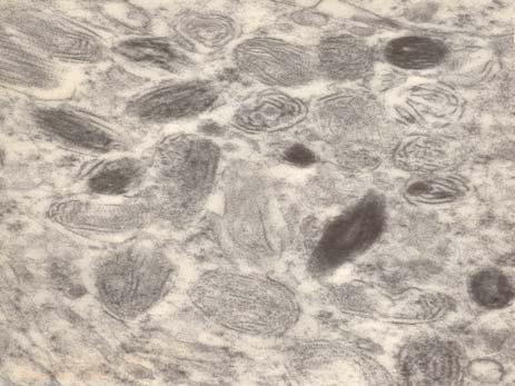
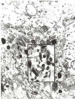
2 ISSUES TO BE ADDRESSED USING ELECTRON MICROSCOPY Epithelial vs mesenchymal DIFFERENTIATIONcrucial in the differential diagnosis of these types of tumors Aberrant expression of antigenic epitopes Lack of specific immunohistochemical profile and/or molecular imprint Need to better characterize a rare tumor CONFIRM A SUSPECTED THOUGH NOT DEFINITIVE DIAGNOSIS OR A RATHER WELL ESTABLISHED DIAGNOSIS TO PROVIDE ADDITIONAL EVIDENCE VALUE OF ELECTRON MICROSCOPY MUST BE VIEWED AS MULTIFACETED EM not only can address the differential diagnosis but may raise new diagnostic possibilities EM can provide a deeper understanding to the problem EM provides solid morphologic evidence to substantiate the diagnosis THE WORK UP OF A SPINDLE CELL / EPITHELIOID NEOPLASM Need to develop a differential diagnosis Judicious use of ancillary diagnostic techniques What is available and what is desired Understanding advantages and limitations of techniques available to address the differential diagnosis Awareness of pitfalls DIFFERENTIAL DIAGNOSIS OF SOFT TISSUE SARCOMAS WITH EPITHELIOID FEATURES Soft tissue sarcomas, especially those poorly differentiated, may mimic a variety of epithelial and EVEN lymphoid neoplasms Diagnosis must be approached by using an intelligent selection of ancillary diagnostic techniques The most reasonable route to address the differential diagnosis and to make a final diagnosis should be taken However, a combination of diagnostic techniques may be needed and should be used in a subset of these cases to arrive to an unequivocal diagnosis WHY TO even CONSIDER EM? Identifying specific combinations of morphologic findings in the neoplastic cells, rather than staining patterns or other nonmorphologic parameters, is generally much more specific and reassuring EM not only can rule out certain diagnoses but IN A SUBSET OF CASES also suggests or provides definitive evidence for a diagnosis that would not have been considered IH IN THE EVALUATION OF SPINDLE / EPITHELIOID TUMORS AND LOOK-ALIKES Often not ONE specific antibody to establish cell type and thus address differential diagnosis Batteries of stains required commonly resulting in expensive work-ups Overlap among IH profiles and confusion with look-alikes 2
 difference most dramatic in high grade tumors IH MORE OFTEN THAN EM DID NOT PROVIDE INFORMATION TO AID IN THE TYPING Kandel et al.")

- positive CHALLENGING SITUATIONS Differentiation of sarcoma vs")
3 IH and EM in the diagnosis of soft tissue tumors 142 consecutive soft tissue sarcomas examined by IH and EM tentative dx in 58 and differential diagnosis in 82 EM more often contributed to diagnosing tumors than IH (80 vs 65%) difference most dramatic in high grade tumors IH MORE OFTEN THAN EM DID NOT PROVIDE INFORMATION TO AID IN THE TYPING Kandel et al. Ultrastruct Pathol 22: , 1998 IH AND EM IN THE DIAGNOSIS OF SOFT TISSUE TUMORS In general, the value of EM was greater in the more diagnostically challenging cases In 47% of the cases in which one of the modalities was non-contributory, the other was helpful inn reaching a diagnosis Both EM and IH are necessary to properly evaluate soft tissue tumors Kandel et al: Ultrastruct Pathol 22: , 1998 Pleural fluid / right pleural mass 60 y/o male VIMENTIN, S-100 protein + keratin 1st panel Calretinin - 2nd panel PAN-KERATIN CK 5/6 - NEGATIVE AE1/AE3 Cell block- pleural mass - FOLLOW UP- MALIGNANT MELANOM SCALP Final diagnosis: Metastatic melanoma with aberrant keratin expression Melan A and HMB-45 + (3 rd panel)- positive CHALLENGING SITUATIONS Differentiation of sarcoma vs carcinoma, including sarcomatoid carcinoma Differentiation of sarcoma vs sarcomatoid mesothelioma Differentiation of pseudomesotheliomatous adenocarcinoma of the lung with florid stromal reaction vs mesothelioma vs sarcoma (i.e. synovial SARCOMA)- biphasic pattern Differentiation pleomorphic sarcoma vs poorly diff. carcinoma / melanoma / lymphoma Evaluation of fine needle aspirates where sarcoma is in the differential 3


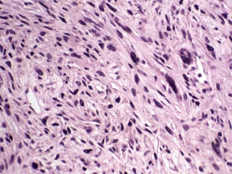
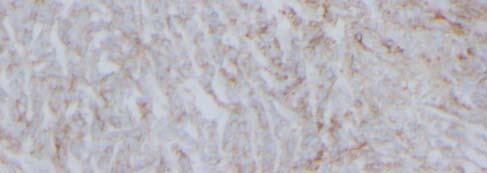
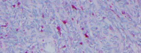
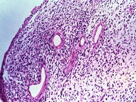
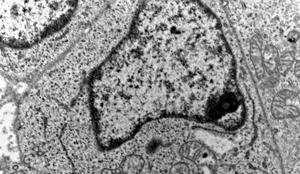
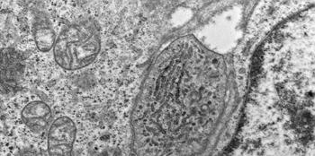
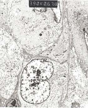
4 DIFFERENTIATION OF SARCOMAS FROM CARCINOMAS Kidney tumor Depending on the situation (site ), the differential diagnosis may vary If sarcomatoid carcinoma is in the differential dx, there are certain areas where this challenge occurs most often: Kidney, bladder, thyroid and upper respiratory tract Proper characterization extremely important to address therapeutic management and defining prognosis LEIOMYOSARCOMA 4 cm back neoplasm / 39 y/o female / no history SYNAPTOPHYSIN Cohesive medium size cells with small processes which interdigitate 4


5 PERINUCLEAR FILAMENT AGGREGATES MERKEL CELL CARCINOMA THIGH TUMOR SARCOMA VS SARCOMATOID MESOTHELIOMAS SPINDLE CELL / EPITHELIOID TUMORS OF THE PLEURA Metastatic RCC (clear cell) with sarcomatoid component 5


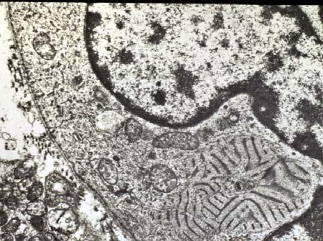
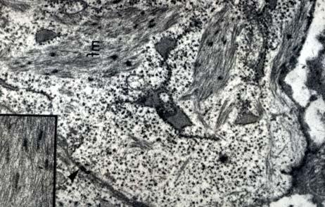
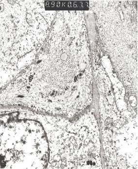
6 SPINDLE CELL TUMORS OF THE PLEURA- DIFFERENTIAL DIAGNOSIS Generally challenging Similar clinical presentation- localized mass or diffuse pleural thickening, histology- spindle cell tumors with variable pleomorphism and cellularity and IMMUNOREACTIVITY Metastatic spindle cell tumors more frequent than malignant mesothelioma Often pleural tumors must be evaluated using a multimodal approach to arrive to the correct diagnosis Rdzanek, M, Fresco, R, Pass, HI, Carbone, M: Spindle cell tumors of the pleura: differential diagnosis. Sem Diagn Pathol 2006; 23: SARCOMA VS SARCOMATOID MESOTHELIOMA MOST IH MARKERS OF EPITHELIAL MESOTHELIOMAS ARE NOT PRESENT IN THE SARCOMATOID VARIETY THE POSITIVE MESOTHELIOMA MARKERS: PODOPLANIN, CALRETININ, KERATINS 5 AND 6 AND WT1 PROTEIN ARE GENERALLY ABSENT or weakly + CALRETININ MOST COMMONLY PRESENT KERATIN IH FREQUENTLY WEAK, FOCAL OR ABSENT and some sarcomas express keratins aberrantly NO OTHER TECHNIQUES AVAILABLE FOR ADDRESSING DIFFERENTIAL DIAGNOSIS, EXCEPT FOR EM, unless specific molecular markers are searched for EM IN THE DIFFERENTIAL DIAGNOSIS OF SARCOMATOID MESOTHELIOMA VS SARCOMA Variable numbers of fibroblastic cells in MOST areas of tumor examined FINDING OF DEFINITIVE EPITHELIAL DIFFERENTIATION, SUCH AS WELL FORMED DESMOSOMES AND TONOFILAMENTS and usually small lumina lined by bushy and tall microvillous borders- requires careful and detailed EM analysis Absence of specific differentiation along specific mesenchymal cell lines (i.e. smooth or skeletal muscle, schwannian differentiation) is very helpful Exclusion of other tumors that may exhibit spindle cell morphology (i.e. melanoma) 6


7 BL MF MF DIFFERENTIAL DIAGNOSIS MESOTHELIOMA / ADENOCARCINOMA VS SYNOVIAL SARCOMA DIFFICULT DIFFERENTIAL DIAGNOSIS- SYNOVIAL SARCOMA ONLY RARELY CONSIDERED- need to suspect ADENOCARCINOMAS- both 1ary and metastatic- MAY EXPRESS MESOTHELIAL MARKERS and SYNOVIAL SARCOMAS HAVE OVERLAPING IH MARKERS with epithelial tumors PLEURAL SYNOVIAL SARCOMAS X;18 translocation IN SS Differential diagnosis from malignant mesothelioma requires a high degree of suspicion and complete immuno- morphologic work-up, including molecular analysis in at least some cases SS are susceptible to chemotherapy and require such tx while MM are resistant and should not be treated with chemo 7
 (X;18) Translocation X and 18 SYNOVIAL")


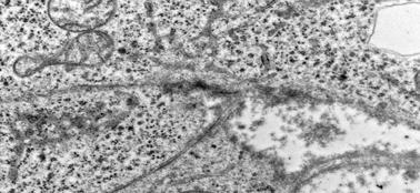
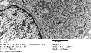
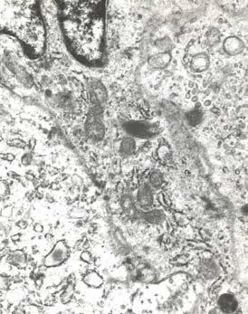
8 ( p11.2;q11.2) (X;18) Translocation X and 18 SYNOVIAL SARCOMAS EM DIAGNOSIS apical surface with short and simple microvilli, sometimes with intestinal features forming glands separated from the spindle shaped cells by a well defined basal lamina prominent lysosomes in neoplastic cells tonofilaments (even keratohyaline granules) junctional complexes- including in spindle cell areas amorphous deposits of epithelial mucin in clefts and luminal spaces 8

 chromosomal translocation in synovial sarcoma may be the only means for establishing a definitive diagnosis Suster, S,")
 + Is it really a sarcoma?")
9 MOLECULAR ANALYSIS IN SS The characteristic X;18 translocation results in the fusion of the SYT gene on chromosome 18 to either SSX1, SSX2, or SSX4 gene on chromosome X Initially felt to be rather specific for SS and present in >90% of SS- CHALLENGED Aubry et al confirmed this translocation in 100% of pleural SS (Am J Surg Pathol 25: , 2001)- purely sarcomatoid SS PLEURAL / MEDIASTINAL SYNOVIAL SARCOMAS In equivocal cases, ultrastructural examination coupled with demonstration of the characteristic (X;18) chromosomal translocation in synovial sarcoma may be the only means for establishing a definitive diagnosis Suster, S, Moran, CA: Primary synovial sarcomas of the mediastinum: A clinicopathologic, immunohistochemical, and ultrastructural study of 15 cases. Am J Surg Pathol 29: , 2005 CHALLENGES IN DIAGNOSIS KERATIN, NSE AND DESMIN (dotlike) + Is it really a sarcoma??? 20 y/o- abdominal mass DESMIN - EWS-WT1 gene fusion + Final dx: Desmoplastic small cell tumor (DSCT) 9


10 Anti-desmin antibodies in the diagnosis of rhabdomyosarcomas Truong, % 282 cases+/359 Leader, % Myogenin is far more sensitive PLEOMORPHIC SARCOMAS DIFFERENTIAL DIAGNOSIS NEED TO BE DIFFERENTIATED FROM POORLY DIFFERENTIATED CARCINOMAS AND MELANOMAS; RARELY FROM HIGH GRADE LYMPHOMAS INITIAL IMMUNOHISTOCHEMISTRY OR ELECTRON MICROSCOPIC EVALUATION? BECAUSE IH is more readily available, the former will happen in most institutions USUAL ANTIBODY BATTERY Keratins Melan A, HMB-45 and S-100 protein Vimentin More specific markers such as TTF-1, Hepar-1, PSA, thyroglobulin etc as needed LUNG, peripheral mass 10


11 Abdominal mass KERATIN + CLEAR CELL SARCOMA? EWS-ATF1 gene fusion NOT DETECTED 11


12 DIFFICULTIES IN THE EVALUATION OF FINE NEEDLE ASPIRATES IN THE DIFFERENTIAL DIAGNOSIS OF SARCOMAS vs LOOK-ALIKES Scanty cellular elements No architectural pattern Difficulties in selecting the appropriate antibody panel in small samples- not much tissue available Problems with IH in FNA samples (background, edge artifact, overstaining) FNA Abdominal mass FNA Pleura Lymphoma vs carcinoma vs sarcoma Sarcoma vs carcinoma 45 y/o female 58 y/o male LOOK-ALIKES EM is a useful technique to separate these look- alikes 12


13 LOOK-ALIKES 82 y/o male with history of prior removal of a tumor in the right leg. Now with abdominal mass Carcinoma Melanoma Sarcoma 18 y/o male with a history of sarcomaleft arm- S/P chemotherapy treatment presenting with subcutaneous nodule on his back (approximately 2 cm in diameter) 13


14 CD-99 EQUIVOCAL Translocation (11;22) EWS/FLI-1 fusion gene 14 y/o girl with history of sarcoma of left femur (? type) who developed a pelvic mass 55 y/o male with abdominal mass. No pertinent history elicited osteosarcoma 14


 and promptly fixed in a fixative adequate for EM If EM scope or expertise in ultrastructural diagnosis is not")
15 Other techniques useful in the diagnosis of spindle / epitheliod tumors and look-alikes in addition to IH and EM FLOW CYTOMETRY extremely useful when hematologic conditions are in the differential diagnosis Keratin -, muscle specific actin +, HMB-45 - CYTOGENETICS certain soft tissue neoplasms have characteristic cytogenetic abnormalities i.e. synovial sarcomas- TRANSLOCATIONS INVOLVING CHROM X AND 18, Ewing s sarcomas (EWS-Fli1), clear cell sarcomas (ews-atf1), myxoid liposarcomas (FUS- CHOP), among others MOLECULAR DIAGNOSTICS GUIDELINES FOR EM Several pieces of tumor from different areas, avoiding necrotic and hemorrhagic areas should be submitted Tissue should be cut in small pieces (1 mm cubes) and promptly fixed in a fixative adequate for EM If EM scope or expertise in ultrastructural diagnosis is not available locally, specimen should be sent to regional laboratory IMPORTANT FACTORS TO BE CONSIDERED WHEN EM IS TO BE USED AVAILABILITY OF EM LOCALLY TURN AROUND TIME TRAINED INDIVIDUALS TO PERFORM EM STUDY CONSIDER SENDING TO A REGIONAL FACILITY WITH EM SERVICES PROPER COLLECTION AND FIXATION OF MATERIAL ESSENTIAL TO MAKE SURE THAT THE BEST POSSIBLE EVALUATION IS PERFORMED MISCONCEPTIONS ABOUT THE ROLE OF EM IN THE EVALUATION OF SOFT TISSUE TUMORS IH has made EM obsolete Only role of EM is to confirm LM impression Only a few sarcomas have specific ultrastructural features Very limited value in poorly differentiated tumors Sampling of tumors too small to produce meaningful information Too slow Too expensive 15


 that may mimic a given diagnosis Clarify atypical ( aberrant ) IH results Should")

16 COMPREHENSIVE DIAGNOSTIC APPROACH USING SEVERAL DIAGNOSTIC TECHNIQUES ELECTRON MICROSCOPY GENERALLY PROVIDES THE QUICKEST TAT- 24 HOURS FROM REQUEST SIGNIFICANT SAVINGS TO THE HEALTH CARE INDUSTRY, EVEN THOUGH IT MAY APPEAR TO BE MORE EXPENSIVE AT FIRST? THE SAFEST IN TERMS OF OBTAINING THE CORRECT DIAGNOSIS ELECTRON MICROSCOPY PITFALLS LACK OF KNOWLEDGE REGARDING SPECTRUM OF ULTRASTRUCTURAL FEATURES OF A GIVEN DIAGNOSIS INTERPRETING NORMAL STRUCTURES SURROUNDING A TUMOR AS PART OF THE TUMOR POOR FIXATION MASKING DIAGNOSTIC FINDINGS INTERPRETING FINDINGS IN A VACUUM THE USE OF EM IN THE DIAGNOSIS OF SPINDLE CELL / EPITHELIOID TUMORS Establish specific diagnosis Rule out other types of tumors (LOOK- ALIKES) that may mimic a given diagnosis Clarify atypical ( aberrant ) IH results Should formalin-fixed or paraffin embedded tumors be considered for EM EVALUATION? CERTAINLY, WITH SOME CAVEATS Formalin fixed is generally better than paraffin embedded- tissue PARAFFIN EMBEDDED TUMORS- QUALITY FOR ELECTRON MICROCOPY DEPENDS ON A NUMBER OF VARIABLES IN SOME CASES PRESERVATION IS EXTREMELY POOR AND EM IS OF NO VALUE 16


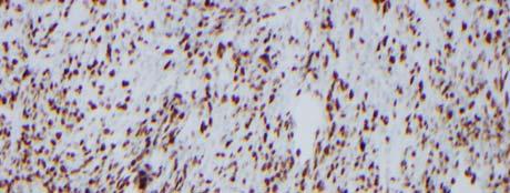
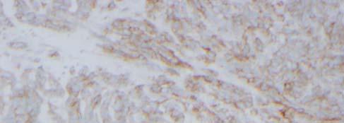

17 Ki67 CD 99 S-100 protein 17


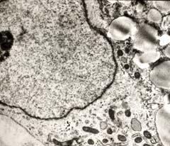
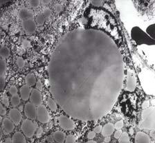
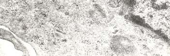
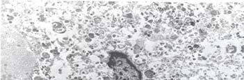
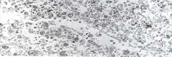
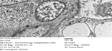
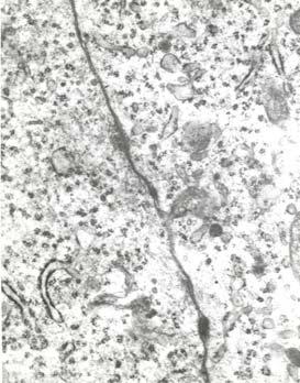
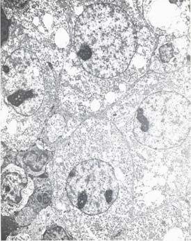
18 Basement membrane Luse body CONCLUSIONS Long spacing collagen LONG-SPACING COLLAGEN EM can certainly play an important role in the differential diagnosis of spindle / epithelioid tumors and look-alikes The right diagnosis may be compromised if EM is not performed A comprehensive approach with use of a number of selective ancillary diagnostic techniques is undoubtedly the best way to approach unusual or difficult diagnostic situations PROPER FIXATION OF SPECIMENS MAXIMIZES THE USEFULNESS OF ELECTRON MICROSCOPY 18


19 19

20 mf 20


21 21


22 Disclosure of Relevant Financial Relationships USCAP requires that all faculty in a position to influence or control the content of CME disclose any relevant financial relationship WITH COMMERCIAL INTERESTS which they or their spouse/partner have, or have had, within the past 12 months, which relates to the content of this educational activity and creates a conflict of interest. Dr. Guillermo Herrera declares he has no conflict(s) of interest to disclose 22
The role of Electron Microscopy in the study of cytologic specimens. Elba A. Turbat-Herrera, MD
 The role of Electron Microscopy in the study of cytologic specimens. Elba A. Turbat-Herrera, MD Louisiana State University Health Sciences Center Shreveport, LA, USA Introduction The field of Cytology
Mesothelioma: diagnostic challenges from a pathological perspective. Naseema Vorajee August 2016
 Mesothelioma: diagnostic challenges from a pathological perspective Naseema Vorajee August 2016 Naseema.vorajee@nhls.ac.za Pleural diseases (whether neoplastic, reactive or infective) may have similar
Lung Tumor Cases: Common Problems and Helpful Hints
 Lung Tumor Cases: Common Problems and Helpful Hints Brandon T. Larsen, MD, PhD Senior Associate Consultant Department of Laboratory Medicine and Pathology Mayo Clinic Arizona Arizona Society of Pathologists
Pathology Mystery and Surprise
 Pathology Mystery and Surprise Tim Smith, MD Director Anatomic Pathology Medical University of South Carolina Disclosures No conflicts to declare Some problem cases Kidney tumor Scalp tumor Bladder tumor
Tumores de células pequeñas, redondas y azules: diagnóstico diferencial cuando el tiempo apremia
 Tumores de células pequeñas, redondas y azules: diagnóstico diferencial cuando el tiempo apremia Sílvia Bagué Servei de Patologia Hospital de Sant Pau Barcelona Soft tissue sarcomas Heterogeneous group
Immunohistochemistry in Bone and Soft Tissue Tumors. Sahar Rassi Zankoul, MD
 Immunohistochemistry in Bone and Soft Tissue Tumors Sahar Rassi Zankoul, MD Introduction Bone tumors represent a wide variety of tumors of various origins and malignant potentials. These different tumor
57th Annual HSCP Spring Symposium 4/16/2016
 An Unusual Malignant Spindle Cell Lesion to Involve the Breast Erinn Downs-Kelly, D.O. Associate Professor of Pathology University of Utah & ARUP Laboratories No disclosures Case 39 y/o female with no
Case 2. Dr. Sathima Natarajan M.D. Kaiser Permanente Medical Center Sunset
 Case 2 Dr. Sathima Natarajan M.D. Kaiser Permanente Medical Center Sunset History 24 year old male presented with a 3 day history of right flank pain, sharp in nature Denies fever, chills, hematuria or
Problem 1: Differential of Neuroendocrine Carcinoma 3/23/2017. Disclosure of Relevant Financial Relationships
 Differential of Neuroendocrine Carcinoma Alain C. Borczuk,MD Weill Cornell Medicine Disclosure of Relevant Financial Relationships USCAP requires that all faculty in a position to influence or control
Klinisch belang van chromosomale translocatie detectie in sarcomen
 Translocations in sarcomas Klinisch belang van chromosomale translocatie detectie in sarcomen Judith V.M.G. Bovée, M.D., Ph.D. Department of Pathology Leiden University Medical Center RNA binding DNA binding
Enterprise Interest Nothing to declare
 Enterprise Interest Nothing to declare Diagnoses one would not like to miss in soft tissue pathology early in your career Marta Sbaraglia, MD Department of Pathology Hospital of Treviso University of Padua
Disclosures. An update on ancillary techniques in the diagnosis of soft tissue tumors. Ancillary techniques. Introduction
 Disclosures An update on ancillary techniques in the diagnosis of soft tissue tumors. I have nothing to disclose. Andrew Horvai, MD, PhD Clinical Professor, Pathology Introduction Ancillary techniques
Biopsy Interpretation of Spindle cell proliferations of the Serosa
 Biopsy Interpretation of Spindle cell proliferations of the Serosa Richard Attanoos, Cardiff. U.K. Disclosure of Relevant Financial Relationships USCAP requires that all planners (Education Committee)
ACCME/Disclosures. Diagnosing Mesothelioma in Limited Tissue Samples. Papanicolaou Society of Cytopathology Companion Meeting March 12 th, 2016
 Diagnosing Mesothelioma in Limited Tissue Samples Papanicolaou Society of Cytopathology Companion Meeting March 12 th, 2016 Sanja Dacic, MD, PhD University of Pittsburgh ACCME/Disclosures GENERAL RULES
Charles Halsey, DVM, PhD, DACVP Pfizer, Inc. IHC Resources
 Charles Halsey, DVM, PhD, DACVP Pfizer, Inc. IHC Resources 1 IHC Identification Targets Specimens Controls 2 Tissue controls Trouble Spots 3 The Key to Description IHC Description 4 Intermediate Filaments
Mesothelioma Pathobasic. Lukas Bubendorf Pathology
 Mesothelioma Pathobasic Lukas Bubendorf Pathology Mechanisms of Asbestos Carcinogenesis in Mesothelioma Asprin High-mobility group protein B1 master switch HMGB1 Initiation/ perpetuation of inflamm. response
Cutaneous Mesenchymal Neoplasms with EWSR1 Rearrangement
 Cutaneous Mesenchymal Neoplasms with EWSR1 Rearrangement By Konstantinos Linos MD, FCAP, FASDP Bone, Soft Tissue and Dermatopathology Assistant Professor of Pathology Dartmouth-Hitchcock Medical Center
Diplomate of the American Board of Pathology in Anatomic and Clinical Pathology
 A 33-year-old male with a left lower leg mass. Contributed by Shaoxiong Chen, MD, PhD Assistant Professor Indiana University School of Medicine/ IU Health Partners Department of Pathology and Laboratory
From Morphology to Molecular Pathology: A Practical Approach for Cytopathologists Part 1-Cytomorphology. Songlin Zhang, MD, PhD LSUHSC-Shreveport
 From Morphology to Molecular Pathology: A Practical Approach for Cytopathologists Part 1-Cytomorphology Songlin Zhang, MD, PhD LSUHSC-Shreveport I have no Conflict of Interest. FNA on Lymphoproliferative
Disclosure of Relevant Financial Relationships
 Ewing and Ewing like sarcomas Using Genetic Signatures in Refining Small Blue Round Cell Tumor Classification Cristina Antonescu, MD Department of Pathology Disclosure of Relevant Financial Relationships
Cutaneous metastases. Thaddeus Mully. University of California, San Francisco Professor, Departments of Pathology and Dermatology
 Cutaneous metastases Thaddeus Mully University of California, San Francisco Professor, Departments of Pathology and Dermatology DISCLOSURE OF RELATIONSHIPS WITH INDUSTRY Thaddeus Mully Course C005 Essential
SELECTED DILEMMAS IN RESPIRATORY CYTOPATHOLOGY (2 CASES)
") SELECTED DILEMMAS IN RESPIRATORY CYTOPATHOLOGY (2 CASES) Dr. Mariamma Joseph Professor of Pathology Division Head Cytopathology Department of Pathology and Laboratory Medicine LHSC and Western University
SELECTED DILEMMAS IN RESPIRATORY CYTOPATHOLOGY (2 CASES) Dr. Mariamma Joseph Professor of Pathology Division Head Cytopathology Department of Pathology and Laboratory Medicine LHSC and Western University
Disclosure of Relevant Financial Relationships
 Evening Specialty Conference - Genitourinary Pathology Case 2 Disclosure of Relevant Financial Relationships Sean R Williamson, MD Henry Ford Health System, Detroit, MI @Williamson_SR USCAP requires that
Applications of IHC. Determination of the primary site in metastatic tumors of unknown origin
 Applications of IHC Determination of the primary site in metastatic tumors of unknown origin Classification of tumors that appear 'undifferentiated' by standard light microscopy Precise classification
Spindle Cell Lesions Of The Breast. Emad Rakha Professor of Breast Pathology and Consultant Pathologist
 Spindle Cell Lesions Of The Breast Emad Rakha Professor of Breast Pathology and Consultant Pathologist * SCLs comprise a wide spectrum of diseases, ranging from reactive processes to aggressive malignant
Financial disclosures
 Cutaneous Mesenchymal Neoplasms with EWSR1 Rearrangement By Konstantinos Linos MD, FCAP, FASDP Bone, Soft Tissue and Dermatopathology Assistant Professor of Pathology Dartmouth-Hitchc Geisel School of
Presentation material is for education purposes only. All rights reserved URMC Radiology Page 1 of 98
 Presentation material is for education purposes only. All rights reserved. 2011 URMC Radiology Page 1 of 98 Radiology / Pathology Conference February 2011 Brooke Koltz, Cytopathology Resident Presentation
Diagnostic Value of Immunohistochemistry in Soft Tissue Tumors
 Original Article DOI: 10.21276/APALM.1637 Diagnostic Value of Immunohistochemistry in Soft Tissue Tumors Sridevi. V*., Susruthan Muralitharan., and Thanka. J Dept of Pathology, SriMuthukumaran Medical
C.L. Davis Foundation Descriptive Veterinary Pathology Course
 C.L. Davis Foundation 2015 Descriptive Veterinary Pathology Course IHC Resources IHC Identification Targets Antibodies Antibodies 1 Antibodies Specimens Antigen Retrieval Unmasks antigen epitopes Methods
Immunohistochemistry on Fluid Specimens: Technical Considerations
 Immunohistochemistry on Fluid Specimens: Technical Considerations Blake Gilks Dept of Pathology University of British Columbia, Vancouver, BC, Canada Disclosures None Learning Objectives At the end of
I have nothing to disclose
 A 47 year old female with multiple lung nodules Disclosure of Relevant Financial Relationships Tamar Giorgadze, MD, PhD Professor of Pathology Medical College of Wisconsin Milwaukee, Wisconsin USCAP requires
Original Article Differential diagnosis of sarcomatoid mesothelioma from true sarcoma and sarcomatoid carcinoma using immunohistochemistry
 Pathology International 2008; 58: 75 83 doi:10.1111/j.1440-1827.2007.02193.x Original Article Differential diagnosis of sarcomatoid mesothelioma from true sarcoma and sarcomatoid carcinoma using immunohistochemistry
Financial disclosures
 An update on immunohistochemical markers in mesenchymal neoplasms By Konstantinos Linos MD, FCAP, FASDP Assistant Professor of Pathology Geisel School of Medicine at Dartmouth Dartmouth-Hitchcock Medical
Mayo Medical Laboratories
 Mayo Medical Laboratories Virtual Lectures 2014 MFMER 2016 MFMER slide-1 Virtual Lectures Planning Committee Disclosure Summary As a provider accredited by ACCME, College of Medicine, Mayo Clinic (Mayo
3/24/2017 DENDRITIC CELL NEOPLASMS: HISTOLOGY, IMMUNOHISTOCHEMISTRY, AND MOLECULAR GENETICS. Disclosure of Relevant Financial Relationships
 DENDRITIC CELL NEOPLASMS: HISTOLOGY, IMMUNOHISTOCHEMISTRY, AND MOLECULAR GENETICS Jason L. Hornick, M.D., Ph.D. Director of Surgical Pathology and Immunohistochemistry Brigham and Women s Hospital Professor
3/27/2017. Disclosure of Relevant Financial Relationships
 Ophthalmic Pathology Evening Specialty Conference USCAP 2017 5 th March, 2017 Mukul K. Divatia, MD Assistant Professor Department of Pathology & Genomic Medicine Weill Cornell Medical College Houston Methodist
Disclosures. An update on ancillary techniques in the diagnosis of soft tissue tumors. Ancillary techniques. Introduction
 Disclosures An update on ancillary techniques in the diagnosis of soft tissue tumors. I have nothing to disclose. Andrew Horvai, MD, PhD Clinical Professor, Pathology Introduction Ancillary techniques
GUT-C 11/30/2017. Debasmita Das, M.D. PGY-1 Danbury Hospital
 GUT-C 11/30/2017 Debasmita Das, M.D. PGY-1 Danbury Hospital CLINICAL SUMMARY 8/2017 59 year old female Presented to the ED with 1 month history of general malaise, fever and weight loss PMH: Significant
ACCME/Disclosures. Case 4 USCAP Pulmonary Panel Case 4 History
 Case 4 USCAP Pulmonary Panel 2016 Andrew Churg, MD Department of Pathology Vancouver General Hospital & University of British Columbia Vancouver, BC achurg@mail.ubc.ca. ACCME/Disclosures The USCAP requires
Classification (1) Classification (3) Classification (2) Spindle cell lesions. Spindle cell lesions of bladder (Mills et al.
 Classification (3) Classification (2) Spindle cell lesions. Spindle cell lesions of bladder (Mills et al.") Non-epithelial tumours and nonepithelial tumour-like lesions of the bladder Dr Jonathan H Shanks The Christie NHS Foundation Trust, Manchester, UK Classification (1) Myofibroblastic proliferations and
Non-epithelial tumours and nonepithelial tumour-like lesions of the bladder Dr Jonathan H Shanks The Christie NHS Foundation Trust, Manchester, UK Classification (1) Myofibroblastic proliferations and
Pathology of Sarcoma ELEANOR CHEN, MD, PHD, ASSISTANT PROFESSOR DEPARTMENT OF PATHOLOGY UNIVERSITY OF WASHINGTON
 Pathology of Sarcoma ELEANOR CHEN, MD, PHD, ASSISTANT PROFESSOR DEPARTMENT OF PATHOLOGY UNIVERSITY OF WASHINGTON Presentation outline Background and epidemiology of sarcomas Sarcoma classification Sarcoma
Update on Cutaneous Mesenchymal Tumors. Thomas Brenn
 Update on Cutaneous Mesenchymal Tumors Thomas Brenn Cutaneous Mesenchymal Tumours Wide morphological and biological spectrum Myofibroblastic, smooth muscle, neural, vascular, apidocytic, undifferentiated;
INTRODUCTION TO PATHOLOGICAL TECHNIQUES. 1. Types of routine biopsy procedures 2. Special exams (IHC, FISH)
") INTRODUCTION TO PATHOLOGICAL TECHNIQUES 1. Types of routine biopsy procedures 2. Special exams (IHC, FISH) Biopsy-Indications Diffuse/multifocal lesions (neoplastic, inflammatory, etc) Etiology of the
INTRODUCTION TO PATHOLOGICAL TECHNIQUES 1. Types of routine biopsy procedures 2. Special exams (IHC, FISH) Biopsy-Indications Diffuse/multifocal lesions (neoplastic, inflammatory, etc) Etiology of the
Case 8 Soft tissue swelling
 Case 8 Soft tissue swelling 26-year-old female presented with a swelling on the back of the left knee joint since the last 6 months and chronic pain in the calf and foot since the last 2 months. Pain in
Primary Synovial Sarcoma of the Thyroid Gland
 J Korean Med Sci 2007; 22 (Suppl): S154-8 ISSN 1011-8934 Copyright The Korean cademy of Medical Sciences Primary Synovial Sarcoma of the Thyroid Gland Synovial sarcoma is a rare but distinct soft tissue
Disclosure. Relevant Financial Relationship(s) None. Off Label Usage None MFMER slide-1
 None. Off Label Usage None MFMER slide-1") Disclosure Relevant Financial Relationship(s) None Off Label Usage None 2013 MFMER slide-1 Case Presentation A 43 year old male, with partial nephrectomy for a right kidney mass 2013 MFMER slide-2 2013
Disclosure Relevant Financial Relationship(s) None Off Label Usage None 2013 MFMER slide-1 Case Presentation A 43 year old male, with partial nephrectomy for a right kidney mass 2013 MFMER slide-2 2013
Immunohistochemical Evaluation of Necrotic Malignant Melanomas
 Anatomic Pathology / EVALUATION OF NECROTIC MALIGNANT MELANOMAS Immunohistochemical Evaluation of Necrotic Malignant Melanomas Daisuke Nonaka, MD, Jordan Laser, MD, Rachel Tucker, HTL(ASCP), and Jonathan
Ascitic Fluid and Use of Immunocytochemistry. Mercè Jordà, University of Miami
 Ascitic Fluid and Use of Immunocytochemistry Mercè Jordà, University of Miami Is It Malignant? Yes? No Ascitic Fluid Cytomorphologic Useful Findings Tight clusters with smooth borders Cellular and nuclear
Disclosure of Relevant Financial Relationships
 Squamous entities of the thyroid: Reactive to Neoplastic Michelle D. Williams Associate Professor Dept of Pathology, Head & Neck Section University of Texas MD Anderson Cancer Center Disclosure of Relevant
Impact of immunostaining of pulmonary and mediastinal cytology
 Impact of immunostaining of pulmonary and mediastinal cytology Harman Sekhon MD, PhD Director of Cytopathology Head of Ottawa-site Ontario Tumour Bank June 20, 2014 Disclaimer Pfizer: Honorarium-Advisory
Aggressive Malignant Mesothelioma In A Patient Without Previous Asbestos Exposure
 ISPUB.COM The Internet Journal of Pathology Volume 19 Number 1 Aggressive Malignant Mesothelioma In A Patient Without Previous Asbestos Exposure A Julai, G Wathuge Citation A Julai, G Wathuge. Aggressive
Case 18. M75. Excision of mass on scalp. Clinically SCC. The best diagnosis is:
 Case 18 M75. Excision of mass on scalp. Clinically SCC. The best diagnosis is: A. Pilomatrical carcinoma B. Adnexal carcinoma NOS C. Metastatic squamous cell carcinoma D.Primary squamous cell carcinoma
Mojca Velikonja Jože Pižem
 Mojca Velikonja Jože Pižem An 81-year old woman presented with an exophytic, wart-like skin lesion on her neck that she had observed for one year. Cryotherapy had been applied twice, but proved unsuccessful.
Value of antimesothelioma HBME 1 in the diagnosis of inflammatory and malignant pleural effusions
 Romanian Journal of Morphology and Embryology 2006, 47(4):351 355 ORIGINAL PAPER Value of antimesothelioma HBME 1 in the diagnosis of inflammatory and malignant pleural effusions LILIANA MOCANU 1), ANCA
The Panel Approach to Diagnostics. Lauren Hopson International Product Specialist Cell Marque Corporation
 The Panel Approach to Diagnostics Lauren Hopson International Product Specialist Cell Marque Corporation Cell Marque Rocklin, California About Cell Marque: IVD primary antibody manufacturer Distributors
Newer soft tissue entities
 Newer soft tissue entities Examples among fibroblastic tumors Turku, May 6, 2010 Markku Miettinen, M.D. AFIP, Washington, DC Fibroblastic neoplasms Solitary fibrous tumor /Hemangiopericytoma Low-grade
Almost any suspected tumor can be aspirated easily and safely. Some masses are more risky to aspirate including:
 DOES THIS PATIENT HAVE CANCER? USING IN-HOUSE CYTOLOGY TO HELP YOU MAKE THIS DIAGNOSIS. Joyce Obradovich, DVM, Diplomate, ACVIM (Oncology) Animal Cancer & Imaging Center, Canton, Michigan Almost every
NEW IHC A n t i b o d i e s
 NEW IHC Antibodies TABLE OF CONTENTS NEW IHC ANTIBODIES from Cell Marque CITED1 (5H6).... 1 Claudin 7 (5D10F3).... 1 GATA1 (4F5).... 1 Transgelin (2A10C2).... 1 NEW IHC ANTIBODIES using RabMAb Technology
IHC Panels as an Aid in Diagnostic Decision Making
 IHC Antibody Test Selection Using a Panel Approach Steven Westra B.S. Reagent Product Specialist Leica Biosystems IHC Panels as an Aid in Diagnostic Decision Making Diagnostic Use of Tumors Using Algorithms
ACCME/Disclosures ALK FUSION-POSITIVE MESENCHYMAL TUMORS. Tumor types with ALK rearrangements. Anaplastic Lymphoma Kinase. Jason L.
 Companion Meeting of the International Society of Bone and Soft Tissue Pathology The Evolving Concept of Mesenchymal Tumors ALK FUSION-POSITIVE MESENCHYMAL TUMORS Jason L. Hornick, MD, PhD March 13, 2016
Differential diagnosis of HCC
 Hepatocellular Carcinoma Quest for an Ideal Immunohistochemical Panel Sanjay Kakar, MD UCSF Differential diagnosis of HCC Hepatocellular lesions Adenoma, FNH, HG dysplasia Adenocarcinoma CholangioCA, metastasis
Gross appearance of nodular hyperplasia in material obtained from suprapubic prostatectomy. Note the multinodular appearance and the admixture of
 Tiền liệt tuyến Tiền liệt tuyến Gross appearance of nodular hyperplasia in material obtained from suprapubic prostatectomy. Note the multinodular appearance and the admixture of solid and microcystic areas.
IMMUNOPROFILES OF THE MAJOR RENAL NEOPLASMS (%staining)
") Stain Clear Cell Papillary IMMUNOPROFILES OF THE MAJOR RENAL NEOPLASMS (%staining) Chromophobe Collecting Duct Carcinom a Sarcomatoid Xp11 Translocat ion Dr Jon Oxley See also www.jonoxley.com Page 1 MTSCC
Stain Clear Cell Papillary IMMUNOPROFILES OF THE MAJOR RENAL NEOPLASMS (%staining) Chromophobe Collecting Duct Carcinom a Sarcomatoid Xp11 Translocat ion Dr Jon Oxley See also www.jonoxley.com Page 1 MTSCC
Gross appearance of peritoneal cysts. They have a thin, translucent wall and contain a clear fluid.
 Gross appearance of peritoneal cysts. They have a thin, translucent wall and contain a clear fluid. So-called multicystic benign mesothelioma. A, Gross appearance. So-called multicystic benign mesothelioma.
Special slide seminar
 Special slide seminar Tomáš Rozkoš The Fingerland Department of Pathology Charles University Medical Faculty and Faculty Hospital in Hradec Králové Czech Republic Case history, 33 years old resistance
Keywords solitary fibrous tumor, dedifferentiation, dedifferentiated solitary fibrous tumor, STAT6, GRIA2, cytokeratin, rhabdomyosarcomatous
 758452IJSXXX10.1177/1066896918758452International Journal of Surgical PathologyCreytens et al research-article2018 Pitfalls in Pathology Multifocal Cytokeratin Expression in a Dedifferentiated Solitary
ARTHUR PURDY STOUT SOCIETY COMPANION MEETING: DIFFICULT NEW DIFFERENTIAL DIAGNOSES IN PROSTATE PATHOLOGY. Jonathan I. Epstein.
 1 ARTHUR PURDY STOUT SOCIETY COMPANION MEETING: DIFFICULT NEW DIFFERENTIAL DIAGNOSES IN PROSTATE PATHOLOGY Jonathan I. Epstein Professor Pathology, Urology, Oncology The Reinhard Professor of Urological
3/27/2017. Pulmonary Pathology Specialty Conference. Disclosure of Relevant Financial Relationships. Clinical History:
 Pulmonary Pathology Specialty Conference Saul Suster, M.D. Medical College of Wisconsin Disclosure of Relevant Financial Relationships USCAP requires that all planners (Education Committee) in a position
Objectives. Salivary Gland FNA: The Milan System. Role of Salivary Gland FNA 04/26/2018
 Salivary Gland FNA: The Milan System Dr. Jennifer Brainard Section Head Cytopathology Cleveland Clinic Objectives Introduce the Milan System for reporting salivary gland cytopathology Define cytologic
1/10/2018. Soft Tissue Tumors Showing Melanocytic Differentiation. Overview. Desmoplastic/ Spindle Cell Melanoma
 2016 MFMER slide-1 2016 MFMER slide-2 2016 MFMER slide-3 Soft Tissue Tumors Showing Melanocytic Differentiation Andrew L. Folpe, M.D. Professor of Laboratory Medicine and Pathology Mayo Clinic, Rochester,
CONSULTATION DURING SURGERY / NOT A FINAL DIAGNOSIS. FROZEN SECTION DIAGNOSIS: - A. High grade sarcoma. Wait for paraffin sections results.
 Pathology Report Date: 3/5/02 A, B. Biopsy right distal femur- high grade spindle cell sarcoma Immunohistochemistry studies are pending to further classify the nature of the tumor. CONSULTATION DURING
Difficult Diagnoses and Controversial Entities in Neoplastic Lung
 Difficult Diagnoses and Controversial Entities in Neoplastic Lung Lynette M. Sholl, M.D. Associate Pathologist, Brigham and Women s Hospital Chief, Pulmonary Pathology Service Associate Professor, Harvard
Update on 2015 WHO Classification of Lung Adenocarcinoma 1/3/ Mayo Foundation for Medical Education and Research. All rights reserved.
 1 Our speaker for this program is Dr. Anja Roden, an associate professor of Laboratory Medicine and Pathology at Mayo Clinic as well as consultant in the Anatomic Pathology Laboratory and co-director of
3/24/2017. Disclosure of Relevant Financial Relationships. Mixed Epithelial Endometrial Carcinoma. ISGyP Endometrial Cancer Project
 Disclosure of Relevant Financial Relationships USCAP requires that all planners (Education Committee) in a position to influence or control the content of CME disclose any relevant financial relationship
Immunohistochemical classification of lung carcinomas and mesotheliomas. Prof. Mogens Vyberg NordiQC Institute of Pathology Aalborg, Denmark
 Immunohistochemical classification of lung carcinomas and mesotheliomas Prof. Mogens Vyberg NordiQC Institute of Pathology Aalborg, Denmark Endobronchial ultrasound guided transbronchial needle biopsy
History A 89 year old gentleman presenting with a scalp/forehead nodule. Patient had squamous cell carcinoma 18 m at same site, excised. Outside diagn
 Case III History A 89 year old gentleman presenting with a scalp/forehead nodule. Patient had squamous cell carcinoma 18 m at same site, excised. Outside diagnoses: Squamous cell carcinoma. R/O: SCC, Melanoma,
The clinically challenging entity of liver metastasis from tumors of unknown primary
 The clinically challenging entity of liver metastasis from tumors of unknown primary Xuchen Zhang, MD, PhD Associate Professor of Pathology Department of Pathology Yale University School of Medicine Liver
Kidney Case 1 SURGICAL PATHOLOGY REPORT
 Kidney Case 1 Surgical Pathology Report February 9, 2007 Clinical History: This 45 year old woman was found to have a left renal mass. CT urography with reconstruction revealed a 2 cm medial mass which
Update on Thyroid FNA The Bethesda System. Shikha Bose M.D. Associate Professor Cedars Sinai Medical Center
 Update on Thyroid FNA The Bethesda System Shikha Bose M.D. Associate Professor Cedars Sinai Medical Center Thyroid Nodules Frequent occurrence Palpable: 4-7% of adults Ultrasound: 10-31% Majority benign
Brief History. Identification : Past History : HTN without regular treatment.
 Brief History Identification : Name : 陳 x - Admission : 94/10/06 Gender : male Age : 75 y/o Chief Complaint : Urinary difficulty for months. Past History : HTN without regular treatment. Brief History
Radiology Pathology Conference
 Radiology Pathology Conference Sharlin Johnykutty,, MD, Cytopathology Fellow Sara Majewski, MD, Radiology Resident Friday, August 28, 2009 Presentation material is for education purposes only. All rights
The role of immunohistochemistry in surgical pathology of the uterine corpus and cervix
 The role of immunohistochemistry in surgical pathology of the uterine corpus and cervix Prof. Ben Davidson, MD PhD Department of Pathology, Norwegian Radium Hospital, Oslo University Hospital, Oslo, Norway
59 yo male with past medical history of prostate carcinoma, presented with upper abdominal pain
 December 2016 59 yo male with past medical history of prostate carcinoma, presented with upper abdominal pain Contributed by: Divya Sharma, MD. Fellow, Gastrointestinal Pathology, Department of Pathology
3/28/2017. Disclosure of Relevant Financial Relationships. GU Evening Subspecialty Case Conference. Differential Diagnosis:
 GU Evening Subspecialty Case Conference Rajal B. Shah, M.D. VP, Medical Director, Urologic Pathology Miraca Life Sciences, Irving, Texas Clinical Associate Professor of Pathology Baylor College of Medicine,
RENAL EPITHELIAL NEOPLASMS: IS THERE A ROLE OF IMMUNOSTAINS IN DIAGNOSIS?
 RENAL EPITHELIAL NEOPLASMS: IS THERE A ROLE OF IMMUNOSTAINS IN DIAGNOSIS? John C. Cheville, M.D. Mayo Clinic and Mayo Foundation Rochester, MN The majority of renal epithelial neoplasms are diagnosed on
Desmoplastic Melanoma R/O BCC. Clinical Information. 74 y.o. man with lesion on left side of neck r/o BCC
 R/O BCC Sabine Kohler, M.D. Professor of Pathology and Dermatology Dermatopathology Service Stanford University School of Medicine Clinical Information 74 y.o. man with lesion on left side of neck r/o
Salivary Gland Cytology
 Salivary Gland Cytology Diagnostic challenges and potential pitfalls Tarik M. Elsheikh, MD Professor and Medical Director Anatomic Pathology Cleveland Clinic FNA Salivary Gland Lesions Indications Distinguish
Immunohistochemical classification of the unknown primary tumour (UPT) Part I. Prof. Mogens Vyberg NordiQC Institute of Pathology Aalborg, Denmark
 Part I. Prof. Mogens Vyberg NordiQC Institute of Pathology Aalborg, Denmark") Immunohistochemical classification of the unknown primary tumour (UPT) Part I Prof. Mogens Vyberg NordiQC Institute of Pathology Aalborg, Denmark Tumours of unknown origin: Histology Brain tumour - biopsy
Immunohistochemical classification of the unknown primary tumour (UPT) Part I Prof. Mogens Vyberg NordiQC Institute of Pathology Aalborg, Denmark Tumours of unknown origin: Histology Brain tumour - biopsy
Sarcomatoid Carcinoma of Prostate: A Rare Entity
 JOURNAL OF CASE REPORTS 2014;4(1):33-37 Sarcomatoid Carcinoma of Prostate: A Rare Entity PHILLIPS Adekoyejo Abiodun 1, DARAMOLA Adetola 1, ANUNOBI Charles 1, TIJANI Kehinde Habbeb 2, MOMOH Martins 1 Department
Case Scenario 1: Thyroid
 Case Scenario 1: Thyroid History and Physical Patient is an otherwise healthy 80 year old female with the complaint of a neck mass first noticed two weeks ago. The mass has increased in size and is palpable.
Enterprise Interest No disclosures.
 Enterprise Interest No disclosures. Secondary Tumours in Uropathology Case 2 PRESENTED AT: EUROPEAN CONGRESS OF PATHOLOGY 18 #ECP2018 Slides are the property of the author. Permission required for reuse.
Maligna Melanoma and Atypical Fibroxanthoma: An Unusual Collision Tumour G Türkcü 1, A Keleş 1, U Alabalık 1, D Uçmak 2, H Büyükbayram 1 ABSTRACT
 Maligna Melanoma and Atypical Fibroxanthoma: An Unusual Collision Tumour G Türkcü 1, A Keleş 1, U Alabalık 1, D Uçmak 2, H Büyükbayram 1 ABSTRACT Two different neoplasia in the same biopsy material called
Financial disclosures
 Mesenchymal Neoplasms with Melanocytic Differentiation By Konstantinos Linos MD, FCAP, FASDP Bone, Soft Tissue and Dermatopathology Assistant Professor of Pathology Dartmouth-Hitchcock Medical Center Geisel
performed to help sway the clinician in what the appropriate diagnosis is, which can substantially alter the treatment of management.
 Hello, I am Maura Polansky at the University of Texas MD Anderson Cancer Center. I am a Physician Assistant in the Department of Gastrointestinal Medical Oncology and the Program Director for Physician
A 25 year old female with a palpable mass in the right lower quadrant of her abdomen
 May 2016 A 25 year old female with a palpable mass in the right lower quadrant of her abdomen Contributed by: Paul Ndekwe, MD, Resident Physician, Indiana University School of Department of Pathology and
Breast - ductal carcinoma CK7 ER PR GATA3 Mammaglobin (50-70%) GCDFP-15 (50-70%) E-cadherin HMWCK CK20 PAX2 ER/PR/HER2 on all newly diagnosed cases
 GCDFP-15 (50-70%) E-cadherin HMWCK CK20 PAX2 ER/PR/HER2 on all newly diagnosed cases") Adrenal cortical carcinoma Inhibin Synap Melan-A Calretinin Vimentin Chromogr CK7 CK20 Breast - ductal carcinoma CK7 ER PR GATA3 Mammaglobin (50-70%) GCDFP-15 (50-70%) E-cadherin HMWCK CK20 PAX2 ER/PR/HER2
Adrenal cortical carcinoma Inhibin Synap Melan-A Calretinin Vimentin Chromogr CK7 CK20 Breast - ductal carcinoma CK7 ER PR GATA3 Mammaglobin (50-70%) GCDFP-15 (50-70%) E-cadherin HMWCK CK20 PAX2 ER/PR/HER2
4/12/2018. MUSC Pathology Symposium Kiawah Island April 18, Jesse K. McKenney, MD
 MUSC Pathology Symposium Kiawah Island April 18, 2018 Jesse K. McKenney, MD 1 Urothelial Carcinoma with Alternative Differentiation 2 Urothelial Carcinoma with Alternative Differentiation Recognition as
أملس عضلي غرن = Leiomyosarcoma. Leiomyosarcoma 1 / 5
 Leiomyosarcoma 1 / 5 EPIDEMIOLOGY Exact incidence is unknown, but older studies suggest that leiomyosarcomas comprise approximately 3 percent of soft-tissue sarcomas. Superficial leiomyosarcoma occurs
Technology from Abcam
 CD2 (EP222) CD2 is one of the earliest T-cell lineage restricted antigens to appear during T-cell differentiation and only rare CD2+ cells can be found in the bone marrow. Anti-CD2 is a pan-t-cell antigen
ACCURACY OF IMMUNOHISTOCHEMISTRY IN EVALUATION
 POL J PATHOL 2011; 2: 95-100 ACCURACY OF IMMUNOHISTOCHEMISTRY IN EVALUATION OF MALIGNANT PLEURAL AND PERITONEAL EFFUSIONS FERESHTEH ENSANI, FARNAZ NEMATIZADEH, GITI IRVANLOU Department of Cytology, Cancer