10/2/17. MELTUMP, SAMPUS, AST.An Algorithmic Approach to Challenging (Often Borderline) Melanocytic Tumors. An Introduction to SNP Arrays
|
|
|
- Toby Campbell
- 5 years ago
- Views:
Transcription
 Why do we need more stuff? A small proportion have ambiguous histology Common nevus Melanoma Common nevus?")
1 MELTUMP, SAMPUS, AST.An Algorithmic Approach to Challenging (Often ) Melanocytic Tumors An Introduction to SNP Arrays Rajiv M. Patel, M.D. RCPA NZ ASM 2017 (11:45-12:30pm, Saturday, ) Why do we need more stuff? A small proportion have ambiguous histology Common nevus Melanoma Common nevus? MELTUMP Molecular studies Any other ways to distinguish nevi from melanoma? 1
2 Molecular studies Chromosome T T T T Genomic instability in melanoma Chromosome Mitosis T T T Chromosome Mitosis T T p53, P53, RB, p16 Checkpoint Cell cycle arrest (Senescence) Chromosome Chromosome Mitosis T T T T T T T Nevi MAPK -B-RAF -N-RAS -H-RAS -GNAQ Chromosome Chromosome Mitosis Melanoma T T T T T T T MAPK -B-RAF -N-RAS -H-RAS -GNAQ Chromosome Mitosis T T p53, RB, p16 Chromosome Mitosis T T p53, P53, RB, p16 p16 inactivation: ~50% of MM Cell cycle arrest ( Oncogene-Induced Senescence) Nevus Chromosome Mitosis Mitosis DNA breaks, fusions -Re-stabilize telomeres -growth advantage Cell Crisis Cell Death Melanoma Gross chromosomal abnormalities Molecular studies Genomic instability in melanoma Detection of numerical abnormalities in the tumor genome (CGH/SNP and FISH) Molecular studies Detection of numerical abnormalities in the tumor genome Mass spectrometry Gene expression profile Identification of mutations (TERT gene promoter) 2

3 Comparative Genomic Hybridization (CGH)/ Single Nucleotide Polymorphism (SNP) arrays SNP arrays Screens the entire genome for gains and losses in DNA material in one experiment Variants: Array based CGH Gains and Losses Array based SNP Gains, Losses and Loss of Heterozygosity SNP arrays SNP arrays Copy number changes Copy number changes 11p gain SNP arrays SNP arrays Copy number changes Allele peak Copy number changes Allele peak 3


 4")
4 SNP arrays Copy number changes Allele peak Mutation data BRAF NRAS PTEN TP53 Advantages of SNP arrays over conventional CGH Detect Copy Neutral LOH Advantages of SNP arrays over CGH Detect Copy Neutral LOH Challenges with FFPE Input DNA Agilent > 500ng Affymetrix CytoScan > 250ng Challenges with FFPE Input DNA Agilent > 500ng Affymetrix CytoScan > 250ng DNA Quality 150 2,000 bp Molecular Inversion Probes (MIP) SNP array OncoScan TM Low DNA input 80ng DNA (as low as 20ng) 4
5 Molecular Inversion Probes (MIP) SNP array OncoScan TM Low DNA input 80ng DNA (as low as 20ng) Works with fragmented samples 40bp fragments Data Gains Losses LOH Mutations BRAF: V600E/K, G469A/E NRAS: Q61L/R/K, G12D/VS/C PTEN: R130*/G/Q/fs, R159S, R233*, P248fs, P267fs TP53: 16 variants University of Michigan Cohort University of Michigan Cohort Melanoma Melanoma Nevus Nevus University of Michigan Cohort University of Michigan Cohort Melanoma Melanoma Nevus Nevus 5


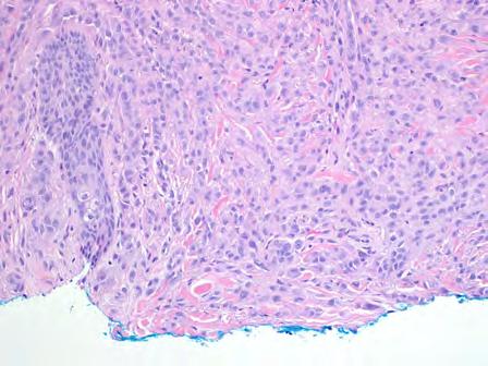
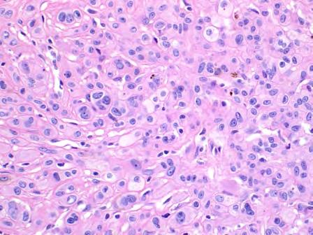
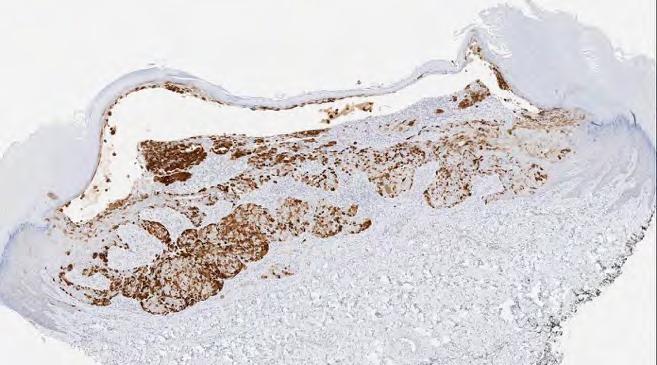
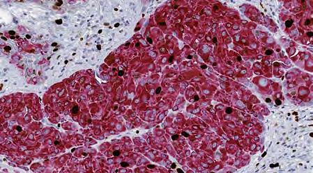
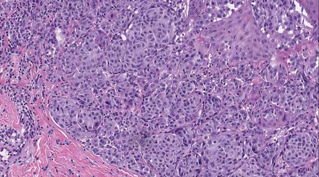
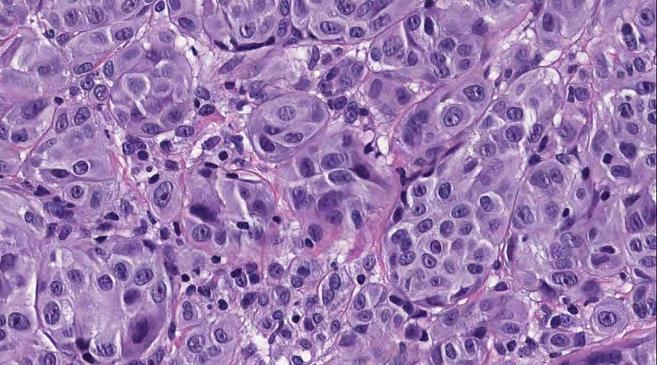
6 Compound nevus Primary melanoma No gains or losses BRAF V600E 18 CNA 11 losses 6 gains 1 CN-LOH BRAF wild NRAS wild PTEN wild TP53 wild Metastatic melanoma 30 CNA 25 gains 2 losses 3 CN-LOH BRAF V600K No DNA copy number changes Test for DNA copy number changes Multiple DNA copy number changes 40-year old woman with a papule on the right shoulder Clinical R/O BCC 35 6


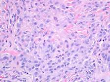
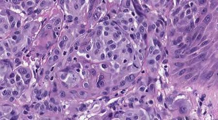
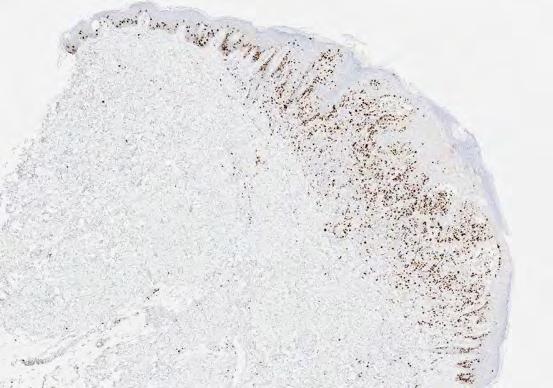
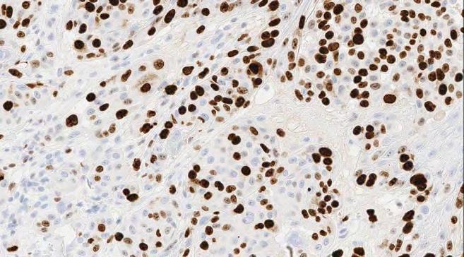
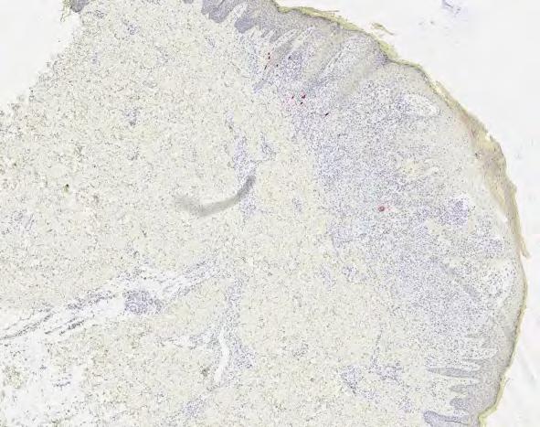
7 HMB-45 Ki-67 Melanocytic neoplasm with borderline features between atypical nevus and melanoma Suspicious for nevoid melanoma p16 Chr 22 CN-LOH Chr 9p21 homozygous loss (CDKN2A) 7


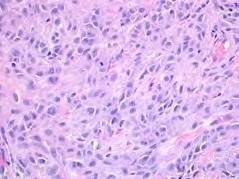
8 Chr 1p gain (NRAS) Chr 13q loss (BRCA2) NRAS Q61R Nevoid melanoma Chr 9p 21 homozygous loss Chr 22 CN-LOH Chr 1p gain (NRAS) Chr 13q loss (BRCA2) NRAS Q61R This seems easy enough No abnormalities GOOD Abnormalities BAD Not so simple! Not all abnormalities are bad Some can be used to classify nevi 48 8

9 32 y/o F, Face Desmoplastic Spitz nevus HRAS mutation 11p gain BRAF wild NRAS wild PTEN wild TP53 wild 28 y/o F, Lt Temple 9
")


 BRAF")


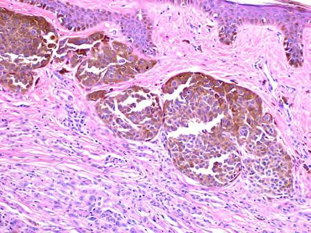
10 BAP-1 negative nevus (BAP-oma) 10-day-old AA newborn with giant congenital nevus with several nodules 3p loss (BAP-1 locus) BRAF V600E Diagnosis: proliferative nodule Copy number Result: Losses of whole chromosomes 3, 4, 5, 10, 11, 13, 14, 16, 17, 18, 21 1 year old More complicating factors What to do with only a few abnormalities? 10


11 46 y/o M, Right chest 11p gain Chr 14 CN-LOH UM Cohort Melanoma Desmoplastic Spitz nevus with atypia Not enough for melanoma 11p gain Chr 14 CN-LOH Nevus 11
12 More problems How many abnormalities do we require for a melanoma diagnosis? Histological classification # of cases with at least one significant copy number variation Average # CNV Nevi 0/6 (0%) 0 Atypical nevi 3/15 (20%) 1.6 (1-2) Ambiguous 15/25 (60%) 6.3 (1-25) Melanoma 35/39 (90%) 21.7 (1-69) Sensitivity: 90% Specificity: 87% Alomari et al. Platform at USCAP meeting, Seattle WA, 2016 How many abnormalities do we require for a melanoma diagnosis? How many abnormalities do we require for a melanoma diagnosis? >=3 abnormalities significant (but with exceptions) Whole chromosomal abnormalities in proliferative nodules Isolated homozygous deletion of 9p21 favors melanoma Others to come. >=3 abnormalities significant (but with exceptions) Whole chromosomal abnormalities in proliferative nodules Isolated homozygous deletion of 9p21 favors melanoma Others to come. Molecular pathologist job: Provide a comprehensive interpretation Your job: Understand the report and communicate with your molecular pathologist Ultimate question Practical algorithm for use of molecular studies Can CNV number and/or pattern predict adverse outcome in borderline lesions? Unfortunately few studies 12
13 Melanocytic lesion Definitive diagnosis Melanocytic lesion Definitive diagnosis No further testing No further testing Molecular testing Mol - Mol - Mol + Mol - Mol + Mol + Mol - Mol - Mol + Mol - Mol + Mol + Mol - Mol - Mol + Mol - Mol + Mol + Nevus Melanoma favor nevus favor melanoma 13


14 Melanocytic lesion Definitive diagnosis No further testing Mol - Mol - Mol + Mol - Mol + Mol + Mol - Mol - Mol + Mol - Mol + Mol + Nevus favor nevus favor melanoma Melanoma Risk assessment Melanocytic lesion Definitive diagnosis Other findings on SNP arrays No further testing Detection of unbalanced rearrangements Mol - Mol - Mol + Mol - Mol + Mol + Nevus favor nevus favor melanoma Melanoma Excision with limited margins Excision with margins appropriate for depth +/- SLN Excision with margins appropriate for depth SLN 82 8 y/o girl, Right ear lobe 14


15 8 y/o girl, Right ear lobe Atypical Spitz nevus/tumor with LMNA- NTRK1 fusion LMNA NTRK1 Summary CGH/ SNP arrays remain valuable diagnostic ancillary tools in selected cases Information beyond CNV can be obtained Copy neutral LOH Mutation data Translocations Challenges Technical 30-50% pure tumor cells Tissue requirements Does not allow histologic correlation Biological More outcome studies are needed to define the CNV patterns associated with adverse outcome 89 15


16 Cases QUESTIONS? Case 1 28-year-old man 2-year history of lesion on the abdomen 1.2 cm pigmented papule with a surrounding rim of light brown pigment Symmetric Well circumscribed Hyperplastic epidermis Spindle melanocytes arranged in nests and fascicles Atypical junctional nevus at the periphery 16


17 Maturation Epithelioid and spindle melanocytes arranged in nests and fascicles Rare mitoses P16 Ki67/Mart1 HMB45 99 Favors nevus - Symmetry, well circumscribed - No significant atypia - Maturation - Stratification with HMB45 - Expression of p16 Favors melanoma - Slight increase in ki-67 - Mitotic activity Atypical Spitz tumor, favor atypical Spitz nevus 17


18 No abnormalities Mol - Nevus Atypical Spitz nevus Case 2 A 14-year old girl presents with a left foot cyst. FISH testing for melanoma is reported as borderline positive Gene expression profile is indeterminate Symmetric Well circumscribed Predominantly dermal Compact nests Epithelioid morphology No maturation 18


19 Lymphocytic infiltrate Mitoses, some deep Spitzoid morphology Cytologic atypia HMB-45 p16 Ki-67 / Mart-1 19
20 Favors nevus - Symmetry - Well circumscribed - Expression of p16 Favors melanoma - Cytological atypia - Lack of maturation - Mitotic activity - Abnormal HMB45 pattern - Increased ki-67 - Positive FISH - Indeterminate Gene Expression Atypical Spitz tumor, favor Spitzoid melanoma No abnormalities Mol Final diagnosis: Atypical Spitz tumor of uncertain malignant potential Initial FISH Repeat FISH Initial FISH 6p % 8q % 11q13 21% Interpretation positive 6p % 6.7% 8q % 0% 11q13 21% 6.7% Tetraploidy 20% Interpretation positive Negative 119 Tetraploidy may increase false-positive FISH results


21 Case 3 Asymmetric Well-circumscribed 60-year old male Left lateral shoulder lesion Clinical impression: irritated benign nevus. Epithelioid cells Eosinophilic cytoplasm Distinct cytoplasmic borders Nuclear atypia No maturation Banal intradermal nevus


22 Banal intradermal nevus HMB45 p16 Ki67 / Mart-1 BAP-1 BAP-1 BRAF V600E Favors nevus - Well circumscribed - No significant atypia - No mitoses - Negative HMB45 - Expression of p16 Favors melanoma - Asymmetric - Focal cytological atypia - Focal increase ki-67 22


 Interactions with BRCA1, polycomb")


23 3p Deletion Atypical Spitz tumor, favor atypical BAP-1 negative nevus 134 BAP1 gene Mol - Nevus Final diagnosis: BAP1 negative nevus 135 BAP1 Nuclear ubiquitin carboxy-terminal hydrolase (UCH) Interactions with BRCA1, polycomb repressor complex, others Germline mutations associated with: Malignant mesothelioma Cutaneous and uveal melanoma Carcinomas (lung, breast, ovarian, renal) Atypical Spitz Tumors 23


24 BAP1 loss in melanocytic lesions Diagnostic/prognostic significance: likely indolent behavior Risk of germline mutation: Suggest genetic consult H&E H&E 33 AST 28% BAP1 25% BRAF+/BAP1- H&E BAP1 Wiesner et al. Am J Surg Pathol, August 2012 Case 4 Small 2 y/o boy Lesion on left thigh Asymmetric Lack of maturation 24
25 Lack of maturation Spitzoid cytology Atypia Mitoses Ki P16 Case 4 FISH: No abnormalities

26 Favor nevus - Small - Infant - Nega*ve FISH Favor melanoma - Asymmetry - No maturation - Cytological atypia - Mitotic activity - Increased ki-67 - Diffuse loss of p16 Atypical Spitz tumor CDKN2A 0.6 MB Homozygous deletion at 9p21 Losses of chormosome 9 and Heterozygous loss of chromosome 9 CDKN2A 9p21 FISH probe 9p21 CEN 9 FISH probe produces a signal: False positive result


27 Case 5 16 y/o male with lesion on the arm Mol + favor melanoma Final diagnosis: Atypical Spitz Tumor, favor spitzoid melanoma Compound melanocytic proliferation Bulges into the subcutis dumbbell profile Extends along neurovascular bundles Courtesy of Dr Guido Massi 160 Junctional component 27


28 Plexiform architecture Epithelioid to spindled Large ovoid nuclei with occasional nucleoli and nuclear pseudoinclusions Irregular nuclear border with hyperchromasia and nuclear pleomorphism Pale cytoplasm with occasional balloon cell changes HMB-45 Ki-67 / Mart-1 28

 MET possible fusion 9 (whole Loss 140.")
 Final diagnosis: DPN-like melanocytic tumor of uncertain malignant potential")
29 P16 BRAF V600E 170 Favor nevus - Symmetric - Well circumscribed - DPN morphology - Expression of p16 Favor melanoma - Cytological atypia - Mitotic activity - Focal increased ki-67 - BRAF V600E Favor atypical DPN. Mol + Chromosome Type or Size Relevant genes abnormality 7q Gain 42.7 BRAF (also with V600E mutation) MET possible fusion 9 (whole Loss CDKN2A chromosome) 14q32.12 Loss 0.25 GOLGA5 possible fusion 21 (whole Gain chromosome) Final diagnosis: DPN-like melanocytic tumor of uncertain malignant potential Possible associated translocation 29


30 Case 6 25 y/o man with nevus on the arm No gains or losses BRAF -wild 30


31 Dermatopathology Molecular Diagnostics and Research Laboratory Dx: Deep penetrating nevus Min Wang Dr. Doru Andea Acknowledgments U of M Dermatopathology Section Douglas Fullen Lori Lowe May Chan Alex Hristov Paul Harms U of M Melanoma Clinic Tim Johnson Chris Bichakjian Alison Durham Kelly Harms QUESTIONS? 31
The Enigmatic Spitz Lesion
 The Enigmatic Spitz Lesion The Dawn of Spitz S Spitz Sophie Spitz Melanomas of Childhood ; Am J Pathol 1948 1910-1956 13 children (18 mo - 12 yrs) 12/13 had a benign clinical course Sophie Spitz Born 1910
21/07/2017. The «gray zone» of diagnosis is visible. Nevus Atypical nevus Melanoma. Melanoma ex-blue nevus
 Update on the Clinico- Pathological and Molecular Diagnosis of Melanocytic Lesions None to declare Conflicts of interest Belfast pathology Arnaud de la Fouchardière MD, PhD Lyon, France What is new? Today
Update on Spitzoid and Blue nevus-like melanocytic lesions Emphasis on molecular studies informing diagnosis, prognosis and therapy
 Update on Spitzoid and Blue nevus-like melanocytic lesions Emphasis on molecular studies informing diagnosis, prognosis and therapy Michael T. Tetzlaff MD, PhD Associate Professor Department of Pathology,
Michael T. Tetzlaff MD, PhD
 Molecular alterations informing the diagnosis of melanocytic tumors Michael T. Tetzlaff MD, PhD Associate Professor Department of Pathology, Section of Dermatopathology Department of Translational and
Molecular Aspects of Melanocytic Neoplasia. Iwei Yeh MD, PhD University of California, San Francisco
 Molecular Aspects of Melanocytic Neoplasia Iwei Yeh MD, PhD University of California, San Francisco Thanks to: Boris Bastian Timothy McCalmont Philip LeBoit Beth Ruben Jeff North Laura Pincus Thaddeus
Dermatopathology. Dr. Rafael Botella Estrada. Hospital La Fe de Valencia
 Dermatopathology Dr. Rafael Botella Estrada. Hospital La Fe de Valencia Melanoma and mimics Dr. Martin Mihm Malignant lesions result from the accumulation of mutations Class I lesions (benign) Class II
Melanoma and the genes: Molecular alterations informing the diagnosis of melanocytic tumors
 Melanoma and the genes: Molecular alterations informing the diagnosis of melanocytic tumors Michael T. Tetzlaff MD, PhD Associate Professor Department of Pathology, Section of Dermatopathology Department
MAPK Pathway. CGH Next Generation Sequencing. Molecular Tools in Care of Patients with Pigmented Lesions 7/20/2017
 Molecular Tools in Care of Patients with Pigmented Lesions Tammie Ferringer, MD Geisinger Medical Center, Danville, PA tferringer@geisinger.edu DISCLOSURE OF RELATIONSHIPS WITH INDUSTRY Tammie Ferringer,
Female 18. Deeply pigmented lesion on trunk.?warty naevus?seborrhoeic keratosis?malignant melanoma. The best diagnosis is:
 Female 18. Deeply pigmented lesion on trunk.?warty naevus?seborrhoeic keratosis?malignant melanoma. The best diagnosis is: A. deep penetrating naevus B. naevoid malignant melanoma C. pigment synthesising
There is NO single Melanoma Stain. > 6000 Mutations in Melanoma. What else can be done to discriminate atypical nevi from melanoma?
 Las Vegas Fall Clinical 2016: The Assessment and Diagnosis of Melanoma Whitney A. High, MD, JD, MEng Associate Professor, Dermatology & Pathology Director of Dermatopathology (Dermatology) University of
Vernon K. Sondak. Department of Cutaneous Oncology Moffitt Cancer Center Tampa, Florida
 Vernon K. Sondak Department of Cutaneous Oncology Moffitt Cancer Center Tampa, Florida Australasian Melanoma Conference 2016 Sydney, NSW, Australia October 29, 2016 Disclosures Dr. Sondak is a compensated
Case 26 Male 37. Right jawline 5mm nodule?keloid. The best diagnosis is:
 Case 26 Male 37. Right jawline 5mm nodule?keloid. The best diagnosis is: A. Desmoplastic Spitz naevus B. Atypical Spitz Tumour C. Spitzoid melanoma D. Deep penetrating naevus E. Spitz naevus Case 26: M
Melanocytic Lesions: Use of Immunohistochemistry and Special Studies Napa Valley 2018
 Melanocytic Lesions: Use of Immunohistochemistry and Special Studies Napa Valley 2018 Victor G. Prieto, MD, PhD Professor Depts. of Pathology and Dermatology University of Texas - MD Anderson Cancer Center
The Pathology of Neoplasia Part II
 The Pathology of Neoplasia Part II February 2018 PAUL BOGNER, MD A S S O C I A T E P R O F E S S O R O F O N C O L O G Y P A T H O L O G Y A N D D E R M A T O L O G Y Clinical goals of cancer pathology
Case RAC7783. M46. Ear. Mole. r/o MM.?Blue naevus RAC7783
 Case RAC7783. M46. Ear. Mole. r/o MM.?Blue naevus RAC7783 Pie Chart Participants N=74 Benign: 48 N=74 Blue naevus: 38 Intradermal: 12 DPN: 10 Compound 3 Clonal: 3; Spitz 2; Special Site: 1; Congenital:
Ways to get into trouble, ideas on avoiding trouble, and diagnostic approaches to keep trouble at bay
 Pitfalls in the diagnosis of melanocytic tumors Timothy McCalmont, MD University of California, San Francisco Ways to get into trouble, ideas on avoiding trouble, and diagnostic approaches to keep trouble
Melanocytic proliferations in sundamaged
 Atypical Spitzoid Tumor: What Does It Mean And How Should It Be Managed? Melanocytic proliferations in sundamaged skin Jane L. Messina, Jane L. Messina MD International Melanoma Pathology Working Group
Guy Perrot (Ги Перро)
") НАУЧНО-ПРАКТИЧЕСКАЯ КОНФЕРЕНЦИЯ (МАСТЕР-КЛАСС) «ПРАКТИЧЕСКИЕ АСПЕКТЫ ДИАГНОСТИКИ И ЛЕЧЕНИЯ МЕЛАНОМЫ КОЖИ» DIAGNOSTIC AND PITFALLS IN MELANOMA Guy Perrot (Ги Перро) MD PHD pathologist, University Hospital
НАУЧНО-ПРАКТИЧЕСКАЯ КОНФЕРЕНЦИЯ (МАСТЕР-КЛАСС) «ПРАКТИЧЕСКИЕ АСПЕКТЫ ДИАГНОСТИКИ И ЛЕЧЕНИЯ МЕЛАНОМЫ КОЖИ» DIAGNOSTIC AND PITFALLS IN MELANOMA Guy Perrot (Ги Перро) MD PHD pathologist, University Hospital
Supplementary Figure 1. Spitzoid Melanoma with PPFIBP1-MET fusion. (a) Histopathology (4x) shows a domed papule with melanocytes extending into the
 Histopathology (4x) shows a domed papule with melanocytes extending into the") Supplementary Figure 1. Spitzoid Melanoma with PPFIBP1-MET fusion. (a) Histopathology (4x) shows a domed papule with melanocytes extending into the deep dermis. (b) The melanocytes demonstrate abundant
Supplementary Figure 1. Spitzoid Melanoma with PPFIBP1-MET fusion. (a) Histopathology (4x) shows a domed papule with melanocytes extending into the deep dermis. (b) The melanocytes demonstrate abundant
Conflict of Interest 9/2/2014. Pathogenesis and Comparison of Atypical Spitz Nevi vs Benign Spitz, and Childhood Melanoma
 Pathogenesis and Comparison of Atypical Spitz Nevi vs Benign Spitz, and Childhood Melanoma Martin C. Mihm Jr., M.D., F.A.C.P. Harvard Medical School Brigham and Women s Hospital Dana Farber Cancer Center
Patricia Chevez-Barrrios AAOOP-USCAP /12/2016
 Biomarkers in Ocular Melanoma Patricia Chévez-Barrios, MD Pathology and Genomic Medicine, Houston Methodist Hospital Professor of Pathology and Laboratory Medicine and Ophthalmology, Weill Cornell Medical
Benign and malignant epithelial lesions: Seborrheic keratosis: A common benign pigmented epidermal tumor occur in middle-aged or older persons more
 Benign and malignant epithelial lesions: Seborrheic keratosis: A common benign pigmented epidermal tumor occur in middle-aged or older persons more common on the trunk; but extremities, head and neck are
Management of pediatric melanocytic lesions
 Open Journal of Clinical & Medical Case Reports Management of pediatric melanocytic lesions Volume 3 (2017) Issue 8 ISSN 2379-1039 Jin Kim, BS; Emmanuel Gabriel MD, PhD; Weiguo Liu MD, PhD; Lin Lin MD,
A PRACTICAL APPROACH TO ATYPICAL MELANOCYTIC LESIONS BIJAN HAGHIGHI M.D, DIRECTOR OF DERMATOPATHOLOGY, ST. JOSEPH HOSPITAL
 A PRACTICAL APPROACH TO ATYPICAL MELANOCYTIC LESIONS BIJAN HAGHIGHI M.D, DIRECTOR OF DERMATOPATHOLOGY, ST. JOSEPH HOSPITAL OBJECTIVES Discuss current trends and changing concepts in our understanding of
Desmoplastic Melanoma R/O BCC. Clinical Information. 74 y.o. man with lesion on left side of neck r/o BCC
 R/O BCC Sabine Kohler, M.D. Professor of Pathology and Dermatology Dermatopathology Service Stanford University School of Medicine Clinical Information 74 y.o. man with lesion on left side of neck r/o
David B. Troxel, MD. Common Medicolegal Situations: Misdiagnosis of Melanoma
 Common Medicolegal Situations: Misdiagnosis of Melanoma David B. Troxel, MD Medical Director, The Doctors Company, Napa, California Clinical Professor Emeritus, University of California at Berkeley Past
Financial disclosures
 Mesenchymal Neoplasms with Melanocytic Differentiation By Konstantinos Linos MD, FCAP, FASDP Bone, Soft Tissue and Dermatopathology Assistant Professor of Pathology Dartmouth-Hitchcock Medical Center Geisel
Genetic Testing: When should it be ordered? Julie Schloemer, MD Dermatology
 Genetic Testing: When should it be ordered? Julie Schloemer, MD Dermatology Outline Germline testing CDKN2A BRCA2 BAP1 Somatic testing Gene expression profiling (GEP) BRAF Germline vs Somatic testing
Hematopathology Service Memorial Sloan Kettering Cancer Center, New York
 SH2017-0334 t(14;18) Negative Follicular Lymphoma with 1p36 abnormality associated with In Situ Follicular Neoplasia with t(14;18) translocation Pallavi Khattar MD, Jennifer Maerki MD, Alexander Chan MD,
Copy number and somatic mutations drive tumors
 Detection of copy number alterations, ploidy and loss of heterozygosity across the genome in FFPE specimens Utility for diagnosis and treatment with comparison to FISH-based and as a complement to sequencing
BAP-oma & BEYOND MICHAEL A NOWAK, MD
 BAP-oma & BEYOND MICHAEL A NOWAK, MD CONFLICTS No conflicts with the content of this lecture BAP-oma Wiesner 2011: Families with multiple tan dome-shaped papules of head, neck, trunk, and extremities.
Diagnoses of Cases 1. Lentigo, other melanosis and the acquired nevus 2. Variations on the acquired nevus 3. Dermal melanocytosis
 Diagnoses of Cases 1. Lentigo, other melanosis and the acquired nevus 1 1A. Lentigo simplex 4 1B. Psoralens and ultraviolet A (PUVA) lentigo 6 1C. Solar lentigo 8 1D. Café au lait macule 10 1E. Ink-spot
6/22/2015. Original Paradigm. Correlating Histology and Molecular Findings in Melanocytic Neoplasms
 6 Correlating Histology and Molecular Findings in Melanocytic Neoplasms Pedram Gerami MD, Associate Professor of Dermatology and Pediatrics at Northwestern University Disclosures: I have been a consultant
I have no relevant conflicts of interest to disclose. John T. Seykora MD PhD Departments of Dermatology & Pathology and Laboratory Medicine
 Molecular Characterization of Stage 1-3 Melanoma: Are we close to accurate prognostication and prediction? I have no relevant conflicts of interest to disclose. John T. Seykora MD PhD Departments of Dermatology
Case 231: F7. Exophytic naevus over left trapezious. Grown over a few weeks. Iniitally flat.?spitz naevus,?malignant
 Case 231: F7. Exophytic naevus over left trapezious. Grown over a few weeks. Iniitally flat.?spitz naevus,?malignant Dermoscopy: coarse vascular structures. c/o A, B, C RAC7750 Case 231: F7. Exophytic
The Relevance of Cytologic Atypia in Cutaneous Neural Tumors
 The Relevance of Cytologic Atypia in Cutaneous Neural Tumors Recent Findings - New Developments New Problems Zsolt B. Argenyi, M.D. Professor of Pathology & Dermatology Director of Dermatopathology Department
An update on molecular alterations in melanocytic tumors with emphasis on Spitzoid lesions
 Review Article on Molecular Oncology age 1 of 17 An update on molecular alterations in melanocytic tumors with emphasis on Spitzoid lesions Emmanouil Dimonitsas 1#, Aliki Liakea 2#, Stratigoula Sakellariou
Update on Genetic Testing for Melanoma
 Update on Genetic Testing for Melanoma Emily Y. Chu, M.D., Ph.D. Assistant Professor of Dermatology & Pathology and Laboratory Medicine Hospital of the University of Pennsylvania February 18, 2018 AAD
DIAGNOSTIC SLIDE SEMINAR: PART 1 RENAL TUMOUR BIOPSY CASES
 DIAGNOSTIC SLIDE SEMINAR: PART 1 RENAL TUMOUR BIOPSY CASES Dr. Andrew J. Evans MD, PhD, FACP, FRCPC Consultant in Genitourinary Pathology University Health Network, Toronto, ON Case 1 43 year-old female,
المركب النموذج--- سبيتز وحمة = Type Spitz's Nevus, Compound SPITZ NEVUS 1 / 7
 SPITZ NEVUS 1 / 7 Epidemiology An annual incidence rate of 1.4 cases of Spitz nevus per 100,000 individuals has been estimated in Australia, compared with 25.4 per 100,000 individuals for cutaneous melanoma
EARLY ONLINE RELEASE
 EARLY ONLINE RELEASE Note: This article was posted on the Archives Web site as an Early Online Release. Early Online Release articles have been peer reviewed, copyedited, and reviewed by the authors. Additional
Integrating Fluorescence in situ Hybridization and Genomic Array Results into the Diagnostic Workup of Melanoma
 Integrating Fluorescence in situ Hybridization and Genomic Array Results into the Diagnostic Workup of Melanoma Association for Molecular Pathology United States and Canadian Academy of Pathology Companion
Malignant tumors of melanocytes: Part 1. Deba P Sarma, MD., Omaha
 Malignant tumors of melanocytes: Part 1 Deba P Sarma, MD., Omaha The melanocytic tumor is one of the most difficult and confusing areas in Dematopathology. It is true that most (95%) of such lesions are
أملس عضلي غرن = Leiomyosarcoma. Leiomyosarcoma 1 / 5
 Leiomyosarcoma 1 / 5 EPIDEMIOLOGY Exact incidence is unknown, but older studies suggest that leiomyosarcomas comprise approximately 3 percent of soft-tissue sarcomas. Superficial leiomyosarcoma occurs
Melanocytic Tumours. Molecular Biology 02/06/2015. Cutaneous Melanocytic Tumours Introduction. Thomas Brenn. Intermediate Malignancy
 Cutaneous Melanocytic Tumours Introduction Melanocytic Tumours: Update on Epidemiology and Molecular Biology Thomas Brenn Wide clinical and morphological spectrum Ranging from benign naevi to melanoma
ACCME/Disclosures ALK FUSION-POSITIVE MESENCHYMAL TUMORS. Tumor types with ALK rearrangements. Anaplastic Lymphoma Kinase. Jason L.
 Companion Meeting of the International Society of Bone and Soft Tissue Pathology The Evolving Concept of Mesenchymal Tumors ALK FUSION-POSITIVE MESENCHYMAL TUMORS Jason L. Hornick, MD, PhD March 13, 2016
1/10/2018. Soft Tissue Tumors Showing Melanocytic Differentiation. Overview. Desmoplastic/ Spindle Cell Melanoma
 2016 MFMER slide-1 2016 MFMER slide-2 2016 MFMER slide-3 Soft Tissue Tumors Showing Melanocytic Differentiation Andrew L. Folpe, M.D. Professor of Laboratory Medicine and Pathology Mayo Clinic, Rochester,
Blue Melanocytic Proliferations
 Blue Melanocytic Proliferations Labib R. Zakka M.D., M.A. Research Fellow Melanoma Program Department of Dermatology Brigham and Women s Hospital Harvard Medical School Conflicts of Interest No conflicts
Assessment of Breast Cancer with Borderline HER2 Status Using MIP Microarray
 Assessment of Breast Cancer with Borderline HER2 Status Using MIP Microarray Hui Chen, Aysegul A Sahin, Xinyan Lu, Lei Huo, Rajesh R Singh, Ronald Abraham, Shumaila Virani, Bal Mukund Mishra, Russell Broaddus,
Chromothripsis: A New Mechanism For Tumorigenesis? i Fellow s Conference Cheryl Carlson 6/10/2011
 Chromothripsis: A New Mechanism For Tumorigenesis? i Fellow s Conference Cheryl Carlson 6/10/2011 Massive Genomic Rearrangement Acquired in a Single Catastrophic Event during Cancer Development Cell 144,
Molecular Methods in the Diagnosis and Prognostication of Melanoma: Pros & Cons
 Molecular Methods in the Diagnosis and Prognostication of Melanoma: Pros & Cons Ben J. Friedman, MD Senior Staff Physician Department of Dermatology Department of Pathology and Laboratory Medicine Henry
Next Generation Sequencing in Clinical Practice: Impact on Therapeutic Decision Making
 Next Generation Sequencing in Clinical Practice: Impact on Therapeutic Decision Making November 20, 2014 Capturing Value in Next Generation Sequencing Symposium Douglas Johnson MD, MSCI Vanderbilt-Ingram
Which melanoma patients benefit from genetic testing?
 Which melanoma patients benefit from genetic testing? Michael A. Marchetti, MD Assistant Attending, Dermatology Service Memorial Sloan Kettering Cancer Center American Academy of Dermatology Annual Meeting
Predisposition of Melanoma
 Predisposition of Melanoma Nelleke Gruis Department of Dermatology Leiden University Medical Center The Netherlands OCTOBER 27TH 2017 Melanoma Risk Factors? Melanoma Predisposition 10% familial Manolio
THE SPITZ NEVUS OFTEN POSES
 OBSERVATION ONLINE FIRST Melanoma Mimic A Case of Multiple Pagetoid Spitz Nevi KaLynne Harris, MD; Scott R. Florell, MD; Jason Papenfuss, MD; Wendy Kohlmann, MS, CGC; Mona Jahromi, BS; Joshua D. Schiffman,
Approximately 2% of the United States population
 Differentiation of Malignant Melanoma From Benign Nevus Using a Novel Genomic Microarray With Low Specimen Requirements Wells M. Chandler, MD; Leslie R. Rowe, MS; Scott R. Florell, MD; Mona S. Jahromi,
Index. Springer-Verlag Berlin Heidelberg 2017 J.A. Plaza, V.G. Prieto, Pathology of Pigmented Skin Lesions, DOI /
 A Acral lentiginous (mucosal lentiginous) melanoma, 483 Acral lentiginous melanoma (ALM) asymmetric and irregular lentiginous junctional growth, 431 clinical features, 427 428 differential diagnosis, 428
A 25 year old female with a palpable mass in the right lower quadrant of her abdomen
 May 2016 A 25 year old female with a palpable mass in the right lower quadrant of her abdomen Contributed by: Paul Ndekwe, MD, Resident Physician, Indiana University School of Department of Pathology and
Application of Whole Genome Microarrays in Cancer: You should be doing this test!!
 Application of Whole Genome Microarrays in Cancer: You should be doing this test!! Daynna Wolff, Ph.D. Director, Cytogenetics and Genomics Disclosures Clinical Laboratory Director and Employee, Medical
A diagnostic algorithm for atypical spitzoid tumors: guidelines for immunohistochemical and molecular assessment
 Modern Pathology (2016), 1 15 2016 USCAP, Inc All rights reserved 0893-3952/16 $32.00 1 A diagnostic algorithm for atypical spitzoid tumors: guidelines for immunohistochemical and molecular assessment
5/21/2018. Disclosures. Consulting: Myriad Genetics SciBase. Superficial Atypical Melanocytic Proliferations. SSM, LMM and (some of) their Simulants
 their Simulants") Disclosures Consulting: Myriad Genetics SciBase Superficial Atypical Melanocytic Proliferations SSM, LMM and (some of) their Simulants 1 Melanomas and Nevi. Nevi are important mainly in relation to melanoma
Disclosures Consulting: Myriad Genetics SciBase Superficial Atypical Melanocytic Proliferations SSM, LMM and (some of) their Simulants 1 Melanomas and Nevi. Nevi are important mainly in relation to melanoma
SOFT TISSUE TUMOR PATHOLOGY: AN UPDATE
 SOFT TISSUE TUMOR PATHOLOGY: AN UPDATE Jason L. Hornick, MD, PhD July 18, 2013 Department of Pathology Brigham and Women s Hospital Harvard Medical School Boston, MA, USA I have no disclosures. New Soft
Characterisation of structural variation in breast. cancer genomes using paired-end sequencing on. the Illumina Genome Analyser
 Characterisation of structural variation in breast cancer genomes using paired-end sequencing on the Illumina Genome Analyser Phil Stephens Cancer Genome Project Why is it important to study cancer? Why
57th Annual HSCP Spring Symposium 4/16/2016
 An Unusual Malignant Spindle Cell Lesion to Involve the Breast Erinn Downs-Kelly, D.O. Associate Professor of Pathology University of Utah & ARUP Laboratories No disclosures Case 39 y/o female with no
MECHANISMS OF HUMAN DISEASE: LABORATORY SESSION PATHOLOGY OF THE SKIN LAB. Friday, February 12, :30 am 11:00 am
 MECHANISMS OF HUMAN DISEASE: LABORATORY SESSION PATHOLOGY OF THE SKIN LAB Friday, February 12, 2012 9:30 am 11:00 am FACULTY COPY GOALS: Describe the basic clinical and morphologic features of various
"Atypical": Criteria and
 "Atypical": Criteria and Controversies Esther Rossi MD PhD MIAC Division of Anatomic Pathology and Cytology Catholic University of Sacred Heart Rome, Italy CASE HISTORY In 2015, 45 y/o woman underwent
MRC-Holland MLPA. Description version 06; 23 December 2016
 SALSA MLPA probemix P417-B2 BAP1 Lot B2-1216. As compared to version B1 (lot B1-0215), two reference probes have been added and two target probes have a minor change in length. The BAP1 (BRCA1 associated
Reviewers' comments: Reviewer #1 (Remarks to the Author):
:") Reviewers' comments: Reviewer #1 (Remarks to the Author): In this study the authors analysed 18 deep penetrating nevi for oncogenic genomic changes (single nucleotide variations, insertions/deletions,
Reviewers' comments: Reviewer #1 (Remarks to the Author): In this study the authors analysed 18 deep penetrating nevi for oncogenic genomic changes (single nucleotide variations, insertions/deletions,
Associate Clinical Professor of Dermatology MUSC
 Re-excision of Moderately Dysplastic Nevi: Should we or shouldn t we? John C. Maize, Jr, M.D. Dermatologist and Dermatopathologist Trident Dermatology, Charleston SC Associate Clinical Professor of Dermatology
F006 Imaging in Dermatology Melanocytic Neoplasia Clinical-Confocal-Pathological-Correlations
 F006 Imaging in Dermatology Melanocytic Neoplasia Clinical-Confocal-Pathological-Correlations Melissa Gill, MD SkinMedical Research and Diagnostics Dobbs Ferry, NY, USA Department of Pathology SUNY Downstate
Dilemmas in Cytopathology and Histopathology
 Dilemmas in Cytopathology and Histopathology Yuri E. Nikiforov, MD, PhD Division of Molecular & Genomic Pathology University of Pittsburgh Medical Center, USA Objectives Discuss new WHO classification
Transform genomic data into real-life results
 CLINICAL SUMMARY Transform genomic data into real-life results Biomarker testing and targeted therapies can drive improved outcomes in clinical practice New FDA-Approved Broad Companion Diagnostic for
Disclosure Information. Lecture Outline. Lecture Outline. Introduction. Molecular Pathology of Cutaneous Melanoma. Nothing to disclose
 Molecular Pathology of Cutaneous Melanoma Disclosure Information Nothing to disclose Jonathan L. Curry, MD Assistant Professor of Pathology and Dermatology University of Texas-MD Anderson Cancer Center
21/07/2017. Hobnail endothelial cells are not the same as epithelioid endothelial cells
 UPDATE IN CUTANEOUS VASCULAR S DERMATOPATHOLOGY SESSION BELFAST PATHOLOGY JUNE 21/2017 Dr E Calonje St John s Institute of Dermatology, London, United Kingdom THE FAMILY OF VASCULAR S WITH EPITHELIOID
Simulators of melanoma
 Simulators of melanoma Philip E. LeBoit, M.D. Depts. of Pathology and Dermatology University of California, San Francisco Simulators of melanoma Simulators of melanoma in situ Melanocytic Non-melanocytic
Genomic analysis of childhood High grade glial (HGG) brain tumors
 brain tumors") Genomic analysis of childhood High grade glial (HGG) brain tumors Linda D Cooley Children s Mercy, Kansas City The Children s Mercy Hospital, 2017 Genomic analysis of childhood High grade glial (HGG) brain
Genomic analysis of childhood High grade glial (HGG) brain tumors Linda D Cooley Children s Mercy, Kansas City The Children s Mercy Hospital, 2017 Genomic analysis of childhood High grade glial (HGG) brain
Whitney A. High, MD, JD, MEng
 ADS Dermatopathology Meeting 2014 Selected Adnexal Tumors Whitney A. High, MD, JD, MEng Associate Professor, Dermatology & Pathology Director of Dermatopathology (Dermatology) University of Colorado School
Special slide seminar
 Special slide seminar Tomáš Rozkoš The Fingerland Department of Pathology Charles University Medical Faculty and Faculty Hospital in Hradec Králové Czech Republic Case history, 33 years old resistance
Disclosure. Relevant Financial Relationship(s) None. Off Label Usage None MFMER slide-1
 None. Off Label Usage None MFMER slide-1") Disclosure Relevant Financial Relationship(s) None Off Label Usage None 2013 MFMER slide-1 Case Presentation A 43 year old male, with partial nephrectomy for a right kidney mass 2013 MFMER slide-2 2013
Disclosure Relevant Financial Relationship(s) None Off Label Usage None 2013 MFMER slide-1 Case Presentation A 43 year old male, with partial nephrectomy for a right kidney mass 2013 MFMER slide-2 2013
Enterprise Interest Nothing to declare
 Enterprise Interest Nothing to declare Diagnoses one would not like to miss in soft tissue pathology early in your career Marta Sbaraglia, MD Department of Pathology Hospital of Treviso University of Padua
K Blessing, J J H Grant, D S A Sanders, M M Kennedy, A Husain, P Coburn
 J Clin Pathol 2000;53:591 595 591 Papers Pathology, Aberdeen University, Foresterhill, Aberdeen AB25 2ZD, K Blessing Pathology, Birmingham University, Birmingham B15 2TT, D S A Sanders Pathology, Heartlands
Pathology of the skin. 2nd Department of Pathology, Semmelweis University
 Pathology of the skin 2nd Department of Pathology, Semmelweis University Histology of the skin Epidermis: Stratum corneum Stratum granulosum Stratum spinosum Stratum basale Dermis: papillary and reticular
ARTICLE INFO ABSTRACT
 Melanocytic Pigmentation: A Single Manifestation of Myriad of Pathologies [PP: 05-09] Dr. Swapna Honwad Department of Oral Pathology dr.swapnahonwad@gmail.com Dr. Elsy P. Simon Department of Endodontics
Selected Pseudomalignant Soft Tissue Tumors of the Skin and Subcutis
 Selected Pseudomalignant Soft Tissue Tumors of the Skin and Subcutis Andrew L. Folpe, M.D. Professor of Laboratory Medicine and Pathology Mayo Clinic, Rochester, MN folpe.andrew@mayo.edu 2016 MFMER slide-1
Dermatologica Sinica
 DERMATOLOGICA SINICA 30 (2012) 57e61 Contents lists available at SciVerse ScienceDirect Dermatologica Sinica journal homepage: http://www.derm-sinica.com CASE REPORT Pigmented epithelioid melanocytoma:
Thyroid Nodules: Understanding FNA Cytology (The Bethesda System for Reporting of Thyroid Cytopathology) Shamlal Mangray, MB, BS
 Shamlal Mangray, MB, BS") Thyroid Nodules: Understanding FNA Cytology (The Bethesda System for Reporting of Thyroid Cytopathology) Shamlal Mangray, MB, BS Attending Pathologist Rhode Island Hospital, Providence, RI DISCLOSURE:
Thyroid Nodules: Understanding FNA Cytology (The Bethesda System for Reporting of Thyroid Cytopathology) Shamlal Mangray, MB, BS Attending Pathologist Rhode Island Hospital, Providence, RI DISCLOSURE:
PDF hosted at the Radboud Repository of the Radboud University Nijmegen
 PDF hosted at the Radboud Repository of the Radboud University Nijmegen The following full text is a publisher's version. For additional information about this publication click this link. http://hdl.handle.net/2066/27367
Among the benign intraepithelial melanocytic proliferations, Inflamed Conjunctival Nevi. Histopathological Criteria. Resident Short Reviews
 Resident Short Reviews Inflamed conjunctival nevi (ICN) may suggest malignancy because of their rapid growth and atypical histology. The objective of this study was to characterize the diagnostic features
Conflicts of Interest
 Challenging Melanocytic Lesions Carlos N. Prieto-Granada M.D. Assistant Professor University of Alabama at Birmingham (UAB) Department of Pathology 2017 AAD Annual Meeting 3/2/17 - Orlando, FL None Conflicts
An Alphabet Soup of Thyroid Neoplasms
 Overall Objectives An Alphabet Soup of Thyroid Neoplasms Lester D. R. Thompson www.lester-thompson.com What is the current management of papillary carcinoma? What are the trends and what can we do differently?
The role of cytogenomics in the diagnostic work-up of Chronic Lymphocytic Leukaemia
 The role of cytogenomics in the diagnostic work-up of Chronic Lymphocytic Leukaemia Adrian Zordan, Meaghan Wall, Ruth MacKinnon, Pina D Achille & Lynda Campbell Victorian Cancer Cytogenetics Service (VCCS)
MECHANISMS OF HUMAN DISEASE: LABORATORY SESSION PATHOLOGY OF THE SKIN LAB. Friday, February 13, :30 am 11:00 am
 MECHANISMS OF HUMAN DISEASE: LABORATORY SESSION PATHOLOGY OF THE SKIN LAB Friday, February 13, 2009 9:30 am 11:00 am FACULTY COPY GOALS: Describe the basic clinical and morphologic features of various
THE FOLLICULAR VARIANT OF PAPILLARY THYROID CARCINOMA AND NIFTP
 THE FOLLICULAR VARIANT OF PAPILLARY THYROID CARCINOMA AND NIFTP FOLLICULAR VARIANT OF PAPILLARY CARCINOMA HISTORICAL PERSPECTIVE FOLLICULAR VARIANT OF PAPILLARY CARCINOMA 1960 described by Dr. Stuart Lindsay
Malignant Peripheral Nerve Sheath Tumor
 C H A P T E R 120 Malignant Peripheral Nerve Sheath Tumor Currently, malignant peripheral nerve sheath tumor (MPNST) is the most commonly used generic name for the neoplasms known in the past as neurosarcoma,
3/27/2017. Pulmonary Pathology Specialty Conference. Disclosure of Relevant Financial Relationships. Clinical History:
 Pulmonary Pathology Specialty Conference Saul Suster, M.D. Medical College of Wisconsin Disclosure of Relevant Financial Relationships USCAP requires that all planners (Education Committee) in a position
The Dermal Melanocytoses. Conflicts of Interest 5/22/2018. The Nevi of Ota and Ito. Martin C. Mihm M.D.
 The Dermal Melanocytoses Martin C. Mihm M.D. Director Mihm Cutaneous Pathology Consultative Service (MCPCS) Brigham and Women s Hospital Director Melanoma Program Brigham and Women s Hospital and Harvard
Page 1 of 3. We suggest the following changes:
 Page 1 of 3 Loren E. Clarke, M.D. Myriad Genetic Laboratories, Inc. 320 Wakara Way, Salt Lake City, UT 84108 Phone: 801.883.3470 Email: lclarke@myriad.com Date of Request: June 2017 NCCN Guidelines Panel:
Less Common Variants of Cutaneous Melanoma
 Less Common Variants of Cutaneous Melanoma Raymond L. Barnhill* 1, G. Peter Sarantopoulos 1, and Kapil Gupta 2 1 Department of Pathology and Laboratory Medicine, University of California, Los Angeles,
2/6/2018. Original Paradigm. Clonal Chromosomal A berrations. Only 20% of Spitz Nevi 95% 6p, 7q, 17q, 20q, 4q,8q, 1q, 11q. Isolated Gain in 11p
 Molecular Diagnostics for Melanocytic Neoplasms: Moving towards a Revolution in the Management of Melanocytic Neoplasms Pedr am Gerami MD Associate Professor of Dermatology, Pathology and Pediatrics at
Division of Pathology
 Case 38 Adult woman with a 35mm right breast lump at the 10 o clock position. Excision performed. (Case contributed by Dr Mihir Gudi, KKH) Division of Pathology Merlion, One Fullerton Singapore Diagnosis