F NaF PET/CT in the Evaluation of Skeletal Malignancy
|
|
|
- Hilary Grant
- 5 years ago
- Views:
Transcription


1 F NaF PET/CT in the Evaluation of Skeletal Malignancy Andrei Iagaru, MD September 26, 2013 School of of Medicine
2 Ø Introduction Ø F NaF PET/CT in Primary Bone Cancers Ø F NaF PET/CT in Bone Metastases Ø Future Directions

3 99m Tc MDP

4 0-5 min min min min Dynamic F NaF PET Diagnostic F NaF PET


5 DJD Single metastasis Multiple metastases
6 Ø F NaF has desirable characteristics (rapid blood clearance and bone uptake) for high quality functional imaging of the skeleton Ø F NaF PET/CT is able to detect osseous lesions with improved results when compared to 99m Tc MDP planar and SPECT bone scintigraphy Ø F NaF PET/CT allows for shorter imaging time, thus improving patients convenience and benefiting the overall workflow of the imaging facility
7 Ø Introduction Ø F NaF PET/CT in Primary Bone Cancers Ø F NaF PET/CT in Bone Metastases Ø Future Directions
8 Ø The utility of F NaF PET and PET/CT in the management of osteosarcoma has been evaluated in preliminary reports, usually with small number of participants included Ø Quantitative F NaF PET/CT may also be useful for monitoring therapy response, including the response to neoadjuvant chemotherapy before surgical resection Ø F NaF PET/CT may allow the detection of viable, nonnecrotic, and, thus, chemotherapy-resistant parts of the tumor, possibly predicting prognosis
9 22 year-old man with Ewing s sarcoma


10 60 year-old man with Ewing s sarcoma

11 41 year-old man with osteosarcoma

12 60 year-old woman with multiple myeloma
13 Ø F NaF images are useful in mapping patterns of bone metabolism, as well as identifying extraosseous site of bone formation or calcification Ø A patient with polyostotic fibrous dysplasia, metastatic osteogenic sarcoma, and a breast mass presented with pulmonary nodules Ø F NaF PET imaging was useful in confirming the nature of the pulmonary nodules
14 Ø Introduction Ø F NaF PET/CT in Primary Bone Cancers Ø F NaF PET/CT in Bone Metastases Ø Future Directions
 might serve as a suitable alternative biomarker of the treatment response Ø Semi-quantitative F-fluoride PET is more accurate than the qualitative comparison of scans and correlates with the")
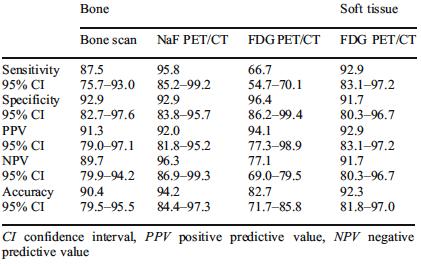
15 Ø Qualitative assessment of conventional bone scintigraphy with 99m Tc MDP is an insensitive method for monitoring the treatment response of bone metastases Ø F-fluoride positron emission tomography (PET) might serve as a suitable alternative biomarker of the treatment response Ø Semi-quantitative F-fluoride PET is more accurate than the qualitative comparison of scans and correlates with the PSA response and ALP activity

16 Qualitative response assessment. MIP images at 0 and 12 weeks in two subjects, subject A and subject B, showing no significant qualitative change.
17 Mean SUVmax, PSA and ALP changes. Mean SUVmax, PSA and ALP changes at 6 and 12 weeks as a percentage of baseline levels in the five subjects (A to E).
18
 Ø 19 sarcoma, prostate cancer, 6 breast cancer, 2 colon cancer, 1 bladder cancer, 1 lung cancer, 1 malignant paraganglioma, 1 lymphoma, 1 gastrointestinal stromal tumor, 1 renal cancer and 1")
19 Ø 52 patients with proven malignancy, referred for evaluation of skeletal metastases Ø 37 men and 15 women, year-old (average: 55.6 ± 15.9) Ø 19 sarcoma, prostate cancer, 6 breast cancer, 2 colon cancer, 1 bladder cancer, 1 lung cancer, 1 malignant paraganglioma, 1 lymphoma, 1 gastrointestinal stromal tumor, 1 renal cancer and 1 salivary gland cancer Ø 99m Tc MDP bone scintigraphy, F NaF PET/CT and F FDG PET/CT were subsequently performed within 1 month

20 99m Tc MDP F FDG F NaF 61 year-old woman with metastatic breast cancer

21 F FDG PET/CT F NaF PET/CT 61 year-old woman with metastatic breast cancer

22 99m Tc MDP F FDG F NaF 73-year-old man with metastatic prostate cancer

23 F FDG PET/CT F NaF PET/CT 73-year-old man with metastatic prostate cancer
24 99m Tc MDP bone scan F NaF PET/CT F FDG PET/CT Skeletal lesions 22/52 24/52 16/52 Other lesions N/A N/A 28/52 Ø The image quality and evaluation of extent of disease was superior by F NaF PET/CT over 99m Tc MDP scintigraphy in all 22 patients with skeletal lesions on both scans and over F FDG PET/CT in 11/16 patients with skeletal metastases on F FDG PET/CT Ø In 2 patients (one with sarcoma and another with prostate cancer), the F NaF PET/CT showed skeletal metastases not seen on either of the other 2 scans Ø Extra-skeletal metastases were identified by F FDG PET/CT in 28/52 participants
25 Diagnostic effectiveness:
, salivary gland cancer (1 participant) and renal cancer (1 participant) Ø F NaF PET/CT, F FDG PET/CT and WBMRI were performed within 1 month for each")
26 Ø 10 participants (5 men, 5 women, year-old) diagnosed with cancer and known osseous metastases Ø The diagnoses included breast cancer (5 participants), prostate cancer (3 participants), salivary gland cancer (1 participant) and renal cancer (1 participant) Ø F NaF PET/CT, F FDG PET/CT and WBMRI were performed within 1 month for each participant
27
28 Ø The image quality and evaluation of extent of disease was superior by F NaF PET/CT compared to 99m Tc-MDP scintigraphy in all patients with skeletal lesions and compared to F FDG PET/CT in 3 of the patients with skeletal metastases Ø F NaF PET/CT showed osseous metastases where F FDG PET/ CT was negative in another 3 participants Ø Extra-skeletal metastases were identified by F FDG PET/CT in 6 participants Ø WBMRI with the combination of IDEAL, STIR and DWI pulse sequences showed fewer lesions than F NaF PET/CT in 5 patients, same number of lesions in 2 patients and more lesions in 1 patient Ø When compared to F FDG, WBMRI showed fewer lesions in 3 patients and the same amount of lesions in 6 patients

29 99m Tc MDP F FDG F NaF 73 year-old man with prostate cancer

30 99m Tc MDP F FDG F NaF 65 year-old man with prostate cancer


31 99m Tc MDP F FDG F NaF 67 year-old man with urothelial cancer

32 99m Tc MDP 65 year-old man with RCC F FDG F NaF
33 99m Tc MDP F FDG F NaF 58 year-old man with prostate cancer
34 99m Tc MDP F FDG F NaF 72 year-old woman with breast cancer
35 Ø Introduction Ø F NaF PET/CT in Primary Bone Cancers Ø F NaF PET/CT in Bone Metastases Ø Future Directions

36 Ø 115 patients with proven malignancy who had separate F NaF PET/CT, F FDG PET/CT and a combined F NaF/ F FDG PET/CT scans for evaluation of malignancy (total of 3 scans each) Ø 63 men and 52 women, year-old (average: 58.5 ± 14.3) Ø Tumor type: prostate cancer (41 participants), breast cancer (39 participants), sarcoma (22 participants), and other cancers (13 participants) Ø The interval between the first and third scan ranged 3-28 days (average: 6.7±4.9 days) Ø A direct comparison for each detected lesion was performed among the 3 scans


37 0-5 min min min min F NaF PET F FDG PET F NaF & FDG PET

38 74 year-old man with metastatic prostate cancer.

39 45 year-old woman with metastatic breast cancer

40 38 year-old woman with metastatic breast cancer

41 F FDG PET/CT F NaF PET/CT F NaF & F FDG PET/CT 68-year-old man with metastatic colon cancer

42 F FDG PET/CT F NaF PET/CT F NaF & F FDG PET/CT 75-year-old man with metastatic prostate cancer
43
44

45 Primary: breast (Denmark) FDG Cocktail FDG Cocktail
46 F FDG PET/CT F NaF PET/CT F NaF & F FDG PET/CT Skeletal lesions 38/115 67/115 67*/115 Ø F NaF PET/CT and F FDG PET/CT scans identified malignant lesions in 82/115 enrolled patients (71.3%) Ø 19 participants: F NaF > F FDG (osseous metastases) Ø 29 patients: F NaF positive, F FDG negative (osseous metastases) Ø participants: F NaF = F FDG (osseous metastases) Ø 1 patient: F FDG positive, FNaFG negative (osseous metastases) Ø 48 participants had no osseous metastases identified on the F NaF PET/CT or the F FDG PET/CT scans *2 skull lesions missed
47 F FDG PET/CT F NaF PET/CT F NaF & F FDG PET/CT Other lesions 48/115 N/A 48**/115 Ø F FDG PET/CT detected lesions outside the skeleton in 48/115 participants (42.2%) Ø The most common extra skeletal sites of metastases were lymph nodes (28/115 patients), lungs (14/115 patients) and the liver (8/115 patients) Ø The combined F NaF/ F FDG PET/CT scans missed three F FDGavid lung nodules in 2 patients and two F NaF-avid skull lesions in another 2 patients. These 4 patients had other sites of metastatic disease in addition to the ones not clearly identified on the combined PET/CT. **subcentimeter lung nodules missed
48 Diagnostic effectiveness:
49 99m Tc MDP bone scan F FDG PET/CT F NaF/ F FDG PET Technical fee: $275 Technical fee: $1,421 Technical fee: $1,421 Professional fee: $48 Professional fee: $140 Professional fee: $140 ($280) 99m Tc MDP: $100 F FDG: $250 F FDG: $250 Total: $423 Total: $11 F NaF: $150 Total: $2,234 Total: $1,961 ($2,101) Ø There are approximately 2 million 99m Tc MDP bone scans performed for detection of cancer annually in the US and approximately 1 million F FDG PET/CT scans performed in the same population Ø This can potentially amount to a total of approximately $130 million saved annually in reimbursement
50 Radiation Dosimetry 99m Tc MDP bone scan 420 mrem F FDG PET/CT 1650 mrem* 1000 mrem* F NaF & F FDG PET/CT 1650 mrem** 500 mrem** 1000 mrem** Radiation Exposure 3070 mrem 3150 mrem * 110 mrem/mci from F FDG and 1000 mrem from the low-dose CT ** 110 mrem/mci from F FDG, 100 mrem/mci from F NaF and 1000 mrem from the low-dose CT
51 Ø The uptake mechanism of F NaF resembles that of 99m Tc- MDP, with better pharmacokinetic characteristics including faster blood clearance and 2-fold higher uptake in bone Ø Uptake of F NaF reflects blood flow and bone remodeling Ø The use of novel hybrid PET/CT systems has significantly improved the specificity of F NaF imaging, because the CT component of the study allows morphologic characterization of the functional lesion and more accurate differentiation between benign lesions and metastases
52 Concerning bone scintigraphy with F NaF, the following statement is correct: a) F NaF is less protein bound in the blood, therefore allowing shorter time from injection to imaging when compared to 99m Tc MDP b) F NaF is the only FDA-approved radiopharmaceutical for skeletal imaging c) F NaF PET/CT imaging requires special patient preparation d) F NaF is most useful for evaluation of lytic skeletal lesions
53 Published results suggest the most appropriate use of F NaF PET/CT is in the following clinical scenarios: a) To differentiate between benign and malignant bone lesions b) To evaluate for skeletal metastases when results of other imaging studies are equivocal c) To evaluate for pulmonary metastases d) To measure tumor hypoxia

54 THANK YOU!
Ryan Niederkohr, M.D. Slides are not to be reproduced without permission of author
 Ryan Niederkohr, M.D. CMS: PET/CT CPT CODES 78814 Limited Area (e.g., head/neck only; chest only) 78815 78816 Regional (skull base to mid-thighs) True Whole Body (skull vertex to feet) SELECTING FIELD
Using PET/CT in Prostate Cancer
 Using PET/CT in Prostate Cancer Legal Disclaimer These materials were prepared in good faith by MITA as a service to the profession and are believed to be reliable based on current scientific literature.
Whole body F-18 sodium fluoride PET/CT in the detection of bone metastases in patients with known malignancies: A pictorial review
 Whole body F-18 sodium fluoride PET/CT in the detection of bone metastases in patients with known malignancies: A pictorial review Poster No.: C-1196 Congress: ECR 2014 Type: Educational Exhibit Authors:
Dr Sneha Shah Tata Memorial Hospital, Mumbai.
 Dr Sneha Shah Tata Memorial Hospital, Mumbai. Topics covered Lymphomas including Burkitts Pediatric solid tumors (non CNS) Musculoskeletal Ewings & osteosarcoma. Neuroblastomas Nasopharyngeal carcinomas
Prof. Dr. NAGUI M. ABDELWAHAB,M.D.; MARYSE Y. AWADALLAH, M.D. AYA M. BASSAM, Ms.C.
 Role of Whole-body Diffusion MR in Detection of Metastatic lesions Prof. Dr. NAGUI M. ABDELWAHAB,M.D.; MARYSE Y. AWADALLAH, M.D. AYA M. BASSAM, Ms.C. Cancer is a potentially life-threatening disease,
Index. Surg Oncol Clin N Am 16 (2007) Note: Page numbers of article titles are in boldface type.
 Note: Page numbers of article titles are in boldface type.") Surg Oncol Clin N Am 16 (2007) 465 469 Index Note: Page numbers of article titles are in boldface type. A Adjuvant therapy, preoperative for gastric cancer, staging and, 339 B Breast cancer, metabolic
Surg Oncol Clin N Am 16 (2007) 465 469 Index Note: Page numbers of article titles are in boldface type. A Adjuvant therapy, preoperative for gastric cancer, staging and, 339 B Breast cancer, metabolic
Clinical indications for positron emission tomography
 Clinical indications for positron emission tomography Oncology applications Brain and spinal cord Parotid Suspected tumour recurrence when anatomical imaging is difficult or equivocal and management will
Joint Comments on Positron Emission Tomography (NaF-18) to Identify Bone Metastasis of Cancer (CAG-00065R1)
 to Identify Bone Metastasis of Cancer (CAG-00065R1)") July 2, 2009 Tamara Syrek Jensen, J.D. Acting Director, Coverage and Analysis Group Centers for Medicare & Medicaid Services 7500 Security Blvd., Mail Stop C1-09-06 Baltimore, MD 21244 Re: Joint Comments
July 2, 2009 Tamara Syrek Jensen, J.D. Acting Director, Coverage and Analysis Group Centers for Medicare & Medicaid Services 7500 Security Blvd., Mail Stop C1-09-06 Baltimore, MD 21244 Re: Joint Comments
Combined 18 F-Fluoride and 18 F-FDG PET/CT Scanning for Evaluation of Malignancy: Results of an International Multicenter Trial
 Combined F-Fluoride and F-FDG PET/CT Scanning for Evaluation of Malignancy: Results of an International Multicenter Trial Andrei Iagaru 1, Erik Mittra 1, Camila Mosci 1, David W. Dick 1, Mike Sathekge
FDG PET/CT STAGING OF LUNG CANCER. Dr Shakher Ramdave
 FDG PET/CT STAGING OF LUNG CANCER Dr Shakher Ramdave FDG PET/CT STAGING OF LUNG CANCER FDG PET/CT is used in all patients with lung cancer who are considered for curative treatment to exclude occult disease.
PET-MRI in malignant bone tumours. Lars Stegger Department of Nuclear Medicine University Hospital Münster, Germany
 PET-MRI in malignant bone tumours Lars Stegger Department of Nuclear Medicine University Hospital Münster, Germany Content From PET to PET/MRI General considerations Bone metastases Primary bone tumours
Indications of PET/CT in oncology
 Monday, August 27, 2012 Session 1, 10:00-10:40 Indications of PET/CT in oncology Helle Westergren Hendel MD, PhD, assistant professor Bacelor in Leadership & Health Ecomomics Head of Clinical PET, Herlev
Nuclear Medicine: Manuals. Nuclear Medicine. Nuclear imaging. Emission imaging: study types. Bone scintigraphy - technique
 Nuclear Medicine - Unsealed radioactive preparations the tracer mixes with the patients body fluids on a molecular level (e.g. after intravenous injection) - 3 main fields: - In vitro : measuring concentrations
Los Angeles Radiological Society 62 nd Annual Midwinter Radiology Conference January 31, 2010
 Los Angeles Radiological Society 62 nd Annual Midwinter Radiology Conference January 31, 2010 Self Assessment Module on Nuclear Medicine and PET/CT Case Review FDG PET/CT IN LYMPHOMA AND MELANOMA Submitted
POSITRON EMISSION TOMOGRAPHY (PET)
") Status Active Medical and Behavioral Health Policy Section: Radiology Policy Number: V-27 Effective Date: 08/27/2014 Blue Cross and Blue Shield of Minnesota medical policies do not imply that members should
Status Active Medical and Behavioral Health Policy Section: Radiology Policy Number: V-27 Effective Date: 08/27/2014 Blue Cross and Blue Shield of Minnesota medical policies do not imply that members should
Bone PET/MRI : Diagnostic yield in bone metastases and malignant primitive bone tumors
 Bone PET/MRI : Diagnostic yield in bone metastases and malignant primitive bone tumors Lars Stegger, Benjamin Noto Department of Nuclear Medicine University Hospital Münster, Germany Content From PET to
Medical Policy An independent licensee of the Blue Cross Blue Shield Association
 PET Scanning: Oncologic Applications Page 1 of 88 Medical Policy An independent licensee of the Blue Cross Blue Shield Association Title: Positron Emission Tomography (PET) Scanning: Oncologic Applications
performed to help sway the clinician in what the appropriate diagnosis is, which can substantially alter the treatment of management.
 Hello, I am Maura Polansky at the University of Texas MD Anderson Cancer Center. I am a Physician Assistant in the Department of Gastrointestinal Medical Oncology and the Program Director for Physician
Whole Body MRI. Dr. Nina Tunariu. Prostate Cancer recurrence, progression and restaging
 Whole Body MRI Prostate Cancer recurrence, progression and restaging Dr. Nina Tunariu Consultant Radiology Drug Development Unit and Prostate Targeted Therapies Group 12-13 Janeiro 2018 Evolving Treatment
1 Introduction. 2 Materials and methods. LI Na 1 LI Yaming 1,* YANG Chunming 2 LI Xuena 1 YIN Yafu 1 ZHOU Jiumao 1
 Nuclear Science and Techniques 20 (2009) 354 358 18 F-FDG PET/CT in diagnosis of skeletal metastases LI Na 1 LI Yaming 1,* YANG Chunming 2 LI Xuena 1 YIN Yafu 1 ZHOU Jiumao 1 1 Department of Nuclear Medicine,
Nuclear medicine in oncology. 1. Diagnosis 2. Therapy
 Nuclear medicine in oncology 1. Diagnosis 2. Therapy Diagnosis - Conventional methods - Nonspecific radiopharmaceuticals cumulating in tumours - Specific radiopharmaceuticals (receptor- and immunoscintigraphy)
Radiology Pathology Conference
 Radiology Pathology Conference Sharlin Johnykutty,, MD, Cytopathology Fellow Sara Majewski, MD, Radiology Resident Friday, August 28, 2009 Presentation material is for education purposes only. All rights
Bone Metastasis. Patient Management with PET/CT
 Bone Metastasis Patient Management with PET/CT Bone Metastasis Patient Management with PET/CT OVERVIEW Bone Metastasis is a process in which cancer cells from the original or primary tumor site break away
Re: Comments on Proposed Decision Memorandum (CAG-00065R2) Positron Emission Tomography (NaF-18) to Identify Bone Metastasis of Cancer
 Positron Emission Tomography (NaF-18) to Identify Bone Metastasis of Cancer") 1300 North 17 th Street Suite 900 Arlington, Virginia 22209 Tel: 703.841.3200 Fax: 703.841.3392 www.medicalimaging.org October 15, 2015 CAGInquiries@cms.hhs.gov Tamara Syrek Jensen, Esq. Director, Coverage
1300 North 17 th Street Suite 900 Arlington, Virginia 22209 Tel: 703.841.3200 Fax: 703.841.3392 www.medicalimaging.org October 15, 2015 CAGInquiries@cms.hhs.gov Tamara Syrek Jensen, Esq. Director, Coverage
Research Article Prevalence of Clinically Significant Extraosseous Findings on Unenhanced CT Portions of 18 F-Fluoride PET/CT Bone Scans
 The Scientific World Journal Volume 2012, Article ID 979867, 5 pages doi:10.1100/2012/979867 The cientificworldjournal Research Article Prevalence of Clinically Significant Extraosseous Findings on Unenhanced
Oncologic Applications of PET Scanning
 6.01.26 Oncologic Applications of PET Scanning Section 6.0 Radiology Subsection Effective Date February 15, 2015 Original Policy Date January 26, 2009 Next Review Date December 2015 Description Positron
PET IMAGING (POSITRON EMISSION TOMOGRAPY) FACT SHEET
 FACT SHEET") Positron Emission Tomography (PET) When calling Anthem (1-800-533-1120) or using the Point of Care authorization system for a Health Service Review, the following clinical information may be needed to
Positron Emission Tomography (PET) When calling Anthem (1-800-533-1120) or using the Point of Care authorization system for a Health Service Review, the following clinical information may be needed to
Chapter 10. Summary, conclusions and future perspectives
 Chapter 10 Summary, conclusions and future perspectives 10.1 SUMMARY In this thesis, a new tumor imaging tracer in nuclear medicine is studied. This 123 tracer, L-3-[ I]Iodo-alpha-methyl-tyrosine (IMT),
objectives Pitfalls and Pearls in PET/CT imaging Kevin Robinson, DO Assistant Professor Department of Radiology Michigan State University
 objectives Pitfalls and Pearls in PET/CT imaging Kevin Robinson, DO Assistant Professor Department of Radiology Michigan State University To determine the regions of physiologic activity To understand
New Visions in PET: Surgical Decision Making and PET/CT
 New Visions in PET: Surgical Decision Making and PET/CT Stanley J. Goldsmith, MD Director, Nuclear Medicine Professor, Radiology & Medicine New York Presbyterian Hospital- Weill Cornell Medical Center
PET/CT Frequently Asked Questions
 PET/CT Frequently Asked Questions General Q: Is FDG PET specific for cancer? A: No, it is a marker of metabolism. In general, any disease that causes increased metabolism can result in increased FDG uptake
Case Reports: Tumor Detection by Diffusion-Weighted MRI and ADC-Mapping with Correlation to PET/CT Results
 Case Reports: Tumor Detection by Diffusion-Weighted MRI and ADC-Mapping with Correlation to PET/CT Results Matthias Philipp Lichy, M.D.; Philip Aschoff, M.D.; Christina Pfannenberg, M.D.; Schlemmer Heinz-Peter,
Quantitative Nuclear Medicine Imaging in Oncology. Susan E. Sharp, MD
 Quantitative Nuclear Medicine Imaging in Oncology Susan E. Sharp, MD Disclosures None Objectives Describe scoring systems used in oncologic nuclear medicine imaging Deauville scoring in lymphoma Curie
Value of true whole-body FDG- PET/CT scanning protocol in oncology and optimization of its use based on primary malignancy
 Value of true whole-body FDG- PET/CT scanning protocol in oncology and optimization of its use based on primary malignancy Ronnie Sebro MD, Ph.D Carina Mari Aparici MD, Miguel Hernandez Pampaloni MD, PhD
PET/CT in breast cancer staging
 PET/CT in breast cancer staging Anni Morsing Consultant, PhD, DMSc Rigshospitalet 1 18F- FDG PET/CT for breastcancer staging Where is the clinical impact? To which women should 18F- FDG PET/CT be offered?
PET/CT imaging and RIT of prostate cancer. Kirsten Bouchelouche, MD, DMSc PET & Cyclotron Unit Rigshospitalet, Copenhagen Denmark
 PET/CT imaging and RIT of prostate cancer Kirsten Bouchelouche, MD, DMSc PET & Cyclotron Unit Rigshospitalet, Copenhagen Denmark Prostate cancer Prostate cancer is the most common malignancy in men Imaging
Page: 1 of 29. For this policy, PET scanning is discussed for the following 4 applications in oncology:
 Emission Tomography Scanning Page: 1 of 29 Last Review Status/Date: June 2015 Description Positron emission tomography (PET) scans are based on the use of positron-emitting radionuclide tracers coupled
Hybrid Imaging SPECT/CT PET/CT PET/MRI. SNMMI Southwest Chapter Aaron C. Jessop, MD
 Hybrid Imaging SPECT/CT PET/CT PET/MRI SNMMI Southwest Chapter 2014 Aaron C. Jessop, MD Assistant Professor, Department of Nuclear Medicine UT MD Anderson Cancer Center, Houston, Texas Complimentary role
Recommendations for cross-sectional imaging in cancer management, Second edition
 www.rcr.ac.uk Recommendations for cross-sectional imaging in cancer management, Second edition Musculoskeletal tumours Faculty of Clinical Radiology www.rcr.ac.uk Contents Primary bone tumours 3 Clinical
Principles of nuclear metabolic imaging. Prof. Dr. Alex Maes AZ Groeninge Kortrijk and KULeuven Belgium
 Principles of nuclear metabolic imaging Prof. Dr. Alex Maes AZ Groeninge Kortrijk and KULeuven Belgium I. Molecular imaging probes A. Introduction - Chemical disturbances will precede anatomical abnormalities
Medical Policy An independent licensee of the Blue Cross Blue Shield Association
 PET Scanning: Oncologic Applications Page 1 of 42 Medical Policy An independent licensee of the Blue Cross Blue Shield Association Title: See also: Positron Emission Tomography (PET) Scanning: Oncologic
FDG-PET/CT in Gynaecologic Cancers
 Friday, August 31, 2012 Session 6, 9:00-9:30 FDG-PET/CT in Gynaecologic Cancers (Uterine) cervical cancer Endometrial cancer & Uterine sarcomas Ovarian cancer Little mermaid (Edvard Eriksen 1913) honoring
Subject: PET Scan With or Without CT Attenuation. Original Effective Date: 11/7/2017. Policy Number: MCR: 610. Revision Date(s): Review Date:
: Review Date:") Subject: PET Scan With or Without CT Attenuation Policy Number: MCR: 610 Revision Date(s): MHW Original Effective Date: 11/7/2017 Review Date: DISCLAIMER This Molina Clinical Review (MCR) is intended to
Subject: PET Scan With or Without CT Attenuation Policy Number: MCR: 610 Revision Date(s): MHW Original Effective Date: 11/7/2017 Review Date: DISCLAIMER This Molina Clinical Review (MCR) is intended to
Detection of Soft Tissue Tumors on Bone Scintigraphy: Report of Four Cases
 Detection of Soft Tissue Tumors on Bone Scintigraphy: Report of Four Cases Fariba Akhzari 1 MD and Mahrokh Daemi MD 2 1 Nuclear Medicine Department, 2 General Surgery Department, Sina Hospital, Faculty
Molecular Imaging of Bone Metastasis Hojjat Ahmadzadehfar
 Molecular Imaging of Bone Metastasis Hojjat Ahmadzadehfar Seite 1 Bone metastases Different tumors % 80 70 68% 73% 60 50 40 40% 35% 42% 36% 30 20 10 0 5% prostate bladder renal breast thyroid lung GI urological
FLUCICLOVINE: 1 ST FDA APPROVED F-18 PET IMAGING AGENT FOR RECURRENT PROSTATE CANCER
 FLUCICLOVINE: 1 ST FDA APPROVED F-18 PET IMAGING AGENT FOR RECURRENT PROSTATE CANCER KEVIN P BANKS, MD SAN ANTONIO MILITARY MEDICAL CENTER ASSISTANT PROFESSOR OF RADIOLOGY, USU I HAVE NO FINANCIAL DISCLOSURES.
Contents. 3 Pneumology Introduction Positron Emission Tomography: Past and Present 1. 2 Fundamentals. xxx
 xxx IX Contents 1 Introduction Positron Emission Tomography: Past and Present 1 1.1 Survey.......................... 1 Physical and Biochemical Fundamentals.... 2 PET in National and International Medical
Assessment of renal cell carcinoma by two PET tracer : dual-time-point C-11 methionine and F-18 fluorodeoxyglucose
 Assessment of renal cell carcinoma by two PET tracer : dual-time-point C-11 methionine and F-18 fluorodeoxyglucose Poster No.: C-0805 Congress: ECR 2015 Type: Scientific Exhibit Authors: S. Ito, K. Kato,
Unusual Osteoblastic Secondary Lesion as Predominant Metastatic Disease Spread in Two Cases of Uterine Leiomyosarcoma
 49 Unusual Osteoblastic Secondary Lesion as Predominant Metastatic Disease Spread in Two Cases of Uterine Leiomyosarcoma Loredana Miglietta a Maria Angela Parodi b Luciano Canobbio b Luca Anselmi c a Medical
Thyroid Cancer: Imaging Techniques (Nuclear Medicine)
") Thyroid Cancer: Imaging Techniques (Nuclear Medicine) Andrei Iagaru, MD MIPS Molecular Imaging Program at Stanford Stanford University School of Medicine Department of Radiology Introduction Ø There are
Thyroid Cancer: Imaging Techniques (Nuclear Medicine) Andrei Iagaru, MD MIPS Molecular Imaging Program at Stanford Stanford University School of Medicine Department of Radiology Introduction Ø There are
PET imaging of cancer metabolism is commonly performed with F18
 PCRI Insights, August 2012, Vol. 15: No. 3 Carbon-11-Acetate PET/CT Imaging in Prostate Cancer Fabio Almeida, M.D. Medical Director, Arizona Molecular Imaging Center - Phoenix PET imaging of cancer metabolism
Radiation Dosimetry for CT Protocols
 Radiation Dosimetry for CT Protocols This document contains radiation dosimetry information from CT scans and can be used by investigators to estimate the dosimetry information required by the JRSC or
Sodium Fluoride F 18 Injection* PET/CT Imaging
 Sodium Fluoride F 18 Injection* PET/CT Imaging for Prostate Cancer Sodium Fluoride F 18 Injection* ( 18 F NaF) PET/CT Imaging of Bone Metastases in Prostate Cancer About Prostate Cancer According to the
Novel Imaging in Advanced Prostate Cancer
 Novel Imaging in Advanced Prostate Cancer Robert J. Hamilton, MD MPH FRCSC Princess Margaret Cancer Centre ICUC Saturday January 21, 2017 Company/Organizati Details Faculty/Presenter on Disclosures I am
Hybrid bone scintigraphy in gastrointestinal malignancies Institutional Experience
 Hybrid bone scintigraphy in gastrointestinal malignancies Institutional Experience Nazia Rashid 1, Saima Riaz 1, Humayun Bashir 1, Shafqat Mehmood 2 1 Nuclear Medicine Department ad 2 Internal Medicine
Immunohistochemistry in Bone and Soft Tissue Tumors. Sahar Rassi Zankoul, MD
 Immunohistochemistry in Bone and Soft Tissue Tumors Sahar Rassi Zankoul, MD Introduction Bone tumors represent a wide variety of tumors of various origins and malignant potentials. These different tumor
PET/CT in lung cancer
 PET/CT in lung cancer Andrei Šamarin North Estonia Medical Centre 3 rd Baltic Congress of Radiology 08.10.2010 Imaging in lung cancer Why do we need PET/CT? CT is routine imaging modality for staging of
Does PET/CT Have an Additional Value in Detection of Osteolytic Bone Metastases.
 Egyptian J. Nucl. Med., Vol 2, No. 2, Dec. 2009 65 ONCOLOGY, Original Article Does PET/CT Have an Additional Value in Detection of Osteolytic Bone Metastases. R. Riad, M.D.*, M. Awad, M.D. **, E. Eldebawy,
Positron Emission Tomography and Bone Metastases
 Positron Emission Tomography and Bone Metastases Ignac Fogelman, BSc, MD, FRCP,* Gary Cook, MSc, MD, FRCP, FRCR, Ora Israel, MD, and Hans Van der Wall, MBBS, PhD, FRACP The use of 2-[ 18 F]fluoro-2-deoxy-D-glucose
FieldStrength. Leuven research is finetuning. whole body staging
 FieldStrength Publication for the Philips MRI Community Issue 40 May 2010 Leuven research is finetuning 3.0T DWIBS for whole body staging The University Hospital of Leuven is researching 3.0T whole body
Nuclear Sciences and Medicine
 Nuclear Sciences and Medicine Rethy Chhem, MD, PhD (Edu), PhD (His), FRCPC Division of Human Health Guest Professor, Medical University of Vienna International Atomic Energy Agency Medical Imaging X-rays
Imaging of bone metastases
 Imaging of bone metastases Antoine Feydy Service de Radiologie B Hôpital Cochin APHP Université Paris Descartes antoine.feydy@aphp.fr MEXICO 2016 INTRODUCTION Diagnostic Imaging Imaging Modalities Strengths,
Austin Radiological Association Nuclear Medicine Procedure PET SODIUM FLUORIDE BONE SCAN (F-18 NaF)
") Austin Radiological Association Nuclear Medicine Procedure PET SODIUM FLUORIDE BONE SCAN (F-18 NaF) Overview Indication Sodium Fluoride F18 injection is a radioactive diagnostic agent for positron emission
Austin Radiological Association Nuclear Medicine Procedure PET SODIUM FLUORIDE BONE SCAN (F-18 NaF) Overview Indication Sodium Fluoride F18 injection is a radioactive diagnostic agent for positron emission
Radiological assessment of neoadjuvent chemotherapy for breast cancer
 XV th Balkan Congress of Radiology Budapest, Hungary, October 12 15, 2017 Radiological assessment of neoadjuvent chemotherapy for breast cancer V. Bešlagić C l i n i c o f R a d i o l o g y, U n i v e
Positron Emission Tomography in Lung Cancer
 May 19, 2003 Positron Emission Tomography in Lung Cancer Andrew Wang, HMS III Patient DD 53 y/o gentleman presented with worsening dyspnea on exertion for the past two months 30 pack-year smoking Hx and
Prostate Cancer Basics: Background Information for Outreach Activities with Oncologists, Urologists and Surgeons
 Prostate Cancer Basics: Background Information for Outreach Activities with Oncologists, Urologists and Surgeons Legal Disclaimer These materials were prepared in good faith by MITA as a service to the
PET/CT Value: Rocky Mountain Cancer Centers
 PET/CT Value: Rocky Mountain Cancer Centers Glenn Balasky Executive Director Rocky Mountain Cancer Centers glenn.balasky@usoncology.com CANM/CAMRT Joint Conference March 22, 2018 Vancouver, British Columbia
Ga68 Imaging. Roland HUSTINX Division of Nuclear Medicine and Oncologic Imaging Centre Hospitalier Universitaire de Liège Belgium
 Ga68 Imaging Roland HUSTINX Division of Nuclear Medicine and Oncologic Imaging Centre Hospitalier Universitaire de Liège Belgium 68 Ga Produced by a 68 Ge/ 68 Ga generator Decays by positron emission
Outcomes Report: Accountability Measures and Quality Improvements
 Outcomes Report: Accountability Measures and Quality Improvements The FH Memorial Medical Center s Cancer Committee ensures that patients with cancer are treated according to the nationally accepted measures.
Nuclear Medicine Head and Neck Region. Bán Zsuzsanna, MD University of Pécs, Department of Nuclear Medicine
 Nuclear Medicine Head and Neck Region Bán Zsuzsanna, MD University of Pécs, Department of Nuclear Medicine Thyroid scintigraphy Parathyroid scintigraphy F18-FDG PET examinations in head and neck cancer
Department of Nuclear Medicine with Positron Emission Tomography
 (PET) Unit [1] Contact information: Registration: +48 41 367 4850 Main office: +48 41 367 4860 Fax: +48 41 367 4887 e-mail: zmnsco@onkol.kielce.pl [2] Head of the Department: Professor Janusz Braziewicz
Department of Radiology, Division of Nuclear Medicine, University of Wisconsin Madison, Madison, WI, USA; 2
 Am J Nucl Med Mol Imaging 2015;5(2):162-168 www.ajnmmi.us /ISSN:2160-8407/ajnmmi0002089 Original Article Differentiation of metastatic vs degenerative joint disease using semi-quantitative analysis with
PSMA PET SCANNING AND THERANOSTICS IN PROSTATE CANCER KEVIN TRACEY, MD, FRCPC PRECISION DIAGNSOTIC IMAGING REGIONAL PET/CT CENTRE
 PSMA PET SCANNING AND THERANOSTICS IN PROSTATE CANCER KEVIN TRACEY, MD, FRCPC PRECISION DIAGNSOTIC IMAGING REGIONAL PET/CT CENTRE DISCLOSURES/CONFLICTS NONE OBJECTIVES Understand current diagnostic role
Assessment of Skeletal Metastases in Prostate Cancer: 68Ga-PSMA PET vs 99mTc-MDP WBBS - A Case Series
 Assessment of Skeletal Metastases in Prostate Cancer: 68Ga-PSMA PET vs 99mTc-MDP WBBS - A Case Series Poster No.: R-0094 Congress: 2016 ASM Type: Scientific Exhibit Authors: O. Bennett, Y.-T. T. Huang;
Emerging Referral Patterns for Whole-Body Diffusion Weighted Imaging (WB-DWI) in an Oncology Center
 in an Oncology Center") Emerging Referral Patterns for Whole-Body Diffusion Weighted Imaging (WB-DWI) in an Oncology Center Poster No.: C-1296 Congress: ECR 2014 Type: Scientific Exhibit Authors: G. Petralia 1, G. Conte 1, S.
Emerging Referral Patterns for Whole-Body Diffusion Weighted Imaging (WB-DWI) in an Oncology Center Poster No.: C-1296 Congress: ECR 2014 Type: Scientific Exhibit Authors: G. Petralia 1, G. Conte 1, S.
NCCN Practice Guidelines Narrative Summary of Indications for FDG PET and PET/CT
 NCCN Practice Guidelines Narrative Summary of Indications for FDG PET and PET/CT NCCN guidelines were reviewed on 2/14/2016 for utilization of 18F-fluorodeoxyglucose (FDG) PET and PET/CT (available at:
Heterogeneous osteoblastic activity in the right ischium of unclear etiology seen on NaF18-PET/CT
 CASE REPORT Heterogeneous osteoblastic activity in the right ischium of unclear etiology seen on NaF18-PET/CT Aung Zaw Win, Carina Mari Aparici Dept. Radiology, Nuclear Medicine section, San Francisco
Repeatability and reproducibility of 18 F-NaF PET quantitative imaging biomarkers
 Repeatability and reproducibility of 18 F-NaF PET quantitative imaging biomarkers Christie Lin, Tyler Bradshaw, Timothy Perk, Stephanie Harmon, Glenn Liu, Robert Jeraj University of Wisconsin Madison,
A 64 y.o. man presents to the hospital with persistent cough and hemoptysis. Fernando Mut Montevideo - Uruguay
 A 64 y.o. man presents to the hospital with persistent cough and hemoptysis Fernando Mut Montevideo - Uruguay Teaching case Bone # 1 A 64 y.o. man presents to the hospital with persistent cough and hemoptysis.
Pathology of Sarcoma ELEANOR CHEN, MD, PHD, ASSISTANT PROFESSOR DEPARTMENT OF PATHOLOGY UNIVERSITY OF WASHINGTON
 Pathology of Sarcoma ELEANOR CHEN, MD, PHD, ASSISTANT PROFESSOR DEPARTMENT OF PATHOLOGY UNIVERSITY OF WASHINGTON Presentation outline Background and epidemiology of sarcomas Sarcoma classification Sarcoma
The Role of PET / CT in Lung Cancer Staging
 July 2004 The Role of PET / CT in Lung Cancer Staging Vlad Vinarsky, Harvard Medical School Year IV Patient AM HPI: 81 yo F p/w hemoptysis x 1 month LLL lesion on CXR, not responsive to Abx 35 pack-year
An Introduction to PET Imaging in Oncology
 January 2002 An Introduction to PET Imaging in Oncology Janet McLaren, Harvard Medical School Year III Basics of PET Principle of Physiologic Imaging: Allows in vivo visualization of structures by their
Phillip J. Koo, MD Division Chief of Diagnostic Imaging Banner MD Anderson Cancer Center, USA
 ADVANCED PET IMAGING IN PROSTATE CANCER Phillip J. Koo, MD Division Chief of Diagnostic Imaging Banner MD Anderson Cancer Center, USA PET, positron-emission tomography DISCLAIMER Please note: The views
The Use of PET Scanning in Urologic Oncology
 The Use of PET Scanning in Urologic Oncology Dr Nicholas C. Buchan Uro-oncology Fellow 1 2 Aims To understand the basic concepts underlying PET scanning. Understand the emerging role of PET Scanning for
Molecular Imaging and Cancer
 Molecular Imaging and Cancer Cancer causes one in every four deaths in the United States, second only to heart disease. According to the U.S. Department of Health and Human Services, more than 512,000
Staging Colorectal Cancer
 Staging Colorectal Cancer CT is recommended as the initial staging scan for colorectal cancer to assess local extent of the disease and to look for metastases to the liver and/or lung Further imaging for
PET/CT in Breast Cancer
 PET/CT in Breast Cancer Rodolfo Núñez Miller, M.D. Nuclear Medicine and Diagnostic Imaging Section Division of Human Health International Atomic Energy Agency Vienna, Austria Overview Introduction Locorregional
ORIGINAL ARTICLE ABSTRACT
 ORIGINAL ARTICLE 99m Tc MDP Bone Scan in Lung Cancer: Predilection Sites for Metastasis Shamim M F Begum, Zeenat Jabin, Rahima Perveen, Nasreen Sultana, Laila S Banu National Institute of Nuclear Medicine
Nuclear Medicine in Thyroid Cancer. Phillip J. Koo, MD Division Chief of Diagnostic Imaging
 Nuclear Medicine in Thyroid Cancer Phillip J. Koo, MD Division Chief of Diagnostic Imaging Financial Disclosures Bayer Janssen Learning Objectives To learn the advantages and disadvantages of SPECT/CT
Diffuse high-attenuation within mediastinal lymph nodes on non-enhanced CT scan: Usefulness in the prediction of benignancy
 Diffuse high-attenuation within mediastinal lymph nodes on non-enhanced CT scan: Usefulness in the prediction of benignancy Poster No.: C-1785 Congress: ECR 2012 Type: Authors: Keywords: DOI: Scientific
1/25/13 Right partial nephrectomy followed by completion right radical nephrectomy.
 History and Physical Case Scenario 1 45 year old white male presents with complaints of nausea, weight loss, and back pain. A CT of the chest, abdomen and pelvis was done on 12/8/12 that revealed a 12
Disclosure. Acknowledgement. What is the Best Workup for Rectal Cancer Staging: US/MRI/PET? Rectal cancer imaging. None
 What is the Best Workup for Rectal Cancer Staging: US/MRI/PET? Zhen Jane Wang, MD Assistant Professor in Residence UC SF Department of Radiology Disclosure None Acknowledgement Hueylan Chern, MD, Department
PET/CT in oncology. Positron emission tomography
 Clinical Medicine 2012, Vol 12, No 4: 368 72 PET/CT in oncology Fahim-Ul-Hassan, SpR Nuclear Medicine, Guy s Hospital, London; Gary J Cook, professor of Clinical PET, KCL Division of Imaging Sciences &
The Egyptian Journal of Hospital Medicine (Apr. 2017) Vol.67 (2), Page
 Vol.67 (2), Page") The Egyptian Journal of Hospital Medicine (Apr. 2017) Vol.67 (2), Page 578-590 Role of Positron Emission Tomography/Computed Tomography (PET/CT) in Detection of Bone Metastases Ahmed Farad, Islam El-Shazley,
The Egyptian Journal of Hospital Medicine (Apr. 2017) Vol.67 (2), Page 578-590 Role of Positron Emission Tomography/Computed Tomography (PET/CT) in Detection of Bone Metastases Ahmed Farad, Islam El-Shazley,
Icd 10 code lung ca with mets to bone
 Icd 10 code lung ca with mets to bone 2018 ICD-10 -CM Diagnosis Code C79.9.. C79.9 is a billable/specific ICD - 10 -CM code that can be used to indicate a diagnosis. Metastasis from malignant tumor of
PET in Prostate Cancer
 PET in Prostate Cancer Tom R. Miller, M.D., Ph.D. Mallinckrodt Institute of Radiology Washington University School of Medicine St. Louis, Missouri, USA Prostate Imaging Bone Scintigraphy primarily for
42 yr old male with h/o Graves disease and prior I 131 treatment presents with hyperthyroidism and undetectable TSH. 2 hr uptake 20%, 24 hr uptake 50%
 Pinhole images of the neck are acquired in multiple projections, 24hrs after the oral administration of approximately 200 µci of I123. Usually, 24hr uptake value if also calculated (normal 24 hr uptake
Case 4: Disseminated bone metastases from differentiated follicular thyroid cancer
 Case 4: Disseminated bone metastases from differentiated follicular thyroid cancer Giuliano Mariani Regional Center of Nuclear Medicine, University of Pisa Medical School, Pisa (Italy) Disseminated bone
Sodium Fluoride PET/CT: An Advanced Imaging Technique to Iden<fy and Predict The Behavior of Painful Osseous Metastases For Early Interven<on
 Sodium Fluoride PET/CT: An Advanced Imaging Technique to Iden
Theranostics in Nuclear Medicine
 Theranostics in Nuclear Medicine Patrick FLAMEN, MD, PhD Head Nuclear Medicine Institut Jules Bordet Université Libre de Bruxelles (U.L.B.) n Theranostics in Nuclear Medicine n A form of (nuclear) diagnostic