DISSERTATION MECHANISM-BASED THRESHOLDS OF TOXICOLOGICAL CONCERN (TTC) FOR DEVELPOMENTAL AND REPRODUCTIVE TOXICITY OF ANTICANCER COMPOUNDS
|
|
|
- Willa Lynch
- 5 years ago
- Views:
Transcription
1 DISSERTATION MECHANISM-BASED THRESHOLDS OF TOXICOLOGICAL CONCERN (TTC) FOR DEVELPOMENTAL AND REPRODUCTIVE TOXICITY OF ANTICANCER COMPOUNDS Submitted by Brad Stanard Department of Environmental and Radiological Health Sciences In partial fulfillment of the requirements For the Degree of Doctor of Philosophy Colorado State University Fort Collins, Colorado Fall 2015 Doctoral Committee: Advisor: William Hanneman Marie Legare Gerrit Bouma Joshua Schaeffer
2 Copyright by Brad Stanard 2015 All Rights Reserved
3 ABSTRACT MECHANISM-BASED THRESHOLDS OF TOXICOLOGICAL CONCERN (TTC) FOR DEVELPOMENTAL AND REPRODUCTIVE TOXICITY OF ANTICANCER COMPOUNDS Pharmaceutical companies have been developing increasingly specialized targeted oncology drugs for the treatment of late stage cancers. The speed at which these drugs are available in the clinic is critical for patients but also presents challenges for determining equipment cleaning limits and occupational exposures limits in the absence of adequate preclinical and clinical toxicology data. The International Conference on Harmonization (ICH) S9 Guideline describes the modified nonclinical study requirements allowed for clinical trials in advanced cancer patients in order to expedite the development process and decrease the time to get new treatments into the clinic for patients not responding to existing therapies. The target patient population for these new drugs is cancer patients whose disease condition is progressive and fatal, so modifications to the standard preclinical trials protocols required of pharmaceuticals for other indications is prudent (ICH, 2008). This is important for drug development considering the need for rapid delivery of life-saving drugs to patients, but can result in inadequate or incomplete data available for the derivation of safe exposure limits for product quality risk and occupational health risk. Worker exposure limits assure safety for the workers that handle the drug during manufacturing operations. Occupational health risk must be determined in the early stages of ii
4 drug development. The methodology adopted by regulatory agencies internationally, uses a standard risk assessment, which begins with the identification of the most sensitive adverse health effects observed in animal or human toxicology studies. The reference level or point of departure (POD) is the selected measure for adverse health effect and may be a no- or lowestobserved adverse health effect (NOAEL or LOAEL). The POD is divided by various safety factors (e.g. target population exposed, route of exposure, duration, human variability and extrapolation from animal data) to derive a safe exposure level. The resulting limit represents the level of exposure considered without appreciable risk for adverse health effects for workers exposed during the manufacturing process (ACGIH, 2013; ISPE, 2010). The primary route of occupational exposure is inhalation, so workplace exposure limits, often referred to as occupational exposure limits (OEL), are the airborne concentration in the workplace environment considered safe for 8 hours/day and 5 days/week. Product quality risk must also be determined for residual drug carryover in manufacturing equipment cleaned and used for the manufacture of another drug to assure patient safety in terms of unintended exposure. Acceptable daily exposure (ADE) limits are calculated to establish acceptance criteria for residual drug that a patient could be exposed every day for a lifetime without appreciable risk of adverse health effects. The ADE is used as the basis for demonstrating that cleaning procedures limit potential carryover to levels considered safe. Cleaning procedures a validated as part of the overall risk management strategy to ensure the safety and quality of medicines to patients. ADEs are health-based limits derived from the same toxicology and pharmacology data used to calculate the OEL. In certain cases, there is limited or insufficient data available to identify sensitive adverse effects, such as carcinogenicity, genotoxicity, developmental or reproductive toxicity. For example, many anticancer compounds iii
5 mechanistically target reproductive function and embryonic development, yet studies for fertility and embryo-fetal toxicity typically will not be available until late in the drug development process when the drug application is submitted for marketing approval (ICH, 2008). Conversely, there is a large amount of preclinical, clinical and post-marketing data published for approved anticancer drugs. Limited data packages in early drug development can present product quality challenges at multi-product manufacturing facilities. Risk management strategies must be developed to provide controls and safeguards (e.g. engineering, process and operation, procedural, etc.) that minimize potential for cross-contamination or other product quality risk. Health-based exposure limits that define acceptable limits for anticancer compounds can be developed; however, the pharmacological and toxicological study data required for these assessments is rarely available for anticancer compounds in the early stages of drug development. Previous studies have shown threshold limits established from analysis of large groups of chemicals with known toxicity that share similar characteristics like chemical structure or mechanism of action. The threshold of toxicological concern (TTC) is a risk assessment tool that establishes a safe limit of exposure, under which adverse effects are unlikely, and can be use an alternative when substance-specific data is not available (Munro et al., 1996; Kroes et al., 2004; Dolan et al., 2005; EMA 2014). This research was designed to evaluate the use of the TTC as an alternative method for establishing protective limits for manufacturing plant workers and for quality acceptance limits for anticancer compounds through the analysis of reproductive and developmental toxicology studies of existing anticancer drugs. Our first hypothesis that a TTC for developmental and reproductive toxicity (DART) can be determined for anticancer compounds and used as the basis for health-based acceptance limits iv
6 of new drug therapies is discussed in Chapter 2. Our second hypothesis that a mechanism-based approach could expand the application of TTC for anticancer compounds through the correlation between mechanism of action, potency and toxicity with developmental and reproductive toxicity is discussed in Chapter 3. Finally, in Chapter 4, we describe how these concepts are applied to developing ADEs for new compounds, with limited data, as part of an effort to harmonize the methodology applied to pharmaceutical ADE derivation with focus on special toxicological endpoints and product-specific characteristics. In Chapter 2, the TTC for DART for anticancer compounds was evaluated. The aim of this chapter was to develop an indication-specific, endpoint-specific exposure threshold that can be used as part of the risk assessment process to evaluate impurities during pharmaceutical drug manufacturing. The TTC concept that is typically applied to general toxicity (including carcinogenicity) can potentially overlook low-dose endpoint toxicity effects (e.g. DART). The existing TTC values were established from databases of industrial chemicals, food substances and environmental contaminants, which tend to over-represent agricultural and industrial chemicals and underrepresent pharmaceuticals, especially anticancer compounds (Munro et al., 1996; Kroes et al., 2000, 2004; Dolan et al., 2005). There are recent examples of endpointspecific TTC for DART (Bernauer et al., 2008; Van Ravenzwaay et al., 2011, 2012; Laufersweiler et al., 2012); however, very few anticancer compounds were included in these analyses. The TTC database was compiled from 108 anticancer compounds populated with preclinical and clinical data (and post-marketing data when available) from studies on male and female reproductive function and fertility as well as developmental toxicity in the offspring. Several sources were cross-referenced to identify all drugs approved for cancer treatment including National Cancer Institute s Cancer Drug list ( and the 2014 NIOSH v
7 List of Antineoplastic Drugs (NIOSH, 2014). For each anticancer compound, NOAEL values were derived from male and reproductive toxicity studies and developmental toxicity studies required to support regulatory approval. These studies included male/female reproductive function and fertility as well as developmental toxicity in offspring (embryofetal; pre- and postnatal). Each compound in the database was categorized based on its mode of action including direct-acting, e.g. DNA alkylating agents, antimetabolites, cytotoxic antibiotics, microtubule-disrupting agents and topoisomerase inhibitors; and indirect-acting, e.g. hormonemodulating agents, kinase inhibitors, immune modulating agents and other miscellaneous targets compounds. The human exposure thresholds for developmental and reproductive effects were derived from the 5 th percentile NOAELs divided by an uncertainty factor of 100, resulting in endpoint-specific human exposure of 6 µg/day for reproductive function/fertility, 1 µg/day for developmental toxicity, and 3 µg/day for the combined developmental and reproductive toxicity. The direct-acting and indirect-acting anticancer compounds had a derived human exposure threshold for DART of 5 µg/day and 1 µg/day, respectively. This analysis has important implications for deriving health-based limits for anticancer compounds in early drug development when there is limited DART data. Pharmaceutical companies and regulators are working to advance anticancer compounds at an accelerated rate to provide life-saving medications to patients with advances cancer. The TTC concept presented supports the use of a health-based approach to ensure negligible cross-contamination of pharmaceutical residues of drug products early in the drug development process when insufficient nonclinical and clinical data are available to more precisely estimate compound-specific levels of safe exposure. In Chapter 3, a mechanism-based approach for TTC was evaluated using characteristics common among anticancer drugs, such mechanism of action (MOA) and potency utilizing the vi
8 database discussed in Chapter 2. Based on the indirect-mechanism-specific effect observed with hormone modulating anticancer drugs, further evaluation was conducted to assess the potential for additional indirect-mechanism-based effects on TTC for anticancer drugs. Protein kinase inhibitors (PKI) were the logical choice for further development of the mechanism-based TTC concept based the regulatory role of protein kinases in reproductive cell function, the toxicity observed, and the high percentage of PKI anticancer compounds. For this analysis, the correlation of toxicological endpoint (development vs reproductive), and MOA (indirect vs direct acting; protein kinase inhibition) was tested against the relationship between toxicity (i.e. NOAEL) and potency (i.e. therapeutic dose (TD)). Mixed models showed a statistically signification correlation (p<0.001) between ln (NOAEL) and ln (TD). The mixed models showed that toxicological endpoints and MOA were related but not statistically (p > 0.001). There was a slightly stronger correlation between indirect-mechanism (p=0.007) and developmental toxicity (p = 0.09) and the relationship between NOAEL and TD; however, despite the small p values, the overall effect on the model was not statistically significant (p= and 0.11, respectively). In a separate analysis, linear regressions of various endpoints and the relationship between potency and toxicity found that the correlation improves relative to the level of specificity for mechanism applied to each endpoint. The correlation with developmental toxicity further improves when considered for specific kinases, EGFR, VEGFR, and ABL, while there was less of a correlation with reproductive toxicity. The 5 th percentile of NOAEL and TD were derived from cumulative distribution resulting in an exposure threshold of 3 µg/day, toxicity threshold of 300 µg/day, and a potency threshold of 2100 µg/day. Mechanism-based thresholds for toxicity and potency were derived from the distribution of NOAELs and TDs from indirect-mechanism compounds, and PKIs. The general- and targeted-mechanism based potency thresholds derived vii
9 from the 5 th percentile cumulative distribution of the TDs from indirect-mechanism compounds and PKIs were 2000 µg/day and 2500 µg/day, respectively. The thresholds of toxicity derived from the 5 th percentile NOAELs for indirect and PKI were 60 µg/day and 1800 µg/day, respectively. The toxicity thresholds were converted to human exposure thresholds using the 100-fold safety factor as described in Chapter 2, resulting in a general-mechanism-based exposure threshold of 6 µg/day and a targeted-mechanism-based exposure threshold of 18 µg/day. The thresholds for toxicity, potency, and exposure for the specific PKs, including EGFR, VEGFR, and Abl were 60 µg/day, 40 µg/day, and 10 µg/day, respectively. The aim of Chapter 3 was to determine if the TD and MOA of anticancer drugs could provide an indication for potential developmental and reproductive toxicity. The results show that there is a statistically significant correlation between potency and toxicity and this is important for the application of TD as a predictor for developmental and reproductive effects when insufficient nonclinical and clinical data are available. The TD is an attractive reference point for a predictive model because it is readily accessible from the prescribing information for marketed drugs and must be established early in the development of investigational drugs to support the initiation of clinical trials, although early dose values are subject to change throughout the clinical process. While there is a convenience to using TD as surrogate indicator for toxicity, several limitations must be considered for applicability to anticancer drugs. Application of a mechanism-based approach for exposure thresholds expands the application of endpoint-specific TTC and can provide a robust risk assessment tool that may be used to evaluate the carryover of drug products early in the drug development process when insufficient nonclinical and clinical data are available that more precisely estimate compound-specific levels of safe exposure. The treatment paradigm for cancer is directly related to the scientific community s understanding of tumor cell complexity and as viii
10 that knowledge base grows, the development of anticancer therapy will likely become increasingly complex and specialized. The approaches used for risk assessment must evolve with the science and the mechanism-based exposure thresholds discussed in this chapter represents another positive step in the evolution of the TTC concept. Although it was determined in chapter 2 that a general-mechanism-based approach (i.e. direct vs. indirect mechanisms) did not significantly change the endpoint-specific exposure threshold for anticancer drugs with regards to risk assessment, this chapter shows that selecting targeted-mechanisms based on the correlation with the potency-toxicity model can have a larger effect than that observed from the general mechanism. Chapter 4 describes a harmonized approach for risk-assessment applied to establishing acceptable product quality limits in pharmaceutical manufacturing facilities for human exposure to residual drug substances that could have special toxicological endpoints, such as cytotoxicity, genotoxicity, developmental and reproductive toxicity, and immunotoxicity, or other special characteristics like antibody drug conjugates (ADC). Changes in the regulatory landscape are requiring pharmaceutical companies with multiple product manufacturing facilities to develop health-based exposure limits to protect against potential adverse health effects from potentially potent active drug product that may be present in other medicinal products produced subsequently in the same equipment or facility. There have been two recently published guidance documents that describe the process for setting health-based exposure limits for active pharmaceutical ingredients (API) in multiproduct facilities: International Society of Pharmaceutical Engineers (ISPE) Risk-MaPP Baseline Guide (2010) and the European Medicines Agency (EMA) Guideline (2014a) on the manufacture of medicinal products in shared facilities. The guidances use different terms for health-based limits. ISPE (2010) uses the ix
11 term ADE and EMA (2014) uses the term PDE but both utilize similar methodology to define the safe dose unlikely to cause adverse effects from daily exposure over a lifetime. These guidances recommend general approaches for determining a safe level of a residual drug for the general patient population (humans and target animals) from unintended exposure due to contamination of another drug. The ADE determination encompasses a standard risk assessment, requiring an understanding of the toxicological effects, the mechanism of action and the dose response as well as the pharmacokinetic properties of the compound and compound classes. The approach for risk assessment and determination of the ADE should be adjusted depending on characteristics of the molecule being assessed. One must consider dose-response, pharmacokinetics, physical/chemical properties, and amount of available information on a compound and current techniques to determine safe ADE/PDEs. In addition to applying the concepts presented in the Chapters 2 and 3 for reproductive and developmental effects, additional guidance is provided for special endpoints including: cytotoxicity, genotoxicity, sensitization, immunogenicity, and immunosuppression. Product-specific considerations must also be used to evaluate special molecules such as antibody drug conjugates (ADCs), large molecules/peptides vs. small molecules, and solvents and metals versus other impurities. The aim of this chapter was to provide harmonized approach to ensure consistency in derivation of ADEs in order to strengthen the overall credibility of the process. Although new guidance s provide a set of general principles, each compound, each data set, and each derivation of an ADE can be different (Hayes et al., 2015; Faria et al., 2015; Bercu et al., 2015). Chapter 4 enhances the EMA guidance by addressing challenging toxicological scenarios and providing suggested approaches to consider when setting an ADE for compounds with special endpoints that included cytotoxicity, genotoxicity, developmental and reproductive toxicity, immune responses, antibody drug x
12 conjugates, immunosuppressant, emerging technologies and compounds with limited datasets. The approach showed careful evaluation of the available information with an overall knowledge of the pharmaceutical class, which is the basis of a risk assessment and development of an ADE at an appropriate, safe level. Taken together, the three chapters demonstrate that a science-based approach can be used to establish human exposure thresholds for sensitive toxicological effects in a group of highly potent chemicals designed to mechanistically target rapid proliferation with a high probability to cause adverse developmental and reproductive effects. Furthermore, it was shown that the TTC concept could be expanded to specific pharmaceutical indications (anticancer) using mechanismbased approaches for developmental and reproductive toxicity and provides a science-based alternative that can be used to protect patients from inadvertent exposures that could occur from cross-contamination from residual drug carry-over in multiple product facilities. The TTC adds a significant contribution to the growing framework for demonstrating compliance in a regulatory environment that continues to demand heath-based approaches for worker exposure and product quality risk. xi
13 ACKNOWLEDGEMENTS My PhD experience has been a long road with many rest stops, detours and missed exits but after reaching the final destination, I reflect back on an amazing trip feeling a tremendous sense of accomplishment, personal growth, and at times, pure exhaustion. This trip would not have been possible driving alone. I am very thankful for the many people that came along for the ride providing guidance, navigation, good tunes and moral support. I am extremely grateful for my mentor, Joel Bercu, who was a constant guiding light helping me find the balance between a busy professional career and difficult doctoral program while supporting a growing family and still finding the time to have a little fun and enjoy the process. He spent countless hours working through two manuscripts and continued to support my efforts through many versions of this dissertation. Thank you Dr. Hanneman for taking a chance and having the vision to challenge the traditional norms of academia and accept a full-time working professional into the PhD program. The pursuit of my PhD would never have started had it not been for your open mind and pushing me to think outside of the box. I cannot begin to express my gratitude for making it all work and helping to navigate through the process after I had to leave Ft. Collins. Thank you to my doctoral committee for the feedback, commitment and flexibility. Thank you Dr. Legare for being a dependable sounding board, keeping things calm and always willing to help find solutions to a variety of problems. Thank you Dr. Bouma for insightful discussions of reproductive biology and providing valuable resources. Finally, thank you Dr. Schaeffer, for coming up big in the end, helping though some challenges and providing a significant contribution to my committee in a short amount of time. Thank you to my employers for allowing the time and resources to make this PhD possible while still keeping a job; Amgen s flexibility for creative scheduling that xii
14 allowed me to complete all the necessary coursework of student life while also sustaining a productive work life; AstraZeneca and MedImmune for supporting my dissertation research remotely from Maryland and Virginia. I am very appreciative of AstraZeneca s Studentship program that provided financial support for many trips to CSU for committee meetings, prelims, defense trips, etc. Most of all, I am endlessly thankful to my amazing wife, Tammy and two awesome kids, Logan and Emily who provided the bedrock on which this long, winding path was laid. Your unconditional support during countless late nights and times of chronic sleep deprivation helped me sustain the drive and desire to cross the finish line. Tammy deserves an honorary PhD for running our family for the last 6 years and allowing me to pursuit my dream, while she did everything else to keep our family happy and healthy, through the very end proofing my dissertation and even humoring me with your fascination of the toxicology of anticancer drugs. We are both blessed with two kids that have been amazingly patient, understanding and supportive. I am looking forward to this next chapter in our lives. xiii
15 TABLE OF CONTENTS ABSTRACT... ii ACKNOWLEDGEMENTS... xii LIST OF TABLES... xix LIST OF FIGURES... xxi CHAPTER Introduction... 1 Goals of dissertation research... 3 Specific Aims... 4 Background and Significance... 6 Worker Exposure and Product Quality Limits... 6 Occupational Exposure Limits (OEL)... 6 Product Quality Risk Limits Daily Exposure... 8 ADE vs. OELs Limited Data Sets Threshold of Toxicological Concern (TTC) Overview of the TTC Concept Evolution of the Threshold of Toxicological Concern TTC Applications to Pharmaceuticals Anticancer Drugs and their Mechanisms of Action Toxicity of Anticancer Drugs Cell Cycle Biology xiv
16 Cancer Biology Direct-Acting and Indirect-Acting Mechanism of Action Direct-Acting Anticancer Drugs Alkylating Agents Topoisomerase Inhibitors Antimetabolites Antimicrotubule Indirect-acting Anticancer Drugs Immune modulators Hormone Disrupting (Agonists and Antagonists) Kinase Inhibitors Hybrid Molecules Antibody Drug Conjugates (ADCs) Developmental and Reproductive Toxicity Male and female reproductive system Hormone Regulation Female Hormone Regulation Male Male and female reproductive toxicity Developmental Toxicity Embryofetal Developmental Effects Windows of susceptibility Windows of Susceptibility of Monoclonal Antibodies Anticancer Drug Development and Regulatory Oversight Tables and Figures xv
17 REFERENCES CHAPTER Summary Introduction Methods Results Discussion Conclusion Tables and Figures Supplemental Data REFERENCES CHAPTER Introduction The Role of Kinases in Cellular Growth and Function Vascular Endothelial Growth Factor (VEGF) Family Epidermal Growth Factor (EGF) Family Non-Receptor Tyrosine Kinases Materials and Methods DART Database Mixed Models Mechanism-Based Regression Analysis Statistical analysis Results xvi
18 Mechanism-Based Regression Tree Discussion Conclusion Tables and Figures REFERENCES CHAPTER Summary Highlights: Introduction Cytotoxicity Genotoxicity Developmental and Reproductive Toxicity (DART) Derivation of the ADE for DART Endpoints Immune System Modulation Types of Immune Reactions Type I Hypersensitivity: IgE-mediated Type II Hypersensitivity: IgG-mediated cytotoxicity Type III Hypersensitivity: Immune complex deposition Type IV Hypersensitivity: T cell mediated Other Idiosyncratic Immune Responses Immunogenicity Reactions to Large Molecule Protein Therapeutics Immunosuppressants Antibody Drug Conjugates (ADCs) xvii
19 Other Novel Therapeutics Limited Datasets Active Pharmaceutical Ingredients Synthetic Intermediates Solvents and metals Conclusion Tables and Figures REFERENCES xviii
20 LIST OF TABLES TABLE 1-1: Examples of anticancer drugs that cause developmental toxicity TABLE 1-2: Examples of anticancer drugs that cause reproductive toxicity TABLE 1-3: Reproductive and Developmental Toxicity Study Parameters (ICH S5(R2), 2005).. 52 TABLE 2-1: Comparison of human thresholds for chronic effects/carcinogenicity and the associated uncertainty factors (UFs) that were applied TABLE 2-2: Routes of exposure and species used for all of the DART NOAEL studies in the anticancer DART database TABLE 2-3: Breakdown of anticancer compounds by mode of action TABLE 2-4: Summary of the 5th percentile NOAELs and the derived human exposure thresholds for DART of anticancer compounds TABLE 2-5: Modes of action and critical effects for the most potent anticancer compounds, i.e. derived human exposure threshold below purposed TTC (3 µg/day) TABLE 2-6: Comparison of endpoint-specific human exposure thresholds for reproductive function/fertility and developmental toxicity and the associated UFs that were applied in each study TABLE 2-7: Comparison of the datasets used to derive human exposure thresholds: number of developmental and reproductive toxicants, number of anticancer compounds and the different human exposure thresholds TABLE 3-1: Summary statistics of variables of interest (DART and MOA), toxicity and potency TABLE 3-2: Outputs from the mixed linear models for potency and toxicity xix
21 TABLE 3-3: Summary of protein kinases and signaling pathways TABLE 3-4: List of Anticancer Compounds that target malignant cells through a mechanism involving inhibition of kinase activity TABLE 3-5 : Mechanism-based thresholds for toxicity, potency and human exposure TABLE 3-6: Therapeutic index summary statistics of anticancer drugs TABLE 4-1: ICH M7 Impurities Classification with Respect to Mutagenic and Carcinogen Potential and Resulting Control Actions TABLE 4-2: Developmental Toxicity Associated with Therapeutic Treatment of Some Drug Classes TABLE 4-3: Types of Hypersensitivity Reactions and Mechanisms xx
22 LIST OF FIGURES FIGURE 1-1: Risk Assessment Decision Tree (Kroes et al., 2004) FIGURE 1-2: Hypothalamus-Pituitary-Gonadal Axis FIGURE 1-3: Calculation for occupational exposure limits (OEL) (Sargent and Kirk, 1988) FIGURE 1-4: Calculation for Allowable Daily Exposure (ADE) (Sargent et al., 2013) FIGURE 2-1: The modes of action of anticancer compounds FIGURE 2-2: Total number of NOAELs by mode of action FIGURE 2-3: Cumulative distribution of the NOAELs for developmental toxicity, male/female reproductive function/fertility and the combined developmental and reproductive toxicity (DART) endpoints FIGURE 2-4 Cumulative distribution of the human thresholds for developmental toxicity, male/female reproductive function/fertility and the combined developmental and reproductive toxicity (DART) endpoints FIGURE 3-1a: Mixed Model of Potency (Therapeutic Dose) and Toxicity (NOAEL) FIGURE 3-1b: Lognormal distribution of ln(noael)=ln(td) residuals FIGURE 3-2: Mechanism-Based Regression Tree FIGURE 3-3: Cumulative distribution of toxicity, potency and the human exposure threshold for Anticancer Compounds FIGURE 3-4a: General-Mechanism-Based Cumulative Distribution of Exposure, Potency, and Toxicity - Indirect-mechanism FIGURE 3-4b: Targeted-Mechanism-Based Cumulative Distribution of Exposure, Potency, and Toxicity - Protein Kinase Inhibitors xxi
23 FIGURE 4-1: Example Decision Tree for Active Pharmaceutical Ingredients (API) with Limited Data FIGURE 4-2: Isolated Intermediates with Limited Toxicology Data xxii
24 CHAPTER 1 INTRODUCTION, GOALS and BACKGROUND Introduction The central purpose of pharmaceutical companies is to develop and deliver medicine to patients in need of treatment. Responsible pharmaceutical drug development and delivery requires that safety is maintained throughout manufacturing operations in terms of both workplace safety and product quality. Risk management strategies must be developed to provide controls and safeguards (e.g. engineering, process and operation, procedural, etc.) that minimize the likelihood of product contamination, while providing workers adequate protection from occupational exposure risks. Safe exposure limits have been established for thousands of chemicals that humans may be exposed to in the workplace (ACGIH, 2013; OSHA, 1970) and in the environment via ingestion in food (FDA, 1995) and drinking water (EPA, 2010). There are also product quality risk limits for certain starting materials that may appear as residual solvent impurities and elemental impurities in the bulk drug (ICH 2011b; 2014a). However, very few exposure limits have been published for pharmaceuticals. As a result, pharmaceutical companies set their own exposure limits for drug products, and process intermediates and impurities to ensure safe workplace exposure and patient safety during pharmaceutical development and manufacturing operations (Sargent and Kirk 1998; Neumann and Sargent 1997). A health-based limit used to define safe levels for worker exposure in the workplace is referred to as an occupational exposure levels (OEL) and product quality acceptance levels for impurities and residual drug in manufacturing equipment are referred to as acceptable daily exposure (ADE) or permitted daily exposure (PDE) limits (EMA, 2014; ISPE, 2010; ICH, 1997). 1
25 An OEL is established for worker safety and is typically defined as an airborne concentration of a substance that workers can be exposed to without any adverse effects through the course of the working life. ADE/PDE values are established for patient safety and represent the daily dose below which there is little risk of adverse effects, for any individual, including sensitive populations (e.g. elderly, children, developing fetus, disease impaired). In order to set these health-based exposure limits, OELs and ADE/PDEs are derived using the standard risk assessment process, which begins with the identification of the most sensitive adverse health effects observed in animal or human toxicology studies. The reference level or point of departure (POD) is the selected measure for adverse health effect and may be a no- or lowest- observed adverse health effect (NOAEL or LOAEL). The POD is divided by various safety factors (e.g. target population exposed, route of exposure, duration, human variability and extrapolation from animal data) to derive a safe exposure level. The resulting limit represents the level of exposure unlikely to cause adverse health effects for workers exposed during the manufacturing process (ACGIH, 2013; ISPE, 2010). In this approach, an exhaustive review of all pertinent pharmacological and toxicological data from in vitro and in vivo animal studies, and human clinical studies must be evaluated to determine critical health effects and the dose that did not cause an effect in the most sensitive health endpoint (NOAEL). The safety factors are determined as a result of the availability and robustness of the data used to determine the POD. For many pharmaceuticals, toxicity and pharmacology generated through throughout the preclinical and clinical process can be used to derive exposure limits. However, there is often limited data available for anticancer drugs in the early stages of drug development, which presents a unique challenge for establishing exposure limits for class of drugs known to be very potent and cause serious adverse health effects. 2
26 Targeted treatment for cancer is becoming increasingly specialized and new products are getting to market at an increased rate. ICH S9 (2010) allows modified testing requirements to expedite speed at which life-saving medications are available to patients with late stage cancer. The patient populations for clinical trials involving new anticancer drugs have disease conditions that are progressive and fatal. There is typically a very narrow margin between the effective dose and toxic dose levels in these clinical studies. For these reasons, the type and timing of nonclinical studies for anticancer compounds can differ from preclinical safety requirements of pharmaceuticals with indications other than cancer. For example, embryofetal toxicity studies are not considered essential to support any stage of clinical trials and are not required submittal for marketing approval, and fertility and reproductive studies may not be required at all. However, anticancer drugs are designed to target rapidly proliferating malignant cells, but can also target also target rapidly dividing healthy cells, such as those in reproductive tissues. Toxicity data regarding developmental and reproductive effects is therefore critical for determining the POD, and without sufficient data, establishing an acceptable exposure limit becomes very difficult and the implications can be quite severe. Alternative approaches are needed to determine safe exposure levels with limited or insufficient data. The threshold of toxicological concern (TTC) is a risk assessment tool that can be used as an alternative approach for safe exposures in the absence of substance-specific data (Munro et al., 1996; Kroes et al., 2004; Dolan et al. 2005; EMA, 2014a). The basic premise behind the TTC concept is that there is a human exposure threshold below which there is minimal risk for adverse human health effects. Goals of dissertation research Advancing technology and the high cost of pharmaceutical development and operations necessitates the use of multiple product manufacturing facilities. Pharmaceutical companies may 3
27 be required to use dedicated production areas for substances with high pharmacological activity or toxicity, such as anticancer compounds, unless it can be demonstrated that product quality risk and workplace exposure can be managed through validated cleaning procedures and process controls (EMA, 2014b Ch. 3; EMA, 2014c Ch. 5; ICH, 2000; ISPE, 2010). Pharmaceutical industry experience has shown that anticancer drugs can manufactured safely in multiple product facilities, and it is not practical to build dedicated facilities for anticancer drugs in the interim until acceptable exposure levels are established. We hypothesize that the TTC concept can be incorporated into a risk management plan applied to anticancer drugs as the basis for establishing safe exposure limits for anticancer compounds early in the drug development process, when there is limited or insufficient data. Furthermore, we can expand this concept to toxicological endpoints and mechanism-based exposure thresholds. This research is significant because it will provide pharmaceutical companies a data-based approach to estimate workplace exposure and product quality risk in multi-product facilities that manufacture anticancer drugs. As a result, risk management plans can be developed that will ensure the safety of workers and patients, and satisfy regulatory requirements for multiple product manufacturing facilities. The implications of this research are particularly timely, as the pharmaceutical industry is currently in the process of adapting to an evolving regulatory climate. Specific Aims: 1. Develop and evaluate the Threshold of Toxicological Concern (TTC) for developmental and reproductive toxicity of anticancer compounds. A database of all anticancer compounds currently or formally approved by FDA and/or EMA was created to analyze reproductive and developmental effects observed in preclinical safety studies conducted for commercial 4
28 approval to treat cancer. A categorization scheme was developed based on the mechanism of action for antitumor effects. Human exposure thresholds will be derived using methodologies well documented in the literature and accepted by regulatory agencies. This study tested two hypotheses: 1) a threshold could be determined for developmental and/or reproductive toxicity of anticancer compounds that can be used to estimate safe exposure levels for early stage drugs with insufficient data available to derive compound specific limits. 2) there are differences between the sensitivity of developmental effects and reproductive effects, related to direct-acting and indirect-acting mechanisms of action, that could warrant a tiered threshold approach. 2. Develop and evaluate a mechanism-based TTC for developmental and reproductive toxicity for anticancer compounds targeting protein kinases and kinase signaling pathways. The database created for the Aim 1 study will be used to further assess how certain mechanisms of action affect the exposure thresholds. We also evaluate the potential to estimate toxicity based on the potency of anticancer compounds, in terms of therapeutic dose. Mixed models and linear regression analysis will be used to analyze the relationship between potency and toxicity, and how the mechanism of action affects the relationship. The study tested two hypotheses: 1) therapeutic dose can be used for an early indicator for potential developmental and/or reproductive effects because the mechanism of action for the therapeutic effect can also cause toxicity; and 2) a mechanism-based TTC can be determined for anticancer drugs and a tiered threshold approach can be applied to the protein kinase inhibitors. 3. Develop guidance for the pharmaceutical industry to harmonize the effort for the Acceptable Daily Exposure (ADE) methodology with focus on special toxicological endpoints and product-specific characteristics. Dealing with data gaps in early stage drugs is a common 5
29 problem across the industry that has led to confusion and inconsistency in how ADEs are calculated. There is currently is no formal guidance available for addressing special toxicological endpoints, such as carcinogenicity, genotoxicity, developmental and reproductive toxicity, immunotoxicity. The significance of this study will enhance regulatory guidance and provide supplemental guidance to ensure ADE values are derived consistently across innovator pharmaceutical companies, contract manufacturers and generics. Background and Significance The purpose of this research is to apply the TTC to anticancer drug development as an alternative approach for risk assessment in the early stages of drug development. This chapter provides an overview of worker exposure limits and product quality limits and the relevance of TTC to anticancer drug development. We will also discuss how the TTC concept can be expanded relative anticancer, mechanism of action and potential for developmental and reproductive toxicity. This chapter provides a detailed review of each of the following key concepts: Worker exposure and product quality limits Threshold of toxicological concern (TTC) Anticancer compounds and their mechanisms of action Developmental and reproductive toxicity Anticancer drug development and regulatory oversight Worker Exposure and Product Quality Limits Occupational Exposure Limits (OEL) Occupational Exposure Limits (OELs) are determined for workplace toxicants to protect workers from adverse health effects related to chemical exposures. OELs are published by 6
30 regulatory agencies throughout the world for industrial commodity chemicals, where there is large potential for workplace exposures. The legally binding standards include: Permissible Exposure Limit (PEL) set by US Occupational Health and Safety Administration (OSHA); Workplace Exposure Limits (WEL) set by UK Health and Safety Executive; Workplace Exposure Standards (WES) set by New Zealand Occupational Safety and Health Service of the Department of Labor; Maximum Workplace Concentration (MAK) set by Germany In addition, Threshold Limit Values (TLV) set by American Conference of Governmental Industrial Hygienists (ACGIH) has been accepted by several countries (e.g. Australia, Mexico, Japan, Hong Kong). The majority of the OELs have been set for industrial chemicals; very few pharmaceuticals have published OELs (e.g. acetylsalicylic acid, disulfiram, essential compounds (Fe, Mn, Mo, Se), nitroglycerine, warfarin) (Nielsen et al., 2008). The individual pharmaceutical companies are the most knowledgeable about the safety/toxicity data, and as a result, it is incumbent upon the innovator company to develop internal OELs for use throughout the drug development process (Sargent and Kirk, 1988). The OEL is the concentration of a toxicant in air, under which there is low likelihood for adverse effects in heathy workers exposed for 8-hours per day over a 40-hour week. Calculation of the OEL requires examination of all relevant toxicology and pharmacology studies conducted in animals, as well as human clinical data when available, for acute and chronic effects, reproductive effects, and potential for genotoxicity. The physiochemical properties of a compound will help determine the exposure potential. Physiochemical properties of interest include characteristics that affect potential for inhalation and dermal exposure, such as physical 7
31 state, vapor pressure, ph, and particle size. Other characteristics such as molecular weight, lipophilicity, and solubility can provide additional indicators for potential toxicity. Pharmacokinetics (absorption, distribution, metabolism and elimination) are needed to assess potential for local and systemic toxicity should an exposure occur. The resulting data package is reviewed collectively in order to identify all of the NOAEL or LOAELs and evaluate critical effects. Additional factors/assumptions used for occupational exposure include a factor to account for volume of air inhaled in a normal work shift (10 m 3 ) and an adjustment for human body weight (generally assume 70 kg male and 50 kg female) (Ku, 2000). Once the NOAEL or LOAEL for the critical toxicological effect (i.e. Point of Departure (POD) has been determined, the OEL is calculated by applying a series of uncertainty or assessment factors to the POD to ensure adequate protection from the adverse effects. The magnitude of these factors used is dependent on the source, relevance, and quality of the data. The typical factors include: interspecies variation (1-10), intraspecies variation (1-10), duration of study (sub chronic vs. chronic)(1-10), whether a NO(A)EL or LO(A)EL was identified (1-10), bioavailability by exposure route of interest (1-10), severity of the adverse effect (1-10) and pharmacokinetics (1-10). Scientifically defensible interpretation of the data and determination of the specific assessment factors should be based on the unique characteristics of individual pharmaceuticals and requires professionals trained in toxicology and risk assessment. An example OEL calculation and different uncertainty/assessment factors that are often used is provided in Figure 3. Product Quality Risk Limits Daily Exposure Product quality risk must be carefully managed to assure patients are protected from contaminants in drug products manufactured in multi-product facilities. Anticancer drugs are 8
32 designed to kill tumor cells but can also indiscriminately target heathy cells. The benefit of life saving treatment outweighs the risk of adverse side effects for patients with advanced cancer but drug manufacturers must ensure other patient populations are not exposed to the risk through cross-contamination. One approach to manage product quality risk is through a robust cleaning process capable of reducing active product residues to safe levels that will not pose a risk patient to safety (ISPE, 2010). If the product quality risk from residue of active product is unknown or below an acceptable level, manufacture in a dedicated facility may be required. Historically, US and European GMP required dedicated facilities for certain types of compounds (e.g., certain antibiotics, certain hormones, certain cytotoxics, and other highly active compounds) and often times, any compound indicated for cancer treatment gets classified as cytotoxic regardless of the mechanism of action (ICH, 2001; EU 2008). The growing trend has been towards scientifically driven health based risk assessment to determine product quality thresholds. Acceptable Daily Exposure (ADE) is a health-based limit that is derived similarly to OELs (as described above), and has been used to establish quality acceptance limits that may be present in products subsequently manufactured (ISPE, 2010). Several guidance documents have been published in recent years with recommended approaches for calculating health-based limits for impurities and residues of active pharmaceutical ingredients. ICH first published several quality guidance s for residual impurities (solvents and organic elements) and degradants in the pharmaceuticals, and described the methodology for calculating a so-called, permitted daily exposure (PDE) (ICH, 2006a; 2006b; 2011b; 2014a). Two additional guidance documents have been recently published to address residual APIs in multi-product facilities. The International Society for Pharmaceutical Engineering (ISPE) published a guidance document for Risk-Based Manufacture of 9
33 Pharmaceutical Products (Risk MaPP) which described a scientific approach to manage risk to product quality and worker safety, using an ADE (ISPE 2010). More recently, the European Medicine Agency (EMA) published a guideline on setting health based exposure limits for use in risk identification in the manufacture of different medicinal products in shared facilities using a permitted daily exposure (PDE) adopted from the ICH Q3C (EMA, 2014; ICH, 2011). While the terminology is different, the ADE and PDE can be effectively used synonymously as both are intended to define the daily dose below which there is little risk of adverse effects, for any individual, including sensitive populations (e.g. elderly, children, developing fetus, disease impaired). For the purposes of simplicity, ADE will be the term used throughout this paper. The ADE is used to define a scientifically based quality acceptance threshold for individual active drug ingredients to be applied to the cross-contamination risk control strategy. The health-based approach is an evolution from measures used in the past, which were arbitrarily set based on some fraction of the clinical dose (e.g. 0.1% of the lowest therapeutic dose), or analytical detection limits (10 ppm), or quality threshold (e.g. visibly clean) (Sargent et al., 2013). The ADE approach provides some harmonization on the methodology used for deriving exposure limits in an industry where quality risk thresholds and rationale for their use has been applied inconsistently across companies. The first step in setting an ADE is reviewing all available animal and human data to determine the NOAEL for the most sensitive pharmacological and/or toxicological effect. There could be multiple critical effects observed in different animal studies, in which case, the NOAEL with the lowest dose for the most severe and humanly relevant effects would be used. For many therapeutic agents, the focus of preclinical animal studies and human clinical studies is demonstrating safety and pharmacology and may not consider doses that are not likely to have an 10
34 effect. For some drug indications, e.g. anticancer drugs, NOAEL may not be identified and the LOAEL can be used. In other cases, based on the potency, mechanism of the drug, and indication of the drug, the pharmacodynamic effect(s) may be considered as the critical effect which would translate to using the highest dose tested that was considered therapeutically inefficacious as the NOAEL (EMA, 2014a). An adjustment for body weight (e.g. 50 kg) is used to convert an NOAEL in mg/kg to a daily dose of mg/day. Uncertainty factors are applied to the NOAEL based on substance-specific characteristics and the data used for the assessment. The selection of uncertainty factors (interindividual variability, interspecies variability, dose response, exposure duration, and database quality) and compound-specific pharmacokinetics and pharmacodynamics must be protective for the most sensitive populations potentially exposed to the compound (Sargent et al., 2013). The bioavailability of the drug must be considered in terms of route of administration of the subsequent product, i.e. if preclinical data is used from an oral study to set the ADE for a contaminant, a correction factor may be applied if the subsequent product made in the same facility is administered intravenously (Naumann and Weideman 1995). ADE vs. OELs There are many similarities in the calculations of ADEs and OELs. Both approaches require identification of critical effect, assessment of dose-response, and adjustment for uncertainty factors; however, different assumptions might be made to correspond to their specific application. OELs are used to define safe airborne exposure for workers and ADEs are used to assess GMP quality risk related to cross contamination (ISPE, 2010). OELs protect healthy workers, aged and exposed over a 40 year working life based on the most frequent occupational route of exposure (inhalation, adjusted for bioavailability) (Naumann et al., 2009; Sargent et al., 2013). ADEs on the other hand, are established to protect patients exposed through 11
35 contaminants in their medication that would include the entire population (e.g. healthy adults and children, elderly and immune-compromised), exposed daily through any route for a lifetime (ISPE 2010). In cases where the same assumptions apply for the critical effect and uncertainty factors, the OEL can be calculated directly from ADE by dividing 10 m 3, to account for the typical volume of air breathed by workers (ISPE 2010). Limited Data Sets ADEs and OELs both require an evaluation of all pharmacological and toxicological studies in animals and humans. Anticancer compounds present unique challenge for deriving a health-based limit because there is often very little drug substance-specific data available during early stage development as compared to those targeted at non-cancer indications. As a result, alternative approaches must be developed to conservatively manage occupational and product quality risk when there is limited data. Many pharmaceutical companies have instituted a banding approach for occupational exposure limits, developed from large databases of internal OELs and other values that have been shared collaboratively between companies. Generally, chemicals with limited data are placed into default bands with very low exposure limits. Strategies for worker protection could include elaborate engineering controls and in some cases, segregation and dedicated facilities. Another approach that is used to address inadequate data is to apply the TTC concept, which is based on the principal that a threshold exists for chemicals under which there is a low probability of risk to human health, even in the absence of chemicalspecific toxicity data. 12
36 Threshold of Toxicological Concern (TTC) Overview of the TTC Concept The TTC is a risk assessment tool used to predict the human exposure below which there is no appreciable risk for adverse human health effects. Implicit in this approach, is that a safe level of chemical exposure can be derived from existing toxicological data from groups of chemicals that correlates with the toxicological potential of similar chemicals with little to no toxicology data available. The application of a TTC to estimate risk-based exposure limits for chemicals with untested toxicity is not a new concept. The FDA established a regulatory precedent when the Threshold of Regulation (TOR) was finalized in 1995 to regulate allowable dietary levels in food (FDA, 1995; Munro, 1990; Rulis, 1986). The TOR was derived from the Gold Carcinogen Database (GCDB) and determined that dietary exposure to 1.5 µg/day (or less) to an indirect food additive of unknown toxicity would not exceed acceptable risk (1 in 10 6 risk of cancer) even if that chemical were later found to be a carcinogen. Chemical substances above the threshold of regulation are exempt from the extensive toxicological testing that is required for chemicals that can be found in dietary levels below the threshold. The TTC approach has since been applied to health-based acceptance limits for indirect food additives (Munro 1990; Munro et al., 1996, 1999), flavoring substances (Kroes et al., 2000, 2004), cosmetics (Cheeseman et al., 1999; Kroes et al., 2004, 2007; Munro et al., 1999) and pharmaceuticals (Bercu and Dolan, 2013; Dolan et al., 2005; ISPE 2010). The TTC calculation is derived from the general risk assessment concept of identifying a no observed effect level (NOEL) divided by a predetermined factor to account for uncertainty. The use of TTC has evolved over time from general toxicity (including carcinogenicity) endpoints to categorizing by classes based on chemical structure. Most recently, endpoint specific TTCs have been developed 13
37 for fertility and developmental toxicity (Bernauer et al. 2008, Laufersweiler et al., 2012, van Ravenzwaay et al., 2011; 2012). Evolution of the Threshold of Toxicological Concern Munro et al. (1996) compiled a database NOAELs from a diverse range of chemicals, excluding those with structural alerts that could suggest carcinogenicity or mutagenicity, divided into classes based on their chemical structure, as defined in the decision tree created by Cramer et al. (1978), known as the Cramer structural classes. The Cramer structural classes are as follows: Class I contains simple structures and metabolism suggesting low oral toxicity, Class II contains structures with apparent moderate toxicity (more than Class I but less than Class III), and Class III contained structures with reactive functional groups or suggested significant toxicity. Human exposure thresholds (e.g. threshold of toxicological concern) were calculated from the 5 th percentile distribution of NOELs for each structural class, which provided 95% confidence that any other chemical of unknown toxicity would not have a NOEL less than at the 5 th percentile. The 5 th percentile NOEL was converted to a human exposure threshold by dividing by a 100-fold uncertainty factor to account for dose extrapolation from animal studies and for variability amongst human response. The result of their analysis was tiered TTC based on chemical structure as defined by their Cramer classification: Cramer Class I: 1800 μg/day Cramer Class II: 540 µg/day Cramer Class III: 90 μg/day 14
38 Cheeseman and colleagues (1999) expanded on the FDA TOR database of carcinogens and identified structural alerts for carcinogenicity as markers for potency. It was recognized that a correlation exists between certain structural alerts and the carcinogenicity potential for chemicals and that structural analysis can be used to classify a chemical as a potent carcinogen, non-potent carcinogen or non-carcinogen based on structural activity, genotoxicity and short-term toxicity data. The result of their analysis was a tiered TOR that could be applied based on classification of structural alerts for carcinogenic potency. Chemicals that do not belong in any of the structure alert classes could have a threshold above 1.5 µg/day. Conversely, there were several structure alert classes with potential for adverse effects at the regulatory threshold level that must be evaluated on a case-by-case basis (Cheeseman et al., 1999). Kroes and colleagues (2004) further expanded on the concept of a tiered TTC (Munro et al., 1996) and created a decision tree that incorporated a Cohort of Concern (COC) based on further analysis of the Cheeseman et al. (1999) structural alerts. The COC includes five structural groups of highly potent carcinogens (e.g. steroids, N-nitroso-, azoxy-, alfatoxin-like, and polyhalogenated dibenzo-p-dioxins and dibenzofurans). The Kroes et al., (2004) decision tree shown in Figure 1 provides a step-wise approach to determine a TTC from a series of decisions that assign the most stringent TTCs to the most potent chemicals based on various structural alerts. The initial steps identify criteria that would exclude chemicals from the TTC approach and require chemical-specific toxicity data. The first step categorically excludes classes of chemicals and structures that were not represented in the databases used for the basis of the TTC (e.g. proteins, heavy metals, TCDD and its analogues). The next step segregates structural alerts for genotoxicity. Chemicals with structural alerts for genotoxicity require further analysis to determine suitability of TTC based on the presence of chemicals with structural alerts for 15
39 genotoxic carcinogenicity (e.g. alphatoxin-like, azoxy-, or N-nitroso compounds). The threshold for the COC is 0.15 µg/day. Genotoxic chemicals expected to exceed the threshold require compound-specific toxicological evaluation. Non-genotoxic chemicals that are not expected to exceed the TTC of 1.5 µg/day are considered safe and require no further evaluation. If there is a potential for exposure greater than the non-genotoxic threshold, then a tiered TTC may be applied. Organophosphates were identified as potent neurotoxicants and assigned a threshold of 18 µg/day. Chemicals that are non-genotoxic and non-organophosphates are assigned thresholds based on the Munro et al. (1996) Cramer classifications (e.g. Class III: 90 µg/day; Class II: 540 µg/day; Class I: 1800 µg/day). (Kroes et al., 2004). The initial TTC work focused on broad endpoints of general toxicity, including carcinogenicity (FDA 1995; Munro et al., 1996; Cheeseman et al. 1999; Kroes et al., 2004). Kroes and colleagues (2000) evaluated endpoint specific toxicity (e.g. neurotoxicity, developmental neurotoxicity, developmental toxicity, immunotoxicity) to determine whether sensitive endpoints might elicit low-dose effects which may not be realized from general toxicity and carcinogenicity studies. The database of chemicals used for the previous TTC analysis was first narrowed based on inclusion criteria specific to neurotoxicity, immunotoxicity and developmental toxicity, and then expanded to include additional chemicals demonstrated to cause endpoint specific toxicity found through literature review and analysis of publically available databases (e.g. EPA IRIS, JECFA). The analysis of databases for the toxicological endpoints for neurotoxicity, developmental neurotoxicity, immunotoxicity, and developmental toxicity indicated that, with the exception of neurotoxicity, there was no difference the derived human exposure thresholds for endpoint specific toxicity compared to structural class III (Munro et al., 1996; Kroes et al. 2000). The exception of neurotoxicity was contributed to the bias from 16
40 organophosphates included in the database, which is reflected in the organophosphate-specific threshold included in the decision tree (Kroes et al., 2004). TTC Applications to Pharmaceuticals The TTC has been adapted from indirect food additives, flavoring and cosmetics has been applied to pharmaceuticals (Dolan et al., 2005; ICH, 2014b; Paskiet et al., 2011). Dolan et al. (2005) applied the TTC concept to quality thresholds for cleaning validation and process contaminants in drug products to support pharmaceutical manufacturing operations using the previously established methodologies (Munro et al., 1996; Kroes et al. 2000). In this analysis, TTC values were derived using Reference Doses (RfD) from US EPA Integrated Risk Information System (IRIS) database, Maximum Recommended Levels (MRLs) from the Agency for Toxic Substances and Disease Registry (ATSDR), and from a database of Allowable Daily Intake (ADI) values for Merck active pharmaceutical ingredients (API). The tiered thresholds of toxicity for pharmaceuticals are grouped into three categories based on indicators for potency that apply to all types of toxicological endpoints including carcinogenicity, immunotoxicity, neurotoxicity and developmental toxicity. The three categories of compounds include the following: Category 1: likely to be carcinogenic (1 μg/day); Category 2: likely to be potent or highly toxic (10 μg/day); and Category 3: not likely to be potent, highly toxic or carcinogenic (100 μg/day) (Dolan et al., 2005). It should be noted that the Dolan et al. (2005) categories are inversely related to the Munro et al., (1996) Cramer class TTCs, i.e. a Dolan Category 1 (likely to be carcinogenic) corresponds with a Cramer Class III (chemical structures that suggest significant toxicity). A TTC has also been established for mutagenic pharmaceutical intermediates and impurities where a lifetime exposure to a dose of 1.5 µg/day corresponding to a 10-5 lifetime 17
41 cancer risk is considered acceptable according to ICH M7 (ICH, 2014b; EMA, 2014a). Higher exposures were recognized to represent the same negligible risk for exposures less than a lifetime, such that a short-term exposure ( 1 month) at 120 µg/day would have the same negligible excess cancer risk as a lifetime exposure of 1.5 µg/day (ICH, 2014b). Recently, there have been several works published that further expand on endpointspecific thresholds for developmental and reproductive toxicity. Bernauer et al. (2008) analyzed 91 chemicals from the finalized EU Risk Assessments, separated into endpoints for fertility and developmental toxicity. Due to the limited size of their database, they used the lowest NOAELs in the fertility and developmental toxicity databases opposed to the statistical distribution and proposed a TTC for fertility (75 µg/day) and developmental toxicity (50 µg/day) (Bernauer et al., 2008). Van Ravenzwaay et al. (2011) later proposed additional endpoint-specific thresholds for developmental toxicity and maternal toxicity, using a proprietary database of BASF developmental toxicity studies. Analysis of the distribution of NOAELs was used to determine the 5 th percentile NOAEL for developmental toxicity and maternal toxicity that was converted to human exposure thresholds of 600 µg/day and 480 µg/day for developmental toxicity and maternal toxicity, respectively (van Ravenzwaay et al., 2011). The developmental toxicity database was combined with developmental database from Kroes et al., (2004), which lowered their TTC for developmental toxicity to equal that determined for maternal toxicity (van Ravenzwaay et al., 2011). The potential for species-specific sensitivity was also evaluated by the same group, who found the difference in developmental toxicity in rabbits (240 µg/day) to be insignificantly different from rodents, concluding that rabbits were not more sensitive than rodents (van Ravenzwaay et al., 2012). Laufersweiler et al., (2012) combined the datasets from Kroes et al., (2004) and Bernauer et al., (2008) with additional chemicals identified in the 18
42 literature and included chemical structure analysis to further expand the concept to structurebased endpoint specific TTCs for developmental and reproductive toxicity. The 5 th percentile NOAEL was calculated from the cumulative distribution of all values, as well as from each of the Cramer classes and converted to human exposure thresholds (100-fold uncertainty factor; 60 kg body weight) resulting in a combined developmental/ reproductive TTC of 342 µg/day, and corresponding structure-specific TTCs of 186 µg/day, 1122 µg/day and 7860 µg/day for class III, II, and I, respectively (Bernauer et al., 2012). An endpoint-specific TTC limit is another step in the continued evolution of the TTC concept. As stated above, the TTC was originally developed for food additives and has since been applied to flavor additives, cosmetics, and pharmaceuticals and has been adopted by the Joint FAO/WHO Expert Committee on Food Additives for flavorings (JECFA, 1999), and more recently applied to the pharmaceutical industry for genotoxic impurities (ICH, 2014; EMA, 2014b). A further expansion of the endpoint-specific approach for DART would correlate directly with anticancer drugs because the mechanism that targets the therapeutic effect also targets the reproductive system and embryofetal development. Anticancer Drugs and their Mechanisms of Action Toxicity of Anticancer Drugs Anticancer drugs are designed to preferentially target rapidly dividing cells and designed to work though different mechanisms of action that varies in potency and specificity towards normal and neoplastic cells. The selective toxicity for malignant cells often results in the offtarget effects in tissues where cell proliferation may occur at rates similar to cancer cells, such as the reproductive tissues, bone marrow, gastrointestinal tract and hair follicles. In healthy tissues, cells are continually cycled through phases of rest, growth and death, which is normally 19
43 regulated through a complex series of molecular switches and checkpoints. Tumor cells can disrupt biological mechanisms that regulate normal cell growth at various stages resulting in neoplastic growth and conversely, anticancer drugs can target specific stages of the tumor cell cycle to restore or reverse control of cell proliferation. The tumor cell cycle closely parallels that of healthy cells, which can cause adverse effects to the heathy cells through the same mechanism targeted by anticancer drugs. In the next sections, the cell cycle will be reviewed in terms of both normal cell function and tumor cell proliferation, growth, division and death. Cell Cycle Biology Mitosis is the process for cell division that involves replication of one identical set of chromosomes that are segregated and split between two identical daughter cells that occurs through distinct phases. During interphase, though technically not a part of mitosis, the cell progressed through 4 steps: S, G1, G2 and M when there is increased synthesis of protein, RNA, and DNA and protein synthesis and growth (Alberts, B., 2015). G1 is comprised of growth, and RNA and protein synthesis, which is controlled by the G1 checkpoint, which ensures preparation for DNA synthesis, when the two complete sets of DNA are synthesized. G2 is a gap phase between DNA synthesis (S phase) and mitosis (M-phase) when the growth continues and active protein synthesis occurs. There is another checkpoint at G2 that verifies DNA has successful replicated and everything is order to progress to the M-phase. In prophase, the duplicated chromosomes produced during the S phase condenses into a tightly bound package of sister chromatids and the centrioles migrate to the opposite ends of the cell. The microtubules assemble from the centrosome and the mitotic spindle begins to form. Next, in prometaphase, kinetochores begin to form a link between centromeres, chromosomes and microtubules providing an anchor to both sides of the cell. The microtubules pull the chromosomes in opposite directions to 20
44 arrange the chromosome along the equatorial plane during metaphase. The most dramatic step occurs at anaphase when the microtubules rapidly depolymerize splitting the sister chromatids between opposites sides of the cell. At telophase, exact copies of the chromosomes reside at opposite sides of the cell. The actual division occurs in the final phase during cytokinesis when actin forms a ring around the cell and contracts to pinch the cell forming identical daughter cells (O'Connor, 2008). Each of the distinct phases are highly controlled and regulated and may be susceptible to toxic insult leading to uncontrolled growth or cell death if disturbed. There are two proteins, cyclin and cyclin-dependent protein kinases (Cdk), which play a critical role in regulation. Cyclins and Cdk associate to form active Cyclin-Cdk complexes that regulate phosphorylation or dephosphorylation and are involved in regulating several different cell cycle transitions and drive progression through the cell cycle. The cyclin-cdk complexes serve as key regulators of the G1 and G2 checkpoints and regulate defects and trigger repair activities or initiate apoptosis (Collins et al., 1997). The cyclin-cdk is complex associated with the signal that initiates transition from quiescence to G1; at this phase, the cell prepares for growth, replicates ribosomes and other cytoplasmic organelles and synthesizes RNA and proteins needed for DNA synthesis. DNA replication is highly controlled and the target of several anticancer alkylating agents (discussed in later section). The first stage of replication is initiated when helicase attaches to DNA and breaks apart the hydrogen bonds of the base pairs separating and uncoiling the double helix. Topoisomerase I and II initiate single and double strand breaks to relieve the torsional stain of unwinding (Sclafani and Holzen, 2011). A copy of each strand is created and DNA polymerase catalyzes strand elongation and the complementary pairing of free nucleotides with their partner nucleotide in the single strand based on the molecular affinity between the purine 21
45 bases (adenine with thymidine) and the pyrimidine bases (cytosine with guanine). DNA ligase joins the fragments together forming two new strands of DNA. Protein synthesis involves transcription, RNA processing and translation. The transcription process is similar to DNA replication whereby helicase unwinds a section of DNA to expose the nucleotide sequence of the gene and RNA polymerase attaches and synthesizes messenger RNA (mrna) (Sclafani and Holzen, 2011). The mrna takes the nucleotide sequence to a ribosome for processing. Translation to protein occurs as transfer RNA (trna) gather amino acids from the cytoplasm with the corresponding nucleotides for the mrna and deliver to the ribosome where the amino acid sequence is assembled via peptide bonds and subsequent protein folding (Alberts, B., 2015). Once the chromosomes have been completely replicated and protein synthesis is complete, the cell prepares to divide in G2 until the checkpoint is cleared for entry into mitosis (M phase). Cancer Biology Malignant tumors can disturb the normal cell cycle process allowing for resistance to apoptosis, and uncontrolled cell division and growth. External signals in the cellular environment stimulate interaction with growth factors and activation of intracellular signaling pathways that regulate critical cell functions such as migration, proliferation and apoptosis (Payne and Miles, 2008). Throughout the cell cycle, kinase family proteins regulate progression through each phase. The G1 and G2 checkpoints are specific stages where progress is assessed and repair mechanisms are triggered to correct errors, mutations, etc. Proto-oncogene are proteins that contribute to regulation by stimulating growth and division usually stimulating progression from a G phase to either DNA synthesis (S) or mitosis (M) (Collins et al., 1997). Oncogenes are mutated forms of these proteins, which can lead to overstimulation, excessive growth and malignancy, i.e. instead of stopping a G phase; the cycle continues resulting in uncontrolled 22
46 division (Alberts et al., 2002). Tumor growth can also result from activation of normal genes causing excessive production of growth factors, overactive growth factors and alterations in intercellular signaling stimulating cell division (Payne and Miles, 2008). In contrast, tumor suppression genes found in healthy cells code for proteins that can restrain abnormal cell growth and proliferation, stimulate apoptosis to maintain balance, and in many cases, inhibit the same pathways stimulated by oncogenes (Chial et al., 2008). Inactivation of tumor suppression genes eliminates negative regulatory signals leading to development of tumors. These proteins are also involved in DNA repair and can help minimize mutations in cancer-related genes. Tumor cells utilize the same mechanism for growth as those used by the healthy cells they destroy. Direct-Acting and Indirect-Acting Mechanism of Action The mechanisms of action of anticancer drugs can be broken down into the several classes of chemicals that either directly target malignant cells or indirectly disrupt tumor cell dependencies. Most direct-acting anticancer drugs preferentially target rapidly proliferating cells and some can disrupt certain phases of the cell cycle such as DNA synthesis or microtubule formation (e.g., methotrexate and vinca alkaloids). Other direct-acting anticancer drugs indiscriminately target cells (normal or malignant) regardless of proliferation rate or phase of cell division (Payne and Miles, 2008). The classic anticancer drugs directly target the tumor cell DNA and/or disrupt the tumor cell cycle and include alkylating agents, e.g. topoisomerase I (Pommier et al., 1998); topoisomerase II (Burden and Osheroff 1998); DNA binding (Gibbs 2000); nitrosourea-related compounds and mustards (Schabel 1976), microtubule inhibitors (Matson and Stukenberg 2011), cytotoxic antibiotics (Geisler et al. 2007) and antimetabolites (Geisler at al., 2007). Anticancer drugs have also been developed that indirectly target tumors such as those that stimulate the immune system, modulate kinase-signaling pathways, disrupt 23
47 hormones or inhibit angiogenesis. In total, anticancer compounds can be classified into ten different categories based on their mechanism; several of which will be reviewed in detail in the next section. Direct-Acting Anticancer Drugs Alkylating Agents Alkylating agents are one of the oldest classes of anticancer compounds that disrupt all stages of the cell cycle working through different mechanisms. As the name implies, this class of compounds contains a reactive alkyl group (R-CH 2 ) that covalently attaches to nucleic acids and proteins which prevents DNA synthesis and RNA transcription (Payne and Miles, 2008). The bipolar structure of many alkylating agents allows for linking together DNA bases between a single strand or double strand of the same DNA or cross-linking 2 different DNA molecules. In addition, alkylation can also result in mispairing of guanine bases with thymine and adenine with cytosine, which can lead to permanent mutations (Trigg and Flanigan-Mikkick 2011). Nitrogen mustards (e.g. mechlorethamine, melphalan, idosfamide, and cyclophosphamide) are one of the oldest and most prevalent groups of alkylating agents, and contain a reactive aziridinium group with multiple alkylating groups per molecule resulting in interstrand and interstrand linking with nucleotides, often guanine. Cross-linking and alkylation of DNA interferes with DNA replication and creates errors, which leads to cell death. Nitrosoureas (e.g. carmustine) are another subclass of alkylating agents that also cause cross-linking of DNA and RNA and disrupts synthesis and replication. Platinum compounds (e.g. cisplatin, carboplatin, oxaliplatin) are a group of platinum salts and their derivatives that are highly reactive platinum complexes capable of covalent binding adenine and guanine to form interstrand, intrastrand and protein cross-linking which ultimately leads to inhibition of DNA synthesis in a non-cell cycle specific manner. 24
48 Cytotoxic antibiotics are a sub-class of anticancer compounds produced from bacteria and fungi that kill tumor cells by disrupting DNA function and synthesis of nucleic acids, similar to alkylating agents and topoisomerase inhibitors. Epirubicin intercalates itself between DNA and RNA nucleotide base pairs inhibiting DNA, RNA and protein synthesis (Trigg and Flanigan- Mikkick 2011). Actinomycins are a group of antibiotics (often produced from Streptomyces) that intercalate between guanine and/or cytosine base pairs and interfere with the transcription of DNA and RNA synthesis in dose dependent manner (Payne and Miles, 2008). Bleomycin consists of a combination of glycopeptides isolated from bacteria, that can inhibit DNA and RNA synthesis via free radicals formed following iron chelation, which leads to single and double strand breaks and DNA fragmentation (Trigg and Flanigan-Mikkick, 2011). Topoisomerase Inhibitors Topoisomerases are enzymes that initiate single and double strand breaks to unwind the DNA double helix that is required during DNA replication, chromatid segregation and RNA transcription. These enzymes are found at elevated concentrations in malignant cells (Burden et al., 1998). Type I topoisomerases cut a single strand of DNA and type II topoisomerases cut both strands of DNA (Pommier et al., 1998; Burden and Osheroff, 1998). Anticancer compounds that fall into this class tend to be phase-specific, arresting tumor cell mitosis preventing entry into G2 through inhibition of topoisomerase activity resulting in inhibition of strand breaks and DNA replication with lethal effects in cells with elevated enzyme concentrations. Camptothecin and its derivatives, irinotecan and topotecan, inhibit topoisomerase I activity through binding with the enzyme-dna complex, stabilizing the DNA structure preventing strand breaks and ultimately DNA replication (Trigg and Flanigan-Mikkick 2011). As mentioned previously, there is some overlap in the anticancer mechanism of action classifications, for example, anthracyclines (e.g. 25
49 doxorubicin, daunorubicin, epirubicin) are a diverse group of antibiotics that target tumors cells through multiple mechanisms of action (Geisler et al., 2007). In addition to intercalating themselves into DNA they can also inhibit DNA and RNA enzymes, topoisomerase II and RNA polymerase, respectively, preventing reconnection of DNA strands during replication and preventing RNA transcription (Geisler et al., 2007; Trigg and Flanigan-Mikkick, 2011). Doxorubicin, specifically, accumulates in the nucleus and mitochondria, causes oxidative stress, which destroys chromosomes via inhibition of topoisomerase-ii (Aharon and Shalgi, 2012). Epipodophyllotoxin derivatives (e.g. etoposide, vespid) are synthesized from wild mandrake (an herbaceous plant), and believed to exert antimitotic effects through inhibition of topoisomerase II (Payne and Miles, 2008). Antimetabolites Reproducing cells are dependent on the availability of nucleotides required for DNA synthesis. Antimetabolites are a class of anticancer drugs that disrupt the metabolism of nucleotides thereby interrupting DNA synthesis. Many drugs in this class are phase-specific, particularly during the S phase of the cell cycle when DNA and protein synthesis is most active. Antimetabolites are structurally similar to endogenous vitamins, nucleotides or amino acids and can interfere with cell cycle and growth through by interrupting steps in the metabolism of nucleotides. Some interact directly with DNA (e.g. 6-mercaptopurine), others by inhibiting nucleic acid synthesis and/or inhibiting enzyme activity required for their synthesis (e.g. methotrexate, 5-fluorouracil, gemcitabine) (Payne and Miles, 2008; Geisler at al., 2007). Methotrexate and 5-flurouricil both inhibit thymidine by blocking the activity of two different enzymes, dihydrofolate reductase and thymidylate synthase, respectively, required for thymidine biosynthesis. Thymidine monophosphate is essential for production of DNA and RNA (Payne 26
50 and Miles, 2008). Methotrexate also inhibits folic acid, which is essential in numerous bodily functions as well as synthesis of both RNA and DNA. Pemetrexed shares structural similarity with folic acid and inhibits enzymes used in purine and pyrimidine synthesis inhibiting DNA and RNA synthesis (Trigg and Flanigan-Mikkick, 2011). Gemcitabine acts as a pyrimidine analog that becomes metabolically bioactivated to diphosphate and triphosphate analogs that both disrupt DNA synthesis. The diphosphate can bind to the active site of ribonucleotide reductase inhibiting the catalyzing reactions required for DNA synthesis and the triphosphate replaces a nucleotide during replication prohibiting attachment of additional nucleotides, which leads to apoptosis (Trigg and Flanigan-Mikkick, 2011). Other antimetabolites that inhibit purine synthesis include cytarabine, which primarily acts thought rapid conversion to cytosine arabinoside triphosphate and inhibition of DNA polymerase, as well as thioguanine and 6- mercaptoputine, which directly incorporate into DNA and inhibit adenine and guanine, respectively (Payne and Miles, 2008). Antimicrotubule Microtubule disrupting agents, i.e. mitotic/spindle poisons, bind to tubulin and disrupt normal polymerization/depolymerization, leading to cell cycle arrest at the spindle assembly checkpoint, i.e. G2-M phase (Matson and Stukenberg 2011). The formation of the mitotic spindle is a crucial step of mitosis to ensure chromosomes are split equally during cell division. Taxanes (e.g. paclitaxel, docetaxel) and vinca alkyloids (e.g. vinblastine, vincristine, vinorelbin and vindesine) are examples of two groups of anticancer compounds that act on tumor cells through disrupting microtubule activity. Taxanes bind to tubulin of formed microtubules and stabilize the structure of the mitotic spindle and prevents depolymerization making the cytoskeleton rigid (Matson and Stukenberg 2011). Paclitaxel is one example of a taxane that 27
51 stabilizes the microtubule through binding the subunit and locking it in place to prevent microtubules from disassembling. Paclitaxel can bind the anti-apoptotic protein expressed by tumors, Bcl-2, which arrests tumor cell function and can bind and sequester free tubulin (Trigg and Flanigan-Mikkick, 2011). Docetaxel also limits availability of free tubulin by promoting the formation of microtubules while at the same time preventing disassembly. The vinca alkyloids are salts of an alkaloid from the periwinkle plant, that disrupt microtubule formation though binding with tubulin monomers, which prevents binding to the microtubule terminus and prevents polymerization (Trigg and Flanigan-Mikkick, 2011). Vincristine and its chemical analog vinblastine bind tubulin and inhibit the assembly of microtubules arresting mitosis in the absence of the critical component of the mitotic spindle responsible for splitting chromosomes evenly during anaphase. Vinorelbin is a synthetic vinca alkyloid with a primary mechanism of binding tubulin and may also elicit antimitotic function through interference with metabolism of certain amino acids and as well as lipid and nucleic acid synthesis (Trigg and Flanigan-Mikkick 2011). Indirect-acting Anticancer Drugs Immune modulators Immune modulators are a fast growing class of anticancer drugs that indirectly kill tumor cells by redirecting the host immune defense against the antineoplastic growth. Many immune modulating agents are Monoclonal Antibodies (mabs) that target overexpressed markers in the tumor microenvironment to enhance innate immune response or reverse suppressive effects caused by the tumor. MAbs kill tumor cells through several mechanisms that include: Directacting effects on the tumor, e.g. through antagonistic and agonistic receptor activity, inducing programmed cell death or targeted drug delivery of potent cytotoxics; and indirect-acting effects 28
52 via stimulation of immune-mediated cytotoxicity, i.e. Complement-Dependent Cytotoxicity (CDC) and Antibody-Dependent Cellular Cytotoxicity (ADCC) and regulation of B-cell and T- cell function (Scott et al., 2012). The immune defense mechanism is initiated with antigen recognition by antigen presenting cells, binding of antigen peptides to major histocompatibility complex, followed by a series of reactions leading to T-cell activation. At the same time inhibitory molecules, such as CTLA4 is co-secreted to down regulate T-cell activation and turn off the response once antigen has been removed. Malignant cells can express antigens that inhibit normal immune response, which can also be used as a tumor specific marker to direct anti-tumor activities of monoclonal antibodies. Rituximab is a monoclonal antibody that binds to CD20, a transmembrane protein expressed on virtually all normal B cells and also highly expressed on malignant B cells (Waldman 2006). The exact mechanism of cell death following CD20 activation is unclear but CDC, ADCC and induction of apoptosis has been observed. Ipilimumab is a monoclonal antibody that targets CTLA4 which blocks the inhibitory down-regulating signal and potentiates T-cell activation. The programmed cell death protein 1 (PD-1) and its ligand (PD-L1) is another inhibitory signaling pathway recently found to be effective against several different types of cancer and has received a lot of attention from pharmaceutical companies and regulatory agencies. Pembrolizumab and nivolumab are two anti-pd-1 mabs that were both recently approved to treat solid tumors. The particular path is a critical checkpoint for T-cell mediated immune response because of the interaction of PD-1 (expressed by lymphocytes) and PD-L1 (expressed by tumors) results in a down-regulation T-cell response and allows tumors to evade natural defenses by turning off the T-cell activation. (Massari et al., 2015). Blocking the activity of either PD-1 or PD-L1, improves the immune response through increased proliferation of cytotoxic T-cells. 29
53 Hormone Disrupting (Agonists and Antagonists) Another class of anticancer drugs targets the availability of testosterone and estrogen, on which prostate cancer and breast cancer (respectively) depend for growth, through agonism and antagonism of the hypothalamus-pituitary axis (Ben-Ahanon and Shalgi, 2012). Goserelin inhibits gonadotropin-releasing hormone, which subsequently reduces serum testosterone and has been shown to cause infertility in males and females (Ward et al., 1989). Letrozole competitively inhibits aromatase, blocking the conversion to estrogen and is an effective treatment for breast cancer but has been shown to produce miscarriages and fetal abnormalities in rats (Tiboni et al., 2008). Tamoxifen works through tissue-specific activation and inhibition of estrogen signaling and exposure during the first trimester has been associated with craniofacial abnormalities (Aharon and Shalgi, 2012). Kinase Inhibitors Cell growth, differentiation, metabolism and apoptosis are tightly controlled through extracellular and intracellular signaling pathways catalyzed by activation of protein kinases. Protein kinases are critically important for the regulation of many normal cellular functions, serve as checkpoints throughout the cell cycle, and act as the on/off switches for progression through each of the phases of cell division. In normal cells, the activities of the protein kinases are tightly regulated to maintain cellular balance. However, mutations in protein kinase genes or oncogenes that signal through protein kinases can interfere with the signaling network, leading to deregulated cellular control that can cause increased proliferation of malignant cells, perturb normal apoptosis, and promote metastasis and angiogenesis (Arora and Scholar 2005; Fabbro et al., 2002). Anticancer drugs are developed to inhibit activation of defective protein kinases with tumorigenic effects. 30
54 Cells contain receptor kinases, which are transmembrane proteins with an extracellular binding site and transduces signals through its intracellular catalytic terminal site. Cells also contain non-receptor kinases that relay intracellular signals (Arora and Scholar 2005). The signaling process involves transfer of a terminal phosphate group of ATP to protein targets that contain serine (Ser), threonine (Thr) or tyrosine (Tyr) residues. The family of protein kinases is subdivided into three categories based on specificity for Tyr or Ser/Thr or both Tyr and Ser/Thr (Zhang et al., 2009). Kinase activation begins with extracellular binding of a ligand with a kinase receptor, which induces dimerization of the receptor kinases leading to phosphorylation of the cytoplasmic domain and activation of the cascade of intracellular signals that activate a molecule or protein that can enter the nucleus and interact with genes responsible for cellular function and division. There are ~ 500 protein kinases in the human genome that encode for approximately 2000 protein kinases (Subramani et al., 2013) all of which could be the target of anticancer drugs that work through a mechanism of action that inhibits protein kinase function. The exact mechanisms of action of anticancer drugs are often unclear and there may be activation through several mechanisms. Anticancer drugs can be selective inhibitors of specific kinases or multi-targeted inhibitors of protein kinases, protein phosphatases and growth factors. The vascular endothelial growth factors (VEGF) receptor kinases are the target of several anticancer drugs and involved in tumor functions including cell cycle regulation and angiogenesis. Sorafenib is an anticancer agent that targets growth factor kinases (e.g. VEGFR, EGFR, PDGFR) and several MAP kinases (e.g., Raf, Mek and Erp pathways) that are thought to play a role in tumor angiogenesis, cell signaling, and apoptosis (Trigg and Flanigan-Mikkick, 2011). Gefitinib and erlotinib are selective inhibitors of epidermal growth factor receptors (EGFR) that regulate proliferation, 31
55 growth and survival and block progression past the GI phase of the cell cycle (Arora and Scholar 2005). EGFR are the used as the target of several anticancer drug because they are expressed on healthy cells and tumor cells and can competitively inhibit the binding epidermal growth factor, as well as other ligands. Human epidermal growth factor 1 (HER1) is a tyrosine kinase expressed on both tumor cells (e.g. head, neck, colon, rectum) and normal hair and skin cells (Baldo 2013). Certuximab and panitumumab selectively bind to HER1, which inhibits cell growth, decreases vasculature growth and production of cytokines, and cell death. HER2 is expressed in healthy cells of the gastrointestinal tract, ovaries and breast and highly expressed on breast tumor cells. Trastuzumab binds to HER2, likely resulting in down-regulation of HER2 receptor, which then interferes with dimerization and disturbs the PI3K pathway, blocking phosphorylation of protein, ultimately allowing allows into the nucleus and inhibition of cyclin-dependent kinase 2 (CDK2) (Trigg and Flanigan-Mikkick, 2011). Hybrid Molecules Antibody Drug Conjugates (ADCs) Antibody-drug conjugates (ADC) are a hybrid molecule that consists of a monoclonal antibody with a tumor specific recognition and highly potent oncolytic compounds, or warhead, fused together with a cleavable linker (warhead + linker = payload ). In theory, the antibody targets a tumor-specific antigen and upon surface binding, the ADC is encapsulated and internalized and endocytosed (Scharma et al., 2006). Inside the tumor cell, the payload is cleaved via factors in the intracellular environment and kills the tumor via direct interaction with DNA or inhibition of mitosis. Most warheads used both commercially or in development are tubulysinderivatives (microtubule inhibitors) or pyrrolobenzodiazepine-derivatives (DNA alkylating) (ibid). Although tumor cytotoxicity is mainly target mediated, there can be unwanted effects from off-target binding or payload disassociation. Ado-trastuzumab emtansine is an antibody 32
56 drug conjugate (ADC), that consists of the trastuzumab antibody conjugated with a highly potent, cytotoxic microtubule inhibitor warhead (emtansine), with high specificity for HER2, resulting in localized delivery of warhead to breast tumors with high expression of HER2 that arrests cell cycle and initiates apoptosis (Baldo 2013). Regardless of mechanism or pathway, direct or indirect, anticancer drugs are designed to kill tumor cells by disrupting cellular function through the same mechanism used to maintain normal cell function, with toxicity often seen at sub-therapeutic dose levels. Anticancer drugs target the high proliferation rate of malignant cells; however, normal cells such as those in the GI tract, bone marrow and reproductive system can display proliferative capacity similar to tumors cells making them susceptible to off-target toxicity. For the purposes of this research, we will focus on the reproductive system due to the potential for transgenerational effects. Developmental and Reproductive Toxicity The male and female reproductive system is uniquely sensitive to toxic insult from all chemicals, especially anticancer drugs. Anticancer drugs are designed to kill malignant cells and preferentially target areas of high cellular activity, such as nucleic acid metabolism in the nucleus, which rapidly increases to support DNA synthesis during mitosis (DeGeorge et al., 1997). However, many healthy tissues, such as bone marrow, gastrointestinal tract lining and reproductive tissues have a proliferative capacity similar to or greater than malignant cells and are highly susceptive to toxic effects. As a result, many anticancer drugs can cause non tissuespecific adverse effects that include: germ cell depletion, loss of reproductive function and fertility, embryofetal developmental toxicity and teratogenic effects (Remesh 2011). Anticancer compounds can cause adverse effects through both direct interactions with the reproductive organs and indirect action with the neuroendocrine system. The next section will review different 33
57 stages of the reproductive cycle in detail and the mechanisms for developmental and reproductive toxicity of anticancer compounds. Male and female reproductive system The reproductive cycle begins with production and release of gametes, followed by fertilization and implantation, through embryogenesis, fetal development, parturition and postnatal development, then more growth and development and finally, sexual maturity (Foster and Gray, 2008). The earliest stage of the reproductive cycle begins with gametogenesis. Male and female haploid germ cells are produced through meiosis. Similar to mitosis, the chromosomes are replicated, spilt and pulled to opposite poles of the cell to form the makings of two daughter cells (i.e. meiosis I). However, unlike mitosis, the chromosomes realign within each pole and split again to form four daughter cells (i.e. meiosis II) that are genetically unique, with each containing half the number of chromosomes from the parent cell (Gray and Foster 2008). Females are born with their lifetime supply of oocytes that mature over a long period. Meiosis begins during early embryonic development, completing the first stage of meiosis, and progressing through meiosis II during ovulation. The final stages of meiosis will only occur if the egg is fertilized. Male germ cell production does not begin until puberty, after which time, spermatogenesis is a continual process of spermatogonia, the primitive male germ cells, differentiating into spermatocytes, spermatids, and finally mature spermatozoa or sperm cells. Hormone Regulation Female Reproductive functions, like germ production, growth, and maturity are tightly regulated by the hypothalamus-pituitary-gonadal axis (HPG) (Figure 2). The HPG works through a feedback system of checks between hormones, stimulatory factors, and inhibitory factors based on signals from the gonads, pituitary and hypothalamus. The gonads release estrogen and 34
58 progesterone, pituitary gland secretes follicle stimulating hormone (FSH) and luteinizing hormone (LH) and the hypothalamus secretes gonadotropin-releasing hormone (GnRH). In the ovary, thecal cells produce androgens (and progesterone following ovulation) in response to LH stimulus, which stimulate granulosa cells convert to estradiol following stimulation by FSH. The steroids then provide feedback to the pituitary and hypothalamus to regulate production and release of GnRH (which provides feedback for FSH and LH). The HPG feedback system regulates the menstrual/ovarian cycle. FSH stimulates follicular development and initiates the menstruation cycle. The stimulation by FSH also leads to increased estrogen production in ovary, which stimulates endometrium growth in the uterus and complementary surge in LH from the hypothalamus that causes ovulation. The release of the ovum triggers the follicle transformation into a corpus lutea, which further produces progesterone and estrogen to stimulate endometrial growth and development. At the end of the menstrual cycle (25-30 days), production of estrogen and progestins rapidly decreases causing the endometrium to break down leading to menstrual bleeding (Gray and Foster, 2008). Hormone Regulation Male The male HPG feedback system works similarly, to regulate steroidogenesis in the Leydig cells and spermatogenesis in the Sertoli cells. Leydig cells receive a stimulatory signal from LH to synthesize testosterone, which provides a stimulatory signal to Sertoli cells and inhibitory signal to the pituitary gland. FSH stimulates Sertoli cells to support spermatogenesis in the seminiferous tubules, which in turn releases inhibin as an inhibitory signal for FSH. Sertoli cells also release the activin as a stimulatory signal for FSH. Unlike the female that has all her germ cells at birth, gametogenesis in males begins at puberty. Spermatogonia are immature stem cells in the seminiferous tubules that become activated at puberty, which initiates rapid 35
59 proliferation followed by meiotic division into primary and secondary spermatocytes, forming spermatids that differentiate into mature spermatozoa in the seminiferous tubules (Gray and Foster, 2008). Male and female reproductive toxicity The US Environmental Protection Agency (EPA) defines reproductive toxicity as alterations to the female or male reproductive organs, the related endocrine system, or pregnancy outcomes. The manifestation of such toxicity may include (but not be limited to): adverse effects on onset of puberty, gamete production and transport, reproductive cycle normality, sexual behavior, fertility, gestation, parturition, lactation, developmental toxicity, premature reproductive senescence, or modifications in other functions that are dependent on the integrity of the reproductive systems (EPA 1996). Male and females are susceptible to toxicants that affect the HPG axis. In males, prior to puberty, the testis contain immature Sertoli cells and spermatogonia, and precursors to Leydig cells which will produce testosterone once mature; however, prepubertal exposure to alkylating agents such as cyclophosphamide, procarbazine, and chlorambucil will cause sterility and germ cell damage (Meistrich, 2009). In the adult male, anticancer drugs may cause different toxicities depending on the timing and duration of dosing, stage of spermatogenesis, and disruption of the HPG axis. Adult exposure to alkylating agents can cause long term/permanent impairments on fertility. In females, anticancer compounds that work through other mechanism, such as microtubule inhibitors (e.g. vinblastine) and anthracyclines (e.g. doxorubicin) can cause adverse effects on fertility that may be also be temporary and reversible (Meistrich, 2009). Paclitaxel has been associated with reversible female reproductive toxicity as evidenced by cases of fertility loss that were not later associated with developmental effects to fetus (Aharon and Shalgi 2012). 36
60 The period of gametogenesis is particularly susceptible to toxic insult from anticancer agents and although the exact mechanism of action is often not known, there are several compounds that are gonadotoxic. There are examples anticancer compounds associated with reproductive toxicity listed in Table 2. Alkylating agents (e.g. busulfan, cyclophosphamide, chlorambucil, and procarbazine) can target germ cells directly and cause long-term damage. Camptothecins (e.g. irinotecan, topotecan) can interfere with DNA replication and protein synthesis through inhibition of topoisomerase I and II leading to germ cell depletion, mutagenic changes in germinal cells and loss of gonadal function resulting in male and female infertility and teratogenic effects in the fetus (Trasler and Doerksen 1999; Remesh, 2012). Developmental Toxicity Embryofetal Developmental Effects The US EPA has defined developmental toxicity by identifying four major manifestations of concern: death of the developing organism; structural abnormalities, which include both malformations (i.e., teratogenicity) and variations; growth alterations; and functional deficits (EPA, 1991). Developmental toxicity is a concern for anticancer compounds due to their mechanisms that can interfere with the hypothalamus-pituitary-gonadal feedback or otherwise disrupt active gene transcription, and DNA metabolism (Rogers 2008). Throughout development, the embryo/fetus undergoes rapid changes through distinct phases, during which the susceptibility to toxic insult from anticancer drugs can vary depending on the timing of exposure. The relationship between the timing of exposure during specific phases of development, mechanism of action of a toxicant and the dose response relationship as important factors for developmental toxicity was first observed and described in Jim Wilson s general principles of teratology (Wilson 1973). Differences between reproductive cycles and 37
61 pharmacokinetics/ pharmacodynamics are additional factors that contribute to potential for developmental toxicity. One of the most sensitive periods for the developing organism occurs during in the first trimester, during organogenesis when the rate of DNA replication increases 1000-fold during early organogenesis and slows down two-fold or lower during the end of this period (Keller and Aggarwal 1983; Vinson and Hales 2002). The increased mitotic activity during this period makes the developing embryo particularly susceptible to the adverse effects of any agent targeting rapid cell growth. Windows of susceptibility There is a critical period of sensitivity for toxicity in each tissue, organ and stage of embryofetal development. During the first 2 weeks (post conception) the developing embryo is in a stage of highly resilient and restorative cell growth when exposure to toxicants would cause embryolethality before malformations (Moore 1998). The most sensitive period is organogenesis when all the tissues and organs are forming, which in humans occurs in the first trimester during weeks 3-8 gestation. Exposure during this period has greatest potential for teratogenic effects and can induce gross anatomic, metabolic or functional defects (Moore 1998). By the end of the organogenesis periods, organs are formed but continue to develop throughout pregnancy and during the 2 nd trimester (weeks 14-26) and 3 rd trimester (weeks 27-40), the fetal development is characterized by rapid growth, tissue differentiation, and physiologic maturation. Exposure during the later stages in gestation are less likely to cause teratogenic effects, but likely effect growth and functional maturation and critical organ systems such as CNS and reproductive system (Moore 1998). There is still risk of major structural alterations during this period; however, these tend to be deformations of previously normal structures opposed to malformations (Foster and Gray 2008). There are several examples of anticancer compounds 38
62 known to cause developmental toxicity (Table 1). Thalidomide has been extensively studied and causes a spectrum of developmental toxicity, including deaths and malformations especially of the arms and legs (e.g., phocomelia) from maternal ingestion of therapeutic doses very early in pregnancy (Lenz and Knapp, 1962). These effects will not occur from exposures later in pregnancy. In contrast, daunorubicin and idarubicin are examples of antineoplastic antibiotics with observed fetal deformations following exposure in the 2nd and 3rd trimester. In addition to the timing of exposure, the dosing frequency and duration, the potential for threshold effects, and pharmacokinetics of the anticancer drug can also affect the potential for developmental toxicity. If the maternal dose were low enough, adverse developmental effects would not be expected. In contrast, high doses can indirectly cause adverse embryofetal developmental effects as a result of maternal toxicity (e.g. low birth weight or mortality). Developmental and teratogenic effects are generally presumed to have a dose-response threshold based on the protective capacity of maternal metabolism and the high restorative capacity of the developing fetus (Foster and Gray 2008). Adverse developmental effects may be observed along a continuum of responses related to the maternal exposure dose-response relationship. The ability for maternal exposure to result in embryofetal toxicity generally requires placental transfer to the fetus in sufficient concentration to cause an effect. Drugs with simple structures and small molecular size may passively diffuse across the placenta and deliver an embryo-fetal dose concentration similar to that in maternal blood. Windows of Susceptibility of Monoclonal Antibodies Proteins and other large molecular weight compounds generally cannot cross the placenta through passive diffusion, with the exceptions being highly charged molecules or certain large molecules like heparin and insulin (FDA 2005b). Protein therapeutics, such as monoclonal 39
63 antibodies, that have an Fc binding region, is able to transfer via active transport through the neonatal Fc receptor (FcRn). Maternal pharmacokinetic parameters of absorption, distribution, metabolism and excretion are dynamic and change throughout the gestation period. For example, the human and primate FcRn do not appear until after organogenesis, so therapeutic mabs would likely not be transported across the placenta during early gestation (Bowman et al., 2013). Trastuzumab acts through HER2 binding and is indicated for the treatment of breast cancer and has been shown to cause developmental toxicity including skeletal abnormalities and neonatal death following exposure during pregnancy (Aharon and Shalgi 2012). Anticancer Drug Development and Regulatory Oversight Preclinical safety assessment is required for all pharmaceuticals to support clinical development and commercial marketing approval in the United States, European Union, Japan and most other developed countries. The primary goals are to establish therapeutic dose range and clinical dosing scheme. The secondary goals are to identify potential for target organ toxicity, and to identify specific parameters to monitor for during clinical trials. Reproductive and/or developmental toxicity studies are required as part of the nonclinical safety package for all drugs submitted for marketing approval. The International Conference on Harmonization (ICH) has published several guidelines that describe the recommended testing strategy to determine the potential to cause developmental and reproductive toxicity that includes fertility, early embryonic, fetal, and peri- through postnatal development. The following ICH guidelines will be reviewed in this section: M3: Guidance on Nonclincal Safety Studies for the Conduct of Human Clinical Trials and Marketing Authorization for Pharmaceuticals (ICH 2009) 40
64 S5 (R2): Detection of Toxicity to Reproduction for Medicinal Products & Toxicity to Male Fertility (ICH 2005) S6 (R1): Preclinical Safety Evaluation of Biotechnology-Derived Pharmaceuticals (ICH 2011) S9: Nonclinical Evaluation for Anticancer Pharmaceuticals (ICH 2010) ICH M3 describes the overall nonclinical safety program for all pharmaceuticals. The purpose the nonclinical safety assessment is to characterize toxicity and pharmacology in order to estimate safe dosing for clinical trials from studies that include pharmacology, general toxicity, toxicokinetics and nonclinical pharmacokinetics, reproductive toxicity, genotoxicity, and carcinogenicity of the drug. The requirements for reproductive toxicity are determined based on the target patient population, indication of the drug (non-cancer) and phase of clinical trials. For Phase I and II clinical trials, male and female fertility can be assessed from repeat-dose toxicity studies; nonclinical studies that specifically address fertility are required to initiate Phase III trials. Women of childbearing potential (WOCBP) can only be used in clinical trials if embryofetal development studies have been completed or in certain circumstances while taking precautions to prevent pregnancy. If WOCBP are excluded from the early studies, then the embryofetal studies must be performed before Phase III clinical trials. Nonclinical studies that evaluate prenatal through postnatal development must be completed to support submittal for marketing approval (i.e. concurrent with Phase III clinical trials). ICH S5 (R2) and S6 (R1) describe the specific testing requirements for developmental and reproductive toxicity for small molecule pharmaceutical and biotechnology derived pharmaceuticals, respectively. At the time that most reproductive studies are planned, there is typically data available from pharmacology, acute and chronic toxicity and kinetic studies that 41
65 are used to set the clinical dosing strategy. Repeated dose toxicity studies are generally used to determine the high dose with the lower doses selected in descending sequence to ideally demonstrate a NOAEL. The purpose of the reproductive and developmental toxicity studies is to reveal any effect on reproduction or development through all stages of life, from conception to sexual maturity. The animal species for the studies must be selected from a relevant model, typically rodent. Two species, often rodent and non-rodent (e.g. rabbit) are required for the embryofetal development studies; accept drugs with a species-specific target. For example, many monoclonal antibodies have a target is only expressed in nonhuman primate. Fertility and early embryonic development studies must evaluate male and female reproductive function, maturation and viability of gametes, mating behavior, fertility, preimplantation development and implantation. Embryofetal development studies to assess adverse effects on maternal toxicity, altered embryofetal growth and structural changes, and embryofetal death. Pre- and postnatal development, including maternal toxicity, studies detects adverse effects on pregnant/lactating female, the development of the fetus, and the offspring from implantation through weaning. Table 3 summarizes the DART study requirement endpoints used to determine toxicity. In contrast, ICH S9 provides modifications to the DART testing required for life-saving medications indicated for treatment of advanced cancer. Embryofetal toxicity studies are not considered essential to support any stage of clinical trials and are not required until regulatory submittal. These studies are not required at all for anticancer drugs that genotoxic or target rapidly dividing cells (e.g. crypt cells, bone marrow). In addition, there is only one species required for small molecule pharmaceuticals that test positive for lethality or teratogenicity. Fertility and early embryonic development studies as well as pre- and postnatal development studies are not required at all for drugs indicated for late stage cancer. The general toxicity 42
66 studies can be used to assess the toxic effect of the compound to reproductive tissues. These expedited procedures are warranted given the potential therapeutic benefit, but have created new challenges to manufacturing drugs in terms of determining and managing risk of both potential occupational exposure and product quality in compliance with Good Manufacturing Practices (GMP). While the rationale for ICH S9 to expedite lifesaving medicine is prudent given the risk benefit for patients with advanced cancer, it creates a gap for data available to derive safe limits at the manufacturing facilities where those same drugs are made. US and European GMP required dedicated facilities for certain types of compounds (e.g., certain antibiotics, certain hormones, certain cytotoxics, and other highly active compounds) and often times, any compound indicated for cancer treatment gets classified as cytotoxic regardless of the mechanism of action (ICH, 2001; EU 2008). In recent years, there has been a shift towards designating the need for segregated or dedicated manufacturing operations based on the use of health-based product quality threshold levels to establish acceptable carryover values as the basis for cleaning limits (ISPE, 2010; EMA, 2014). However, limited or insufficient data available creates a conundrum for the developers of anticancer drugs. In conclusion, we have identified an opportunity that may significantly challenge anticancer drug development and manufacturer in the future. Anticancer drugs provide lifesaving treatment for patients with advance cancer; however, unintended patient exposure (via residue of anticancer drug product A as a contaminant of non-cancer drug product B) can result in severe reproductive effects. There are existing methodologies to quantify risk but the required data for anticancer compounds is not available in early stage drug development, and if safe limits cannot be determined, dedicated equipment and/or facilities may be required. One viable 43
67 alternative to address this challenge is a TTC for anticancer drugs, which does not currently exist. The goals of this research are to develop a comprehensive solution that can be applied across the pharmaceutical industry. 44
68 Tables and Figures Figure 1-1 Risk Assessment Decision Tree (Kroes et al. 2004) Figure 1-2 Figure 1-3 Hypothalamus-Pituitary-Gonadal Axis Occupational Exposure Limit Calculation and Uncertainty Factors (Sargent and Kirk, 1988) Figure 1-4 Calculation for Allowable Daily Exposure (ADE) (Sargent et al., 2013) Table 1-1 Table 1-2 Table 1-3 Examples of anticancer drugs that cause developmental toxicity Examples of anticancer drugs that cause reproductive toxicity Reproductive and Developmental Toxicity Study Parameters (ICH S5(R2), 2005) 45

69 FIGURE 1-1: Risk Assessment Decision Tree (Kroes et al., 2004) 46
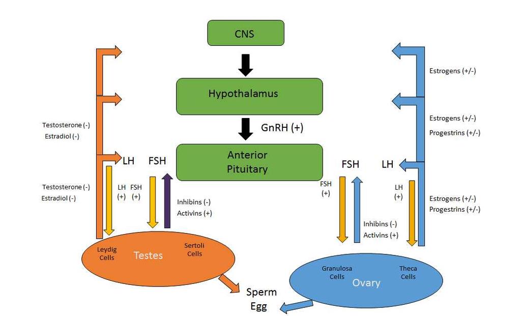
70 FIGURE 1-2: Hypothalamus-Pituitary-Gonadal Axis 47
71 / 3 = Where: NOAEL: No Observed Adverse Effect Level (mg/kg/day) Body Weight (BW): Body weight - 70 kg male; 50 kg female V = volume of air breathed in 8=hr shift (m 3 ) = bioavailability adjustment MF = Modifying Factor UF c = composite uncertainty factor Uncertainty Factor Common factors Reference UF1 Interspecies 2 mouse ICH, 2011b variability 5 rat (extrapolate human 2.5 rabbit exposure from 2 dog animal data) 3 monkey UF2 Human variability (default) Dourson et al., 1996 UF3 Exposure duration (default) Dourson et al., Study lasts ½ lifetime ICH, 2011b 1 Developmental study that covers period of organogenesis 2 6 month study in rodents; 3.5 yr. study in non-rodents 5 3-mo study in rodents; 2 yr. study non-rodents 3 high quality studies or if exposure is > sub-chronic 10 Shorter duration studies UF4 Dose-response (default if LOAEL only) Dourson et al., 1996 UF5 Severity of effects 1 Embryofetal w/ maternal toxicity ICH, 2011b 5 Embryofetal toxicity w/out maternal toxicity 5 Teratogenic w/ maternal toxicity 10 Teratogenic w/out maternal toxicity PK Route-to-route 20 mabs: assume 5% bioavailable via Pfister et al adjustment inhalation (default) assume 100% absorption via lung Naumann and Weideman, 1995 MF Modifying Factor (default) Account for residual uncertainty Sargent & Kirk, 1988 FIGURE 1-3: Calculation for occupational exposure limits (OEL) (Sargent and Kirk, 1988) 48
72 Where: / = NOAEL: No Observed Adverse Effect Level (mg/kg/day) Body Weight (BW): 50 kg PK = pharmacokinetics MF = Modifying Factor UF c = composite uncertainty factor Uncertainty Factor Common factors Reference UF1 Interspecies 2 mouse ICH, 2011b variability 5 rat (extrapolate human 2.5 rabbit exposure from 2 dog animal data) 3 monkey UF2 Human variability (default) Dourson et al., 1996 UF3 Exposure duration (default) Dourson et al., Study lasts ½ lifetime ICH, 2011b 1 Developmental study that covers period of organogenesis 2 6 month study in rodents; 3.5 yr. study in non-rodents 5 3-mo study in rodents; 2 yr. study non-rodents 3 high quality studies or if exposure is > sub-chronic 10 Shorter duration studies UF4 Dose-response (default if LOAEL only) Dourson et al., 1996 UF5 Severity of effects 1 Embryofetal w/ maternal toxicity ICH, 2011b 5 Embryofetal toxicity w/out maternal toxicity 5 Teratogenic w/ maternal toxicity 10 Teratogenic w/out maternal toxicity PK Route-to-route adjustment 20 mabs: assume 5% bioavailable via inhalation Pfister et al (default) clinical route (oral, IV) vs. inhalation MF Modifying Factor (default) Account for residual uncertainty Naumann and Weideman, 1995 Sargent & Kirk, 1988 FIGURE 1-4: Calculation for Allowable Daily Exposure (ADE) (Sargent et al., 2013) 49
73 TABLE 1-1: Examples of anticancer drugs that cause developmental toxicity Agent Effect Mechanism of Action Description of Toxicity Cyclophosphamide Fetal toxicity; DNA alkylation Increase pre- and malformations postimplantation loss; increased abnormal and growth retarded fetuses 5-Fluorouracil Malformations Antimetabolite; Reduced fetal interferes with weight; DNA synthesis; developmental incorporates anomalies into RNA and (edema, skull cell dysmorphology, proliferation orbital hemorrhage, wavy ribs, cleft palate, brachygnathia and hind limb defects Methotrexate Malformations Antimetabolite; Craniofacial folic acid defects, limb inhibition deformities, and decreased fetal weights Thalidomide Malformation Antiangiogenic deaths and malformations (especially of the arms and legs, e.g., phocomelia) Valproic Acid Malformation Anticonvulsant; inhibits histone deacetylase Various developmental defects: neuraltube-closure defects, spina bifida Reference Trasler and Doerksen, 1999 Casserett and Douhl, 7th edition, 2008 Newman, Johnson, and Staples, 1993 Lenz & Knapp, 1962 Casserett and Douhl, 7th edition,
74 TABLE 1-2: Examples of anticancer drugs that cause reproductive toxicity Agent Busulfan Cisplatin (platinum compounds) Target Effect male and female gonad toxicity male fertility Cyclophosphamide male and female gonad toxicity Mechanism of Action DNA alkylation DNA alkylation DNA alkylation Doxorubicin amenorrhea Antibiotic; Inhibits topoisomerase II Flutamide Purine analogs prostate and seminal vesicle toxicity ovary toxicity GnRH agonist and antagonist Structural similarity to endogenous purines Description of Toxicity spermatogonial death with secondary depletion of postspermatogonial germ cells seminiferous epithelium toxicity; disrupts hormonal regulation of spermatogenesis female: may deplete the follicular pool; can affect cells that are not actively dividing such as oocytes or primordial follicles male: oligospermia and azoospermia in following treatment accumulate in both the nucleus and mitochondria, initiates both oxidative stress, and induce chromosomal obliteration androgen receptor blockage resulting in secretory inhibition and atrophy adversely affect ovary development. Potential for malemediated developmental toxicity Reference Creasy, D., 2001 Boekelheide, K., 2005 Ben-Aharon and Shalg, 2012 Ben-Aharon and Shalg, 2012 Creasy, D., 2001 Mattison and Thomford, 1989 Trasler and Doerksen,
75 TABLE 1-3: Reproductive and Developmental Toxicity Study Parameters (ICH S5(R2), 2005) No. of Species Preferred Species Dosing Period Endpoint of Toxicity Fertility and Early Embryonic Development 1 rat Premating Maternal body weight, body weight change and food consumption (2-3 weeks) Effects on mating or precoital time through Day 13- General maternal toxicity 15 pregnancy Estrous cycle Gross necropsy of all adults Histopathological evaluation of testes, epididymides, ovaries, and uteri Male libido, sperm count in epididymides and sperm viability Tubal transport, numbers of corpora lutea and implantation sites Preimplantation stages of embryo and survival Embryofetal Development 1*/2 Rat and Implantation to Maternal body weight, body weight change and food consumption rabbit birth, including General maternal toxicity *nonhuman organogenesis Gross necropsy of all adults primate only Numbers of corpora lutea, live and dead implantations (biologics) Fetal body weight Gross evaluation of placenta External inspection, visceral and/or skeletal exam for anomalies and deformations Pre- and Postnatal Development, including maternal function 1 rat Implantation to Maternal body weight, body weight change, and food consumption end of lactation Duration of pregnancy and parturition Maternal toxicity (relevant to non-pregnant females) Gross necropsy of all adults Number of implantations Pre- and postnatal death of offspring Fetal body weight, altered fetal growth and development Pre- and postnatal survival, growth/body weight, maturation and fertility Physical development, sensory function and reflexes, and behavior 52
76 REFERENCES American Conference of Governmental Industrial Hygienists (ACGIH) (2013), Documentation of threshold limit values and biological exposure indices 7th edition. Cincinnati, OH. Alberts, B., Johnson, A., Lewis, J., Raff, M., Roberts, K., Walter, P. (2002) Molecular biology of the cell. (4th ed.). New York: Garland Publishing. Arora, A. and Scholar, E.M. (2005) Role of tyrosine kinase inhibitors in cancer therapy. Perspectives in Pharmacology. 315, Ben-Aharon, I., Shalgi, R. (2012). What lies behind chemotherapy-induced ovarian toxicity. Reproduction. 144, Bercu, J. P., & Dolan, D. G. (2013). Application of the threshold of toxicological concern concept when applied to pharmaceutical manufacturing operations intended for shortterm clinical trials. Regulatory Toxicology and Pharmacology, 65(1), Bercu, J. P., Sharnez, R., & Dolan, D. G. (2013). Advancing toxicology in RiskMAPP: Setting ADEs based on the subsequent drug substance. Regulatory Toxicology and Pharmacology, 65(1), doi: Bernauer, U., Heinemeyer, G., Heinrich-Hirsh, B., Ulbrich, B., Gundert-Remy, U. (2008). Exposure-triggered reproductive toxicity testing under REACH legislation: a proposal to define significant/relevant exposure. Toxicology Letters. 176, Burden, D. A., & Osheroff, N. (1998). Mechanism of action of eukaryotic topoisomerase II and drugs targeted to enzyme. Biochim Biophys Acta, 1400, Boekelheide, K. (2005). Mechanisms of toxic damage to spermatogenesis. Journal of the national Cancer Institute Monographs. 34, 6-8 Cheeseman, M. A., Machuga, E. J., & Bailey, A. B. (1999). A Tiered Approach to Threshold of Regulation. Food and Chemical Toxicology, 37(4), Chial, H. (2008). Tumor suppressor (TS) genes and the two-hit hypothesis. Nature Education. 1(1):177 Collins, K. Jacks, T., Pavletich, N. (1997). The cell cycle and cancer. Proceedings of the National Academy of Sciences. 94, Cramer, G.M., Ford, R.A., Hall, R.L. (1978). Estimation of toxic hazard-a decision tree approach. Food and Cosmetics Toxicology. 16,
77 Creasy, D. M., Pathogenesis of male reproductive toxicity. Toxicol Pathol. 29, DeGeorge, J.J., Ahn, C., Andrews, P.A., Brower, M.E., Giorgio, D.W., Goheer, M.E., Lee-Ham, D.Y., McGuinn, W.D., Schmidt, W., Sun, C.J., Tripathi, S.C. (1997). Regulatory considerations for preclinical development of anticancer drugs. Cancer Chemotherapy and Pharmacology. 41, Dolan, D. G., Naumann, B. D., Sargent, E. V., Maier, A., & Dourson, M. (2005). Application of the threshold of toxicological concern concept to pharmaceutical manufacturing operations. Regulatory Toxicology and Pharmacology, 43(1), 1-9. doi: Dourson, M., et al., Differential Sensitivity of Children and Adults to Chemical Toxicity; II. Risk and Regulation. Regulatory Toxicology and Pharmacology. 35, EPA, Guidelines for Developmental Toxicity Risk Assessment. Federal Register, Vol. 56, 1991, pp EPA, Guidelines for Reproductive Toxicity Risk Assessment. Federal Register, Vol. 61. U.S. Environmental Protection Agency, Risk Assessment Forum, Washington, DC, 1996, pp EPA (2002). A review of the reference dose and reference concentration processes. US Environmental protection Agency, Risk Assessment Forum, Washington DC, EPA/630/P-02/002F Espinosa, E., Zamora, P., Feliu, J., & González Barón, M. (2003). Classification of anticancer drugs a new system based on therapeutic targets. Cancer Treatment Reviews, 29(6), doi: /s (03) EU EudraLex (The Rules Governing Medicinal Products in the European Union) (2008). Volume 4 EU Guidelines to Good Manufacturing Practice Medicinal Products for Human and Veterinary Use. Fabbro, D., Ruetz, S., Buchdunger, E., Cowan-Jacob, S., Fendrich, G., Liebetanz, J., Mestan, J., O Reilly, T., Traxler, P., Chaudhuri, B., Fretz, H., Zimmermann, J., Meyer, T., Caravatti, G., Furet, P., Manley, P. (2002). Protein kinases as targets for anticancer agents: from inhibitors to useful drugs. Pharmacology & Therapeutics. 93, Food and Drug Administration (1995) Food Additives: Threshold of regulation of substances used in food-contact articles: Final rule. Federal Register 60, Food and Drug Administration (2004) Pharmaceutical cgmps for the 21st Century A Risk- Based Approach Final Report. 54
78 Food and Drug Administration (2005a). Guidance for Industry: Estimating the maximum safe starting dose in initial clinical trials for therapeutics in adult healthy volunteers. Center for Drug Evaluation and Research (CDER) Food and Drug Administration (2005b) Reviewer Guidance: Evaluating the risks of drug exposure in human pregnancies. Foster, P. M. D., & Gray, L. E., Jr. (2008). Chapter 20. Toxic Responses of the Reproductive System. In C. D. Klaassen (Ed.), Casarett And Doull's Toxicology: The Basic Science of Poisons (Seventh ed., pp ): McGraw-Hill Professional. Geisler, J., Manahan, K., Linnemeier, G., & Weinmann, M. (2007). Mechanism of action of cytotoxic drugs in gynecologic oncology. Journal of Gynecologic oncology, 12, Gibbs, J. (2000). Mechanism-based target identification and drug discovery in cancer research. Science, 287, ICH (International Conference on Harmonisation). (2005). S5 (R2): Detection of Toxicity to Reproduction for Medicinal Products & Toxicity to Male Fertility ICH (International Conference on Harmonisation). (2006a). ICH Guidance for Industry: Q3A (R2) Impurities in New Drug Substances. International Conference on Harmonisation. ICH (International Conference on Harmonisation). (2006b). ICH Guidance for Industry: Q3B (R2) Impurities in New Drug Products. ICH (International Conference on Harmonisation). (2009). M3: Guidance on Nonclincal Safety Studies for the Conduct of Human Clinical Trials and Marketing Authorization for Pharmaceuticals. ICH (International Conference on Harmonisation). (2010). S9 Nonclincal Evaluation for Anticancer Pharmaceuticals. ICH (International Conference on Harmonisation). (2011a). S6 (R1): Preclinical Safety Evaluation of Biotechnology-Derived Pharmaceuticals ICH (International Conference on Harmonisation). (2011b). ICH Topic Q3C (R5) Impurities: Guideline for Residual Solvents. ICH Topic Q3C (R5) Impurities: Guideline for Residual Solvents. ICH (International Conference on Harmonisation). (2014a). ICH Q3D Technical Document Q3D: Guideline for Elemental Impurities. International Conference on Harmonization. Current Step 4 version; Dated: 16 Dec
79 ICH (International Conference on Harmonisation). (2014b). ICH Guideline M7 on Assessment and Control of DNA Reactive (Mutagenic) Impurities in Pharmaceuticals to Limit Potential Carcinogenic Risk. ISPE (International Society of Pharmaceutical Engineers). (2010). Risk-Based Manufacture of Pharmaceutical Products: Guide to Managing Risks Associated with Cross- Contamination, vol 7. JEFCA, (1999). Evaluation of certain food additives and contaminants. Procedure for the safety evaluation of flavouring agents. Forty-ninth Report of the Joint FAO/WHO Expert Committee on Food Additives. WHO Technical Report Series 884. World Health Organization (WHO) Kroes, R., Galli, C., Munro, I., Schilter, B., Tran, L.A., Walker, R., Würtzen, G. (2000). Threshold of toxicological concern for chemical substances present in the diet: a practical tool for assessing the need for toxicity testing. Food and Chemical Toxicology, 38, Kroes, R., Renwick, A. G., Cheeseman, M., Kleiner, J., Mangelsdorf, I., Piersma, A., Schilter, B., Schlatter, F., van Schothorth, F., Vos, J.G., Würtzen, G. (2004). Structure-based thresholds of toxicological concern (TTC): guidance for application to substances present at low levels in the diet. Food and Chemical Toxicology, 42(1), doi: Kroes, R., Renwick, A. G., Feron, V., Galli, C. L., Gibney, M., Greim, H., Guy, R.H., Lhuguenot, J.C., van de Sandt, J. J. M., Application of the threshold of toxicological concern (TTC) to the safety evaluation of cosmetic ingredients. Food and Chemical Toxicology, 45(12), doi: Ku, R. (2000). An overview of setting occupational exposure limits (OELs) for pharmaceuticals. Chemical Health and Safety Laufersweiler, M. C., Gadagbui, B., Baskerville-Abraham, I. M., Maier, A., Willis, A., Scialli, A. R., Daston, G. (2012). Correlation of chemical structure with reproductive and developmental toxicity as it relates to the use of the threshold of toxicological concern. Regulatory Toxicology and Pharmacology, 62(1), doi: Lenz, W., Knapp, K., Thalidomide embryopathy. Arch Environ Health. 5, Matson, D., & Stukenberg, T. (2011). Spindle Poisons and Cell Fate: A tale of two pathways. Molecular Interventions, 11(2), 141. Massari, F., Santoni, M., Ciccarese, C., Santini, D., Alfieri. S., Martignoni, G., Brunelli, M., Piva, F., Berardi, R., Montironi, R., Porta, C., Cascinu, S., Tortora, G. (2015) PD-1 56
80 blockade therapy in renal cell carcinoma: current studies and future promises. Cancer Treatment Reviews. 41 (2), Mattison, D., Thomford, P. (1989). The mechanism of action of reproductive toxicants. Toxicologic Pathology. 17(2), Moore, KL and TVL Persaud. (1998) The developing human: clinically orientated embryology, 6 th edition, Philadelphia: W.B. Munro, I. C. (1990). Issues to be considered in the safety evaluation of fat substitutes. Food and Chemical Toxicology, 28(11), Munro, I. C., Ford, R. A., Kennepohl, E., & Sprenger, J. G. (1996). Correlation of structural class with no-observed-effect levels: a proposal for establishing a threshold of concern. Food and Chemical Toxicology, 34(9), Munro, I., Kennepohl, E., Kroes, R. (1999). A procedure for the safety evaluation of flavouring substances. Food and Chemical Toxicology. 37, Nielsen, GD., Øverbø, S. (2008). Background, approaches and recent trends for setting healthbased occupational exposure limits: a minireview. Regulatory Toxicology and Pharmacology, (51), Naumann, B.D., Sargent, E.V. (1997). Setting occupational exposure limits for pharmaceuticals. Occupational Medicine. 12(1), Naumann, B.D., Weideman, P.A. (1995) Scientific basis for uncertainty factors used to establish occupational exposure limits for pharmaceutical activie ingredients. Human Ecological Risk Assessment. 1(5), Naumann, B., Weideman, P., Sarangapani, R., Hu, S., Dixit, R., Sargent, E. (1999). Investigations of the use of bioavailability data to adjust occupational exposure limits for active pharmaceutical ingredients. Toxicological Sciences. 112, NIOSH, NIOSH list of antineoplastic and other hazardous drugs in healthcare settings By Connor TH, MacKenzie BA, DeBord DG, Trout DB, O Callaghan JP. Cincinnati, OH: U.S. Department of Health and Human Services, Center for Disease Control and Prevention, National Institute for Occupational Safety and Health, DHHS (NIOSH) Publication No (Supersedes ) O'Connor, C. (2008) Cell Division: Stages of Mitosis. Nature Education 1(1):188 Occupational Safety and Health Act (OSHA). (1970). Toxic and hazardous substances, air contaminants standard, CFR, Chapter XVII, part , subpart Z. 57
81 Paskiet, D., Jenke, D., Ball, D., Houston, C., Norwood, D.L., Markovic, I. (2013). The product quality research institute (PQRI). Leachables and extractables working group initiatives for parenteral and ophthalmic drug product (PODP). PDA Journal of Pharmaceutical Science Technology. 67(5), Payne, S. and D. Miles. (2008). Chapter 4 Mechanisms of anticancer drugs. In G. Gleeson (Ed.) Scott-Brown's Otorhinolaryngology: Head and Neck Surgery (Seventh ed., pp 34-46): CRC Press. Pfister, T., et al., Estimation of uncertainty factors covering the risk of EFD toxicity based on general toxicity data a data based statistical approach. Albuquerque, New Mexico, Occupational Toxicology Roundtable, October 7-9, 2013, 2013 Pommier, Y., Pourquier, P., & Fan, Y., Strumberg. (1998). Mechanism of action of eukaryotic DNA topoisomerase I and drugs targeted to the enzyme. Biochim Biophys Acta, 1400, Remesh, A. (2003). Toxicities of anticancer drugs and its management. International Journal of Basic and Clinical Pharmacology. 1, 1-12 Rogers, J. (2008). Chapter 10. Developmental Toxicity. In C. D. Klaassen (Ed.), Casarett And Doull's Toxicology: The Basic Science of Poisons (Seventh ed., pp ): McGraw- Hill Professional. Rulis, A.M. (1986). De minimus and the threshold of regulation. In: Felix, C.W. (ed.), Food Protection Technology. Lewis Publishers Inc., Chelsea, MI Sargent, E.V., Kirk, G.D. (1998). Establishing airborne exposure control limits in the pharmaceutical industry. American Industrial Hygiene Association Journal. 49, Sargent, E. V., Faria, E., Pfister, T., & Sussman, R. G. (2013). Guidance on the establishment of acceptable daily exposure limits (ADE) to support Risk-Based Manufacture of Pharmaceutical Products. Regulatory Toxicology and Pharmacology, 65(2), doi: Sclafani, R. A., & Holzen, T. M. (2007). Cell Cycle Regulation of DNA Replication. Annual Review of Genetics, 41, SCCS, SCHER, SCENIHR, Joint Opinion on the Use of the Threshold of Toxicological Concern (TTC) Approach for Human Safety Assessment of Chemical Substances with focus on Cosmetics and Consumer Products, 8 June 2012 Subramani, S., Jayapalan, S., Kalpana, R., Natarajan, J. (2013). Homokinase: a curated database of human protein kinases. ISRN Computational Biology. 2013, Article ID , 5 pages. 58
82 Tiboni GM, Marotta F, Rossi C, Giampietro F. (2008). Effects of the aromatase inhibitor Letrozole on in utero development in rats. Human Reproduction. 23, Trasler, J., Doerksen, T. (1999). Teratogen Update: Paternal Exposures-Reproductive Risks. Teratology. 60, Trigg, M. and Flanigan-Minnick, A. (2011). Mechanisms of action of commonly used drugs to treat cancer. Community Oncology. 8(8); Van Ravenzwaay, B., Dammann, M., Buesen, R., & Schneider, S. (2011). The threshold of toxicological concern for prenatal developmental toxicity. Regulatory Toxicology and Pharmacology, 59(1), doi: Van Ravenzwaay, B., Dammann, M., Buesen, R., Flick, B., & Schneider, S. (2012). The threshold of toxicological concern for prenatal developmental toxicity in rabbits and a comparison to TTC values in rats. Regulatory Toxicology and Pharmacology, 64(1), 1-8. doi: Waldman, T. (2006). Effective cancer therapy through immunomodulation. Annual Reviews of Medicine. 57, Ward, JA., Furr BJ., Valcaccia B., Curry B., Bardin CW., Gunsalus GL., Morris, ID Prolonged suppression of rat testis function by a depot formulation of Zoladex, a GnRH agonist. Journal of Andrology. 10,
83 CHAPTER 2 THRESHOLD OF TOXICOLOGICAL CONCERN (TTC) FOR DEVELOPMENTAL AND REPRODUCTIVE TOXICITY OF ANTICANCER 1 Summary Pharmaceutical companies develop specialized therapies to treat late stage cancer. In order to accelerate life-saving treatments and reduce animal testing, compounds to treat lifethreatening malignancies are allowed modified requirements for preclinical toxicology testing. Limited data packages in early drug development can present product quality challenges at multi-product manufacturing facilities. The present analysis established an endpoint-specific Threshold of Toxicological Concern (TTC) for developmental and reproductive toxicity (DART) for anticancer compounds. A comprehensive database was created consisting of over 300 noobserved adverse effect levels (NOAELs) for DART of 108 anticancer compounds. The 5 th percentile NOAEL for developmental and reproductive toxicity was mg/kg/day (300 µg/day), resulting in a human exposure threshold of 3 µg/day assuming standard uncertainty factors and a 60 kg human bodyweight. The analysis shows this threshold is protective for developmental and reproductive toxicity of highly potent groups of anticancer compounds. There were similar TTC values calculated for direct-acting and indirect-acting anticancer compounds. It was confirmed that the 1.5 µg/day threshold for mutagenic impurities is protective for developmental and reproductive toxicity of pharmaceutical agents designed to target tumors. 1 This chapter is published as: Stanard, B., Dolan, D.G., Hanneman, H., Legare, M., Bercu, J.P. (2015). Threshold of toxicological concern (TTC) for developmental and reproductive toxicity of anticancer compounds. Regulatory Toxicology and Pharmacology. 72,
84 Key words: Threshold of toxicological concern (TTC); anticancer; reproductive toxicity; developmental toxicity; direct-acting; indirect-acting; antineoplastic; Permitted Daily Exposure (PDE); Acceptable Daily Exposure (ADE) Introduction Pharmaceutical companies may be required to use dedicated production areas for agents with high pharmacological activity or toxicity unless validated cleaning procedures are in place (EMA, 2014 Ch. 3; EMA 2014 Ch. 5; ICH, 2000). Health-based values, such as Acceptable Daily Exposure (ADE) or Permitted Daily Exposure (PDE), are used to determine product quality acceptance levels for residual drug in manufacturing equipment after cleaning prior to the manufacture of another drug product (EMA, 2014; ISPE 2010; ICH, 1997a). The terms ADE and PDE are both health-based limits, are used interchangeably, and are the daily doses of drugs below which there is little risk of adverse effects, including members of sensitive populations like the elderly, children, developing fetus, and disease-impaired individuals. A health-based limit is derived by dividing the no-observable adverse effect level for the critical effect by uncertainty or uncertainty factors; with different consideration for target population, route of exposure, duration, and acceptable risk. Pharmaceutical companies have been developing increasingly targeted therapies to treat cancers based on advancing knowledge of the molecular causes of cancer (Chabner et al., 2012). Anticancer compounds are allowed modifications to the standard nonclinical testing protocols and procedures required of active pharmaceutical ingredients intended for other therapeutic indications to accelerate the availability of new, and potentially lifesaving treatments while minimizing adverse clinical effects and unnecessary animal testing (ICH, 2009). These modified regulatory requirements are prudent given the potential risk-benefit of the drugs, but can create 61
85 data gaps for critical endpoints such as developmental and reproductive toxicity (DART) of anticancer compounds in early drug development. The resulting data limitations have created new challenges to developing health-based limits for anticancer drugs in multiple product facilities as compared to those targeted for non-cancer indications. The threshold of toxicological concern (TTC) is a method for deriving limits for compounds with limited or no toxicity data is (Kroes et al., 2004; Munro et al., 1996). The TTC is a risk assessment tool derived from a distribution of toxicity data to establish a safe limit of exposure, under which adverse effects are unlikely. When developing any database to support a threshold of toxicological concern, it is important to understand the chemicals in the database and the limitations of its application (SCCS, SCHER, SCENIHR, 2012). This concept is not intended to replace chemical-specific hazard assessment and should be utilized as an interim control in lieu of having the data required to establish a substance specific limit. As with any risk assessment tool, one must consider the potential limitations of probabilistic models and consider application of the TTC on a case-by-case basis (SCCS, SCHER, SCENIHR, 2012). The TTC has long been utilized in industries such as food (Kroes et al., 2004; Munro et al., 1996) or cosmetics (Kroes et al., 2007) and has also been used for residual active pharmaceutical ingredients in manufacturing facilities. Dolan et al. (2005) applied the TTC concept to pharmaceutical manufacturing, looking at all endpoints including carcinogenicity, immunotoxicity, neurotoxicity and developmental toxicity for Merck active pharmaceutical ingredients following tiered category thresholds as defined as: Category 1: likely to be carcinogenic (1 μg/day); Category 2: likely to be potent or highly toxic (10 μg/day); and Category 3: not likely to be potent, highly toxic or carcinogenic (100 μg/day). Table 1 provides a summary of the human exposure threshold values for general toxicity and the uncertainty factors used. The TTC methodology as described by Dolan et al., 62
86 2005 has been referenced in guidance documents for health-based limits (EMA, 2014; ISPE, 2010). The purpose of this paper is to determine if thresholds specific to systemic DART effects can be established from a database constructed for these effects for anticancer compounds. Other potential adverse health effects (e.g., local effects such as irritation) were excluded from this analysis. Many anticancer drugs are potent reproductive toxicants because they target rapidly proliferating cells. Although fertility and developmental toxicity have been previously evaluated for end-point specific thresholds (Bernauer et al., 2008; Laufersweiler et al., 2012; Ravenzwaay et al., 2011; 2012), the present analysis utilized a database of pharmaceuticals that have an inherent effect on developmental and reproductive toxicity. The target pharmacologic effect for anticancer compounds is to stop or slow the growth of rapidly proliferating cells (i.e., tumor cells) through direct interaction with DNA (e.g., DNA alkylation, mitotic disruption, etc.) or indirect interaction (e.g., kinase inhibitors, hormone modulation) with tumor cells. Consequently, we will also be evaluating if there is a difference in the exposure threshold of direct-acting vs. indirect interaction with tumor cells. Methods The database used for the present analysis was compiled from a reference dataset of anticancer compounds (current and/or formerly approved by FDA/EMA) populated with preclinical and clinical data (and post-marketing data when available) from studies on male and female reproductive function and fertility as well as developmental toxicity in the offspring. Several sources were cross-referenced to identify all drugs approved for cancer treatment including: National Cancer Institute s Cancer Drug list ( and the 2014 NIOSH List of Antineoplastic Drugs (NIOSH, 2014). The database included both chemically synthesized 63
87 (i.e., small molecule) and biologically derived (i.e., large molecule) anticancer compounds with DART studies publically available. It should be noted that only substances indicated for the treatment of cancer as either a single agent or part of a combination therapy were considered. We excluded supportive medications approved to treat chemotherapy-related adverse effects (e.g., anemia or neutropenia) or other conditions related to cancer (e.g., nausea and vomiting). For each anticancer compound, NOAEL (no-observed-adverse-effect-level) values were derived from studies on male/female reproductive function and fertility as well as developmental toxicity in offspring (embryofetal; pre- and postnatal) conducted to support regulatory approval. If a NOAEL for developmental and/or reproductive effects could not be determined, a LOAEL for the DART endpoint was considered for inclusion in the database. It is recognized that some sponsors may refer to a dose that did not cause reproductive effects as the No-Observed Effect Level (NOEL) because low dose effects can be difficult to translate into human effects. To ensure consistency in terminology with comparative papers, any effects associated with reproduction, reproductive function/fertility or developmental toxicity were considered an adverse effect. Our use of NOAEL does not suggest that there may be other effects at lower doses. The database of NOAELs includes toxicity studies following dose administration by any relevant route for anticancer drugs; which included oral, intramuscular (IM), intraperitoneal (IP), intravenous (IV) and subcutaneous (SC). The most common species used in DART studies are rodents and rabbits; however, studies from other species (i.e., dogs and monkeys) were also included when available. In total there were 3 NOAELs for DART in dogs (3 different drugs) and 14 NOAELs for DART in monkeys (11 different compounds). A list of all species and routes of exposures for the NOAELs in the anticancer database is included in Table 2. 64
88 In total, there were 150 substances that had been approved (currently or formerly) by the US FDA and/or EMA for the treatment of cancer (as of April 2014). The data entered into the database included name of the anticancer compound, study type, species, sex, route of exposure, dose levels, study duration, endpoints reported, NOAEL and/or LOAEL and references. Application of the above criteria resulted in a final dataset of 108 anticancer compounds. For many compounds included in the database, multiple NOAELs were identified as a result of testing requirements in multiple species and sex as well as different reproductive endpoints that were investigated. The final database contained 320 NOAELs/LOAELs that were used for the present analysis. Each compound in the dataset was categorized based on its mode of action. Drugs that kill cancer cells by disrupting mitotic function or through direct interaction with DNA were classified as direct-acting. Examples of direct-acting anticancer compounds include: DNA alkylating agents, antimetabolites, cytotoxic antibiotics, microtubule-disrupting agents and topoisomerase inhibitors. Drugs that disrupt the support, maintenance or defense functions of cancer cells were defined as indirect-acting. Examples of indirect-acting anticancer compounds include: hormone-modulating agents, kinase inhibitors, immune modulating agents and other miscellaneous targets compounds. Anticancer compounds target a wide variety of direct and indirect-acting modes of action. As shown in Figure 1, direct and indirect-acting anticancer compounds were sub-classified into 10 different categories based on the specific mode of action. Table 3 shows the number of anticancer compounds in each mode of action category. In total, there were 81 cancer drugs in the reference dataset classified as direct-acting and 69 classified as indirect-acting. The final database contains 52 direct-acting anticancer compounds with 149 NOAEL values and 56 indirect-acting anticancer compounds with 171 NOAELs for 65
89 developmental and reproductive toxicity. It should be noted that the exact mode of action is often not fully understood, so the classification system was based on the presumed mode given the current information available. There were 4 compounds classified as Other targets (misc.) and 5 classified as unknown because there was insufficient data available to classify into the one of the ten categories. Figure 2 is a bar graph showing the number of NOAELs by mode of action. It can be seen that kinase inhibitors have the largest number of NOAELs. This is a function of the large number of anticancer drugs in this subclass and their relatively more recent approvals by regulatory agencies (1998 to 2014). The database contains adverse effects on reproduction as well as effects in all stages of development that include resorptions, intrauterine and perinatal deaths, structural abnormalities, altered growth (fetal birth weight and post-natal growth), neonatal survival and viability of prenatally exposed offspring. The results from studies on male/female reproductive function and/or male/female fertility were combined into one category for reproductive function/fertility. Developmental toxicity represented 66% of the NOAELs and male/female reproductive function/fertility represented the remaining 34% of the NOAELs in the database. Consistent with previous TTC calculations by Munro et al. (1996) and Kroes et al. (2004), a human exposure threshold was derived from the 5 th percentile NOAEL divided by 100 (accounting for animal data extrapolation and human variability) and assuming a 60 kg human. Results The cumulative distribution of the NOAELs and derived NOAELs for reproductive function/fertility and for developmental toxicity were plotted to evaluate the endpoint-specific toxicity of anticancer compounds (Figure 3). The cumulative distribution of the NOAELs from 66
90 the combined reproductive function/fertility and developmental was also plotted. There is slight difference in the distribution of NOAELs for reproductive function/fertility compared to the distribution of NOAELs for developmental toxicity, which suggests that endpoints for developmental toxicity are slightly more sensitive than those for reproductive function/fertility. However; this difference was within an order of magnitude and therefore not considered significant for the purposes of risk assessment. The 5 th percentile NOAELs were mg/kg/day and mg/kg/day for reproductive function/fertility and developmental toxicity, respectively. The 5 th percentile NOAEL for the combined DART endpoints was mg/kg/day. The distribution of human exposure threshold values derived from the 5 th centile NOAELs were also plotted (Figure 4). The endpoint-specific human exposure thresholds calculated from the distributions were 6 µg/day for reproductive function/fertility and 1 µg/day for developmental toxicity, as shown in Table 3. The combined human exposure threshold for developmental and reproductive toxicity was 3 µg/day. The thresholds values for direct-acting and indirect-acting anticancer compounds were calculated using the same assumptions (Table 4). The direct-acting and indirect-acting anticancer compounds had a derived human exposure threshold for DART of 5 µg/day and 1 µg/day, respectively. The threshold value for indirectacting anticancer compounds was slightly lower than the direct-acting compounds but also within an order of magnitude and therefore not considered independently. Within the group of indirect-acting anticancer compounds, it was observed that a subgroup of drugs, those that effect hormone activity, had the lowest NOAEL values. However, excluding the hormone modulating compounds had a relatively minor effect on the human exposure threshold values (Table 4). The threshold for developmental toxicity was increased 67
91 from 1 µg/day to 6 µg/day and the threshold for reproductive function/fertility was unaffected; the threshold for the combined developmental and reproductive toxicity increased only 2-fold from 3 µg/day to 6 µg/day. There were 11/108 (10%) anticancer compounds in the database with a human exposure threshold less than 3 µg/day (Table 5). Of the 11 compounds below this threshold value, 8 were indirect-acting and 3 were direct-acting. Seven of the indirect-acting agents inhibit sex hormone activity (aromatase, GnRH, and estrogen). Discussion The goal of this analysis was to develop an indication-specific, endpoint-specific exposure threshold that can be used as part of the risk assessment process to evaluate impurities during pharmaceutical drug manufacturing. Pharmaceutical companies and regulators are working to advance anticancer compounds through the drug development process at an accelerated rate to provide life-saving medications to market more quickly to patients. The TTC concept presented in this paper may be utilized to evaluate the carryover of drug products early in the drug development process when insufficient nonclinical and clinical data are available to more precisely estimate compound-specific levels of safe exposure. Analyses were conducted to determine whether a threshold could be developed for anticancer compounds that was protective for systemic developmental and reproductive toxicity and to determine whether there were any differences between the distribution of NOAELs from animal studies for individual adverse outcomes (reproductive function/fertility, developmental toxicity), and the combined distribution of DART. The results show that an exposure threshold of 3 µg/day is protective for systemic developmental and reproductive effects for most classes of anticancer compounds with limited data (e.g., pretesting, or early development). 68
92 When developing a database for a TTC value, it is important to understand its limitations and the chemical domain for its application (SCCS, SCHER, SCENIHR, 2012). In this case, the database was limited to systemic developmental and reproductive toxicity of anticancer compounds to address a data-gap that is common for oncology drugs in clinical development (EMA, 2014). There are other databases that can be used to address different endpoints such as mutagens in the diet (Kroes et al., 2014) or effects from leachables and extractables in orally inhaled and nasal drug product (Ball et al., 2007). The intention is not to provide a TTC for all other applications, but provide scientific support when an anticancer compound is devoid of developmental/reproductive toxicity data. We also evaluated whether the mode of action (direct-acting and indirect-acting) had an effect on the exposure threshold value. The results show that indirect-acting anticancer compounds had a slightly lower exposure threshold than direct-acting compounds (within an order of magnitude) but the difference was not considered significant for the purposes of risk assessment. In addition, there was a slight shift in threshold between hormonal compounds and other anticancer compounds. These findings have a potentially significant impact on the pharmaceutical manufacturing of anticancer compounds. Functional definitions are critical to ensuring consistent regulatory oversight; however, the findings from this current study reinforces the belief that decisions around dedicated equipment and segregation should be based on the ability to control below an established threshold value for human exposure and not be arbitrarily made based on a definition alone (i.e. cytotoxicity). The TTC values that are currently published were derived from databases of large numbers of chemicals representing broad classes of chemicals, such as food additives, industrial chemicals, cosmetics and pharmaceuticals (Dolan et al. 2005; Kroes et al. 2004; Munro et al. 69
93 1996). There have been variations in exposure threshold values and uncertainty factors. The human exposure threshold of 3 µg/day presented in this paper was calculated from the 5th percentile NOAEL divided by a composite uncertainty factor of 100 to account for extrapolation of animal data to human effects (10x) and human variability (10x) and an average human body weight of 60 kg. The 100-fold uncertainty factor is sufficiently protective because the 5th percentile NOAEL was derived from a database of DART studies from anticancer compounds with a high probability for developmental and reproductive toxicity. The uncertainty factors that were used are also consistent with the 100-fold factor used to derive thresholds for different types of toxicological endpoints including carcinogenicity, immunotoxicity, neurotoxicity and developmental toxicity (Kroes et al., 2000; Munro et al., 1996), as well as the one proposed threshold for reproductive toxicity (Laufersweiler et al. 2012). The threshold value described in the present analysis is lower than the corresponding thresholds suggested by Munro et al. (1996) and the recently published endpoint-specific thresholds suggested by Bernauer et al., 2008; van Ravenzwaay et al., 2011; and Laufersweiler et al Additional uncertainty factors have been used to calculate thresholds for fertility and developmental toxicity compensating for uncertainty in the data. Bernauer et al. (2008) applied an additional factor of 10x to account for their small dataset (composite factor: 1000) and van Ravenzwaay et al. (2011) applied an additional factor of 5x to account for potentially incomplete chemical classes in their proprietary database of nonpharmaceutical industrial chemicals (composite factor: 500). The wide variety of chemical classes previously evaluated (e.g., industrial chemicals, food chemicals, pesticides, drugs) and the variability in the uncertainty factors applied has contributed to the wide range in human exposure threshold values for developmental and reproductive toxicity. However, it is believed that the modes of action of the anticancer compounds significantly contributed to the lower 70
94 human exposure threshold value determined in the present analysis. The human exposure thresholds for fertility and developmental toxicity and uncertainty factors used in each of the corresponding studies are summarized in Table 6. We analyzed the published datasets used to derive the different TTCs in the literature and found the numbers of developmental and reproductive toxicants in the databases to be similar; however, anticancer compounds as a chemical class have been underrepresented. Furthermore, steroid hormones, large molecular weight compounds and agents with high pharmacologic activity were largely excluded. The summary of this analysis in included in Table 7. The Munro et al. (1996) database contained over 600 chemicals representing a wide spectrum of chemicals from industrial and agriculture to cosmetics, food additives and consumer products all separated into Cramer Structural Classes (Cramer et al., 1978). Anticancer compounds would likely fall into Cramer Class III chemicals, or those with chemical structures that suggests toxicity. Their database contains 448 Cramer Class III chemicals, of which 132 had NOAELs reported for reproductive or teratogenic endpoints. The only anticancer compound in their database was cyclophosphamide. The database used by Kroes et al. (2000) was an expanded subset of the Munro et al. (1996) database, which contained 81 chemicals with reproductive toxicity endpoints. Only 2 additional anticancer compounds were included: azacitidine and hydroxyurea. Cheeseman et al. (1999) analyzed 3306 chemicals with NOAELs for reproductive toxicity endpoints from the RTECS database that contained 2 additional anticancer compounds: dacarbazine and prednimustine. There were no anticancer compounds included in the Bernauer et al. (2008) database of 91 industrial chemicals from the EU Risk Assessment Program for Existing Chemicals or the Laufersweiler et al. (2012) database of 283 industrial chemicals. Bernauer et al. (2008) also described a database of over 500 pharmaceuticals; however, the 71
95 database was not published. Likewise, van Ravenzwaay et al. (2011) published their dataset of 111 BASF chemicals; however the chemical names and CAS numbers were omitted and therefore could not be reviewed, nor could the Cramer classification be determined. In total, there were only 5 unique anticancer compounds included in the aforementioned TTC databases. The database created for the present analysis is made entirely of chemical compounds, which directly and indirectly target rapidly dividing cells and are known to cause adverse reproductive and developmental effects. Anticancer compounds that modulate hormone levels (antagonists and agonists) as well as monoclonal antibodies and other large molecular weight compounds were included in the database. The potential for developmental and reproductive toxicity of this subset of chemicals is reflected in our proposed human exposure threshold. The thresholds values listed in Table 4 are lower than those proposed in other TTC analyses for non-genotoxic, non-carcinogenic compounds. This is likely reflecting the fact that the database used in the present study was composed of chemicals with modes of action expected to most effect rapidly proliferating cells, i.e. reproductive tissues and the developing embryofetus. Alkylating agents and hormone modulators are two examples of classes of anticancer compounds that adversely impact reproduction and the developing embryo-fetus. Alkylating agents such as cisplatin, doxorubicin and cyclophosphamide work through direct interaction with DNA via DNA binding and cross-linking with RNA, DNA and protein synthesis inhibition leading to germ cell depletion, mutagenic changes in germinal cells and loss of gonadal function resulting in male and female infertility and teratogenic effects in the fetus (Trasler and Doerksen, 1999; Remesh, 2012). Hormone modulating agents target the availability of testosterone and estrogen, on which prostate cancer and breast cancer cells (respectively) depend for growth, through agonism and antagonism of the hypothalamus-pituitary axis (Ben-Ahanon and Shalgi, 72
96 2012). Goserelin inhibits gonadotropin-releasing hormone (GnRH), which subsequently reduces serum testosterone and has been shown to cause infertility in males and females (Ward et al., 1989). Letrozole can effectively treat breast cancer by competitively inhibiting aromatase, blocking the conversion to estrogen, but has been shown to produce miscarriages and fetal abnormalities in rats (Tiboni et al., 2008). As shown in Table 5, anticancer compounds that target reproductive hormones cause adverse developmental and reproductive effects at very low doses because both tumor cells and healthy hormone-dependent cells are highly sensitive to changes in sex hormones (i.e., testosterone, estrogen, aromatase, GnRH). Hormone modulating compounds represent approximately 10% of all existing drugs approved for treatment of cancer and within this class, 7/14 compounds in the database had NOAELs less than the proposed threshold. Excluding the hormone modulating agents from the human exposure threshold calculation resulted in a slight increase in threshold value, within a degree of magnitude. There was no clear relationship between the intended pharmacologic effect or therapeutic dose and the potency for reproductive toxicity. For example, anastrozole, exemestane, and letrozole are indicated for treatment of breast cancer and all work through a similar mode of action (aromatase inhibition) but NOAELs ranged from mg/kg/day to 100 mg/kg/day (12 µg/day 6 x 10 6 µg/day) for developmental and reproductive toxicity. There were other outliers that do not target reproductive hormones, such as pentostatin, which is indicated for the treatment of hairy cell leukemia. Pentostatin is a direct-acting anticancer compound that inhibits adenosine deaminase with a NOAEL of mg/kg/day (30 µg/day) for embryofetal development. It is clear that some anticancer compounds are very potent reproductive toxicants but the reason why some are more potent than others requires further exploration. These outliers highlight some gaps that 73
97 were identified with some anticancer compounds in the database. Further exploration of the pathways of toxicity for specific mechanisms of action and other chemical descriptors may improve the predictive value of the current models. The TTC concept should only be used to estimate safe exposure levels for systemic toxicity in the absence of sufficient data to determine compound-specific limits. The potential for local effects, e.g. irritation, etc. were excluded from this analysis. Exceeding the TTC does not necessarily increase the risk of reproductive or developmental effects because of the conservative assumptions used to derive the threshold values. The TTC value can and should be adjusted based on the data available at the time of the assessment. The TTC is a tool and should be applied on a case-by-case basis. As new products move through the drug development process, a toxicologist must review the data-package continually and consider adjustments to the approach, as applicable, based on available data. Conclusion This analysis has important implications for deriving health-based limits for anticancer compounds in development when there is limited DART data. Based on the data herein a TTC of 6 µg/day is suggested for anticancer molecules when the developmental and reproductive toxicity is unknown and there is no evidence of hormone modulation. The threshold for anticancer compounds with known potential to modulate hormones or when the mode of action is unknown is 6 µg/day for effects on reproductive function/fertility, 1 µg/day for developmental toxicity and 3 µg/day for the combined developmental and reproductive toxicity. These thresholds are specific to non-mutagenic effects; different limits may be needed for other applications, such as the 1.5 µg/day for mutagenic impurities (EMA, 2014; ICH M7, 2014). Finally, this analysis supports the use of a risk -based approach (i.e., deriving a health-based 74
98 limit) to ensure negligible cross-contamination of pharmaceutical residues. Our results show the hazard of the compound (i.e., cytotoxic ( direct acting ) versus non-cytotoxic ( indirect acting ) had no meaningful impact on the overall human exposure threshold value for developmental and reproductive effects of anticancer compounds. 75
99 Tables and Figures Figure 2-1: The modes of action of anticancer compounds Figure 2-2: The total number of NOAELs by modes of action Figure 2-3: Cumulative distribution of the NOAELs for developmental toxicity, male/female reproductive function/fertility and the combined developmental and reproductive toxicity (DART) endpoints. Figure 2-4: Cumulative distribution of the human thresholds for developmental toxicity, male/female reproductive function/fertility and the combined developmental and reproductive toxicity (DART) endpoints. Table 2-1: Comparison of human thresholds for chronic effects/carcinogenicity and the associated uncertainty factors (UFs) that were applied Table 2-2: Routes of exposure and species used for all of the DART NOAEL studies in the anticancer DART database Table 2-3: Breakdown of anticancer compounds by mode of action Table 2-4: Summary of the 5 th percentile NOAELs and the derived human exposure thresholds for DART of anticancer compounds Table 2-5: Modes of action and critical effects for the most potent anticancer compounds, i.e. derived human exposure threshold below purposed TTC (3 µg/day) Table 2-6: Comparison of endpoint-specific human exposure thresholds for reproductive function/fertility and developmental toxicity and the associated UFs that were applied in each study Table 2-7: Comparison of the datasets used to derive human exposure thresholds: number of developmental and reproductive toxicants, number of anticancer compounds and the different human exposure thresholds 76

100 FIGURE 2-1: The modes of action of anticancer compounds 77

101 Kinase Inhibitors Anti-metabolites Hormone Agonists/Antagonists Alkylating Agents Misc. Cytotoxic Antimicrotubule Antibotics Immunomodulators Topoisomerase Inhibitors Osteo-Active NOAELs FIGURE 2-2: Total number of NOAELs by mode of action 78
102 1.00 DART (combined) Developmental Toxicity 0.80 Reproductive Function/Fertility Cumulative Distribution (%) NOAEL (mg/kg/day) FIGURE 2-3: Cumulative distribution of the NOAELs for developmental toxicity, male/female reproductive function/fertility and the combined developmental and reproductive toxicity (DART) endpoints. 79
103 DART (combined) Developmental Toxicity Reproduction Function/Fertility Cumulative Distribution (%) Human Exposure Threshold (µg/day) FIGURE 2-4: Cumulative distribution of the human thresholds for developmental toxicity, male/female reproductive function/fertility and the combined developmental and reproductive toxicity (DART) endpoints 80
104 TABLE 2-1: Comparison of human thresholds for chronic effects/carcinogenicity and the associated uncertainty factors (UFs) that were applied End Point Carcinogenicity (Threshold of Regulation for Food Additives and Pharmaceutical Mutagenic Impurities) Chronic Toxicity: Cramer Class I (Food Additives) Chronic Toxicity: Cramer Class II (Food Additives) Chronic Toxicity: Cramer Class III (Food and Cosmetics) Chronic Toxicity: Cramer Class III (Class III excluding organophosphates, carbamates, and organohalogens) Chronic Toxicity: Organophosphates (Food and Cosmetics) Genotoxic carcinogenicity (Food and Cosmetics) Carcinogenic (Pharmaceuticals) Highly potent or toxic (Pharmaceuticals) Not highly potent or toxic (Pharmaceuticals) Threshold (µg/day) 1.5 NA UF Applied Reference US FDA (1996); ICH (2014) Munro et al. (1996) Munro et al. (1996) Munro et al. (1996) Leeman et al. (2014) Kroes et al. (2004) 0.15 NA Kroes et al. (2004) 1 NA Dolan et al. (2005) 10 NA Dolan et al. (2005) 100 NA Dolan et al. (2005) 81
105 TABLE 2-2: Routes of exposure and species used for all of the DART NOAEL studies in the anticancer DART database IM IP IV Oral SC Species Totals Dog 3 3 Hamster 1 1 Monkeys Mouse Rabbit Rat Route of Admin Totals Abbreviations: Intramuscular (IM); intraperitoneal (IP); intravenous (IV); subcutaneous (SC) TABLE 2-3: Breakdown of anticancer compounds by mode of action Direct Acting Total Total w/ DART data Indirect Acting Total Alkylating Agents Hormone Agonists/Antagonists Antimetabolites Kinase Inhibitors Antimicrotubule Agents 13 8 Immunomodulatory Agents Antineoplastic Antibiotics Other Targeted Topoisomerase 5 4 Inhibitors Misc. Cytotoxic 11 6 Total Total Total w/ DART data 82
106 TABLE 2-4: Summary of the 5th percentile NOAELs and the derived human exposure thresholds for DART of anticancer compounds 5th Percentile NOAEL (mg/kg/d) 5th Percentile NOAEL (µg/day) Threshold (w/hormones) (µg/day) Threshold (W/out hormones) (µg/day) Endpoint Reproductive Function/fertility Developmental DART (combined) Direct acting Indirect acting
107 TABLE 2-5: Modes of action and critical effects for the most potent anticancer compounds, i.e. derived human exposure threshold below purposed TTC (3 µg/day) Mode of Action NOAEL (mg/kg/d) NOAEL (µg/day) Threshold (µg/day) Critical Effects Indirect-acting (N = 8) Non-Hormone Modulating (N = 1) Zoledronic acid Osteo-active 0.001* Skeletal variations and malformations. Hormone Modulating (N = 7) Histrelin Hormone modulator * marked change in repro organs, ovaries, uterus, mammary gland, cervix and vagina Letrozole Hormone modulator * Increased resorptions and postimplantation loss; increased dead fetuses Abarelix Hormone modulator Increased resorptions. Increased incidence fetal malformations Degarelix Hormone modulator Post-implantation loss, developmental abnormalities Fulvesant Hormone modulator Decreased live fetuses; increased incidence of torsal flexure Anastrazole Hormone modulator Reduced live fetuses; delayed fetal development Toremifene Hormone modulator 0.002* Increased weights of placenta and fetus. Fetal anomalies Direct-acting (N = 3) Pentostatin Antimetabolite * Abortions, early deliveries, and deaths. (slight maternal toxicity) Gemtuzumab ozogamicin Misc. ADC 0.001* Decreased fetal weight; increased embryofetal mortality; digit malformations Topotecan Topoisomerase Inhibitor 0.001* Decrease maternal body weight gain; increased resorptions 84
108 TABLE 2-6: Comparison of endpoint-specific human exposure thresholds for reproductive function/fertility and developmental toxicity and the associated UFs that were applied in each study End Point Threshold (µg/day) UF Applied Reference Developmental and Reproductive toxicity This analysis Fertility Bernauer et al. (2008) Developmental toxicity Bernauer et al. (2008) Developmental toxicity Van Ravenzwaay et al. (2011) Reproductive toxicity (Overall) Laufersweiler et al. (2012) Reproductive toxicity (Class III) Laufersweiler et al. (2012) Reproductive toxicity (Class II) Laufersweiler et al. (2012) Reproductive toxicity (Class I) Laufersweiler et al. (2012) 85
109 TABLE 2-7: Comparison of the datasets used to derive human exposure thresholds: number of developmental and reproductive toxicants, number of anticancer compounds and the different human exposure thresholds Reference Database Total Chemicals This Analysis Laufersweiler et al. (2012) Van Ravenzwaay et al. (2011) Bernauer et al. (2008) Kroes et al. (2004) Munro et al. (1996) Anticancer database Kroes et al. (2004); Bernauer et al. (2008) BASF (proprietary); Kroes et al (2004). EU Risk Assessment Reports (RAR) for Existing Chemicals Munro et al. (1996) Munro et al. (1996) Total Developmental and Reproductive Toxicants Chemical Class Anticancer Drugs Industrial Agriculture Food Additives Industrial Number of Anticancer Compounds Threshold (µg/day) N/A chemical names not published Industrial Industrial Agriculture Food Additives Industrial Agriculture Food Additives Pharmaceutical Environmental 3 [Azacitidine, Cyclophosphamide, Hydroxyurea] 1 [Cyclophosphamide ] 342 (overall) 186 (Class III) 1122 (Class II) 7860 (Class I) (Fertility) 60 (DevTox) 0.15 (genotoxic) 1.5 (nongenotoxic) 90 (Class III) 540 (Class II) 1800 (Class I) Endpoint Developmental and Reproductive Toxicity Reproductive Toxicity Developmental Toxicity Fertility and Developmental Toxicity Carcinogenicity Neurotoxicity Immunotoxicity Developmental Toxicity Chronic toxicity Cramer Classes Cheeseman et al (1999) RTECS CPDB Carcinogens 3 [Cyclophosphamide Dacarbazine, Prednimustine] 1.5 (struc. alerts) 15 (Ames+) 45 (Ames- and low acute toxicity) Carcinogenicity 86
110 Supplemental Data 87
111 88
112 COMPOUND NAME INDICATION CAS # SPECIES ROUTE STUDY EFFECT NOAEL (mg/kg/day) REFERENCES Pemetrexed malignant pleural Mouse IP slightly reduced fertility rates, testicular atrophy, and epididmal hypospermia CDER (2004). Alimta (pemetrexed) Pharmacology Reviews mesothelioma Pentostatin hairy cell leukemia Rabbit IV maternal toxicity, abortions, early deliveries, and deaths Nipent (pentostain) [package insert]. Hospira, Inc., Lake Forest, IL Pertuzumab breast cancer (HER2+) Monkeys IV reduced fetal weight and growth; malformations and variations. 0.3 CDER (2012). Perjeta (pertuzumab) Pharmacology Reviews Orig1s000 Pomalidomide multiple myeloma Rabbit Oral increased fetal cardiac anaomalies. increased gross exteral, visceral, and skeletal malformations. 0.1 CDER (2013). Pomalyst (pomalidomide) Pharmacology Reviews Orig1s000 Ponatinib chronic myeloid leukemia Rat Oral increased resorptions and implantation loss. decreased number of live fetuses. Increased gross external alterations; skeletal and soft tissue alternations. 0.3 CDER (2012). Inclusig (ponatinib) Pharmacology Reviews Orig1s000 Pralatrexate peripheral T- cell lymphoma Rabbit IV decrease in uterine weight and early resorptions. increased postimplantation loss and decreased number of live fetuses CDER (2009). Fototyn (pralatrexate) Pharmacology Reviews Procarbazine Hodgkin's disease Rat IV disruption of spermatocytic maturation; reduced sperm count. 100 Johnson, F.E., Doubke, W.G., Tolman, K.C., Janney, C.G. (1993). Testicular cytotoxicity of IV procarbazine in rats. Surgical Oncology, 2; Regorafenib colorectal cancer Rat Oral increased late resorptions; reduced ossification. 0.3 CDER (2012). Stivarga (regorafenib) Pharmacology Reviews Orig1s000 Rituximab B-cell non- Hodgkin's lymphoma Monkeys IV fetal B-cell depletion. 50 Vaidyanathan, A., McKeever, K., Anand, B., Eppler, S., Weinbauer, G., Beyer, J. (2011). Developmental immunotoxicology assessment of rituximab in cynomolgus monkeys. Toxicological Sciences; 119(1): Romidepsin cutaneous T- cell lymphoma Rat IV atrophy of mammary glands, uterus, ovary and vagina; ovarian maturation arrest CDER (2012). Istodax (romidepsin) Pharmacology Reviews Ruxolitinib myelofibrosis Rat Oral no adverse effects on male reproduction was observed. 10 CDER (2011). Jakafi (ruxolitinib) Pharmacology Reviews Orih1s000 Sorafenib tosylate Sunitinib hepatocellular and renal carcinomas renal cell carcinoma Rabbit Oral increased post-implantation loss; decreased litter size, necrosis of placenta; fetal malformations and skeletal deviations. 0.3 CDER (2005). Nexavar (sorafenib) Pharmacology Reviews Rabbit Oral embryolethality; cleft lip and cleft palate 1 CDER (2006). Sutent (sunitinib) Pharmacology Reviews Tamoxifen citrate breast cancer Rat Oral atrophy in testes, reduced ovarian follicles, decreased spermatozoa 0.1 CDER (2002). Nolvadex (Tamoxifen citrate). Pharmacology Reviews /S050 Temozolomide glioblastoma multiforme Dog Oral increase in the syncytial cells/immature sperm, and testicular atrophy CDER (1998). Temodar (temozolomide) Pharmacology Reviews /S000 Temsirolimus renal cell carcinoma Rabbit Oral increased late resorptions and post-implantation loss. Decreased fetal weight. delayed skeletal ossification CDER (2007). Torisel (temsirolimus) Pharmacology Reviews Thalomide multiple myeloma Rabbit Oral no apparent effects on fertility or sexual function. reduced embryonic survival. 1 CDER (2005). Thalomid (thalidomide) Pharmacology Reviews /S000 Topotecan small cell lung Rat IV increased pre- and post-implantation loss, decreased litter size and gravid CDER (1995). Hycamtin (topotecan hydrochloride) cancer uterine weight. decreased fetal body weight. increased skeletal malformations. Pharmacology Reviews Toremifene breast cancer Rat Oral increased fetal and placental weights and fetal size. fetal anomalies CDER (1995). Fareston (Toremifene citrate). Pharmacology Reviews Trametinib unresectable or metastatic melanoma Rat Oral increased post-implantation loss. decreased fetal body weight CDER (2013). Mekinist (trametinib) Pharmacology Reviews Orig1s000 Trastuzumab breast cancer Monkeys IV no evidence of maternal toxicity. No developmental effects observed in offspring. 25 CDER (2013). Kadcyla (ado-trastuzumab emtansine) Pharmacology Reviews s000 Tretinoin acute promyelocytic leukemia Rat Dermal increase resorptions; decreased number of live fetuses 0.5 CDER (2007). Atralin (tretinoin) Pharmacology Reviews S-1 (tegafur, CDHP and various; head Rabbit Oral fetal lethality and inhibition of fetal growth. 1 Shinomiya, M., Yukiyama, S., Ikebuchi. (1996). Reproductive and potassium oxonate) and neck developmental toxicity study of a new antineoplastic agent, S-1 (III) Teratological Study in rabbits by oral admin. Journal of Toxicological Sciences; 21, Suppl III: Valrubicin bladder cancer Rat IV decreased fetal body weight. embryofetal death, fetal malformations and skeletal variations. 0.6 CDER (1998). Valstar (valrubicin) Pharmacology Reviews Vandetanib thyroid cancer Rat Oral increased pre- and post-implantation loss; reduction in live fetuses; delayed ossification and heart vessel abnormalities. 0.1 CDER (2011). Caprelsa (Vandetanib) Pharmacology Reviews Orig1s000 Vemurafenib V600E mutation Rat Oral no adverse maternal effects. no observations developmental toxicity. 250 CDER (2011). Zelboraf (vemurafinib) Pharmacology Reviews Orig1s000 melanoma Vinorelbine non small-cell lung cancer Rabbit IV decreased fetal body weight and delayed skeletal ossification. 0.1 CDER (1993). Navelbine (vinorelbine) Pharmacology Reviews Vismodegib metastatic basal cell carcinoma Vorinostat cutaneous T- cell lymphoma ziv-aflibercept metastatic colorectal cancer Rat Oral fetal malformations; absent and/or fused digits; fetal retardations and variations 1 CDER (2012). Erivedge (vismodegib) Pharmacology Reviews Orig1s Rabbit Oral decreased fetal weight, incomplete ossification; increased incidence of 13th rib; gall bladder malformations. 2 CDER (2006). Zolinza (vorinostat) Pharmacology Reviews Rabbit IV decreased fetal weight; external, visceral and skeletal malformations. 3 CDER (2012). Zaltrap (ziv-aflibercept) Pharmacology Reviews Orig1s000 Zoledronic Acid bone cancer Rat SC increase in % still births. decrease viable fetuses. skeletal malformations and variations CDER (2001). Zometa (zoledronic acid) Pharmacology Reviews
113 REFERENCES Ball, D., Blanchard, J., Jacobson-Kram, D., McClellan, RO., McGovern, T., Norwood, DL., Vogel, W., Wolff, R., Nagao, L Development of safety qualification thresholds and their use in orally inhaled and nasal drug product evaluation. Toxicological Sciences. 92 (2), Ben-Aharon, I., Shalgi, R., What lies behind chemotherapy-induced ovarian toxicity. Reproduction. 144, Bercu, J. P., & Dolan, D. G., Application of the threshold of toxicological concern concept when applied to pharmaceutical manufacturing operations intended for short-term clinical trials. Regulatory Toxicology and Pharmacology, 65(1), Bernauer, U., Heinemeyer, G., Heinrich-Hirsh, B., Ulbrich, B., Gundert-Remy, U., Exposure-triggered reproductive toxicity testing under REACH legislation: a proposal to define significant/relevant exposure. Toxicology Letters. 176, Chabner, B. A., Barnes, J., Neal, J., Olson, E., Mujagic, H., Sequist, L., Wilson, W., Longo, D. L., Mitsiades, C., & Richardson, P. (2011). Chapter 62. Targeted Therapies: Tyrosine Kinase Inhibitors, Monoclonal Antibodies, and Cytokines. In L. L. Brunton & B. A. Chabner & B. C. Knollmann (Eds.), Goodman and Gilman's The Pharmacological Basis of Therapeutics (12th ed., pp ). New York: McGraw-Hill Professional. Cheeseman, M. A., Machuga, E. J., & Bailey, A. B., A Tiered Approach to Threshold of Regulation. Food and Chemical Toxicology, 37(4), Cramer, G.M., Ford, R.A., Hall, R.L., Estimation of toxic hazard-a decision tree approach. Food and Cosmetics Toxicology. 16, Dolan, D. G., Naumann, B. D., Sargent, E. V., Maier, A., & Dourson, M., Application of the threshold of toxicological concern concept to pharmaceutical manufacturing operations. Regulatory Toxicology and Pharmacology, 43(1), 1-9. doi: EMA, Guideline on the limits of genotoxic impurities. EMEA/CHMP/QWP/251344/2006. EMA 2012 European Medicines Agency. Guideline on setting health based exposure limits for use in risk identification in the manufacture of different medicinal products in shared facilities. [EMA/CHMP/CVMP/SWP/169430/2012] Draft from 13 December 2012 for public consultation closed 30 June 2013.Draft. WC pdf 90
114 EMA 2012b European Medicines Agency. Guideline on the evaluation of anticancer medicinal products in man [EMA/CHMP/205/94/Rev.4] 13 December 2012 effective 1 July WC pdf EMA 2014a. Guideline on setting health base exposure limits for use in risk identification in manufacture of different medical products in shared facilities. WC pdf (last accessed January 11, 2015) EMA, 2014b. EU Guidelines for Good Manufacturing Practice for Medicinal Products for Human and Veterinary Use. Part 1, Chapter 3: Premises and Equipment. (last accessed January 11, 2015) EMA, 2014c. EU Guidelines for Good Manufacturing Practice for Medicinal Products for Human and Veterinary Use. Part 1, Chapter 5: Production (last accessed January 11, 2015) EMEA, Guideline on the Limits of Genotoxic Impurities. CPMP/SWP/5199/02; EMEA/CHMP/QWP/251344/2006. European Medicines Evaluation Agency, Committee for Medicinal Products for Human Use (CHMP), London. EMEA, Question & Answers on the CHMP Guideline on the Limits of Genotoxic Impurities, Revision 1. EMEA/CHMP/SWP/431994/2007. European Medicines Evaluation Agency, Committee for Medicinal Products for Human Use (CHMP), London. Food and Drug Administration, Food Additives: Threshold of regulation of substances used in food-contact articles: Final rule. Federal Register 60, Food and Drug Administration, Draft Guidance for Industry: Genotoxic and Carcinogenic Impurities in Drug Substances and Products: Recommended Approaches. International Conference on Harmonisation (ICH)., M7 Assessment and Control of DNA Reactive (Mutagenic) Impurities in Pharmaceuticals to Limit Potential Carcinogenic Risk International Conference on Harmonisation (ICH)., M3 (R2) Guidance on Nonclincal Safety Studies for the Conduct of Human Clinical Trials and Marketing Authorization for Pharmaceuticals. International Conference on Harmonisation (ICH)., S5 (R2) Detection of Toxicity to Reproduction for Medicinal products and Toxicity to Male Fertility. 91
115 International Conference on Harmonisation (ICH)., S9 Nonclincal Evaluation for Anticancer Pharmaceuticals. International Conference on Harmonisation (ICH)., 1997a. Q3C: Impurities in New Drug Substances. International Conference on Harmonization on Technical Requirements for Registration of Pharmaceuticals for Human Use., ICH Harmonized Tripartite Guideline. Good Manufacturing Practice Guidance for Active Pharmaceutical Ingredients. Q7. Current Step 4 version dated 10 November p4/q7_guideline.pdf International Society of Pharmaceutical Engineers, Risk-Based Manufacture of Pharmaceutical Products: A Guide to Managing Risks Associated with Cross-Contamination, vol. 7, 2010 Keller KA, Aggarwal SK., Embryotoxicity of cisplatin in rats and mice. Toxicol Appl Pharmacol 69 (2), Kroes, R., Renwick, A. G., Cheeseman, M., Kleiner, J., Mangelsdorf, I., Piersma, A., Schilter, B., Schlatter, J., van Schothorst, F., Vos, J.G., Würtzen, G., Structure-based thresholds of toxicological concern (TTC): guidance for application to substances present at low levels in the diet. Food and Chemical Toxicology, 42(1), doi: Kroes, R., Renwick, A. G., Feron, V., Galli, C. L., Gibney, M., Greim, H., Guy, R.H., Lhuguenot, J.C., van de Sandt, J. J. M., Application of the threshold of toxicological concern (TTC) to the safety evaluation of cosmetic ingredients. Food and Chemical Toxicology, 45(12), doi: Laufersweiler, M. C., Gadagbui, B., Baskerville-Abraham, I. M., Maier, A., Willis, A., Scialli, A. R., Daston, G., Correlation of chemical structure with reproductive and developmental toxicity as it relates to the use of the threshold of toxicological concern. Regulatory Toxicology and Pharmacology, 62(1), doi: Leeman WR, Krul L, Houben GF., Reevaluation of the Munro dataset to derive more specific TTC thresholds. Regul Toxicol Pharmacol 69(2), Muller, L., Mauthe, R. J., Riley, C. M., Andino, M. M., Antonis, D. D., Beels, C., DeGeorge, J., De Knaep, A. G., Ellison, D., Fagerland, J. A., Frank, R., Fritschel, B., Galloway, S., Harpur, E., Humfrey, C. D., Jacks, A. S., Jagota, N., Mackinnon, J., Mohan, G., Ness, D. K., O'Donovan, M. R., Smith, M. D., Vudathala, G., & Yotti, L. (2006). A rationale for determining, testing, and controlling specific impurities in pharmaceuticals that possess potential for genotoxicity. Regul Toxicol Pharmacol, 44(3),
116 Munro, I. C., Issues to be considered in the safety evaluation of fat substitutes. Food Chem Toxicol, 28(11), Munro, I. C., Ford, R. A., Kennepohl, E., & Sprenger, J. G., 1996a. Correlation of structural class with no-observed-effect levels: a proposal for establishing a threshold of concern. Food Chem Toxicol, 34(9), Munro, I. C., Ford, R. A., Kennepohl, E., & Sprenger, J. G., 1996b. Thresholds of toxicological concern based on structure-activity relationships. Drug Metab Rev, 28(1-2), doi: / NIOSH, NIOSH list of antineoplastic and other hazardous drugs in healthcare settings By Connor TH, MacKenzie BA, DeBord DG, Trout DB, O Callaghan JP. Cincinnati, OH: U.S. Department of Health and Human Services, Center for Disease Control and Prevention, National Institute for Occupational Safety and Health, DHHS (NIOSH) Publication No (Supersedes ) Remesh, A., Toxicities of anticancer drugs and its management. International Journal of Basic and Clinical Pharmacology. 1, 1-12 SCCS, SCHER, SCENIHR, Joint Opinion on the Use of the Threshold of Toxicological Concern (TTC) Approach for Human Safety Assessment of Chemical Substances with focus on Cosmetics and Consumer Products, 8 June 2012 Tiboni GM, Marotta F, Rossi C, Giampietro F., Effects of the aromatase inhibitor Letrozole on in utero development in rats. Human Reproduction. 23, Trasler, J., Doerksen, T., Teratogen Update: Paternal Exposures-Reproductive Risks. Teratology. 60, Van Ravenzwaay, B., Dammann, M., Buesen, R., Flick, B., & Schneider, S., The threshold of toxicological concern for prenatal developmental toxicity in rabbits and a comparison to TTC values in rats. Regulatory Toxicology and Pharmacology, 64(1), 1-8. doi: Van Ravenzwaay, B., Dammann, M., Buesen, R., & Schneider, S., The threshold of toxicological concern for prenatal developmental toxicity. Regulatory Toxicology and Pharmacology, 59(1), doi: Ward JA, Furr BJ, Valcaccia B, Curry B, Bardin CW, Gunsalus GL, Morris ID., Prolonged suppression of rat testis function by a depot formulation of Zoladex, a GnRH agonist. Journal of Andrology. 10, Winkler, G.C., Barle, E.L., Galati, G., Kluwe, W.M., Functional differentiation of cytotoxic cancer drugs and targeted cancer therapeutics, Regulatory Toxicology and Pharmacology, doi: 93
117 CHAPTER 3 MECHANISM-BASED TTC FOR DEVELOPMENTAL AND REPRODUCTIVE TOXICITY FOR ANTICANCER COMPOUNDS TARGETING RECEPTOR TYROSINE KINASES AND SIGNALING PATHWAYS 2 Introduction Pharmaceutical companies develop specialized therapies to treat late stage cancer. In order to accelerate life-saving treatments and reduce animal testing, compounds to treat lifethreatening malignancies are allowed modified requirements for preclinical toxicology testing. Per, ICH S9, requirements for developmental and reproductive toxicity (DART) testing may be reduced or delayed to expedite development of life-saving medications indicated for treatment of advanced cancer (ICH, 2009). Embryofetal toxicity studies are not considered essential to support any stage of clinical trials and are not required until submittal of the marketing application; these studies are not required at all for anticancer drugs that genotoxic or target rapidly dividing cells (e.g. crypt cells, bone marrow). Fertility and early embryonic development studies as well as pre- and postnatal development studies are not required at all for for drugs indicated for late stage cancer. These modified preclinical testing requirements expedite the speed of anticancer treatment to market, which is prudent considering the risk-benefit for providing life-saving treatment to patients, but in turn, has created new challenges in terms of determining and managing product quality risk in compliance with Good Manufacturing 2 Prepared for submission as: Stanard, B. and Bercu, J. (2015). Mechanism-based threshold of toxicological concern (TTC) for developmental and reproductive toxicity for anticancer compounds targeting receptor tyrosine kinases and signaling pathways. Regulatory Toxicology and Pharmacology 94
118 Practices (GMP) during the development and manufacturing when there is limited data available for critical endpoints such as DART for anticancer drugs as compared to those targeted for noncancer indications. An effective quality risk management system provides the means to proactively identify and control quality issues during drug development and manufacturing to ensure safe drug products are delivered to patients (ICH Q9, 2005). A critical aspect of the management system involves determining the potential risk from the product, process intermediates, and impurities in order to establish safe product quality limits. Dedicated equipment and/or facilities may be required if product quality risk associated with residue of active product is unknown or found below an acceptable. Historically, US and European GMP required dedicated facilities for certain types of compounds (e.g., certain antibiotics, certain hormones, certain cytotoxics, and other highly active compounds) and often times, any compound indicated for cancer treatment gets classified as cytotoxic regardless of the mechanism of action (ICH, 2000; EMA, 2014b; EMA, 2014c; EU 2008). In recent years, there has been a shift towards designating the need for segregated or dedicated manufacturing operations based on the use of health-based product quality threshold levels to establish acceptable carryover values as the basis for cleaning limits (ISPE, 2010; EMA, 2014a). An acceptable daily exposure (ADE) is a health-based limit that has been used to establish quality acceptance limits of residual drug from a previous that may be present in products subsequently manufactured at multiple product facilities (ISPE, 2010). The ADE determination encompasses a standard risk assessment, requiring an understanding of the toxicological effects, the mechanism of action and the dose-response as well as the pharmacokinetic properties of the compound, and drug compound class (ISPE, 2010). The ADE for anticancer compounds in early development is required despite limited preclinical data. In 95
119 lieu of the complete data package required to set a health-based limit, default approaches can be utilized to establish interim threshold limits. One common default approach is to derive the acceptable carryover limit from 0.1% of the therapeutic dose (TD), that is used for the cleaning validation (Fourman and Mullen, 1993). This approach can provide a conservative limit if the TD was derived from a fraction of the dose that causes adverse effects. However, anticancer compounds are generally known to cause toxicity through the same mechanism as the therapeutic effect, resulting in a TD at or above the toxic dose (Muller and Milton, 2012). An alternative approach uses a health-based threshold of toxicological concern (TTC) to establish safe carryover limits for drug products early in the drug development process when insufficient nonclinical and clinical data are available to more precisely estimate compound-specific levels of safe exposure. The TTC approach is a risk assessment tool used predict the human exposure below which there is no appreciable risk for adverse health effects that can be derived from existing toxicological data from groups of chemicals that correlates with the toxicological potential of similar chemicals with little to no toxicology data available. The TTC is a well-established model originally developed to estimate cancer risk from food contaminants (FDA, 1995), and subsequently expanded to include a broad range of health effects applied to industrial and agrichemicals (Munro et al., 1996; Kroes et al., 2004), and more recently to pharmaceuticals (Dolan et al., 2005). The toxicological endpoints have also expanded from general toxicity to include specific endpoints, such as fertility and developmental toxicity (Bernauer et al., 2008; van Ravenzwaay et al., 2011; Laufersweiler et al., 2012). The endpointspecific approach was expanded further by Stanard et al., by deriving a DART-based TTC from a database of anticancer compounds and proposed several thresholds specific to anticancer 96
120 compounds, including: DART(combined) (3 µg/day), reproductive toxicity (6 µg/day) and developmental toxicity (1 µg/day) (Stanard et al., 2015). There were also to include indicationspecific anticancer cancer TTC for DART. In addition to the toxicological effects, the analysis also considered the mechanism of action for anticancer compounds in terms of a directmechanism (i.e. acting upon tumor DNA or mitosis) or indirect-mechanism (i.e. disruption of tumor support, maintenance, or defense) and reported a lower threshold for indirect-mechanism (1 µg/day) compared to direct-mechanism (5 µg/day), where the 5x difference was attributed to a highly active subclass (e.g. hormone modulators) of the indirect-mechanism (Stanard et al. 2015). Many tumors of the male and female reproductive system (e.g. breast, ovaries, prostate, and testes) depend on reproductive hormones for growth, including oestrogen, progesterone and testosterone, depending on the type of cancer (Dos Santos Silva and Swerdlow, 1993). The treatment for these types of tumors often antagonize normal levels of hormones to disrupt hormone-dependent tumor growth, however, these effects are non-specific and can adversely affect healthy reproductive cells that depend on the same pathway disrupted by the antitumor effects. For this reason, one would expect hormone modulating anticancer drugs to have a mechanism-based effect on the exposure threshold due to sensitive endocrine feedback and high probability for adverse reproductive effects at or below the TD. Another subclass of indirectmechanism anticancer drugs is the protein kinase inhibitors (PKI) which target kinase receptor interaction and signal transduction pathways to disrupt tumor cell proliferation, division and differentiation, and apoptosis; however, protein kinases play critical role in the regulation of the same cellular functions in healthy cells, including those of the reproduction system. PKI are a logical choice to further develop the mechanism-based TTC concept based on toxicity caused by 97
121 the therapeutic mechanism of action, regulatory role of protein kinases in reproductive cell function and high percentage of PKI anticancer compounds. The Role of Kinases in Cellular Growth and Function Protein kinases are a family of enzymes, which use ATP to catalyze phosphorylation of select tyrosine residues in target proteins and work at several levels of the cell. Kinase signaling pathways consist of transmembrane protein receptor kinases with an extracellular binding site that transduces signals through its intracellular catalytic terminal site where non-receptor kinases relay intracellular signals (Arora and Scholar, 2005). The signaling process involves transfer of a terminal phosphate group of ATP to protein targets that contain serine (Ser), threonine (Thr) or tyrosine (Tyr) residues. The family of protein kinases is subdivided into three categories based on specificity for Tyr or Ser/Thr or both Tyr and Ser/Thr (Zhang et al. 2009). Kinase activation begins with extracellular binding of a ligand with a kinase receptor which induces dimerization of the receptor kinases leading to phosphorylation of the cytoplasmic domain and activation of the cascade of intracellular signals that activate a molecule or protein that can enter the nucleus and interact with genes responsible for cellular function and division (Alberts et al., 2002). There are ~ 500 protein kinases in the human genome that encode for approximately 2000 protein kinases (Subramani et al., 2013) all of which could be the target of anticancer drugs that work through a mechanism of action that inhibits protein kinase function. Protein kinases are critically important for the regulation of many normal cellular functions and serve as checkpoints throughout the cell cycle serving as on/off switches for progression through each of the phases of cell proliferation, growth, differentiation, metabolism and apoptosis (Bononi et al., 2011; Collins et al., 1997). In normal cells, these activities are tightly regulated through extracellular and intracellular signaling pathways catalyzed by 98
122 activation of protein kinases. However, genomic rearrangements of unrelated proteins resulting in activated fusion proteins (e.g. Bcr-Abl), and mutations that can lead to constitutively activated kinases, activation of oncogenes leading to deregulation of kinases activity, and over-expression of growth factors (e.g. EGFR, VEGFR) (Arora and Scholar, 2005; Fabbro et al., 2002). Disturbing kinases activity can lead to deregulated cellular control resulting in proliferation of malignant cells, interfere with normal apoptosis, and promote metastasis and angiogenesis (Collins et al., 1997). Anticancer drugs are developed to reverse these effects by inhibiting the activation of mutated or defective protein kinases with tumorigenic activity. The complex functionality of the tumor cell requires equally complex drug development for PKI that may involve multiple receptor kinases and signaling pathways (Shoshan and Linder, 2012). Although the exact mechanism is often not fully understood, EGFR, VEGF and Bcr-Abl have been frequently selected as the targets for anticancer drugs. Vascular Endothelial Growth Factor (VEGF) Family The vascular endothelial growth factor (VEGF) is expressed by many cells including macrophages, platelets, keratinocytes, and many different types of tumor cells and is plays critical role in the vasculogenesis (i.e. formation of new blood vessels) and angiogenesis (i.e. growth of new vessels from existing vessels) formation of blood vessels (Ferrara et al., 2013). The growth and formation of new vessels is a highly complex and coordinated process requiring activation of numerous receptors and ligands; however, in simple terms, the process consists of an initiating signal, activation of endothelial cell proliferation and migration to form new lining of vessel, and finally remodeling of the endothelial cell membrane (Ferrara et al., 2013). VEGF signaling pathway is of critical importance for placental and ovarian angiogenesis, embryo implantation, fetal development and skeletal growth (Lambertini et al., 2015). VEGF also plays a 99
123 critical role in tumor angiogenesis and is the target of several anticancer drugs that block the formation of vasculature production needed to support tumor growth. Developmental and reproductive effects have been observed with VEGF inhibitors. Bevacizumab is a monoclonal antibody indicated for treatment of different solid tumors caused decreased fetal body weight, fetal death, and gross and skeletal alterations in preclinical studies. Vandetanib is indicated for thyroid cancer had nonclinical findings associated with paternalmediated embryotoxicity (e.g. slight decrease in number of live embryos; increased preimplantation loss) and increased estrous cycle irregularity and increased implantation loss following maternal exposure (Thorton et al., 2011). Axitinib and pazopanib are both indicated for the treatment of renal cell carcinoma and resulted in impaired reproductive function and fertility in males (e.g. testicular degeneration, decreased testes and epididymis weights, abnormal sperm growth and function); however, fertility as affected from pazopanib. Female reproductive effects included reduced/absent corpora lutea, decreased uterine weights and atrophy; preimplantation loss and early resorptions. Epidermal Growth Factor (EGF) Family Epidermal growth factor (EGF) family is a group of receptor tyrosine kinase epidermal growth factors that activate multiple signaling pathways that promote induction of mitosis, and cell survival, differentiation and migration (Matthews and Gerritsen, 2011). Of the EGF family of kinases, EGFR and HER receptor are both targeted for anticancer treatment. Activation of EGFR leads to intracellular signal transduction and activation of several signal transduction pathways, including Ras/Raf/MAPK stimulating cell proliferation, PI(3)K/Akt which is important for cell survival, and STAT, which plays an important role in epithelial cell polarity and adhesion (Wieduwit et al., 2008). 100
124 EGFR EGF-receptor (EGFR) is a cell-surface receptor for members of the EGF-family and is the used as the target for several anticancer drugs because they are expressed on healthy cells and tumor cells and can competitively inhibit the binding EGF, as well as other ligands (Wieduwit et al., 2008). Gefitinib and erlotinib are selective inhibitors of epidermal growth factor receptors (EGFR) tyrosine kinase-dependent cell proliferation, growth and survival and block progression past the GI phase of the cell cycle (Arora and Scholar 2005). A preclinical toxicity study with Gefitinib found increased incidence of irregular estrous, decreased corpora lutea, and decreased uterine implants and live embryos per litter (Cohen et al., 2004). Preclinical toxicity for findings for erlotinib reported no impaired male or female fertility findings. HER Human epidermal growth factor (e.g. HER1 and HER2) is a tyrosine kinase expressed on both tumor cells (e.g. head, neck, colon, rectum) and normal hair and skin cells (Baldo 2013). Panitumumab selectively bind to HER1 which inhibits cell growth, decreases vasculature growth and production of cytokines, and cell death (Nayak et al., 2010). HER2 is expressed in healthy cells of gastrointestinal tract, ovaries and breast and highly expressed on breast tumor cells (Baldo 2013). Preclinical studies conducted for panitumumab in monkeys reported increased incidence of embryolethality and abortions there were no observed fetal malformations (Bugelski and Martin, 2012). Trastuzumab binds to HER2, likely resulting in down-regulation of HER2 receptor, which then interferes with dimerization and disturbs the PI3K pathway, blocking phosphorylation of protein, ultimately allowing allows into the nucleus and inhibition of cyclindependent kinase 2 (CDK2) (Trigg and Flanigan-Mikkick, 2011). Preclinical studies for 101
125 trastuzumab were also conducted in monkeys that revealed no evidence of impaired fertility in females (males not tested) (Bugelski and Martin, 2012). Non-Receptor Tyrosine Kinases Abelson murine leukemia (ABL) is a non-receptor tyrosine kinase that is widely expressed and activated through various signals including cytokines, growth factors, cell adhesion, DNA damage that stimulate cell proliferation or differentiation, cell adhesion, survival, apoptosis and cytoskeleton function (Matthews and Gerritsen, 2011). Abl involved in regulation of specialized functions in lymphocytes, neurons, and intestinal epithelial cells (Wang, J., 2014). ABL is a proto-oncogene that can fuse with a BCR gene to form the BCR-ABL fusion proteins in chronic myeloid leukemia (CML) causing constitutively activated Abl resulting in enhanced proliferation of malignant cells and resistance to programmed cell death (Deininger et al., 2000). Bcr-Abl is an intracellular protein that activates several signaling pathways, including MAPK, JAK, and EGFR (McCubrey et al., 2011). There has been evidence of reproductive and developmental effects in preclinical toxicity studies for anticancer compounds that target Abl. Bosutinib, dasatinib is indicated for CML and there were no apparent effects on male or female fertility; however, there were decreased implantations and reduced number of viable embryos observed in animals dosed with bosutinib and embryolethality observed following dosing with dasatinib. Although fertility was not impaired from treatment with dasatinib, the reproductive effects observed in males (e.g. reduced size and secretion of seminal vesicles, and immature prostate, seminal vesicle, and testis) and female reproductive effects (cystic ovaries and ovarian hypertrophy). EGFR, VEGF and/or Bcr-Abl are targeted by a large proportion of the PKI anticancer drugs, but as shown in Table 4, the several additional kinase receptors and pathways targeted by 102
126 a total of 32 PKI, and many have been associated with impaired reproductive function/fertility and developmental effects ranging from mild pharmacological effects to embryotoxicity, premature death and multiple malformations. The adverse development and reproductive effects are consistent with those expected from interference with cell growth, proliferation, division and differentiation, survival, migration, vasculogenesis, angiogenesis, and apoptosis. These effects have been reported at or above TD, which indicates a relationship between the therapeutic effect and preclinical developmental and reproductive effects observed with treatment of PKI. The TD selected for preclinical and clinical studies is a reflection the efficacy/potency (i.e. pharmacokinetics, receptor/ligand binding, etc.) of the therapeutic effect and the dose levels vary widely across the spectrum of anticancer compounds. As a result of the variability between adverse effects and mechanism of anticancer drugs, TD on its own has limited predictive value in terms of toxicity; however, evaluating TD as a function of a specific mechanism should improve the relationship between TD (potency) and NOAEL (toxicity). The aim of this paper is to further this concept by identifying specific factors that contribute to the variability between potency and toxicity create an appropriate model for TD and NOAEL that accounts for toxicological endpoints and the mechanisms of action. The factors with the greatest effect on the model will be identified as endpoint-specific factors applied to enhanced TTC for DART of anticancer drugs. This Aim will extend the TTC concept discussed in Chapter 2 and identify additional characteristics that can be used estimate toxicity of anticancer compounds relative to the therapeutic potency, toxicological endpoints and mechanism of action. Utilizing the TTC database discussed in the Chapter 2, we will show that the correlation of characteristics common among anticancer drugs and the relationship between toxicological endpoints, mechanisms of action, potency, and toxicity anticancer drugs can be utilized to improve risk assessment 103
127 methodologies for determining safe levels of residual drug in the multiple product pharmaceutical manufacturing facilities. The current approach towards establishing ADEs for early stage cancer drugs is to extrapolate toxicity from similar drugs, apply uncertainty factors or use a TTC in the absence of toxicology studies. The availability of new tools to estimate toxicity based on mechanism and therapeutic potency would significantly improve risk assessment. This aim will provide an enhanced mechanism-based approach can be applied to the TTC model for anticancer compounds. Materials and Methods DART Database The anticancer substances used for the present analysis were drawn from the anticancer DART database previously developed by Stanard et al. (2015) and included 108 anticancer compounds with a total of 320 NOAELs for DART. The NOAEL values were derived from developmental (n=212) and reproductive (n=108) toxicity studies performed in six different species (e.g. rat, n=197, rabbit, n=79; mouse, n=26; monkey, n=14; dog, n=3; hamster, n=1). The therapeutic dose (TD) of each compound was obtained from the prescribing information of the marketed product. The NOAELs and TDs were converted to the total amount per day (mg/day; 60 kg person) to adjust for therapeutic dosing regimen. The variables of interest were species, toxicological endpoints (DART), and mechanism endpoints. Mechanism of action (MOA) was defined as either direct-mechanism or indirect-mechanism. The compounds with a pharmacologic action that kills cancer cells by disrupting mitotic function or through direct interaction with DNA were classified as direct-mechanism. Compounds that disrupt the support, maintenance, or defense functions of malignant cells are defined as indirect-mechanism. 104
128 Next, the indirect-mechanism compounds were subdivided based on their specific mechanisms of action target that included: protein kinase inhibitors (PKI), hormone-modulating agents and immune modulating agents. The anticancer compounds were almost evenly split between direct-mechanism and indirect-mechanism, totally 52 and 56 anticancer compounds, respectively. The total number of studies were 149 for direct-mechanism, and 179 for indirectmechanism compounds. The PKI constituted the largest proportion of compounds classified as indirect (66%), and were selected to evaluate the mechanism-specific effects. The protein kinases (n = 113) were classified based on receptor interaction or signaling cascade. PKI were categorized by kinase family according to the phosphorylation site (e.g. tyrosine, serine/threonine), and were subcategorized based on the interaction with specific surface receptors or intracellular signaling pathways. The target protein kinase (PK) were identified from the pharmacology section of the package insert for the marketed product. For most compounds, the therapeutic target could be narrowed to a single PK group; however, there were 4 anticancer compounds that interact with numerous PKs with no single target distinguished and were assigned to a generic category (e.g. Multiple). The PK groups were indiscriminately selected for correlation analysis based on those with the highest numbers of compounds (n = 5). The following PKs were selected for this analysis: EGFR (n = 5), VEGFR (n = 5), and ABL (n = 5). Mixed Models Mixed models were used to determine whether potency of anticancer drugs predict toxicity. Potency is measured by TD and toxicity is measured by NOAEL, both in mg/day. In addition, we are interested in knowing whether certain study variables influence the relationship between toxicity and potency. The study variables of interest include species, toxicological 105
129 endpoint (development vs reproductive effects), and mechanism of action (indirect vs direct). NOAEL is the outcome and TD is the main covariate of interest in the model. Toxicological endpoint (developmental or reproductive effects) and mechanism (direct or indirect) were analyzed as fixed effects. Species, reproductive effects, and direct mechanism were included as random effects. A within-compound correlation will be included in the model to account for the correlation structure of compound and being included more than once in the study. The mixed models test whether there is relationship between toxicity and potency and that the relationship varies by the selected study variables. Mechanism-Based Regression Analysis The endpoints for toxicity and MOA were evaluated in a step-wise fashion in terms of the specificity for cause (MOA) and effect (toxicity) and the correlation with the relationship between potency and toxicity. Level 1 Specificity looked at the correlation of the effects (developmental and reproductive effects combined (DART), developmental effects, and reproductive effects). Level 2 Specificity looked the correlation of the effects as a function of a general cause (direct-mechanism or indirect-mechanism). Level 3 Specificity looked at the correlation of the effects as a function of a targeted cause (PKI and individual PK). Mechanism-Based Thresholds exposure, potency and toxicity There were two approaches used to evaluate the MOA as a function potency and toxicity: general- and target-based. The general mechanism-based approach looked at mechanism in terms broad interaction with the tumor, as defined as direct-mechanism: interaction with tumor DNA or mitotic function; or indirect-mechanism: disruption of tumor support, maintenance or defense. The indirect-mechanism was selected as the basis for the general mechanism-based approach. The targeted mechanism-based approach looked at PKI, the target of a subgroup of indirect- 106
130 mechanism anticancer drugs. The NOAELs and TDs all groups of indirect-mechanism anticancer drugs (e.g. hormone modulating, PKI, and immune modulating) were used to derive the general mechanism-based thresholds of toxicity and potency, respectively. The NOAELs and TDs of the PKIs were used to derive the targeted mechanism-based thresholds. The cumulative distribution of NOAELs from the general-mechanism (indirect) compounds and the targeted-mechanism (PKI) compounds were plotted separately and fit to a lognormal distribution for toxicity. The TDs of the general mechanism (indirect) and targeted mechanism (PKI) were plotted separately and fit to a lognormal distribution for potency. The 5 th percentile NOAEL for general mechanism (indirect), targeted mechanism (PKI), and specific PK groups was calculated from the respective distribution curve and was divided by a safety factor of 100 to derive the generaland targeted-mechanism-based human exposure thresholds. The potency thresholds for the general- and targeted-mechanism-based approaches were derived from the 5 th percentile therapeutic dose for general mechanism (indirect), targeted mechanism (PKI), and specific PK groups. The selection of the 5 th percentile to convert NOAEL and TD to general- and targetedmechanism-based human exposure, and therapeutic potency thresholds is consistent with the methodology that has been used to derive TTCs for general toxicity (Munro et al, 1996; Dolan et al., 2005) and toxicological endpoint-specific effects (Bernauer et al., 2008; Laufersweiler et al., 2012; Stanard et al., 2015). Statistical analysis Summary statistics of TD and NOAEL are reported by the potential variables of interest (toxicological endpoint and mechanism) Summary statistics included are means, standard deviation, median, and minimum and maximum. A Wilcoxon-rank test was performed to indicate if there is an unadjusted relationship between the potential covariates ((toxicological 107
131 endpoint and mechanism) and the variables of interest (NOAEL and TD). P values 0.05 indicate a statistically significant relationship. SAS software package (PROC UNIVARIATE, PROC NPar1Way, SAS 9.3) was used for the summary statistics. Mixed Models: PROC GLM, SAS 9.3 was used to assess whether the residuals are normally distributed and require a transformation. Local regression and the loess method (Proc LOESS SAS 9.3) were employed to produce a loess plot and assess the linearity assumption between ln (NOAEL) and therapeutic dose. The loess plot indicates the relationship is not linear and suggests a log transformation. Mixed models were performed using PROC MIXED, SAS 9.3. For each model we report Akaike s Information Criterion (AIC) and the coefficient estimates, standard errors, p-values, and the lower and upper confidence limits. To account for some of the anticancer compounds with NOAELs from more than 1 study a within correlation of compound was included. Compound symmetry was selected based on a smaller AIC value than the model without a within-compound correlation. Statistical significance was set at p Mechanism-based regression analysis: The Pearson correlation coefficient (r 2 ) was calculated with linear regression analysis to determine correlation between specific endpoints and the relationship between potency and toxicity. The standard error of the estimates, r 2, and upper and lower confidence limits were calculated from the linear regression analysis using Microsoft Excel 2010 Analysis ToolPak (Microsoft Corporation, Seattle, WA). Significance was set at p Results A simple linear model and mixed models were used to compare the relationship of potency (TD) and toxicity (NOAEL). The simple linear model showed a nonlinear relationship and residuals that were not normally distributed. However, when the log transformed NOAEL 108
132 was plotted with the log transformed TD (Figure 1a), the local regression (loess) analysis showed a linear trend between ln(noael) and ln(td) and the residuals were evenly dispersed across the lognormal distribution (Figure 1b). Mixed models were used to assess how different variables affected the relationship between potency and toxicity while accounting for the correlation variability within and between the anticancer compounds with more than one NOAEL value assigned for different DART endpoints (fertility and developmental toxicity) and/or different species tested. The variables of interest were species, toxicological endpoints (developmental and reproductive), and MOA (direct and indirect). Table 1 reflects the summary statistics for the variables of interest (e.g. toxicological endpoint and MOA), potency and toxicity and the outputs from the mixed models. There were 320 NOAELs for the toxicological endpoints with 212 NOAELs (66%) for developmental toxicity and 108 NOAELs for reproductive toxicity (34%). The mechanism-based endpoints were evenly dispersed with 149 NOAELs for direct-mechanism and 171 NOAELs for indirect-mechanism. The output from the mixed models reported in Table 2 show a statistically signification correlation (p<0.001) between ln(noael) and ln(td). The variables of interest are related and there was a correlation between both toxicological endpoint and MOA but neither were statistically significant. There was a slightly stronger correlation between indirectmechanism and developmental toxicity and the relationship between NOAEL and TD; however, despite the small p values, the effect was not statistically significant (p.0.05). Species was found to be unrelated and remained in the mixed models as a random effect. Mechanism-Based Regression Tree Regression analysis of various endpoints and the relationship between potency and toxicity found that the correlation improves relative to the level of specificity for mechanism was 109
133 applied to each endpoint, as illustrated in Figure 2. Level 1 specificity considered only the toxicological endpoints, and showed that neither DART(combined) nor reproductive were strongly correlated with the relationship between potency and toxicity. In comparison, the developmental correlation was slightly higher but still considered as a weak correlate to the relationship between potency and toxicity. Level 2 specificity added the general MOA endpoints direct and indirect. There is an increased correlation of indirect MOA with the relationship between potency and toxicity, which further improved when considering developmental vs. reproductive. Specificity for direct MOA + DART had no apparent effect on the correlation as compared to Level 1 specificity for toxicological endpoints alone. Level 3 specificity added targeted MOA, which showed an increased correlation with protein kinases compared to the general MOA in Level 2. Developmental and reproductive is also more strongly correlated with potency and toxicity when considered relative to the protein kinases. The correlation with developmental toxicity further improves when considered for specific kinases, EGFR, VEGFR, and ABL, while there was less of a correlation with reproductive toxicity. The endpoint specific regression tree shows how the correlation with the relationship of potency and toxicity increases with specificity down to the level of individual groups of protein kinases. Table 3 represents all of the different families of protein kinases and signaling cascades that are targeted for cancer treatment and the numbers of NOAELs and unique anticancer compounds within each group of PKs. Table 4 describes the specific anticancer compounds, the range of therapeutic doses and NOAELs, summaries of reproductive and developmental effects and the correlations that were observed. Although the correlation of potency and toxicity was related to Raf/MEK reproductive and developmental effects, it was not included in Figure 2 because of the limited number of unique compounds (Raf, n=2; MEK, n=1) and NOAELs (n=9). 110
134 EGFR had the lowest correlation for reproductive which could be attributed to a lack of reproductive effects, with irregular estrous cycles and decreased pregnancy observed with one compound. Unlike EGFR and VEGR, there was an apparent correlation between the Abl reproductive endpoint and the relationship of potency and toxicity, and there was also a correlation associated with developmental, although not as high as EGFR or VEGFR. The different observations in Abl may be partially explained by the large variation between of TD and NOAEL with a 13-fold difference between TD range and a 6000-fold difference NOAEL range. Overall, therapeutic doses for the PKI ranged between mg/day with an average TD of 280 mg/day. The NOAELs ranged between ,000 mg/day. The cumulative distribution of the NOAELs and therapeutic doses from all of the anticancer compounds were plotted to evaluate the thresholds for toxicity and potency, respectively (Figures 3 and 4). The NOAELs and TDs were both converted to mg/day by adjusting for a 60 kg person. Stanard et al. (2015) derived thresholds for human exposure by applying a 100-fold safety factor to the NOAELs (adjusted for 60 kg person) for DART and these values (NOAEL (mg/day) /100) were also plotted to compare the thresholds for human exposure with the thresholds for toxicity and potency. As can be seen in Figure 3, there is clear difference between the exposure threshold values and the distribution of NOAELs and TD as indicated by the distinct separation from the distributions of potency and toxicity. The difference between the potency and toxicity thresholds is less apparent defined. There is some separation with doses <50 mg/day where the distribution of NOAELs is shifted toward lower does. However, the dose-response curves begin to overlap at does 50 mg/day until curves converge and cross each other ~ 100 mg/day. 111
135 The 5 th percentile of NOAEL and TD were derived from cumulative distribution resulting in an exposure threshold of 3 µg/day, toxicity threshold of 300 µg/day, and a potency threshold of 2100 µg/day. As mentioned, the exposure threshold was derived using the NOAELs in the Stanard database, so the 100-fold difference between the human exposure threshold and the threshold of toxicity is directly proportional to the 100-fold safety factor that was applied to convert the 5 th percentile NOAELs to the exposure threshold. The potency threshold was derived from the 5 th percentile TD to provide a reference relative to toxicity and no safety factors applied. Mechanism-based thresholds for toxicity and potency were derived from the distribution of NOAELs and TDs from indirect-mechanism compounds, and PKIs. The general-mechanismbased thresholds seen in the Figure 4a were derived from NOAELs and TDs of the indirectmechanism compounds and Figure 4b shows the targeted-mechanism-based threshold for potency and toxicity derived from the distribution of NOAELs and TDs from the protein kinase inhibitors. The general- and targeted-mechanism based potency thresholds derived from the 5 th percentile cumulative distribution of the TDs from indirect-mechanism compounds (Figure 4a) and PKIs (Figure 4b) were 2000 µg/day and 2500 µg/day, respectively. The thresholds of toxicity derived from the 5 th percentile NOAELs for indirect and PKI were 60 µg/day and 1800 µg/day, respectively. The toxicity thresholds were converted to human exposure thresholds using the 100-fold safety factor as described above, resulting in a general-mechanism-based exposure threshold of 6 µg/day and a targeted-mechanism-based exposure threshold of 18 µg/day. The thresholds for potency and toxicity, and the derived human exposure thresholds are described in Table 5. Also shown in Table 5 are the thresholds for toxicity, potency, and exposure for the 112
136 specific PKs, including EGFR, VEGFR, and Abl. The exposure thresholds were 60 µg/day, 40 µg/day, and 10 µg/day for EGFR, Abl, and VEGFR, respectively. Discussion The goal of these analyses was to determine if the therapeutic dose and mechanism of action of anticancer drugs could provide an indication for potential developmental and reproductive toxicity. The results show that there is a statistically significant correlation between potency and toxicity and this is important for the application of TD as a predictor for developmental and reproductive effects when insufficient nonclinical and clinical data are available. The therapeutic dose is an attractive reference point for a predictive model because it is readily accessible from the prescribing information for marketed drugs and also must be established early in the development of investigational drugs to support the initiation of clinical trials, although early dose values are subject to change throughout the clinical process. While there is a convenience to using TD as surrogate indicator for toxicity, there are several limitations that must be considered for applicability to anticancer drugs. Therapeutic dose has long been used as a reference point in the pharmaceutical industry to establish quality based limits for cleaning validation where acceptable carryover limits are derived from the lowest TD divided by an arbitrary safety factor of 1000 (Fourman and Mullen, 1993). As general rule, 0.1% of the therapeutic dose can provide a conservative approach for establishing safe limits of exposure for many pharmaceuticals because the therapeutic dose is usually a fraction of the dose that caused adverse effects in the preclinical toxicology studies (FDA, 2005). The common goal for all pharmaceutical developmental programs is to identify candidate drugs that demonstrate the largest therapeutic index (TI) (i.e. NOAEL/TD), to maximize clinical benefit and minimize risk of adverse effects for the patient. 113
137 However, for anticancer drugs, there is an expected dose-toxicity relationship because the mechanism for the therapeutic effect is often the same for the adverse effect, where toxicity can actually serve as a surrogate marker for efficacy (Narang and Divyakant, 2009). The dose selection for anticancer drugs is based on a maximum tolerance for severe adverse effects with maximum clinical benefit relative to the severity of the disease condition. This approach is in stark contrast to the model for non-cancer indications where the goal is least amount of side effects, regardless of severity. As a result, the therapeutic index for anticancer drugs is very tight because the mechanism targeted for therapeutic activity may also target healthy cell function leading to adverse effects on reproductive function and embryofetal development, as shown in Table 6. When applying the margin of safety that we defined as: sub-therapeutic dose (Sub-TD) (TI < 1.0), narrow (TI 1.0 and <5.0) and wide (TI 5.0), it was found that only 18% of anticancer drugs have a wide margin of safety and more than half (55%) can elicit DART effects at sub-td. These margins are further reduced for the direct-mechanism drugs with a wide margin and sub-td of 12% and 62%, respectively. However, the TIs improve incrementally within the indirect-mechanism with a wide margin of safety in 22% of the drugs and sub-td toxicity in 50%. The TI profile increasingly improves within the targeted-mechanism PKI and with greater variation in TI profile within specific groups of protein kinases, which can be attributed to the interactions of the individual kinases with the reproductive process, route of administration, and pharmacokinetic and pharmacodynamic properties. For the reasons described, therapeutic dose on its own is not an appropriate reference point to directly predict toxicity of anticancer. The TD provides information about relative potency in terms of receptor binding and other pharmacological activity, but it s the mechanism that provides an indication as to what toxicity might be expected relative to the intended clinical 114
138 effect. Our analysis has shown that the combined effect of mechanism relative to the potency has a greater potential to predict adverse effects than when accounting for either endpoint individually. The significant relationship between the therapeutic dose and toxicity provides an important model that we used to evaluate factors that were more applicable to anticancer drugs. The endpoints for toxicity (developmental, reproductive), general-mechanisms (direct, indirect) and targeted-mechanisms (PKIs, PKs) are directly related, and in order to evaluate their correlation with potency and toxicity, we tested the interaction between each endpoint and therapeutic dose to determine the magnitude by which the relationship between TD and NOAEL varied by that endpoint. While the output from the models shown in Table 1b did not find endpoints that had a statistically significant effect on the model, the low p-values indicated that variation in the relationship between potency and toxicity was at least partially attributed to DART and MOA and variability within those endpoints. The correlation of the individual DART and mechanism endpoints, illustrated in the Figure 2 provide a greater understanding of which endpoints are most associated with the relationship between potency and toxicity and how the correlation changes with adding levels specificity. For example, DART(combined) or as individual developmental and reproductive endpoints has a weak correlation with the potency-toxicity relationship. Mechanism of action is weakly correlated at the general-mechanism level, where direct-mechanism had no apparent correlation but indirect had a slightly higher correlation than both DART and direct. Further analysis of the indirect-mechanism demonstrated that the correlation increased 2x when evaluated relative to developmental; whereas addition of the toxicological endpoints added as function of direct did not affect the correlation. The narrowing of the indirect-mechanism to group of PKIs and further to individual PKs improves the correlation with the highest correlations related to the 115
139 developmental effects in the two growth factor kinases, EGFR and VEGFR. Interestingly, the correlation of reproductive-pkis decreased when compared with the individual PKs. These phenomena observed with EGFR and VEGFR can be explained by the toxicity profile of individual protein kinase targets and the function of those kinase pathways in the reproductive process. The targeted mechanism-based approach was further supported though analyses of the cumulative distribution the NOAELs and TDs for indirect-mechanism and targeted mechanism in Figures 4a and 4b compared to the distribution of NOAEL and TD for all mechanisms shown in the Figure 3. The results show that the separation between the potency and toxicity curves is more distinct with all mechanism compared to either of the mechanism-based models and supports the finding that variation in the relationship between potency-toxicity can be explained by the mechanism. There was 100-fold threshold shift in the PKI distribution of potency and toxicity with the low dose range starting around 1 mg/day as compared to the 0.01 mg/day in Figures 3 and 4a. These findings closely align with the therapeutic index values in Table 6 that shows a higher percentage of incidences of toxicity at or below the therapeutic dose for all anticancer drugs compared to the indirect or PKIs. Stanard et al. (2015) reported exposure thresholds for anticancer compounds overall, direct-, and indirect-mechanisms and found that the indirect-mechanism threshold was 5x lower than the direct-mechanism threshold. The difference was attributed to the highly active hormone modulating subclass of the indirect-mechanism and re-analysis excluding the hormonal compounds resulted in a derived threshold value for overall anticancer compounds that was no different from direct or indirect suggesting that a mechanismbased approach may not be applicable for anticancer compounds. In contrast, Table 5 shows our exposure threshold of 18 µg/day derived from a targeted-mechanism based approach, which is 116
140 considerably higher than the 1 µg/day, 3 µg/day, or 6 µg/day, reported previously. The 18-fold difference reflects a greater understanding of targeted-mechanisms and the correlation with the relationship between potency and toxicity. Further applying this approach to specific kinases resulted in the threshold values of 10 µg/day, 40 µg/day, and 60 µg/day for VEGFR, ABL, EGFR, respectively, which shows variation within the class of PKI. The exposure thresholds for the PKs were provided as a reference to illustrate the differences that can be observed within a large subset of anticancer compounds. Some of the variability between the class of PKIs and individual PKs are directly related to limited sample sizes. Further analysis with consideration of additional compounds, pharmacokinetics, and pharmacodynamics, is required before kinasespecific thresholds should be considered for risk assessment. Conclusion Application of a mechanism-based approach for exposure thresholds expands the application of endpoint-specific TTC and can provide a robust risk assessment tool that may be utilized to evaluate the carryover of drug products early in the drug development process when insufficient nonclinical and clinical data are available to more precisely estimate compoundspecific levels of safe exposure. The treatment paradigm for cancer is directly related to the scientific community s understanding of tumor cell complexity and as that knowledge base grows, the development of anticancer therapy will likely become increasingly complex and specialized. The approaches used for risk assessment must evolve with the science and the mechanism-based exposure thresholds discussed in this paper represents another positive step in the evolution of the TTC concept. Although Stanard et al. (2015) determined that a generalmechanism-based approach (i.e. direct vs. indirect mechanisms) did not significantly change the endpoint-specific exposure threshold for anticancer drugs with regards to risk assessment, this 117
141 analysis shows that selecting targeted-mechanisms based on the correlation with the potencytoxicity model can have a larger effect than that observed from the general mechanism. 118
142 Tables and Figures Figure 3-1a: Figure 3-1b: Figure 3-2: Figure 3-3: Figure 3-4a: Figure 3-4b: Table 3-1: Table 3-2: Table 3-3 Table 3-4: Table 3-5: Table 3-6: ln(noael) = ln(td) model Lognormal distribution of residuals Mechanism-based regression Tree Cumulative Distribution of Potency and Toxicity of Anticancer Compounds General-Mechanism-Based Cumulative Distribution of Exposure, Potency, and Toxicity - indirect-mechanism Targeted-Mechanism-Based Cumulative Distribution of Exposure, Potency, and Toxicity - Protein Kinase Inhibitors Summary statistics for the potency, toxicity and variables of interest Mixed Model outputs Summary of protein kinases and signaling pathways List of anticancer compounds with that target malignant cells through a mechanism of action of involving inhibition of kinase activity, classified by kinase group Mechanism-based thresholds for exposure, potency and toxicity Therapeutic Index summary statistics for anticancer drugs 119
143 FIGURE 3-2a: Mixed Model of Potency (Therapeutic Dose) and Toxicity (NOAEL) FIGURE 3-1b: Lognormal distribution of ln(noael)=ln(td) residuals 120
 Developmental (r 2 = 0.16) Level 2 Toxicological endpoint + general MOA Direct (r 2 = 0.")
 Developmental (r 2 = 0.")
 Kinase Inhibitors (r 2 = 0.41) Developmental (r 2 = 0.44) EGFR (r 2 = 0.005) EGFR (r 2 = 0.")
144 Level 1 Toxicological endpoint Therapeutic Dose: NOAEL DART (r 2 = 0.09) Reproductive (r 2 = 0.07) Developmental (r 2 = 0.16) Level 2 Toxicological endpoint + general MOA Direct (r 2 = 0.09) Reproductive (r 2 = 0.003) Indirect (r 2 = 0.21) Reproductive (r 2 = 0.12) Developmental (r 2 = 0.12) Developmental (r 2 = 0.41) Level 3 Toxicological endpoint + general MOA + targeted MOA Reproductive (r 2 = 0.37) Kinase Inhibitors (r 2 = 0.41) Developmental (r 2 = 0.44) EGFR (r 2 = 0.005) EGFR (r 2 = 0.55) VEGF (r 2 = 0.14) VEGF (r 2 = 0.43) ABL (r 2 = 0.32) ABL (r 2 = 0.29) FIGURE 3-2: Mechanism-Based Regression Tree 121
145 Cumulative Distribution of Toxicity and Potentcy 1.00 Toxicity (NOAEL) Potency (Therapeutic Dose) 0.80 Exposure Threshold (Toxicty/100) Cumulative Distribution (%) mg/day FIGURE 3-4: Cumulative distribution of toxicity, potency and the human exposure threshold for Anticancer Compounds 122
146 Cumulative Distribution (%) Cumulative Distribution of Toxicity and Potentcy InDirect-mechanism (mg/day) Toxicity (NOAEL) Potency (Therapeutic Dose) mg/day FIGURE 3-4a: General-Mechanism-Based Cumulative Distribution of Exposure, Potency, and Toxicity - Indirect-mechanism 123
147 Cumulative Distribution of Toxicity and Potency Kinase Inhibitors (mg/day) Cumulative Distribution (%) Toxicity (NOAEL) Potency (Therapeutic Dose) mg/day FIGURE 3-3b: Targeted-Mechanism-Based Cumulative Distribution of Exposure, Potency, and Toxicity - Protein Kinase Inhibitors 124
148 TABLE 3-1: Summary statistics of variables of interest (DART and MOA), toxicity and potency N Mean SD Median (Min, Max) Anticancer Compounds (1,7) mg/day Toxicity (NOAEL) (0.006, 30000) Toxicological Endpoint Developmental (0.012, 27000) Reproductive (0.006, 30000) Mechanism of Action Direct (0.030, 15000) Indirect (0.006, 30000) Therapeutic dose (TD) (0.005, 4050) Toxicological Endpoint Developmental (0.110, 4050) Reproductive (0.005, 1920) Mechanism of Action Direct (0.257, 4050) Indirect (0.005,1920) 125
149 TABLE 3-2: Outputs from the mixed linear models for potency and toxicity Model of interest Estimate SE P value CL (lower, upper) AIC ln(noael)= ln(td) Fixed Effects Intercept (-6.70, 9.87) ln(td) < (0.30, 0.75) ln(noael)= ln(td) + DART Fixed effects ln(td) < (0.31, 0.75) Development (-0.24, 2.65) Reproduction (Intercept) (-6.15, 10.04) Dev vs Repro (-1.24, -.24) ln(noael)= ln(td) + MOA Fixed effects ln(td) < (0.30, 0.75) Direct (-0.32, 2.37) Indirect (Intercept) (0.61, 3.33) Direct vs Indirect (-2.13, 0.25) 126
150 TABLE 3-3: Summary of protein kinases and signaling pathways Kinase Family/ Signaling Cascade Kinase Sub-Family/ Total NOAELs Unique Compounds Group Receptor Tyrosine Kinases Growth Factors Epidermal Growth Factor (EGF) EGFR 21 5 HER2 3 2 Vascular Endothelial Growth Factor (VEGF) VEGFR 19 5 Hepatocyte Growth Factor (HGF) ALK 4 2 Tyrosine Kinases RAF/MEK/ERK Cascade PI3K/AKT/MTOR Cascade Multiple Kinase Families and Pathways RET, VEGF, KIT, PDGF, FGFR, TIE, DDR, TRK, EPH, RAF, SAP, PTK, and ABL Hedgehog Signaling ABL 20 5 BTK 2 1 JAK 4 1 MEK 2 1 RAF 7 2 Phosphatidylinositol mtor 9 2 Multiple growth factor families pathways 18 4 Hedgehog pathway 1 1 Grand Total
151 TABLE 3-4: List of Anticancer Compounds that target malignant cells through a mechanism involving inhibition of kinase activity Protein Kinase Abl EGFR VEGFR Raf/MEK ALK BTK Anticancer Compounds Bosutinib Dasatinib Imatinib Nilotinib Ponatinib Afatinib Erlotinib Gefitinib Lapatinib Panitumumab Axitinib Bevacizumab Pazopanib Vandetanib ziv-aflibercept Dabrafenib Trametinib Vemurafenib Crizotinib Ceritinib Ibrutinib TD mg/day (min - max) NOAEL mg/day (min - max) , ,000 Reproductive effects r 2 Developmental Effects r 2 reduce fecundity measures; reduced sperm count and motility estrous irregularity; decreased pregnancy (found in only 1 out of 5 compounds) 0.01 impaired fertility; decrease in sex organ size and weight; reductions in sperm production and motility in F0 males reduced maternal body weight; decreased gravid uterine weight; impaired fertility (male and female) no data Reduced gravid uterine increased resorptions and post implantation loss; skeletal malformations abortifacient; fetal lethality; teratogenic increased implantation loss; malformations, cleft pallet; variation in skeletal ossification increased post-implantation loss; decreased fetal weights, external malformations and skeletal variations increased resorptions and post implantation loss; malformations increased resorptions and post implantation loss; malformations Hedgehog Vismodegib no data - fetal variations; malformations - HER2 Trastuzumab Pertuzumab 1500 none - none - JAK Ruxolitinib reduced implantation sites; none - reduced number of pups delivered (at maternally toxic doses) - mtor Everomilus increased resorptions and post Termsirolimus impaired fertility (male) - implantation loss; malformations; incomplete ossification - Multiple PIK3 Cabozantinib Sorafenib Sunitinib Regorafenib Idelalisib impaired fertility (male and female) decreased weight of epididymis and testes; reduced sperm count - increased resorptions and post implantation loss; skeletal variations; malformations decreased fetal weights, external malformations and skeletal variations
152 TABLE 3-5 : Mechanism-based thresholds for toxicity, potency and human exposure Mechanism- Based Endpoint n (NOAEL) n (anticancer drugs) 5 th percentile NOAEL 5 th percentile TD 5 th percentile Exposure Threshold µg/day* µg/day* µg/day Reference Overall Stanard et al Indirect Stanard et al Kinase This Analysis EGFR This Analysis VEGFR This Analysis ABL This Analysis * adjusted for 60 kg body weight 129
153 TABLE 3-6: Therapeutic index summary statistics of anticancer drugs Therapeutic Index = NOAEL/TD < 1 = Toxicity at sub-therapeutic doses < 5 = Narrow safety margin 5 = Wide safety margin Therapeutic Index #TI values/category Range Sub-TD Narrow Wide n Min Max < 1 % < 5 % 5 % All Anticancer Drugs * % 88 28% 56 18% General Mechanism n Min Max < 1 % < 5 % 5 % Direct % 39 26% 18 12% Indirect % 49 29% 37 22% Targeted Mechanism n Min Max < 1 % < 5 % 5 % Kinases % 40 35% 26 23% ABL % 12 60% 4 20% ALK % EGFR % 13 62% 4 19% Hedgehog % HER % JAK % MEK % 1 50% - - mtor % 2 22% 1 11% Multiple % 4 22% - - PIK % 1 33% 2 67% Raf % 3 43% 3 43% VEGF % 3 16% 4 21% * excluded interferon alpha-2a due to skewed TI (293,000) 130
154 REFERENCES Alberts, B., Johnson, A., Lewis, J., Raff, M., Roberts, K., Walter, P. (2002). Signaling through enzyme-linked cell-surface receptors. Molecular Biology of the Cell. 4th edition. New York: Garland Science. Available from: Arora, A. and Scholar, E.M. (2005) Role of tyrosine kinase inhibitors in cancer therapy. Perspectives in Pharmacology. 315, Baldo, B. (2013). Adverse events of monoclonal antibodies used for cancer therapy. OncoImmunology. 2(10), e e Bononi, A., Agnoletto, A., De Marchi, E. Patergnani, S., Bonora, M., Giorgi, C., Missiroli, S., Poletti, F., Rimessi, A., Pinton, P. (2011). Protein kinases and phosphatases in the control of cell fate. Enzyme Research. 2011, Article ID , 26 pages, doi: /2011/ Bernauer, U., Heinemeyer, G., Heinrich-Hirsh, B., Ulbrich, B., Gundert-Remy, U., Exposure-triggered reproductive toxicity testing under REACH legislation: a proposal to define significant/relevant exposure. Toxicology Letters. 176, Bugelski, P. J., & Martin, P. L. (2012). Concordance of preclinical and clinical pharmacology and toxicology of therapeutic monoclonal antibodies and fusion proteins: cell surface targets. British journal of pharmacology, 166(3), Cohen, M. H., Williams, G. A., Sridhara, R., Chen, G., McGuinn, W. D., Morse, D., Abraham, S., Rahman, A., Liang, C., Lostritto, R., Baird, A., Pazdur, R. (2004). United States Food and Drug Administration drug approval summary gefitinib (ZD1839; Iressa) tablets. Clinical Cancer Research,10(4), Deininger, M.W.N., Vieira, S., Mendiola, R., Schultheis, B., Goldman, J, and Junia, V.M. (2000). BCR-ABL Tyrosine Kinase Activity Regulates the Expression of Multiple Genes Implicated in the Pathogenesis of Chronic Myeloid Leukemia. Cancer Research. 60, Dolan, D. G., Naumann, B. D., Sargent, E. V., Maier, A., & Dourson, M., Application of the threshold of toxicological concern concept to pharmaceutical manufacturing operations. Regulatory Toxicology and Pharmacology, 43(1), 1-9. doi: Dos Santos, S., Swerdlow, A.J. (1993). Sex differences in the risks of hormone dependent cancers. American Journal of Epidemiology. 138(1),
155 EMA 2014a. Guideline on setting health base exposure limits for use in risk identification in manufacture of different medical products in shared facilities. EMA, 2014b. EU Guidelines for Good Manufacturing Practice for Medicinal Products for Human and Veterinary Use. Part 1, Chapter 3: Premises and Equipment. (last accessed January 11, 2015) EMA, 2014b. EU Guidelines for Good Manufacturing Practice for Medicinal Products for Human and Veterinary Use. Part 1, Chapter 5: Production (last accessed January 11, 2015) EU EudraLex (The Rules Governing Medicinal Products in the European Union) (2008). Volume 4 EU Guidelines to Good Manufacturing Practice Medicinal Products for Human and Veterinary Use. Fabbro, D., Ruetz, S., Buchdunger, E., Cowan-Jacob, S., Fendrich, G., Liebetanz, J., Mestan, J., O Reilly, T., Traxler, P., Chaudhuri, B., Fretz, H., Zimmermann, J., Meyer, T., Caravatti, G., Furet, P., Manley, P. (2002). Protein kinases as targets for anticancer agents: from inhibitors to useful drugs. Pharmacology & Therapeutics. 93, Ferrara, N., Gerber, HP., LeCouter, J. (2013). The biology of VEGF and its receptors. Nature Medicine: Angiogenesis Focus. 6(9), Food and Drug Administration, Food Additives: Threshold of regulation of substances used in food-contact articles: Final rule. Federal Register 60, Food and Drug Administration, Estimating the maximum safe starting dose in initial clinical trials for therapeutics in adult healthy volunteers. Fourman, G., and Mullin, M. (1993). Determining Cleaning Validation Acceptance Limits for Pharmaceutical Manufacturing Operations. Pharmaceutical Technology. International Conference on Harmonisation (ICH)., S9 Nonclincal Evaluation for Anticancer Pharmaceuticals. International Conference on Harmonization (ICH)., Q7 Good Manufacturing Practice Guidance for Active Pharmaceutical Ingredients. Current Step 4 version dated 10 November International Conference on Harmonization (ICH)., Q9 Quality Risk Management. Current Step 4 version dated 9 November
156 International Society of Pharmaceutical Engineers, Risk-Based Manufacture of Pharmaceutical Products: A Guide to Managing Risks Associated with Cross-Contamination, vol. 7, 2010 Kroes, R., Renwick, A. G., Cheeseman, M., Kleiner, J., Mangelsdorf, I., Piersma, A., Schilter, B., Schlatter, J., van Schothorst, F., Vos, J.G., Würtzen, G., Structure-based thresholds of toxicological concern (TTC): guidance for application to substances present at low levels in the diet. Food and Chemical Lambertini, M., Peccatori, F., Azim Jr., H.A. (2015). Targeted agents for cancer treatment during pregnancy. Cancer Treatment Reviews. Laufersweiler, M. C., Gadagbui, B., Baskerville-Abraham, I. M., Maier, A., Willis, A., Scialli, A. R., Daston, G., Correlation of chemical structure with reproductive and developmental toxicity as it relates to the use of the threshold of toxicological concern. Regulatory Toxicology and Pharmacology, 62(1), doi: Matthews, D. and Gerritsen, M. (2011). Ch 1: Kinases and Cancer. Targeting Protein Kinases for Cancer Therapy. Wiley and Sons; Hoboken, NJ. Maziasz, T., Kadambi, V.J., Silverman, L, Fedyk, E., Alden, C.L. (2010). Predictive Toxicology Approaches for Small Molecule Oncology Drugs. Toxicological Pathology. 38, McCubrey, J.A., Steelman, L., Chappell, W., Abrams, S., Wong, E., Fumin, C., Lehmann, B., Terrian, D., Milella, M., Tafuri, A., Stivala, M., Basecke, J., Evangelisti, C., Martelli, M., Franklin, R. (2007). Roles of Raf/MEK/ERK pathway in cell growth, malignant transformation and drug resistance. Biochimica et Biophysica Acta. 1773, Muller, L., Mauthe, R. J., Riley, C. M., Andino, M. M., Antonis, D. D., Beels, C., DeGeorge, J., De Knaep, A. G., Ellison, D., Fagerland, J. A., Frank, R., Fritschel, B., Galloway, S., Harpur, E., Humfrey, C. D., Jacks, A. S., Jagota, N., Mackinnon, J., Mohan, G., Ness, D. K., O'Donovan, M. R., Smith, M. D., Vudathala, G., & Yotti, L. (2006). A rationale for determining, testing, and controlling specific impurities in pharmaceuticals that possess potential for genotoxicity. Regul Toxicol Pharmacol, 44(3), Muller, P. and Milton, M. (2012) The determination and interpretation of the therapeutic index. Nature Reviews: Drug Discovery. 11, Munro, I. C., Ford, R. A., Kennepohl, E., & Sprenger, J. G., Correlation of structural class with no-observed-effect levels: a proposal for establishing a threshold of concern. Food Chem Toxicol, 34(9), Nayak, T. K., Garmestani, K., Baidoo, K. E., Milenic, D. E., & Brechbiel, M. W. (2010). Preparation, biological evaluation, and pharmacokinetics of the human anti-her1 monoclonal antibody panitumumab labeled with 86Y for quantitative PET of carcinoma. Journal of Nuclear Medicine, 51(6),
157 NIOSH, NIOSH list of antineoplastic and other hazardous drugs in healthcare settings By Connor TH, MacKenzie BA, DeBord DG, Trout DB, O Callaghan JP. Cincinnati, OH: U.S. Department of Health and Human Services, Center for Disease Control and Prevention, National Institute for Occupational Safety and Health, DHHS (NIOSH) Publication No (Supersedes ). Narang, A. and Desai, D. (2009). Chapter 2: Anticancer Drug Development. Pharmaceutical Perspectives of Cancer Therapeutics. Springer-Verlag, New York Patyna, S., Haznedar, J., Morris, D., Freshwater, K., Peng, G., Sukbuntherng, J., Chmielewski, G., Matsumoto, D. (2009). Evaluation of the Safety and Pharmacokinetics of the Multi- Targeted Receptor Tyrosine Kinase Inhibitor Sunitinib During Embryo Fetal Development in Rats and Rabbits. Birth Defects Research (Part B) 86: Pratt, W., Ruddon, R., Ensminger, W., Maybaum. (1994). The Anticancer Drugs, 2 nd edition. Oxford University Press, Inc. New York, NY. Pommier, Y., Yu, O., Kohn, K. (2002). Chapter 2: Novel targets in the cell cycle and cell cycle checkpoints. Anticancer Drug Development. editors: Baguley, B., and Kerr, D. Academic Press Remesh, A., Toxicities of anticancer drugs and its management. International Journal of Basic and Clinical Pharmacology. 1, 1-12 SCCS, SCHER, SCENIHR, Joint Opinion on the Use of the Threshold of Toxicological Concern (TTC) Approach for Human Safety Assessment of Chemical Substances with focus on Cosmetics and Consumer Products, 8 June 2012 Shoshan, M., Linder, S. (2004). Promiscuous and specific anti-cancer drugs: combatting biological complexity with complex therapy. Cancer Therapy. 2, Stanard, B., Dolan, D.G., Hanneman, H., Legare, M., Bercu, J.P. (2015). Threshold of toxicological concern (TTC) for developmental and reproductive toxicity of anticancer compounds. Regulatory Toxicology and Pharmacology. 72, Subramani, S., Jayapalan, S., Kalpana, R., Natarajan, J. (2013). Homokinase: a curated database of human protein kinases. ISRN Computational Biology. 2013, Article ID , 5 pages. Thornton, K., Kim, G., Maher, V. E., Chattopadhyay, S., Tang, S., Moon, Y. J. Song, P., Marathe, A., Balakrishnan, S., Zhu, H., Garnett, C., Liu, Q., Booth, B., Gehrke, B., Dorsam, R., Verbois, L., Ghosh, D., Wilson, W., Duan, J., Sarker, H., Miksinski, S.P., Skarupa, S., Ibrahim, A., Justice, R., Murgo,A., Pazdur, R. (2012). Vandetanib for the treatment of symptomatic or progressive medullary thyroid cancer in patients with unresectable locally advanced or metastatic disease: US Food and Drug Administration drug approval summary. Clinical Cancer Research, 18(14),
158 Tiboni, GM., Marotta, F., Rossi, C., Giampierto, F. (2008). Effects of the aromatase inhibitor Letrozole on in utero development in rats. Human Reproduction. 23, Tiecher, B. (2004). Ch 2: Molecular cancer therapeutics: will the promise be fulfilled? Molecular Cancer Therapeutics: Strategies for Drug Discovery and Development. Ed. Prendergast, G.C. Trasler, J., Doerksen, T., Teratogen Update: Paternal Exposures-Reproductive Risks. Teratology. 60, Trigg, M. and Flanigan-Minnick, A. (2011). Mechanisms of action of commonly used drugs to treat cancer. Community Oncology. 8(8); Van Ravenzwaay, B., Dammann, M., Buesen, R., Flick, B., & Schneider, S., The threshold of toxicological concern for prenatal developmental toxicity in rabbits and a comparison to TTC values in rats. Regulatory Toxicology and Pharmacology, 64(1), 1-8. doi: Van Ravenzwaay, B., Dammann, M., Buesen, R., & Schneider, S., The threshold of toxicological concern for prenatal developmental toxicity. Regulatory Toxicology and Pharmacology, 59(1), doi: Wang, J. (2014). The capable Abl: what is the biological function? Molecular and Cell Biology. 34(7), Weiduwilt, MJ., Moasser, MM. (2008). The epidermal growth factor receptor family: biology driving targeted therapeutics. Cellular and Molecular Life Sciences. 65(10), Zhang, J., Yang, P., Gray, N. (2009). Targeting cancer with small kinase inhibitors. Nature Reviews: Cancer. 9;
159 CHAPTER 4 HARMONIZE EFFORT FOR ACCEPTABLE DAILY EXPOSURE (ADE) METHODOLOGY APPLIED TO PHARMACEUTICAL ADE DERIVATION WITH FOCUS ON SPECIAL TOXICOLOGICAL ENDPOINTS AND PRODUCT-SPECIFIC CHARACTERISTICS 3 Summary Guidelines have been published by the European Medicines Agency (EMA) on setting safe limits, permitted daily exposures (PDE) [also called acceptable daily exposures (ADE)] for medicines manufactured in multi-product facilities. The ADE provides a safe exposure limit for inadvertent exposure of a drug due to cross contamination in manufacturing. The ADE determination encompasses a standard risk assessment, requiring an understanding of the toxicological effects, the mechanism of action, and the dose-response as well as the pharmacokinetic properties of the compound and drug compound class. Here we discuss considerations for setting ADEs when the following specific adverse health endpoints may constitute the critical effect: cytotoxicity, genotoxicity, developmental and reproductive toxicity (DART), immune system modulation (immunostimulation or immunosuppression), antibody drug conjugates (ADCs), emerging medicinal therapeutic compounds, and compounds with limited datasets. These are challenging toxicological scenarios that require a careful evaluation of all of the available information in order to establish a health-based safe level. 3 Accepted as part of a collection of papers in supplemental issue of Regulatory Toxicology and Pharmacology: Gould, J., Callis, C., Dolan, D., Stanard, B., Weideman, P. (2015). Harmonize effort for acceptable daily exposure (ADE) methodology applied to pharmaceutical ADE derivation with focus on special toxicological endpoints and product specific characteristics. Regulatory Toxicology and Pharmacology. 136
160 Highlights: Pharmaceuticals can cause a wide-range of toxicity. The approach for risk assessment and determination of the acceptable daily exposure (ADE) should be adjusted depending on characteristics of the molecule being assessed. One must consider dose-response, pharmacokinetics, physical/chemical properties, and amount of available information on a compound and current techniques to determine safe ADEs; Additional consideration should be given for special endpoints including: cytotoxicity, genotoxicity, reproductive and developmental toxicity, sensitization, immunogenicity, and immunosuppression; There are often limited datasets for some active pharmaceutical ingredients (APIs) and synthetic intermediates; however, approaches exist to assess the hazards and manage risks in the absence of critical data; Product-specific considerations are used to evaluate special molecules such as: antibody drug conjugates (ADCs), large molecules/peptides vs. small molecules, and solvents and metals versus other impurities. Introduction The EMA published final guidance on cross-contamination in multiproduct facilities in December 2014 (EMA, 2014). The purpose of the guidance is to ensure the safety of medicinal products by addressing the potential concern from cross-contamination of medicines that are manufactured in multiproduct facilities. It recommends general approaches for determining a safe level of a residual drug for the general patient population (humans and target animals) from unintended exposure due to contamination of another drug. EMA terms this scientifically-based safe threshold value a permitted daily exposure (PDE), although herein we use the synonymous 137
161 term acceptable daily exposure (ADE), which was adopted by ISPE in its Risk-MaPP guidance (ISPE, 2010). Although EMA provides a set of general principles, each compound, each data set, and each derivation of an ADE can be different (Hayes et al., 2015; Faria et al., 2015; Bercu et al., 2015). This paper supplements the EMA guidance by addressing special toxicological endpoints or data that need additional consideration or interpretation in the ADE determination process. The following topics are defined, general issues for the topics are discussed, and risk assessment approaches are provided. Cytotoxicity Genotoxicity Developmental and Reproductive Toxicity (DART) Immune system modulation (immunostimulation or immunosuppression) Novel medicinal treatments [Antibody-Drug Conjugates (ADCs) and other novel therapeutics] Limited Datasets Cytotoxicity Use of the term cytotoxicity has been problematic in regulatory guidances for cross contamination and ADE-setting. Historically, segregated facilities were required for compounds exhibiting cytotoxicity, but no definition of this term was provided, leading to confusion about what types of compounds required segregated facilities under these guidances (Sargent et al., 2015). Most guidances seem to be moving away from use of this term, however, international guidances are not uniform in this movement (Sargent et al., 2015). The definition of cytotoxicity according to the Merriam Webster Dictionary or the MedicineNet medical dictionary is 138
162 something that is toxic to cells; cell-toxic, cell-killing or any agent or process that kills cells, respectively. Since the dose makes the poison, one could accurately say that every chemical is cytotoxic because at some concentration it will kill cells. Consequently, without some qualifiers, the term cytotoxicity becomes meaningless and precludes meaningful discussion about one manifestation of toxicity. In relation to pharmaceutical manufacturing, the term is generally used to identify a specific subset of drugs, particularly classic oncology drugs that act by a directacting DNA mechanism and cause significant, indiscriminant, non-specific toxicity to non-target cells, especially rapidly dividing cells (e.g., those of the hematopoietic system, reproductive/development systems, gastrointestinal tract, and hair follicles). Winkler et al. recently defined cytotoxicity in relation to mechanism of action and used the term cytotoxic cancer drugs for direct-acting DNA mechanisms as differentiated from targeted cancer therapies for other oncology agents (Winkler et al., 2014). The authors provided three (3) specific criteria required to meet the definition of a cytotoxic cancer drug: a therapeutic agent, whose primary activity is to indiscriminately and directly kill both healthy and cancerous cells in an effort to control the spread of cancer in the human body is considered to be cytotoxic if: the mechanism of action is to directly disrupt DNA structure or mitotic function (e.g., intercalation, clastogenicity, spindle destruction) causing cell death; the above mechanism of action does not selectively target tumor cells or differentiate in susceptibility between tumor and non-tumor cells; and results of cell culture assays, genotoxicity, and experimental animal studies or human clinical studies demonstrate that the drug s toxicity is not specific to nor displays 139
163 substantially different susceptibility to tumor cells in comparison to non-tumor cells in living tissue. Other pharmaceutical companies have adopted similar definitions. It is important to recognize that the cytotoxicity label identifies only an inherent hazard, and not the risk, which requires an evaluation of the dose-response and pharmacokinetic data on a compound. Risk characterization can be addressed via derivation of ADEs even for such extremely toxic, directacting DNA mutagenic compounds pursuant to recent ICH guidance (ICH, 2014a). Ideally, a standard risk assessment taken to develop an ADE for a cytotoxic drug includes an evaluation of the pharmacological mechanism of action, dose-response in animals for a variety of toxicity endpoints [e.g., target organ, systemic toxicity, and developmental and reproductive toxicity (DART)], as well as human therapeutic and sub-therapeutic doses. Genotoxicity In recent years, the adjective genotoxic has entered into the vernacular of risk assessment toxicologists for describing mutagenic chemicals. However, genotoxicity is a much broader term than mutagenicity as it also encompasses endpoints such as structural chromosomal damage (clastogenic activity) and numerical chromosome aberrations (aneugenic activity), sister chromatid exchanges, and unscheduled DNA synthesis. The term mutagenicity more specifically describes chemicals with direct impact on DNA (e.g., alkylating agents, intercalators), for which the Ames bacterial reverse mutation assay is the primary assay used to identify these type of responses. In silico and in vitro mutagenicity assays are conducted for APIs and impurities in early drug development. Aside from drugs with an oncology or other grievous illness indication, Ames positive compounds are usually terminated early in development. For mutagenic and/or genotoxic oncology drugs, refer to the cytotoxicity section 140
164 above. The following section is intended to apply to mutagenic and/or genotoxic process impurities and intermediates. A recent International Conference on Harmonisation (ICH) guideline entitled, Assessment and Control of DNA Reactive (Mutagenic) Impurities in Pharmaceuticals to Limit Potential Carcinogenic Risk (M7) differentiates mutagenicity from genotoxicity and makes the clarification that mutagenic compounds directly cause DNA damage that leads to mutations with the potential to result in cancer (ICH, 2014a; U.S. Food and Drug Administration, 2015). It also establishes DNA reactive impurity levels as a function of total number of days of dosing over a lifetime with excess lifetime cancer risk levels capped at 1 in 100,000 (10-5) in both clinical drug candidates and commercial drugs. The guideline supports application of higher DNA reactive impurity levels for compounds (e.g., investigational compounds) with less-than-lifetime exposure scenarios (ICH, 2014b; Faria et al., 2015; Olson et al., 2015). The goal of human health risk assessment of mutagenic chemicals is to limit carcinogenic risk associated with lowlevel patient exposure. To this end, it is appropriate to follow ICH M7 as a guideline for human health risk assessment of chemicals by applying the ICH M7 threshold of toxicological concern (TTC) for compounds demonstrated to be mutagenic in a bacterial assay in the absence of sufficient in vivo rodent carcinogenicity data, for compounds for which no threshold of genotoxicity can be identified, and for compounds predicted to be mutagenic in a bacterial assay via in silico models for which no mutagenicity data are available. Note that in the absence of in vitro data ICH M7 requires two orthogonal computer modeling methods (e.g., statistical-based and expert system) to predict mutagenicity on the basis of structural features. For non-mutagenic genotoxic compounds and for those with no structural alerts for mutagenicity, ICH M7 directs the use of risk assessment approaches that assume a threshold mechanism [i.e., adjustment 141
165 factors applied to a point of departure (PoD)], since exposure below the threshold would not be associated with an increase in carcinogenic risk (ICH M7, 2014). Compounds that test positive in in vitro and/or in vivo clastogenicity assays and test negative in the in vitro Ames mutagenicity assay likely do not react directly with DNA. For compounds with positive in vitro cytogenetic toxicity data only, further evaluation of the available data for the compound and/or chemical class is needed prior to a final ADE determination. For compounds that test positive in the in vitro Ames mutagenicity assay and for which sufficient in vivo rodent carcinogenicity data are available, linear extrapolation from the PoD (e.g., BMDL10) to an accepted excess lifetime cancer risk (1 x 10⁻⁵) should be applied as part of ADE development. The ICH M7 classifications and control actions for impurities are summarized in Table 1 (ICH, 2014a) and in Figure 2 on limited datasets. Consideration of emerging science around threshold-based mutagenicity may also be appropriate. It is recognized that several methods/approaches to quantitative analysis of doseresponse data from in vitro and in vivo genotoxicity assays, including the benchmark dose (BMD) level, the no-observed-genotoxic-effect level (NOGEL), and the threshold or break-point dose, are currently under evaluation by other working groups for PoD determination (Gollapudi et al., 2013; Johnson et al., 2014; MacGregor et al., 2015a; MacGregor et al., 2015b). In cases where a threshold mechanism of mutagenicity can be identified, the measured or conservatively estimated NOAEL can be used as a PoD in deriving the ADE. For example, Müller and Gocke present a case-specific ADE for a direct-acting DNA alkylating agent, ethyl methanesulfonate (EMS), by applying the ICH Q3C(R5) methodology to observed thresholds for both in vivo mutagenicity and clastogenicity in mice that were attributed to the saturation of in vivo DNA repair processes (Müller and Gocke, 2009). EMS was shown to follow a linear threshold model 142
166 for DNA-alkylation. An ADE of 104 μg/day was derived from the no-observed-effect-level (NOEL) for induction of mutations based on in vivo genotoxicity data divided by standard adjustment factors (ibid). Developmental and Reproductive Toxicity (DART) The reproductive cycle is a very complex cycle of growth and development, sexual maturation, gamete production and release, fertilization, zygote transport, implantation, embryogenesis, fetal development, parturition, lactation, and postnatal development. Disturbances to these processes can reduce the potential for reproductive success (Foster and Gray, 2008). The U.S. Environmental Protection Agency (EPA) has defined developmental toxicity by identifying four major manifestations of concern as (EPA, 1991): 1. Death of the developing organism; 2. Structural abnormalities, which include both malformations (i.e., teratogenicity) and variations; 3. Growth alterations; and 4. Functional deficits are of concern. It is important to note that adverse developmental or reproductive effects may be detected at any point in the life span of the organism (Selevan et al., 2000). These effects may not show the classical monotonic dose-response because the spectrum of adverse effects may be associated with timing and dose and thus some adverse effects may mask those of others. For instance, a low dose exposure that could result in congenital malformations observable at birth could go undetected if higher doses result in early fetal loss or infertility (Selevan and Lemasters, 1987). 143
167 Developmental toxicity may be a concern for drugs that interfere with the sensitive hormone regulation/feedback, rapid cell proliferation, active gene transcription, and high rate of DNA metabolism present in the developing organism, and in particular during the period of organogenesis that occurs in the first trimester (Vinson and Hales, 2002). Jim Wilson s general principals of teratology noted the existence and importance of exposure during highly susceptible period(s) of development, and explicitly organogenesis, over 50 years ago (Wilson, 1973). Broad windows of sensitivity have been identified for many target organ systems (e.g., respiratory, immune, reproductive, nervous, cardiovascular, and endocrine systems) (Adams et al., 2000; Barr et al., 2000; Dietert et al., 2000; Lemasters et al., 2000; Olshan et al., 2000), and considerations for the health risk assessment of early-life exposures have been published (Dourson et al., 2002; Felter et al., 2015; Scheuplein et al., 2002). A number of pharmaceutical agents have been identified to have the potential to cause developmental toxicity through their mechanism of action. For example, thalidomide, which came to worldwide attention in the early 1960s as a sleep aid, was discovered to cause a spectrum of developmental toxicity in pregnant women, including fetal deaths and malformations especially of the arms and legs (e.g., phocomelia) from maternal ingestion of therapeutic doses very early in pregnancy (Lenz and Knapp, 1962). In contrast, daunorubicin and idarubicin are examples of antineoplastic antibiotics with observed fetal malformations following exposure in the 2nd and 3rd trimester. Other examples are provided in Table 2. EPA defines reproductive toxicity as (EPA, 1996): 1. Alterations to the female or male reproductive organs, the related endocrine system, or pregnancy outcomes; 144
168 2. The manifestation of such toxicity may include, but not be limited to, adverse effects on onset of puberty, gamete production and transport, reproductive cycle normality, sexual behavior, fertility, gestation, parturition, lactation, developmental toxicity, premature reproductive senescence, or modifications in other functions that are dependent on the integrity of the reproductive systems. Developmental and reproductive toxicity (DART) effects cannot generally be predicted unless specifically examined. Direct toxicity to adult reproductive organs can be identified in general toxicity studies but it isn t until an examination of the reproductive function or specific time period that certain DART effects can be detected. Currently, the potential for drugs to cause developmental and reproductive toxicity from evaluating fertility, early embryonic, fetal, and peri- through postnatal development in a battery of studies in rats and rabbits follow ICH or OECD guidelines (ICH, 2005; ICH, 2008). For certain biopharmaceuticals, where the pharmacological target is not present in traditional species, the DART evaluation is conducted in nonhuman primates (ICH, 2005; ICH, 2008). Pharmacokinetics can impact the potential for developmental effects to occur. Small molecule APIs may passively diffuse across the placenta or into the milk and result in embryofetal or neonatal exposure at concentrations similar to that in maternal blood. For large proteins (e.g., monoclonal antibodies) where passive diffusion is not likely, the presence of an Fc region in the molecule allows for active transport through the Fc receptor (FcRn). Since the human and nonhuman primate receptors do not appear until after organogenesis has ended (Bowman et al., 2013; DeSesso et al., 2012), there is less concern for early 1st trimester effects. Nonetheless, following administration of proteins with a long half-life, such as with those with an Fc region, adverse effects in primates have been observed either in the late stage fetus after early trimester 145
169 maternal exposure (Bowman et al., 2010) or among neonates following maternal prenatal treatment (Boyce et al., 2014; Bussiere et al., 2013; Vaidyanathan et al., 2011). Examples of known male and female reproductive toxicants: DNA alkylators, such as busulfan and cyclophosphamide that cause male and female gonadotoxicity (Creasy, 2001; Schardein and Macina, 2007a; Ben-Aharon and Shalgi, 2012); the anthracycline antibiotic doxorubicin that inhibits topoisomerase II leading to amenorrhea in females (Ben-Aharon et al., 2012) and gonadal toxicity in males possibly by acute vascular toxicity (Ben Aharon et al., 2013); and the nonsteroidal antiandrogen flutamide causing prostate and seminal vesicle toxicity in males (Creasy, 2001). Derivation of the ADE for DART Endpoints Several approaches may be used to derive ADEs for reproductive and developmental toxicants depending on the stage of drug development and the available data. A standard risk assessment can be applied for data rich chemicals such as those described above. ICH Q3C suggests a default uncertainty factor of up to 10 to account for potential for severe effects such as developmental and reproductive toxicity (ICH, 2011). However, depending on the understanding of the potential for the DART effect at the PoD and the severity or irreversibility of the effect, the severity factor may be reduced (Sussman et al., 2015). It is rare to have a noobserved-adverse-effect-level (NOAEL) and a good understanding of the dose-response in humans. When these are not available, the ADE may be derived from animal data or from a human lowest-observed-adverse-effect-level (LOAEL) for pharmacological effects after application of appropriate uncertainty factors (EMA, 2014; ICH, 2011; Sussman et al., 2015). The EMA guidance (EMA, 2014) recognizes that potential data gaps can exist for drugs in early stage development and allows for adjustment to the standard risk assessment model. Discussion 146
170 of different approaches is provided below; each of which has limitations, so it is important to treat each scenario based on the available data and make adjustments accordingly. An evaluation begins with a review of the mechanism of action and comparison to other compounds and their potential for DART effects. In some cases, one can use read-across to compare a molecule with structurally or pharmacologically similar compounds (Bercu et al., 2015). Obvious examples would be steroids, some estrogenic compounds, and retinoids. One should be cautious when reading-across potency as in vivo pharmacodynamics and pharmacokinetics may be very compound-specific (Reichard et al., 2015). Structure-activity relationship (SAR) models can suggest potential for developmental or reproductive toxicity; however the predictive value can be limited (Arena et al., 2004; Gombar et al., 1995; Maślankiewicz et al., 2005; Matthews et al., 2007). In most cases, it is reasonable to assume that the pharmacological target is the most sensitive endpoint, and that no adverse effects, including developmental or reproductive toxicity, would be observed below a pharmacologically effective dose. If DART data are not available, it may be appropriate to use pharmacological potency as a guide to dose-response (Reichard et al., 2015). Early in vitro and in vivo pharmacology studies may give an indication of potency (e.g., concentrations associated with tumor size reduction, percent receptor occupancy, etc.). For compounds that are suspected of DART potential, an additional uncertainty factor (10x) could be applied these surrogate NOAELs to account for potential developmental and reproductive toxicity (EMA, 2014; Pfister et al., 2013). The use of the default limits or applying a TTC is another approach that may be used for setting ADEs (Hayes et al., 2015; Bercu et al., 2015). The TTC concept has been expanded beyond carcinogenicity and chronic systemic toxicity, and more recently applied to 147
171 developmental and reproductive toxicity (Bernauer et al., 2008; Laufersweiler et al., 2012; van Ravenzwaay et al., 2012; van Ravenzwaay et al., 2011). Dolan et al. (2005) proposed a tiered TTC approach for pharmaceutical quality operations (Dolan et al., 2005). These TTC values can also be applied to DART effects and ADEs where an ADE of 100 µg/day applies for compounds with low potential for DART based on mechanism of action, chemical structure, and pharmacologic activity; 10 µg/day for moderate potential for DART effects; and 1 µg/day for compounds with high potential for adverse DART effects; i.e., mechanisms targeting cell proliferation or reproductive/ developmental function. Recently, based on an analysis of DART data for a large number of antineoplastics, Stanard and colleagues proposed a DART TTC value of 3 µg/day when the developmental and reproductive toxicity is unknown and 6 µg/day if the drug was not a hormone modulator (Stanard et al., 2015). In summary, as with all of the approaches described, individual adjustments can and should be made based on the compound-specific data or company experience. In many cases, there are other endpoints of concern (i.e., mutagenicity/carcinogenicity) that may drive the ADE lower than those determined from reproductive and/or developmental toxicity. The suggested approach for ADE derivation for chemicals with DART data, assuming the DART effect is the most sensitive effect, is to use a NOAEL/LOAEL as the PoD in the standard ADE derivation. Other suggested approaches for chemicals with no DART data include: use and application of a TTC, or application of an additional uncertainty factor to account for missing data using a weight-of-evidence analysis based on read-across or pharmacological dose-response and potency data. In each instance, expert toxicological judgment, internal company policy, and external regulatory guidance should be utilized to make the appropriate decision. 148
172 Immune System Modulation In evaluating the types of adverse drug reactions (ADRs), there are those that are Type A (predictable) which may account for 85-90% of such reactions, and the balance are Type B (unpredictable) reactions that account for 10-15% of such reactions (Hausmann et al., 2012; Yates and deshazo, 2003). Type A ADRs occur at usual therapeutic doses due to the known pharmacologic or toxicological effects of a drug that may occur from either (1) overdose, (2) pharmacological effects in a sensitive individual, (3) drug-drug interactions, (4) underlying illness or disease state (e.g., impaired metabolism or excretion), or (5) unintended secondary effects, such as teratogenicity (ibid). For these Type A adverse effect, an ordinary risk assessment approach to establish an ADE that will prevent such effects is appropriate and adequate (Bercu et al., 2015). More troublesome from an ADE-setting perspective are the Type B unpredictable or idiosyncratic ADRs. These reactions may be via either a nonspecific mechanism or through a specific immune reaction. Some Type B immunological reactions may occur in response to drug exposures at levels that may be far lower than those established to protect against the pharmacological or toxicological effects. Nonetheless, Type B reactions are routinely managed in the clinical setting. These reactions are described in greater detail below. Types of Immune Reactions Some pharmaceuticals have been identified as having the potential to cause either Type I immediate- or Type IV delayed-hypersensitivity. Far less common are Type II and Type III hypersensitivity reactions. Based on the classifications proposed by Coombs and Gell, there are four major types of hypersensitivity reactions that are classified by the type of immune response and mechanism (Abbas and Lichtman, 2005a). Type I, the most commonly occurring type of hypersensitivity, is an immediate hypersensitivity reaction that is IgE-mediated. It is a true 149
173 allergic reaction (Abbas and Lichtman, 2005b). Types II, III, and IV are non-immediate hypersensitivity reactions (Hausmann et al., 2012). The vast majority of true drug allergies are considered to be either Ig-E (Type I) or T-cell mediated (Type IV) delayed-type hypersensitivity reactions (Table 4). All four types of hypersensitivity require multiple exposures, an induction phase to sensitize the individual, and then a challenge exposure to elicit the hypersensitivity response. Type I, II, and III hypersensitivity reactions involve production of specific antibodies against the foreign compound, whereas Type IV generates memory T cells (Hausmann et al., 2012). A compound either has potential to react with endogenous protein to form a hapten or can act alone as an antigen that is recognized by the specific antibody or T memory cell. Each type of hypersensitivity will be discussed below with the primary focus on Type I reactions. Type I Hypersensitivity: IgE-mediated In some individuals, exposure to some chemicals or their metabolites (e.g., some pharmaceuticals) triggers hapten formation and sensitization (induction phase). In a sensitized individual, Type I reactions occur after subsequent exposure and involve antigen binding to IgE, which then stimulates mast cell degranulation and release of histamine and other prostaglandins and leukotrienes. The response is immediate with signs and symptoms ranging from a minor runny nose or skin rash to anaphylaxis and death (WHO, 1999). Due to the severity of the potential response, historically compounds that are highly sensitizing with the potential to induce Type I hypersensitivity have been a major point of focus for regulators and the pharmaceutical manufacturing in regard to segregation, and more recently of interest for cleaning limits (Sargent et al., 2015). The prevalence of Type 1 hypersensitivity is high in the general population to the common therapeutic use of penicillin and cephalosporin molecules with the β-lactam ring. It is estimated that 1-3% of the population is allergic to 150
174 has allergies to penicillin (Albin and Agarwal, 2014; Macy, 2014). The concern for crosscontamination of penicillin and β-lactams in manufacturing has existed for over 50 years (Carter, 1977; Pedersen-Bjergaard, 1967). There are a number of reports of allergic reactions, including anaphylaxis and death due to unintended non-therapeutic exposures to penicillins and cephalosporins (Blanca et al., 1996; Kelkar and Li, 2001). There are several pragmatic reasons why stringent regulations imposing separation (dedication of equipment and segregation of operations) for β-lactam manufacturing, processing, and packaging operations have been instituted. These include: (1) the difficulty in determining a safe exposure limit in sensitized individuals; 2) the lack of a suitable animal or an in vitro testing model that could predict the human threshold dose for elicitation of a Type I hypersensitivity reaction; and (3) the elicitation dose at which hypersensitivity reactions occur may be extremely low, which may pose an analytical or operational challenge. The complexity of the response and the species differences have made it difficult to identify animal models for identifying Type I allergens (Basketter et al., 2010; United Nations, 2013). Consequently, Type I allergens are primarily identified by human experience (Nielsen et al., 2012). A toxicologist must consider several factors when setting a ADE as described in the EMA guidance (EMA, 2014) when Type I hypersensitivity is the critical effect. First of all, immediate allergic response is considered a threshold effect, as thresholds have been reported for peanut, soy, and other food and respiratory allergens (Ballmer-Weber et al., 2015; Ballmer- Weber et al., 2007; Basketter et al., 2010; Hourihane et al., 1997; Taylor et al., 2002; Wensing et al., 2002). Therefore, at least in theory, it should be possible to set an ADE using standard risk assessment methodology. The basis of the PoD should be explained based on the identification 151
175 of the dose for induction or elicitation of the Type I hypersensitivity response. If a NOAEL cannot be determined for these effects, additional adjustment factors may be applied to the LOAEL, depending on the magnitude difference believed to exist between the available LOAEL to the NOAEL and knowledge of doses that did or did not elicit a hypersensitivity reaction in sensitized individuals (Sussman et al., 2015). The potency of allergens can vary substantially. For some food allergens, the lowest effect exposures for eliciting an allergic response was mg for peanut and fish; mg for hazelnut; and 132 mg for shrimp (Ballmer-Weber et al., 2015). For many respiratory sensitizing enzymes, respiratory effects have been described in response to exposures to rat urinary proteins in the pg/m3 range; fungal α-amylase in the ng/m3 range; and wheat, pig, and cow proteins in the μg/m3 range (Brant et al., 2009; Nielsen et al., 2012; Zhang et al., 2004). There is evidence that peak exposure concentrations to an antigen may more profoundly increase the risk for sensitization in non-atopic individuals than lower, longer duration, exposures (Baur et al., 1998; Maestrelli et al., 2009). There is large variability in the toxicodynamics of the Type I hypersensitivity response in the human population when one considers the range of doses that would cause a response; the range and intensity of responses; or if an individual will respond. Once sensitization occurs, allergic responses may occur after exposure by additional routes [e.g., for penicillin (oral, dermal, and injection); for peanut (oral and inhalation)]. For route-to-route extrapolation, considerations for dermal penetration and a detailed understanding of the actual exposure by the considered routes would be needed (Reichard et al., 2015). If a protein is demonstrated to degrade under acid conditions, then the oral route may be of less importance. Likewise, if a large molecule cannot penetrate the skin then dermal route would be of minimal 152
176 concern. Generally, one would expect parenteral injections to have greater potential for a Type I hypersensitivity response (Lenz, 2007). Mechanistic quantitative structure activity relationship (QSAR) models to generate structural alerts for hazard identification purposes have been developed for respiratory sensitization based on covalent binding of a chemical to protein in the lung (Enoch et al., 2010; Enoch et al., 2012). The most recent model was based on a set of 104 respiratory sensitizing chemicals identified from the literature. It led to the development of 52 structural alerts that encompass different electrophilic mechanistic chemical domains. This information can be used for APIs in early development to screen for potential concern for residual carryover. Alternatively, positive results from the LLNA may be used as presumptive evidence of a respiratory sensitization hazard (Boverhof et al., 2008). Type II Hypersensitivity: IgG-mediated cytotoxicity Type II hypersensitivity reactions result when antigen binds to the surface of a normal cell and a specific antibody (IgG) binds and induces the complement dependent lysis. While these type of reactions caused by drugs, including monoclonal antibodies, are quite rare, examples do exist, such as autoimmune hemolytic anemia induced by rituximab and alemtuzumab (Baldo and Pagani, 2014). It is believed that reducing exposures below pharmacologically effective doses should minimize concerns with regard to compounds manifesting Type II hypersensitivity. Type III Hypersensitivity: Immune complex deposition Type III hypersensitivity reactions result when an antibody (IgG) binds to an antigen, the complex deposits in tissue, and platelets aggregate to form microthrombi resulting in complement activation and release of inflammatory factors and tissue damage. These types of 153
177 reactions are also quite rare. Vasculitis may be induced by some monoclonal antibodies and chimeric monoclonal antibodies (e.g., rituximab) and may cause serum sickness (Baldo and Pagani, 2014). It is believed that reducing exposures below pharmacologically effective doses should minimize concerns with regard to compounds manifesting Type III hypersensitivity. Type IV Hypersensitivity: T cell mediated Dermal Type IV hypersensitivity responses are different from the immediate hypersensitivity in that the dermal response is through an immune cell-mediated mechanism. Type IV-delayed hypersensitivity reactions involve processing of the hapten by the Langerhans cells, which migrate from the skin to the lymph nodes. The memory T cells secrete chemoattractant cytokines that induce inflammatory cells to migrate to the skin. The Type IV response tends to be less severe than Type I and will manifest locally within hours after an exposure. These reactions are primarily limited to dermatitis, redness, and swelling of the skin. A well-known example is the dermal sensitization response to poison ivy (urushiol). Small molecule drugs that have demonstrated potential to cause delayed contact sensitization include saxagliptin, ixabepilone, and ß-lactams. There is the ability to use animal models and structural activity relationship (SAR) to predict skin sensitization and potency. There are various tools available both commercially and publically (e.g. DEREK, OECD Toolbox, etc.) that can be used. For example, QSAR models have been developed to predict the dermal hypersensitivity potential of chemicals based on their ability to react covalently with skin proteins (Natsch et al., 2011; Roberts and Aptula, 2014; Roberts et al., 2011). Uniquely from a hazard identification and risk assessment perspective is the availability of predictive tests such as the local lymph node assay (LLNA; OECD 429) and 154
178 Guinea pig models (OECD 406). Recently, OECD has published methods for in chemico and in vitro identification of potential skin sensitizers (OECD, 2015a; OECD, 2015b). Historically, drug manufacturing has not segregated drugs with dermal Type IV sensitization potential. However, it would be reasonable to include prevention of the delayed hypersensitivity response in setting ADEs for products with dermal applications. The potential for dermal sensitization or allergy is a common concern in the occupational setting, and LLNA testing data or predictive tools would support product specific ADEs for topical medication. Other Idiosyncratic Immune Responses Three rare, but recognized cutaneous manifestations of Type B delayed drug hypersensitivity reactions after systemic dosing at therapeutic levels are: the Stevens-Johnson syndrome/toxic epidermal necrolysis (SJS/TEN), the drug reaction with eosinophilia and systemic symptoms (DRESS syndrome), also known as DIHS (drug-induced hypersensitivity syndrome), and acute generalized exanthematous pustulosis (AGEP) (Ye et al., 2014). These syndromes are associated with certain small molecule drugs. For instance, (SJS/TEN) in certain individuals may become manifested 2-6 weeks after initial dosing at therapeutic levels with anticonvulsants, antibiotics (sulfonamides), allopurinol, and non-steroidal anti-inflammatory drugs (NSAIDs) (Kasemsarn et al., 2011; Knowles et al., 2000; Perucca and Gilliam, 2012). Similarly, the DRESS syndrome, which has an incidence of 1 in 1,000 to 1 in 10,000 therapeutic drug exposures (Avancini et al., 2015), generally appears 3 weeks to 3 months after therapeutic dosing of certain drugs, especially with certain anticonvulsants, antidepressants, sulfonamides and sulfone antibiotics, NSAIDs, anti-infectives, ACE inhibitors, or beta-blockers (Criado et al., 2012). Rare Type IV reactions have been observed with some monoclonal antibodies (e.g., brentuximab, rituximab) and others (e.g., bevacizumab) may cause Type IV reactions but the 155
179 mechanisms are not established (Baldo and Pagani, 2014). Nonetheless, and in contrast to Type I hypersensitivity reactions, it is believed that exposures below pharmacologically effective doses should minimize concerns with regard to compounds manifesting these idiosyncratic immune responses. Immunogenicity Reactions to Large Molecule Protein Therapeutics Biopharmaceuticals ( biologics ) represent a diverse group of pharmaceuticals, including peptides and proteins. With each of these types of drugs, an immunogenic reaction is a theoretical possibility, as described in the prescribing information, and for most biologics a real possibility at clinical therapeutic doses as the body responds to the administered foreign therapeutic protein (Singh, 2011). Under clinical treatment protocols (e.g., dose, dose frequency, and route of exposure) immunogenic responses may occur from treatment with biopharmaceuticals. These immunogenic responses may pose a higher risk of adverse reactions or lower therapeutic efficacy to patients. There is a spectrum of severity of adverse events, ranging from rapid onset anaphylaxis, delayed or other allergic reactions including rash and injection site reactions, to no apparent clinical effects. Immune responses may be simplistically classified into (1) activation of the classical immune system by foreign proteins, (2) breach of B and T cell tolerance to autologous proteins (De Groot and Scott, 2007). From a residual drug product carryover perspective, biopharmaceuticals are considered differently than small molecules. In theory, ADEs for biopharmaceuticals could be set in a fashion similar to that for small molecule APIs from a pharmacological PoD, after adjusting for the potentially longer pharmacokinetic or pharmacodynamic half-life (Reichard et al., 2015). First and foremost, proteins and their metabolic products, amino acids, are endogenous to all living things. Consistent with this, proteins are considered to have low topical irritation potential 156
180 and mucous membrane reactivity. Although biopharmaceuticals may have high potency and high specificity for their therapeutic target, their metabolic degradation products are amino acids with no pharmacological activity (unlike many small molecule APIs whose metabolic product may still possess significant pharmacological activity). In addition, it should be noted that most biologics are also exquisitely sensitive to environmental conditions (e.g., temperature and light), and thus the aggressive cleaning chemicals and conditions used to clean process equipment should denature these proteins, destroying the pharmacologically active biophore. While this presumption should be validated (Sharnez et al., 2012); if a validated cleaning method demonstrates the destruction of biological activity of a large molecule protein therapeutic, generally no additional steps need be taken. Recently, an acceptance limit of 0.65 mg (650 μg) for inactivated biopharmaceutical drug product was calculated based on a gelatin protein as a reference (surrogate) residual impurity (Sharnez et al., 2013). ADEs would generally only be needed for routes where there is potential for systemic exposure to intact protein as the larger mass (>1 kda) and size of biopharmaceuticals results in low to negligible bioavailability from oral and dermal exposure, and inhalation systemic bioavailability is very low ( 5%) for large molecular weight biopharmaceuticals (Pfister et al., 2014), as well as for low molecular weight (1 6 kda) peptides where peptidases in the lung that would degrade them. Small peptides (<4 kda) have low bioavailability via nonparenteral routes and are believed to be poor immunogens (Diao and Meibohm, 2013; Pernot et al., 2011). Concerns with immunogenic reactions observed in patients receiving clinical doses of a biopharmaceutical [e.g., anti-drug antibodies (ADA)], in particular neutralizing antibodies to endogenous proteins (e.g., erythropoietin) as a consequence of residual carryover are no longer 157
181 relevant if a validated cleaning methodology that inactivates the biopharmaceutical is implemented. Immunosuppressants A number of different classes of drugs with immunosuppressant properties, both large and small molecules, are used therapeutically to treat serious health conditions such as multiple sclerosis, rheumatoid arthritis, or to reduce the risk of rejection after an organ transplant. Their general mechanism can be described as a decrease in one or more of the body s natural immune surveillance systems. As a result, treatment at therapeutic levels with such medications may increase the risk of serious infection and cancer. These effects are considered to have a threshold-based mechanism. In this case, significant immunosuppressive pharmacological response must be maintained occur over time for adverse effects (e.g., tumors) to become manifested. Recently, the derivation of an occupational exposure limit for cyclosporine (a typical immunosuppressant) was published based on an exposure level that does not induce the immunosuppressive pharmacological effect of the drug, and thus would not cause an increased risk of cancer (Lovsin Barle et al., 2014). For determining an ADE, a standard risk assessment approach would be appropriate. A PoD for a compound where the most sensitive effect is immunosuppression would need to be selected by first identifying a dose that does not adversely affect an individual s immune response. This could be evaluated by examining changes in baseline function (e.g., B cells, T cells, T helper cells, T cytotoxic cells, NK cells, monocytes). Once the critical effect dose is selected, adjustment factors would be applied based on the data available. 158
182 Antibody Drug Conjugates (ADCs) Antibody-drug conjugates (ADCs) are hybrid molecules consisting of a monoclonal antibody with a specific tumor antigen recognition element, a cleavable linker, and the highly toxic oncolytic (a.k.a., payload, warhead, toxin or conjugate ) (Iyer and Kadambi, 2011). As the monoclonal antibody portion of the ADC should be inactivated during cleaning, the toxicological concern relates to any active warhead. Consequently, the ADE for an ADC is based on the potency of any residual, pharmacologically active warhead. Since the moiety of concern is the highly toxic agent, the validated cleaning method should be able to detect this molecule either as free toxin or bound to protein, either denatured or intact. If no validated cleaning method is available, a health-based cleaning limit could be calculated from standard risk assessment methodology based on a PoD for the most sensitive health adverse health outcome, which may be based on the warhead or the monoclonal antibody. ADCs share the same immunogenicity concerns as with monoclonal antibodies, which are discussed in section 5.2.1, above. Historically, warheads have been compounds that are very potent with a narrow therapeutic index, meaning low levels could result in effects such as direct damage to DNA, inhibition of tubulin polymerization, or interference with enzymes such as topoisomerase and RNA polymerase II. After the monoclonal antibody recognition element locates a specific tumor antigen located on the surface of the tumor cell, the ADC is encapsulated, internalized, and delivered to the lysosome in the tumor cell (e.g., endocytosis) (Schrama et al., 2006). The payload is then released within the cell via mechanisms such as cleavage of the linker, degradation of the protein, acid hydrolysis, or disulfide reduction. While tumor cytotoxicity is mainly target-mediated, unwanted ADC-mediated cytotoxic events can occur. These events can 159
183 be due to linker instability, off-target binding of the ADC to normal tissues, and non-antigen mediated ADC uptake via Fc recognition or pinocytosis. Disassociation of the payload from the monoclonal antibody is a major concern, and the science around dissociation is largely uncertain. In clinical studies, pharmacokinetic assessment has revealed a higher level of free drug (payload) in the blood over the monitoring period (days) in comparison with predicted levels based on monkey studies (Lin and Tibbitts, 2012; Younes et al., 2010). Typically, ADCs have molecular weights in excess of 150 kilodaltons (kda). One commonly used health-based approach to derive ADEs for ADCs in early development considers the molecular weight percent contribution of the warhead to the ADC, the drug-antibody ratio (DAR), and the percentage dissociation of the linker from the warhead (percent free warhead). The ADE is then based on the proportional mass contribution of the warhead. This molecular weight contribution would be applied as an adjustment factor to the PoD determined from the data on the warhead. During the course of development, the nonclinical and clinical data on the ADC are reviewed to determine whether statistically significant increases in effects occur at even lower doses than those manifested from the naked warhead, which would drive adoption of a revised ADE. Since the protein may be degraded over time and during the cleaning process, a risk assessment may consider the potential for immunogenicity from protein degradation. See immunogenicity section above for the discussion of ADEs for degraded proteins. Other Novel Therapeutics Some novel investigational anticancer drugs (e.g., chimeric antigen receptor-modified T- cells (CAR T-cells, oncolytic viruses) are in essence living drugs that reproduce in the body of the patient after administration. Such novel treatments pose a challenge to clinicians in 160
184 estimating effective doses for treatment and toxicologists for developing health-based exposure limits. However, as a living biological agent, destruction and removal of these cells in equipment cleaning would appear to be the most reasonable approach, and would follow the methods outlined above, in which case, no ADE would need to be established if a validated cleaning method were used that could demonstrate inactivation/killing of these cells. Limited Datasets The EMA guideline acknowledges that it may be difficult to estimate PDE (ADE) values for molecules with limited datasets. This section describes alternative approaches for compounds without robust datasets, as may be the case for investigational medicinal products (IMPs) early in development (Ph I/II) and for isolated synthetic intermediates. As advocated by EMA, the approach described herein adopts concepts proposed by Kroes et al. (2004), Munro et al. (2008), and Dolan et al. (2005). Active Pharmaceutical Ingredients Only a limited data set may be available for the initial risk assessment of an investigational API introduced into shared manufacturing facilities for the first time. For instance, while in vitro pharmacological dose-response data may be available and expected pharmacology (on-target) is understood, in vivo data may be limited or not available. In vivo toxicology data may include abbreviated investigative toxicology studies, but off-target effects may not be well understood. In addition, judgment of the potential for reproductive or developmental toxicity will be based solely on the mechanism of action or the pharmacological class. Therefore, it is necessary to use alternative methodology until more data are obtained later in development (Hayes et al., 2015; Olson et al., 2015). Refer to other sections of this paper for further guidance for addressing 161
185 limited datasets for specific endpoints of concern in greater detail. A summary of some of said alternatives may include: 1. Alignment with the ICH M7 (2014) approach for DNA reactive impurities; 2. The Dolan et al. (2005) strategy for acceptable daily exposure values for compounds likely to be carcinogenic, potent or highly toxic, or not likely potent, highly toxic, or carcinogenic ; 3. Use of comparator information if available (especially to address the potential for reproductive and/or developmental toxicity); 4. Default assumptions for limited datasets are discussed in a companion manuscript (Faria et al., 2015). Alignment with the Dolan et al. (2005) approach may include an assessment of historical knowledge of the company s APIs and typical occupational performance-based control limit bands (Dolan et al., 2005). Regardless, the ADE for an investigational compound should be set conservatively knowing the uncertainties so that the limit increases as more data become available. A drawback of this approach may be the requirement of re-validation for analytical methods as the ADE values change. Figure 2: Example Decision Tree for Active Pharmaceutical Ingredients (API) with Limited Datasets Synthetic Intermediates Figure 1 provides an example for how limited data could be approached for development of an ADE for isolated synthetic intermediates. A risk-specific dose based on cancer potency data can be used to establish an ADE for an isolated intermediate that is structurally similar to a known carcinogen and tests positive in the Ames assay. If the isolated intermediate tests positive but is not structurally similar to a known carcinogen, then the ICH M7 guideline should be 162
186 followed (e.g., an ADE of 1.5 µg/day for the intermediate as an impurity in a drug substance anticipated to be a treatment extending >10 years). In determining the appropriate limit for a positive Ames in either case, adjustments might be made for duration, frequency, route of exposure, and indication. If Ames data are not available, but in silico assessment indicates an alert for mutagenicity, the structure should be considered for further evaluation. If the structure is unrelated to the API, then the ICH M7 guideline should be followed. However, if the compound is related to the API and the API has been shown to be negative in the Ames, if subsequent testing of the intermediate demonstrates negative results in the Ames assay, or if there are no in silico alerts, then the intermediate should be treated as a non-mutagen and other endpoints should be considered. A limited dataset including read across from that of structurally-similar molecules, the molecule s structural similarity to the API and potential for pharmacological activity, and additional SAR endpoints should be considered. If the compound is thought to be highly potent or highly toxic, then the process outlined by Dolan et al. (2005) would indicate that an ADE of 10 µg/day would be appropriately protective. Bercu and Dolan describe adjustments that could be made to modify an ADE for known exposure scenarios (e.g., intermittent clinical trial dosing), to support Product Specific ADEs (Bercu et al., 2013). If an intermediate of API A is likely to be a residual in another intermediate of API A, it is important to consider whether the impurity is qualified via ICH Q3A, whether the process has been shown to purge the intermediate, and whether the intermediate is already specified. For an intermediate of API A as a residual in API B, the chemist or cleaning validation expert should be engaged to determine what limits are applied based on Quality parameters such as visibly clean and what is known about the disposition of the batch. For instance, is the batch going to be used for short-term clinical trials? 163
187 These questions will help implement the Dolan et al. (2005) thresholds in establishing ADE values for residual intermediates. Figure 1 shows how these concepts could be applied in a decision tree for chemicals with limited datasets. Solvents and metals This paper predominantly addresses drugs with an abundant amount of data on mechanism and potency in addition to pharmacological and toxicological data in animals and humans. For solvents and other impurities there may be a much more limited dataset, and in many cases, the dataset may be limited to preclinical data. Several authorized expert agencies have developed permissible daily exposure limits for common solvents or metals. Acceptable safe exposure values may be listed by groups including: ICH QC (solvents); U.S. EPA, WHO, ATSDR (MRLs), ACGIH (TLVs), and Occupational Alliance for Risk Science (WEELs). Acceptable exposure values for inorganic elements may be listed by: ICH Q3D (24 unique elements) (ICH, 2014b); EMA metal catalysts guideline (3 unique elements) (EMEA, 2008); IOM Dietary Reference Intake series (7 unique elements) (Institute of Medicine, 2006); U.S. FDA parenteral aluminum limits (1 unique element) (U.S. Food and Drug Administration, 2013); ATSDR Toxicity Profile MRLs (36 elements) and, ignoring valence state unless otherwise noted, adjusted by route of exposure and other bioavailability considerations (Reichard et al., 2015). If a published safe limit is not available or other types of impurities are being assessed, an ADE/PDE may be derived following ICH Q3C methodology or U.S. EPA risk assessment methodologies, which are consistent with the EMA (2014) guideline. This can be used in combination with TTC methodology which is described in more detail elsewhere in the journal (Faria et al., 2015). 164
188 Conclusion This paper was intended to enhance the existing guidance for ADE/PDE development by providing a discussion of several toxicological endpoints of special concern for small molecule APIs and biopharmaceuticals and suggesting approaches that can be applied to establish greater consistency for risk assessment. More specifically, it describes different considerations and methods that may be used to establish safe residual carryover amounts for compounds with potential toxicity of special concern, such as cytotoxicity, genotoxicity, DART, immune response, and immunosuppressants. Furthermore, several approaches were provided for the challenges associated with limited datasets for some APIs and intermediates, as well as productspecific considerations that can be used to evaluate special molecules like ADCs and other novel therapeutics. The pharmaceutical industry explores the frontier of science to develop the best medicines to support human health and meet health needs. There will always be challenges to industry and regulators to ensure that products are safe and effective for patients. Based on an understanding of the chemistry and biology of a molecule and its available clinical and nonclinical safety data, well-understood risk assessment principles may be applied to establish safe limits to protect patients from inadvertent nontherapeutic exposures. 165
189 Tables and Figures Figure 4-1: Example decision tree for active pharmaceutical ingredients (API) with limited data Figure 4-2: Isolated Intermediates with Limited Toxicology Data Table 4-1: ICH M7 Impurities Classification With Respect to Mutagenic and Carcinogen Potential and Resulting Control Actions Table 4-2: Developmental Toxicity Associated with Therapeutic Treatment of Some Drug Classes Table 4-3: Types of Hypersensitivity Reactions and Mechanisms 166
190 FIGURE 4-4: Example Decision Tree for Active Pharmaceutical Ingredients (API) with Limited Data 167

191 FIGURE 4-5: Isolated Intermediates with Limited Toxicology Data 168
CHARACTERIZING THE IMPACTS OF UNCERTAINTY AND SCIENTIFIC JUDGMENT IN EXPOSURE LIMIT DEVELOPMENT
 CHARACTERIZING THE IMPACTS OF UNCERTAINTY AND SCIENTIFIC JUDGMENT IN EXPOSURE LIMIT DEVELOPMENT Andrew Maier, Ph.D., CIH, DABT TERA Robert Sussman, Ph.D., DABT SafeBridge Consultants, Inc. Bruce Naumann,
Toxicological tool. Sarah O Meara, PhD, MSc PharmMed Non-clinical Assessor. GMP Conference 12 th November 2014
 Guideline on setting health based exposure limits for use in risk identification in the manufacture of different medicinal products in shared facilities Toxicological tool Sarah O Meara, PhD, MSc PharmMed
Proactive Integration of Occupational Toxicology Assessments into the Drug Development Process
 Proactive Integration of Occupational Toxicology Assessments into the Drug Development Process Greg Erexson, PhD, DABT, ATS 14 July 2015 AbbVie, Inc. North Chicago, IL Disclosure Statement This presentation
Robert G. Sussman, Ph.D., DABT Managing Principal, Eastern Operations. SafeBridge Consultants, Inc. Mountain View, CA New York, NY Liverpool, UK
 Robert G. Sussman, Ph.D., DABT Managing Principal, Eastern Operations SafeBridge Consultants, Inc. Mountain View, CA New York, NY Liverpool, UK Paracelsus (1493 1541) All substances are poisons; there
Part 2. Chemical and physical aspects
 Part 2. Chemical and physical aspects 12. Chemical and physical aspects: introduction 12.1 Background information used The assessment of the toxicity of drinking-water contaminants has been made on the
General Chapter/Section: <232> Elemental Impurities - Limits Expert Committee(s): General Chapters Chemical Analysis No.
: General Chapters Chemical Analysis No.") General Chapter/Section: Elemental Impurities - Limits Expert Committee(s): General Chapters Chemical Analysis No. of Commenters: 18 Editorial changes suggested by commenters have been reviewed by
General Chapter/Section: Elemental Impurities - Limits Expert Committee(s): General Chapters Chemical Analysis No. of Commenters: 18 Editorial changes suggested by commenters have been reviewed by
Methodologies for development of human health criteria and values for the lake Erie drainage basin.
 3745-1-42 Methodologies for development of human health criteria and values for the lake Erie drainage basin. [Comment: For dates of non-regulatory government publications, publications of recognized organizations
STUDIES TO EVALUATE THE SAFETY OF RESIDUES OF VETERINARY DRUGS IN HUMAN FOOD: GENERAL APPROACH TO ESTABLISH AN ACUTE REFERENCE DOSE
 VICH GL54 (SAFETY) ARfD November 2016 For Implementation at Step 7 STUDIES TO EVALUATE THE SAFETY OF RESIDUES OF VETERINARY DRUGS IN HUMAN FOOD: GENERAL APPROACH TO ESTABLISH AN ACUTE REFERENCE DOSE (ARfD)
Provisional Translation Original: Japanese
 Provisional Translation Original: Japanese Regarding Establishment of the level to be determined by the Minister of Health, Labour and Welfare, at the Pharmaceutical Affairs and Food Sanitation Council
Human Health Risk Assessment Overview [For the APS/OPP Roundtable]
![Human Health Risk Assessment Overview [For the APS/OPP Roundtable]](/thumbs/74/69622095.jpg "Human Health Risk Assessment Overview [For the APS/OPP Roundtable]") Human Health Risk Assessment Overview [For the APS/OPP Roundtable] Christina Swartz, USEPA Nov. 12, 2008 [Slides Courtesy of Mike Metzger, USEPA] The Risk Assessment Paradigm: The Red Book Hazard Identification
Human Health Risk Assessment Overview [For the APS/OPP Roundtable] Christina Swartz, USEPA Nov. 12, 2008 [Slides Courtesy of Mike Metzger, USEPA] The Risk Assessment Paradigm: The Red Book Hazard Identification
Contamination & Cross-contamination best practices New EU & PIC/S requirements. Presented by Gordon Farquharson August 2015
 Contamination & Cross-contamination best practices New EU & PIC/S requirements Presented by Gordon Farquharson August 2015 Where and why did all this begin? It all began in 2005, 10 years ago! First we
Step by Step Approach for GPS Risk Assessment
 Step by Step Approach for GPS Risk Assessment 1) Tier Categorization and Prioritization 2) Hazard Characterization By Mr. Krittanon Yuen-ying (Product Regulatory) Dow Chemical Thailand Limited Email: krittanon.yuen-ying@dow.com
UPDATE ON A SAFE OCCUPATIONAL EXPOSURE LEVEL FOR 1-BROMOPROPANE. Prepared for EnviroTech Europe, Ltd.
 UPDATE ON A SAFE OCCUPATIONAL EXPOSURE LEVEL FOR 1-BROMOPROPANE Prepared for EnviroTech Europe, Ltd. Prepared by Dr. Mark Stelljes SLR International Corporation Executive Summary This paper re-evaluates
Development of NJ Human Health-based Criteria and Standards
 Development of NJ Human Health-based Criteria and Standards Gloria Post NJDEP Office of Science Presented to: Public Health Standing Committee October 18, 2010 Human Health-based Criteria and Standards
Regarding Establishment of a Uniform Limit in a Positive List System concerning Agricultural Chemicals Residues in Food etc.
 Regarding Establishment of a Uniform Limit in a Positive List System concerning Agricultural Chemicals Residues in Food etc. (Final Draft) In introducing a positive list system concerning agricultural
Threshold of Toxicological Concern (TTC):
:") Threshold of Toxicological Concern (TTC): Introduction into the Tiered TTC Concept and regulatory status globally Dr Kirstin Kosemund, Procter & Gamble, Central Product Safety Eurotox 2015 CEC, September,
Threshold of Toxicological Concern (TTC): Introduction into the Tiered TTC Concept and regulatory status globally Dr Kirstin Kosemund, Procter & Gamble, Central Product Safety Eurotox 2015 CEC, September,
PQRI PODP Extractables & Leachables Workshop Leachable Evaluation of a Container Closure System - What to do When Above the Threshold
 PQRI PODP Extractables & Leachables Workshop Leachable Evaluation of a Container Closure System - What to do When Above the Threshold William P. Beierschmitt, PhD, DABT, FATS Drug Safety Research and Development
Cross Contamination & the EU GMP Guide. Bryan J Wright July 2017
 Cross Contamination & the EU GMP Guide Bryan J Wright July 2017 Slide 1 PharmOut 2017 Outline PICs GMP (v13) and Cross Contamination (CC) EU GMP Guide changes to Chapters 3 & 5 Other related EU regulatory
Dose response relationships: biological and modeling aspects
 Dose response relationships: biological and modeling aspects Jason Aungst, Ph.D. Office of Food Additive Safety Center for Food Safety and Applied Nutrition U.S. Food and Drug Administration The findings
Development of safe levels of elemental impurities
 Development of safe levels of elemental impurities ICH Q3D MASSET Dominique Head of Pharmaceutical Quality Non Clinical and Viral Safety Department Evaluation division 5 april 2016 EMA London Safe Levels
Evaluation of Potential Human Health Effects from Environmental Exposure to Human Pharmaceuticals
 Evaluation of Potential Human Health Effects from Environmental Exposure to Human Pharmaceuticals Eileen Hayes, Sc.D. DABT Bristol-Myers Squibb Company for Water Environment Federation Conference - July,
TLVs for Nanoparticles. Terry Gordon, PhD Chair, TLV Committee
 TLVs for Nanoparticles Terry Gordon, PhD Chair, TLV Committee OELs for Nanoparticles? No EU OELs National initiatives Germany OEL for silica dioxide UK benchmark levels pragmatic guidance 0.066 x OEL insoluble
Regulation of Genotoxic and Carcinogenic Impurities in
 Regulation of Genotoxic and Carcinogenic Impurities in Pharmaceuticals Impurities in Drugs: Monitoring, Safety and Regulation The Israel Chapter of PDA July, 15 16, 2008 David Jacobson-Kram, Ph.D. DABT
SILICA, CRYSTALLINE (RESPIRABLE DUST)
") SILICA, CRYSTALLINE (RESPIRABLE DUST) CAS number: Cristobalite 14464-46-1 Quartz 14808-60-7 Tridymite 15468-32-3 Tripoli 1317-95-9 Synonyms: Chemical formula: α quartz, crystallized silicon dioxide, silica,
SILICA, CRYSTALLINE (RESPIRABLE DUST) CAS number: Cristobalite 14464-46-1 Quartz 14808-60-7 Tridymite 15468-32-3 Tripoli 1317-95-9 Synonyms: Chemical formula: α quartz, crystallized silicon dioxide, silica,
Thresholds of Toxicological Concern
 Thresholds of Toxicological Concern Introduction to the Concept Heli M Hollnagel (hmhollnagel@dow.com) Next 20 Minutes TTC Databases Grouping schemes Risk assessment approach What is excluded from TTC
Quantitative Risk Assessment: An Overview and Discussion of Emerging Issues. Anne-Marie Nicol, PhD
 Quantitative Risk Assessment: An Overview and Discussion of Emerging Issues Anne-Marie Nicol, PhD Today s talk Broader overview of Risk Assessment process (at the US EPA) Explanation of the 4 step formal
PQRI PODP Extractables & Leachables Workshop Derivation of the Parenteral Safety Concern Threshold (SCT)
") PQRI PODP Extractables & Leachables Workshop Derivation of the Parenteral Safety Concern Threshold (SCT) Presented by: Douglas J Ball, MS, DABT Toxicology Consultant April 2018 Agenda TTC Cramer Classification
PQRI PODP Extractables & Leachables Workshop Derivation of the Parenteral Safety Concern Threshold (SCT) Presented by: Douglas J Ball, MS, DABT Toxicology Consultant April 2018 Agenda TTC Cramer Classification
DOSE SELECTION FOR CARCINOGENICITY STUDIES OF PHARMACEUTICALS *)
") DOSE SELECTION FOR CARCINOGENICITY STUDIES OF PHARMACEUTICALS *) Guideline Title Dose Selection for Carcinogenicity Studies of Pharmaceuticals *) Legislative basis Directive 75/318/EEC as amended Date
DOSE SELECTION FOR CARCINOGENICITY STUDIES OF PHARMACEUTICALS *) Guideline Title Dose Selection for Carcinogenicity Studies of Pharmaceuticals *) Legislative basis Directive 75/318/EEC as amended Date
Considerations in Toxicology Study Design and Interpretation: An Overview Gradient Corporation: Lewis, AS; Beyer, LA; Langlois, CJ; Yu, CJ; Wait, AD
 Considerations in Toxicology Study Design and Interpretation: An Overview Gradient Corporation: Lewis, AS; Beyer, LA; Langlois, CJ; Yu, CJ; Wait, AD Introduction The use of animal data to evaluate the
MATERIAL SAFETY DATA SHEET
 Page 1 of 7 1. IDENTIFICATION OF THE SUBSTANCE/PREPARATION AND THE COMPANY/UNDERTAKING Pfizer Inc Pfizer Pharmaceuticals Group 235 East 42nd Street New York, New York 10017 1-212-573-2222 Emergency telephone
MATERIAL SAFETY DATA SHEET
 Page 1 of 7 1. IDENTIFICATION OF THE SUBSTANCE/PREPARATION AND THE COMPANY/UNDERTAKING Pfizer Inc Pfizer Pharmaceuticals Group 235 East 42nd Street New York, New York 10017 1-212-573-2222 Emergency telephone
Ethylene Oxide
 Ethylene Oxide 75-21-8 Hazard Summary The major use of ethylene oxide is as a chemical intermediate in the manufacture of ethylene glycol. Ethylene oxide is also used as a sterilizing agent for medical
Introduction to principles of toxicology and risk assessment
 Introduction to principles of toxicology and risk assessment SEAC Training Helsinki, 29-30/06/2009 Kimmo Louekari Unit B4, Evaluation Disclaimer: This presentation does not represent ECHA s position on
MATERIAL SAFETY DATA SHEET
 Page 1 of 6 1. IDENTIFICATION OF THE SUBSTANCE/PREPARATION AND THE COMPANY/UNDERTAKING Pfizer Inc Pfizer Pharmaceuticals Group 235 East 42nd Street New York, New York 10017 1-212-573-2222 Emergency telephone
THE NON PENICILLIN BETA LACTAM DRUG CROSS CONTAMINATION PREVENTION; USFDA PERSPECTIVE
 THE NON PENICILLIN BETA LACTAM DRUG CROSS CONTAMINATION PREVENTION; USFDA PERSPECTIVE An overview by Sarah Vugigi, M. Pharm, Elys Chemical Industries Ltd, Nairobi, Kenya INTRODUCTION This guidance describes
FDA Expectations and Evaluation of Inhalation Toxicology Studies
 FDA Expectations and Evaluation of Inhalation Toxicology Studies Presented by Timothy McGovern, Ph.D. SciLucent, LLC Herndon, Virginia Development of inhalation products has unique regulatory aspects My
ICH Topic S1C(R2) Dose Selection for Carcinogenicity Studies of Pharmaceuticals. Step 5
 Dose Selection for Carcinogenicity Studies of Pharmaceuticals. Step 5") European Medicines Agency October 2008 EMEA/CHMP/ICH/383/1995 ICH Topic S1C(R2) Dose Selection for Carcinogenicity Studies of Pharmaceuticals Step 5 NOTE FOR GUIDANCE ON DOSE SELECTION FOR CARCINOGENICITY
European Medicines Agency October 2008 EMEA/CHMP/ICH/383/1995 ICH Topic S1C(R2) Dose Selection for Carcinogenicity Studies of Pharmaceuticals Step 5 NOTE FOR GUIDANCE ON DOSE SELECTION FOR CARCINOGENICITY
Environmental Risk Assessment Toxicity Assessment
 Environmental Risk Assessment Toxicity Assessment UT Evening School Course Spring Semester 2000 Class #07 on February 29, 2000 Environmental Engineering EV595 Martin Clauberg, Ph.D. 1 Outline of discussion
Dose and Response for Chemicals
 Dose and Response for Chemicals 5 5 DOSE AND RESPONSE FOR CHEMICALS All substances are poisons; there is none which is not a poison. The right dose differentiates a poison and a remedy. Paracelsus, 16th
TTC NON-CANCER ORAL DATABASES
 TTC NON-CANCER ORAL DATABASES Dr Sue Barlow Consultant in toxicology & risk assessment suebarlow@mistral.co.uk EUROTOX CEC on TTC 13 September 2015 Overview of presentation Rationale for TTC values for
Basic Principles of API Risk Assessment
 RISK IDENTIFICATION FOR MANUFACTURE IN SHARED FACILITIES Basic Principles of API Risk Assessment Prof. Andrew Bartholomaeus School of Pharmacy, University of Canberra Therapeutic Research Centre, School
Threshold Establishment and Rationale. Douglas J Ball, MS, DABT Research Fellow Pfizer Worldwide R&D
 Threshold Establishment and Rationale Douglas J Ball, MS, DABT Research Fellow Pfizer Worldwide R&D 1 Objectives Basic Definitions Background on the use of safety thresholds in risk assessment Application
Thought Starter Combined Exposures to Multiple Chemicals Second International Conference on Risk Assessment
 Thought Starter Combined Exposures to Multiple Chemicals Second International Conference on Risk Assessment M.E. (Bette) Meek & A. Kortenkamp 1 Outline State of the Art Assessment of Mixtures (aka Combined
Risk Assessment of Chemicals in Foods- WHO Principles and Methods
 Risk Assessment of Chemicals in Foods- WHO Principles and Methods Presented by Dr Debabrata Kanungo DK 31-07-2018 Seminar on Food Additives: A Global Perspect on Safety Evaluation and Use July 19-20, 2018
Poly-fluoroalkyl substances (PFASs), also called perfluoroalkyl substances (PFASs)
, also called perfluoroalkyl substances (PFASs)") Poly-fluoroalkyl substances (PFASs), also called perfluoroalkyl substances (PFASs) Perfluorooctane sulphonic acid (PFOS), and perfluorooctanoic acid (PFOA) belong to a group of man-made compounds called
Poly-fluoroalkyl substances (PFASs), also called perfluoroalkyl substances (PFASs) Perfluorooctane sulphonic acid (PFOS), and perfluorooctanoic acid (PFOA) belong to a group of man-made compounds called
Threshold of Toxicological Concern Overview of Ongoing Scientific Developments
 Threshold of Toxicological Concern Overview of Ongoing Scientific Developments TTC threshold of toxicological concern derives a limit value (thresholds) below which a lifetime risk for human health is
Risk Assessment Approaches for Nanomaterials
 Risk Assessment Approaches for Nanomaterials Eileen D. Kuempel, PhD Nanotechnology Research Center Education and Information Division National Institute for Occupational Safety and Health The findings
Science Policy Notice
 Science Policy Notice SPN2002-01 Children s Health Priorities within the Pest Management Regulatory Agency (publié aussi en français) January 3, 2002 This document is published by the Submission Coordination
Case Study Application of the WHO Framework for Combined Exposures. Presented by: M.E. (Bette) Meek University of Ottawa
 Meek University of Ottawa") Case Study Application of the WHO Framework for Combined Exposures Presented by: M.E. (Bette) Meek University of Ottawa bmeek@uottawa.ca WHO IPCS Framework for Combined Exposures Objectives Building on
Case Study Application of the WHO Framework for Combined Exposures Presented by: M.E. (Bette) Meek University of Ottawa bmeek@uottawa.ca WHO IPCS Framework for Combined Exposures Objectives Building on
Conflict of Interest Statement
 Specific Aspects and Approaches for Regulatory Evaluation of Pharmaceuticals in Two-Year Rodent Carcinogenicity Studies James A. Popp Stratoxon LLC Morgantown, PA Tel: 610.286.7592 popp@stratoxon.com Conflict
Chapter 6 Physical and chemical quality of drinking water
 Chapter 6 Physical and chemical quality of drinking water Chapter 6 Physical and Chemical Quality of Drinking Water Chapter 6 Chapter 6 Physical and chemical quality of drinking water 6.1 Introduction
APPLICATION FOR AUTHORISATION: ESTABLISHING REFERENCE DNELs FOR 1-BROMOPROPANE (1-BP)
") 1 (10) Helsinki, 09 September 2016 RAC/38/2016/09 rev 1 Final APPLICATION FOR AUTHORISATION: ESTABLISHING REFERENCE DNELs FOR 1-BROMOPROPANE (1-BP) Background At the 22 nd meeting of the Committee for
1 (10) Helsinki, 09 September 2016 RAC/38/2016/09 rev 1 Final APPLICATION FOR AUTHORISATION: ESTABLISHING REFERENCE DNELs FOR 1-BROMOPROPANE (1-BP) Background At the 22 nd meeting of the Committee for
MATERIAL SAFETY DATA SHEET
 Page 1 of 6 1. IDENTIFICATION OF THE SUBSTANCE/PREPARATION AND THE COMPANY/UNDERTAKING Pfizer Inc Pfizer Pharmaceuticals Group 235 East 42nd Street New York, New York 10017 1-212-573-2222 Emergency telephone
MATERIAL SAFETY DATA SHEET
 Page 1 of 7 1. IDENTIFICATION OF THE SUBSTANCE/PREPARATION AND THE COMPANY/UNDERTAKING Pfizer Inc Pfizer Pharmaceuticals Group 235 East 42nd Street New York, New York 10017 1-212-573-2222 Emergency telephone
Joint Pacific Alaska Range Complex
 Joint Pacific Alaska Range Complex Environmental Impact Statement for the Modernization and Enhancement of Ranges, Airspace, and Training Areas in the Joint Pacific Alaska Range Complex in Alaska Volume
Mathematical Framework for Health Risk Assessment
 Mathematical Framework for Health Risk Assessment Health Risk Assessment Does a substance pose a health hazard and, if so how is it characterized? A multi-step process Risk Characterization MSOffice1 Hazard
E11(R1) Addendum to E11: Clinical Investigation of Medicinal Products in the Pediatric Population Step4
 Addendum to E11: Clinical Investigation of Medicinal Products in the Pediatric Population Step4") E11(R1) Addendum to E11: Clinical Investigation of Medicinal Products in the Step4 October 2017 International Council for Harmonisation of Technical Requirements for Pharmaceuticals for Human Use 1 Legal
E11(R1) Addendum to E11: Clinical Investigation of Medicinal Products in the Step4 October 2017 International Council for Harmonisation of Technical Requirements for Pharmaceuticals for Human Use 1 Legal
TNsG on Annex I Inclusion Revision of Chapter 4.1: Quantitative Human Health Risk Characterisation
 TNsG on Annex I Inclusion Revision of Chapter 4.1: Quantitative Human Health Risk Characterisation These Technical Notes for Guidance were adopted during the 34 th meeting of representatives of Members
MATERIAL SAFETY DATA SHEET
 Page 1 of 6 1. IDENTIFICATION OF THE SUBSTANCE/PREPARATION AND THE COMPANY/UNDERTAKING Pfizer Inc Pfizer Pharmaceuticals Group 235 East 42nd Street New York, New York 10017 1-212-573-2222 Emergency telephone
1st SETAC Europe Special Science Symposium
 1st SETAC Europe Special Science Symposium Integrated Testing Strategies for REACH Brussels, October 23-24, 2008 Thresholds of Toxicological Concern (TTC): Human Toxicology H. Greim Technical University
PPD S EXPERT HEMATOLOGY AND ONCOLOGY TEAM
 HEMATOLOGY ONCOLOGY PPD S EXPERT HEMATOLOGY AND ONCOLOGY TEAM COMMITTED TO ADVANCING DRUG DEVELOPMENT IN ONCOLOGY $ MARKETPLACE COMPLEXITIES increasingly competitive marketplace and rising cost pressures
The European Commission non-food Scientific Committees Scientific Committee on consumer safety - SCCS
 The European Commission non-food Scientific Committees Scientific Committee on consumer safety - SCCS Health and Consumers Threshold of toxicological concern (TTC) Cosmetics a special case? Thomas Platzek,
Graduate Survey - May 2014 (Human Services)
") 1a. To earn an Associate Degree, Diploma, or Certificate 1b. To prepare for getting a new job Yes 10 100.00 Yes 6 60.00 Missing 0 0.00 Missing 4 40.00 1c. To improve existing job skills 1d. To transfer
1a. To earn an Associate Degree, Diploma, or Certificate 1b. To prepare for getting a new job Yes 10 100.00 Yes 6 60.00 Missing 0 0.00 Missing 4 40.00 1c. To improve existing job skills 1d. To transfer
MATERIAL SAFETY DATA SHEET
 Page 1 of 6 1. IDENTIFICATION OF THE SUBSTANCE/PREPARATION AND THE COMPANY/UNDERTAKING Pfizer Inc Pfizer Pharmaceuticals Group 235 East 42nd Street New York, New York 10017 1-212-573-2222 Emergency telephone
COMMENTS. Submitted by The International Pharmaceutical Aerosol Consortium
 COMMENTS on a draft Guidance for Industry Nasal Spray and Inhalation Solution, Suspension, and Spray Drug Products Chemistry, Manufacturing, and Controls Documentation (Docket No. 99D-1454) Submitted by
MATERIAL SAFETY DATA SHEET
 Page 1 of 6 1. IDENTIFICATION OF THE SUBSTANCE/PREPARATION AND THE COMPANY/UNDERTAKING Pfizer Inc Pfizer Pharmaceuticals Group 235 East 42nd Street New York, New York 10017 Emergency telephone number:
MATERIAL SAFETY DATA SHEET
 Page 1 of 6 1. IDENTIFICATION OF THE SUBSTANCE/PREPARATION AND THE COMPANY/UNDERTAKING Pfizer Animal Health Pfizer Inc 235 East 42nd Street New York, NY 10017 Poison Control Center Phone: 1-866-531-8896
RISK MANAGEMENT OPTION ANALYSIS CONCLUSION DOCUMENT
 RISK MANAGEMENT OPTION ANALYSIS CONCLUSION DOCUMENT for Substance name: 2,4,6-trimethyl-2,4,6-tris(3,3,3- trifluoropropyl)cyclotrisiloxane, F-D3 EC number: 219-154-7 CAS number: 2374-14-3 Member State(s):
The Industry s Views on Older Old Patients
 The Industry s Views on Older Old Patients Susanna Del Signore and Philippe Guillet Global Regulatory Policy and Ageing Therapeutic Strategic Unit SANOFI R&D 1 Outline Introduction EFPIA Survey: Overview
MATERIAL SAFETY DATA SHEET
 Page 1 of 7 1. IDENTIFICATION OF THE SUBSTANCE/PREPARATION AND THE COMPANY/UNDERTAKING Pfizer Inc Pfizer Pharmaceuticals Group 235 East 42nd Street New York, New York 10017 1-212-573-2222 Emergency telephone
MATERIAL SAFETY DATA SHEET
 Page 1 of 7 1. IDENTIFICATION OF THE SUBSTANCE/PREPARATION AND THE COMPANY/UNDERTAKING Pfizer Inc Pfizer Pharmaceuticals Group 235 East 42nd Street New York, New York 10017 1-212-573-2222 Emergency telephone
SCIENTIFIC / TECHNICAL REPORT submitted to EFSA
 SCIENTIFIC / TECHNICAL REPORT submitted to EFSA Applicability of thresholds of toxicological concern in the dietary risk assessment of metabolites, degradation and reaction products of pesticides 1 Prepared
Case Study Summary: Appendix: Evaluation of Hazard Range for Three Additional Chemicals: Tetrachloroethylene, Chromium (VI) and Arsenic.
 and Arsenic.") Case Study Summary: Practical Guidance on the Development of a Non-cancer Hazard Range for Effective Risk Assessment and Risk Management of Contaminated Sites: A Case Study with Trichloroethylene and Other
Case Study Summary: Practical Guidance on the Development of a Non-cancer Hazard Range for Effective Risk Assessment and Risk Management of Contaminated Sites: A Case Study with Trichloroethylene and Other
Rationale for TEL, WES and NOAEC values for ethanedinitrile, Draft 1
 Rationale for TEL, WES and NOAEC values for ethanedinitrile, Draft 1 Prepared by Adam Jonas, Ph.D. A) Summary of relevant chronic studies 1) Ethanedinitrile: In Lewis et al. 1984 study, they exposed animals,
Outline: risk assessment. What kind of environmental risks do we commonly consider? 11/19/2013. Why do we need chemical risk assessment?
 Outline: Human health h and ecological l risk assessment Purpose of risk assessment Methodology for quantifying risk Case study: Children s exposure to As from CCA wood staircases Issues practical and
MATERIAL SAFETY DATA SHEET
 Page 1 of 6 1. IDENTIFICATION OF THE SUBSTANCE/PREPARATION AND THE COMPANY/UNDERTAKING Pfizer Inc Pfizer Pharmaceuticals Group 235 East 42nd Street New York, New York 10017 1-212-573-2222 Emergency telephone
Nonclinical Safety Evaluation of Inhalation Drug Products
 Nonclinical Safety Evaluation of Inhalation Drug Products February 13, 2002 Life Science Research Organization Bethesda, Maryland Luqi Pei, Ph.D. Division of Pulmonary and Allergy Drug Products Center
New Annex 15 Updated Requirements & Approach to Validation. Presented by Ashley Isbel 10 th August 2015
 New Annex 15 Updated Requirements & Approach to Validation Presented by Ashley Isbel 10 th August 2015 EU GMP Guide Annex 15 Qualification & Validation 2001 Current version of Annex 15 published Nov 2012
MATERIAL SAFETY DATA SHEET
 Page 1 of 5 1. IDENTIFICATION OF THE SUBSTANCE/PREPARATION AND THE COMPANY/UNDERTAKING Pfizer Inc Pfizer Pharmaceuticals Group 235 East 42nd Street New York, New York 10017 1-212-573-2222 Emergency telephone
DEVELOPMENT OF THE CHROMIUM PUBLIC HEALTH GOAL
 DEVELOPMENT OF THE CHROMIUM PUBLIC HEALTH GOAL Robert A. Howd, Ph.D. Chief, Water Toxicology Unit Office of Environmental Health Hazard Assessment, California Environmental Protection Agency Oakland and
The Inhalation DNEL- Challenge
 The Inhalation DNEL- Challenge Jürgen Pauluhn Rome, October 1, 2010 Wuppertal, Germany First Step of Risk Characterization: Reference Concentration for Workers DNEL = derived no effect level - the level
CHAPTER 7 TOXICITY ASSESSMENT
 CHAPTER 7 TOXICITY ASSESSMENT The purpose of the toxicity assessment is to assessment, the amount of new toxicological weigh available evidence regarding the potential evaluation of primary data required
HEALTH CONSULTATION. Tom Lea Park EL PASO COUNTY METAL SURVEY EL PASO, EL PASO COUNTY, TEXAS EPA FACILITY ID: TX
 HEALTH CONSULTATION Tom Lea Park EL PASO COUNTY METAL SURVEY EL PASO, EL PASO COUNTY, TEXAS EPA FACILITY ID: TX0000605388 September 6, 2002 Prepared by: The Texas Department of Health Under a Cooperative
SAFETY ASSESSMENT OF FOOD CONTACT MATERIALS. Threshold of Toxicological Concern Lisette Krul
 SAFETY ASSESSMENT OF FOOD CONTACT MATERIALS Threshold of Toxicological Concern Lisette Krul OUTLINE Challenges safety assessment Food Contact Materials Threshold of Toxicological Concern (TTC): principles,
MATERIAL SAFETY DATA SHEET
 Page 1 of 5 1. IDENTIFICATION OF THE SUBSTANCE/PREPARATION AND THE COMPANY/UNDERTAKING Pfizer Inc Pfizer Pharmaceuticals Group 235 East 42nd Street New York, New York 10017 1-212-573-2222 Emergency telephone
MATERIAL SAFETY DATA SHEET
 Page 1 of 6 1. IDENTIFICATION OF THE SUBSTANCE/PREPARATION AND THE COMPANY/UNDERTAKING Pfizer Inc Pfizer Pharmaceuticals Group 235 East 42nd Street New York, New York 10017 1-212-573-2222 Emergency telephone
PIC/S GUIDANCE ON CLASSIFICATION OF GMP DEFICIENCIES
 PHARMACEUTICAL INSPECTION CONVENTION PHARMACEUTICAL INSPECTION CO-OPERATION SCHEME PI 040-1 3 Appendices 1 January 2019 PIC/S GUIDANCE ON CLASSIFICATION OF GMP DEFICIENCIES PIC/S January 2019 Reproduction
ToxStrategies, Inc. and Summit Toxicology
 Deborah Proctor Chad Thompson, Mark Harris, Mina Suh, Laurie Haws, Chris Kirman and Sean Hays ToxStrategies, Inc. and Summit Toxicology November 2012 Research Project funded by the Cr 6 Panel of the American
MATERIAL SAFETY DATA SHEET
 Page 1 of 6 1. IDENTIFICATION OF THE SUBSTANCE/PREPARATION AND THE COMPANY/UNDERTAKING Pfizer Inc Pfizer Pharmaceuticals Group 235 East 42nd Street New York, New York 10017 1-212-573-2222 Emergency telephone
MATERIAL SAFETY DATA SHEET
 Page 1 of 6 1. IDENTIFICATION OF THE SUBSTANCE/PREPARATION AND THE COMPANY/UNDERTAKING Pfizer Inc Pfizer Pharmaceuticals Group 235 East 42nd Street New York, New York 10017 1-212-573-2222 Emergency telephone
Justification for the selection of a candidate CoRAP substance UPDATE
 Justification for the selection of a candidate CoRAP substance UPDATE Substance Name (Public Name): Chemical Group: Tetrapropylenebenzene Branched alkylbenzenes EC Number: 246-772-4 CAS Number: 25265-78-5
SAFETY DATA SHEET DATE REVISED: January 1, 2017
 SAFETY DATA SHEET DATE REVISED: January 1, 2017 61470 Product Name: Superior Sal Ammoniac Forney SKU: 61470 Manufacturer: Forney Industries, Inc. 2057 Vermont Drive, Fort Collins CO 80525 Emergency Phone
Module 34: Legal aspects, ADI and GRAS status of food additives
 Paper No.: 13 Paper Title: FOOD ADDITIVES Module 34: Legal aspects, ADI and GRAS status of food additives 34.1 Legal Aspects of Food Additives The data provided by Joint Expert Committee on Food Additives
EVALUATION OF POLYCYCLIC AROMATIC HYDROCARBONS IN CLAY TARGET FRAGMENTS AND SURFACE SOIL AT SHOT GUN RANGE SITES Presenter: Glenn Hoeger and Brian
 EVALUATION OF POLYCYCLIC AROMATIC HYDROCARBONS IN CLAY TARGET FRAGMENTS AND SURFACE SOIL AT SHOT GUN RANGE SITES Presenter: Glenn Hoeger and Brian Magee ARCADIS/Malcolm Pirnie May 11, 2011 Objectives 1.
Zinc: Issues and Update. Craig Boreiko, Ph.D. Ottawa May 2008
 Zinc: Issues and Update Craig Boreiko, Ph.D. Ottawa May 2008 Topics Basics of zinc deficiency and essentiality Nutrition vs Toxicology Effects of elevated zinc intake Comparison of risk assessments Concluding
MATERIAL SAFETY DATA SHEET
 Page 1 of 7 1. IDENTIFICATION OF THE SUBSTANCE/PREPARATION AND THE COMPANY/UNDERTAKING Pfizer Inc Pfizer Pharmaceuticals Group 235 East 42nd Street New York, New York 10017 1-212-573-2222 Emergency telephone
SAFETY DATA SHEET (SDS): COASTAL BRAND SHELL
: COASTAL BRAND SHELL") SAFETY DATA SHEET (SDS): COASTAL BRAND SHELL SECTION I IDENTIFICATION PRODUCT IDENTIFIER TRADE NAME OTHER SYNONYMS Carbonate Coastal Brand Shell Aglime, Mineral Filler RECOMMENDED USE AND RESTRICTION ON
SAFETY DATA SHEET (SDS): COASTAL BRAND SHELL SECTION I IDENTIFICATION PRODUCT IDENTIFIER TRADE NAME OTHER SYNONYMS Carbonate Coastal Brand Shell Aglime, Mineral Filler RECOMMENDED USE AND RESTRICTION ON
Chemical Name: Metolachlor ESA CAS: Synonyms: Ethanesulfonate degradate of metolachlor; CGA
 2011 Health Risk Limits for Groundwater Health Risk Assessment Unit, Environmental Health Division 651-201-4899 651-201-5797 TDD Web Publication Date: March 21, 2011 Chemical Name: Metolachlor ESA CAS:
ICH Topic S1B Carcinogenicity: Testing for Carcinogenicity of Pharmaceuticals. Step 5
 European Medicines Agency March 1998 CPMP/ICH/299/95 ICH Topic S1B Carcinogenicity: Testing for Carcinogenicity of Pharmaceuticals Step 5 NOTE FOR GUIDANCE ON CARCINOGENICITY: TESTING FOR CARCINOGENICITY
1 OJ L 354, , p OJ L 80, , p. 19.
 Call for scientific and technical data on the permitted food additives sulphur dioxide (E 220), sodium sulphite (E 221), sodium bisulphite (E 222), sodium metabisulphite (E 223), potassium metabisulphite