Indications of PET/CT in oncology
|
|
|
- Maryann Clark
- 5 years ago
- Views:
Transcription

1 Monday, August 27, 2012 Session 1, 10:00-10:40 Indications of PET/CT in oncology Helle Westergren Hendel MD, PhD, assistant professor Bacelor in Leadership & Health Ecomomics Head of Clinical PET, Herlev Hospital Department of Clinical Physiology & Nuclear Medicine, PET & cyclotron Institute of Diagnostic Science University of Copenhagen Denmark
 Dynamic contrast-enhanced")
 MR spectroscopy (MRS) Structural imaging")
2 Case study of a famous American 18F FDG-PET and beyond Diffusion-weighted imaging (DWI) Dynamic contrast-enhanced MRI Perfusion CT Blood oxygenation level dependent MRI (BOLD-MRI) MR spectroscopy (MRS) Structural imaging Functional imaging


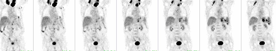
3 Why functional imaging? The lesion was suspected by FDG-PET in January With current PET systems the limit of resolution is cm which translates into a tumor size of g or cells Cancers are usually not diagnosed until they reach a size of g, or cells The lesion was visible on CT in April

4 Learning point Metabolic changes precede structural changes
5 History of FDG-PET Glucose transporter 2-deoxy-D-Glucose 2-deoxy-D-glucose was developed as a chemotherapeutic agent hexokinase 2-deoxy-D Glucose-6-phosphate Lazlo J et al. Effects of glucose analogues (2-deoxy-D-glucose, 2-deoxy-D-galactose) on experimental tumors J Natl Cancer Inst 1960;24: Metabolic trapping TCA cyclus Oxidativ phosphylat ion Cancer cell

6 Metabolic trapping 2-deoxy-D-glucose 18F-FDG 18F-FDG 6 phosphate
7 History of FDG-PET Glucose transporter 2-deoxy-D-Glucose 2-deoxy-D-glucose was developed as a chemotherapeutic agent hexokinase 2-deoxy-D Glucose-6-phosphate Lazlo J et al. Effects of glucose analogues (2-deoxy-D-glucose, 2-deoxy-D-galactose) on experimental tumors J Natl Cancer Inst 1960;24: Metabolic trapping TCA cyclus Oxidativ phosphylat ion Cancer cell

8 History of FDG-PET 1976 & 1977 Glucose transporter 18F-FDG F-FDG was developed to study cerebral glucose metabolism based on PET Brookhaven National Laboratories hexokinase 1977 First PET brain imaging studies UCLA 18F-FDG-6-phosphate Metabolic trapping TCA cyclus Oxidativ phosphylat ion Warburg effect in a cancer cell
9 Biologic Current characteristics and future research of tracer Isotopes 18F 11C 13N 15O 64Cu 68Ga Others
10 PET/CT in Western Europe 2010 Providers: mobile centres 64% public facilities PET/CT 87%
11 Indications Outline for FDG-PET Diagnosis and screening Staging (re-staging) Recurrence Treatment monitoring Treatment planning

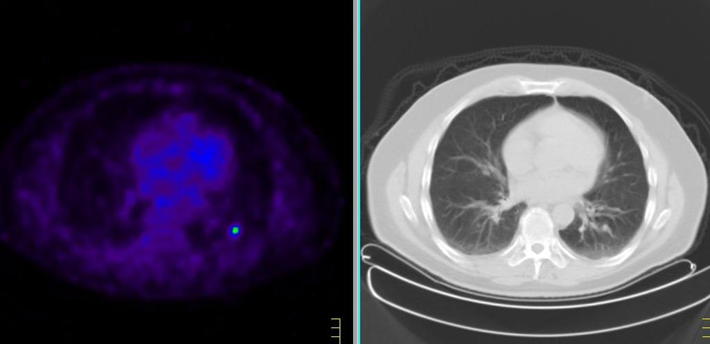
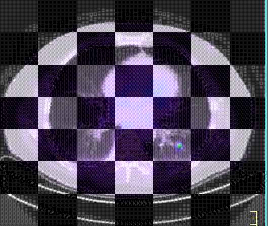
12 The normal FDG-PET Physiological high uptake in the brain In the heart (variable) Excretion into urine bladder The liver has inhomogenous moderately high uptake Spleen is just visible (less activity than liver)
13 Indications An abnormal for FDG-PET P998, S0
 Thyroid cancer")
14 An abnormal FDG-PET 2 years later (after operation) Thyroid cancer P998, S0

 P998,")
15 An abnormal FDG-PET 2 years later (after operation) P998, S0 Breast cancer

16 An abnormal FDG-PET 2 years later Pitt fall P998, S0 Diverticulosis - colonoscopy P998, S0
17 An abnormal FDG-PET 2 years later Hodgkin s lymphoma P998, S0

18 Learning point Learning point Thyroid cancer Breast cancer FDG is an unspecific all round tracer Diverticulosis Hodgkin P998, S0


19 Diagnosis and screening Unknown primary SPN Unknown cancer 2-5% disease - economy radiation exposure - ethics
20 Screening Unknown with primary PET Unknown primary Known secondary (metastases) Lymph node (neck) Liver metastases Lung metastases Primary is identified in app 20% of the cases


21 Screening Single pulmonary with PET nodule Highly suspicious of malignancy

22 Single pulmonary nodule


23 Single pulmonary nodule FDG-negative tumor 111In-octreotide positive carcinoid Sensitivity 80%

24 Unknown cancer?

25 Unknown cancer?

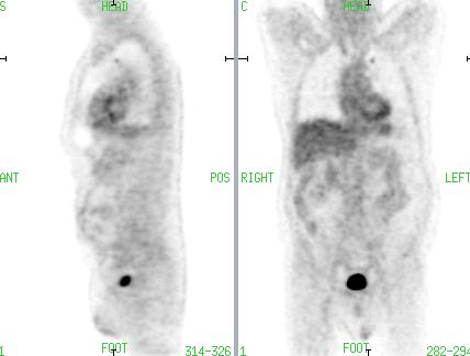
26 Unknown cancer?

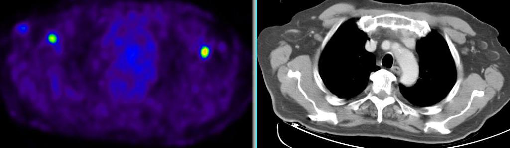
27 Unknown cancer?

28 Unknown cancer?
29 Indications FDG-PET Diagnosis and screening Staging (re-staging) Recurrence Treatment monitoring Treatment planning
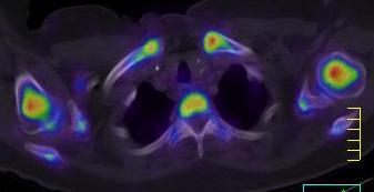
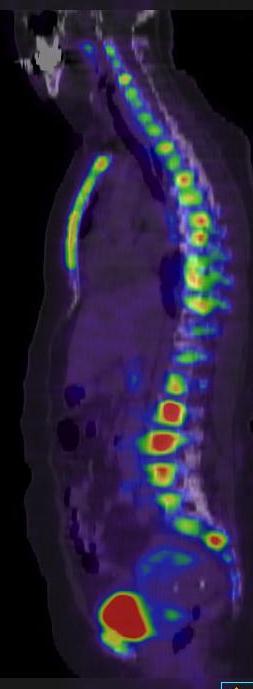
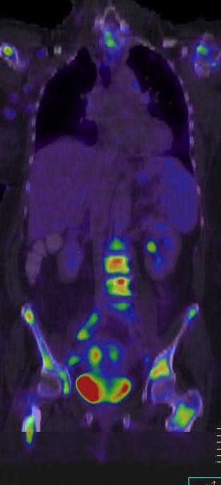
30 TNM Staging T = tumor N = node Sentinel node M = metastase Example: T2N3M1

31 Staging TNM Staging 17 y male. Two months of common cold and pain in left ear. Now bleeding from the nose Cancer of the rhinopharynx MRI has assessed the tumor to be in operable

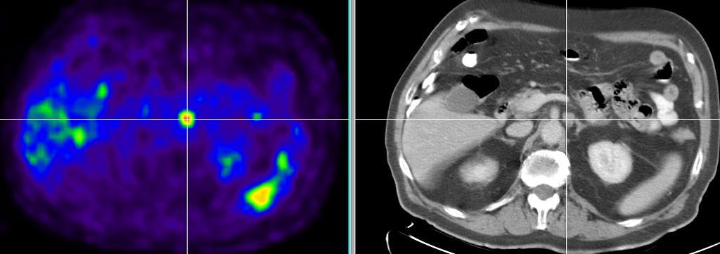
32 T-stage T Staging Tumor in rhinopharynx

33 N-stage N Staging Lymph nodes on both sides of the neck


34 M-stadium M Staging Metastases anteriorly in L4 Metastases in illiac bone

35 Learning point point Total body examination has same radiation exposure to the patient as regional examination Same tracer used for primary and secondaries in different organ systems
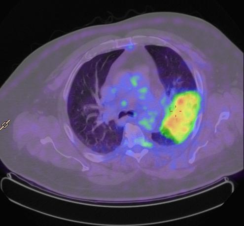
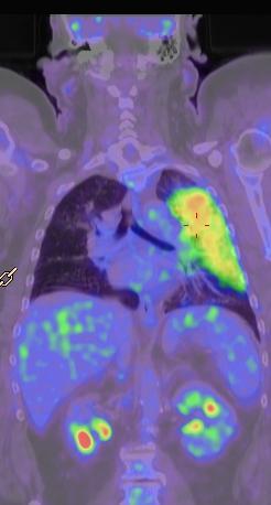
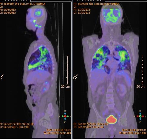
36 Indications for FDG-PET Diagnosis and screening Staging (re-staging) Recurrence Treatment monitoring Treatment planning
37 Colo-rectal cancer. Increased CEA
38 Colo-rectal Lung metastases cancer. Increased CEA
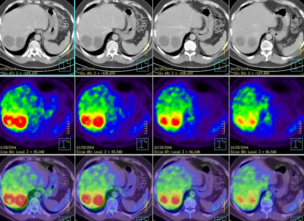
39 Liver metastasis

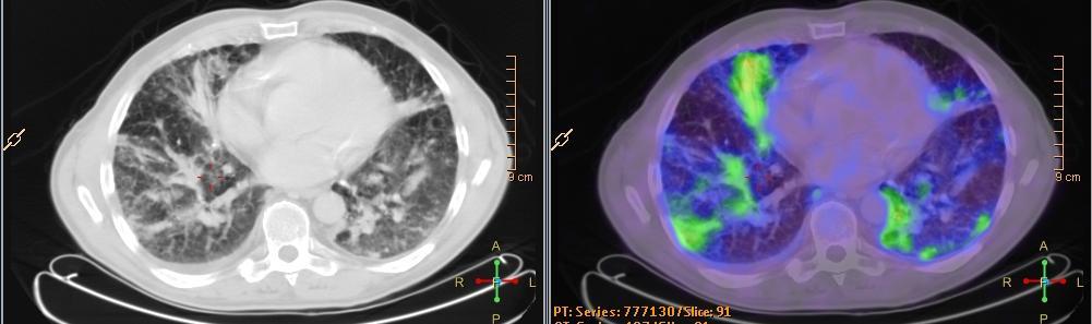
40 Recurrence Colo-rectal cancer. Increased CEA The metastasis in the liver and the left lung were resected. Check-up scanning two years later


41 Colo-rectal cancer. Increased CEA Recurrence in the rectum (stapler line)
42 Collective review : The Emerging Role of 18 F- Fluorodeoxyglucose Positron Emission Tomography in the Management of Primary and Recurrent Rectal Cancer Presented in part at the Colon and Rectal Surgery Specialty Session, American College of Surgeons 90 th Annual Clinical Congress, New Orleans, LA, October David B. Chessin MD, Ravi P. Kiran MD, Timothy Akhurst MD and Jose G. Guillem

43 Colo-rectal cancer. Increased CEA Benign uptake due to operation The former lung metastases

44 First scan: Liver and lung metastasis Second scan: Local recurrence
45 Learning point PET has high sensitivity and specificity in many common cancers incl metastasis and relapse
46 Indications for FDG-PET Diagnosis and screening Staging (re-staging) Recurrence Treatment monitoring Treatment planning
47 Treatment monitoring 76 y male, large celle B cell lymphoma

48 Before After

49 Før Before Before After After

50 Learning point Morphologic lesions can be FDG- positive or FDG-negative
51 PET in early response evaluation CT kan ikke forudse om Behandlingen hjælper
52 Learning point Change in FDG-metabolism can predict treatment response/survival
53 Indications for FDG-PET Diagnosis and screening Staging (re-staging) Recurrence Treatment monitoring Treatment planning

54 Treatment planning 74 y male with lung cancer. RTP

55 Change of GTV actelasis necrosis tumor
 Recurrence Treatment monitoring Treatment")
56 Indications for FDG-PET Diagnosis and screening Staging (re-staging) Recurrence Treatment monitoring Treatment planning

57 Indications Other tracers for than FDG-PET Prostate cancer Bone PET Hypoxia

58 Other tracers than FDG Prostate cancer 99mTc HDP planar bone scan
59 Lipid synthesis prostate cancer [ 11 C] Acetate Acetate Acetate AcetylCoA synthetase (mithochondria) Acetyl CoA TCA Fatty Acid Synthase Malonyl CoA TCA cyclus Oxidativ phosphylation Fatty Acids (phosphatidylcholin) CO 2 Acetate is activated to acetyl-coa which is oxidized to CO 2 and H 2 O in normal cells
 in the prostate (b) in three pelvic lymph nodes (c)")
60 18F choline/acetate Axial-fused PET/CT images demonstrate pathological 18F-choline uptake (a) in the prostate (b) in three pelvic lymph nodes (c) in bones
![Bone metabolism [ 18 F]NaF 18F NaF is chemisorbed onto bone surface by exchanging with OHgroups in](/docs-images/82/85805105/images/61-0.jpg "hydroxapatite crystal of bone to form fluoroapatite.")
61 Bone metabolism [ 18 F]NaF 18F NaF is chemisorbed onto bone surface by exchanging with OHgroups in hydroxapatite crystal of bone to form fluoroapatite. Same uptake mechanism as 99mTc-MDP TCA cyclus Oxidativ phosphylation

62 99mTc MDP bone scan vs 18FNaF PET Examination time: 3-4 h min
![Oxygenation treatment resistance [ 18 F]MISO Nitroreductase Re-oxidation TCA cyclus Oxidativ phosphylation](/docs-images/82/85805105/images/63-0.jpg "Hypoxia is a key factor in Tumour aggression/progression Resistance to therapy Independent negative predictive")
63 Oxygenation treatment resistance [ 18 F]MISO Nitroreductase Re-oxidation TCA cyclus Oxidativ phosphylation Hypoxia is a key factor in Tumour aggression/progression Resistance to therapy Independent negative predictive factor
 and in the left neck nodal")

.")
64 F-MISO in radiotherapy The 18FDG-PET image: Increased uptake in both the oropharyngeal tumour (arrow) and in the left neck nodal metastasis (asterix). The 18F- MISO images The left neck nodal metastasis is shown to be hypoxic Dose escalation map superimposed on CT with Planning Target Volume (red). Isodose lines show conformality to hypoxic lymph node with a maximum dose increase by 20%.
 Generator product Simple labelling")

65 Neuroendocrine cell - tagets tumors for functional imaging 111In Octreotid 123I MIBG 68Ga Octreotid High affinity (SSTR2) Generator product Simple labelling L-Tyrosine 18F-DOPA 11C-HTP* *hyrdoxy trhptophan L-DOPA Dopamine Norepinephrine NET GLUT Epinephrine Loss of NET or VMAT in tumor cell dedifferentiation 18F-FDG 18F DOPA
66
F NaF PET/CT in the Evaluation of Skeletal Malignancy
 F NaF PET/CT in the Evaluation of Skeletal Malignancy Andrei Iagaru, MD September 26, 2013 School of of Medicine Ø Introduction Ø F NaF PET/CT in Primary Bone Cancers Ø F NaF PET/CT in Bone Metastases
PET imaging of cancer metabolism is commonly performed with F18
 PCRI Insights, August 2012, Vol. 15: No. 3 Carbon-11-Acetate PET/CT Imaging in Prostate Cancer Fabio Almeida, M.D. Medical Director, Arizona Molecular Imaging Center - Phoenix PET imaging of cancer metabolism
Nuclear Medicine in Oncology
 Radiopharmaceuticals Nuclear Medicine in Oncology Practice Pharmaceutical Radionuc lide Function Tumor type Diphosphonates Tc-99m Osteoblast Bone tumor & metast. Ga-citrate Ga-67 Fe-analogue Bronchogenous
PET/CT in oncology. Positron emission tomography
 Clinical Medicine 2012, Vol 12, No 4: 368 72 PET/CT in oncology Fahim-Ul-Hassan, SpR Nuclear Medicine, Guy s Hospital, London; Gary J Cook, professor of Clinical PET, KCL Division of Imaging Sciences &
LYMPHATIC DRAINAGE IN THE HEAD & NECK
 LYMPHATIC DRAINAGE IN THE HEAD & NECK Like other parts of the body, the head and neck contains lymph nodes (commonly called glands). Which form part of the overall Lymphatic Drainage system of the body.
Los Angeles Radiological Society 62 nd Annual Midwinter Radiology Conference January 31, 2010
 Los Angeles Radiological Society 62 nd Annual Midwinter Radiology Conference January 31, 2010 Self Assessment Module on Nuclear Medicine and PET/CT Case Review FDG PET/CT IN LYMPHOMA AND MELANOMA Submitted
Principles of nuclear metabolic imaging. Prof. Dr. Alex Maes AZ Groeninge Kortrijk and KULeuven Belgium
 Principles of nuclear metabolic imaging Prof. Dr. Alex Maes AZ Groeninge Kortrijk and KULeuven Belgium I. Molecular imaging probes A. Introduction - Chemical disturbances will precede anatomical abnormalities
Clinical indications for positron emission tomography
 Clinical indications for positron emission tomography Oncology applications Brain and spinal cord Parotid Suspected tumour recurrence when anatomical imaging is difficult or equivocal and management will
Index. Surg Oncol Clin N Am 16 (2007) Note: Page numbers of article titles are in boldface type.
 Note: Page numbers of article titles are in boldface type.") Surg Oncol Clin N Am 16 (2007) 465 469 Index Note: Page numbers of article titles are in boldface type. A Adjuvant therapy, preoperative for gastric cancer, staging and, 339 B Breast cancer, metabolic
Surg Oncol Clin N Am 16 (2007) 465 469 Index Note: Page numbers of article titles are in boldface type. A Adjuvant therapy, preoperative for gastric cancer, staging and, 339 B Breast cancer, metabolic
POSITRON EMISSION TOMOGRAPHY (PET)
") Status Active Medical and Behavioral Health Policy Section: Radiology Policy Number: V-27 Effective Date: 08/27/2014 Blue Cross and Blue Shield of Minnesota medical policies do not imply that members should
Status Active Medical and Behavioral Health Policy Section: Radiology Policy Number: V-27 Effective Date: 08/27/2014 Blue Cross and Blue Shield of Minnesota medical policies do not imply that members should
FDG PET/CT STAGING OF LUNG CANCER. Dr Shakher Ramdave
 FDG PET/CT STAGING OF LUNG CANCER Dr Shakher Ramdave FDG PET/CT STAGING OF LUNG CANCER FDG PET/CT is used in all patients with lung cancer who are considered for curative treatment to exclude occult disease.
Nuclear medicine in oncology. 1. Diagnosis 2. Therapy
 Nuclear medicine in oncology 1. Diagnosis 2. Therapy Diagnosis - Conventional methods - Nonspecific radiopharmaceuticals cumulating in tumours - Specific radiopharmaceuticals (receptor- and immunoscintigraphy)
Disclosure. Acknowledgement. What is the Best Workup for Rectal Cancer Staging: US/MRI/PET? Rectal cancer imaging. None
 What is the Best Workup for Rectal Cancer Staging: US/MRI/PET? Zhen Jane Wang, MD Assistant Professor in Residence UC SF Department of Radiology Disclosure None Acknowledgement Hueylan Chern, MD, Department
Positron Emission Tomography in Lung Cancer
 May 19, 2003 Positron Emission Tomography in Lung Cancer Andrew Wang, HMS III Patient DD 53 y/o gentleman presented with worsening dyspnea on exertion for the past two months 30 pack-year smoking Hx and
Subject: PET Scan With or Without CT Attenuation. Original Effective Date: 11/7/2017. Policy Number: MCR: 610. Revision Date(s): Review Date:
: Review Date:") Subject: PET Scan With or Without CT Attenuation Policy Number: MCR: 610 Revision Date(s): MHW Original Effective Date: 11/7/2017 Review Date: DISCLAIMER This Molina Clinical Review (MCR) is intended to
Subject: PET Scan With or Without CT Attenuation Policy Number: MCR: 610 Revision Date(s): MHW Original Effective Date: 11/7/2017 Review Date: DISCLAIMER This Molina Clinical Review (MCR) is intended to
The Use of PET Scanning in Urologic Oncology
 The Use of PET Scanning in Urologic Oncology Dr Nicholas C. Buchan Uro-oncology Fellow 1 2 Aims To understand the basic concepts underlying PET scanning. Understand the emerging role of PET Scanning for
Whole Body MRI. Dr. Nina Tunariu. Prostate Cancer recurrence, progression and restaging
 Whole Body MRI Prostate Cancer recurrence, progression and restaging Dr. Nina Tunariu Consultant Radiology Drug Development Unit and Prostate Targeted Therapies Group 12-13 Janeiro 2018 Evolving Treatment
Using PET/CT in Prostate Cancer
 Using PET/CT in Prostate Cancer Legal Disclaimer These materials were prepared in good faith by MITA as a service to the profession and are believed to be reliable based on current scientific literature.
PET/CT Frequently Asked Questions
 PET/CT Frequently Asked Questions General Q: Is FDG PET specific for cancer? A: No, it is a marker of metabolism. In general, any disease that causes increased metabolism can result in increased FDG uptake
PET in Prostate Cancer
 PET in Prostate Cancer Tom R. Miller, M.D., Ph.D. Mallinckrodt Institute of Radiology Washington University School of Medicine St. Louis, Missouri, USA Prostate Imaging Bone Scintigraphy primarily for
An Introduction to PET Imaging in Oncology
 January 2002 An Introduction to PET Imaging in Oncology Janet McLaren, Harvard Medical School Year III Basics of PET Principle of Physiologic Imaging: Allows in vivo visualization of structures by their
New imaging techniques: let there be light. Felix M. Mottaghy Department of Nuclear Medicine University Hospital KU Leuven
 New imaging techniques: let there be light Felix M. Mottaghy Department of Nuclear Medicine University Hospital KU Leuven Medical imaging and the pathology cascade Molecular/Cellular disturbance Alterations
New Visions in PET: Surgical Decision Making and PET/CT
 New Visions in PET: Surgical Decision Making and PET/CT Stanley J. Goldsmith, MD Director, Nuclear Medicine Professor, Radiology & Medicine New York Presbyterian Hospital- Weill Cornell Medical Center
2004 SNM Mid-Winter Educational Symposium
 1 2 20 Numeric values 15 10 5 0 SUV corr hs 18FDG Slope corr hs SUV corr hs Slope corr hs 11C-methionin 3 11C-methionine Grading and delineation of brain tumors Differentiation of malignant from benign
Page: 1 of 29. For this policy, PET scanning is discussed for the following 4 applications in oncology:
 Emission Tomography Scanning Page: 1 of 29 Last Review Status/Date: June 2015 Description Positron emission tomography (PET) scans are based on the use of positron-emitting radionuclide tracers coupled
Molecular Imaging and Cancer
 Molecular Imaging and Cancer Cancer causes one in every four deaths in the United States, second only to heart disease. According to the U.S. Department of Health and Human Services, more than 512,000
performed to help sway the clinician in what the appropriate diagnosis is, which can substantially alter the treatment of management.
 Hello, I am Maura Polansky at the University of Texas MD Anderson Cancer Center. I am a Physician Assistant in the Department of Gastrointestinal Medical Oncology and the Program Director for Physician
What Radiologists do?
 Multimodality Imaging in Oncology 2018 March 5 th 9th Diagnostic Imaging in Oncology What Radiologists do? Chikako Suzuki, MD, PhD Department of Diagnostic Radiology, KS Solna Department of Molecular Medicine
objectives Pitfalls and Pearls in PET/CT imaging Kevin Robinson, DO Assistant Professor Department of Radiology Michigan State University
 objectives Pitfalls and Pearls in PET/CT imaging Kevin Robinson, DO Assistant Professor Department of Radiology Michigan State University To determine the regions of physiologic activity To understand
Dr Sneha Shah Tata Memorial Hospital, Mumbai.
 Dr Sneha Shah Tata Memorial Hospital, Mumbai. Topics covered Lymphomas including Burkitts Pediatric solid tumors (non CNS) Musculoskeletal Ewings & osteosarcoma. Neuroblastomas Nasopharyngeal carcinomas
Medical Policy An independent licensee of the Blue Cross Blue Shield Association
 PET Scanning: Oncologic Applications Page 1 of 42 Medical Policy An independent licensee of the Blue Cross Blue Shield Association Title: See also: Positron Emission Tomography (PET) Scanning: Oncologic
Bone PET/MRI : Diagnostic yield in bone metastases and malignant primitive bone tumors
 Bone PET/MRI : Diagnostic yield in bone metastases and malignant primitive bone tumors Lars Stegger, Benjamin Noto Department of Nuclear Medicine University Hospital Münster, Germany Content From PET to
Medical Policy An independent licensee of the Blue Cross Blue Shield Association
 PET Scanning: Oncologic Applications Page 1 of 88 Medical Policy An independent licensee of the Blue Cross Blue Shield Association Title: Positron Emission Tomography (PET) Scanning: Oncologic Applications
Oncologic Applications of PET Scanning
 6.01.26 Oncologic Applications of PET Scanning Section 6.0 Radiology Subsection Effective Date February 15, 2015 Original Policy Date January 26, 2009 Next Review Date December 2015 Description Positron
Chapter 10. Summary, conclusions and future perspectives
 Chapter 10 Summary, conclusions and future perspectives 10.1 SUMMARY In this thesis, a new tumor imaging tracer in nuclear medicine is studied. This 123 tracer, L-3-[ I]Iodo-alpha-methyl-tyrosine (IMT),
Nuclear Medicine Head and Neck Region. Bán Zsuzsanna, MD University of Pécs, Department of Nuclear Medicine
 Nuclear Medicine Head and Neck Region Bán Zsuzsanna, MD University of Pécs, Department of Nuclear Medicine Thyroid scintigraphy Parathyroid scintigraphy F18-FDG PET examinations in head and neck cancer
PET IMAGING (POSITRON EMISSION TOMOGRAPY) FACT SHEET
 FACT SHEET") Positron Emission Tomography (PET) When calling Anthem (1-800-533-1120) or using the Point of Care authorization system for a Health Service Review, the following clinical information may be needed to
Positron Emission Tomography (PET) When calling Anthem (1-800-533-1120) or using the Point of Care authorization system for a Health Service Review, the following clinical information may be needed to
Positron emission tomography/computer tomography in the evaluation of head and neck cancer treatment
 Positron emission tomography/computer tomography in the evaluation of head and neck cancer treatment Severina Šedienė 1, Ilona Kulakienė 1, Viktoras Rudžianskas 2 1 Lithuanian University of Health Sciences,
PET-MRI in malignant bone tumours. Lars Stegger Department of Nuclear Medicine University Hospital Münster, Germany
 PET-MRI in malignant bone tumours Lars Stegger Department of Nuclear Medicine University Hospital Münster, Germany Content From PET to PET/MRI General considerations Bone metastases Primary bone tumours
Short summary of published results of PET with fluoromethylcholine (18F) in prostate cancer
 in prostate cancer") Short summary of published results of PET with fluoromethylcholine (18F) in prostate cancer JN TALBOT and all the team of Service de Médecine Nucléaire Hôpital Tenon et Université Pierre et Marie Curie,
Short summary of published results of PET with fluoromethylcholine (18F) in prostate cancer JN TALBOT and all the team of Service de Médecine Nucléaire Hôpital Tenon et Université Pierre et Marie Curie,
FieldStrength. Leuven research is finetuning. whole body staging
 FieldStrength Publication for the Philips MRI Community Issue 40 May 2010 Leuven research is finetuning 3.0T DWIBS for whole body staging The University Hospital of Leuven is researching 3.0T whole body
Whole body F-18 sodium fluoride PET/CT in the detection of bone metastases in patients with known malignancies: A pictorial review
 Whole body F-18 sodium fluoride PET/CT in the detection of bone metastases in patients with known malignancies: A pictorial review Poster No.: C-1196 Congress: ECR 2014 Type: Educational Exhibit Authors:
PET/CT Value: Rocky Mountain Cancer Centers
 PET/CT Value: Rocky Mountain Cancer Centers Glenn Balasky Executive Director Rocky Mountain Cancer Centers glenn.balasky@usoncology.com CANM/CAMRT Joint Conference March 22, 2018 Vancouver, British Columbia
PET/MR:Techniques, Indications and Applications
 PET/MR:Techniques, Indications and Applications Franz Wolfgang Hirsch Professor and Head of the Department of Pediatric Radiology University Hospital Leipzig / Germany Children s Hospital University Leipzig
Molecular Imaging Guided Therapy: The Perfect Storm. David M Schuster, MD Emory University Department of Radiology Atlanta, GA
 Molecular Imaging Guided Therapy: The Perfect Storm David M Schuster, MD Emory University Department of Radiology Atlanta, GA Talk can be found at radiology.emory.edu Let s start with a case 74 year
Nuclear Medicine and PET. D. J. McMahon rev cewood
 Nuclear Medicine and PET D. J. McMahon 150504 rev cewood 2018-02-15 Key Points Nuclear Medicine and PET: Imaging: Understand how Nuc Med & PET differ from Radiography & CT by the source of radiation. Be
Multiparametric imaging in oncology
 Multiparametric imaging in oncology p1 T p2 p2 T T p3 p1 p3 T Marco Ravanelli Roberto Maroldi The goal of traditional imaging is high spatial and contrast resolution diagnosis, tumor extent treatment planning,
FDG-PET/CT for cancer management
 195 REVIEW FDG-PET/CT for cancer management Hideki Otsuka, Naomi Morita, Kyo Yamashita, and Hiromu Nishitani Department of Radiology, Institute of Health Biosciences, The University of Tokushima, Graduate
Lung Cancer Imaging. Terence Z. Wong, MD,PhD. Department of Radiology Duke University Medical Center Durham, NC 9/9/09
 Lung Cancer Imaging Terence Z. Wong, MD,PhD Department of Radiology Duke University Medical Center Durham, NC 9/9/09 Acknowledgements Edward F. Patz, Jr., MD Jenny Hoang, MD Ellen L. Jones, MD, PhD Lung
Ga68 Imaging. Roland HUSTINX Division of Nuclear Medicine and Oncologic Imaging Centre Hospitalier Universitaire de Liège Belgium
 Ga68 Imaging Roland HUSTINX Division of Nuclear Medicine and Oncologic Imaging Centre Hospitalier Universitaire de Liège Belgium 68 Ga Produced by a 68 Ge/ 68 Ga generator Decays by positron emission
Assessment of renal cell carcinoma by two PET tracer : dual-time-point C-11 methionine and F-18 fluorodeoxyglucose
 Assessment of renal cell carcinoma by two PET tracer : dual-time-point C-11 methionine and F-18 fluorodeoxyglucose Poster No.: C-0805 Congress: ECR 2015 Type: Scientific Exhibit Authors: S. Ito, K. Kato,
ROLE OF PET-CT IN BREAST CANCER, GUIDELINES AND BEYOND. Prof Jamshed B. Bomanji Institute of Nuclear Medicine UCL Hospitals London
 ROLE OF PET-CT IN BREAST CANCER, GUIDELINES AND BEYOND Prof Jamshed B. Bomanji Institute of Nuclear Medicine UCL Hospitals London CANCER Key facts Estimated 15.2 million new cases per year in 2015 worldwide
The Drug Development Paradigm in Oncology: Role of Imaging Hedvig Hricak MSKCC
 The Drug Development Paradigm in Oncology: Role of Imaging Hedvig Hricak MSKCC Precision Medicine: Imaging in Clinical Trials and Drug Development* Critical elements: Patient Selection, Dose Finding &
Regional Training Course on PET/CT in Oncology. (RER-6.035) PROGRAM
 PROGRAM") Regional Training Course on PET/CT in Oncology. (RER-6.035) Riga and Liepaja (Latvia), October 3rd- 6th, 2016 Venue: Nuclear Medicine Centre, Gardenes Street 13, Riga, LATVIA (NOTE: Learning objectives
Regional Training Course on PET/CT in Oncology. (RER-6.035) Riga and Liepaja (Latvia), October 3rd- 6th, 2016 Venue: Nuclear Medicine Centre, Gardenes Street 13, Riga, LATVIA (NOTE: Learning objectives
Colorectal Cancer and FDG PET/CT
 Hybrid imaging in colorectal & esophageal cancer Emmanuel Deshayes IAEA WorkShop, November 2017 Colorectal Cancer and FDG PET/CT 1 Clinical background Cancer of the colon and rectum is one of the most
Nuclear Medicine in the Diabetic Foot
 26.11.2015, Uniklinik Balgrist Nuclear Medicine in the Diabetic Foot Martin Hüllner Nuklearmedizin und Neuroradiologie, USZ / UZH Outline A. Imaging modalities brief technical overview B. Nuclear medicine
Prostate Cancer Basics: Background Information for Outreach Activities with Oncologists, Urologists and Surgeons
 Prostate Cancer Basics: Background Information for Outreach Activities with Oncologists, Urologists and Surgeons Legal Disclaimer These materials were prepared in good faith by MITA as a service to the
PSMA PET SCANNING AND THERANOSTICS IN PROSTATE CANCER KEVIN TRACEY, MD, FRCPC PRECISION DIAGNSOTIC IMAGING REGIONAL PET/CT CENTRE
 PSMA PET SCANNING AND THERANOSTICS IN PROSTATE CANCER KEVIN TRACEY, MD, FRCPC PRECISION DIAGNSOTIC IMAGING REGIONAL PET/CT CENTRE DISCLOSURES/CONFLICTS NONE OBJECTIVES Understand current diagnostic role
Department of Nuclear Medicine with Positron Emission Tomography
 (PET) Unit [1] Contact information: Registration: +48 41 367 4850 Main office: +48 41 367 4860 Fax: +48 41 367 4887 e-mail: zmnsco@onkol.kielce.pl [2] Head of the Department: Professor Janusz Braziewicz
Appendix 1: Regional Lymph Node Stations for Staging Esophageal Cancer
 Appendix 1: Regional Lymph Node Stations for Staging Esophageal Cancer Locoregional (N stage) disease was redefined in the seventh edition of the AJCC Cancer Staging Manual as any periesophageal lymph
Contents. 3 Pneumology Introduction Positron Emission Tomography: Past and Present 1. 2 Fundamentals. xxx
 xxx IX Contents 1 Introduction Positron Emission Tomography: Past and Present 1 1.1 Survey.......................... 1 Physical and Biochemical Fundamentals.... 2 PET in National and International Medical
Nuclear Sciences and Medicine
 Nuclear Sciences and Medicine Rethy Chhem, MD, PhD (Edu), PhD (His), FRCPC Division of Human Health Guest Professor, Medical University of Vienna International Atomic Energy Agency Medical Imaging X-rays
Case Reports: Tumor Detection by Diffusion-Weighted MRI and ADC-Mapping with Correlation to PET/CT Results
 Case Reports: Tumor Detection by Diffusion-Weighted MRI and ADC-Mapping with Correlation to PET/CT Results Matthias Philipp Lichy, M.D.; Philip Aschoff, M.D.; Christina Pfannenberg, M.D.; Schlemmer Heinz-Peter,
στη σταδιοποίηση του καρκίνου του προστάτη Γ. Αρσος, Γ Εργ. Πυρηνικής Ιατρικής ΑΠΘ, ΓΝΘ Παπαγεωργίου
 Η θέση του PET/CT στη σταδιοποίηση του καρκίνου του προστάτη Γ. Αρσος, Γ Εργ. Πυρηνικής Ιατρικής ΑΠΘ, ΓΝΘ Παπαγεωργίου 2014 : the Guidelines year. PRINCIPLES OF IMAGING Imaging is performed for the detection
Table 1. Some other positron tracers with applications in oncology.
 CME: CLINICAL PRACTICE AND ITS BASIS Nuclear Medicine Edited by Peter J Ell FMedSci, University Chair and Director, Institute of Nuclear Medicine, UCL; Clinical Director, University College London Hospitals
PET/CT in breast cancer staging
 PET/CT in breast cancer staging Anni Morsing Consultant, PhD, DMSc Rigshospitalet 1 18F- FDG PET/CT for breastcancer staging Where is the clinical impact? To which women should 18F- FDG PET/CT be offered?
The role of appropriateness criteria in the planning of health services: the case of FDG-PET in oncology
 V Annual Meeting of Health Tecnology Assessment International (HTAi) MONTREAL 8 july 2008 The role of appropriateness criteria in the planning of health services: the case of FDG-PET in oncology L. BALLINI
PET/CT F-18 FDG. Objectives. Basics of PET/CT Imaging. Objectives. Basic PET imaging
 Basics of PET/CT Imaging Kevin Robinson, DO Department of Radiology Michigan State University Objectives Basic PET imaging Evaluating the therapeutic response Evaluating the big 5 Lymphoma Breast Lung
Staging Colorectal Cancer
 Staging Colorectal Cancer CT is recommended as the initial staging scan for colorectal cancer to assess local extent of the disease and to look for metastases to the liver and/or lung Further imaging for
The Role of PET / CT in Lung Cancer Staging
 July 2004 The Role of PET / CT in Lung Cancer Staging Vlad Vinarsky, Harvard Medical School Year IV Patient AM HPI: 81 yo F p/w hemoptysis x 1 month LLL lesion on CXR, not responsive to Abx 35 pack-year
Novel Imaging in Advanced Prostate Cancer
 Novel Imaging in Advanced Prostate Cancer Robert J. Hamilton, MD MPH FRCSC Princess Margaret Cancer Centre ICUC Saturday January 21, 2017 Company/Organizati Details Faculty/Presenter on Disclosures I am
Functional aspects of anatomical imaging techniques
 Functional aspects of anatomical imaging techniques Nilendu Purandare Associate Professor & Consultant Radiologist Tata Memorial Centre Functional/metabolic/molecular imaging (radioisotope scanning) PET
FDG-PET/CT in Gynaecologic Cancers
 Friday, August 31, 2012 Session 6, 9:00-9:30 FDG-PET/CT in Gynaecologic Cancers (Uterine) cervical cancer Endometrial cancer & Uterine sarcomas Ovarian cancer Little mermaid (Edvard Eriksen 1913) honoring
Phillip J. Koo, MD Division Chief of Diagnostic Imaging Banner MD Anderson Cancer Center, USA
 ADVANCED PET IMAGING IN PROSTATE CANCER Phillip J. Koo, MD Division Chief of Diagnostic Imaging Banner MD Anderson Cancer Center, USA PET, positron-emission tomography DISCLAIMER Please note: The views
COLORECTAL CARCINOMA
 QUICK REFERENCE FOR HEALTHCARE PROVIDERS MANAGEMENT OF COLORECTAL CARCINOMA Ministry of Health Malaysia Malaysian Society of Colorectal Surgeons Malaysian Society of Gastroenterology & Hepatology Malaysian
Radionuclides in Medical Imaging. Danielle Wilson
 Radionuclides in Medical Imaging Danielle Wilson Outline Definitions History and development Radionuclide applications & techniques in imaging Conclusion Definition #1 : Radionuclide An unstable nucleus
Thyroid Cancer: Imaging Techniques (Nuclear Medicine)
") Thyroid Cancer: Imaging Techniques (Nuclear Medicine) Andrei Iagaru, MD MIPS Molecular Imaging Program at Stanford Stanford University School of Medicine Department of Radiology Introduction Ø There are
Thyroid Cancer: Imaging Techniques (Nuclear Medicine) Andrei Iagaru, MD MIPS Molecular Imaging Program at Stanford Stanford University School of Medicine Department of Radiology Introduction Ø There are
IMAGING GUIDELINES - COLORECTAL CANCER
 IMAGING GUIDELINES - COLORECTAL CANCER DIAGNOSIS The majority of colorectal cancers are diagnosed on colonoscopy, with some being diagnosed on Ba enema, ultrasound or CT. STAGING CT chest, abdomen and
Positron emission tomography scanning is coming to a hospital near you soon!
 PROFESSIONAL ISSUES Positron emission tomography scanning is coming to a hospital near you soon! Humayun Bashir, Gregory Shabo and TO Nunan Humayun Bashir MB BS FCPS, Specialist Registrar in Nuclear Medicine
PET-CT for radiotherapy planning in lung cancer: current recommendations and future directions
 PET-CT for radiotherapy planning in lung cancer: current recommendations and future directions Gerry Hanna Centre for Cancer Research and Cell Biology Queen s University of Belfast @gerryhanna Talk Outline
Research Article Prevalence of Clinically Significant Extraosseous Findings on Unenhanced CT Portions of 18 F-Fluoride PET/CT Bone Scans
 The Scientific World Journal Volume 2012, Article ID 979867, 5 pages doi:10.1100/2012/979867 The cientificworldjournal Research Article Prevalence of Clinically Significant Extraosseous Findings on Unenhanced
M etastatic disease influences patient management; Whole body PET/CT for initial staging of choroidal melanoma SCIENTIFIC REPORT
 1270 SCIENTIFIC REPORT Whole body PET/CT for initial staging of choroidal P T Finger, M Kurli, S Reddy, L B Tena, A C Pavlick... Aim: To investigate the value of whole body positron emission tomography/computed
Hybrid systems in Medical Imaging
 Hybrid systems in Medical Imaging from PET/CT to PET/MR Osman Ratib, MD, PhD, FAHA Professor and chair Department of Medical Imaging and Information Sciences Head of division of Nuclear Medicine University
Detection of Soft Tissue Tumors on Bone Scintigraphy: Report of Four Cases
 Detection of Soft Tissue Tumors on Bone Scintigraphy: Report of Four Cases Fariba Akhzari 1 MD and Mahrokh Daemi MD 2 1 Nuclear Medicine Department, 2 General Surgery Department, Sina Hospital, Faculty
Laura Tormoehlen, M.D. Neurology and EM-Toxicology Indiana University
 Laura Tormoehlen, M.D. Neurology and EM-Toxicology Indiana University Disclosures! No conflicts of interest to disclose Neuroimaging 101! Plain films! Computed tomography " Angiography " Perfusion! Magnetic
NUCLEAR MEDICINE Molecular Imaging + Endo-Radiotherapy
 NUCLEAR MEDICINE Molecular Imaging + Endo-Radiotherapy Istvan Szilvási Dept. of Nuclear Medicine Semmelweis University and HDF Medical Centre 2016 DEFINITION OF NUCLEAR MEDICINE Medical applications of
Hybrid Imaging SPECT/CT PET/CT PET/MRI. SNMMI Southwest Chapter Aaron C. Jessop, MD
 Hybrid Imaging SPECT/CT PET/CT PET/MRI SNMMI Southwest Chapter 2014 Aaron C. Jessop, MD Assistant Professor, Department of Nuclear Medicine UT MD Anderson Cancer Center, Houston, Texas Complimentary role
Theranostics in Nuclear Medicine
 Theranostics in Nuclear Medicine Patrick FLAMEN, MD, PhD Head Nuclear Medicine Institut Jules Bordet Université Libre de Bruxelles (U.L.B.) n Theranostics in Nuclear Medicine n A form of (nuclear) diagnostic
ANZUP SURVEILLANCE RECOMMENDATIONS FOR METASTATIC TESTICULAR CANCER POST-CHEMOTHERAPY
 ANZUP SURVEILLANCE RECOMMENDATIONS FOR METASTATIC TESTICULAR CANCER POST-CHEMOTHERAPY Note: These surveillance recommendations are provided as recommendations only. Clinicians should take into account
Tumor Imaging in Nuclear Medicine: Current Status and
 Tumor Imaging in Nuclear Medicine: Current Status and Future Prospects Bennett S. Greenspan, MD SNM MWM Albuquerque, NM January 30, 2010 Tumor Imaging Part I Current Status Current status agents that are
Lugano classification: Role of PET-CT in lymphoma follow-up
 CAR Educational Exhibit: ID 084 Lugano classification: Role of PET-CT in lymphoma follow-up Charles Nhan 4 Kevin Lian MD Charlotte J. Yong-Hing MD FRCPC Pete Tonseth 3 MD FRCPC Department of Diagnostic
Prof. Dr. NAGUI M. ABDELWAHAB,M.D.; MARYSE Y. AWADALLAH, M.D. AYA M. BASSAM, Ms.C.
 Role of Whole-body Diffusion MR in Detection of Metastatic lesions Prof. Dr. NAGUI M. ABDELWAHAB,M.D.; MARYSE Y. AWADALLAH, M.D. AYA M. BASSAM, Ms.C. Cancer is a potentially life-threatening disease,
CLINICAL PRESENTATION AND RADIOLOGY QUIZ QUESTION
 Donald L. Renfrew, MD Radiology Associates of the Fox Valley, 333 N. Commercial Street, Suite 100, Neenah, WI 54956 4/30/2011 Radiology Quiz of the Week # 18 Page 1 CLINICAL PRESENTATION AND RADIOLOGY
Nuclear Medicine in Australia. Shaun Jenkinson
 Nuclear Medicine in Australia Shaun Jenkinson Landmark Infrastructure for Australian Science OPAL Research Reactor Australian Synchrotron Camperdown Cyclotron Bragg Institute Centre for Accelerator Science
Essentials of Clinical MR, 2 nd edition. 73. Urinary Bladder and Male Pelvis
 73. Urinary Bladder and Male Pelvis Urinary bladder carcinoma is best locally staged with MRI. It is important however to note that a thickened wall (> 5 mm) is a non-specific finding seen in an underfilled
Optimized. clinical pathway. propels high utilization of PET/MR at Pitié-Salpêtrière Hospital
 Optimized propels high utilization of PET/MR at Pitié-Salpêtrière Hospital clinical pathway As one of Europe s largest teaching hospitals, Pitié-Salpêtrière Hospital is renowned for its innovative research
Evaluation of Whole-Field and Split-Field Intensity Modulation Radiation Therapy (IMRT) Techniques in Head and Neck Cancer
 Techniques in Head and Neck Cancer") 1 Charles Poole April Case Study April 30, 2012 Evaluation of Whole-Field and Split-Field Intensity Modulation Radiation Therapy (IMRT) Techniques in Head and Neck Cancer Abstract: Introduction: This study
1 Charles Poole April Case Study April 30, 2012 Evaluation of Whole-Field and Split-Field Intensity Modulation Radiation Therapy (IMRT) Techniques in Head and Neck Cancer Abstract: Introduction: This study
Emerging Referral Patterns for Whole-Body Diffusion Weighted Imaging (WB-DWI) in an Oncology Center
 in an Oncology Center") Emerging Referral Patterns for Whole-Body Diffusion Weighted Imaging (WB-DWI) in an Oncology Center Poster No.: C-1296 Congress: ECR 2014 Type: Scientific Exhibit Authors: G. Petralia 1, G. Conte 1, S.
Emerging Referral Patterns for Whole-Body Diffusion Weighted Imaging (WB-DWI) in an Oncology Center Poster No.: C-1296 Congress: ECR 2014 Type: Scientific Exhibit Authors: G. Petralia 1, G. Conte 1, S.
Radiofrequency ablation combined with conventional radiotherapy: a treatment option for patients with medically inoperable lung cancer
 Radiofrequency ablation combined with conventional radiotherapy: a treatment option for patients with medically inoperable lung cancer Poster No.: C-0654 Congress: ECR 2011 Type: Scientific Paper Authors:
VIII. 9. FDG-PET for Diagnosis of an Advanced Jejunal Adenocarcinoma with Distant Metastases, Compared with Gallium Scintigraphy
 CYRIC Annual Report 2003 VIII. 9. FDG-PET for Diagnosis of an Advanced Jejunal Adenocarcinoma with Distant Metastases, Compared with Gallium Scintigraphy Yamaura G., Yoshioka T., Yamaguchi K. *, Fukuda