PET/CT in Breast Cancer
|
|
|
- Liliana Woods
- 5 years ago
- Views:
Transcription

1 PET/CT in Breast Cancer Rodolfo Núñez Miller, M.D. Nuclear Medicine and Diagnostic Imaging Section Division of Human Health International Atomic Energy Agency Vienna, Austria
2 Overview Introduction Locorregional staging Distant (Sistemic) staging FDG PET in skeletal metastasis Monitoring response to neoadjuvant therapy Monitoring response to metastatic disease Positron Emission Mammography (PEM) and BSGI Metabolic Flare Future directions: molecular imaging
3 Breast Cancer Is the second leading cause of cancer mortality in women. Although the incidence continues to rise, mortality has declined over the past several years. This decline has been attributed to both, early diagnosis and more effective treatment. Advances in molecular cancer biology, are bringing a better understanding of breast cancer pathogenesis and progression, which translates into new and effective treatments. Personalized target therapy.

4 GLOBOCAN 2012


5 Locoregional Staging with FDG PET/CT There is currently no clinical role for routine FDG PET/CT axillary staging in women with newly diagnosed breast cancer.

6 Locoregional Staging with FDG PET/CT in patients at high risk for nodal disease FDG PET is highly specific for axillary nodal metastases. There may be a certain role for preoperative FDG PET/CT in certain patient populations, such as LABC, IBC, plexopathy and symptomatic metastasis. In these cases, if confirmed by PET, US guided tissue sampling of abnormal findings can confirm the diagnosis.
7 FDG PET of the Internal Mammary Nodes Is seen in approximately 25% of patients with LABC. Is predictive of both, likelihood and pattern of treatment failure, consistent with IM nodal disease involvement and progression.
, FDG PET/CT was positive for")
.")
8 PET/CT in Inflammatory Breast Cancer (IBC) Study by Carkaci et al., demonstrated that in newly diagnosed unilateral IBC (n=41), FDG PET/CT was positive for ipsilateral axillary nodal involvement in 90%, ipsilateral subpectoral nodes in 44%, and distant metastasis in 49% (17% unknown before PET/CT). FDG PET/CT should be considered in the initial staging of IBC. Carkaci et al. JNM 2009; 50:


9 Retrospective Analysis of 18F-FDG PET/CT for Staging Asymptomatic Breast Cancer Patients Younger Than 40 Years. Riedl C.C. et al. JNM October 2014 PET/CT revealed distant metastases in 17% of asymptomatic stage IIB breast cancer patients younger than 40 y. Although guidelines of the National Comprehensive Cancer Network recommend against systemic staging in patients with stage II disease, our data suggest that PET/CT might be valuable in younger patients with stage IIB and III disease.
10 Distant (Systemic) Staging with FDG PET NCCN practice guidelines, recommend FDG PET as an option for patients with stage 4 disease. In this setting it has been shown to be both, sensitive and specific. FDG PET can be particularly helpful in identifying occult sites of disease; thus affecting therapeutic options. FDG PET or PET/CT can reveal in this setting unknown metastasis in locoregional and mediastinal nodal basins, which are not obtimally evaluated by conventional imaging.
 followed")

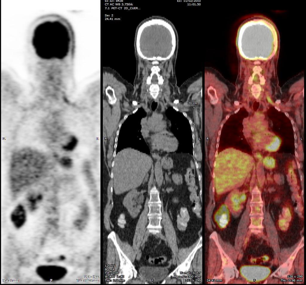
11 39 year old woman with left breast cancer, treated with surgery (Febr, 2010) followed by chemo. There is known liver involvement. Referred for re-staging after initial therapy.
12 Systemic Staging with FDG PET FDG PET and PET/CT can improve staging and alter therapeutic options in patients suspected to have recurrent breast cancer and distant metastatic disease. FDG PET can change or affect treatment in up to 44% of patients suspected to have locorregional recurrence.


13 43 year old woman with left breast cancer, treated initially with surgery, followed by chemo and RT during PET/CT requested to re-stage after initial therapies. October 2009


14 43 year old woman with left breast cancer, treated initially with surgery, followed by chemo and RT during PET/CT requested to re-stage after initial therapies. October 2009


15 She gets treated with additional chemo. PET/CT is requested 6 months later to assess response to therapy. October 2009 April 2010


16 She gets treated with additional chemo. PET/CT is requested 6 months later to assess response to therapy. October 2009 April 2010
17 Staging with FDG PET and PET/CT: When does it help? Is not recommended for routine staging of breast cancer. FDG PET can provide additional information in staging or re-staging cases when results of conventional imaging are equivocal or conflicting. Evaluating asymptomatic patients with rising serum tumor markers without clinical symptoms. FDG PET in this scenario is more accurate than conventional imaging. In a study from 2006* FDG PET/CT was 90% sensitive for diagnosing recurrent tumor, affecting clinical management in 51% of patients. * Randan L et al. Cancer 2006; 107(11):


18 54 year old woman with breast cancer. Referred for re-staging purposes.
19 FDG PET and PET/CT of Skeletal Metastases FDG PET is complementary to bone scintigraphy, which remains the standard imaging modality for surveying the skeleton for metastatic disease. FDG PET better for osteolitic. BS better for osteoblastic. F18 Fluoride PET may offer improve bone metastasis detection compared to FDG and BS.

20 FDG FDG Na 18 F Na 18 F
21 Monitoring Response to Therapy with FDG PET and PET/CT Neoadjuvant Metastatic Disease
22 Monitoring response to Neoadjuvant Therapy Limitations of conventional anatomic imaging. Neoadjuvant Therapy can improve surgical options and provide prognostic information. One of the main objectives is to assess response of the primary tumor to the treatment regime. Most studies measure the change in FDG uptake to midtherapy compared to baseline. 50% or more decline in FDG uptake.

23 Monitoring response to Neoadjuvant Therapy FDG PET may serve as an earlier predictor of response. Absence of FDG uptake is not a reliable indicator of pcr.
24 Dedicated Molecular Breast Imaging Devices PEM (Positron Emission Mammography) BSGI (Breast Specific Gamma Imaging)

25 BSGI The concept Greater access, close to chest wall, able to image breast only with no background activity and scatter from surrounding organs. Greater maneuverability Less intimidating Enabling Technology CZT detectors, low dead space. Registered colimation Optimized pixel size
")
26 Positron Emission Mammography (PEM)


27 The latest design allows for 12 tomographic slices and PEM directed biopsies.
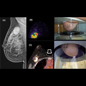
28
29 Advantages in the detection with PEM The advantages are: - Greater spatial resolution. - Improved geometric sensitivity. - Shorter time of image acquisition. - Less attenuation in comparison with PET Fairly small size, with possibility of doing biopsies directed by PEM.

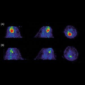
30 62 y-o IDC 2.4cm mass, PEM ratio 3.4, 0.6 cm satellite RMLO LMLO Images of PEM courtesy of Dr. Javier Villanueva-Meyer

31 43 y old IDC + DCIS index lesion 1,7 cm. PEM ratio 7.7. Multifocal + cysts RMLO LMLO

32 46 y-o IDC triple negative, R lumpectomy, 8 year FU in remission RCC LCC
33 Limitations of PEM False Negatives Posterior Lesions Tale of the breast Precision IDC > ILC > DCIS False Positives Recent Biopsy Fat necrosis Fibroadenoma Other benign lesions
34 PEM as alternative to MR Patients with doubtful / equivocal lesions, which cannot be evaluated by MR (claustrophobia, pacemakers, which happens in approx. 15% of cases). Assessment of response to Neoadjuvant therapy.
. Changes in FDG can be prognostic. Combination of FDG and Na-18F can be helpful to assess the skeleton.")
35 Monitoring Response to Metastatic Disease FDG PET can be particularly useful in this setting to evaluate the response to systemic therapy, since conventional imaging is often challenging in these cases (e.g. skeleton). Changes in FDG can be prognostic. Combination of FDG and Na-18F can be helpful to assess the skeleton. However, Na-18F can also have a flare response.
36
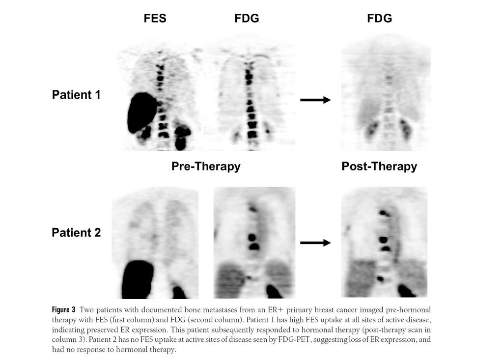
37 Metabolic flare ER+ Tamoxifen Transient increase in metabolic activity of breast cancer within 1 to 2 wks after starting tamoxifen The increase is about +25% in responders while non responders have no change Seen with FDG and FES Dehdashty & Mortimer

38 Metabolic Flare Dehdashti et al. Eur J Nucl Med :51-56.
39 Future Directions: Molecular Imaging Tumor Perfusion and Angiogenesis Imaging. - 15Oxigen water, imbalance of metabolism and perfusion (high mb, and poor perfusion) is associated with poor response and early relapse. - Assess tumor neovasculature, PET probes (RGD peptides) bind to integrins (α 5 β 3 ) in neovessels Tumor Receptor Imaging. - 18Fluoro 17-β-estradiol (FES) - 68Ga-labelled F(ab )2 fragment of trastuzumab, measuring regional HER2 expression in animal models. Early Response Imaging. - FLT. Lilkely to become important in the future I Annexin V for apoptosis.
40 CONCLUSIONS FDG PET and PET/CT have been shown to be most helpful in staging recurrent or metastatic breast cancer, and in evaluating the response of LABC and metastatic disease to treatment. Emerging data support the use of FDG PET/CT in advance axillary disease and evaluation of regional nodal spread in LABC. Currently FDG and occasionally Fluoride PET are used in clinical practice. It is likely that future studies will benefit from tracers other than FDG, for example FES and FLT. As breast cancer diagnosis and therapy become increasingly molecular and individualized, PET/CT imaging will play a progressively more important role in breast cancer patient care.


41 Thank You
ROLE OF PET-CT IN BREAST CANCER, GUIDELINES AND BEYOND. Prof Jamshed B. Bomanji Institute of Nuclear Medicine UCL Hospitals London
 ROLE OF PET-CT IN BREAST CANCER, GUIDELINES AND BEYOND Prof Jamshed B. Bomanji Institute of Nuclear Medicine UCL Hospitals London CANCER Key facts Estimated 15.2 million new cases per year in 2015 worldwide
PET/CT in breast cancer staging
 PET/CT in breast cancer staging Anni Morsing Consultant, PhD, DMSc Rigshospitalet 1 18F- FDG PET/CT for breastcancer staging Where is the clinical impact? To which women should 18F- FDG PET/CT be offered?
Clinical Utility of Positron Emission Tomography Scanning in Breast Cancer Management
 Clinical Utility of Positron Emission Tomography Scanning in Breast Cancer Management David Schuster, MD Director, Division of Nuclear Medicine and Molecular Imaging U N I V E R S I T Y S C H O O L O F
Dr Sneha Shah Tata Memorial Hospital, Mumbai.
 Dr Sneha Shah Tata Memorial Hospital, Mumbai. Topics covered Lymphomas including Burkitts Pediatric solid tumors (non CNS) Musculoskeletal Ewings & osteosarcoma. Neuroblastomas Nasopharyngeal carcinomas
Role of PEM in Breast Cancer Management. Judy Kalinyak, MD, PhD Chief Medical Officer Naviscan, Inc (San Diego, CA)
") Role of PEM in Breast Cancer Management Judy Kalinyak, MD, PhD Chief Medical Officer Naviscan, Inc (San Diego, CA) Role of PEM in Breast Cancer Management Introduction to Positron Emission Mammography
Role of PEM in Breast Cancer Management Judy Kalinyak, MD, PhD Chief Medical Officer Naviscan, Inc (San Diego, CA) Role of PEM in Breast Cancer Management Introduction to Positron Emission Mammography
Breast Cancer. Most common cancer among women in the US. 2nd leading cause of death in women. Mortality rates though have declined
 Breast Cancer Most common cancer among women in the US 2nd leading cause of death in women Mortality rates though have declined 1 in 8 women will develop breast cancer Breast Cancer Breast cancer increases
Breast Cancer. Saima Saeed MD
 Breast Cancer Saima Saeed MD Breast Cancer Most common cancer among women in the US 2nd leading cause of death in women 1 in 8 women will develop breast cancer Incidence/mortality rates have declined Breast
When do you need PET/CT or MRI in early breast cancer?
 When do you need PET/CT or MRI in early breast cancer? Elizabeth A. Morris MD FACR Chief, Breast Imaging Service Memorial Sloan-Kettering Cancer Center NY, NY Objectives What is the role of MRI in initial
Breast Cancer Staging. Physiology Trumps Anatomy Author: Maxine Jochelson, MD, FSBI
 Breast Cancer Staging. Physiology Trumps Anatomy Author: Maxine Jochelson, MD, FSBI The purpose of this paper is to address the importance of physiologic imaging for the staging and follow up of patients
The Role of Radiotracer Imaging in the Diagnosis and Management of Patients with Breast Cancer: Part 1 Overview, Detection, and Staging*
 CONTINUING EDUCATION The Role of Radiotracer Imaging in the Diagnosis and Management of Patients with Breast Cancer: Part 1 Overview, Detection, and Staging* Jean H. Lee 1, Eric L. Rosen 2, and David A.
New Imaging Modalities for better Screening and Diagnosis
 New Imaging Modalities for better Screening and Diagnosis Miri Sklair-Levy, MD Department of Diagnostic Imaging Sheba Medical Center, Sackler School of Medicine, Tel Aviv University Department of Diagnostic
PET/CT for Therapy Assessment in Oncology
 PET/CT for Therapy Assessment in Oncology Rodolfo Núñez Miller, M.D. Nuclear Medicine Section Division of Human Health International Atomic Energy Agency Vienna, Austria Clinical Applications of PET/CT
Clinical Utility of Positron Emission Tomography Scanning in Breast Cancer Management
 Clinical Utility of Positron Emission Tomography Scanning in Breast Cancer Management David Schuster, MD Director, Division of Nuclear Medicine and Molecular Imaging Department of Radiology and Imaging
FDG PET/CT STAGING OF LUNG CANCER. Dr Shakher Ramdave
 FDG PET/CT STAGING OF LUNG CANCER Dr Shakher Ramdave FDG PET/CT STAGING OF LUNG CANCER FDG PET/CT is used in all patients with lung cancer who are considered for curative treatment to exclude occult disease.
Expert Review The Role of Molecular Imaging in Response Prediction in Metastatic Breast Cancer
 Expert Review The Role of Molecular Imaging in Response Prediction in Metastatic Breast Cancer Geraldine Gebhart, MD Institut Jules Bordet Brussels, Belgium Discussants Moderator Lee Lokey, MD prime Oncology
performed to help sway the clinician in what the appropriate diagnosis is, which can substantially alter the treatment of management.
 Hello, I am Maura Polansky at the University of Texas MD Anderson Cancer Center. I am a Physician Assistant in the Department of Gastrointestinal Medical Oncology and the Program Director for Physician
Molecular Imaging and Breast Cancer
 Molecular Imaging and Breast Cancer Breast cancer forms in tissues of the breast usually in the ducts, tubes that carry milk to the nipple, and lobules, the glands that make milk. It occurs in both men
PET/CT Frequently Asked Questions
 PET/CT Frequently Asked Questions General Q: Is FDG PET specific for cancer? A: No, it is a marker of metabolism. In general, any disease that causes increased metabolism can result in increased FDG uptake
PET/CT in oncology. Positron emission tomography
 Clinical Medicine 2012, Vol 12, No 4: 368 72 PET/CT in oncology Fahim-Ul-Hassan, SpR Nuclear Medicine, Guy s Hospital, London; Gary J Cook, professor of Clinical PET, KCL Division of Imaging Sciences &
Molecular Imaging and Cancer
 Molecular Imaging and Cancer Cancer causes one in every four deaths in the United States, second only to heart disease. According to the U.S. Department of Health and Human Services, more than 512,000
BREAST MRI. Elizabeth A. Rafferty, M.D. Avon Comprehensive Breast Center Massachusetts General Hospital Harvard Medical School
 BREAST MRI Elizabeth A. Rafferty, M.D. Avon Comprehensive Breast Center Massachusetts General Hospital Harvard Medical School BREAST MRI Any assessment of the breast parenchyma requires the administration
New Visions in PET: Surgical Decision Making and PET/CT
 New Visions in PET: Surgical Decision Making and PET/CT Stanley J. Goldsmith, MD Director, Nuclear Medicine Professor, Radiology & Medicine New York Presbyterian Hospital- Weill Cornell Medical Center
FDG-PET/CT in Gynaecologic Cancers
 Friday, August 31, 2012 Session 6, 9:00-9:30 FDG-PET/CT in Gynaecologic Cancers (Uterine) cervical cancer Endometrial cancer & Uterine sarcomas Ovarian cancer Little mermaid (Edvard Eriksen 1913) honoring
Breast Cancer Imaging
 Breast Cancer Imaging I. Policy University Health Alliance (UHA) will cover breast imaging when such services meet the medical criteria guidelines (subject to limitations and exclusions) indicated below.
PET/CT in Gynaecological Cancers. Stroobants Sigrid, MD, PhD Departement of Nuclear Medicine University Hospital,Antwerp
 PET/CT in Gynaecological Cancers Stroobants Sigrid, MD, PhD Departement of Nuclear Medicine University Hospital,Antwerp Cervix cancer Outline of this talk Initial staging Treatment monitoring/guidance
Here are examples of bilateral analog mammograms from the same patient including CC and MLO projections.
 Good afternoon. It s my pleasure to be discussing Diagnostic Breast Imaging over the next half hour. I m Wei Yang, Professor of Diagnostic Radiology and Chief, the Section of Breast Imaging as well as
Low Dose Molecular Breast Imaging
 Low Dose Molecular Breast Imaging Dr. M.K. O Connor Conflict of Interest Royalties - Gamma Medica Research funding GE Healthcare Research support MTTI Michael O Connor, Ph.D Dept. of Radiology Mayo Clinic
The Cure Starts Here 2/9/2015. You can get up and use the restroom anytime, but be discreet please. Objectives
 Objectives The Cure Starts Here Deborah Thames R.T. (R)(M)(QM) Staging a Patient through Imaging Modalities Knowing the Choices with BI-RADs and the Lexicon Treatment Options and Managing Patient Care
An Introduction to PET Imaging in Oncology
 January 2002 An Introduction to PET Imaging in Oncology Janet McLaren, Harvard Medical School Year III Basics of PET Principle of Physiologic Imaging: Allows in vivo visualization of structures by their
Courtesy of Dan Kopans, M.D.
 Molecular Breast Imaging New Modality for Diagnosing i Breast Cancer Don t we have enough tools for diagnosing i Breast Cancer??? By Lillian H. Stern, M.D. Current Modalities Mammograms (analogue v. digital)
Bone PET/MRI : Diagnostic yield in bone metastases and malignant primitive bone tumors
 Bone PET/MRI : Diagnostic yield in bone metastases and malignant primitive bone tumors Lars Stegger, Benjamin Noto Department of Nuclear Medicine University Hospital Münster, Germany Content From PET to
PET/CT in Breast Cancer
 PET/CT in Breast Cancer Hossein Jadvar, MD, PhD, MPH, MBA Associate Professor of Radiology and Vice Chair of Research Associate Professor of Biomedical Engineering President, Society of Nuclear Medicine
Index. Surg Oncol Clin N Am 16 (2007) Note: Page numbers of article titles are in boldface type.
 Note: Page numbers of article titles are in boldface type.") Surg Oncol Clin N Am 16 (2007) 465 469 Index Note: Page numbers of article titles are in boldface type. A Adjuvant therapy, preoperative for gastric cancer, staging and, 339 B Breast cancer, metabolic
Surg Oncol Clin N Am 16 (2007) 465 469 Index Note: Page numbers of article titles are in boldface type. A Adjuvant therapy, preoperative for gastric cancer, staging and, 339 B Breast cancer, metabolic
F NaF PET/CT in the Evaluation of Skeletal Malignancy
 F NaF PET/CT in the Evaluation of Skeletal Malignancy Andrei Iagaru, MD September 26, 2013 School of of Medicine Ø Introduction Ø F NaF PET/CT in Primary Bone Cancers Ø F NaF PET/CT in Bone Metastases
Staging and restaging for distant metastatic disease in breast cancer: Has anything changed?
 Staging and restaging for distant metastatic disease in breast cancer: Has anything changed? Sarah J Vinnicombe Clinical Senior Lecturer in Cancer Imaging Dundee Cancer Centre s.vinnicombe@dundee.ac.uk
Mammographic imaging of nonpalpable breast lesions. Malai Muttarak, MD Department of Radiology Chiang Mai University Chiang Mai, Thailand
 Mammographic imaging of nonpalpable breast lesions Malai Muttarak, MD Department of Radiology Chiang Mai University Chiang Mai, Thailand Introduction Contents Mammographic signs of nonpalpable breast cancer
Appendix 1: Regional Lymph Node Stations for Staging Esophageal Cancer
 Appendix 1: Regional Lymph Node Stations for Staging Esophageal Cancer Locoregional (N stage) disease was redefined in the seventh edition of the AJCC Cancer Staging Manual as any periesophageal lymph
Molecular Imaging as a Cancer Biomarker:
 Molecular Imaging as a Cancer Biomarker: Imaging to Guide Targeted Cancer Therapy 51 st Annual AAPM Meeting, Anaheim CA David A. Mankoff, MD, PhD Seattle Cancer Care Alliance University of Washington Seattle,
Physics of MBI (~10 slides)
") Physics of MBI (~10 slides) Molecular Breast Imaging (MBI) physics and performance testing JW Hugg, BR Simrak, PD Smith, BE Patt Gamma Medica, Inc., Northridge, CA Molecular Breast Imaging (MBI) is an
Physics of MBI (~10 slides) Molecular Breast Imaging (MBI) physics and performance testing JW Hugg, BR Simrak, PD Smith, BE Patt Gamma Medica, Inc., Northridge, CA Molecular Breast Imaging (MBI) is an
BREAST MRI. Elizabeth A. Rafferty, M.D. Avon Comprehensive Breast Center Massachusetts General Hospital Harvard Medical School
 BREAST MRI Elizabeth A. Rafferty, M.D. Avon Comprehensive Breast Center Massachusetts General Hospital Harvard Medical School BREAST MRI Any assessment of the breast parenchyma requires the administration
PET-MRI in malignant bone tumours. Lars Stegger Department of Nuclear Medicine University Hospital Münster, Germany
 PET-MRI in malignant bone tumours Lars Stegger Department of Nuclear Medicine University Hospital Münster, Germany Content From PET to PET/MRI General considerations Bone metastases Primary bone tumours
High-Resolution Breast PET
 The Leader in Organ-Specific Molecular Imaging High-Resolution Breast PET L0350 Rev 17 MLO PEM Naviscan PET Scanner High (1.6 mm) intrinsic resolution Short 4-10 minute scan time Compact, portable, easy
Clinical indications for positron emission tomography
 Clinical indications for positron emission tomography Oncology applications Brain and spinal cord Parotid Suspected tumour recurrence when anatomical imaging is difficult or equivocal and management will
Imaging in gastric cancer
 Imaging in gastric cancer Gastric cancer remains a deadly disease because of late diagnosis. Adenocarcinoma represents 90% of malignant tumors. Diagnosis is based on endoscopic examination with biopsies.
The Use of PET Scanning in Urologic Oncology
 The Use of PET Scanning in Urologic Oncology Dr Nicholas C. Buchan Uro-oncology Fellow 1 2 Aims To understand the basic concepts underlying PET scanning. Understand the emerging role of PET Scanning for
CT PET SCANNING for GIT Malignancies A clinician s perspective
 CT PET SCANNING for GIT Malignancies A clinician s perspective Damon Bizos Head, Surgical Gastroenterology Charlotte Maxeke Johannesburg Academic Hospital Case presentation 54 year old with recent onset
Oncologic Applications of PET Scanning
 6.01.26 Oncologic Applications of PET Scanning Section 6.0 Radiology Subsection Effective Date February 15, 2015 Original Policy Date January 26, 2009 Next Review Date December 2015 Description Positron
Breast Imaging Update: Old Dog New Tricks
 Breast Imaging Update: Old Dog New Tricks Claire McKay, DO M&S Imaging Assoc. San Antonio, TX cmckayhart@juno.com Goals Describe modalities available, old and new Provide understanding of pros and cons
Radionuclide detection of sentinel lymph node
 Radionuclide detection of sentinel lymph node Sophia I. Koukouraki Assoc. Professor Department of Nuclear Medicine Medicine School, University of Crete 1 BACKGROUND The prognosis of malignant disease is
Case Scenario 1. 2/15/2011 The patient received IMRT 45 Gy at 1.8 Gy per fraction for 25 fractions.
 Case Scenario 1 1/3/11 A 57 year old white female presents for her annual mammogram and is found to have a suspicious area of calcification, spread out over at least 4 centimeters. She is scheduled to
Using PET/CT in Prostate Cancer
 Using PET/CT in Prostate Cancer Legal Disclaimer These materials were prepared in good faith by MITA as a service to the profession and are believed to be reliable based on current scientific literature.
Case 1. BREAST CANCER From Diagnosis to Treatment: The Role of Primary Care
 BREAST CANCER From Diagnosis to Treatment: The Role of Primary Care Leah Karliner, MD MAS University of California San Francisco Primary Care Medicine Update 2009 April 2009 Case 1 AR, a 60 year old African
THE ROLE OF CONTEMPORARY IMAGING AND HYBRID METHODS IN THE DIAGNOSIS OF CUTANEOUS MALIGNANT MELANOMA(CMM) AND MERKEL CELL CARCINOMA (MCC)
 AND MERKEL CELL CARCINOMA (MCC)") THE ROLE OF CONTEMPORARY IMAGING AND HYBRID METHODS IN THE DIAGNOSIS OF CUTANEOUS MALIGNANT MELANOMA(CMM) AND MERKEL CELL CARCINOMA (MCC) I.Kostadinova, Sofia, Bulgaria CMM some clinical facts The incidence
THE ROLE OF CONTEMPORARY IMAGING AND HYBRID METHODS IN THE DIAGNOSIS OF CUTANEOUS MALIGNANT MELANOMA(CMM) AND MERKEL CELL CARCINOMA (MCC) I.Kostadinova, Sofia, Bulgaria CMM some clinical facts The incidence
Cases presented with teleradiology and telepathology by the local team
 Cases presented with teleradiology and telepathology by the local team W. Demey (oncology), P. Van Dam (surgery), I. Biltjes (radiology), P. Dirix (radiation oncology), C. Colpaert (pathology) 1 Case 1:
Feasibility of Preoperative Axillary Lymph Node Marking with a Clip in Breast Cancer Patients before Neoadjuvant Chemotherapy: A Preliminary Study
 [ABS-0078] GBCC 2018 Feasibility of Preoperative Axillary Lymph Node Marking with a Clip in Breast Cancer Patients before Neoadjuvant Chemotherapy: A Preliminary Study Eun Young Kim 1, Kwan Ho Lee 1, Yong
Work-up/Follow-up: Baseline and Surveillance Studies for Cutaneous Melanoma Patients
 2018 AAD Annual Meeting, San Diego, CA Work-up/Follow-up: Baseline and Surveillance Studies for Cutaneous Melanoma Patients Susan M. Swetter, MD, FAAD Professor of Dermatology Director, Pigmented Lesion
Page: 1 of 29. For this policy, PET scanning is discussed for the following 4 applications in oncology:
 Emission Tomography Scanning Page: 1 of 29 Last Review Status/Date: June 2015 Description Positron emission tomography (PET) scans are based on the use of positron-emitting radionuclide tracers coupled
Los Angeles Radiological Society 62 nd Annual Midwinter Radiology Conference January 31, 2010
 Los Angeles Radiological Society 62 nd Annual Midwinter Radiology Conference January 31, 2010 Self Assessment Module on Nuclear Medicine and PET/CT Case Review FDG PET/CT IN LYMPHOMA AND MELANOMA Submitted
Maria João Cardoso, MD, PhD
 Locally Advanced Breast Cancer Specific Issues in LocorregionalTreatment Surgery, MD, PhD Head Breast Surgeon Breast Unit, Champalimaud Foundation Lisbon, Portugal 1 Conflict of Interest Disclosure No
Esophageal Cancer. What is the value of performing PET scan routinely for staging of esophageal cancers
 Esophageal Cancer What is the value of performing PET scan routinely for staging of esophageal cancers What is the sensitivity and specificity of PET scan for metastatic lesions When should PET scan be
MEASUREMENT OF EFFECT SOLID TUMOR EXAMPLES
 MEASUREMENT OF EFFECT SOLID TUMOR EXAMPLES Although response is not the primary endpoint of this trial, subjects with measurable disease will be assessed by standard criteria. For the purposes of this
Molecular Breast Imaging
 Molecular Breast Imaging Development of a Low-Dose Screening Test for Dense Breasts Conflict of Interest Royalties for technologies licensed to Gamma Medica Ideas Carrie B. Hruska, Ph.D. Department of
PET IMAGING (POSITRON EMISSION TOMOGRAPY) FACT SHEET
 FACT SHEET") Positron Emission Tomography (PET) When calling Anthem (1-800-533-1120) or using the Point of Care authorization system for a Health Service Review, the following clinical information may be needed to
Positron Emission Tomography (PET) When calling Anthem (1-800-533-1120) or using the Point of Care authorization system for a Health Service Review, the following clinical information may be needed to
Theranostics in Nuclear Medicine
 Theranostics in Nuclear Medicine Patrick FLAMEN, MD, PhD Head Nuclear Medicine Institut Jules Bordet Université Libre de Bruxelles (U.L.B.) n Theranostics in Nuclear Medicine n A form of (nuclear) diagnostic
Is There a Role for Imaging as a Predictive Biomarker?
 Is There a Role for Imaging as a Predictive Biomarker? National Cancer Policy Forum October 5, 2007 Daniel C. Sullivan, M.D. Duke University Medical Center Radiological Society of North America Questions
Medical Policy An independent licensee of the Blue Cross Blue Shield Association
 PET Scanning: Oncologic Applications Page 1 of 42 Medical Policy An independent licensee of the Blue Cross Blue Shield Association Title: See also: Positron Emission Tomography (PET) Scanning: Oncologic
POSITRON EMISSION TOMOGRAPHY (PET)
") Status Active Medical and Behavioral Health Policy Section: Radiology Policy Number: V-27 Effective Date: 08/27/2014 Blue Cross and Blue Shield of Minnesota medical policies do not imply that members should
Status Active Medical and Behavioral Health Policy Section: Radiology Policy Number: V-27 Effective Date: 08/27/2014 Blue Cross and Blue Shield of Minnesota medical policies do not imply that members should
Standard Breast Imaging Modalities. Lilian Wang, M.D. Breast Imaging Section Department of Radiology Northwestern Medicine
 Standard Breast Imaging Modalities Lilian Wang, M.D. Breast Imaging Section Department of Radiology Northwestern Medicine Overview Standard breast imaging modalities Mammography Ultrasound MRI Imaging
στη σταδιοποίηση του καρκίνου του προστάτη Γ. Αρσος, Γ Εργ. Πυρηνικής Ιατρικής ΑΠΘ, ΓΝΘ Παπαγεωργίου
 Η θέση του PET/CT στη σταδιοποίηση του καρκίνου του προστάτη Γ. Αρσος, Γ Εργ. Πυρηνικής Ιατρικής ΑΠΘ, ΓΝΘ Παπαγεωργίου 2014 : the Guidelines year. PRINCIPLES OF IMAGING Imaging is performed for the detection
Hybrid Imaging SPECT/CT PET/CT PET/MRI. SNMMI Southwest Chapter Aaron C. Jessop, MD
 Hybrid Imaging SPECT/CT PET/CT PET/MRI SNMMI Southwest Chapter 2014 Aaron C. Jessop, MD Assistant Professor, Department of Nuclear Medicine UT MD Anderson Cancer Center, Houston, Texas Complimentary role
Molecular Breast Imaging: History and Recent Developments
 Molecular Breast Imaging: History and Recent Developments Associate Professor, Department of Imaging Physics The University of Texas MD Anderson Cancer Center, Houston, Texas Educational Objectives 1.
Case Conference: Post-Mastectomy Radiotherapy
 Case Conference: Post-Mastectomy Radiotherapy Outline - Case Intro Guidelines Studies - Case Conclusion Summary Outline Case Intro to PMRT Guidelines Studies Case conclusion Summary Outline - Case Intro
PSMA PET SCANNING AND THERANOSTICS IN PROSTATE CANCER KEVIN TRACEY, MD, FRCPC PRECISION DIAGNSOTIC IMAGING REGIONAL PET/CT CENTRE
 PSMA PET SCANNING AND THERANOSTICS IN PROSTATE CANCER KEVIN TRACEY, MD, FRCPC PRECISION DIAGNSOTIC IMAGING REGIONAL PET/CT CENTRE DISCLOSURES/CONFLICTS NONE OBJECTIVES Understand current diagnostic role
Innovations in Breast Molecular Imaging and Targeted Therapy
 Innovations in Breast Molecular Imaging and Targeted Therapy Professor Jason S. Lewis, PhD Emily Tow Jackson Endowed Chair in Oncology Memorial Sloan Kettering Cancer Center Disclosure No relevant financial
ROLE OF MRI IN SCREENING, DIAGNOSIS AND MANAGEMENT OF BREAST CANCER. B.Zandi Professor of Radiology
 ROLE OF MRI IN SCREENING, DIAGNOSIS AND MANAGEMENT OF BREAST CANCER B.Zandi Professor of Radiology Introduction In the USA, Breast Cancer is : The Most Common Non-Skin Cancer The Second Leading cause of
4/13/2010. Silverman, Buchanan Breast, 2003
 Tailoring Breast Cancer Treatment: Has Personalized Medicine Arrived? Judith Luce, M.D. San Francisco General Hospital Avon Comprehensive Breast Care Center Outline First, treatment of DCIS Sorting risk
Imaging Surveillance in Women with a History of Treated Breast Cancer. Wei Tse Yang, M.D.
 Imaging Surveillance in Women with a History of Treated Breast Cancer Wei Tse Yang, M.D. Breast Cancer 1. Extent 2. Response 3. Recurrence Surveillance Breast Cancer 1. Extent 2. Response Surveillance
The prognosis of patients with locally advanced breast
 F-FDG PET/CT in Staging Patients with Locally Advanced or Inflammatory Breast Cancer: Comparison to Conventional Staging David Groheux 1,2, Sylvie Giacchetti 3, Marc Delord 4, Elif Hindié 2,5, Laetitia
Carcinoma mammario: le istologie non frequenti. Valentina Guarneri Università di Padova IOV-IRCCS
 Carcinoma mammario: le istologie non frequenti Valentina Guarneri Università di Padova IOV-IRCCS Histological diversity of breast adenocarcinomas Different histological types are defined according to specific
What is Molecular Breast Imaging?
 What is Molecular Breast Imaging? The basics of Functional Imaging Functional imaging is the detection of breast abnormality based on the altered characteristic of the tissue, rather than its altered morphology
Breast Cancer Basics. Clinical Oncology for Public Health Professionals. Ben Ho Park, MD, PhD
 This work is licensed under a Creative Commons Attribution-NonCommercial-ShareAlike License. Your use of this material constitutes acceptance of that license and the conditions of use of materials on this
ACRIN 6666 Therapeutic Surgery Form
 S1 ACRIN 6666 Therapeutic Surgery Form 6666 Instructions: Complete a separate S1 form for each separate area of each breast excised with the intent to treat a cancer (e.g. each lumpectomy or mastectomy).
Targeting Surgery for Known Axillary Disease. Abigail Caudle, MD Henry Kuerer, MD PhD Dept. Surgical Oncology MD Anderson Cancer Center
 Targeting Surgery for Known Axillary Disease Abigail Caudle, MD Henry Kuerer, MD PhD Dept. Surgical Oncology MD Anderson Cancer Center Nodal Ultrasound at Diagnosis Whole breast and draining lymphatic
Nuclear Medicine related studies for Prostate cancer
 Nuclear Medicine related studies for Prostate cancer ผศ. พญ. ว ชชนา จาร ญร ตน Wichana Chamroonrat, MD 14 กรกฎาคม 2560 เวลา 15:15-15:40น. ณ ห องประช มท านผ หญ ง ช น 5 อาคารศ นย การแพทย ส ร ก ต Courtesy
It is a malignancy originating from breast tissue
 59 Breast cancer 1 It is a malignancy originating from breast tissue including both early stages which are potentially curable, and metastatic breast cancer (MBC) which is usually incurable. Most breast
Ryan Niederkohr, M.D. Slides are not to be reproduced without permission of author
 Ryan Niederkohr, M.D. CMS: PET/CT CPT CODES 78814 Limited Area (e.g., head/neck only; chest only) 78815 78816 Regional (skull base to mid-thighs) True Whole Body (skull vertex to feet) SELECTING FIELD
Emerging Techniques in Breast Imaging: Contrast-Enhanced Mammography and Fast MRI
 Emerging Techniques in Breast Imaging: Contrast-Enhanced Mammography and Fast MRI Lilian Wang, M.D. Breast Imaging Section Department of Radiology Northwestern Medicine Overview Rationale for new imaging
Index. Note: Page numbers of article titles are in boldface type. A Age as factor in melanoma, Anorectal melanoma RT for, 1035
 Index Note: Page numbers of article titles are in boldface type. A Age as factor in melanoma, 947 948 Anorectal melanoma RT for, 1035 B Bacille Calmette-Guerin (BCG) in melanoma, 1008 BCG. See Bacille
PET/CT in lung cancer
 PET/CT in lung cancer Andrei Šamarin North Estonia Medical Centre 3 rd Baltic Congress of Radiology 08.10.2010 Imaging in lung cancer Why do we need PET/CT? CT is routine imaging modality for staging of
Biases affecting tumor uptake measurements in FDG-PET
 Biases affecting tumor uptake measurements in FDG-PET M. Soret, C. Riddell, S. Hapdey, and I. Buvat Abstract-- The influence of tumor diameter, tumor-tobackground activity ratio, attenuation, spatial resolution,
Breast Cancer Diagnosis, Treatment and Follow-up
 Breast Cancer Diagnosis, Treatment and Follow-up What is breast cancer? Each of the body s organs, including the breast, is made up of many types of cells. Normally, healthy cells grow and divide to produce
RSNA, /radiol Appendix E1. Methods
 RSNA, 2016 10.1148/radiol.2016151097 Appendix E1 Methods US and Near-infrared Data Acquisition Four optical wavelengths (740 nm, 780 nm, 808 nm, and 830 nm) were used to sequentially deliver the light
Nuclear Medicine in Oncology
 Radiopharmaceuticals Nuclear Medicine in Oncology Practice Pharmaceutical Radionuc lide Function Tumor type Diphosphonates Tc-99m Osteoblast Bone tumor & metast. Ga-citrate Ga-67 Fe-analogue Bronchogenous
Nuclear medicine in oncology. 1. Diagnosis 2. Therapy
 Nuclear medicine in oncology 1. Diagnosis 2. Therapy Diagnosis - Conventional methods - Nonspecific radiopharmaceuticals cumulating in tumours - Specific radiopharmaceuticals (receptor- and immunoscintigraphy)
PET CT for Staging Lung Cancer
 PET CT for Staging Lung Cancer Rohit Kochhar Consultant Radiologist Disclosures Neither I nor my immediate family members have financial relationships with commercial organizations that may have a direct
Lymphoma Read with the experts
 Lymphoma Read with the experts Marc Seltzer, MD Associate Professor of Radiology Geisel School of Medicine at Dartmouth Director, PET-CT Course American College of Radiology Learning Objectives Recognize
Lugano classification: Role of PET-CT in lymphoma follow-up
 CAR Educational Exhibit: ID 084 Lugano classification: Role of PET-CT in lymphoma follow-up Charles Nhan 4 Kevin Lian MD Charlotte J. Yong-Hing MD FRCPC Pete Tonseth 3 MD FRCPC Department of Diagnostic
PET imaging of cancer metabolism is commonly performed with F18
 PCRI Insights, August 2012, Vol. 15: No. 3 Carbon-11-Acetate PET/CT Imaging in Prostate Cancer Fabio Almeida, M.D. Medical Director, Arizona Molecular Imaging Center - Phoenix PET imaging of cancer metabolism
Evolving Practices in Breast Cancer Management
 Evolving Practices in Breast Cancer Management The Georgia Tumor Registrars Association 2016 Priscilla R. Strom, MD, FACS Objectives 1. understand newer indications for neoadjuvant treatment 2. understand
Typical PET Image. Elevated uptake of FDG (related to metabolism) Lung cancer example: But where exactly is it located?
 Lung cancer example: But where exactly is it located?") Typical PET Image Elevated uptake of FDG (related to metabolism) Lung cancer example: But where exactly is it located? PET/CT Oncology Imaging Anatometabolic fusion images are useful in the management
Typical PET Image Elevated uptake of FDG (related to metabolism) Lung cancer example: But where exactly is it located? PET/CT Oncology Imaging Anatometabolic fusion images are useful in the management