Melanoma and the genes: Molecular alterations informing the diagnosis of melanocytic tumors
|
|
|
- Eustace Wheeler
- 5 years ago
- Views:
Transcription

1 Melanoma and the genes: Molecular alterations informing the diagnosis of melanocytic tumors Michael T. Tetzlaff MD, PhD Associate Professor Department of Pathology, Section of Dermatopathology Department of Translational and Molecular Pathology The University of Texas MD Anderson Cancer Center Executive Officer Translational Research Program The Alliance for Clinical Trials
2 Disclosure In the past 12 months, I have not had a significant financial interest or other relationship with the manufacturer(s) of the product(s) or provider(s) of the service(s) that will be discussed in my presentation. Advisory board Myriad Genetics and Seattle Genetics.
3 Molecular studies informing the diagnosis of melanocytic tumors Comparative Genomic Hybridization (CGH) demonstrates discrete chromosomal gains/losses in melanoma Fluorescence in situ hybridization (FISH) is a surrogate of genomic instability described by CGH First generation FISH Sensitivity, specificity and pitfalls How does FISH inform our diagnosis? Second-generation FISH testing FISH and prognosis htert promoter mutations BAP1 associated Spitzoid lesions Translocation associated Spitzoid lesions Molecular alterations distinguishing Melanoma Blue nevus type

4 Comparative genomic hybridization (CGH) systematically interrogates the whole genome The relative hybridization of tumor derived probe compared to normal defines the relative composition of tumor DNA throughout the genome Gains show net green signal Losses show net red signal
5 CGH confirms widespread genomic instability in melanoma compared to nevi Applied CGH to 186 melanocytic tumors 132 Melanomas 54 Melanocytic nevi 96% of melanomas carry some Can chromosomal we exploit this copy difference aberration: between melanoma Gains or and losses nevi in in a discrete diagnostic fragments assay? of chromosomes
6 CGH is the gold standard, but there are limitations to CGH as a routine diagnostic test Expensive, labor intensive and technical expertise. Requires large amount of pure tumor cell population. If the relative tumor content of the tissue chosen for CGH is small compared to nontumor cells (inflammatory, adnexal, etc) then gains and losses in the tumor genome will be masked by the contamination of normal DNA. Need a technically less demanding test that specifically interrogates the tumor cells.


7 Fluorescence in situ hybridization (FISH): as a surrogate of CGH Hybridize fluorescently labeled probes: 11q13 (CCND1) 6p25 (RREB1) 6q23 (MYB) Cen6 11q13 6p25 6q23 Cen 6 Assess tumor cell nuclei for abnormalities in the number of fluorescent signals. Important advantage: Specifically interrogate these surrogate loci in the tumor cells. Geram i P et al. Am J Surg Pathology (8):
8 FISH assay as a surrogate of the genomic instability captured by CGH Cen 6 6p25 6q23 11q13 Probe Set UCSF/Northwestern Neogenomics RREB (6p25)>2 >29% >16% RREB (6p25)>CEN6 >55% >53% MYB (6q23)<CEN6 >40% >42% CCND1 (11q13)>2 >38% >19%
9 FISH literature: unambiguous lesions Author Year Journal Gerami P 2009 AJSP Melanomas Tested Melanomas Positive SENSITIVITY Nevi Tested Nevi negative SPECIFICITY Type of case tested Gerami P 2010 Archives % Unambiguous MM vs Nevus Morey 2009 Pathology % % Unambiguous MM vs Nevus Newmann MD 2009 Modern Pathology % % Unambiguous MM vs Nevus Gaiser T 2010 Modern Pathology % % Unambiguous MM vs Nevus Moore MW 2011 Diagnostic Pathology % % Unambiguous MM vs Nevus Vergier B 2011 Modern Pathology % % Unambiguous MM vs Nevus Fang 2012 IJSP Unambiguous melanomas versus nevi Overall Sensitivity= 82.2% (479/583) Range: 43%-100% Overall Specificity= 95.3% (686/720) Range: 93%-100% % % Unambiguous MM vs Nevus % Metastases from these lesions Newmann MD 2009 Modern Pathology % % Lentiginous nevus of the elderly vs MM Pennachia 2012 Virchows % % Lentiginous nevus of the elderly vs MM Pouryazdanparast P 2009 AJSP % % Blue nevus-like MM vs Epithelioid Blue nevus Gammon B 2011 JCP % % Blue nevus-like MM vs Epithelioid Blue nevus Gerami P 2011 JCP % % Desmoplastic melanoma vs Desmoplastic nevus IMPORTANT LIMITATION: Most of these studies used unambiguous nevi Gerami P 2010 AJSP % % Superficial melanocytic neoplasms with pagetoid melanocytosis vs MM Gerami P 2009 AJSP % % Nevoid Melanoma vs nevus Busam KJ 2010 JCP % % Conjunctival melanoma vs Nevus % Dalton SR 2010 AJSP Metastatic MM vs nodal nevus; primary MM and unambiguous melanomas, 83% where 94% FISH is Requena 2012 Histopathology 12 (8) 7 88% 6 (5) 5 100% Spitzoid MM vs Spitzoid nevus not needed. Kiuru 2012 JCP % Sclerosing melanocytic nevus Diaz 2011 AJSP % % Spindle cell MM vs PSCN (Reed) Pouryazdanparast P 2011 AJSP % Nevus with atypical epithelioid cell component
10 FISH is less sensitive but highly specific in diagnostically challenging melanocytic lesions Author Year Journal Melanomas Tested Melanomas Positive SENSITIVITY Nevi Tested Nevi negative SPECIFICITY Type of case tested Gerami P 2009 AJSP % % DCML Vergier B 2011 Modern Pathology % % DCML For DCML: Melanoma versus Nevi Gaiser T 2010 Modern Pathology % 7 (3) 1 33% DCML Overall Sensitivity= 50.6% (43/85) Range: 43%-100% Zembowicz A 2012 Archives of Path Lab Med % % DCML Tetzlaff MT 2013 AJSP % % DCML Al-Rohil 2016 Human Overall Pathology Specificity= % 44% 19 (167/206) 84% DCML Range: 33%-89%
.")

11 The utility of FISH in diagnostically challenging melanocytic lesions Studied the largest series of ambiguous melanocytic tumors with clinically annotated outcomes (minimum 5 years follow-up). Histopathology versus clinical outcome Metastasis No Metastasis Favor benign 1 31 Favor melanoma Sensitivity=95% Specificity=52% FISH versus clinical outcome Metastasis No Metastasis FISH negative FISH positive 9 14 Sensitivity=43% Specificity=80% Vergier B et al. Modern Pathology :
12 The utility of FISH in diagnostically challenging melanocytic lesions Histopathologic Review FISH RESULT Metastasis No Metastasis Favor Malignant FISH NEG 8 23 n=49 FISH POS 9 9 Favor Benign FISH NEG 1 26 n=32 FISH POS 0 3 Vergier B et al. Modern Pathology :
13 Most lesions with benign morphology and benign follow-up are FISH-NEGATIVE Histopathologic Review FISH RESULT Metastasis No Metastasis Favor Malignant FISH NEG 8 23 n=49 FISH POS 9 9 Favor Benign FISH NEG 1 26 n=32 FISH POS 0 3 High specificity of FISH is reassuring in lesions where a benign diagnosis is favored Vergier B et al. Modern Pathology :
14 FISH-NEGATIVE lesions can still metastasize Histopathologic Review FISH RESULT Metastasis No Metastasis Favor Malignant FISH NEG 8 23 n=49 FISH POS 9 9 Favor Benign FISH NEG 1 26 n=32 FISH POS 0 3 FISH negative lesions produce metastasis. Vergier B et al. Modern Pathology :
15 There are FISH-POSITIVE lesions with a mostly benign morphology and follow-up Histopathologic Review FISH RESULT Metastasis No Metastasis Favor Malignant FISH NEG 8 23 n=49 FISH POS 9 9 Favor Benign FISH NEG 1 26 n=32 FISH POS 0 3 Benign lesions can be FISH positive. Vergier B et al. Modern Pathology :
16 Second generation FISH: New probes expand the diagnostic utility of FISH Sensitivity of standard FISH probes in ambiguous lesions LOW. Only 70% of Spitzoid melanomas FISH+ using standard probes. Tetraploidy becoming an increasingly frequent pitfall in FISH application. Thus, collected a series of FISH negative melanomas and asked if there were additional informative loci with copy number changes in these lesions that would improve the sensitivity and specificity of conventional FISH. Gerami P et al. Am. J. Surg Pathol (6):


17 Second generation FISH: New probes include homozygous loss of 9p21 and gains in 8q24 9p21 Best discriminators comprise new FISH Probe set: Look for homozygous deletion of 9p21 8q24 Gain 11q13 Gain 6p25 Gain 8q24 Homozygous deletion 9p21 Assessed with cen9 Look for gains of 8q24 Gerami P et al. Am. J. Surg Pathol (6):


 >2 (Gain) >29% CCND1")
18 Second generation FISH: New probes expand and improve the diagnostic utility of FISH Gerami P et al. Am. J. Surg Pathol (6): cen9 9p21 6p25 8q24 11q13 Probe Cut-off RREB (6p25)>2 (Gain) >29% CDKN2A (9p21) Homozygous Deletion >29% MYC (8q24) >2 (Gain) >29% CCND1 (11q13)>2 (Gain) >29%
19 Second generation FISH: New probes expand and improve the diagnostic utility of FISH 9p21 Cen6 Cen9 6p25 6p25 6q23 8q24 11q13 11q13 Probe Set Sensitivity Specificity FISH #1 75% 96% FISH #2 94% 98% Gerami P et al. Am. J. Surg Pathol (6):

20 FISH positivity correlates with worse outcome In a series of histopathologically unambiguous melanomas (n=144), compared clinical outcome between FISH+ (n=118) and FISH- (n=26) melanomas. NO correlation between FISH status and Breslow, ulceration, SLN status. Multivariate analysis: FISH+ Melanomas have increased risk for subsequent metastasis when controlled for: Age, Breslow thickness, Ulceration, SLN

21 Gains at 8q24 and 11q13 correlate with worse outcomes


22 Gains at 8q24 and 11q13 correlate with worse outcomes
23 Second generation FISH: New probes expand and improve the diagnostic utility of FISH Assessed 75 Atypical Spitz Tumors by FISH (First and Second generation probes)
24 Second generation FISH: New probes expand and improve the diagnostic utility of FISH Assessed 75 Atypical Spitz Tumors by FISH (First and Second generation probes) SLN- SLN+ SLNx SLN+/LN+ Death
 SLN- SLN+ SLNx SLN+/LN+ Death")
25 Second generation FISH: New probes expand and improve the diagnostic utility of FISH Assessed 75 Atypical Spitz Tumors by FISH (First and Second generation probes) SLN- SLN+ SLNx SLN+/LN+ Death

26 Homozygous deletion of 9p21 correlates with aggressive behavior in Spitzoid melanoma 9p21 9/11 patients with spread beyond sentinel node and or death, showed homozygous deletion of 9p21 compared to 3/64 in group 1.
27 Homozygous deletion of 9p21 correlates with aggressive behavior in Spitzoid melanoma 9p21 9/11 patients with spread beyond sentinel node and or death, showed homozygous deletion of 9p21 compared to 3/64 in group 1.
28 htert promoter mutations as a surrogate of clinical outcome among Spitzoid lesions Clinical Features Favorable (n=52) Unfavorable (n=4) Age (years) (P =0.03)* < (60%) 21 (40%) 0 (0%) 4 (100%) Gender (P =0.64) Female Male 30 (60%) 22 (40%) 3 (75%) 1 (25%) 56 patients with Atypical Spitz Tumor and follow-up 21/40 (50%) who underwent SLN biopsy with at least one positive SLN 9 patients with extensive nodal metastases 4 patients with hematogenous metastases who died of disease Mean follow up for remaining 52 patients=32.5 months Location (P=0.27) Lower extremity Upper extremity Face Ear Scalp Trunk Race (P=0.70) White Black Other Not specified Lesional diameter (p=0.054) 5 mm 6-10 mm >10 mm (81%) 2 (5%) 5 (14%) (43%) 23 (45%) 6 (12%) (100%) 0 (0%) 0 (0%) 0 1 (33%) 0 (0%) 2 (67%) 1


29 htert promoter mutations as a surrogate of clinical outcome among Spitzoid lesions All 4 patients who died of disease had TERT promoter mutations 0/52 patients alive had TERT promoter mutations 12/49 with favorable outcome had homozygous deletion of 9p21 2/4 patients who died of disease had homozygous deletion of 9p21 All 4 had loss of p16 protein by IHC
30 htert promoter mutations as a surrogate of clinical outcome among Spitzoid lesions TERT promoter mutations are not always associated with poor prognosis in atypical spitzoid tumors. Requena C, Heidenreich B, Kumar R, Nagore E. Pigment Cell Melanoma Res htert promoter mutations Spitz/Reed Nevi (n=15) AST/SMM (n=9) 0/15 2/9 1 of 2 AST/SMMs with htert promoter mutations had positive SLN Both patients alive and without disease (60 and 66 months) htert promoter mutations as marker of bad prognosis could not be demonstrated in our series

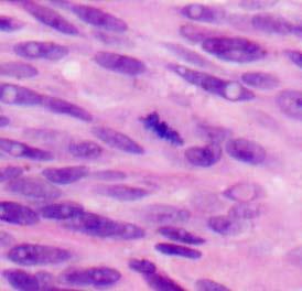
31 Case Presentation 33 year old woman with an enlarging brown lesion on the thigh.
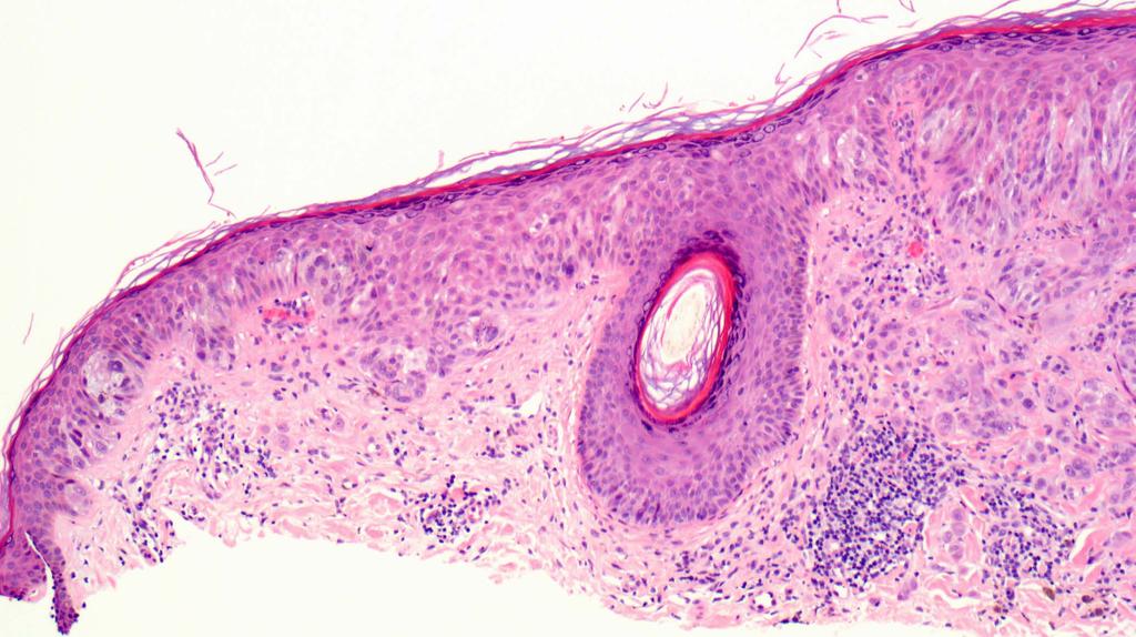
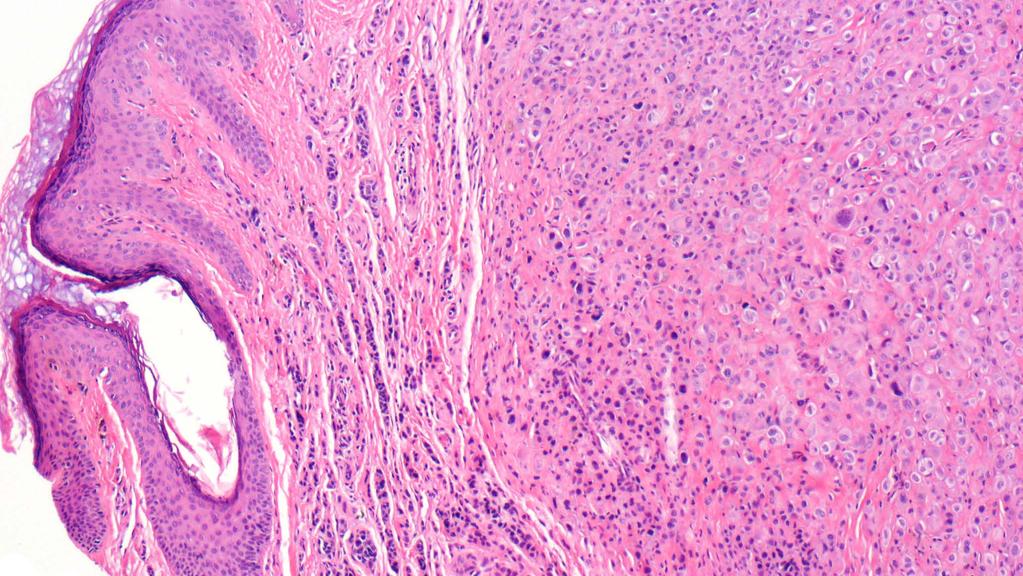
32
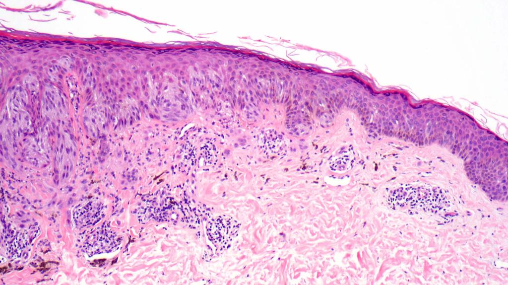
33
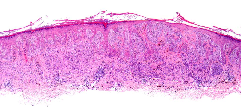
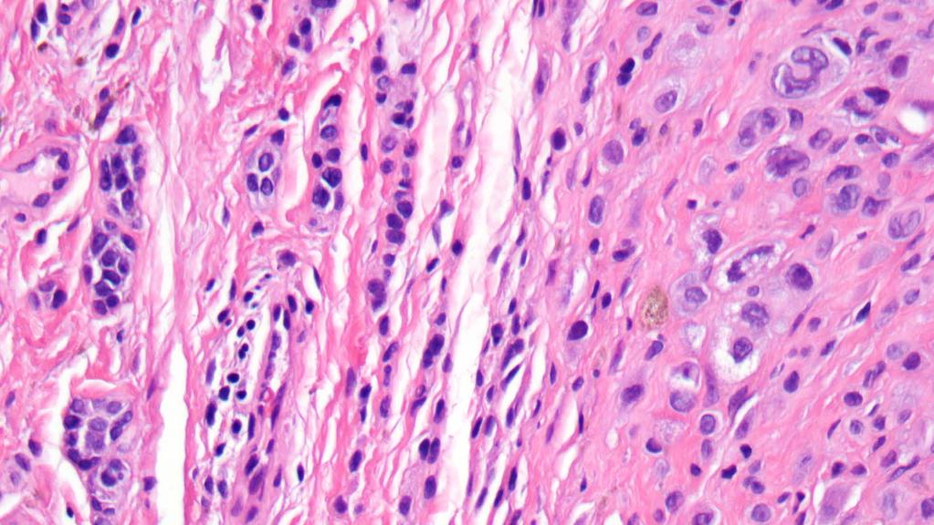
34
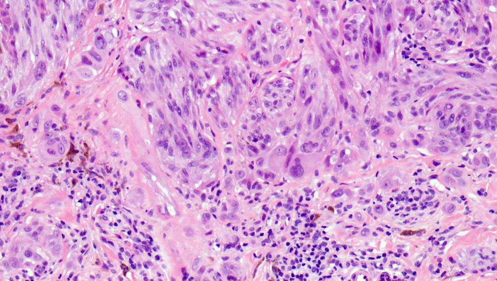
35
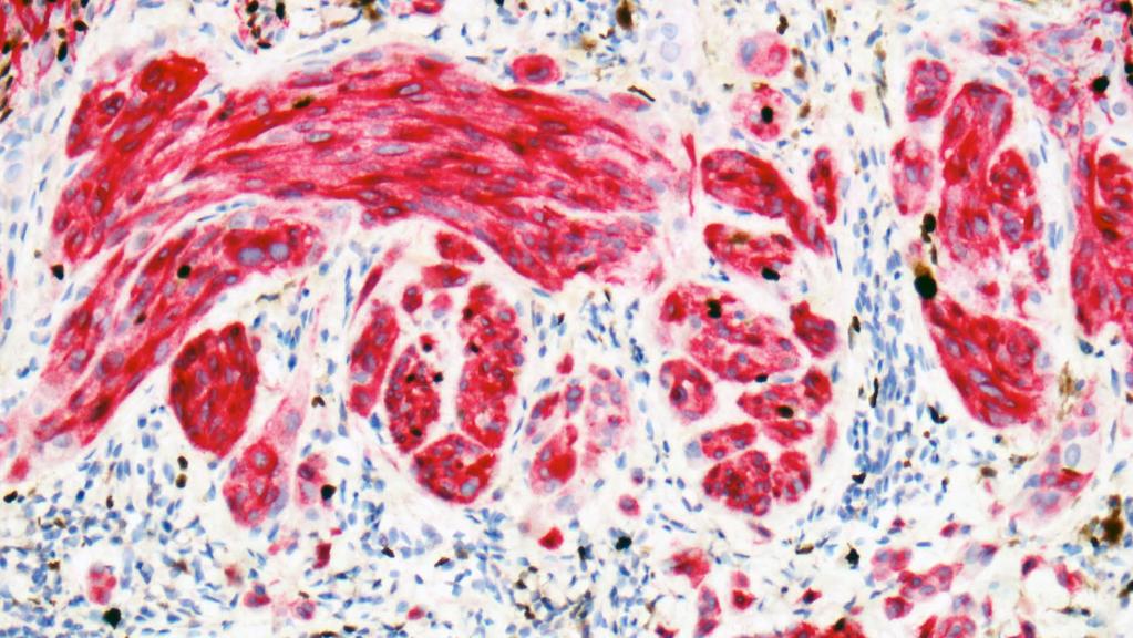
36 Mart-1/Ki-67


37 FISH results 9p21 cen9 6p25 8q24 11q13


, 1")

38 Final Diagnosis INVASIVE MELANOMA, CLARK LEVEL IV (AT LEAST), BRESLOW THICKNESS 0.98 MM (AT LEAST), 1 MITOTIC FIGURES/mm 2 WITH SPITZOID FEATURES 9p21 cen9
39 Molecular studies informing the diagnosis of melanocytic tumors Comparative Genomic Hybridization (CGH) demonstrates discrete chromosomal gains/losses in melanoma Fluorescence in situ hybridization (FISH) is a surrogate of genomic instability described by CGH First generation FISH Sensitivity, specificity and pitfalls How does FISH inform our diagnosis? Second-generation FISH testing FISH and prognosis htert promoter mutations BAP1 associated Spitzoid lesions Translocation associated Spitzoid lesions Molecular alterations distinguishing Melanoma Blue nevus type


40 Germline BAP1 loss confers risk to develop melanocytic tumors with distinct morphology Two families with multiple members bearing numerous papular melanocyic neoplasms with typical Spitzoid morphology and occasional uveal melanomas. Haplotype analyses determined that affected Analyzed lesions by acgh and identified frequent Sequencing individuals analyses inherited of the same minimally maternal deleted deletions of variable portions of chromosome 3p chromosome region identified 3. The deleted mutations copy in was BAP1. always in the lesions of both families. paternal.


41 BAP1 loss also seen in sporadically occurring melanocytic tumors with distinct morphology Assessed 32 Atypical Spitz tumors by BAP1 IHC: BAP1 negative in 9/32 cases assessed Characteristic features: Papular lesions on the trunk Predominantly dermal Epithelioid cells Prominent TILS BRAF V600E (8/9 cases)


42 BAP1 loss also seen in sporadically occurring melanocytic tumors with distinct morphology

43 Case Presentation Case Summary A 45 year-old man presented with a papular brown lesion on abdomen.

44
45
46
47
48 BAP-1
49 BRAF-V600E


50 Final Diagnosis Combined melanocytic nevus, with features of Spitz nevus and loss of BAP1, present at tissue edges.
51 Molecular studies informing the diagnosis of melanocytic tumors Comparative Genomic Hybridization (CGH) demonstrates discrete chromosomal gains/losses in melanoma Fluorescence in situ hybridization (FISH) is a surrogate of genomic instability described by CGH First generation FISH Sensitivity, specificity and pitfalls How does FISH inform our diagnosis? Second-generation FISH testing FISH and prognosis htert promoter mutations BAP1 associated Spitzoid lesions Translocation associated Spitzoid lesions Molecular alterations distinguishing Melanoma Blue nevus type


52 Kinase fusions are frequent in melanocytic lesions with Spitzoid morphology Different fusion partner gene Spanned the spectrum of Spitzoid neoplasia Each fusion occurred in mutually exclusive fashion of others Constitutively active kinase activity IHC+ Kinase

53 Spitzoid lesions with ALK kinase fusions have distinctive clinical and histopathologic features ALK kinase fusion lesions (n=49) Median Age Gender Anatomic Location Diagnosis ALK IHC Clinical outcome 14y (Range: y) 29 female: 20 male 26 Extremities 12 trunk 9 Head/Neck 11 Spitz nevus; 34 AST; 4 Spitzoid MM 100% positive 2 with SLN deposits (All alive; NED)
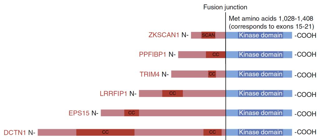
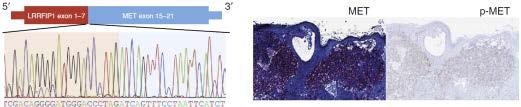
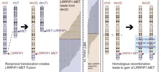
54 Activating MET kinase rearrangements in Spitzoid lesions

55 Spitzoid lesions characterized by many distinctive genetic abnormalities HRAS mutation and 11p amplification Homozygous deletion of 9p21 Loss of 6q23 Kinase fusions: o ALK o ROS1 o NTRK1 o BRAF o RET o MET o NTRK3 Spitzoid proliferation BAP1 loss with BRAFV600E htert promoter mutation
56 Molecular studies informing the diagnosis of melanocytic tumors Comparative Genomic Hybridization (CGH) demonstrates discrete chromosomal gains/losses in melanoma Fluorescence in situ hybridization (FISH) is a surrogate of genomic instability described by CGH First generation FISH Sensitivity, specificity and pitfalls How does FISH inform our diagnosis? Second-generation FISH testing FISH and prognosis htert promoter mutations BAP1 associated Spitzoid lesions Translocation associated Spitzoid lesions Molecular alterations distinguishing Melanoma Blue nevus type


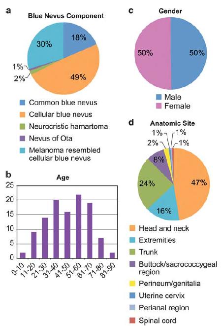
57 Melanoma, blue nevus type
58 Distinguishing among blue nevus-like lesions remains a challenge Location Cellularity Blue nevus Mid- to upper dermis Dendritic melanocytes with coarse pigment Cellular Blue nevus Pan-dermal Biphasic with dendritic and epithelioid melanocytes Atypical Cellular Blue nevus Pan-dermal Biphasic with dendritic and epithelioid melanocytes Melanoma, Blue nevus type Pan-dermal and may involve subcutis May contain or resemble blue nevus/cellular blue nevus Size Small (<10 mm) Can be >10 mm Can be >10 mm >10 mm Symmetry Present Present Present/Absent Usually absent Cytologic atypia Absent Absent Increased Prominent Mitotic figures Usually absent Infrequent Increased High, atypical Necrosis Absent Absent Absent Present


59 Chromosomal instability is typical of blue nevuslike melanomas Copy number changes Cellular Blue nevus Melanoma, Blue nevus type 3/17 (17%) 6/9 (67%) In comparison to cellular blue nevi, melanoma blue nevus type exhibits significant chromosomal instability
 Blue nevi")
 Blue nevus like metastases")
60 Chromosomal instability typical of blue nevuslike melanomas detected by FISH 100% (22/22) Blue nevi FISH negative 100% (5/5) Melanoma, blue nevus type FISH positive 90% (9/10) Blue nevus like metastases FISH positive


61 GNAQ and GNA11 mutations in blue nevus and blue nevus-like melanomas
 7/11 (64%) 16/18 (89%) 1/11 (9%) 1/18 (6%) 8/11 (73%) GNAQ/GNA11 mutations in both benign and malignant blue nevus-like proliferations BAP1 loss typical in melanoma, blue nevus")
62 GNAQ and GNA11 mutations, BAP1 loss and genomic instability in blue nevus-like melanomas Loss of nuclear BAP1 IHC GNAQ mutation (Exon 5) GNA11 mutation (Exon 5) Cellular Blue nevus Melanoma, Blue nevus type 0/24 (0%) 7/11 (64%) 16/18 (89%) 1/11 (9%) 1/18 (6%) 8/11 (73%) GNAQ/GNA11 mutations in both benign and malignant blue nevus-like proliferations BAP1 loss typical in melanoma, blue nevus type Chromosomal abnormalities frequent in melanoma, blue nevus type
63 Thank you Questions Victor Prieto MD PhD Jonathan L. Curry MD Carlos A. Torres-Cabala MD Priya Nagarajan MD PhD Phyu Aung MD PhD
64 The FISH experience at UTMDACC First and Second Generation FISH Favored diagnosis Number of cases Gender Age (Years) FISH RESULT FINAL DIAGNOSIS Favor Benign male 20 female 30 (4-64) 37 Negative 6 Positive Nevus with atypical features Nevus with atypical features Favor melanoma 28 9 male: 19 female 26 (2-53) 16 Negative 12 Positive Invasive Melanoma Invasive Melanoma Tetzlaff MT et al. Am J Surg Pathol (12): Al-Rohil RA et al. Hum Pathol :73-81.
65 The FISH experience at UTMDACC First and Second Generation FISH Favored diagnosis Number of cases Gender Age (Years) FISH RESULT FINAL DIAGNOSIS Favor Benign male 20 female 30 (4-64) 37 Negative 6 Positive Nevus with atypical features Nevus with atypical features Favor melanoma 28 9 male: 19 female 26 (2-53) 16 Negative 12 Positive Invasive Melanoma Invasive Melanoma Tetzlaff MT et al. Am J Surg Pathol (12): Al-Rohil RA et al. Hum Pathol :73-81.
:1783-96. Al-Rohil RA et al. Hum Pathol. 2016.")
66 The FISH experience at UTMDACC First and Second Generation FISH Tetzlaff MT et al. Am J Surg Pathol (12): Al-Rohil RA et al. Hum Pathol :73-81.
Michael T. Tetzlaff MD, PhD
 Molecular alterations informing the diagnosis of melanocytic tumors Michael T. Tetzlaff MD, PhD Associate Professor Department of Pathology, Section of Dermatopathology Department of Translational and
Update on Spitzoid and Blue nevus-like melanocytic lesions Emphasis on molecular studies informing diagnosis, prognosis and therapy
 Update on Spitzoid and Blue nevus-like melanocytic lesions Emphasis on molecular studies informing diagnosis, prognosis and therapy Michael T. Tetzlaff MD, PhD Associate Professor Department of Pathology,
Melanocytic Lesions: Use of Immunohistochemistry and Special Studies Napa Valley 2018
 Melanocytic Lesions: Use of Immunohistochemistry and Special Studies Napa Valley 2018 Victor G. Prieto, MD, PhD Professor Depts. of Pathology and Dermatology University of Texas - MD Anderson Cancer Center
Vernon K. Sondak. Department of Cutaneous Oncology Moffitt Cancer Center Tampa, Florida
 Vernon K. Sondak Department of Cutaneous Oncology Moffitt Cancer Center Tampa, Florida Australasian Melanoma Conference 2016 Sydney, NSW, Australia October 29, 2016 Disclosures Dr. Sondak is a compensated
Molecular Aspects of Melanocytic Neoplasia. Iwei Yeh MD, PhD University of California, San Francisco
 Molecular Aspects of Melanocytic Neoplasia Iwei Yeh MD, PhD University of California, San Francisco Thanks to: Boris Bastian Timothy McCalmont Philip LeBoit Beth Ruben Jeff North Laura Pincus Thaddeus
Update on 8 th Edition Cutaneous AJCC Staging of Primary Cutaneous Melanoma. Michael T. Tetzlaff MD, PhD
 Update on 8 th Edition Cutaneous AJCC Staging of Primary Cutaneous Melanoma Michael T. Tetzlaff MD, PhD Associate Professor Departments of Pathology (Dermatopathology) and Translational and Molecular Pathology
6/22/2015. Original Paradigm. Correlating Histology and Molecular Findings in Melanocytic Neoplasms
 6 Correlating Histology and Molecular Findings in Melanocytic Neoplasms Pedram Gerami MD, Associate Professor of Dermatology and Pediatrics at Northwestern University Disclosures: I have been a consultant
Case 26 Male 37. Right jawline 5mm nodule?keloid. The best diagnosis is:
 Case 26 Male 37. Right jawline 5mm nodule?keloid. The best diagnosis is: A. Desmoplastic Spitz naevus B. Atypical Spitz Tumour C. Spitzoid melanoma D. Deep penetrating naevus E. Spitz naevus Case 26: M
Michael T. Tetzlaff MD, PhD
 American Joint Cancer Committee (AJCC) staging system for primary cutaneous melanoma (8 th Edition) and principles of sentinel lymph node evaluation Emphasis on concise and accurate reporting of primary
There is NO single Melanoma Stain. > 6000 Mutations in Melanoma. What else can be done to discriminate atypical nevi from melanoma?
 Las Vegas Fall Clinical 2016: The Assessment and Diagnosis of Melanoma Whitney A. High, MD, JD, MEng Associate Professor, Dermatology & Pathology Director of Dermatopathology (Dermatology) University of
The Enigmatic Spitz Lesion
 The Enigmatic Spitz Lesion The Dawn of Spitz S Spitz Sophie Spitz Melanomas of Childhood ; Am J Pathol 1948 1910-1956 13 children (18 mo - 12 yrs) 12/13 had a benign clinical course Sophie Spitz Born 1910
21/07/2017. The «gray zone» of diagnosis is visible. Nevus Atypical nevus Melanoma. Melanoma ex-blue nevus
 Update on the Clinico- Pathological and Molecular Diagnosis of Melanocytic Lesions None to declare Conflicts of interest Belfast pathology Arnaud de la Fouchardière MD, PhD Lyon, France What is new? Today
The Pathology of Neoplasia Part II
 The Pathology of Neoplasia Part II February 2018 PAUL BOGNER, MD A S S O C I A T E P R O F E S S O R O F O N C O L O G Y P A T H O L O G Y A N D D E R M A T O L O G Y Clinical goals of cancer pathology
MAPK Pathway. CGH Next Generation Sequencing. Molecular Tools in Care of Patients with Pigmented Lesions 7/20/2017
 Molecular Tools in Care of Patients with Pigmented Lesions Tammie Ferringer, MD Geisinger Medical Center, Danville, PA tferringer@geisinger.edu DISCLOSURE OF RELATIONSHIPS WITH INDUSTRY Tammie Ferringer,
Dermatopathology. Dr. Rafael Botella Estrada. Hospital La Fe de Valencia
 Dermatopathology Dr. Rafael Botella Estrada. Hospital La Fe de Valencia Melanoma and mimics Dr. Martin Mihm Malignant lesions result from the accumulation of mutations Class I lesions (benign) Class II
I have no relevant conflicts of interest to disclose. John T. Seykora MD PhD Departments of Dermatology & Pathology and Laboratory Medicine
 Molecular Characterization of Stage 1-3 Melanoma: Are we close to accurate prognostication and prediction? I have no relevant conflicts of interest to disclose. John T. Seykora MD PhD Departments of Dermatology
10/2/17. MELTUMP, SAMPUS, AST.An Algorithmic Approach to Challenging (Often Borderline) Melanocytic Tumors. An Introduction to SNP Arrays
 Melanocytic Tumors. An Introduction to SNP Arrays") MELTUMP, SAMPUS, AST.An Algorithmic Approach to Challenging (Often ) Melanocytic Tumors An Introduction to SNP Arrays Rajiv M. Patel, M.D. RCPA NZ ASM 2017 (11:45-12:30pm, Saturday, 23-09-17) Why do we
MELTUMP, SAMPUS, AST.An Algorithmic Approach to Challenging (Often ) Melanocytic Tumors An Introduction to SNP Arrays Rajiv M. Patel, M.D. RCPA NZ ASM 2017 (11:45-12:30pm, Saturday, 23-09-17) Why do we
Integrating Fluorescence in situ Hybridization and Genomic Array Results into the Diagnostic Workup of Melanoma
 Integrating Fluorescence in situ Hybridization and Genomic Array Results into the Diagnostic Workup of Melanoma Association for Molecular Pathology United States and Canadian Academy of Pathology Companion
Michael T. Tetzlaff MD, PhD
 Update on American Joint Cancer Committee (AJCC) staging system for primary cutaneous melanoma Emphasis on concise and accurate reporting of primary and metastatic melanoma for effective risk stratification
Genetic Testing: When should it be ordered? Julie Schloemer, MD Dermatology
 Genetic Testing: When should it be ordered? Julie Schloemer, MD Dermatology Outline Germline testing CDKN2A BRCA2 BAP1 Somatic testing Gene expression profiling (GEP) BRAF Germline vs Somatic testing
A PRACTICAL APPROACH TO ATYPICAL MELANOCYTIC LESIONS BIJAN HAGHIGHI M.D, DIRECTOR OF DERMATOPATHOLOGY, ST. JOSEPH HOSPITAL
 A PRACTICAL APPROACH TO ATYPICAL MELANOCYTIC LESIONS BIJAN HAGHIGHI M.D, DIRECTOR OF DERMATOPATHOLOGY, ST. JOSEPH HOSPITAL OBJECTIVES Discuss current trends and changing concepts in our understanding of
Diagnosis of melanocytic proliferations remains one of
 Update on Fluorescence In Situ Hybridization in Melanoma State of the Art Pedram Gerami, MD; Artur Zembowicz, MD, PhD N Context. Recent advances in understanding the molecular basis of melanoma have resulted
Melanoma Update: 8th Edition of AJCC Staging System
 Melanoma Update: 8th Edition of AJCC Staging System Rosalie Elenitsas, M.D. Professor of Dermatology Director, Dermatopathology University of Pennsylvania DISCLOSURE OF RELATIONSHIPS WITH INDUSTRY None
Page 1 of 3. We suggest the following changes:
 Page 1 of 3 Loren E. Clarke, M.D. Myriad Genetic Laboratories, Inc. 320 Wakara Way, Salt Lake City, UT 84108 Phone: 801.883.3470 Email: lclarke@myriad.com Date of Request: June 2017 NCCN Guidelines Panel:
Melanocytic proliferations in sundamaged
 Atypical Spitzoid Tumor: What Does It Mean And How Should It Be Managed? Melanocytic proliferations in sundamaged skin Jane L. Messina, Jane L. Messina MD International Melanoma Pathology Working Group
Ways to get into trouble, ideas on avoiding trouble, and diagnostic approaches to keep trouble at bay
 Pitfalls in the diagnosis of melanocytic tumors Timothy McCalmont, MD University of California, San Francisco Ways to get into trouble, ideas on avoiding trouble, and diagnostic approaches to keep trouble
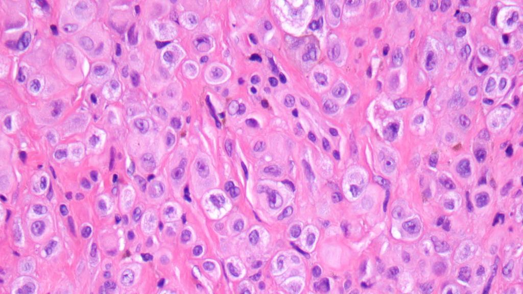
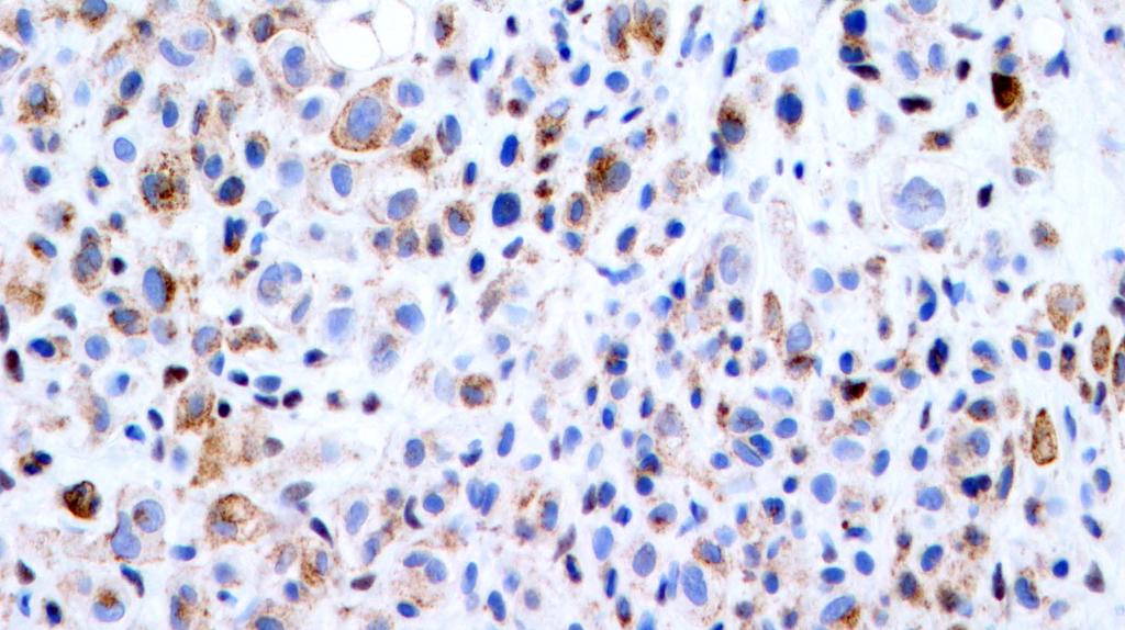
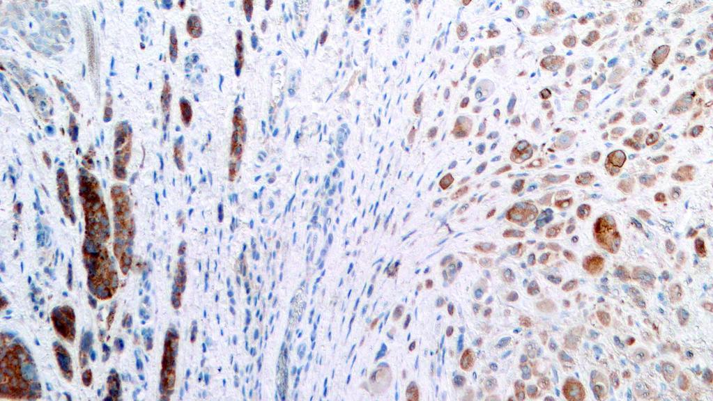
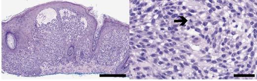
Supplementary Figure 1. Spitzoid Melanoma with PPFIBP1-MET fusion. (a) Histopathology (4x) shows a domed papule with melanocytes extending into the
 Histopathology (4x) shows a domed papule with melanocytes extending into the") Supplementary Figure 1. Spitzoid Melanoma with PPFIBP1-MET fusion. (a) Histopathology (4x) shows a domed papule with melanocytes extending into the deep dermis. (b) The melanocytes demonstrate abundant
Supplementary Figure 1. Spitzoid Melanoma with PPFIBP1-MET fusion. (a) Histopathology (4x) shows a domed papule with melanocytes extending into the deep dermis. (b) The melanocytes demonstrate abundant
Patricia Chevez-Barrrios AAOOP-USCAP /12/2016
 Biomarkers in Ocular Melanoma Patricia Chévez-Barrios, MD Pathology and Genomic Medicine, Houston Methodist Hospital Professor of Pathology and Laboratory Medicine and Ophthalmology, Weill Cornell Medical
> 6000 Mutations in Melanoma. Tests That Cay Be Employed. FISH for Additions/Deletions. Comparative Genomic Hybridization
 Winter Clinical 2017: The Assessment and Diagnosis of Melanoma Whitney A. High, MD, JD, MEng Associate Professor, Dermatology & Pathology Director of Dermatopathology (Dermatology) University of Colorado
Desmoplastic Melanoma R/O BCC. Clinical Information. 74 y.o. man with lesion on left side of neck r/o BCC
 R/O BCC Sabine Kohler, M.D. Professor of Pathology and Dermatology Dermatopathology Service Stanford University School of Medicine Clinical Information 74 y.o. man with lesion on left side of neck r/o
Artur Zembowicz, MD, PhD; Sung-Eun Yang, MD; Antonios Kafanas, MD; Stephen R. Lyle, MD, PhD
 Correlation Between Histologic Assessment and Fluorescence In Situ Hybridization Using MelanoSITE in Evaluation of Histologically Ambiguous Melanocytic Lesions Artur Zembowicz, MD, PhD; Sung-Eun Yang,
Female 18. Deeply pigmented lesion on trunk.?warty naevus?seborrhoeic keratosis?malignant melanoma. The best diagnosis is:
 Female 18. Deeply pigmented lesion on trunk.?warty naevus?seborrhoeic keratosis?malignant melanoma. The best diagnosis is: A. deep penetrating naevus B. naevoid malignant melanoma C. pigment synthesising
Approximately 2% of the United States population
 Differentiation of Malignant Melanoma From Benign Nevus Using a Novel Genomic Microarray With Low Specimen Requirements Wells M. Chandler, MD; Leslie R. Rowe, MS; Scott R. Florell, MD; Mona S. Jahromi,
Melanocytic Tumours. Molecular Biology 02/06/2015. Cutaneous Melanocytic Tumours Introduction. Thomas Brenn. Intermediate Malignancy
 Cutaneous Melanocytic Tumours Introduction Melanocytic Tumours: Update on Epidemiology and Molecular Biology Thomas Brenn Wide clinical and morphological spectrum Ranging from benign naevi to melanoma
Diagnoses of Cases 1. Lentigo, other melanosis and the acquired nevus 2. Variations on the acquired nevus 3. Dermal melanocytosis
 Diagnoses of Cases 1. Lentigo, other melanosis and the acquired nevus 1 1A. Lentigo simplex 4 1B. Psoralens and ultraviolet A (PUVA) lentigo 6 1C. Solar lentigo 8 1D. Café au lait macule 10 1E. Ink-spot
Conflict of Interest 9/2/2014. Pathogenesis and Comparison of Atypical Spitz Nevi vs Benign Spitz, and Childhood Melanoma
 Pathogenesis and Comparison of Atypical Spitz Nevi vs Benign Spitz, and Childhood Melanoma Martin C. Mihm Jr., M.D., F.A.C.P. Harvard Medical School Brigham and Women s Hospital Dana Farber Cancer Center
2/6/2018. Original Paradigm. Clonal Chromosomal A berrations. Only 20% of Spitz Nevi 95% 6p, 7q, 17q, 20q, 4q,8q, 1q, 11q. Isolated Gain in 11p
 Molecular Diagnostics for Melanocytic Neoplasms: Moving towards a Revolution in the Management of Melanocytic Neoplasms Pedr am Gerami MD Associate Professor of Dermatology, Pathology and Pediatrics at
Cutaneous Mesenchymal Neoplasms with EWSR1 Rearrangement
 Cutaneous Mesenchymal Neoplasms with EWSR1 Rearrangement By Konstantinos Linos MD, FCAP, FASDP Bone, Soft Tissue and Dermatopathology Assistant Professor of Pathology Dartmouth-Hitchcock Medical Center
Update on Merkel cell carcinoma
 Merkel cell carcinoma: Diagnosis, staging, sentinel lymph node biopsy and prognostic markers Michael T. Tetzlaff MD, PhD Associate Professor Departments of Pathology (Dermatopathology) and Translational
Merkel cell carcinoma: Diagnosis, staging, sentinel lymph node biopsy and prognostic markers
 Merkel cell carcinoma: Diagnosis, staging, sentinel lymph node biopsy and prognostic markers Michael T. Tetzlaff MD, PhD Associate Professor Departments of Pathology (Dermatopathology) and Translational
Which melanoma patients benefit from genetic testing?
 Which melanoma patients benefit from genetic testing? Michael A. Marchetti, MD Assistant Attending, Dermatology Service Memorial Sloan Kettering Cancer Center American Academy of Dermatology Annual Meeting
David B. Troxel, MD. Common Medicolegal Situations: Misdiagnosis of Melanoma
 Common Medicolegal Situations: Misdiagnosis of Melanoma David B. Troxel, MD Medical Director, The Doctors Company, Napa, California Clinical Professor Emeritus, University of California at Berkeley Past
Molecular Methods in the Diagnosis and Prognostication of Melanoma: Pros & Cons
 Molecular Methods in the Diagnosis and Prognostication of Melanoma: Pros & Cons Ben J. Friedman, MD Senior Staff Physician Department of Dermatology Department of Pathology and Laboratory Medicine Henry
A diagnostic algorithm for atypical spitzoid tumors: guidelines for immunohistochemical and molecular assessment
 Modern Pathology (2016), 1 15 2016 USCAP, Inc All rights reserved 0893-3952/16 $32.00 1 A diagnostic algorithm for atypical spitzoid tumors: guidelines for immunohistochemical and molecular assessment
Springer Healthcare. Staging and Diagnosing Cutaneous Melanoma. Concise Reference. Dirk Schadendorf, Corinna Kochs, Elisabeth Livingstone
 Concise Reference Staging and Diagnosing Cutaneous Melanoma Dirk Schadendorf, Corinna Kochs, Elisabeth Livingstone Extracted from Handbook of Cutaneous Melanoma: A Guide to Diagnosis and Treatment Published
Reviewers' comments: Reviewer #1 (Remarks to the Author):
:") Reviewers' comments: Reviewer #1 (Remarks to the Author): In this study the authors analysed 18 deep penetrating nevi for oncogenic genomic changes (single nucleotide variations, insertions/deletions,
Reviewers' comments: Reviewer #1 (Remarks to the Author): In this study the authors analysed 18 deep penetrating nevi for oncogenic genomic changes (single nucleotide variations, insertions/deletions,
Diploma Examination. Dermatopathology: First paper. Tuesday 20 March Candidates must answer FOUR questions. Time allowed: 3 hours
 Dermatopathology: First paper Tuesday 20 March 2018 Candidates must answer FOUR questions Time allowed: 3 hours 1. Give an account of the genetic aberrations encountered in Spitzoid neoplasms and how these
Financial disclosures
 Mesenchymal Neoplasms with Melanocytic Differentiation By Konstantinos Linos MD, FCAP, FASDP Bone, Soft Tissue and Dermatopathology Assistant Professor of Pathology Dartmouth-Hitchcock Medical Center Geisel
Malignant tumors of melanocytes: Part 1. Deba P Sarma, MD., Omaha
 Malignant tumors of melanocytes: Part 1 Deba P Sarma, MD., Omaha The melanocytic tumor is one of the most difficult and confusing areas in Dematopathology. It is true that most (95%) of such lesions are
Disclosure Information. Lecture Outline. Lecture Outline. Introduction. Molecular Pathology of Cutaneous Melanoma. Nothing to disclose
 Molecular Pathology of Cutaneous Melanoma Disclosure Information Nothing to disclose Jonathan L. Curry, MD Assistant Professor of Pathology and Dermatology University of Texas-MD Anderson Cancer Center
Financial disclosures
 Cutaneous Mesenchymal Neoplasms with EWSR1 Rearrangement By Konstantinos Linos MD, FCAP, FASDP Bone, Soft Tissue and Dermatopathology Assistant Professor of Pathology Dartmouth-Hitchc Geisel School of
Impact of Prognostic Factors
 Melanoma Prognostic Factors: where we started, where are we going? Impact of Prognostic Factors Staging Management Surgical intervention Adjuvant treatment Suraj Venna, MD Assistant Clinical Professor,
Benign and malignant epithelial lesions: Seborrheic keratosis: A common benign pigmented epidermal tumor occur in middle-aged or older persons more
 Benign and malignant epithelial lesions: Seborrheic keratosis: A common benign pigmented epidermal tumor occur in middle-aged or older persons more common on the trunk; but extremities, head and neck are
Blue Melanocytic Proliferations
 Blue Melanocytic Proliferations Labib R. Zakka M.D., M.A. Research Fellow Melanoma Program Department of Dermatology Brigham and Women s Hospital Harvard Medical School Conflicts of Interest No conflicts
BAP-oma & BEYOND MICHAEL A NOWAK, MD
 BAP-oma & BEYOND MICHAEL A NOWAK, MD CONFLICTS No conflicts with the content of this lecture BAP-oma Wiesner 2011: Families with multiple tan dome-shaped papules of head, neck, trunk, and extremities.
Case year old female presented with asymmetric enlargement of the left lobe of the thyroid
 Case 4 22 year old female presented with asymmetric enlargement of the left lobe of the thyroid gland. No information available relative to a prior fine needle aspiration biopsy. A left lobectomy was performed.
Case 4 Diagnosis 2/21/2011 TGB
 Case 4 22 year old female presented with asymmetric enlargement of the left lobe of the thyroid gland. No information available relative to a prior fine needle aspiration biopsy. A left lobectomy was performed.
Case RAC7783. M46. Ear. Mole. r/o MM.?Blue naevus RAC7783
 Case RAC7783. M46. Ear. Mole. r/o MM.?Blue naevus RAC7783 Pie Chart Participants N=74 Benign: 48 N=74 Blue naevus: 38 Intradermal: 12 DPN: 10 Compound 3 Clonal: 3; Spitz 2; Special Site: 1; Congenital:
Diploma examination. Dermatopathology: First paper. Tuesday 21 March Candidates must answer FOUR questions ONLY. Time allowed: Three hours
 Dermatopathology: First paper Tuesday 21 March 2017 1. Discuss the role of fluorescent in-situ hybridization (FISH) and emerging molecular techniques in the diagnosis of cutaneous melanocytic lesions,
- Selected Tumors of the Skin Appendages - Primary vs. Metastasis
 - Selected Tumors of the Skin Appendages - Primary vs. Metastasis Napa Valley 2018 Victor G. Prieto, MD, PhD Chair of Pathology UT MD Anderson Cancer Center vprieto@mdanderson.org Napa Valley in May Introduction
Management of pediatric melanocytic lesions
 Open Journal of Clinical & Medical Case Reports Management of pediatric melanocytic lesions Volume 3 (2017) Issue 8 ISSN 2379-1039 Jin Kim, BS; Emmanuel Gabriel MD, PhD; Weiguo Liu MD, PhD; Lin Lin MD,
ACCME/Disclosures ALK FUSION-POSITIVE MESENCHYMAL TUMORS. Tumor types with ALK rearrangements. Anaplastic Lymphoma Kinase. Jason L.
 Companion Meeting of the International Society of Bone and Soft Tissue Pathology The Evolving Concept of Mesenchymal Tumors ALK FUSION-POSITIVE MESENCHYMAL TUMORS Jason L. Hornick, MD, PhD March 13, 2016
Patient age and cutaneous malignant melanoma: Elderly patients are likely to have more aggressive histological features and poorer survival
 MOLECULAR AND CLINICAL ONCOLOGY 7: 1083-1088, 2017 Patient age and cutaneous malignant melanoma: Elderly patients are likely to have more aggressive histological features and poorer survival FARUK TAS
Case 231: F7. Exophytic naevus over left trapezious. Grown over a few weeks. Iniitally flat.?spitz naevus,?malignant
 Case 231: F7. Exophytic naevus over left trapezious. Grown over a few weeks. Iniitally flat.?spitz naevus,?malignant Dermoscopy: coarse vascular structures. c/o A, B, C RAC7750 Case 231: F7. Exophytic
THE SPITZ NEVUS OFTEN POSES
 OBSERVATION ONLINE FIRST Melanoma Mimic A Case of Multiple Pagetoid Spitz Nevi KaLynne Harris, MD; Scott R. Florell, MD; Jason Papenfuss, MD; Wendy Kohlmann, MS, CGC; Mona Jahromi, BS; Joshua D. Schiffman,
Time to reconsider Spitzoid neoplasms?
 DERMATOLOGY PRACTICAL & CONCEPTUAL www.derm101.com Time to reconsider Spitzoid neoplasms? Carmelo Urso 1 1 Department of Anatomic Pathology, Dermatopathology Section, SM Annunziata Hospital, AUSL Toscana
Pathology of Sarcoma ELEANOR CHEN, MD, PHD, ASSISTANT PROFESSOR DEPARTMENT OF PATHOLOGY UNIVERSITY OF WASHINGTON
 Pathology of Sarcoma ELEANOR CHEN, MD, PHD, ASSISTANT PROFESSOR DEPARTMENT OF PATHOLOGY UNIVERSITY OF WASHINGTON Presentation outline Background and epidemiology of sarcomas Sarcoma classification Sarcoma
Protocol applies to melanoma of cutaneous surfaces only.
 Melanoma of the Skin Protocol applies to melanoma of cutaneous surfaces only. Procedures Biopsy (No Accompanying Checklist) Excision Re-excision Protocol revision date: January 2005 Based on AJCC/UICC
Histopathology of Melanoma
 THE YALE JOURNAL OF BIOLOGY AND MEDICINE 48, 409-416 (1975) Histopathology of Melanoma G. J. WALKER SMITH Department ofpathology, Yale University School ofmedicine, 333 Cedar Street, New Haven, Connecticut
Whitney A. High, MD, JD, MEng
 ADS Dermatopathology Meeting 2014 Selected Adnexal Tumors Whitney A. High, MD, JD, MEng Associate Professor, Dermatology & Pathology Director of Dermatopathology (Dermatology) University of Colorado School
Melanoma Underwriting Presented at 2018 AHOU Conference. Hank George FALU
 Melanoma Underwriting Presented at 2018 AHOU Conference Hank George FALU MELANOMA EPIDEMIOLOGY 70-80,000 American cases annually Majority are in situ or thin > 20% are diagnosed age 45 8-9,000 melanoma
K Blessing, J J H Grant, D S A Sanders, M M Kennedy, A Husain, P Coburn
 J Clin Pathol 2000;53:591 595 591 Papers Pathology, Aberdeen University, Foresterhill, Aberdeen AB25 2ZD, K Blessing Pathology, Birmingham University, Birmingham B15 2TT, D S A Sanders Pathology, Heartlands
Epithelial Cancer- NMSC & Melanoma
 Epithelial Cancer- NMSC & Melanoma David Chin MB, BCh, BAO, LRCP, LRCS (Ireland) MCh(MD), PhD (UQ), FRCS, FRACS (Plast) Plastic & Reconstructive Surgeon Visiting Scientist Melanoma Genomic Group & Drug
Enterprise Interest Nothing to declare
 Enterprise Interest Nothing to declare Diagnoses one would not like to miss in soft tissue pathology early in your career Marta Sbaraglia, MD Department of Pathology Hospital of Treviso University of Padua
Melanoma. Kaushik Mukherjee MD A. Scott Pearson MD
 Melanoma Kaushik Mukherjee MD A. Scott Pearson MD Disclosures You still have to study Not all inclusive No Western blots Extensive use of Google Image Search and Sabiston Melanoma Basics 8 th most common
Pathology of the skin. 2nd Department of Pathology, Semmelweis University
 Pathology of the skin 2nd Department of Pathology, Semmelweis University Histology of the skin Epidermis: Stratum corneum Stratum granulosum Stratum spinosum Stratum basale Dermis: papillary and reticular
3/23/2017. Disclosure of Relevant Financial Relationships. Pathologic Staging Updates in Lung Cancer T STAGE OUTLINE SURVIVAL ACCORDING TO SIZE ONLY
 Pathologic Staging Updates in Lung Cancer Disclosure of Relevant Financial Relationships USCAP requires that all planners (Education Committee) in a position to influence or control the content of CME
Rare melanoma: Are the options improving? Dr Neil Steven Consultant in Medical Oncology University Hospital Birmingham University of Birmingham
 Rare melanoma: Are the options improving? Dr Neil Steven Consultant in Medical Oncology University Hospital Birmingham University of Birmingham Classifying melanoma Melanoma (site of origin, thickness,
Evening Specialty Conference Bone and Soft Tissue Pathology. Diagnostic pitfalls in bone and soft tissue pathology
 Evening Specialty Conference Bone and Soft Tissue Pathology. Case 1 Elizabeth G Demicco, MD, PhD Mount Sinai Hospital, New York Disclosure of Relevant Financial Relationships USCAP requires that all planners
Dermatologica Sinica
 DERMATOLOGICA SINICA 30 (2012) 57e61 Contents lists available at SciVerse ScienceDirect Dermatologica Sinica journal homepage: http://www.derm-sinica.com CASE REPORT Pigmented epithelioid melanocytoma:
21/07/2017. Hobnail endothelial cells are not the same as epithelioid endothelial cells
 UPDATE IN CUTANEOUS VASCULAR S DERMATOPATHOLOGY SESSION BELFAST PATHOLOGY JUNE 21/2017 Dr E Calonje St John s Institute of Dermatology, London, United Kingdom THE FAMILY OF VASCULAR S WITH EPITHELIOID
PODIATRIC PATHOLOGY REPORT IS;RL;MMR; 1 of 1. A Copy was sent to: DR. JANE DOE 456 SAMPLE BLVD NEW YORK, NY DIAGNOSIS
 Batch#: 10388 Tel#: 1-888-CUPTH ccession: Obtained: Received: 12:02 pm PTIENT: JCK JONES 12548 MIN ST FLUSHING, NY 11365 (718) 555-2541 DOB: 01/02/19XX Specimen Final Report Date cct#: 1018 ge: 57 Sex:
57th Annual HSCP Spring Symposium 4/16/2016
 An Unusual Malignant Spindle Cell Lesion to Involve the Breast Erinn Downs-Kelly, D.O. Associate Professor of Pathology University of Utah & ARUP Laboratories No disclosures Case 39 y/o female with no
Update: Morphologic Considerations in Mesothelioma within the Pleural and Peritoneal Cavities. Douglas J. Hartman, MD June 7, 2018
 Update: Morphologic Considerations in Mesothelioma within the Pleural and Peritoneal Cavities Douglas J. Hartman, MD June 7, 2018 Objectives Review Historical Features associated with prognosis Present
Update on Genetic Testing for Melanoma
 Update on Genetic Testing for Melanoma Emily Y. Chu, M.D., Ph.D. Assistant Professor of Dermatology & Pathology and Laboratory Medicine Hospital of the University of Pennsylvania February 18, 2018 AAD
Society for Pediatric Pathology Spring Meeting Joint Symposium with American Society of Dermatopathology
 Society for Pediatric Pathology 2013 Spring Meeting Joint Symposium with American Society of Dermatopathology Update on Cutaneous Melanocytic, Mesenchymal and Lymphoproliferative Lesions in Children Melanocytic
Next Generation Sequencing in Clinical Practice: Impact on Therapeutic Decision Making
 Next Generation Sequencing in Clinical Practice: Impact on Therapeutic Decision Making November 20, 2014 Capturing Value in Next Generation Sequencing Symposium Douglas Johnson MD, MSCI Vanderbilt-Ingram
المركب النموذج--- سبيتز وحمة = Type Spitz's Nevus, Compound SPITZ NEVUS 1 / 7
 SPITZ NEVUS 1 / 7 Epidemiology An annual incidence rate of 1.4 cases of Spitz nevus per 100,000 individuals has been estimated in Australia, compared with 25.4 per 100,000 individuals for cutaneous melanoma
The Relevance of Cytologic Atypia in Cutaneous Neural Tumors
 The Relevance of Cytologic Atypia in Cutaneous Neural Tumors Recent Findings - New Developments New Problems Zsolt B. Argenyi, M.D. Professor of Pathology & Dermatology Director of Dermatopathology Department
Conflicts of Interest
 Challenging Melanocytic Lesions Carlos N. Prieto-Granada M.D. Assistant Professor University of Alabama at Birmingham (UAB) Department of Pathology 2017 AAD Annual Meeting 3/2/17 - Orlando, FL None Conflicts
Dermatopathology: The tumor is composed of keratinocytes which show atypia, increase mitoses and abnormal mitoses.
 Squamous cell carcinoma (SCC): A common malignant tumor of keratinocytes arising in the epidermis, usually from a precancerous condition: 1- UV induced actinic keratosis, usually of low grade malignancy.
MELANOMA IN ADOLESCENTS AND YOUNG ADULTS
 Cancer in Adolescents and Young Adults (AYA) Working Group MELANOMA IN ADOLESCENTS AND YOUNG ADULTS Emmanouil Saloustros MD, DSc General Hospital of Heraklion Venizelio Heraklion, Crete, Greece ESMO Preceptorship
An update on molecular alterations in melanocytic tumors with emphasis on Spitzoid lesions
 Review Article on Molecular Oncology age 1 of 17 An update on molecular alterations in melanocytic tumors with emphasis on Spitzoid lesions Emmanouil Dimonitsas 1#, Aliki Liakea 2#, Stratigoula Sakellariou
Malignant tumors of melanocytes : Part 3. Deba P Sarma, MD., Omaha
 Malignant tumors of melanocytes : Part 3 Deba P Sarma, MD., Omaha Let s go over one case of melanoma using the following worksheet. Of the various essential information that needs to be included in the
LUNG CANCER. pathology & molecular biology. Izidor Kern University Clinic Golnik, Slovenia
 LUNG CANCER pathology & molecular biology Izidor Kern University Clinic Golnik, Slovenia 1 Pathology and epidemiology Small biopsy & cytology SCLC 14% NSCC NOS 4% 70% 60% 50% 63% 62% 61% 62% 59% 54% 51%
Disclosure. Relevant Financial Relationship(s) None. Off Label Usage None MFMER slide-1
 None. Off Label Usage None MFMER slide-1") Disclosure Relevant Financial Relationship(s) None Off Label Usage None 2013 MFMER slide-1 Case Presentation A 43 year old male, with partial nephrectomy for a right kidney mass 2013 MFMER slide-2 2013
Disclosure Relevant Financial Relationship(s) None Off Label Usage None 2013 MFMER slide-1 Case Presentation A 43 year old male, with partial nephrectomy for a right kidney mass 2013 MFMER slide-2 2013
1/10/2018. Soft Tissue Tumors Showing Melanocytic Differentiation. Overview. Desmoplastic/ Spindle Cell Melanoma
 2016 MFMER slide-1 2016 MFMER slide-2 2016 MFMER slide-3 Soft Tissue Tumors Showing Melanocytic Differentiation Andrew L. Folpe, M.D. Professor of Laboratory Medicine and Pathology Mayo Clinic, Rochester,
Primary Cutaneous Melanoma Pathology Reporting Proforma DD MM YYYY. *Tumour site. *Specimen laterality. *Specimen type
 Primary Cutaneous Melanoma Pathology Reporting Proforma Includes the International Collaboration on Cancer reporting dataset denoted by * Family name Given name(s) Date of birth DD MM YYYY Sex Male Female
Primary Cutaneous CD30-Positive T-cell Lymphoproliferative Disorders
 Primary Cutaneous CD30-Positive T-cell Lymphoproliferative Disorders Definition A spectrum of related conditions originating from transformed or activated CD30-positive T-lymphocytes May coexist in individual
Simulators of melanoma
 Simulators of melanoma Philip E. LeBoit, M.D. Depts. of Pathology and Dermatology University of California, San Francisco Simulators of melanoma Simulators of melanoma in situ Melanocytic Non-melanocytic