CONTRACTING ORGANIZATION: University of Michigan Ann Arbor, Michigan
|
|
|
- Stuart Hill
- 5 years ago
- Views:
Transcription
1 AD AWARD NUMBER: W81XWH TITLE: Chemo Resistance of Breast Cancer Stem Cells PRINCIPAL INVESTIGATOR: Max S. Wicha, M.D. CONTRACTING ORGANIZATION: University of Michigan Ann Arbor, Michigan REPORT DATE: May 2006 TYPE OF REPORT: Annual PREPARED FOR: U.S. Army Medical Research andmateriel Command Fort Detrick, Maryland DISTRIBUTION STATEMENT: Approved for Public Release; Distribution Unlimited The views, opinions and/or findings contained in this report are those of the author(s) and should not be construed as an official Department of the Army position, policy or decision unless so designated by other documentation.
2 REPORT DOCUMENTATION PAGE Form Approved OMB No Public reporting burden for this collection of information is estimated to average 1 hour per response, including the time for reviewing instructions, searching existing data sources, gathering and maintaining the data needed, and completing and reviewing this collection of information. Send comments regarding this burden estimate or any other aspect of this collection of information, including suggestions for reducing this burden to Department of Defense, Washington Headquarters Services, Directorate for Information Operations and Reports ( ), 1215 Jefferson Davis Highway, Suite 1204, Arlington, VA Respondents should be aware that notwithstanding any other provision of law, no person shall be subject to any penalty for failing to comply with a collection of information if it does not display a currently valid OMB control number. PLEASE DO NOT RETURN YOUR FORM TO THE ABOVE ADDRESS. 1. REPORT DATE (DD-MM-YYYY) 2. REPORT TYPE Annual 3. DATES COVERED (From - To) 2 Apr Apr TITLE AND SUBTITLE 5a. CONTRACT NUMBER Chemo Resistance of Breast Cancer Stem Cells 5b. GRANT NUMBER W81XWH c. PROGRAM ELEMENT NUMBER 6. AUTHOR(S) 5d. PROJECT NUMBER Max S. Wicha, M.D. mwicha@umich.edu 5e. TASK NUMBER 5f. WORK UNIT NUMBER 7. PERFORMING ORGANIZATION NAME(S) AND ADDRESS(ES) 8. PERFORMING ORGANIZATION REPORT NUMBER University of Michigan Ann Arbor, Michigan SPONSORING / MONITORING AGENCY NAME(S) AND ADDRESS(ES) 10. SPONSOR/MONITOR S ACRONYM(S) U.S. Army Medical Research and Materiel Command Fort Detrick, Maryland SPONSOR/MONITOR S REPORT NUMBER(S) 12. DISTRIBUTION / AVAILABILITY STATEMENT Approved for Public Release; Distribution Unlimited 13. SUPPLEMENTARY NOTES 14. ABSTRACT This past year was the second year of the DOD award. Over this year we have made substantial progress in achieving the goals outlined in the proposal. In addition, we have found that the enzyme aldehyde dehydrogenase 1 is an excellent marker for cancer stem cells which can be readily detected in fixed histological sections. Development of this new tool will greatly facilitate future studies. Preliminary results both in xenograft models as well as in neoadjuvant trial are providing strong support for our hypothesis for resistance of cancer cells to chemotherapy. We have also made excellent progress at elucidating the pathways which regulate the cells including Hedgehog signaling and Bmi-1. Together these studies provide a rationale for the combination compounds which inhibit stem cell renewal pathways such as Hedgehog with chemotherapeutic agents. 15. SUBJECT TERMS No subject terms provided. 16. SECURITY CLASSIFICATION OF: 17. LIMITATION OF ABSTRACT a. REPORT U b. ABSTRACT U 18. NUMBER OF PAGES c. THIS PAGE U UU 47 19a. NAME OF RESPONSIBLE PERSON USAMRMC 19b. TELEPHONE NUMBER (include area code) Standard Form 298 (Rev. 8-98) Prescribed by ANSI Std. Z39.18
3 Table of Contents Cover 1 SF Introduction Body. 4-8 Key Research Accomplishments. 8 Appendices 8 3
4 Department of Defense Annual Progress Report BC Award Number W81XWH Chemo Resistance of Breast Cancer Stem Cells Introduction The proposal BC030314, Chemoresistance of Breast Cancer Stem Cells, was based on our previous description of the isolation of tumor cells from human breast cancers that have stem cell properties. These properties include the ability to self-renew as well as to differentiate into the non-tumorigenic cells which form the bulk of the tumor. The objectives of this study were to test the hypothesis that breast cancer stem cells are relatively resistant to chemotherapy compared to the differentiated cells which form the bulk of the tumor and thus may contribute to relapse following therapy. This was to be accomplished by utilizing mouse xenograft models as well as markers for stem cells in a clinical neoadjuvant chemotherapy study. The neoadjuvant chemotherapy studies are being performed in collaboration with Dr. Jenny Chang at Baylor University. Over the past year we have made significant progress in both aspects of this study. In addition, we have developed new technology which for the first time allows us to assess and quantitate tumor stem cells in sections of fixed tissue. This assay involves the expression of the enzyme aldehyde dehydrogenase 1 (see below). The development of and validation of this new marker should greatly facilitate present and future clinical studies. Our preliminary results both in xenograft models as well as in the neoadjuvant trial support our hypothesis for relative resistance of breast cancer stem cells to chemotherapy. In light of this, it will be most important to develop new approaches to target this cell population. Although not originally included within the statement of work for this grant, we have made substantial progress in elucidating pathways which regulate stem cell behavior. These pathways include both Notch and Hedgehog signaling. The role of Hedgehog signaling as well as the polycomb gene Bmi-1 in regulating the self-renewal of both normal and malignant human mammary stem cells was published in Cancer Research. Together the results of our studies regarding the resistance of breast cancer stem cells to chemotherapy and the reliance of these cells on Hedgehog signaling, suggests that the use of the combination of chemotherapy to target the differentiated cells and Hedgehog inhibitors to target the cancer stem cells represents a rationale therapy strategy. Body 1. Creation of New Xenografts As initially proposed, we have created several new breast cancer xenografts derived from patient samples. In particular, one of the new xenografts is estrogen receptor positive which will allow us to broaden our interpretation of our studies to this subset of breast cancer. We currently have over twenty tumor specimens 4

5 implanted in NOD-SCID mice which should be more than adequate to complete these studies. 2. Development of ALDH-1 As a Stem Cell Marker We had proposed utilizing the cell surface markers CD44 and CD24 as markers of cancer stem cells since these had been the markers that we had initially described for these cells. One limitation of utilizing these markers is that they are not suitable for immunochemical detection of tumor stem cells since they require flow cytometric analysis with lineage positive cells being gated out. The need to utilize flow cytometry to assay stem cell phenotypes was a major limitation for neoadjuvant clinical studies since only small amounts of tumor tissue is obtained in biopsies. We thus sought to develop an alternative approach for identification of tumor stem cells which could also be used for immunochemical detection. Previously, aldehyde dehydrogenase had been described to be expressed in both normal and transformed hematopoietic stem cells. Based on this, we have developed evidence that both the activity and immunochemical localization of ALDH-1 is a powerful marker of tumor stem cells. Aldehyde dehydrogenase in tumor stem cells is an important enzyme which regulates the conversion of retinol to retinol acid which is involved in stem cell differentiation. A fluorescent probe Alduflor, which is commercially available, can be used to detect this enzyme activity. Fluorescently labeled Alduflor is freely permeable into cells, but becomes ionized and trapped in cells that contain ALDH activity. As show in Figure 1, ALDH positive tumor cells can be isolated and when tested in Aldefluor+ Cells Have Characteristics of Cancer Stem Cells ALDH- ALDH+ ALDH- ALDH+ DEAB (Inhibitor of ALDH1) No DEAB Tumor size (c Days after injection 50,000 cells ALDEFLUOR + 5,000 cells ALDEFLUOR cells ALDEFLUOR + 50,000 cells ALDEFLUOR - 5,000 cells ALDEFLUOR cells ALDEFLUOR - 50,000 unsorted cells 500 unsorted cells Figure 1. Tumorigenicity of the ALDH+ population in NOD/scid mice. A. Identificaiton of the ALDH+ population in cells derived from a human breast tumor orthotopically xenpotransplanted in NOD/scid mice. A. ALDH activity was identified by flow cytometry, which shows enzymatically active cells retaining the fluorescent substrate in the right panel. This population is absent when inhibitor of the enzyme is added (left panel). B. Tumor progression and latency of tumor formation correlates with the number of unsorted cancer cells or ALDH+ cells injected. B, C. ALDH- cells are not tumorigenic. Figure 1 5


6 xenografts as little as 500 ALDH positive cells form tumors, whereas 50,000 ALDH negative cells do not. Consistent with ALDH identifying tumor stem cells, ALDH positive cells generate tumors composed of both ALDH positive and negative cells thus recapitulating the phenotypic heterogeneity found in the initial tumor. In tumor xenografts between 1% and 5% of cells are ALDH positive. Using an antibody to ALDH-1 we have been able to detect ALDH positive cells in both normal mammy gland terminal end buds as well as in breast tumor specimens. We have validated the importance of expression of ALDH in tissue microarrays of over 300 human breast tumors. As shown in Figure 2, expression ALDH1 Expression in Human Breast Cancer is Associated With Poor Prognosis A B C D Figure 2. Examples of TMA breat carcinoma cores positive (A,B) and negative for ALDH1 expression. C. Kapplan-Meyer survival curve shows ALDH positive tumors associated with Poor Prognosis. Figure 2 of ALDH-1 was present in approximately 29% of tumors. Patients whose tumors contained ALDH detectable cells had a considerably worse prognosis than patients whose tumors failed to express ALDH-1. These studies (manuscript in preparation) demonstrate that ALDH is a valid marker of tumor stem cells. Furthermore, since we can detect this in situ it will allow us to further the proposed studies in the chemoresistance of breast cancer stem cells, since we can determine the percent of ALDH positive cells pre and post treatment facilitating their quantitation. 3. Chemoresistance of Breast Cancer Stem Cells We have continued our studies of chemoresistance of breast cancer stem cells both in xenograft models as well as in collaboration with Dr. Jenny Chang at 6
7 Baylor University in patients undergoing neoadjuvant chemotherapy. As shown below, we have treated xenograft bearing mice with three weekly courses of Taxotere and assessed stem cell number before and after therapy. Preliminary results support our hypothesis that the percent of stem cells is increased after chemotherapy since these cells are more resistant to chemotherapy than are the differentiated cells comprising the bulk of the tumor. Dr. Jenny Chang at Baylor University has been collaborating with us to test this hypothesis in patients undergoing neoadjuvant chemotherapy. Patient tumors are biopsied before chemotherapy and assayed for stem cell markers, including CD44+ CD24- lin- by flow cytometry following chemotherapy residual tumor is assayed. As shown in Figure 3 below in illustrating two patients, the percent of Increase in Proportion of Stem Cells in Breast Cancer Patients Responding to Neoadjuvant Chemotherapy Pre- Chemo Post- Chemo CD44-/CD24- CD44-/CD24+ CD44+/CD24+ CD44+/CD24- Pre- Chemo Post- Chemo CD44-/CD24- CD44-/CD24+ CD44+/CD24+ CD44+/CD24- Patient 1 Patient 2 Note: CD44-/CD24- percentages 80-99%. Axis is truncated at 20% Figure 3 J. Chang, Baylor University tumor cells bearing stem cell markers significantly increases following chemotherapy consistent with our hypothesis. To date, 12 patients have been accrued to this study and in all patients in which there is a shrinkage of tumors Dr. Chang has found an increase in the cancer stem cell percentage following chemotherapy. These studies will be continued and we will add ALDH as an additional stem cell marker. The above work supports our hypothesis that breast cancer stem cells are inherently more resistant to chemotherapy than are the differentiated cells derived from these tumors. This suggests that new strategies will be necessary to target this cancer stem cell population. Over the past year we have made considerable progress elucidating other pathways which regulate stem cell behavior including the Hedgehog pathway and Bmi-1. We find that both normal and cancer stem cells utilize Hedgehog signaling pathway and Bmi-1 for self-renewal (Cancer 7
8 Research, 66:(12), June 15, 2006). These studies suggest that combinations of Hedgehog inhibitors with chemotherapy may be a rationale therapeutic strategy. We will pursue such strategies in future studies. Key Research Accomplishments Generation of new xenografts from breast cancer patients, including estrogen receptor positive xenograft. Development and validation of ALDH as a marker for breast cancer stem cells. Use of ALDH expression to detect breast cancer stem cells in situ in fixed tissues. Tested affects of Taxotere on stem cells in xenograft models. Continued collaboration with Dr. Jenny Chang at Baylor to demonstrate in a new neoadjuvant study that chemotherapy treatment results in an increase in the proportion of surviving cancer stem cells supporting our original hypothesis. Demonstrated importance of Hedgehog signaling and Bmi-1 in regulation of selfrenewal of both normal and breast cancer stem cells (Reference Breast Cancer Research). Based on above results, development of strategy combining chemotherapy and Hedgehog inhibitors for future studies. Appendices Liu S, Dontu G, Wicha M, Mammary stem cells, self-renewal pathways, and carcinogenesis. Breast Cancer Research, Vol. 7, No. 3, May Dontu G, Wicha M, Survival of mammary stem cells in suspension culture: implications for stem cell biology and neoplasia. Journal of Mammary Gland Biology and Neoplasia, March Wicha M, Liu S, Dontu G, Cancer stem cells: an old idea a paradigm shift. Cancer Research, 66:4, February Liu S, Dontu G, Mantle I, Patel S, Ahn N, Jackson K, Suri P, Wicha W, Hedgehog Signaling and Bmi-1 regulates self-renewal of normal and malignant human mammary stem cells, Cancer Research, 66:12, June
9 Available online Review Mammary stem cells, self-renewal pathways, and carcinogenesis Suling Liu, Gabriela Dontu and Max S Wicha Comprehensive Cancer Center, Department of Internal Medicine, University of Michigan, Ann Arbor, MI, USA Corresponding author: Suling Liu, sulingl@med.umich.edu Published: X Month 2005 This article is online at BioMed Central Ltd Breast Cancer Res 2005, 7:???-??? (DOI /bcr1021) Abstract The mammary gland epithelial components are thought to arise from stem cells that undergo both self-renewal and differentiation. Self-renewal has been shown to be regulated by the Hedgehog, Notch, and Wnt pathways and the transcription factor B lymphoma Mo-MLV insertion region 1 (Bmi-1). We review data about the existence of stem cells in the mammary gland and the pathways regulating the self-renewal of these cells. We present evidence that deregulation of the self-renewal in stem cells or their progenitors [AU: edit OK?] might be a key event in mammary carcinogenesis. If tumor stem cells are inherently resistant to current therapies, targeting stem cell self-renewal pathways might provide a novel approach for breast cancer treatment. Introduction The mammary gland in humans and in other mammals is a dynamic organ that undergoes significant developmental changes during pregnancy, lactation, and involution. It is likely that the cellular repertoire of the human mammary gland is generated by a stem cell component. These stem cells have a unique capacity for self-renewal as well as for generating the three lineages that comprise the lobulo-alveolar structure of the adult gland: myoepithelial cells forming the basal layer of ducts and alveoli, ductal epithelial cells lining the lumen of ducts, and alveolar epithelial cells synthesizing milk proteins [1,2]. Under the regulation of systemic hormones, as well as local stromal epithelial interactions, these cells proliferate extensively, differentiate during each pregnancy and lactation, and undergo apoptosis during mammary involution [2]. It has been shown previously that a subset of the luminal epithelial cells could convert to myoepithelial cells in culture, signifying the possible existence of a progenitor cell [3]. Recently, Stingl and colleagues characterized the multipotent epithelial cells in the normal adult breast [4]. In their experimental system, two distinct types of human breast epithelial cell (HBEC) progenitor population could be distinguished on the basis of their differential expression of the MUC-1 glycoprotein CALLA/CD10 and epithelial-specific antigen (ESA). MUC-1 + /CALLA /ESA + progenitors (luminal restricted progenitor, or alveolar progenitor) expressed typical luminal epitopes (keratin 8/18, keratin 19, MUC-1, and ESA) and showed low levels of expression of myoepithelial epitopes (keratin 14 and CD44v6). The second type of progenitor, MUC-1 to ± /CALLA ± to + /ESA + (bipotent progenitor, or ductal progenitor), generated mixed colonies of both luminal and myoepithelial cells when seeded in two-dimensional and three-dimensional cultures. Furthermore, they suggested that the MUC-1 + /CALLA /ESA + and the MUC-1 to ± /CALLA ± to + / ESA + progenitors are candidate in vivo alveolar and ductal progenitors, respectively [4]. HBEC clonal heterogeneity has also been reported by others [5]. Such clonal heterogeneity might be indicative of an underlying stochastic mechanism regulating HBEC differentiation independently of the presence of factors (such as epidermal growth factor and insulin [AU: definitions OK?]) that might be required to support the viability and/or stimulate the proliferation of these cells [4]. There is also increasing evidence that stem cells might be the targets of transformation during carcinogenesis. Carcinomas are believed to arise through a series of mutations that occur over many years. Adult stem cells are slowly dividing, longlived cells, which by their very nature are exposed to damaging agents for long periods. They may therefore accumulate mutations that result in transformation [6]. In favor of the role of stem cells in carcinogenesis comes the observation that normal stem cells and cancer stem cells share several important properties such as the capacity for self-renewal, the ability to differentiate, active telomerase and anti-apoptotic pathways, increased membrane transporter activity, anchorage independence and ability to migrate and Bmi-1 = B lymphoma Mo-MLV insertion region 1; Dsh = Dishevelled; [AU: please confirm capitalization/lower-case initials on all protein names; there is inconsistency between the abbreviations list and throughout the paper] ESA = epithelial-specific antigen; Fu = Fused; GSK = glycogen synthase kinase; HBEC = human breast epithelial cell; Ihh = indian hedgehog; Ptch = patched; Shh = sonic hedgehog; Smo = smoothened; SuFu = suppressor of fused. 1
10 Breast Cancer Research May 2005 Vol 7 No 3 Liu et al. 2 form metastasis. The transformation of mammary stem and progenitor cells also contributes to the generation of tumor heterogeneity. There is now evidence for the existence of tumor stem cells in human leukemias, myeloma, and brain tumors, as well as in breast carcinomas [7 12]. A unique property of stem cells is their ability to undergo selfrenewal divisions. In normal organogenesis this process is tightly regulated. The deregulation of self-renewal might be one of the key events involved in carcinogenesis. Indeed, pathways involving cell signaling pathways and transcription factors involved in the self-renewal of normal stem cells have all been implicated in carcinogenesis. These pathways include hedgehog, [AU: please confirm the capitalization of these and all other protein names, and their abbreviations] Notch and Wnt, as well as the transcription factor B lymphoma Mo-MLV insertion region 1 (Bmi-1). In this article we review evidence that these pathways are involved in both stem cell self-renewal and carcinogenesis, which provides support for the concept that breast carcinogenesis results from the deregulation of self-renewal pathways of normal mammary stem cells. We then discuss the implications of these studies for the development of novel therapies that target these self-renewal pathways. Mammary stem cells Stem cells are defined by their ability to undergo self-renewal, as well as multi-lineage differentiation. This self-renewal can be either asymmetric or symmetric. Self-renewal is distinguished from other proliferative processes in that at least one of the progeny of self-renewal is identical to the initial stem cell. In all other replicative processes, the progeny of division undergo a series of differentiation events [13]. In asymmetric stem cell self-renewal, one of the two progeny is identical to the initial stem cell, whereas the other cell is a committed progenitor cell, which undergoes cellular differentiation. Because the product of an asymmetric selfrenewal division is one stem cell and one differentiated cell, this process maintains stem cell number. In contrast, symmetric self-renewal results in the production of two stem cells; by its very nature this results in stem cell expansion. The processes that regulate the balance between asymmetric and symmetric divisions of stem cells are poorly defined, but recent evidence indicates a role for p53 and inosine monophosphte dehydrogenase [14]. Although stem cells themselves are slowly dividing, progenitor cells derived from them are highly proliferative [15]. This expanding progenitor cell also has the ability to differentiate into the lineages comprising the adult tissue. The existence of self-renewing multipotent mammary stem cells has been clearly demonstrated by transplantation studies in mice and rats [16 18]. Fragments of mammary epithelium marked with mouse mammary tumor virus were able to regenerate a new gland after transplantation into a mammary fat pad cleared of its epithelial components [19]. Serial transplantation of the clonally derived outgrowth recapitulated the entire functional repertoire of the gland, demonstrating the existence of self-renewing and multipotent mammary stem cells. A recent study in mice combining longterm labeling in vivo using bromodeoxyuridine with immunosorting and transplantation showed that mammary stem cell antigen-1 (SCA-1)-positive population is enriched in progenitor cells able to regenerate the gland in vivo [20]. The cultivation of normal mammary stem and progenitor cells has been limited by the lack of suitable systems that permit the propagation of these cells in an undifferentiated state. When primary cultures of mammary epithelium from rodents or humans are cultured on solid substrata, they undergo limited replication and differentiate in a process that is regulated by hormonal factors, extracellular matrix, and cell cell interactions [21 23]. A major advance in neural stem cell research was achieved when it was found that an undifferentiated multipotent population of neural cells can be grown in suspension as neurospheres [24]. On the basis of the hypothesis that stem cells might be able to grow in anchorage-independent conditions, we developed a novel culture system for human mammary epithelial stem and progenitor cells. We demonstrated that human mammary epithelial cells, isolated from reduction mammoplasties, when grown on non-adherent substrata in the presence of growth factors, generate spherical colonies that we have termed mammospheres [25], which are different from the threedimensional structured mammospheres cultured from mammary organoids plated on extracellular matrix [26]. In our culture system in vitro, mammospheres are grown in suspension and are enriched in mammary stem/progenitor cells capable of self-renewal and multi-lineage differentiation (Fig. 1). We have also shown that mammospheres contain cells capable of clonally generating complex functional ductal alveolar structures in reconstituted three-dimensional culture systems in Matrigel (Fig. 1), and when combined with human mammary fibroblasts they are able to reconstitute the mammary tree in the cleared mammary fat pad of NOD/SCID mice (Fig. 1; [AU: please give the names (with initials) of all those whose unpublished work is being cited], manuscript in preparation). The use of this culture system has enabled us to begin to elucidate the pathways that regulate the self-renewal and differentiation of normal mammary stem and progenitor cells (see below). Tumor stem cells There is increasing evidence that both stem and progenitor cells may be the targets of transformation during carcinogenesis. As described above, normal stem cells and cancer cells share several important properties, including the ability to self-renew and undergo differentiation. However, the mutations and/or epigenetic events involved in carcinogenesis may disregulate these pathways. Ensuing aberrant differentiation might in turn contribute to the phenotypic cellular heterogeneity found in tumors. Using different
11 Available online Figure 1 Experimental design for assessing self-renewal and differentiation potential of cells grown as mammospheres. (a) Self-renewal is assessed by evaluating the ability of mammosphere-derived cells to form new spheres, containing multipotent cells. (b) Differentiation into all the three mammary lineage types on collagen in the presence of serum (immunostained with lineage-specific markers: brown, ductal epithelial (ESA); purple, myoepithelial (CD10); red, alveolar (β-casein)). [AU: punctuation OK within parentheses now?] (c) Generate complex ductal-alveolar structures in three-dimensional Matrigel culture. (d) Differentiation and self-renewal in vivo are tested by implanting human mammary epithelial cells into the cleared mammary fat pads of immunodeficient mice (NOD/SCID mice). EGF, epidermal growth factor. systems, several investigators have demonstrated that only a minority of cells in human cancers are capable of selfrenewal. This has been most convincingly demonstrated by examining the ability of subpopulations of tumor cells identified by cell surface markers to form tumors when transplanted into immunosuppressed NOD/SCID mice. This approach was first successfully used to demonstrate the existence of leukemic stem cells [27]. We have used a similar approach to identify a subpopulation of human mammary cancer cells bearing the phenotype ESA + CD44 + CD25 /low Lineage that have the properties of breast cancer stem cells. As few as 100 of these cells, isolated from primary human breast carcinomas or metastatic lesions, are able to form tumors reproducibly in NOD/SCID mice. In contrast, tens of thousands of cells that do not bear this phenotype are unable to generate tumors in this model. Furthermore, consistent with a stem cell model is the observation that tumor stem cells are able to be serially passaged in NOD/SCID mice, each time generating a stem cell population, as well as the more differentiated nontumorigenic cells forming the bulk of the tumor [27]. These tumor stem cells thus share the properties of self-renewal and differentiation with their normal stem cell counterparts, although in tumors these processes are dysregulated. [AU: dysregulated OK, or do you mean deregulated?] Recent studies have provided evidence for the existence of tumor stem cells in human multiple myeloma and brain tumors in addition to acute leukemias and breast cancer [28,29]. An alternative model to the tumor stem cells model is that cancers arise and evolve through stochastic mutations that are then expanded through clonal selection. Genetic instability and clonal selection undoubtedly do contribute to tumor heterogeneity and progression. However, the tumor stem cell model does not exclude the importance of these stochastic or selective events in tumor evolution. Both may in fact be operative in both tumorigenesis and tumor progression, and contribute to the heterogeneity found in cancer. There has been some controversy about the nature of the cells that serve as targets of transformation. In a variety of malignancies, evidence for the clonal generation of tumors that display markers of multiple lineages has provided evidence for the stem cell as the cell of origin. However, in other cases, such as acute promyelocitic leukemia and chronic myelogenous leukemia, there is evidence for the transformation of progenitor cells. The transformation of progenitor cells might require mutations that allow them to undergo self-renewal, normally a process limited to stem cells. Indeed, we have recently proposed that the transformation of mammary stem and/or progenitor cells 3
, Indian Hedgehog (Ihh), and Desert Hedgehog (Dhh), are secreted by signaling cells and bind the transmembrane receptor patched (Ptch) in Hh [AU: but abbreviation")
12 Breast Cancer Research May 2005 Vol 7 No 3 Liu et al. Figure 2 A schematic diagram for the hedgehog (HH) signaling pathway. Ligands, such as Sonic Hedgehog (Shh), Indian Hedgehog (Ihh), and Desert Hedgehog (Dhh), are secreted by signaling cells and bind the transmembrane receptor patched (Ptch) in Hh [AU: but abbreviation above is HH please confirm] responding cells. In the absence of ligands, Ptch binds to Smoothened (Smo) and blocks Smo s function, whereas this inhibition is relieved in the presence of ligands, and Smo initiates a signaling cascade that results in the release of transcription factors Glis from cytoplasmic proteins Su [AU: do you mean fused (Fu)?[ and suppressor of fused (SuFu). In the inactive situation, SuFu prevents Glis from translocating to the nucleus; in the active situation, Fu inhibits SuFu and Glis are released. Gli proteins translocate into the nucleus and control target gene transcription. The red lines and the agents in red show the inhibitors of this pathway with potential therapeutic value. 4 might result in the heterogeneity of breast cancer types between different patients, reflected in molecular profiling data [6]. The molecular profile of tumors might be determined by both the cell of origin as well as the particular mutation profile, in turn determining the differentiation pattern of these cells, which comprise the bulk of the tumor. These categories defined by molecular profiling might have important diagnostic and prognostic implications. Regardless of the cells of origin, the common feature that might be required for transformation is the ability of the target cell to undergo selfrenewal and subsequent expansion. Thus, an understanding of the pathways that govern the selfrenewal of normal stem cells, and the ways in which these pathways are dysregulated [AU: dysregulated OK, or do you mean deregulated?] during carcinogenesis, is of utmost importance. Several pathways found to have important roles in development and a transcription factor Bmi-1 have been shown to be involved in the regulation of stem cell self-renewal and differentiation. These pathways include Hedgehog, Notch, [AU: please check capitalization see below] and Wnt. We review the role of these signaling pathways in stem cell self-renewal as well as evidence that these same pathways are important in the normal development of the mammary gland. We then discuss evidence that deregulation of these pathways is important in mammary carcinogenesis. Hedgehog signaling The hedgehog signaling pathway was first identified in Drosophila, where it is required for early embryo patterning. In recent years, great progress has been made in understanding the hedgehog signaling network [30,31]. This pathway is depicted graphically in Fig. 2. Three hedgehog ligands have been identified in mammals: Sonic Hedgehog (Shh), Desert Hedgehog (Dhh), and Indian Hedgehog (Ihh), all of which are secreted glycoproteins. After secretion, these ligands bind to the hedgehog-interacting protein 1 (Hip1) and Patched (Ptch), which are transmembrane receptors for these ligands. Two transmembrane proteins, Ptch and Smoothened (Smo), form the receptor complex in the absence of ligands. Ptch binds to Smo and blocks its function. This inhibition is relieved in the presence of ligands, and Smo interacts in a signaling cascade that results in activation of the transcription factors Gli1, Gli2, and Gli3. Gli proteins in turn translocate into the nucleus and control target gene transcription. In the absence of ligands, Gli proteins are tethered to the cytoskeleton by interacting with a multiprotein complex that includes Fused (Fu) and Suppressor of Fused (SUFU [AU: this is SuFu in abbreviations list and Fig. 2 legend please check which is correct]) [32]. Gli regulates the
13 Available online transcription of several genes, including those controlling cell proliferation such as cyclin D, cyclin E, Myc, components of the epidermal growth factor pathway, and angiogenesis components including platelet derived-growth factor and vascular endothelial growth factor. Recent studies have indicated that hedgehog signaling is important in embryonic mammary gland induction, ductal morphogenesis, and alveolar development. A critical role for hedgehog signaling in mediating epithelial stromal interactions during ductal development has been shown by the genetic analysis of two hedgehog signal transduction network genes, Ptch1 and Gli-2. Disruption of either gene leads to similar, yet distinct, defects in ductal morphogenesis that are mainly ductal dysplasias [AU: dysplasias OK?] similar to the hyperplasias of the human breast. We have used the mammosphere-based culture system to examine the role of hedgehog signaling in mammary cell fate determination. Our data show that the addition of recombinant Shh can stimulate the formation of primary and secondary mammospheres and can increase mammosphere size, a process that can be blocked by the Smo inhibitor cyclopamine ([AU: please give the names (with initials) of all those whose unpublished work is being cited], manuscript in preparation). These studies suggest that hedgehog signaling is involved in mammary stem cell selfrenewal. The importance of hedgehog signaling in carcinogenesis has been demonstrated by the fact that many of the genes involving hedgehog signaling are known oncogenes, including Smo, Shh, Gli-1, and Gli-2, or that Ptch1 can function as a tumor suppressor. Mutations in these genes have been linked to the development of many common cancers, which were shown to be dependent on activated hedgehog [AU: lower-case OK?] signaling [31]. Mutations in hedgehog signaling were first described in Gorlin syndrome and basal carcinomas of the skin. More recently, an important role for hedgehog signaling has been shown in medulloblastoma, prostate, and pancreatic carcinomas [33,34]. Similarities between hedgehog mutation-induced ductal dysplasias [AU: dysplasias OK?] and human breast pathologies suggest a role for altered hedgehog signaling in the development of mammary cancer. There is also evidence that altered hedgehog signaling has a direct role in the neoplastic progression of the mammary gland. One study showed Ptch1 mutation [AU: edit OK?] in two of seven human breast cancers [35]. Recently, a natural polymorphism in the 3 end of the Ptch1 coding region (C3944T; Pro1315 Leu) has been linked to increased breast cancer risk associated with oral contraceptive use [36]. Evidence for a role in breast cancer also comes from published genetic studies in mice showing hyperplastic defects in the mammary gland of Ptch1 plus and Gli1 [AU: Greek letters OK? Should plus be +?] mutants [37]. Recently, Kubo and colleagues showed that a specific inhibitor of hedgehog signaling, cyclopamine, is able to inhibit the growth of mammary carcinoma cells in vitro [38]. Notch signaling Notch transmembrane receptors are part of signaling pathways that are crucial in the regulation of the fate of cells in a variety of tissues [39]. The Notch [AU: please check whether this should be capital or lower-case, throughout] proteins, represented by four homologues in mammals, Notch 1 to Notch 4, are expressed in a variety of stem or early progenitor cells. They interact with several surface-bound ligands (DSL ligands: Delta, Delta like, Jagged1 and Jagged2 in vertebrates) [39]. These interactions are in turn regulated by a number of modifiers that form the fringe family [40]. Upon ligand binding, Notch receptors are activated by serial cleavage events involving members of the ADAM (for a disintegrin and metalloproteinase ) protease family, as well as an intramembrane cleavage regulated by γ-secretase (presenilin [AU: presenilin OK?]). This intramembrane cleavage is followed by translocation of the intracellular domain of Notch to the nucleus, where it acts on downstream targets (Fig. 3). Activation of the Notch pathway results in changes in cell fate, including self-renewal of stem cells or differentiation along a particular lineage [41]. The Notch pathway was shown to be involved in the normal development of the mammary gland. In vitro, overexpression of the constitutively active form of Notch4 inhibits the differentiation of normal breast epithelial cells. Smith and colleagues also demonstrated that, in vivo, Notch4 has an important role both in normal mammary development and in carcinogenesis. Transgenic mice harboring a constitutively active Notch4 under the regulation of mouse mammary tumor virus promoter exhibited arrested mammary gland development, and eventually developed poorly differentiated adenocarcinomas. Notch1 is also a downstream effecter of oncogenic Ras and its signaling activation maintains the neoplastic phenotype in human Ras-transformed cells [42]. We have recently used the mammosphere system described above to study the role of Notch signaling in mammary cell fate determination. Our findings suggested that Notch signaling is active in several distinct developmental stages of the mammary gland and that Notch acts as a regulator of asymmetric cell fate decisions. Notch activation promoted the self-renewal of stem cells, whereas in later stages of development it biased cell fate decisions in mammary progenitor cells toward the adoption of a myoepithelial cell fate versus an epithelial cell fate [6]. Musashi is a positive regulator of Notch signaling through an interaction with Numb mrna and repression of its translation [43]. More recently, Musashi-1 and Notch1 were shown to be the two key regulators of asymmetric cell division in human breast epithelial stem cells [44,45]. These findings about the role of Notch in promoting the self-renewal of mammary stem cells, in addition to previous observations that it can function as a proto-oncogene [46,47], suggest that abnormal Notch 5

14 Breast Cancer Research May 2005 Vol 7 No 3 Liu et al. Figure 3 A schematic diagram for the Notch signaling pathway. Upon binding of the DSL ligand, [AU: edit OK?] Notch signaling is modulated by Fringe, [AU: lower-case fringe in the text] and Notch receptors are activated by serial cleavage events involving members of the ADAM (for a disintegrin and metalloproteinase ) protease family, as well as an intramembrane cleavage regulated by γ-secretase (presenilin [AU: presenilin OK?]). This intramembrane cleavage is followed by translocation of the intracellular domain on Notch to the nucleus, where it acts on downstream targets. [AU: please define CBF, HDAC and HAT] 6 signaling might be involved in carcinogenesis, through the deregulation of normal mammary stem cell self-renewal. Wnt signaling The Wnt pathway regulates cell fate determination in a number of tissues, including the mammary gland. The Wnts are a family of secreted proteins. So far, the most wellcharacterized Wnt signaling pathway is called the canonical Wnt pathway, in which Wnt ligands signal through the stabilization of β-catenin. More recently, several β-cateninindependent Wnt signaling pathways, known as noncanonical, have been shown to be crucial for different aspects of vertebrate embryo development [48]. In the canonical Wnt pathway, Wnt proteins bind to a family of Frizzled receptors in a complex with the low-density lipoprotein receptor-related proteins 5 and 6 (LRP5/6) [49]. Activation of these receptors results in the accumulation of intracellular β-catenin. In the absence of Wnt signaling, β- catenin remains in the cytoplasm, where it forms a complex with other proteins, including the tumor suppressor adenomatous polyposis coli and axin, and well as glycogen synthase kinase (GSK)-3β. GSK-3β is able to phosphorylate β-catenin, which targets the protein for ubiquitin-mediated degradation. When the Wnt pathway is activated, GSK-3β is inhibited, blocking β-catenin phosphorylation. Unphosphorylated β-catenin is stable and translocates to the nucleus, where it binds to and activates the transcription factors T cell factor/lymphoid enhancer factor (TCF/LEF), which then activate a variety of downstream target genes (Fig. 4a). The noncanonical Wnt signaling pathway [48] involves Frizzled receptors and the proteoglycan co-receptor Knypek. A cytoplasmic signal transduction protein Dishevelled (Dsh) localizes to the cell membrane through its DEP domain. Dsh activates Rho through the bridging molecule Daam1. The precise roles of Rho versus other Rho-family small GTPases such as Rac and Cdc42 remain unclear, as is the potential role of the JNK pathway. Dsh can also stimulate calcium flux and sequentially activates the calcium-sensitive kinases protein kinase C and calmodulin-dependent protein kinase II (Fig. 4b). Recently, several studies have provided evidence for a direct role of Wnt signaling in the self-renewal of hematopoietic, epidermal, and gut stem cells [50,51]. Retroviral transduction of activated β-catenin results in increased epidermal stem cell self-renewal and decreased differentiation. A direct role for dysfunction of this pathway in cancer was established by experiments in transgenic mice that showed that activation of the Wnt signaling pathway in epidermal stem cells leads to epithelial cancers [52]. Furthermore, in breast cancers, it has been demonstrated that there is upregulation of the
 and low-density lipoprotein receptor-related proteins 5 and 6 (LRP5/6) co-receptors to activate Dishevelled (Dsh).")
15 Available online Figure 4 A schematic diagram for the Wnt signaling pathway. (a) The canonical Wnt/β-catenin pathway. Canonical Wnt signaling requires the Frizzled (Fz) and low-density lipoprotein receptor-related proteins 5 and 6 (LRP5/6) co-receptors to activate Dishevelled (Dsh). Then Dsh inhibits the activity of the β-catenin destruction complex (adenomatous polyposis coli (APC), axin, and glycogen synthase kinase-3 (GSK-3)), which phosphorylates β- catenin in the absence of the ligands. β-catenin is stabilized and translocated to the nucleus, where it recruits transactivators to HMG-box [AU: please define HMG ] DNA-binding proteins of the lymphoid enhancer factor/t cell factor (LEF/TCF) family. (b) The noncanonical Wnt signaling pathway. Noncanonical Wnt signaling requires Frizzled receptors and the proteoglycan co-receptor Knypek. In this pathway, Dsh localizes to the cell membrane through its DEP domain. A main branch downstream of Dsh involves the small GTPases of the Rho family. Dsh activation of Rho requires the bridging molecule Daam1. Dsh can also stimulate calcium flux and the activation of the calcium-sensitive kinases protein kinase C (PKC) and calmodulin-dependent protein kinase II (CanKII). At the end, the activation of this pathway induces the complex and dynamic cellular response. uncomplexed transcriptionally active form of β-catenin without mutations afflicting downstream components [53]. A role for Wnt signaling in stem cell self-renewal of mammary stem cells was suggested by recent studies of Alexander and colleagues, who used transgenic mice to show that overexpression of Wnt ligands in mammary stroma or activated β-catenin in mammary epithelium leads to increased numbers of mammary stem cells [54]. Studies linking this process to mammary carcinogenesis include those showing that mammary stem cells and progenitors might be targets for oncogenesis by Wnt 1 signaling elements [55]. Bmi-1 Bmi-1 is a transcriptional repressor belonging to the polycomb (PCG) group of transcription factors. It was first identified in a B-cell lymphoma [56]. Recently, Bmi-1 has been shown to be a key regulator of the self-renewal of both normal and leukemic stem cells [57,58]. Bmi-1 has also been shown to be important in neuronal stem cell self-renewal [59]. Several recent studies have suggested a link between Bmi-1 and mammary carcinogenesis. Bmi-1 was shown to be overexpressed in several human breast cancer cell lines. Furthermore, it was found that Bmi-1 regulates telomerase expression in mammary epithelial cells. These studies suggest that Bmi-1 might have a role in mammary carcinogenesis [60]. Although the mechanisms by which Bmi-1 regulates stem cell self-renewal remain unclear, one important gene silenced by Bmi-1 might be P-16 [58]. However, P-16 only partly mediated the effects of Bmi-1 proteins in neural stem cells, thereby suggesting that other factors might participate in Bmi-1 s effects on stem cell self-renewal. Recent studies by Tlsty and colleagues [61] have suggested that the epigenetic silencing of P-16 might be an important event in early mammary carcinogenesis. Together, these studies suggest that normal stem cell self-renewal might be regulated through Bmi-1, partly mediated through the repression of P-16. During carcinogenesis, this process might be dysregulated [AU: dysregulated OK, or do you mean deregulated?] by the epigenetic silencing of P-16 through methylation of the P-16 promoter [61]. Interaction between self-renewal pathways Although we have described signaling pathways that regulate stem cell self-renewal, individually it is clear that in vivo there are extensive interactions between the pathways. For instance, there is evidence for interaction between Hedgehog signaling and Notch signaling. One study provided evidence that secreted Shh might be involved in reinforcing the cell fate switch executed by Notch [62]. Moreover, a recent study presented intriguing evidence that Notch signaling regulates Gli-2 expression in mouse skin, and inactivation of the Notch- 7
![Breast Cancer Research May 2005 Vol 7 No 3 Liu et al. 8 1 gene in epidermis induces sustained expression of Gli-2 resulting in the formation of basal carcinoma-like tumors [63].](/docs-images/86/94324241/images/16-0.jpg "Recently we used our mammosphere-derived culture systems to examine the relationship between the hedgehog pathway and the Notch pathway, and we found that the activation of the Notch pathway resulted")
16 Breast Cancer Research May 2005 Vol 7 No 3 Liu et al. 8 1 gene in epidermis induces sustained expression of Gli-2 resulting in the formation of basal carcinoma-like tumors [63]. Recently we used our mammosphere-derived culture systems to examine the relationship between the hedgehog pathway and the Notch pathway, and we found that the activation of the Notch pathway resulted in the subsequent activation of the hedgehog pathway, including increased expression of Ptch and Gli. This activation could be blocked by γ-secretase inhibitor, which inhibits Notch signaling ([AU: please give the names (with initials) of all those whose unpublished work is being cited], manuscript in preparation). These studies suggest that hedgehog acts downstream of Notch. In contrast, one study showed that Shh acts upstream of Notch to determine arterial cell fate during arterial endothelial differentiation [64]. Furthermore, we have evidence that activation of hedgehog pathway by the hedgehog ligands (Shh or Ihh) increased the expression of the Notch pathway target, HES1, in the mammospheres, and this effect could be blocked by the hedgehog inhibitor cyclopamine ([AU: please give the names (with initials) of all those whose unpublished work is being cited], manuscript in preparation). Together, these studies indicate that Hedgehog and Notch might form a feedback loop regulating normal development. Furthermore, deregulation of this loop might be involved in cancer formation. In the skin, the activation of two markers of active Wnt signaling, β-catenin and LEF-1, are associated with Notch-dependent transformation [65]. The activation of Smo might initiate processes during which transcription factors belonging to the Gli family are activated, and modify the transcription of Ptch and Wnt [65]. Wnt regulation has previously been observed in human basal carcinomas, indicating that tumor progression is mediated by interactions of distinct signaling pathways that regulate organ development during embryogenesis. All of these pathways are also intimately involved in the regulation of stem cell self-renewal. Interestingly, Bmi-1 expression was rapidly increased after the addition of Shh or after the overexpression of the Shh target Gli in cerebellar granular cells, which implies that Bmi- 1 is a downstream target in the Shh pathway [66]. Overexpression of Bmi-1 correlated with overexpression of Ptch and SUFU, [AU: SuFu elsewhere. Please also confirm that the comma is now correctly placed] which suggests at least a partial activation of the Hedgehog pathway in Bmi-1 overexpression tumors [66]. In our preliminary data we showed that both the activation of Hedgehog pathway by Shh or Ihh and the activation of the Notch pathway by DSL resulted in the expression of Bmi-1 in the mammosphere culture system, and the induction of Bmi-1 expression could be blocked by the pathway-specific inhibitors cyclopamine and γ-secretase inhibitor, respectively [AU: please clarify what respectively is referring to] ([AU: please give the names (with initials) of all those whose Figure 5 A hypothetic interacting model in the regulation of stem cell selfrenewal by the Hedgehog [AU: but lower-case below] signaling pathway, the Notch signaling pathway, the Wnt signaling pathway, and B lymphoma Mo-MLV insertion region 1 (Bmi-1). Interactions between the hedgehog, Notch, and Wnt pathways and Bmi-1 are shown by solid arrows; interactions between stem cell self-renewal regulation by the pathways and Bmi-1 [AU: edit OK?] are shown by dashed arrows; the question marks represent the postulated interactions. unpublished work is being cited], manuscript in preparation). Together, these studies demonstrate extensive interaction between the signaling pathways that regulate stem cell selfrenewal. These interactions are depicted graphically in Fig. 5. In this model, Hedgehog and Notch signalings form a loop regulating normal development; both of these pathways might regulate the stem cell self-renewal by upregulating the expression of Bmi-1, which has been identified as a regulator of stem cell self-renewal. It has also been shown that the Wnt pathway can act downstream of both the Hedgehog pathway and the Notch pathway, and the Wnt pathway has been shown to be a regulator of stem cell self-renewal. However, it has not been determined whether the Hedgehog pathway and the Notch pathway can regulate stem cell self-renewal through downstream targets other than Bmi-1. Further elucidation of this model will be required for an understanding of the elements that regulate normal and malignant mammary stem cell self-renewal. Conclusions and clinical implications In this review we have presented evidence that carcinogenesis in the mammary gland, and in other organs, might result from transformation of stem and/or progenitor cells by the deregulation of self-renewal pathways. These pathways include Hedgehog, Notch, Wnt, and the transcription factor Bmi-1. The hypothesis that mammary carcinogenesis results from the deregulation of normal stem cell self-renewal pathways suggests that components of these pathways
17 Available online might provide attractive targets for therapeutic development. This is of great importance because current therapies may be limited in their effectiveness by virtue of the fact that they might selectively target the more differentiated cells in a tumor. Tumor stem cells, by virtue of their slow cell cycle kinetics, transporter proteins, and anti-apoptotic mechanisms, might be resistant to these treatments (reviewed in [67]). The targeting of self-renewal pathways might provide a more specific approach to the elimination of cancer stem cells. A potential challenge in this regard is the development of therapies that selectively affect cancer stem cells while sparing normal stem cells that may rely on similar mechanisms for self-renewal. Recent studies have shown that inhibitors of hedgehog signaling, such as cyclopamine, can inhibit mammary tumor cells in vitro [38]. Furthermore, a small-molecule inhibitor of the Shh pathway a Hedgehog antagonist (HhAntag) has recently been reported to eliminate medulloblastoma in transgenic mice without apparent systemic toxicity [68]. These studies suggest that strategies aimed at targeting cancer stem cell self-renewal might provide a novel therapeutic approach for the treatment of breast and other cancers. Competing interests MW has financial holdings in OncoMed Pharmaceuticals, which has applied for a patent on cancer stem cell technologies. Acknowledgements Thanks are due to D. Thomas Giordano for tissue procurement, Dr Michael Clarke for technical advice, and Dr Ilia Mantle and Nam-shik Ahn for critical review of the paper. This work was supported by NIH grants CA66233 and CA and in part by the University of Michigan Cancer Center NIH Support Grant (5 P 30 CA46592). References 1. Rudland PS, Barraclough R, Fernig DG, Smith JA: Mammary stem cells in normal development and cancer. In Stem Cells. Edited by Potten CS. San Diego: Academic Press; 1997: [AU: please confirm the corrections to this reference, taken from ref. 25] 2. Hennighausen L, Robinson GW: Signaling pathways in mammary gland development. Dev Cell 2001, 1: Gudjonsson T, Villadsen R, Nielsen HL, Rønnov-Jessen L, Bissell MJ, Petersen OW: Isolation, immortalization, and characterization of a human breast epithelial cell line with stem cell properties. Genes Dev 2002, 16: Stingl J, Eaves CJ, Kuusk U, Emerman JT: Phenotypic and functional characterization in vitro of a multipotent epithelial cell present in the normal adult human breast. Differentiation 1998, 63: Karsten U, Papsdorf G, Pauly A, Vojtesek B, Moll R, Lane EB, Clausen H, Stosiek P, Kasper M: Subtypes of non-transformed human mammary epithelial cells cultured in vitro: histo-blood group antigen H type 2 defines basal cell-derived cells. Differentiation 1993, 54: Dontu G, Al-Hajj M, Abdallah WM, Clarke MF, Wicha MS: Stem cells in normal breast development and breast cancer. Cell Prolif 2003, 36(Suppl 1): Bonnet D, Warren EH, Greenberg PD, Dick JE, Riddell SR: CD8 + minor histocompatibility antigen-specific cytotoxic T lymphocyte clones eliminate human acute myeloid leukemia stem cells. Proc Natl Acad Sci USA 1999, 96: Dorrell C, Takenaka K, Minden MD, Hawley RG, Dick JE: Hematopoietic cell fate and the initiation of leukemic properties in primitive primary human cells are influenced by Ras activity and farnesyltransferase inhibition. Mol Cell Biol 2004, 25: Singh SK, Clarke ID, Terasaki M, Bonn VE, Hawkins C, Squire J, Dirks PB: Identification of a cancer stem cell in human brain tumors. Cancer Res 2003, 63: Singh SK, Clarke ID, Hide T, Dirks PB: Cancer stem cells in nervous system tumors. Oncogene 2004, 23: Warner JK, Wang JC, Hope KJ, Jin L, Dick JE: Concepts of human leukemic development. Oncogene 2004, 23: Al-Hajj M, Wicha MS, Benito-Hernandez A, Morrison SJ, Clarke MF: Prospective identification of tumorigenic breast cancer cells. Proc Natl Acad Sci USA 2003, 100: Smalley M, Ashworth A: Stem cells and breast cancer: a field in transit. Nat Rev Cancer 2003, 3: Sherley JL: Asymmetric cell kinetics genes: the key to expansion of adult stem cells in culture. Stem Cells 2002, 20: [AU: page range added please check] 15. Dontu G, El-Ashry D, Wicha MS: Breast cancer, stem/progenitor cells and the estrogen receptor. Trends Endocrinol Metab 2004, 15: DeOme KB, Medina D: A new approach to mammary tumorigenesis in rodents. Cancer 1969, 25: Smith GH, Chepko G: Mammary epithelial stem cells. Microsc Res Tech 2001, 52: Kim ND, Oberley TD, Yasukawa-Barnes J, Clifton KH: Stem cell characteristics of transplanted rat mammary clonogens. Exp Cell Res 2000, 260: Kordon EC, Smith GH: An entire functional mammary gland may comprise the progeny from a single cell. Development 1998, 125: Welm BE, Tepera SB, Venezia T, Graubert TA, Rosen JM, Goodell MA: Sca-1 pos cells in the mouse mammary gland represent an enriched progenitor cell population. Dev Biol 2002, 255: Muschler J, Lochter A, Roskelley CD, Yurchenco P, Bissell MJ: Division of labor among the α6β4 integrin, β1 integrins, and an E3 laminin receptor to signal morphogenesis and β-casein expression in mammary epithelial cells. Mol Biol Cell 1999, 10: Romanov SR, Kozakiewicz BK, Holst CR, Stampfer MR, Haupt LM, Tlsty TD: Normal human mammary epithelial cells spontaneously escape senescence and acquire genomic changes. Nature 2001, 409: Reynolds BA, Weiss S: Clonal and population analyses demonstrate that an EGF-responsive mammalian embryonic CNS precursor is a stem cell. Dev Biol 1996, 175: Weiss S, Reynolds BA, Vescovi AL, Morshead C, Craig CG, van der Kooy D: Is there a neural stem cell in the mammalian forebrain? Trends Neurosci 1996, 19: Dontu G, Abdallah WM, Foley JM, Jackson KW, Clarke MF, Kawamura MJ, Wicha MS: In vitro propagation and transcriptional profiling of human mammary stem/progenitor cells. Genes Dev 2003, 17: Lochrie JD, Phillips K, Shand JH, Price NC, Allan GJ, Flint DJ, Beattie J: The insulin-like growth factor binding protein-5 (IGFBP-5) profile in primary cultures of differentiated mouse mammary epithelial cells. Endocr Abstr 8-GO Dick JE: Normal and leukemic human stem cells assayed in SCID mice. Semin Immunol 1996, 8: Pellat-Deceunynck C, Bataille R: Normal and malignant human plasma cells: proliferation, differentiation, and expansions in relation to CD45 expression. Blood Cells Mol Dis 2004, 32: Singh SK, Clarke ID, Terasaki M, Bonn VE, Hawkins C, Squire J, Dirks PB: Identification of a cancer stem cell in human brain tumors. Cancer Res 2003, 63: Cohen MM Jr: The hedgehog signaling network. Am J Med Genet 2003, 123A: Lewis MT, Veltmaat JM: Next stop, the twilight zone: hedgehog network regulation of mammary gland development. J Mamm Gland Biol Neoplasia 2004, 9: Pasca di Magliano M, Hebrok M: Hedgehog signalling in cancer formation and maintenance. Nat Rev Cancer 2003, 3: Olsen CL, Hsu PP, Glienke J, Rubanyi GM, Brooks AR: Hedgehog-interacting protein is highly expressed in endothelial 9
18 Breast Cancer Research May 2005 Vol 7 No 3 Liu et al. 10 cells but down-regulated during angiogenesis and in several human tumors. BMC Cancer 2004, 4:43. [AU: only one page?] 34. Karhadkar SS, Bova GS, Abdallah N, Dhara S, Gardner D, Maitra A, Isaacs JT, Berman DM, Beachy PA: Hedgehog signalling in prostate regeneration, neoplasia and metastasis. Nature 2004, 431: Xie J, Johnson RL, Zhang X, Bare JW, Waldman FM, Cogen PH, Menon AG, Warren RS, Chen LC, Scott MP, Epstein EH Jr: Mutations of the PATCHED gene in several types of sporadic extracutaneous tumors. Cancer Res 1997, 57: Chang-Claude J, Dunning A, Schnitzbauer U, Galmbacher P, Tee L, Wjst M, Chalmers J, Zemzoum I, Harbeck N, Pharoah P, Hahn H: The patched polymorphism Pro1315Leu (C3944T) may modulate the association between use of oral contraceptives and breast cancer risk. Int J Cancer 2003,103: Lewis MT: Hedgehog signaling in mouse mammary gland development and neoplasia. J Mammary Gland Biol Neoplasia 2001, 6: Kubo M, Nakamura M, Tasaki A, Yamanaka N, Nakashima H, Nomura M, Kuroki S, Katano M: Hedgehog signaling pathway is a new therapeutic target for patients with breast cancer. Cancer Res 2004, 64: Mumm JS, Kopan R: Notch signaling: from the outside in. Dev Biol 2000, 228: Wu JY, Rao Y: Fringe: defining borders by regulating the notch pathway. Curr Opin Neurobiol 1999, 9: Krause DS: Regulation of hematopoietic stem cell fate. Oncogene 2002, 21: Weijzen S, Rizzo P, Braid M, Vaishnav R, Jonkheer SM, Zlobin A, Osborne BA, Gottipati S, Aster JC, Hahn WC, et al.: Activation of Notch-1 signaling maintains the neoplastic phenotype in human Ras-transformed cells. Nat Med 2002, 8: Okano H, Imai T, Okabe M: Musashi: a translational regulator of cell fate. J Cell Sci 2002, 115: Clarke RB, Anderson E, Howell A, Potten CS: Regulation of human breast epithelial stem cells. Cell Prolif 2003, 36(Suppl 1): Clarke RB, Spence K, Anderson E, Howell A, Okano H, Potten CS: A putative human breast stem cell population is enriched for steroid receptor-positive cells. Dev Biol 2005, 277: Uyttendaele H, Soriano JV, Montesano R, Kitajewski J: Notch4 and Wnt-1 proteins function to regulate branching morphogenesis of mammary epithelial cells in an opposing fashion. Dev Biol 1998, 196: Soriano JV, Uyttendaele H, Kitajewski J, Montesano R: Expression of an activated Notch4(int-3) oncoprotein disrupts morphogenesis and induces an invasive phenotype in mammary epithelial cells in vitro. Int J Cancer 2000, 86: Veeman MT, Axelrod JD, Moon RT: A second canon. Functions and mechanisms of beta-catenin-independent Wnt signaling. Dev Cell 2003, 5: Schweizer L, Varmus H: Wnt/Wingless signaling through betacatenin requires the function of both LRP/Arrow and frizzled classes of receptors. BMC Cell Biol 2003, 4: Brittan M, Wright N: Gastrointestinal stem cells. J Pathol 2002, 197: Reya T, Duncan AW, Ailles L, Domen J, Scherer DC, Willert K, Hintz L, Nusse R, Weissman IL: A role for Wnt signalling in selfrenewal of haematopoietic stem cells. Nature 2003, 423: Honeycutt KA, Roop DR: c-myc and epidermal stem cell fate determination. J Dermatol 2004, 31: Bafico A, Liu G, Goldin L, Harris V, Aaronson SA: An autocrine mechanism for constitutive Wnt pathway activation in human cancer cells. Cancer Cell 2004, 6: Liu BY, McDermott SP, Khwaja SS, Alexander CM: The transforming activity of Wnt effectors correlates with their ability to induce the accumulation of mammary progenitor cells. Proc Natl Acad Sci USA 2004, 101: Li Y, Welm B, Podsypanina K, Huang S, Chamorro M, Zhang X, Rowlands T, Egeblad M, Cowin P, Werb Z, et al.: Evidence that transgenes encoding components of the Wnt signaling pathway preferentially induce mammary cancers from progenitor cells. Proc Natl Acad Sci USA 2003, 100: Alkema MJ, Wiegant J, Raap AK, Berns A, van Lohuizen M: Characterization and chromosomal localization of the human proto-oncogene BMI-1. Human Mol Genet 1993, 2: Lessard J, Sauvageau G: Bmi-1 determines the proliferative capacity of normal and leukaemic stem cells. Nature 2003, 423: Park IK, Qian D, Kiel M, Becker MW, Pihalja M, Weissman IL, Morrison SJ, Clarke MF: Bmi-1 is required for maintenance of adult self-renewing haematopoietic stem cells. Nature 2003, 423: Molofsky AV, Pardal R, Iwashita T, Park IK, Clarke MF, Morrison SJ: Bmi-1 dependence distinguishes neural stem cell selfrenewal from progenitor proliferation. Nature 2003, 425: Dimri GP, Martinez JL, Jacobs JJ, Keblusek P, Itahana K, Van Lohuizen M, Campisi J, Wazer DE, Band V: The Bmi-1 oncogene induces telomerase activity and immortalizes human mammary epithelial cells. Cancer Res 2002, 62: Crawford YG, Gauthier ML, Joubel A, Mantei K, Kozakiewicz K, Afshari CA, Tlsty TD: Histologically normal human mammary epithelia with silenced p16 INK4a overexpress COX-2, promoting a premalignant program. Cancer Cell 2004, 5: Lopez SL, Paganelli AR, Siri MV, Ocana OH, Franco PG, Carrasco AE: Notch activates sonic hedgehog and both are involved in the specification of dorsal midline cell-fates in Xenopus. Development 2003, 130: Nicolas M, Wolfer A, Raj K, Kummer JA, Mill P, van Noort M, Hui CC, Clevers H, Dotto GP, Radtke F: Notch1 functions as a tumor suppressor in mouse skin. Nat Genet 2003, 33: Lawson ND, Vogel AM, Weinstein BM: sonic hedgehog and vascular endothelial growth factor act upstream of the Notch pathway during arterial endothelial differentiation. Dev Cell 2002, 3: Kopper L, Hajdu M: Tumor stem cells. Pathol Oncol Res 2004, 10: Leung C, Lingbeek M, Shakhova O, Liu J, Tanger E, Saremaslani P, Van Lohuizen M, Marino S: Bmi1 is essential for cerebellar development and is overexpressed in human medulloblastomas. Nature 2004, 428: Al-Hajj M, Becker MW, Wicha M, Weissman I, Clarke MF: Therapeutic implications of cancer stem cells. Curr Opin Genet Dev 2004, 14: Romer JT, Kimura H, Magdaleno S, Sasai K, Fuller C, Baines H, Connelly M, Stewart CF, Gould S, Rubin LL, et al.: Suppression of the Shh pathway using a small molecule inhibitor eliminates medulloblastoma in Ptc1 +/ p53 / mice. Cancer Cell 2004, 6:
19 Research Article Hedgehog Signaling and Bmi-1 Regulate Self-renewal of Normal and Malignant Human Mammary Stem Cells Suling Liu, Gabriela Dontu, Ilia D. Mantle, Shivani Patel, Nam-shik Ahn, Kyle W. Jackson, Prerna Suri, and Max S. Wicha Comprehensive Cancer Center, Department of Internal Medicine, University of Michigan, Ann Arbor, Michigan Q2 Abstract The epithelial components of the mammary gland are thought to arise from stem cells with a capacity for self-renewal and multilineage differentiation. Furthermore, these cells and/or their immediate progeny may be targets for transformation. We have used both in vitro cultivation and a xenograft mouse model to examine the role of hedgehog signaling and Bmi-1 in regulating self-renewal of normal and malignant human mammary stem cells. We show that hedgehog signaling components PTCH1, Gli1, and Gli2 are highly expressed in normal human mammary stem/progenitor cells cultured as mammospheres and that these genes are down-regulated when cells are induced to differentiate. Activation of hedgehog signaling increases mammosphere-initiating cell number and mammosphere size, whereas inhibition of the pathway results in a reduction of these effects. These effects are mediated by the polycomb gene Bmi-1. Overexpression of Gli2 in mammosphere-initiating cells results in the production of ductal hyperplasia, and modulation of Bmi-1 expression in mammosphere-initiating cells alters mammary development in a humanized nonobese diabetic-severe combined immunodeficient mouse model. Furthermore, we show that the hedgehog signaling pathway is activated in human breast cancer stem cells characterized as CD44 + CD24 /low Lin. These studies support a cancer stem cell model in which the hedgehog pathway and Bmi-1 play important roles in regulating selfrenewal of normal and tumorigenic human mammary stem cells. (Cancer Res 2006; 66(12): 1-9) Introduction Stem cells are characterized by their ability to self-renew as well as generate differentiated cells within each organ. There is increasing evidence that these cells or their immediate progeny may be targets for transformation. We have hypothesized that an early event in carcinogenesis may involve dysregulation of stem cell self-renewal leading to a clonal expansion of initiated stem cells (1, 2). A number of developmental signaling pathways, such as Wnt, Notch, and hedgehog, have been found to play a role in regulating Note: Supplementary data for this article are available at Cancer Research Online ( M.S. Wicha is a consultant for and has financial holdings in OncoMed Pharmaceuticals. Requests for reprints: Max S. Wicha, Comprehensive Cancer Center, Department of Internal Medicine, University of Michigan, Room 7110, 1500 East Medical Center Drive, 6303 CCGC, Ann Arbor, MI Phone: ; Fax: ; mwicha@med.umich.edu. I2006 American Association for Cancer Research. doi: / can the self-renewal of normal stem cells in the hematopoietic system, the skin, the nervous system, and the breast (1, 3, 4). In normal breast development, the epithelial components of the mammary gland are generated by a stem cell able to give rise to the lineages found in the adult gland, including myoepithelial cells, ductal epithelial cells, and alveolar epithelial cells (5). In the past, characterization of the pathways that regulate self-renewal of mammary stem cells has been limited by the lack of systems that support propagation of these cells in an undifferentiated state in vitro. When primary cultures of mammary epithelium from rodents or humans are cultured on solid substrata, they undergo limited replication and terminally differentiate (6 8). Moreover, the in vivo study of human mammary stem cells has been precluded by the lack of xenotransplantation mouse models. We have recently described an in vitro system for the propagation of human mammary stem and progenitor cells in suspension culture. We showed that human mammary stem cells isolated from reduction mammoplasties generate spherical colonies in suspension culture. These colonies, which we have termed nonadherent mammospheres, are highly enriched in mammary stem and progenitor cells capable of both self-renewal and multilineage differentiation (9). We have previously used this culture system to show that the Notch pathway plays a role in cell fate determination of human mammary stem cells (10). The characterization of mouse mammary stem cells and study of mammary development has been greatly facilitated by the use of transplantation models in which mammary cells can be transplanted into the cleared mammary fatpads of syngenic mice (5, 11). Recently, Kuperwasser et al. (12) described a system in which the fatpads of nonobese diabetic-severe combined immunodeficient mouse (NOD-SCID) mice, humanized by implantation of immortalized human mammary fibroblasts, were able to support the growth of human mammary cells. The use of in vitro human mammosphere cultures and their transplantation into humanized NOD-SCID mouse fatpads has allowed us to further elucidate the pathways that regulate self-renewal of normal human mammary stem cells. In addition to addressing the cell involved in tumor initiation, the cancer stem cell hypothesis postulates that tumors are driven by a cellular subpopulation retaining stem cell properties (2, 3, 13). Consistent with this model, we recently identified a subpopulation of cells in human breast cancers with the phenotype CD44 + CD24 lineage that display stem cell properties. As few as 200 cells that display this phenotype were capable of generating tumors in NOD-SCID mice, whereas the bulk of the tumor population was not tumorigenic. Furthermore, consistent with a stem cell model, these tumor-initiating cells produce tumors that recapitulate the phenotype of the initial tumor. Thus, these tumorinitiating cells display the stem cell characteristics of self-renewal and differentiation. Over the past several years, tumorigenic stem 1 Cancer Res 2006; 66: (12). June 15, 2006
20 Cancer Research cells have been detected in myeloma, brain cancer, sarcoma, and prostate cancers (14 16), lending support to the cancer stem cell hypothesis. However, it remain unclear how pathways such as Hh regulate the self-renewal of normal stem cells and the role that deregulation of these pathways plays in carcinogenesis. In the present studies, we have used both in vitro and mouse model systems to elucidate the role of hedgehog signaling and the polycomb gene Bmi-1 in regulating the self-renewal of normal human mammary stem cells. Furthermore, we have examined the activation of these pathways in breast cancer stem cells. These studies provide support for the cancer stem cell hypothesis in which dysregulation of normal stem cell self-renewal pathways generates tumors driven by cells that maintain stem cell characteristics. Materials and Methods Dissociation of mammary tissue and mammosphere culture. One hundred to 200 g normal breast tissue from reduction mammoplasties were minced and dissociated, and single cells were cultured in suspension as described previously (9). Primary mammospheres were dissociated enzymatically and mechanically, and then cultured in suspension to produce mammospheres or on a collagen substratum, as described previously (9). After mammospheres were formed in suspension culture or cells reached 85% confluency on the collagen plate (f7 days), total RNA Q3 was isolated using RNeasy Mini kit (Qiagen) and used for real-time quantitative reverse transcription-pcr (qrt-pcr) assays in a ABI PRISM 7900HT sequence detection system with 384-well block module and Q4 automation accessory (Applied Biosystems) as described in Supplementary Data. Treatments of mammospheres with hedgehog agonists and antagonist. Single cells from epithelial organoids were plated in six-well ultra-low Q5 attachment plates (Corning) as described previously (9). Biologically active, unmodified amino-terminal recombinant human Sonic hedgehog (Shh) and 6 Q7 mouse Indian hedgehog (Ihh; R&D Systems), cyclopamine (TRC, Inc. ) were used. We tested different concentrations of Shh and determined the optimum stimulation or inhibition was obtained with 3 Ag/mL Shh (17) or 300 nmol/l cyclopamine (18) in our studies. Tomatidine was used as a negative control for cyclopamine. Mammospheres were then collected at days 1, 3, 5, or 7. All of these collected mammospheres were used for RNA extraction and qrt-pcr and the mammospheres treated for 7 days were also used for in vitro self-renewal assays as described in Supplementary Data. Immunostaining. To assess lineage composition of the colonies, singlecell suspensions were plated on collagen-coated dishes and cultured as described previously (9) for 7 days. Cells were fixed on plates in 20jC methanol for 20 minutes and stained using Peroxidase Histostain-Plus and Q8 Alkaline-Phosphatase Histostain-Plus kits (Zymed), according to the protocol of the manufacturer. The primary antibodies, cytokeratin 18 for Q9 epithelial cells and cytokeratin 14 (Novocastra) for myoepithelial cells, were used at the dilutions indicated by the manufacturer. AEC and 3,3 - diaminobenzidine (Zymed) were used as substrates for peroxidase and nitroblue tetrazolium/5-bromo-4-chloro-3-indolyl phosphate (Life Technologies) for alkaline phosphatase. Q10 Virus production, infection, and cell culture. The retroviral plasmid DNAs for Vector only (SIN-IP-EGFP), Gli1 (SIN-GLI1-EGFP; ref. 19), and Gli2 (SIN-GLI2-EGFP; ref. 20) were generous gifts from Dr. Graham W. Neil. Retroviruses for SIN-IP-EGFP, SIN-GLI1-EGFP, and SIN-GLI2-EGFP were produced by stable transfection of 293 cells and were used to infect the single cells isolated from primary mammosphere (see Supplementary Data FN1 for details). A highly efficient lentiviral expression system (plentilox 3.7) was used to generate Bmi-1-expressing (hbmi-1-gfp) and green fluorescent protein (GFP) expressing (GFP alone) lentiviruses in University of Michigan Vector Core Facility. Small interfering RNA constructions. Three human Bmi-1 (hbmi-1) sirna oligos were purchased from Ambion, Inc. (Silencer Predesigned sirnas) and were used to confirm the knockdown of Bmi-1 expression in primary human mammary epithelial cells. All the sirna sequences were converted to small hairpins (shrna) and inserted into lentivirus vector LentiLox 3.7. The GFP is expressed in lentivirus-infected cells as the marker to indicate that the cells express the shrna for hbmi-1. In our experiments, >90% of cells were infected with the control (GFP alone) or sirna lentiviruses (hbmi-1-sirna1-gfp, hbmi-1-sirna2-gfp, and hbmi-1-sirna3- GFP). Mammosphere implantation into the cleared fatpads of NOD-SCID mice. Three-week-old female NOD-SCID mice were anesthetized by an i.p. injection (21). The no. 4 inguinal mammary glands were cleared and humanized with nonirradiated telomerase immortalized human mammary fibroblasts (a generous gift from John Stingl and Connie Eaves, Terry Fox Laboratory, Vancouver, British Columbia, Canada) and irradiated (4 Gy) fibroblasts as previously described (12), following a previously established protocol (22). A 60-day release estrogen pellet (0.72 mg/pellet, Innovative Research of America) was placed s.c. on the back of the neck of the mouse by using a trocar, and 400 mammospheres were mixed with normal human mammary fibroblasts and resuspended in 10 AL of 1:1 Matrigel: 5% serum Ham s F-12 and injected into each of the cleared fatpads. All of the implantation experiments were repeated five times using mammospheres from different patients with three mice implanted per patient sample. Preparation of mammary fatpad sections. Approximately 8 weeks after the implantation, the fatpads were removed and fixed in Carnoy s solution for 1 hour and subsequently stained with carmine alum overnight. The tissue was then defatted through graded ethanol and cleared in 5 ml of xylene for 1 hour. The tissue was then embedded in the paraffin and sectioned for H&E staining. Preparation of single-cell suspensions of tumor cells, xenografts, and flow cytometry. Human mammary tumors were passaged in NOD- SCID mice as previously described (21). Following tumor growth, which took 1 to 2 months, tumors were removed and single cells were obtained by collagenase digestion as described previously (21). One part of the single cells was used for flow cytometry to sort out the H2K d- CD44 + CD24 /low lineage population and H2K d CD44 + CD24 + lineage population as described previously (21). RNA was extracted from these two populations and real-time RT-PCR was used to quantitate gene expression. Statistical analysis. Results are presented as the mean F SD for at least three repeated individual experiments for each group. Analysis was done using Minitab statistical software for Windows (Minitab, Inc. ). Statistical differences were determined by using one-way ANOVA for independent samples. P < 0.05 was considered statistically significant. Results Components of the hedgehog pathway are highly expressed in mammary stem/progenitor cells. We have previously described the development of an in vitro culture system and a xenograft mouse model for the propagation of mammary stem/ progenitor cells. This system is outlined in Fig. 1A. When primary human mammary epithelium isolated from reduction mammoplasties are cultured in nonadhering conditions, the vast majority of cells undergo anoikis. However, a small number (mammosphereinitiating cells; f4 per 1,000 cells) are able to form floating spherical colonies (mammospheres). Utilizing retroviral marking studies, we showed that these mammospheres could be dissociated and serially passaged at clonal density, with secondary and subsequent generation of mammospheres generated from single cells (9), maintaining a relatively constant number of mammospheres over a Q11 Q12 Q13 F1 Cancer Res 2006; 66: (12). June 15,
21 Regulation of Mammary Stem Cell Self-renewal Q1 Figure 1. A, experimental strategies for testing self-renewal and differentiation of human mammary stem cells in vitro and in vivo. I, self-renewal is assessed by evaluating the ability of mammosphere-derived cells to form mammospheres containing multipotent cells. II, lineage-specific differentiation potential is assessed by culturing cells on a collagen-coated substratum in the presence of serum. III, differentiation and self-renewal in vivo are tested by implanting human mammary epithelial cells into the humanized cleared mammary fatpads of NOD/SCID mice. EGF, epidermal growth factor. B to D, components of the hedgehog pathway are highly expressed in mammary stem/progenitor cells. Mammary epithelial cells were cultured as mammospheres in suspension or as differentiated mammary cells on collagen substrata, and mammary fibroblasts from the same patient were cultured on collagen substrata. Total RNA was isolated and mrna was quantitated by real-time RT-PCR. Columns, means; bars, SD. *, P < 0.05, statistically significant differences from the differentiated cells. B, mrna expression of hedgehog ligands Shh, Ihh, and Desert hedgehog (Dhh). C, mrna expression of hedgehog receptors PTCH1, PTCH2, and SMO. D, mrna expression of transcription factors Gli1 and Gli2. number of generations. The lineage-specific differentiation potential was assessed by plating these cells at clonal density on collagen substrata (9). These studies suggest that mammospheres are composed of a small number of stem cells capable of mammosphere formation and progenitors capable of multilineage differentiation, but not sphere formation. Similar findings have been reported for neural stem cells in neurospheres (1, 3, 4, 6). Attachment of cells to collagen substrata induces irreversible differentiation of these cells (9). We compared expression of the genes in the hedgehog pathway in mammary stem/progenitor cells to that of differentiated mammary cells, using mammosphere-derived cells grown in suspension culture versus mammosphere-derived cells cultured on a collagen substratum. As shown in Fig. 1B, Ihh is the major Hh ligand expressed in mammary epithelial cells and its expression level is f9-fold higher in stem/progenitor cells in mammospheres than in differentiated cells cultured on a collagen substratum. During normal mammary development, hedgehog signaling is present in the stroma as well as the epithelium (23). We found that mammary fibroblasts produce Hh ligands although at lower level than in mammospheres. Figure 1C shows that hedgehog receptors PTCHs and SMO are expressed in both cell populations. However, mammosphere-derived mammary stem/progenitor cells express f4-fold higher levels of PTCH mrna, and 3-fold higher levels of SMO mrna than differentiated mammary cells. We measured the expression of transcription factors Gli1 and Gli2, which are downstream components of the hedgehog pathway, and found that mammary stem/progenitor cells have almost 25-fold higher levels of Gli1 mrna and 6-fold higher levels of Gli2 mrna than differentiated mammary cells (Fig. 1D). Taken together, these results indicate that hedgehog signaling pathway is activated in mammary stem/progenitor cells and is down-regulated during differentiation. Hedgehog signaling agonists and antagonist regulate mammary stem cell self-renewal and multilineage differentiation. We have previously shown that mammosphere number upon multiple passages reflects stem cell self-renewal, whereas mammosphere size reflects progenitor cell proliferation (9, 10). We examined the effects of the hedgehog ligand Shh and hedgehog signaling inhibitor cyclopamine on primary and secondary mammosphere formation. We found that 3 Ag/mL Shh increased primary mammosphere formation by 57% and the average cell number in these mammospheres by 62% (Fig. 2A). In contrast, cyclopamine decreased primary mammosphere formation by 45% and the average cell number in the primary mammospheres by 51% (Fig. 2A). The specificity of Shh stimulation was shown by reduction of this effect by cyclopamine (Fig. 2A), but not by tomatidine, an inactive cyclopamine analogue (Supplementary Fig. S1). The degree of reversal was dependent on the concentrations of cyclopamine and Shh. This F2 SF1 3 Cancer Res 2006; 66: (12). June 15, 2006
22 Cancer Research Figure 2. Hedgehog signaling regulates mammosphere number and size. Columns, mean; bars, SD. *, P < 0.05, statistically significant differences from the control group. Bar, 100 Am. A, effects of hedgehog agonist and antagonist on primary and secondary mammosphere formation. Primary mammospheres were grown in suspension for 7 to 10 days in the presence or absence of 3 Ag/mL Shh, 300 nmol/l cyclopamine (CP), or both. Single cells dissociated from each group were grown as secondary mammospheres in suspension for 7 to 10 days without treatment. The number of mammospheres represents total mammospheres formed from 10,000 single cells; the number of cells represents the total cells per mammosphere. B, effect of hedgehog activation and inhibition on lineage specification of human mammary progenitor cells. C, effect of hedgehog agonist and antagonist treatment on differentiated mammary epithelial cells cultured on a collagen substratum. Primary mammary epithelial cells cultured on collagen-coated six-well plates were treated with 3 Ag/mL Shh or 300 nmol/l of cyclopamine for 7 days, after which the cells were collected and counted. D, effects of Gli1 and Gli2 overexpression on mammosphere number and size. Secondary mammospheres were infected with SIN-IP-EGFP virus, SIN-GLI1-EGFP virus, SIN-GLI2-EGFP virus, or none as the control. suggests that at low concentration of cyclopamine, inhibition of Smoothened was incomplete. Modulation of hedgehog signaling had an even greater effect on secondary mammosphere formation. Shh-treated primary mammospheres formed 100% more secondary mammospheres and the average cell numbers per secondary mammosphere were increased 67% (Fig. 2A). This stimulation could be reversed by addition of 300 nmol/l cyclopamine (Fig. 2A). Single cells from primary mammospheres treated with cyclopamine generated 54% less secondary mammospheres and the average cell numbers per secondary mammosphere were decreased 56% (Fig. 2A) compared with controls. To show that Hh stimulated self-renewal of undifferentiated cells, we examined the differentiation potential of cells treated with Hh ligand. If the hedgehog pathway acts on primitive cells, then stimulation of this pathway should increase the number of primitive mammary cells capable of multilineage differentiation. To assess this, we plated mammosphere-derived cells at clonal density on collagen plates in the presence of FCS, conditions that we have previously determined promote cell differentiation (9). We used cytokeratin 14 as a marker of myoepithelial cells and cytokeratin 18 as marker of epithelial cells. Similar results were obtained with the markers ESA and CD10, respectively (data not shown). Addition of Shh resulted in a 3.5-fold increase in the number of multipotent cells, whereas cyclopamine decreased the number of these cells by 1.8-fold (Fig. 2B), demonstrating that Hh activation increased the generation of undifferentiated cells. Because Ihh was the main hedgehog ligand expressed in the mammospheres as assayed by real-time quantitative RT-PCR, we also determined the effects of recombinant Ihh on the system. Consistent with the previously reported interchangeability of hedgehog ligands, Ihh had same effects as Shh on mammosphere Cancer Res 2006; 66: (12). June 15,
23 Regulation of Mammary Stem Cell Self-renewal F3 SF2 formation and production of multilineage progenitors (data not shown). In contrast to the effect on mammospheres, addition of 3 Ag/mL Shh or 300 nmol/l cyclopamine to mammary epithelial cells cultured on a collagen substratum had no effect on the proliferation of these cells (Fig. 2C). This suggests that the Hh pathway primarily affects undifferentiated cell proliferation. Mammary stem cell self-renewal is regulated by Gli transcription factors. To determine whether the effects of Hh signaling on self renewal of stem cells and proliferation of progenitor cells was mediated by the Gli transcription factors, we infected mammosphere-initiating cells with retroviral vectors containing Gli1 or Gli2 and determined the effect of constitutive expression of these transcription factors on mammosphere formation. A highly efficient retroviral expression system (19, 24) was used to generate Gli1-, Gli2-, and EGFP-expressing human mammospheres. We found that compared with uninfected controls or the EGFP-expressing cells, overexpression of Gli1 and Gli2 in mammary epithelial cells in primary suspension culture stimulated mammosphere formation by 49% and 66%, respectively (Fig. 2D). Furthermore, overexpression of Gli1 and Gli2 increased the number of cells per mammosphere by 77% and 100%, respectively (Fig. 2D). Thus, Gli1 or Gli2 overexpression recapitulates the effects of hedgehog activation in this system. Hedgehog effects on mammary stem cell self-renewal are mediated by the polycomb gene Bmi-1. Bmi-1 is a polycomb gene, which has recently been shown to play a role in the regulation of hematopoietic (25) and neural stem cell self-renewal (26). Interestingly, we found that Bmi-1 mrna levels are increased f3.5-fold in mammospheres compared with differentiated mammary cells (Fig. 3A). We hypothesized that Bmi-1 might function as a downstream target of the hedgehog pathway. To test this hypothesis we investigated the effect of hedgehog activation on Bmi-1 expression. We found that activation of the hedgehog pathway by addition of Shh resulted in a 6-fold increase in expression of Bmi-1 in mammospheres, an effect that was blocked by the hedgehog pathway specific inhibitor cyclopamine (Fig. 3B). Furthermore, both Gli1-overexpressing and Gli2-overexpressing mammospheres displayed a 6-fold higher Bmi-1 expression compared with control cultures (Fig. 3B). Together, these results suggest that Bmi-1 expression can be up-regulated by Hh signaling in human mammary stem/progenitor cells. To test whether Bmi-1 plays a role in regulating mammary stem cell self-renewal, we infected mammosphere-initiating cells with lentiviral vectors containing Bmi-1. We found that compared with uninfected controls or GFP-expressing cells, overexpression of Bmi- 1 stimulated mammosphere formation by 80% and increased the number of cells per mammosphere by 67% (Fig. 3C). To provide further evidence that Hh effects on stem cell selfrenewal are mediated by Bmi-1, we used sirna delivered in a lentiviral vector tagged with GFP to down regulate Bmi-1 expression in mammospheres. Two different sirna lentiviruses significantly reduced the Bmi-1 expression at both the mrna (over 80% reduction) and protein levels (over 70% reduction; Supplementary Fig. S2). We used these vectors to examine the effect of down-regulation of Bmi-1 on mammosphere formation in the presence or absence of Hh activation. Down-regulation of Bmi-1 expression reduced primary and secondary mammosphere formation by 80% (Fig. 3D) and 70% (Fig. 3D), respectively; and reduced the primary and secondary mammosphere size by 60% (Fig. 3D) and 70% (Fig. 3D), respectively. Furthermore, the effects of Hh activation on both primary and secondary mammosphere formation were significantly reduced by Bmi-1 down-regulation (Fig. 3D, Supplementary Fig. S3). Taken together, these studies suggest that Hh effects on mammary stem/progenitor cells are mediated by the polycomb gene Bmi-1. Effects of Gli2 and Bmi-1 expression on mammary development in humanized NOD-SCID xenotransplants. Because Hh signaling modulates both Glis and Bmi-1, we determined the effects of Gli and Bmi-1 expression on mammary development. This was accomplished using a modification of the model described recently by Kuperwasser et al. (12) in which irradiated and nonirradiated human mammary fibroblasts are implanted into the cleared fatpads of NOD-SCID mice to support the growth of normal human mammary epithelial cells. The cleared fatpads of 3- week-old NOD-SCID mice were humanized with telomerase immortalized human mammary fibroblasts. Subsequently, they were implanted with control mammospheres, mammospheres overexpressing Gli2, or mammospheres with Bmi-1 overexpression or Bmi-1 down-regulation. After 8 weeks, the mammary glands were removed and examined by histologic analysis. Dense human mammary stroma was apparent in the humanized NOD-SCID mouse fatpad that expressed GFP (data not shown). Control mammospheres (SIN-IP-EGFP, GFP alone) produced limited ductal growth in these areas (Fig. 4A and C). In contrast, both Gli2- overxpressing mammospheres (SIN-GLI2-EGFP) and Bmi-1-overxpressing mammospheres (hbmi-1-gfp) developed substantially more outgrowths (Fig. 4B and D) than control mammospheres. Furthermore, down-regulation of Bmi-1 expression in mammospheres by sirnas inhibited the mammary development (Fig. 4E). Microscopic examination indicated that Gli2-transfected mammospheres but not control mammospheres produced ductal hyperplasia (Fig. 4B). Gli2-transfected mammospheres produced ductal hyperplasia in f90% of ductal structures, and these were not detected with the implantation of control mammospheres. The human origin of these cells was confirmed by immunostaining with human specific antibodies, such as ESA and cytokeratins (data not shown). These results show that mammospheres can generate human ductal/alveolar structures when implanted into the humanized cleared fatpad of NOD-SCID mice. Furthermore, generation of these mammary outgrowths is modulated by the expression of Gli2 and Bmi-1. The hedgehog pathway and Bmi-1 are activated in breast tumor-initiating cells. We have recently reported that human breast cancers are driven by a small subset of cancer stem cells that are characterized by the cell surface phenotype CD44 + CD24 /low lin. These cells functionally resemble normal stem cells in that they are able to self-renew generating tumors in NOD- SCID mice, as well as to differentiate into nontumorigenic cells that form the bulk of tumors (21). To determine whether the Hh pathway is activated in these cells, we used flow cytometry to isolate CD44 + CD24 /low lineage cells from a metastatic human breast carcinoma xenografted in NOD-SCID mice. CD44 is an adhesion receptor for extracellular matrix ligands, such as hyaluronic acid, whose expression has been linked to aggressive behavior and tumor metastasis (27). CD24 may regulate cell adhesion by downregulation of CXCR4 an important receptor in stem cell homing and tumor metastasis (28). Mouse cells are eliminated in these studies by eliminating H2K-positive cells. Lin cells were depleted of cells displaying mammary differentiation antigens using a cocktail of monoclonal antibodies as previously described (21). The levels of SF3 F4 5 Cancer Res 2006; 66: (12). June 15, 2006
24 Cancer Research F5 mrnas for Hh pathway components and Bmi-1 were measured by qpcr. As indicated in Fig. 5A, CD44 + CD24 /low lin cells displayed increased expression of Hh pathway components PTCH1, Gli1, and Gli2 by f1.7-fold, 30-fold, and 6-fold, respectively, as well as 5-fold increase in Bmi-1 compared with the cells isolated from the same tumor, which lacked these cancer stem cell markers (Fig. 5). Discussion There is increasing evidence that stem cells or their immediate progeny may be the targets of transformation during carcinogenesis. Carcinomas are believed to arise through a series of mutations that occur over many years. Adult stem cells are slowly dividing long-lived cells, which by their very nature are exposed to Figure 3. Hh effects on mammospheres are mediated by hbmi-1. Columns, mean; bars, SD. *, P < 0.05; &, P < 0.05, statistically significant differences from the control group or untreated group, respectively. A, hbmi-1 mrna level in mammospheres compared with that in differentiated mammary cells. B, effects of hedgehog signaling on hbmi-1 mrna level. C, effects of Bmi-1 overexpression on mammosphere number and size. D, effects of Bmi-1 knockdown on mammosphere number and size. Primary mammospheres were infected with the control virus (GFP alone) or sirna lentiviruses (hbmi-1-sirna1-gfp, hbmi-1-sirna2-gfp, and hbmi-1-sirna3- GFP), or uninfected (Non) as the control, and cultured in suspension in the absence (untreated) or presence of 3 Ag/mL Shh for 7 days. The total number of mammospheres formed from 10,000 single cells and the total cells per mammosphere were counted. The single cells dissociated from each group of primary mammospheres were grown as secondary mammospheres in suspension for 7 to 10 days without treatment. Cancer Res 2006; 66: (12). June 15,
, SIN-GLI2-EGFP virus infected mammosphere xenograft outgrowth (B), GFP")
25 Regulation of Mammary Stem Cell Self-renewal Figure 4. Gli2 and Bmi-1 moderates mammary gland development in humanized NOD-SCID mice. H&E staining of sections through the mouse fatpads for SIN-IP-EGFP control virus infected mammosphere xenograft outgrowth (A), SIN-GLI2-EGFP virus infected mammosphere xenograft outgrowth (B), GFP alone control virus infected mammosphere xenograft outgrowth (C), hbmi-1-gfp virus infected mammosphere xenograft outgrowth (D), and hbmi-1-sirna-gfp virus infected mammosphere xenograft outgrowth (E). Arrow, ductal hyperplasia. Bar, 100 Am. damaging agents over long periods of time. Therefore, they may accumulate mutations that result in transformation (1). Stem cells are characterized by their ability to undergo self-renewal divisions, as well as to differentiate into cell lineages that form adult organs. The property of self-renewal in which a stem cell can produce one or two exact copies of itself is a property that is unique to stem cells. The development of in vitro culture system that maintain human mammary stem and progenitor cells in an undifferentiated state as well as NOD-SCID mouse models has permitted a more direct analysis of these pathways in normal and tumorigenic mammary stem and progenitor cells. We have shown that components of Hh signaling (PTCH1, Gli1, and Gli2) are highly expressed in normal mammary stem/progenitor cells compared with differentiated cells on a collagen substratum. Furthermore, we show that activation of this pathway with Hh ligands promotes the self-renewal of mammary stem cells, as evidenced by increased number of mammosphere-initiating cells. This effect was blocked by cyclopamine, a specific inhibitor of this pathway. Hh activation also increases the proliferation of mammary progenitor cells as reflected by increased mammosphere size. We have used this system to investigate the downstream targets of Hh signaling responsible for mediating these effects. Addition of Hh ligands increases the expression of the transcription factors Gli1 and Gli2, which was inhibited by cyclopamine. Because Gli1 and Gli2 are positive mediators of Hh signaling, whereas Gli3 functions as a negative regulator of this pathway (29, 30), we focused on Gli1 and Gli2 in the current studies. Forced overexpression of Gli1 or Gli2 in mammosphere-initiating cells by Figure 5. Activation of hedgehog signaling in breast tumor-initiating cells. Hh signaling components and Bmi-1 expression in breast tumor stem cells. Tumor cells were isolated from the mouse xenografts, both CD44 + CD24 /low lin population (tumorigenic stem cells) and CD44 + CD24 + lin (nontumorigenic cells) were sorted by flow cytometry. Total RNA was isolated and mrna for Hh components and Bmi-1 were quantitated by real-time RT-PCR. Columns, mean; bars, SD. *, P < 0.05, statistically significant differences from the control group. 7 Cancer Res 2006; 66: (12). June 15, 2006
26 Cancer Research retroviral transduction recapulated the effects of Hh ligands. These effects were unaffected by cyclopamine, an observation consistent with previous reports that Gli1 and Gli2 act downstream of smoothened, the target of cyclopamine (23). It has recently been reported that the polycomb gene Bmi-1 plays an important role in the regulation of self-renewal of hematopoitic (25) and neuronal stem cells (26). Bmi-1 is a transcriptional repressor that may regulate stem cell self-renewal through the repression of important cell cycle regulatory genes in the INK-4A/ADP ribosylation factor (ARF) complex, P16 INK-4A, and P19 ARF (25, 26). These studies have recently been confirmed and extended using mouse knockout models of INK-4A and P19 ARF (31). We have shown that Bmi-1 is expressed at increased levels in undifferentiated compared with differentiated mammary cells. Activation of Hh signaling increases Bmi-1 expression, and Bmi-1 overexpression promotes mammary stem cell self-renewal and proliferation as indicated by an increase in mammosphere number and size in vitro and increases ductal/alveolar development in humanized NOD-SCID mammary fatpads. In contrast, down-regulation of Bmi-1 using sirna abrogates the effects of Hh signaling on mammosphere formation in vitro and inhibited ductal/alveolar development in NOD-SCID mice. These studies suggest that the effects of Hh signaling on mammary stem cell selfrenewal may be mediated by Bmi-1. We have previously proposed that deregulation of self-renewal may be one of the key events involved in the initial stages of carcinogenesis (1). Activation of the Hh signaling pathway as well as Bmi-1 has been shown to result in the generation of mammary carcinomas in vitro or in transgenic models (32, 33). PTCH mutations have been found in a subset of human breast cancers (33). A specific mutation in PTCH1 was linked to increased risk of breast cancers with oral contraceptives (34). Hh signaling was also shown to be activated in a subset of human cancers based on immunohistochemical staining of a set of 52 invasive breast cancers (35). We have found that overexpression of the Hh target Gli2 in mammospheres produces ductal hyperplasias when these cells are implanted into the humanized fatpads of NOD-SCID mice. These findings are consistent with a stem cell model of carcinogenesis in which early events involve deregulation of Hh signaling resulting in clonal expansion of stem or progenitor cells. These cells in turn may undergo further mutation to acquire a fully malignant phenotype. We have recently described the existence of a cancer stem cell population in human breast cancers (21). In the present study, we show that these cancer stem cells display activation of Hh signaling components as well as increased expression of Bmi-1. Taken together, these studies lend support to the cancer stem cell hypothesis in which carcinogenesis results from deregulation of self-renewal pathways in normal stem cells generating a cancer stem cell population that drives tumorigenesis. In normal mammary development, Hh and the downstream transcription factor Bmi-1 play an important role in regulating stem cell selfrenewal. These processes are tightly regulated by factors in the stem cell niche. Deregulation of these processes during carcinogenesis may result in stem cell expansion, a key event in carcinogenesis. A hypothetical model depicting the role of Hh and Bmi-1 in the regulation of mammary stem cell self-renewal and deregulation of this pathway in cancer stem cells is shown in Fig. 6. The clinical importance of this is highlighted by a recent report demonstrating a strong correlation between the expression of an 11-gene Bmi-1 stem cell signature and poor prognosis in patients with a wide variety of malignancies (36). Recently, inhibitors of Hh signaling, such as cyclopamine and related compounds, have been shown to have antitumor activity with minimal systemic toxicity in mouse tumor models (37, 38). Our studies highlight the importance of the Hh signaling pathway and Bmi-1 in the regulation of normal F6 Figure 6. Hypothetical model depicting mammary stem cell self-renewal pathways in normal and cancer stem cells. Arrows, activation of the pathways by ligands; cyclopamine is an inhibitor of hedgehog signaling. Cancer Res 2006; 66: (12). June 15,
27 Regulation of Mammary Stem Cell Self-renewal and malignant stem cell self-renewal and suggest that strategies aimed at inhibiting these pathways represent a rationale therapeutic approach to target cancer stem cells. Acknowledgments Received 1/6/2006; revised 4/12/2006; accepted 4/17/2006. Grant support: NIH grant CA (M.S. Wicha), Department of Defense grant BC (M.S. Wicha), University of Michigan Cancer Center NIH Support grant 5 P 30 CA46592 (M.S. Wicha), and Susan G. Komen Postdoctoral Fellowship grant PDF (S. Liu). The costs of publication of this article were defrayed in part by the payment of page charges. This article must therefore be hereby marked advertisement in accordance with 18 U.S.C. Section 1734 solely to indicate this fact. We thank Dr. Thomas Giordano for tissue procurement, Dr. Michael Clarke for technical advice, Dr. Celina G. Kleer for examining the H&E staining slides, Zhifen Wu for helping with the Western blotting, the University of Michigan Cancer Center Flow Cytometry and Vector Core Facilities, and Dr. Graham W. Neill for generously providing the retroviral plasmid DNAs. Q14 References 1. Dontu G, Al-Hajj M, Abdallah WM, Clarke MF, Wicha MS. Stem cells in normal breast development and breast cancer. Cell Prolif 2003;36: Wicha MS, Liu S, Dontu G. Cancer stem cells: an old idea a paradigm shift. Cancer Res 2006;66: Kopper L, Hajdu M. Tumor stem cells. Pathol Oncol Res 2004;10: Taipale J, Beachy PA. The hedgehog and Wnt signalling pathways in cancer. Nature 2001;411: Smith GH, Chepko G. Mammary epithelial stem cells. Microsc Res Tech 2001;52: Reynolds BA, Weiss S. Clonal and population analyses demonstrate that an EGF-responsive mammalian embryonic CNS precursor is a stem cell. Dev Biol 1996;175: Muschler J, Lochter A, Roskelley CD, Yurchenco P, Bissell MJ. Division of labor among the a 6 h 4 integrin, h 1 integrins, and an E3 laminin receptor to signal morphogenesis and h-casein expression in mammary epithelial cells. Mol Biol Cell 1999;10: Romanov SR, Kozakiewicz BK, Holst CR, Stampfer MR, Haupt LM, Tisty TD. Normal human mammary epithelial cells spontaneously escape senescence and acquire genomic changes. Nature 2001;409: Dontu G, Abdallah W, Foley J, et al. In vitro propagation and transcriptional profiling of human mammary stem/progenitor cells. Genes Dev 2003;17: Dontu G, Jackson KW, McNicholas E, Kawamura MJ, Abdallah WM, Wicha MS. Role of Notch signaling in cell-fate determination of human mammary stem/ progenitor cells. Breast Cancer Res 2004;6: Stingl J, Eirew P, Ricketson I, et al. Purification and unique properties of mammary epithelial stem cells. Nature 2006;439: Kuperwasser C, Chavarria T, Wu M, et al. Reconstruction of functionally normal and malignant human breast tissues in mice. Proc Natl Acad Sci U S A 2004; 101: Reya T, Morrison SJ, Clarke MF, Weissman IL. Stem cells, cancer, and cancer stem cells. Nature 2001;414: Pellat-Deceunynck C, Bataille R. Normal and malignant human plasma cells: proliferation, differentiation, and expansions in relation to CD45 expression. Blood Cells Mol Dis 2004;32: Singh SK, Clarke ID, Terasaki M, et al. Identification of a cancer stem cell in human brain tumors. Cancer Res 2003;63: Xin L, Lawson DA, Witte ON. The Sca-1 cell surface marker enriches for a prostate-regenerating cell subpopulation that can initiate prostate tumorigenesis. Proc Natl Acad Sci U S A 2005;102: Kenney AM, Rowitch DH. Sonic hedgehog promotes G(1) cyclin expression and sustained cell cycle progression in mammalian neuronal precursors. Mol Cell Biol 2000;20: Chen JK, Taipale J, Cooper MK, Beachy PA. Inhibition of Hedgehog signaling by direct binding of cyclopamine to Smoothened. Genes Dev 2002;16: Regl G, Neill GW, Eichberger T, et al. Human GLI2 and GLI1 are part of a positive feedback mechanism in basal cell carcinoma. Oncogene 2002;21: Ikram MS, Neill GW, Regl G, et al. GLI2 is expressed in normal human epidermis and BCC and induces GLI1 expression by binding to its promoter. J Invest Dermatol 2004;122: Al-Hajj M, Wicha MS, Benito-Hernandez A, Morrison SJ, Clarke MF. Prospective identification of tumorigenic breast cancer cells. Proc Natl Acad Sci U S A 2003;100: Ip MM, Asch BB. Introduction: an histology atlas of the rodent mammary gland and human breast during normal postnatal development and in cancer. J Mammary Gland Biol Neoplasia 2000;5: Lewis MT, Veltmaat JM. Next stop, the twilight zone: hedgehog network regulation of mammary gland development. J Mammary Gland Biol Neoplasia 2004;9: Eichberger T, Regl G, Ikram MS, et al. FOXE1, a new transcriptional target of GLI2 is expressed in human epidermis and basal cell carcinoma. J Invest Dermatol 2004;122: Park IK, Qian D, Kiel M, et al. Bmi-1 is required for maintenance of adult self-renewing haematopoietic stem cells. Nature 2003;423: Molofsky AV, Pardal R, Iwashita T, et al. Bmi-1 dependence distinguishes neural stem cell self-renewal from progenitor proliferation. Nature 2003;425: Avigdor A, Goichberg P, Shivtiel S, et al. CD44 and hyaluronic acid cooperate with SDF-1 in the trafficking of human CD34 + stem/progenitor cells to bone marrow. Blood 2004;103: Zhang M, Rosen JM. Stem cells in the etiology and treatment of cancer. Curr Opin Genet Dev 2006;16: Stecca B, Mas C, Ruiz i Altaba A. Interference with HH-GLI signaling inhibits prostate cancer. Trends Mol Med 2005;11: Ingham PW, McMahon AP. Hedgehog signaling in animal development: paradigms and principles. Genes Dev 2001;15: Molofsky AV, He S, Bydon M, Morrison SJ, Pardal R. Bmi-1 promotes neural stem cell self-renewal and neural development but not mouse growth and survival by repressing the p16ink4a and p19arf senescence pathways. Genes Dev 2005;19: Dimri GP, Martinez JL, Jacobs JJ, et al. The Bmi-1 oncogene induces telomerase activity and immortalizes human mammary epithelial cells. Cancer Res 2002;62: Xie J, Johnson RL, Zhang X, et al. Mutations of the PATCHED gene in several types of sporadic extracutaneous tumors. Cancer Res 1997;57: Strange RC, El-Genidy N, Ramachandran S, et al. Susceptibility to basal cell carcinoma: associations with PTCH polymorphisms. Ann Hum Genet 2004;68: Kubo M, Nakamura M, Tasaki A, et al. Hedgehog signaling pathway is a new therapeutic target for patients with breast cancer. Cancer Res 2004;64: Glinsky GV, Berezovska O, Glinskii AB. Microarray analysis identifies a death-from-cancer signature predicting therapy failure in patients with multiple types of cancer. J Clin Invest 2005;115: Karhadkar SS, Bova GS, Abdallah N, et al. Hedgehog signalling in prostate regeneration, neoplasia and metastasis. Nature 2004;431: Romer JT, Kimura H, Magdaleno S, et al. Suppression of the Shh pathway using a small molecule inhibitor eliminates medulloblastoma in Ptc1(+/ )p53( / ) mice. Cancer Cell 2004;6: Cancer Res 2006; 66: (12). June 15, 2006

28
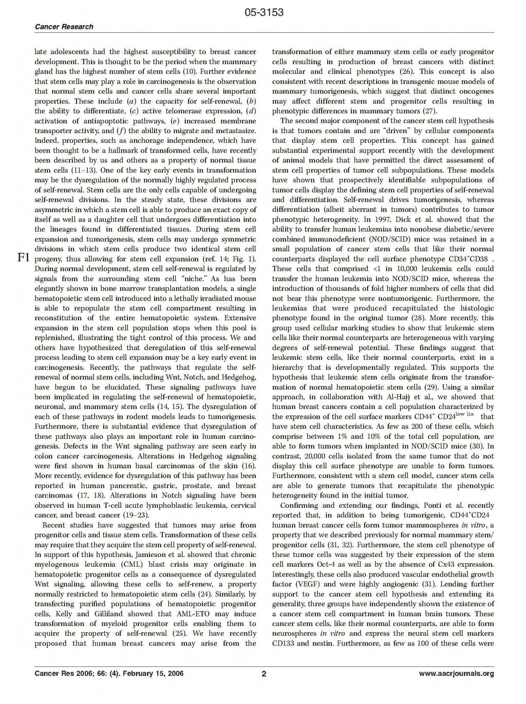
29

30
31

32

33

34

35
36 Journal of Mammary Gland Biology and Neoplasia, Vol. 10, No. 1, January 2005 ( C 2005) DOI: /s Survival of Mammary Stem Cells in Suspension Culture: Implications for Stem Cell Biology and Neoplasia Gabriela Dontu 1,2 and Max S. Wicha 1 There is increasing evidence that a variety of neoplasms including breast cancer may result from transformation of normal stem and progenitor cells. In the past, isolation and characterization of mammary stem cells has been limited by the lack of suitable culture systems able to maintain these cells in an undifferentiated state in vitro. We have recently described a culture system in which human mammary stem and progenitor cells are able to survive in suspension and produce spherical colonies composed of both stem and progenitor cells. Recent observation that adult stem cells from other tissues may also retain the capacity for growth under anchorage independent conditions suggests a common underlying mechanism. We propose that this mechanism involves the interaction between the canonical Wnt signal pathway and E-cadherin. The Wnt pathway has been implicated in normal stem cell self-renewal in vivo. Furthermore, there is evidence that deregulation of this pathway in the mammary gland and other organs may play a key role in carcinogenesis. Thus, the development of in vitro suspension culture systems not only provides an important new tool for the study of mammary cell biology, but also may have important implications for understanding key molecular pathways in both normal and neoplastic stem cells. KEY WORDS: stem cells; cancer stem cells; breast cancer; mammary gland development; Wnt signaling; self-renewal; differentiation. INTRODUCTION The isolation and characterization of tissue specific stem and progenitor cells has gained impetus due to the important insights that their study can provide into normal development and carcinogenesis. Adult stem cells are defined by their capacity for self-renewal and differentiation into cell lineages present in a specific tissue (1,2). Self-renewal ensures the propagation of the stem cell compartment, which in turn sustains morphogenesis, tissue repair and maintenance. Differentiation generates the specialized cells that form each organ and ensure its normal 1 Department of Internal Medicine, Hematology Oncology, Comprehensive Cancer Center, University of Michigan, Ann Arbor, Michigan. 2 To whom correspondence should be addressed at 1500 E Medical Center Dr., 7110 CCGC, Ann Arbor, Michigan 48109; gdontu@umich.edu. functioning. In many organs, slowly dividing stem cells give rise to a transit, amplifying population of progenitor cells that proliferate actively in response to specific signals. These undifferentiated cells are multipotent and generate the lineage-restricted progenitors that subsequently undergo terminal differentiation (3). Whereas mammary stem cells have not yet been isolated and characterized, their existence in vivo in mice was unequivocally demonstrated by Abbreviations used: ASMA, alpha smooth muscle actin; APC, adenomatosis polyposis coli; BCRP, breast cancer resistance protein; BMI, B-lymphoma MO-MLV insertion region 1; BMP4, bone morhogenetic protein 4; EGF, epidermal growth factor; ESA, epithelial specific antigen; ER, estrogen receptor; FACS, fluorescence-activated cell sorting; FITC, fluorescein isothyocyanate; bfgf, basic fibroblast growth factor; LIF, leukemia inhibitory factor; Muc 1, mucin 1; MMP7, matrix metalloproteinase 7; MTA3, metastasis associated gene 3; NuRD, Nuclear remodeling and deacetylation complex; PR, progesterone receptor; PTEN, phosphatase and tensin homolog; SP, side population /05/ /0 C 2005 Springer Science+Business Media, Inc.
37 76 Dontu and Wicha serial transplantation studies (4,5). Evidence for the existence of adult mammary stem cells and the strategies employed for their isolation and characterization were thoroughly reviewed in several recent papers (6,7). The focus of this review will be on the development of methods for the cultivation of adult mammary stem/progenitor cells and the potential application of these systems in stem cell biology. IN VITRO PROPAGATION OF NORMAL STEM CELLS IN SUSPENSION CULTURE Ideally stem cells are purified by isolation directly from tissue to avoid any artifacts introduced by in vitro manipulation (2). Purification of the stem cells is based on finding a number of morphological and molecular traits, including surface antigens, that distinguish them from all the other cells in the tissue of interest (8). Most stem cell enrichment/purification protocols rely on fluorescenceactivated cell sorting (FACS) or magnetic immunosorting and use sets of antibodies against cell surface proteins. When the molecular signature is not known, which is the case for mammary stem cells, the task of isolating the stem cells involves a tedious process of screening large numbers of putative markers to identify the ones that co-segregate with functional attributes of stemness. As a minimal working definition, these attributes include self-renewal and ability to generate differentiated progeny. In vivo generation of a functional tissue is the ultimate proof of stemness of any candidate marker. However, screening large numbers of candidate markers in in vivo transplantation assays in order to find the stem cell signature is impractical. On the other hand, until recently, the lack of a cultivation system that allows for the propagation of mammary progenitors in an undifferentiated state has precluded the testing of self-renewal in vitro. We have developed a culture system, based on previous work in neural stem cell biology, which overcomes this inability of previous systems to maintain and propagate mammary stem and progenitor cells. In 1992 Reynolds and Weis published a study in which they showed that neural cells isolated from the subventricular zone of rat embryos can proliferate in suspension culture, clonally generating spherical colonies, which they termed neurospheres (9). A clonal analysis of neurospheres demonstrated that 20% of these cells, proliferating in vitro in response to EGF and bfgf stimulation, were capable of both self-renewal and differentiation along multiple lineages, the defining characteristics of stem cells (10). Two years later the same group showed that neural stem/progenitor cells from adult animals have the same ability to propagate in vitro as neurospheres (11). Since then, numerous studies utilizing cells derived from either the central or peripheral nervous system, from embryonic and adult tissue showed that the ability to generate neurospheres in vitro correlates with the number of stem cells in the tissue of origin (12 14). Implanted into the nervous system of an animal host, neurospheres generate cells that selfrenew as well as differentiate and contribute to the various cellular compartments of the recipient animal s brain (12,13). Therefore, these cells are capable of self-renewal and multilineage differentiation, both in vitro and in vivo. This experimental system has been an extremely useful tool in neural stem cell biology. It was used as an in vitro model for early development (15,16), to identify factors involved in cell fate determination and to unravel signaling pathways active in stem and early progenitor cells (17 19). A series of studies utilized this system for comparative transcriptional profiling in order to identify gene expression changes underlying self renewal or lineage specific differentiation (20 23). Moreover, neurospheres were utilized in a number of in vivo studies to repair neuronal loss in mice with spinal cord and sciatic nerve injuries and in rat models with ischemic cerebral cortex (24), partial Parkinson s disease (25) and Huntington s disease (26). These studies demonstrated successful engraftment, migration to the site of the lesion and differentiation of sphere-derived cells into functional neurons with variable degrees of behavioral and anatomical recovery. These results make the case for the potential use of these progenitor cells, propagated in vitro in suspension culture, in repairing damaged tissue in vivo. The neurosphere culture is also routinely used now to assess the enrichment of stem cells in experiments using cell sorting for identifying markers of stem cells (12,13). Neurosphere formation was also used to assess the size of the stem cell population in neural tissue of genetically manipulated mice in experiments aiming to investigate the role of genes such as BMI (23), PTEN (27), LIF and Notch (28,29) in self-renewal. NONADHERENT MAMMOSPHERES Recently we adopted the suspension culture as a strategy for the in vitro enrichment and propagation
38 Survival of Mammary Stem Cells in Suspension Culture 77 of human mammary stem/progenitor cells (30). Based on the model of neurospheres, we hypothesized that a small population of mammary cells with stem cell properties would be able to survive and proliferate in the absence of attachment to an exogenous substratum. We developed a culture system in which human mammary epithelial cells, isolated from reduction mammoplasties, are cultured on a non-adhesive substratum in serum-free medium in the presence of EGF and/or bfgf. Under these conditions the vast majority of cells undergo anoikis. This term specifically applies to the apoptosis of nontransformed cells which occurs in the absence of anchorage to a substratum. We hypothesized that anoikis was a property of differentiated cells, but that stem cells could survive anchorage independent conditions. Consistent with this hypothesis, we found that approximately four in 1000 freshly isolated cells are able to survive and proliferate, and form multicellular spheroids. We termed these spheroids mammospheres by virtue of their resemblance to neurospheres cultured from primary neural cells. As is the case for neurospheres, we demonstrated that mammospheres are highly enriched in undifferentiated cells, as demonstrated by the ability of single cells isolated from mammospheres to generate multilineage colonies when cultured in the presence of serum on a collagen substratum which promotes their differentiation (Fig. 1(a)). Primary mammospheres contain eight times more bi-lineage progenitor cells than freshly cultured human mammary cells. Secondary and later passaged mammospheres consist of virtu- Fig. 1. Mammospheres are composed of stem/progenitor cells. Mammosphere-derived cells plated at clonogenic densities can: (a) self renew, (b) generate mixed colonies containing cells of all the three mammary lineage types (immunostained with lineage specific markers, ductal epithelial ESA, brown; myoepithelial CD10, purple; alveolar, beta-casein red) (c) generate complex structures in 3D Matrigel culture, (d) generate ductal alveolar outgrowths in vivo.
39 78 Dontu and Wicha ally 100% bipotent progenitors. Furthermore, the majority of bipotent progenitors are able to generate colonies that contain all three lineages of the adult mammary gland, myoepithelial, ductal epithelial, and alveolar epithelial cells. We have also shown that mammospheres contain cells capable of clonally generating complex functional structures in reconstituted 3-D culture systems in Matrigel (Figs. 1(c) and 2). Self-renewal of a cell population within mammospheres was demonstrated utilizing an assay in which single cells from mammospheres are able to generate second and later generation spheres (Fig. 1(b)). We demonstrated that mammospheres were clonally generated using retroviral marking. Furthermore, we demonstrated that mammospheres derived from these passaged cells also have multipotent differentiation potential. These results resemble those reported for neurospheres (10,31) and are consistent with a model in which the mammosphereforming cell represents a mammary stem cell which undergoes limited self renewal and then gives rise to mammary progenitors still capable of multi-lineage differentiation (32). Clonal experiments in which spheres were grown from single cells and single mammospheres were passaged, suggest that one or two self-renewal divisions are involved in the formation of a single mammosphere. This limited number of self-renewal divisions is in agreement with the majority of studies involving adult stem cells, which indicate that expansion of the adult stem population does not readily occur ex vivo, presumably due to asymmetric cell kinetic divisions that result in a large number of progenitors and differentiated cells and a small fixed number of stem cells. Our findings indicate that mammary stem cells are contained in the small population of cells that are anchorage independent and survive suspension culture to proliferate and differentiate into mammary progenitor cells (30). To determine if the ability of cells to form mammospheres correlates with enrichment in progenitor cells, we tested the sphere formation capability of the side population (SP) of mammary epithelial cells (30). SP represents a subpopulation of cells capable of excluding dyes, such as Rhodamine and Hoechst, due to the expression of transporter proteins, such as BCRP (breast cancer resistance protein) and P-glycoproteins (33). It has been shown that the SP fraction of hematopoietic and neural cells contains the long-term repopulating stem cells (34). This phenomenon has also recently been demonstrated for mouse mammary cells with SP properties, which can regenerate the gland upon transplantation (35). In our study SP and non-sp staining population from uncultured cells were separated by FACS and placed in suspension culture. Only cells contained in the SP fraction were capable of mammosphere formation in suspension culture as well as generation of multilineage colonies on collagen substrata (30). In order to assess the cellular composition of mammospheres we used immunostaining with markers specific for the differentiated mammary cells of luminal epithelial (ESA, Muc1, cytokeratin 18) and myoepithelial lineages (CD10, ASMA, cytokeratin 14, alpha 6 integrin). Some of these markers were identified in previous studies as being associated with bi-potent progenitor cells (ESA, alpha 6 integrin) (36,37) or mammary stem/progenitor cells (cytokeratin 5). Mammospheres contained cells positive for alpha 6 integrin, cytokeratin 5 and CD10, ESA and cytokertain 14 (30). ER and PR expression was also detectable by immunostaining in mammospheres and in colonies generated from mammospheres plated on a collagen substratum. A subpopulation of the ER positive cells was also positive for the Ki67 proliferation marker, indicating that these cells can divide in vitro. After 5 7 days of cultivation on a collagen substratum ER expression was no longer detected, suggesting that it is downregulated during differentiation in vitro (unpublished observations). We have recently found that, when transplanted in the cleared mammary fat pad of NOD/SCID mice, mammospheres generated limited outgrowths with the morphological and cellular characteristics of a human mammary ductal alveolar structure (Figs. 1(d) and 2). As few as 500 transplanted mammospheres (10,000 25,000 cells) generated outgrowths, in the absence of human fibroblasts. Improved engraftment was obtained by using mammospheres combined with human mammary fibroblasts, as described by Kuperwasser et al. (38). Titration experiments will be required to determine the absolute enrichment in gland reconstituting activity of mammosphere derived cells. We have utilized the mammosphere cultivation system to delineate signaling pathways involved in cell fate specification of mammary stem/progenitor cells, such as Notch and Sonic Hedgehog (32). Using assays for in vitro self-renewal and differentiation that we developed with this system, we showed that Notch signaling regulates cell fate decisions in the mammary gland at several distinct developmental stages. Notch activation increases selfrenewal of mammary stem cells, as well as acting on
 Fully developed ductal-alveolar structure grown in Matrigel from a secondary mammosphere, immunostained with lineage specific markers (myoepithelial cells are stained red with CD10-Texas red and")
40 Survival of Mammary Stem Cells in Suspension Culture 79 Fig. 2. (a) Fully developed ductal-alveolar structure grown in Matrigel from a secondary mammosphere, immunostained with lineage specific markers (myoepithelial cells are stained red with CD10-Texas red and ductal epithelial cells are stained green with ESA-FITC) (b) Same as a, higher magnification. (c-k)mammospheres generate outgrowths in the cleared mammary fat pad. Whole mount staining (carmine-red) and H&E tissue sections through a mouse mammary fat pad: not cleared [(c) and (f)], cleared, non-implanted [(d), (g)], cleared and implanted with mammospheres derived from human mammary epithelial cells [(e), (h), (k)]. Immunostaining utilizing human specific ESA antibody - tissue section through a mouse mammary gland (i), human mammary gland (j), mouse cleared fat-pad implanted with human mammospheres (k).
TITLE: Neural Protein Synuclein (SNCG) in Breast Cancer Progression
 in Breast Cancer Progression") AD AWARD NUMBER: DAMD17-01-1-0352 TITLE: Neural Protein Synuclein (SNCG) in Breast Cancer Progression PRINCIPAL INVESTIGATOR: Yangfu Jiang, M.D., Ph.D. CONTRACTING ORGANIZATION: Long Island Jewish Medical
AD AWARD NUMBER: DAMD17-01-1-0352 TITLE: Neural Protein Synuclein (SNCG) in Breast Cancer Progression PRINCIPAL INVESTIGATOR: Yangfu Jiang, M.D., Ph.D. CONTRACTING ORGANIZATION: Long Island Jewish Medical
PREPARED FOR: U.S. Army Medical Research and Materiel Command Fort Detrick, Maryland
 AD Award Number: W81XWH-04-1-0618 TITLE: Are Breast Tumor Stem Cells Responsible for Metastasis and Angiogenesis PRINCIPAL INVESTIGATOR: Quintin Pan, Ph.D. CONTRACTING ORGANIZATION: University of Michigan
TITLE: Evaluation of DNA Methylation as a Target for Intraductal Therapy for Ductal Carcinoma in Situ of the Breast
 AD AWARD NUMBER: DAMD17-02-1-0569 TITLE: Evaluation of DNA Methylation as a Target for Intraductal Therapy for Ductal Carcinoma in Situ of the Breast PRINCIPAL INVESTIGATOR: Kristin A. Skinner, M.D. CONTRACTING
TITLE: A PSCA Promoter Based Avian Retroviral Transgene Model of Normal and Malignant Prostate
 AD Award Number: DAMD17-03-1-0163 TITLE: A PSCA Promoter Based Avian Retroviral Transgene Model of Normal and Malignant Prostate PRINCIPAL INVESTIGATOR: Robert Reiter, M.D. CONTRACTING ORGANIZATION: The
CONTRACTING ORGANIZATION: University of California Lawrence Berkeley National Laboratory Berkeley, CA 94720
 AD Award Number: W81XWH-05-1-0526 TITLE: Effects of Extracellular Matix on DNA Repair in Vivo PRINCIPAL INVESTIGATOR: Aylin Rizki, Ph.D. CONTRACTING ORGANIZATION: University of California Lawrence Berkeley
TITLE: Notch in Pathological Angiogenesis and Lymphangiogenesis
 Award Number: W81XWH-10-1-0304 TITLE: Notch in Pathological Angiogenesis and Lymphangiogenesis PRINCIPAL INVESTIGATOR: Minji Kim CONTRACTING ORGANIZATION: Columbia University New York, NY 10032 REPORT
TITLE: Properties of Leukemia Stem Cells in a Novel Model of CML Progression to Lymphoid Blast Crisis
 AD Award Number: W81XWH-05-1-0608 TITLE: Properties of Leukemia Stem Cells in a Novel Model of CML Progression to Lymphoid Blast Crisis PRINCIPAL INVESTIGATOR: Craig T. Jordan, Ph.D. CONTRACTING ORGANIZATION:
PREPARED FOR: U.S. Army Medical Research and Materiel Command Fort Detrick, Maryland
 AD Award Number: W81XWH-06-1-0524 TITLE: Elucidating and Modeling Irradiation Effects on Centrosomal and Chromosomal Stability within Breast Cancer PRINCIPAL INVESTIGATOR: Christopher A. Maxwell, Ph.D.
TITLE: Role of ADAM15 in Tumor/Endothelial Interactions Prostate Cancer Regression
 AD Award Number: W81XWH-07-1-0030 TITLE: Role of ADAM15 in Tumor/Endothelial Interactions Prostate Cancer Regression PRINCIPAL INVESTIGATOR: Mark L. Day CONTRACTING ORGANIZATION: University of Michigan
TITLE: Enhancing the Anti-Tumor Activity of ErbB Blockers with Histone Deaccetylase (HDAC) Inhibition in Prostate Cancer Cell Lines
 Inhibition in Prostate Cancer Cell Lines") AD Award Number: W81XWH-05-1-0040 TITLE: Enhancing the Anti-Tumor Activity of ErbB Blockers with Histone Deaccetylase (HDAC) Inhibition in Prostate Cancer Cell Lines PRINCIPAL INVESTIGATOR: Prakash Chinnaiyan,
AD Award Number: W81XWH-05-1-0040 TITLE: Enhancing the Anti-Tumor Activity of ErbB Blockers with Histone Deaccetylase (HDAC) Inhibition in Prostate Cancer Cell Lines PRINCIPAL INVESTIGATOR: Prakash Chinnaiyan,
CONTRACTING ORGANIZATION: Vanderbilt University Medical Center Nashville, TN
 AD Award Number: W81XWH-04-1-0867 TITLE: A Myc-Driven in Vivo Model of Human Prostate Cancer PRINCIPAL INVESTIGATOR: Simon W. Hayward, Ph.D. CONTRACTING ORGANIZATION: Vanderbilt University Medical Center
CONTRACTING ORGANIZATION: Mount Sinai School of Medicine New York, New York
 AD AWARD NUMBER: W81XWH-05-1-0475 TITLE: Restoration of Epithelial Polarity in Metastatic Tumors PRINCIPAL INVESTIGATOR: Sergei Sokol, Ph.D. CONTRACTING ORGANIZATION: Mount Sinai School of Medicine New
TITLE: Dietary Genistein and Prostate Cancer Chemoprevention
 AD AWARD NUMBER: DAMD17-02-1-0662 TITLE: Dietary Genistein and Prostate Cancer Chemoprevention PRINCIPAL INVESTIGATOR: Coral A. Lamartiniere, Ph.D. CONTRACTING ORGANIZATION: The University of Alabama at
TITLE: Crosstalk Between Cancer Cells and Bones Via the Hedgehog Pathway Determines Bone Metastasis of Breast Cancer
 AD Award Number: W81XWH-07-1-0400 TITLE: Crosstalk Between Cancer Cells and Bones Via the Hedgehog Pathway Determines Bone Metastasis of Breast Cancer PRINCIPAL INVESTIGATOR: Dr. Lalita Shevde-Samantrese
TITLE: The Role of hcdc4 as a Tumor Suppressor Gene in Genomic Instability Underlying Prostate Cancer
 AD Award Number: TITLE: The Role of hcdc4 as a Tumor Suppressor Gene in Genomic Instability Underlying Prostate Cancer PRINCIPAL INVESTIGATOR: Audrey van Drogen, Ph.D. CONTRACTING ORGANIZATION: Sidney
La Jolla, CA Approved for Public Release; Distribution Unlimited
 AD Award Number: TITLE: Suppression of Breast Cancer Progression by Tissue Factor PRINCIPAL INVESTIGATOR: Wolfram Ruf, M.D. CONTRACTING ORGANIZATION: The Scripps Research Institute La Jolla, CA 92037 REPORT
TITLE: Investigation of the Akt/PKB Kinase in the Development of Hormone-Independent Prostate Cancer
 AD Award Number: TITLE: Investigation of the Akt/PKB Kinase in the Development of Hormone-Independent Prostate Cancer PRINCIPAL INVESTIGATOR: Linda A. degraffenried, Ph.D. CONTRACTING ORGANIZATION: The
TITLE: HER2/neu Antisense Therapeutics in Human Breast Cancer. CONTRACTING ORGANIZATION: Washington University School of Medicine St.
 AD Award Number: DAMD17-99-1-9436 TITLE: HER2/neu Antisense Therapeutics in Human Breast Cancer PRINCIPAL INVESTIGATOR: Jeffrey A. Drebin, M.D., Ph.D. CONTRACTING ORGANIZATION: Washington University School
PREPARED FOR: U.S. Army Medical Research and Materiel Command Fort Detrick, Maryland
 AD Award Number: W81XWH-06-1-0093 TITLE: An Epigenetic Link to Prostate Cancer PRINCIPAL INVESTIGATOR: Raphael F Margueron, Ph.D. CONTRACTING ORGANIZATION: University of Medicine and Dentistry of New Jersey
Award Number: W81XWH TITLE: Dietary Fish Oil in Reducing Bone Metastasis of Breast Cancer
 AD Award Number: W81XWH-04-1-0693 TITLE: Dietary Fish Oil in Reducing Bone Metastasis of Breast Cancer PRINCIPAL INVESTIGATOR: Nandini Ghosh-Choudhury, Ph.D. CONTRACTING ORGANIZATION: University of Texas
TITLE: Prevention and Treatment of Neurofibromatosis Type 1-Associated Malignant Peripheral Nerve Sheath Tumors
 AWARD NUMBER: W81XWH-14-1-0073 TITLE: Prevention and Treatment of Neurofibromatosis Type 1-Associated Malignant Peripheral Nerve Sheath Tumors PRINCIPAL INVESTIGATOR: Kevin A. Roth, MD, PhD CONTRACTING
TITLE: Inhibitors of Histone Deacetylases for Radiosensitization of Prostate Cancer
 AD Award Number: W81XWH-04-1-0170 TITLE: Inhibitors of Histone Deacetylases for Radiosensitization of Prostate Cancer PRINCIPAL INVESTIGATOR: Mira O. Jung, Ph.D. CONTRACTING ORGANIZATION: Georgetown University
AD (Leave blank) PREPARED FOR: U.S. Army Medical Research and Materiel Command Fort Detrick, Maryland
 PREPARED FOR: U.S. Army Medical Research and Materiel Command Fort Detrick, Maryland") AD (Leave blank) Award Number: W81XWH-06-2-0038 TITLE:T Cell Lipid rafts and complement Ligands for Diagnosis and Monitoring PRINCIPAL INVESTIGATOR: George C. Tsokos, MD CONTRACTING ORGANIZATION: Beth
AD (Leave blank) Award Number: W81XWH-06-2-0038 TITLE:T Cell Lipid rafts and complement Ligands for Diagnosis and Monitoring PRINCIPAL INVESTIGATOR: George C. Tsokos, MD CONTRACTING ORGANIZATION: Beth
Award Number: W81XWH TITLE: Characterizing an EMT Signature in Breast Cancer. PRINCIPAL INVESTIGATOR: Melanie C.
 AD Award Number: W81XWH-08-1-0306 TITLE: Characterizing an EMT Signature in Breast Cancer PRINCIPAL INVESTIGATOR: Melanie C. Bocanegra CONTRACTING ORGANIZATION: Leland Stanford Junior University Stanford,
TITLE: Investigating the Role of TBX2 in the Inhibition of Senescence in Prostate Cancer
 AD Award Number: W81XWH-07-1-0155 TITLE: Investigating the Role of TBX2 in the Inhibition of Senescence in Prostate Cancer PRINCIPAL INVESTIGATOR: Srinivas Nandana CONTRACTING ORGANIZATION: Vanderbilt
PREPARED FOR: U.S. Army Medical Research and Materiel Command Fort Detrick, Maryland
 AD Award Number: TITLE: Suppression of Breast Cancer Progression by Tissue Factor PRINCIPAL INVESTIGATOR: Wolfram Ruf, M.D. CONTRACTING ORGANIZATION: The Scripps Research Institute La Jolla, CA 92037 REPORT
CONTRACTING ORGANIZATION: Rush University Medical Center Chicago, IL 60612
 AD Award Number: W81XWH-11-1-0302 TITLE: Yin and Yang of Heparanase in breast Tumor Initiation PRINCIPAL INVESTIGATOR: Xiulong Xu, Ph.D. CONTRACTING ORGANIZATION: Rush University Medical Center Chicago,
PREPARED FOR: U.S. Army Medical Research and Materiel Command Fort Detrick, Maryland
 AD Award Number: W81XWH-12-1-0212 TITLE: Wnt/Beta-Catenin, Foxa2, and CXCR4 Axis Controls Prostate Cancer Progression PRINCIPAL INVESTIGATOR: Xiuping Yu CONTRACTING ORGANIZATION: Vanderbilt University
TITLE: 64Cu-DOTA-trastuzumab PET imaging in women with HER2 overexpressing breast cancer
 AD Award Number: W81XWH-10-1-0824 TITLE: 64Cu-DOTA-trastuzumab PET imaging in women with HER2 overexpressing breast cancer PRINCIPAL INVESTIGATOR: Joanne Mortimer, M.D. CONTRACTING ORGANIZATION: City of
TITLE: A Tissue Engineering Approach to Study the Progression of Breast Tumor Metastasis in Bone
 AD AWARD NUMBER: W81XWH-04-1-0749 TITLE: A Tissue Engineering Approach to Study the Progression of Breast Tumor Metastasis in Bone PRINCIPAL INVESTIGATOR: Mingxin Che, M.D., Ph.D. Daotai Nie, Ph.D. CONTRACTING
PREPARED FOR: U.S. Army Medical Research and Materiel Command Fort Detrick, Maryland
 AD Award Number: W81XWH-12-1-0212 TITLE: Wnt/beta-Catenin, Foxa2, and CXCR4 Axis Controls Prostate Cancer Progression PRINCIPAL INVESTIGATOR: Xiuping Yu CONTRACTING ORGANIZATION: Vanderbilt University
Roles of microrna-mediated Drug Resistance in Tumor Stem Cells of Small Cell Lung Carcinoma
 AD Award Number: W81XWH-12-1-0479 TITLE: Roles of microrna-mediated Drug Resistance in Tumor Stem Cells of Small Cell Lung Carcinoma PRINCIPAL INVESTIGATOR: Kounosuke Watabe, Ph.D. CONTRACTING ORGANIZATION:
TITLE: Induction of Ephs/Ephrins-Mediated Tumor Cells-Endothelial Cells Repulsion as an Anti-Cancer Therapeutic Approach
 AD Award Number: W81XWH-04-1-0661 TITLE: Induction of Ephs/Ephrins-Mediated Tumor Cells-Endothelial Cells Repulsion as an Anti-Cancer Therapeutic Approach PRINCIPAL INVESTIGATOR: Gerald Batist, M.D. CONTRACTING
TITLE: The Role of Constitutively Active Prolactin Receptors in the Natural History of Breast Cancer
 AD Award Number: W81XWH-06-1-0448 TITLE: The Role of Constitutively Active Prolactin Receptors in the Natural History of Breast Cancer PRINCIPAL INVESTIGATOR: Kuang-tzu Huang CONTRACTING ORGANIZATION:
TITLE: "Impact of Obesity on Tamoxifen Chemoprevention in a Model of Ductal Carcinoma in Situ"
 AD Award Number: W81XWH-09-1-0720 TITLE: "Impact of Obesity on Tamoxifen Chemoprevention in a Model of Ductal Carcinoma in Situ" PRINCIPAL INVESTIGATOR: Sarah Dunlap-Smith, Ph.D. CONTRACTING ORGANIZATION:
TITLE: Overcoming Resistance to Inhibitors of the Akt Protein Kinase by Modulation of the Pim Kinase Pathway
 AWARD NUMBER: W81XWH-12-1-0560 TITLE: Overcoming Resistance to Inhibitors of the Akt Protein Kinase by Modulation of the Pim Kinase Pathway PRINCIPAL INVESTIGATOR: Andrew S. Kraft, MD CONTRACTING ORGANIZATION:
CONTRACTING ORGANIZATION: Cold Spring Harbor Laboratory Cold Spring Harbor, NY 11724
 AD Award Number: W81XWH-10-1-0450 TITLE: The role of NF1 in memory retrieval PRINCIPAL INVESTIGATOR: Yi Zhong, Ph.D. CONTRACTING ORGANIZATION: Cold Spring Harbor Laboratory Cold Spring Harbor, NY 11724
TITLE: Genes Associated with Food Allergy and Eosinophilic Esophagitis
 AD Award Number: W81XWH-1-1-75 TITLE: Genes Associated with Food Allergy and Eosinophilic Esophagitis PRINCIPAL INVESTIGATOR: Dr. David Broide CONTRACTING ORGANIZATION: University of California, San Diego
TITLE: The Role of HOX Proteins in Androgen-Independent Prostate Cancer
 AD Award Number: W81XWH-6-1-64 TITLE: The Role of HOX Proteins in Androgen-Independent Prostate Cancer PRINCIPAL INVESTIGATOR: Sunshine Daddario, B.A. CONTRACTING ORGANIZATION: University of Colorado Health
PREPARED FOR: U.S. Army Medical Research and Materiel Command Fort Detrick, Maryland
 AD Award Number: W81XWH- TITLE: PRINCIPAL INVESTIGATOR: CONTRACTING ORGANIZATION: REPORT DATE: TYPE OF REPORT: Annual PREPARED FOR: U.S. Army Medical Research and Materiel Command Fort Detrick, Maryland
PREPARED FOR: U.S. Army Medical Research and Materiel Command Fort Detrick, Maryland
 AD Award Number: W81XWH-12-1-0337 TITLE: Molecular Innovations Toward Theranostics of Aggressive Prostate Cancer PRINCIPAL INVESTIGATOR: Hsieh, Jer-Tsong CONTRACTING ORGANIZATION: University of Texas Southwestern
TITLE: Identification of New Serum Biomarkers for Early Breast Cancer Diagnosis and Prognosis Using Lipid Microarrays.
 AD Award Number: W81XWH-06-1-0690 TITLE: Identification of New Serum Biomarkers for Early Breast Cancer Diagnosis and Prognosis Using Lipid Microarrays. PRINCIPAL INVESTIGATOR: Guangwei Du, Ph.D. CONTRACTING
TITLE: Investigation of the Akt/Pkb Kinase in the Development of Hormone- Independent Prostate Cancer
 AD Award Number: TITLE: Investigation of the Akt/Pkb Kinase in the Development of Hormone- Independent Prostate Cancer PRINCIPAL INVESTIGATOR: Linda A. degraffenried, PhD CONTRACTING ORGANIZATION: University
TITLE: Prostate Expression Databases: Gene Expression Resources for Comparative Studies of Prostate Carcinogenesis
 AD Award Number: W81XWH-05-1-0110 TITLE: Prostate Expression Databases: Gene Expression Resources for Comparative Studies of Prostate Carcinogenesis PRINCIPAL INVESTIGATOR: Peter S. Nelson, Ph.D. CONTRACTING
TITLE: Development of Antigen Presenting Cells for adoptive immunotherapy in prostate cancer
 AD Award Number: W8XWH-5-- TITLE: Development of Antigen Presenting Cells for adoptive immunotherapy in prostate cancer PRINCIPAL INVESTIGATOR: Mathias Oelke Ph.D. CONTRACTING ORGANIZATION: Johns Hopkins
TITLE: Role of CDK5 as a Tumor Suppressor Gene in Non-Small Cell Lung Cancer
 AWARD NUMBER: W81XWH-13-1-0157 TITLE: Role of CDK5 as a Tumor Suppressor Gene in Non-Small Cell Lung Cancer PRINCIPAL INVESTIGATOR: Barry D. Nelkin, Ph.D. CONTRACTING ORGANIZATION: Johns Hopkins University
Sphingosine-1-Phosphate Receptor Subtype 3: A Novel Therapeutic Target of K-Ras Mutant Driven Non-Small Cell Lung Carcinoma
 AWARD NUMBER: W81XWH-14-1-0346 TITLE: Sphingosine-1-Phosphate Receptor Subtype 3: A Novel Therapeutic Target of K-Ras Mutant Driven Non-Small Cell Lung Carcinoma PRINCIPAL INVESTIGATOR: Lee, Menq-Jer,
TITLE: New Advanced Technology to Improve Prediction and Prevention of Type 1 Diabetes
 AD Award Number: DAMD17-01-1-0009 TITLE: New Advanced Technology to Improve Prediction and Prevention of Type 1 Diabetes PRINCIPAL INVESTIGATOR: Robert A. Vigersky CONTRACTING ORGANIZATION: Children s
TITLE: Effect of COX-2 (PGE2) and IL-6 on Prostate Cancer Bone Mets
 and IL-6 on Prostate Cancer Bone Mets") AD Award Number: W81XWH-05-1-0166 TITLE: Effect of COX-2 (PGE2) and IL-6 on Prostate Cancer Bone Mets PRINCIPAL INVESTIGATOR: Alice C. Levine, M.D. CONTRACTING ORGANIZATION: Mount Sinai School of Medicine
AD Award Number: W81XWH-05-1-0166 TITLE: Effect of COX-2 (PGE2) and IL-6 on Prostate Cancer Bone Mets PRINCIPAL INVESTIGATOR: Alice C. Levine, M.D. CONTRACTING ORGANIZATION: Mount Sinai School of Medicine
TITLE: Targeting the Immune System s Natural Response to Cell Death to Improve Therapeutic Response in Breast Cancers
 AD Award Number: W81XWH-13-1-0158 TITLE: Targeting the Immune System s Natural Response to Cell Death to Improve Therapeutic Response in Breast Cancers PRINCIPAL INVESTIGATOR: Rebecca S. Cook CONTRACTING
PREPARED FOR: U.S. Army Medical Research and Materiel Command Fort Detrick, Maryland
 AD Award Number: W81XWH-05-1-0262 TITLE: Role of CDK4 in Breast Development and Cancer PRINCIPAL INVESTIGATOR: Haritha Reddy CONTRACTING ORGANIZATION: Temple University Philadelphia PA 19122-6024 REPORT
TITLE: TREATMENT OF ENDOCRINE-RESISTANT BREAST CANCER WITH A SMALL MOLECULE C-MYC INHIBITOR
 AD Award Number: W81XWH-13-1-0159 TITLE: TREATMENT OF ENDOCRINE-RESISTANT BREAST CANCER WITH A SMALL MOLECULE C-MYC INHIBITOR PRINCIPAL INVESTIGATOR: Qin Feng CONTRACTING ORGANIZATION: Baylor College of
TITLE: Effect of MUC1 Expression on EGFR Endocytosis and Degradation in Human Breast Cancer Cell Lines
 AD AWARD NUMBER: W81XWH-06-1-0464 TITLE: Effect of MUC1 Expression on EGFR Endocytosis and Degradation in Human Breast Cancer Cell Lines PRINCIPAL INVESTIGATOR: Rachid M El Bejjani CONTRACTING ORGANIZATION:
PREPARED FOR: U.S. Army Medical Research and Materiel Command Fort Detrick, Maryland
 AD Award Number: DAMD17-03-1-0392 TITLE: The Role of Notch Signaling Pathway in Breast Cancer Pathogenesis PRINCIPAL INVESTIGATOR: Annapoorni Rangarajan, Ph.D. CONTRACTING ORGANIZATION: Indian Institute
TITLE: Targeted Eradication of Prostate Cancer Mediated by Engineered Mesenchymal Stem Cells
 AD AWARD NUMBER: TITLE: Targeted Eradication of Prostate Cancer Mediated by Engineered Mesenchymal Stem Cells PRINCIPAL INVESTIGATOR: CONTRACTING ORGANIZATION: Louisiana State University New Orleans, Louisiana
Award Number: W81XWH TITLE: Regenerative Stem Cell Therapy for Breast Cancer Bone Metastasis
 AD Award Number: W81XWH-11-1-593 TITLE: Regenerative Stem Cell Therapy for Breast Cancer Bone Metastasis PRINCIPAL INVESTIGATOR: Selvarangan Ponnazhagan, Ph.D. CONTRACTING ORGANIZATION: University of Alabama
TITLE: Breast Tumor-Generated Type 1 Collagen Breakdown Fragments Act as Matrikines to Drive Osteolysis
 AD Award Number: W81XWH-08-1-0639 TITLE: Breast Tumor-Generated Type 1 Collagen Breakdown Fragments Act as Matrikines to Drive Osteolysis PRINCIPAL INVESTIGATOR: Ching Hua William Wu PhD. CONTRACTING ORGANIZATION:
TITLE: Systemic Oncolytic Cytokine HSV Therapy of Prostate Cancer. CONTRACTING ORGANIZATION: Massachusetts General Hospital Boston, MA
 AD Award Number: W81XWH-05-1-0367 TITLE: Systemic Oncolytic Cytokine HSV Therapy of Prostate Cancer PRINCIPAL INVESTIGATOR: Susan Varghese, Ph.D. CONTRACTING ORGANIZATION: Massachusetts General Hospital
CONTRACTING ORGANIZATION: University of Illinois Chicago, IL
 AD Award Number: W81XWH-05-1-0009 TITLE: Selenoproteins and Prostate Cancer PRINCIPAL INVESTIGATOR: Veda Navsariwala, Ph.D. CONTRACTING ORGANIZATION: University of Illinois Chicago, IL 60612-7205 REPORT
Expression and Promoter Methylation of P16INK4A During Estrogen-Induced Mammary Carcinogenesis in the ACI Rat. Omaha, NE
 AD Award Number: DAMD17-03-1-0466 TITLE: Expression and Promoter Methylation of P16INK4A During Estrogen-Induced Mammary Carcinogenesis in the ACI Rat PRINCIPAL INVESTIGATOR: Dr. Lois M. Bartsch CONTRACTING
CONTRACTING ORGANIZATION: University of Texas M.D. Anderson Cancer Center Houston, TX 77030
 AD Award Number: W81XWH-7-1-345 TITLE: Second-Generation Therapeutic DNA Lymphoma Vaccines PRINCIPAL INVESTIGATOR: Larry W. Kwak, M.D., Ph.D. CONTRACTING ORGANIZATION: University of Texas M.D. Anderson
PREPARED FOR: U.S. Army Medical Research and Materiel Command Fort Detrick, Maryland
 AD Award Number: W81XWH-05-1-0045 TITLE: MT 2A Phosphorylation by PKC Mu/PKD Influences Chemosensitivity to Cisplatin in Prostate Cancer PRINCIPAL INVESTIGATOR: Kethandapatti Balaji, M.D. CONTRACTING ORGANIZATION:
PREPARED FOR: U.S. Army Medical Research and Materiel Command Fort Detrick, Maryland
 AD Award Number: W81XWH-09-1-0129 TITLE: Targeting Ovarian Carcinoma Stem Cells PRINCIPAL INVESTIGATOR: Richard Jones, M.D. CONTRACTING ORGANIZATION: Johns Hopkins University Baltimore, MD 21205 REPORT
TITLE: Adipose Estrogen and Increased Breast Cancer Risk in Obesity: Regulation by Leptin and Insulin
 AD Award Number: W81XWH-05-1-0497 TITLE: Adipose Estrogen and Increased Breast Cancer Risk in Obesity: Regulation by Leptin and Insulin PRINCIPAL INVESTIGATOR: Fahumiya Samad CONTRACTING ORGANIZATION:
TITLE: Selective Oncolytic Therapy for Hypoxic Breast Cancer Cells. CONTRACTING ORGANIZATION: Ordway Research Institute Albany, New York 12208
 AD Award Number: W81XWH-06-1-0586 TITLE: Selective Oncolytic Therapy for Hypoxic Breast Cancer Cells PRINCIPAL INVESTIGATOR: Michael T. Fasullo, Ph.D. CONTRACTING ORGANIZATION: Ordway Research Institute
CONTRACTING ORGANIZATION: Fred Hutchinson Cancer Research Center Seattle, WA 98109
 AWARD NUMBER: W81XWH-10-1-0711 TITLE: Transgenerational Radiation Epigenetics PRINCIPAL INVESTIGATOR: Christopher J. Kemp, Ph.D. CONTRACTING ORGANIZATION: Fred Hutchinson Cancer Research Center Seattle,
PREPARED FOR: U.S. Army Medical Research and Materiel Command Fort Detrick, Maryland
 AD Award Number: W81XWH-12-1-0225 TITLE: Epstein Barr virus and blood brain barrier in Multiple Sclerosis PRINCIPAL INVESTIGATOR: Luwen Zhang CONTRACTING ORGANIZATION: University of Nebraska Lincoln, NE
Award Number: W81XWH TITLE: Synthetic Lethal Gene for PTEN as a Therapeutic Target. PRINCIPAL INVESTIGATOR: Kounosuke Watabe, Ph.D.
 AD Award Number: W81XWH-12-1-0151 TITLE: Synthetic Lethal Gene for PTEN as a Therapeutic Target PRINCIPAL INVESTIGATOR: Kounosuke Watabe, Ph.D. CONTRACTING ORGANIZATION: University of Mississippi Medical
AD (Leave blank) TITLE: Proteomic analyses of nipple fluid for early detection of breast cancer
 TITLE: Proteomic analyses of nipple fluid for early detection of breast cancer") AD (Leave blank) Award Number: DAMD17-03-1-0575 TITLE: Proteomic analyses of nipple fluid for early detection of breast cancer PRINCIPAL INVESTIGATOR: Judy Garber CONTRACTING ORGANIZATION: Dana-Farber
AD (Leave blank) Award Number: DAMD17-03-1-0575 TITLE: Proteomic analyses of nipple fluid for early detection of breast cancer PRINCIPAL INVESTIGATOR: Judy Garber CONTRACTING ORGANIZATION: Dana-Farber
~ PRINCIPAL INVESTIGATOR: Yashaswi Shrestha. PREPARED FOR: U.S. Army Medical Research and Materiel Command Fort Detrick, Maryland
 AD (Leave blank) Award Number: W81XWH-08-1-0767 TITLE: Identifying Breast Cancer Oncogenes ~ PRINCIPAL INVESTIGATOR: Yashaswi Shrestha CONTRACTING ORGANIZATION: Dana-Farber Cancer Institute Boston, MA
AD (Leave blank) TITLE: Genomic Characterization of Brain Metastasis in Non-Small Cell Lung Cancer Patients
 TITLE: Genomic Characterization of Brain Metastasis in Non-Small Cell Lung Cancer Patients") AD (Leave blank) Award Number: W81XWH-12-1-0444 TITLE: Genomic Characterization of Brain Metastasis in Non-Small Cell Lung Cancer Patients PRINCIPAL INVESTIGATOR: Mark A. Watson, MD PhD CONTRACTING ORGANIZATION:
AD (Leave blank) Award Number: W81XWH-12-1-0444 TITLE: Genomic Characterization of Brain Metastasis in Non-Small Cell Lung Cancer Patients PRINCIPAL INVESTIGATOR: Mark A. Watson, MD PhD CONTRACTING ORGANIZATION:
TITLE: Can Reproductive Hormones Modulate Host Immunity to Breast Cancer Antigens
 AD AWARD NUMBER: W81XWH-04-1-0668 TITLE: Can Reproductive Hormones Modulate Host Immunity to Breast Cancer Antigens PRINCIPAL INVESTIGATOR: R. Todd Reilly, Ph.D. CONTRACTING ORGANIZATION: Johns Hopkins
TITLE: MicroRNA in Prostate Cancer Racial Disparities and Aggressiveness
 AWARD NUMBER: W81XWH-13-1-0477 TITLE: MicroRNA in Prostate Cancer Racial Disparities and Aggressiveness PRINCIPAL INVESTIGATOR: Cathryn Bock CONTRACTING ORGANIZATION: Wayne State University REPORT DATE:
PREPARED FOR: U.S. Army Medical Research and Materiel Command Fort Detrick, Maryland
 AD Award Number: TITLE: PRINCIPAL INVESTIGATOR: Jeremy Chien, PhD CONTRACTING ORGANIZATION: Mayo Clinic, REPORT DATE: September 2012 TYPE OF REPORT: Annual PREPARED FOR: U.S. Army Medical Research and
PREPARED FOR: U.S. Army Medical Research and Materiel Command Fort Detrick, Maryland
 Page 1 09/10/2013 AD Award Number: W81XWH-12-1-0453 TITLE: The cytoplasm translocation of the androgen receptor cofactor p44 as a target for prostate cancer treatment PRINCIPAL INVESTIGATOR: Zhengxin Wang
AD (Leave blank) Award Number: W81XWH TITLE: Targeting Autophagy for the Treatment of TSC and LAM. PRINCIPAL INVESTIGATOR: Elizabeth Henske
 Award Number: W81XWH TITLE: Targeting Autophagy for the Treatment of TSC and LAM. PRINCIPAL INVESTIGATOR: Elizabeth Henske") AD (Leave blank) Award Number: W81XWH-12-1-0578 TITLE: Targeting Autophagy for the Treatment of TSC and LAM PRINCIPAL INVESTIGATOR: Elizabeth Henske CONTRACTING ORGANIZATION: Brigham and Women s Hospital
AD (Leave blank) Award Number: W81XWH-12-1-0578 TITLE: Targeting Autophagy for the Treatment of TSC and LAM PRINCIPAL INVESTIGATOR: Elizabeth Henske CONTRACTING ORGANIZATION: Brigham and Women s Hospital
TITLE: Oxidative Stress, DNA Repair and Prostate Cancer Risk. PRINCIPAL INVESTIGATOR: Hua Zhao, Ph.D.
 AD Award Number: W81XWH-08-1-0416 TITLE: Oxidative Stress, DNA Repair and Prostate Cancer Risk PRINCIPAL INVESTIGATOR: Hua Zhao, Ph.D. CONTRACTING ORGANIZATION: Health Research Inc Buffalo, NY 14263 REPORT
Fort Detrick, Maryland
 Award Number: W81XWH-15-1-0636 TITLE: Effect of Diet on Gulf War Illness: A Pilot Study PRINCIPAL INVESTIGATOR: Ashok Tuteja, M.D. M.P.H CONTRACTING ORGANIZATION: Western Institute for Biomedical Research
Targeting of the MUC1-C Oncoprotein in Colitis-Associated Colorectal Cancer
 AD Award Number: W81XWH-12-1-0322 TITLE: Targeting of the MUC1-C Oncoprotein in Colitis-Associated Colorectal Cancer PRINCIPAL INVESTIGATOR: Kufe, Donald W., M.D. CONTRACTING ORGANIZATION: Boston, MA 02215-5450
TITLE: The Role Of Alternative Splicing In Breast Cancer Progression
 AD Award Number: W81XWH-06-1-0598 TITLE: The Role Of Alternative Splicing In Breast Cancer Progression PRINCIPAL INVESTIGATOR: Klemens J. Hertel, Ph.D. CONTRACTING ORGANIZATION: University of California,
PREPARED FOR: U.S. Army Medical Research and Materiel Command Fort Detrick, Maryland
 AWARD NUMBER: W81XWH-15-1-0644 TITLE: Targeting Cell Polarity Machinery to Exhaust Breast Cancer Stem Cells PRINCIPAL INVESTIGATOR: Chun-Ju Chang CONTRACTING ORGANIZATION: Purdue University West Lafayette,
PREPARED FOR: U.S. Army Medical Research and Materiel Command Fort Detrick, MD
 AD Award Number: W81XWH-11-1-0126 TITLE: Chemical strategy to translate genetic/epigenetic mechanisms to breast cancer therapeutics PRINCIPAL INVESTIGATOR: Xiang-Dong Fu, PhD CONTRACTING ORGANIZATION:
Award Number: W81XWH
 Award Number: W81XWH-14-2-0193 TITLE: Prevention of Bone Loss after Acute SCI by Zoledronic Acid: Durability, Effect on Bone Strength, and Use of Biomarkers to Guide Therapy PRINCIPAL INVESTIGATOR: Thomas
CONTRACTING ORGANIZATION: Johns Hopkins Bloomberg School of Public Health
 AD Award Number: W81XWH-12-1-0170 TITLE: Prospective Evaluation of Intraprostatic Inflammation and Focal Atrophy as a Predictor of Risk of High-Grade Prostate Cancer and Recurrence after Prostatectomy
CONTRACTING ORGANIZATION: Sloan-Kettering Institute for Cancer Research New York, NY 10021
 AD Award Number: DAMD17-94-J-4376 TITLE: Position Emitter I124 Iododeoxyuridine as a Tracer to Follow DNA Metabolism on Scans and in Tumor Samples in Advanced Breast Cancer: Comparison of 18F 2-Fluror-2-Deoxy-(D)-Glucose,
CONTRACTING ORGANIZATION: Johns Hopkins University School of Medicine Baltimore, MD 21205
 AD Award Number: DAMD7---7 TITLE: Development of Artificial Antigen Presenting Cells for Prostate Cancer Immunotherapy PRINCIPAL INVESTIGATOR: Jonathan P. Schneck, M.D., Ph.D. Mathias Oelke, Ph.D. CONTRACTING
CONTRACTING ORGANIZATION: University of Mississippi Medical Center Jackson, MS 39216
 AD Award Number: W81XWH-12-1-0201 TITLE: Role of long non-coding RNAs in prostate cancer PRINCIPAL INVESTIGATOR: Yin-Yuan Mo CONTRACTING ORGANIZATION: University of Mississippi Medical Center Jackson,
TITLE: Interchromosomal Associations that Alter NF1 Gene Expression Can Modify Clinical Manifestations of Neurofibromatosis 1. Palo Alto, CA 94304
 AD AWARD NUMBER: W81XWH-06-1-0695 TITLE: Interchromosomal Associations that Alter NF1 Gene Expression Can Modify Clinical Manifestations of Neurofibromatosis 1 PRINCIPAL INVESTIGATOR: Andrew R. Hoffman,
CONTRACTING ORGANIZATION: Regents of the University of Michigan Ann Arbor, MI 48109
 AWARD NUMBER: W81XWH-13-1-0463 TITLE: The Ketogenic Diet and Potassium Channel Function PRINCIPAL INVESTIGATOR: Dr. Geoffrey Murphy CONTRACTING ORGANIZATION: Regents of the University of Michigan Ann Arbor,
S Award Number: DAMD TITLE: Potential Role of Chibby as a Tumor Suppressor in Breast Cancer PRINCIPAL INVESTIGATOR:
 AD Award Number: DAMD17-03-1-0748 TITLE: Potential Role of Chibby as a Tumor Suppressor in Breast Cancer PRINCIPAL INVESTIGATOR: Richard W. Carthew, Ph.D. CONTRACTING ORGANIZATION: Northwestern University
U.S. Army Medical Research and Materiel Command Fort Detrick, Maryland
 AWARD NUMBER: W81XWH-14-1-0503 TITLE: Effect of Diabetes and Obesity on Disparities in Prostate Cancer Outcomes PRINCIPAL INVESTIGATOR: Bettina F. Drake, MPH, PhD CONTRACTING ORGANIZATION: Washington University
TITLE: Development of Antigen Presenting Cells for adoptive immunotherapy in prostate cancer
 AD Award Number: W8-XWH-5-- TITLE: Development of Antigen Presenting Cells for adoptive immunotherapy in prostate cancer PRINCIPAL INVESTIGATOR: Mathias Oelke, Ph.D. CONTRACTING ORGANIZATION: Johns Hopkins
TITLE: MiR-146-SIAH2-AR Signaling in Castration-Resistant Prostate Cancer
 AWARD NUMBER: W81XWH-14-1-0387 TITLE: MiR-146-SIAH2-AR Signaling in Castration-Resistant Prostate Cancer PRINCIPAL INVESTIGATOR: Dr. Goberdhan Dimri, PhD CONTRACTING ORGANIZATION: George Washington University,
CONTRACTING ORGANIZATION: North Eastern Ohio Universities Rootstown OH 44202
 AD Award Number: DAMD17-03-1-0082 TITLE: Prevalence and Outcomes of Restless Legs Syndrome among Veterans PRINCIPAL INVESTIGATOR: Claire C. Bourguet, Ph.D. CONTRACTING ORGANIZATION: North Eastern Ohio
CONTRACTING ORGANIZATION: University of Minnesota St Paul, MN 55108
 AD Award Number: W81XWH-06-1-0778 TITLE: Dietary Fat, Eicosanoids and Breast Cancer Risk PRINCIPAL INVESTIGATOR: Lindsay Orr CONTRACTING ORGANIZATION: University of Minnesota St Paul, MN 55108 REPORT DATE:
Award Number: W81XWH TITLE: A Novel Membrane-Permeable, Breast-Targeting, Pro-Apoptotic Peptide for Treatment of Breast Cancer
 AD Award Number: W81XWH-04-1-0705 TITLE: A Novel Membrane-Permeable, Breast-Targeting, Pro-Apoptotic Peptide for Treatment of Breast Cancer PRINCIPAL INVESTIGATOR: Bin Guo, Ph.D. CONTRACTING ORGANIZATION:
CONTRACTING ORGANIZATION: Children s Hospital Los Angeles Los Angeles, CA 90027
 AD Award Number: TITLE: Studies of the Tumor Microenvironment in Pathogenesis of Neuroblastoma PRINCIPAL INVESTIGATOR: Shahab Asgharzadeh, M D CONTRACTING ORGANIZATION: Children s Hospital Los Angeles
TITLE: MODULATION OF T CELL TOLERANCE IN A MURINE MODEL FOR IMMUNOTHERAPY OF PROSTATIC ADENOCARCINOMA
 AD Award Number: DAMD17-01-1-0085 TITLE: MODULATION OF T CELL TOLERANCE IN A MURINE MODEL FOR IMMUNOTHERAPY OF PROSTATIC ADENOCARCINOMA PRINCIPAL INVESTIGATOR: ARTHUR A HURWITZ, Ph.d. CONTRACTING ORGANIZATION:
Detection of Prostate Cancer Progression by Serum DNA Integrity
 AD Award Number: TITLE: Detection of Prostate Cancer Progression by Serum DNA Integrity PRINCIPAL INVESTIGATOR: Dave S.B. Hoon, Ph.D. CONTRACTING ORGANIZATION: John Wayne Cancer Institute Santa Monica,
CONTRACTING ORGANIZATION: Western Institute For Biomedical Research Salt Lake City, UT
 AD Award Number: W81XWH-10-1-0593 TITLE: Probiotic (VSL#3) for Gulf War Illness PRINCIPAL INVESTIGATOR: Ashok Tuteja, M.D., M.P.H. CONTRACTING ORGANIZATION: Western Institute For Biomedical Research Salt