Financial disclosures
|
|
|
- Melinda Lawson
- 5 years ago
- Views:
Transcription

1 An update on immunohistochemical markers in mesenchymal neoplasms By Konstantinos Linos MD, FCAP, FASDP Assistant Professor of Pathology Geisel School of Medicine at Dartmouth Dartmouth-Hitchcock Medical Center Lebanon, NH, USA Financial disclosures Book Royalties A general truth! Some of these markers may prove to be more useful in clinical practice than others With time it is generally appreciated that significant overlap in staining patterns can be seen in different tumor types, some of which share similar biology or can be explained by known biologic mechanisms 1

2 Outline ERG PAX7 DUX4/ETV4 BCOR Claudin 4 STAT6 MUC4 ERG Member of the ETS family of regulatory transcription factors with diverse biological roles Regulates endothelial cell differentiation, angiogenesis, and expression of several endothelial-specific antigens Also required for embryonic stem cells to differentiate into endothelial cells Regulatory gene of cartilage skeletogenesis May have crucial role in permanent cartilage develpment 2


3 ERG in benign vascular tumors ERG in Hemangioendotheliomas 3


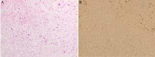
4 ERG in angiosarcoma in comparison with CD31 ERG in non epithelial Mesenchymal, Neuroectodermal and Hematopoietic tumors ERG in Epithelial Neoplams 4


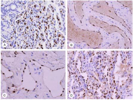
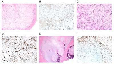
5 ERG in nonvascular tumors A hemorrhagic poorly differentiated carcinoma simulating angiosarcoma 5


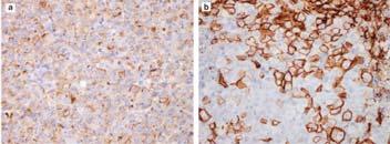
6 ERG From these studies ERG is positive in >95% of angiosarcomas, with a greater sensitivity compared to CD31 and CD34 ERG usually shows a diffuse pattern of nuclear staining, which facilitates its interpretation in this context. 6


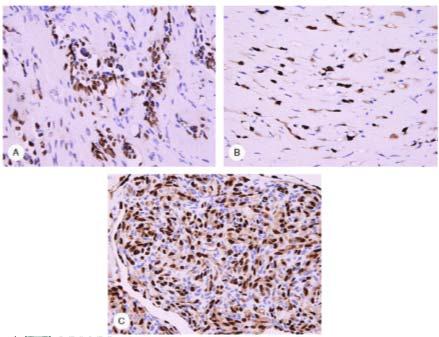
7 Ewing Sarcoma with EWSR1- ERG FLI1 ERG 7


8 Ewing sarcoma with EWSR1-FLI1 EWSR1, FLI1, ERG and their fusion proteins in Ewing Sarcoma 8

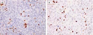
9 Leukemia cutis cases Reactive Leukocytic Infiltrates 9
10 10


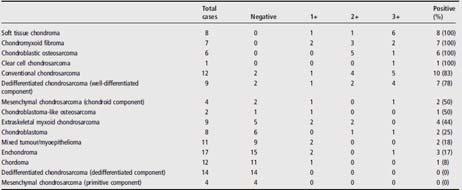
11 Soft Tissue Chondroma Convetional Chondrosarcoma ERG 11


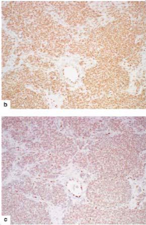
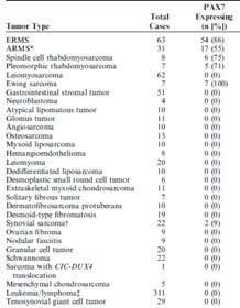
12 FLI1 ERG Conclusion ERG is therefore a useful marker for confirming endothelial differentiation in both benign and malignant neoplasms Potentially useful marker to distinguish Leukemia Cutis vs reactive myeloid infiltrates Expression can also be seen in a subset of epithelioid sarcomas and a small percentage of Ewing sarcomas, as well as approximately 45% to 50% of prostatic carcinomas. ERG can be seen in selected bone and soft tissue tumor with cartilaginous differentiation Outline ERG PAX7 DUX4-ETV4 BCOR Claudin 4 STAT6 MUC4 12

13 PAX7 Immunohistochemical detection in tumor cell nuclei of myogenin and MYOD1 currently benchmark for rhabdomyosarcoma diagnosis Sensitivity and specificity even in combination is imperfect Negative or focal myogenin in a considerable number of embryonal rhabdomyosarcomas MYOD1: high background and cytoplasmic staining PAX7 PAX7 is a paired box transcription factor required for mammalian skeletal muscle stem cells (aka satellite cells) It plays a critical role in mammalian myogenesis Controls early lineage specification, whereas MYOG and MYOD1 regulate subsequent lineage commitment 13
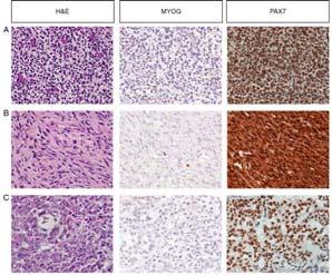
14 Embryonal Rhabdomyosarcoma 14
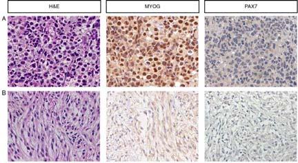
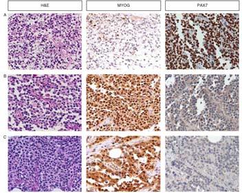
15 Embryonal Rhabdomyosarcoma Alveolar Rhabdomyosarcoma 15


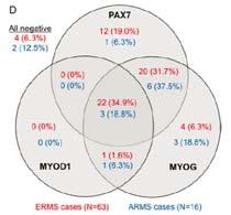
16 PAX7 and Rhabdomyosarcoma Potential diagnostic value of PAX7 IHC in the evaluation of rhabdomyosarcoma, especially embryonal rhabdomyosarcoma Less sensitive in ARMS Could be used secondarily in desmin+, MYOG/MYOD1 - cases 16


17 Ewing Sarcoma CIC-DUX4 PAX7 a sensitive marker for Ewing Sarcoma Positive in NKX2-2 negative cases Also positive in both common and variant forms of Ewing Sarcoma PAX7 expression differentiates Ewing Sarcoma from CIC-DUX4 sarcoma What accounts for the robust PAX7 expression in Ewing Sarcoma? EWSR1-FLI fusion protein binds at position 5 to the PAX7 promoter 17

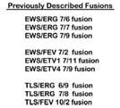
 STAT6 in SFT (NAB2-STAT6) FLI1 in Ewing Sarcoma (ESWR1-FLI1) BCOR in undifferentiated sarcomas PAX7 in ES exploits a characteristic transcriptional")
18 Undifferentiated small round cell or spindle-cell tumor PAX7 positivity: Rhabdo vs Ewing Sarcoma Positivity for desmin, MYOG, MYOD1 would strongly favor rhabdomyosarcoma Rare cases of ES can express myogenic features Diffuse membranous CD99 would favor ES Can be positive in rhabdomyosarcomas NKX2.2 would support ES WT1+, PAX7- would support CIC-DUX4 To date IHC focused on the aberrant expression of one of the genes involved in the pathogenic translocation WT1 in DSMRCT (EWSR1-WT1) STAT6 in SFT (NAB2-STAT6) FLI1 in Ewing Sarcoma (ESWR1-FLI1) BCOR in undifferentiated sarcomas PAX7 in ES exploits a characteristic transcriptional read-out of the diseasecausing translocation 18


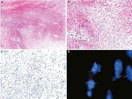
19 Outline ERG PAX7 DUX4 & ETV4 BCOR Claudin 4 STAT6 MUC4 DUX4 IHC Emerging undifferentiated sarcomas resembling but distinct from Ewing Sarcoma t(4;19) translocation involving CIC-DUX4 t(10;19) translocation CIC-DUX4L Distinct transcriptional signature with poor clinical outcomes 19


20 CD99 WT1 FISH CIC Ewing Sarcoma Malignant Rhabdoid Synovial Sarcoma Tumor 20
21 21


22 ETV4 (ETS Variant 4) Member of the PEA3 subgroup in the ETS transcription factor family Upregulation of WT1 and ETV1/4/5 in CICrearranged sarcomas 22
23 23

24 ETV4 and round cell sarcomas Sensitive but not entirely specific for CICrearranged sarcomas Diffuse, moderate-to-strong expression in ~90% sensitive and 95% specific Outline ERG PAX7 DUX4 & ETV4 BCOR Claudin 4 STAT6 MUC4 24


25 BCOR (BCL-6 interacting corepressor) A subset of undifferentiated round cell sarcomas: BCOR-CCNB3 BCOR-MAML3 ZC3H7B-BCOR YWHAE-NUTM2B BCOR internal tandem duplication (ITD) Clear cell sarcoma of the kidney Primitive myxoid mesenchymal tumor of infancy 25


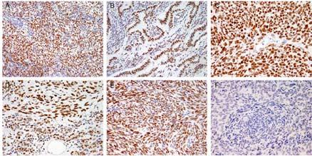
26 BCOR and Synovial Sarcoma 26
27 27


28 BCOR BCOR IHC is highly sensitive in identifying sarcomas with BCOR abnormality Triage for further molecular tests Avoid extensive IHC and molecular workups in small specimens Synovial sarcoma should be included in the DDx of round cell sarcomas with BCOR+ 28
29 Outline ERG PAX7 DUX4 & ETV4 BCOR Claudin 4 STAT6 MUC4 Claudin 4 and SWI/SNF complex-deficient tumors Claudins are integral components of tight junctions which for barriers in epithelial, endothelial and perineurial cells Claudin 4 is expressed in most epithelial cells and carcinomas Has been validated as a useful marker in the distinction of mesothelioma (lack of expression) from metastatic adenocarcinoma (consistently +) Claudin 4 It has been observed: Epithelial component of biphasic synovial sarcoma Subset of Desmoplastic Small Round Cell Tumor 29
 Nearly all cases of ovarian small cell carcinomas of hypercalcemic")
30 SWI/SNF complex Fundamental role in regulation of gene expression Tumor suppressor properties SMARCB1 (INI-1) SMARCA2 (BRM) SMARCA4 (BRG1) ARID1A SWI/SNF complex SMARCB1 deficiency (INI1 loss) Malignant Rhabdoid Tumor Epithelioid Sarcoma Subset of myoepithelial carcinomas Epithelioid MPNST SMARCA4 deficiency (BRG1 loss) Nearly all cases of ovarian small cell carcinomas of hypercalcemic type ARID1A deficiency ~half of ovarian clear cell carcinomas 30
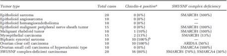
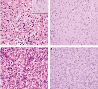
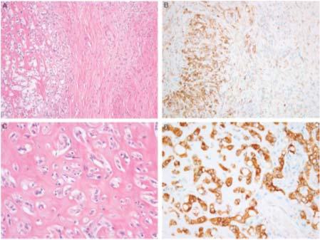
31 Epithelioid Sarcoma Epithelioid Angiosarcoma Epithelioid MPNST Biphasic Synovial Sarcoma 31


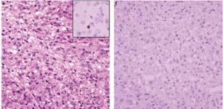
32 Ovarian Clear Cell Carcinoma Pancreatic Rhabdoid Carcinoma Cecal Rhabdoid Carcinoma INI1- deficient Sinonasal Carcinoma Claudin 4 It may serve as a useful diagnostic adjunct in the distinction of SWI/SNF complex deficient carcinomas from sarcomas with epithelioid morphology Strongly associated with true epithelial differentiation However absence of claudin 4 staining does not entirely exclude carcinoma 32


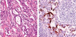
33 Keratin CD34 SMARCA4 33
 transcription factor")
34 Outline ERG PAX7 DUX4 & ETV4 BCOR Claudin 4 STAT6 MUC4 STAT6 and Solitaty Fibrous Tumor STAT6 is a member of the STAT family of cytoplasmic transcription factors, which regulate gene expression by transmitting signals to the nucleus and binding to specific DNA promoter sequences NAB2 is a transcriptional corepressor, a regulator of the early growth response 1 (EGR1) transcription factor 34


35 Variable truncation of the repressor domain of NAB2 with replacement by the transcriptional activation domain of STAT6 Subsequent translocation to the nucleus, where it acts as a transcriptional activator resulting in increased proliferation 35


36 Synovial sarcoma STAT6 Cellular spindle cell lipoma STAT6 36


37 Dedifferentiated liposarcoma Pitfall! Presence of STAT6 expression in approximately 7% to 15% of dedifferentiated liposarcomas The staining pattern in dedifferentiated liposarcoma is variable and may be focal or diffuse, weak or medium to strong in intensity, unlike the generally strong diffuse pattern seen in solitary fibrous tumor 37
38 Unlike the predominantly nuclear pattern of staining seen in solitary fibrous tumor, both cytoplasmic and nuclear expression is common in dedifferentiated liposarcoma. Diffuse expression of MDM2 and CDK4 helps favor a diagnosis of dedifferentiated liposarcoma If doubt persists, FISH for MDM2 amplification may also be useful FISH vs PCR Because STAT6 and NAB2 are located close together on the long arm of chromosome 12, FISH to demonstrate rearrangement of the genes is technically challenging and not diagnostically useful Outline ERG PAX7 DUX4 & ETV4 BCOR Claudin 4 STAT6 MUC4 38


 and")

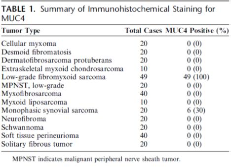
39 MUC4 MUC4 is a useful marker for low-grade fibromyxoid sarcoma (LGFMS) and sclerosing epithelioid fibrosarcoma 39
40 Sclerosing epithelioid sarcoma 40

41 Monophasic synovial sarcoma and MUC4 Sclerosing epithelioid sarcoma, MUC4 and carcinoma Importantly, regarding SEF, in which a poorly differentiated carcinoma may fall into the differential diagnosis it should be remembered that MUC4 expression is seen in a variety of different carcinomas, such as pancreaticobiliary carcinomas, breast carcinoma, and colonic adenocarcinoma. NEVER FORGET! The use of all of these markers requires careful clinical correlation and knowledge of the spectrum of staining in other tumor types, as no one marker is 100% sensitive or specific for a given diagnosis 41


42 KonstantinosLin 42
Cutaneous Mesenchymal Neoplasms with EWSR1 Rearrangement
 Cutaneous Mesenchymal Neoplasms with EWSR1 Rearrangement By Konstantinos Linos MD, FCAP, FASDP Bone, Soft Tissue and Dermatopathology Assistant Professor of Pathology Dartmouth-Hitchcock Medical Center
Financial disclosures
 Cutaneous Mesenchymal Neoplasms with EWSR1 Rearrangement By Konstantinos Linos MD, FCAP, FASDP Bone, Soft Tissue and Dermatopathology Assistant Professor of Pathology Dartmouth-Hitchc Geisel School of
Disclosures. An update on ancillary techniques in the diagnosis of soft tissue tumors. Ancillary techniques. Introduction
 Disclosures An update on ancillary techniques in the diagnosis of soft tissue tumors. I have nothing to disclose. Andrew Horvai, MD, PhD Clinical Professor, Pathology Introduction Ancillary techniques
Disclosure of Relevant Financial Relationships
 Ewing and Ewing like sarcomas Using Genetic Signatures in Refining Small Blue Round Cell Tumor Classification Cristina Antonescu, MD Department of Pathology Disclosure of Relevant Financial Relationships
Surgical Pathology Evening Specialty Conference USCAP 2015
 Surgical Pathology Evening Specialty Conference USCAP 2015 John R. Goldblum, M.D. Chairman, Department of Pathology, Cleveland Clinic Professor of Pathology, Cleveland Clinic Lerner College of Medicine
Clinical History. Pediatric Tumors with Involvement of the Head & Neck
 Pediatric Tumors with Involvement of the Head & Neck John Hicks Texas Children s Hospital Baylor College of Medicine Houston, TX NO DISCLOSURES Clinical History 10 Yr-Old Hispanic Male From Mexico with
Enterprise Interest Nothing to declare
 Enterprise Interest Nothing to declare Diagnoses one would not like to miss in soft tissue pathology early in your career Marta Sbaraglia, MD Department of Pathology Hospital of Treviso University of Padua
The last 10 years have seen the description of many
 An Update on the Application of Newly Described Immunohistochemical Markers in Soft Tissue Pathology George Lin, MD, PhD; Leona A. Doyle, MD Context. During the last 5 to 10 years, significant progress
Tumores de células pequeñas, redondas y azules: diagnóstico diferencial cuando el tiempo apremia
 Tumores de células pequeñas, redondas y azules: diagnóstico diferencial cuando el tiempo apremia Sílvia Bagué Servei de Patologia Hospital de Sant Pau Barcelona Soft tissue sarcomas Heterogeneous group
Molecular pathology in soft tissue tumors. Sylvia Höller Pathologie
 Molecular pathology in soft tissue tumors Sylvia Höller Pathologie When do we perform molecular testing? Morphology and IHC are not clearly fitting with an entity some translocations are entity specific
Immunohistochemistry in Bone and Soft Tissue Tumors. Sahar Rassi Zankoul, MD
 Immunohistochemistry in Bone and Soft Tissue Tumors Sahar Rassi Zankoul, MD Introduction Bone tumors represent a wide variety of tumors of various origins and malignant potentials. These different tumor
Molecular Diagnosis of Soft Tissue Tumors: Avoid Pitfalls
 Molecular Diagnosis of Soft Tissue Tumors: Avoid Pitfalls Cristina Antonescu, MD Department of Pathology Memorial Sloan-Kettering Cancer Center, New York Overview I. When should we rely on the help of
Klinisch belang van chromosomale translocatie detectie in sarcomen
 Translocations in sarcomas Klinisch belang van chromosomale translocatie detectie in sarcomen Judith V.M.G. Bovée, M.D., Ph.D. Department of Pathology Leiden University Medical Center RNA binding DNA binding
Rhabdomyomas and Rhabdomyosarcomas (RMS) David M. Parham, MD Chief of Anatomic Pathology
 David M. Parham, MD Chief of Anatomic Pathology") Rhabdomyomas and Rhabdomyosarcomas (RMS) David M. Parham, MD Chief of Anatomic Pathology Tumors of skeletal muscle: Rhabdomyomas and rhabdomyosarcomas Embryonal muscle 2 3 4 5 6 7 8 Rhabdomyoma Benign
Rhabdomyomas and Rhabdomyosarcomas (RMS) David M. Parham, MD Chief of Anatomic Pathology Tumors of skeletal muscle: Rhabdomyomas and rhabdomyosarcomas Embryonal muscle 2 3 4 5 6 7 8 Rhabdomyoma Benign
Lung Tumor Cases: Common Problems and Helpful Hints
 Lung Tumor Cases: Common Problems and Helpful Hints Brandon T. Larsen, MD, PhD Senior Associate Consultant Department of Laboratory Medicine and Pathology Mayo Clinic Arizona Arizona Society of Pathologists
Evening Specialty Conference Bone and Soft Tissue Pathology. Diagnostic pitfalls in bone and soft tissue pathology
 Evening Specialty Conference Bone and Soft Tissue Pathology. Case 1 Elizabeth G Demicco, MD, PhD Mount Sinai Hospital, New York Disclosure of Relevant Financial Relationships USCAP requires that all planners
Financial disclosures
 Mesenchymal Neoplasms with Melanocytic Differentiation By Konstantinos Linos MD, FCAP, FASDP Bone, Soft Tissue and Dermatopathology Assistant Professor of Pathology Dartmouth-Hitchcock Medical Center Geisel
Diplomate of the American Board of Pathology in Anatomic and Clinical Pathology
 A 33-year-old male with a left lower leg mass. Contributed by Shaoxiong Chen, MD, PhD Assistant Professor Indiana University School of Medicine/ IU Health Partners Department of Pathology and Laboratory
Recent Advances In Select Round Cell Sarcomas
 Recent Advances In Select Round Cell Sarcomas Rajiv M. Patel, M.D. Associate Professor of Pathology & Dermatology University of Michigan, Ann Arbor, MI rajivpat@med.umich.edu 1 Translocation Associated
An Overview of Genital Stromal Tumors
 An Overview of Genital Stromal Tumors By Konstantinos Linos MD, FCAP, FASDP Bone, Soft Tissue and Dermatopathology Assistant Professor of Pathology Dartmouth-Hitchcock Medical Center Geisel School of Medicine
Protocol for the Examination of Biopsy Specimens From Pediatric Patients With Ewing Sarcoma
 Protocol for the Examination of Specimens From Pediatric Patients With Ewing Sarcoma Version: EwingSarcoma 4.0.0.0 Protocol Posting Date: February 2019 Accreditation Requirements The use of this protocol
Pathology Mystery and Surprise
 Pathology Mystery and Surprise Tim Smith, MD Director Anatomic Pathology Medical University of South Carolina Disclosures No conflicts to declare Some problem cases Kidney tumor Scalp tumor Bladder tumor
Case 1. Disclosure. Imaging. Clinical history 5/10/2016. USCAP 2016 Annual Meeting Evening Specialty Conference Bone and Soft tissue Pathology
 Disclosure Dr. Agaram has nothing to disclose Case 1 Narsi Agaram, MBBS USCAP 2016 Annual Meeting Evening Specialty Conference Bone and Soft tissue Pathology Clinical history Imaging 1998 A three month
5/10. Pathology Soft tissue tumors. Farah Bhani. Mohammed Alorjani
 5/10 Pathology Soft tissue tumors Mohammed Alorjani Farah Bhani Slides are included in this sheet. Objectives: Soft tissue tumors 1. Describe soft tissue tumors. 2. Understand the classification of soft
NEW IHC A n t i b o d i e s
 NEW IHC Antibodies TABLE OF CONTENTS NEW IHC ANTIBODIES from Cell Marque CITED1 (5H6).... 1 Claudin 7 (5D10F3).... 1 GATA1 (4F5).... 1 Transgelin (2A10C2).... 1 NEW IHC ANTIBODIES using RabMAb Technology
Keywords solitary fibrous tumor, dedifferentiation, dedifferentiated solitary fibrous tumor, STAT6, GRIA2, cytokeratin, rhabdomyosarcomatous
 758452IJSXXX10.1177/1066896918758452International Journal of Surgical PathologyCreytens et al research-article2018 Pitfalls in Pathology Multifocal Cytokeratin Expression in a Dedifferentiated Solitary
Aspen conference on pediatric disease. July through August Bone and Soft Tissue Update. David M. Parham, MD. Rhabdomyoma and rhabdomyosarcoma
 Aspen conference on pediatric disease July through August 2014 Bone and Soft Tissue Update David M. Parham, MD Rhabdomyoma and rhabdomyosarcoma Embryonic rhabdomyogenesis is a highly conserved process
Technology from Abcam
 CD2 (EP222) CD2 is one of the earliest T-cell lineage restricted antigens to appear during T-cell differentiation and only rare CD2+ cells can be found in the bone marrow. Anti-CD2 is a pan-t-cell antigen
Diagnostic Value of Immunohistochemistry in Soft Tissue Tumors
 Original Article DOI: 10.21276/APALM.1637 Diagnostic Value of Immunohistochemistry in Soft Tissue Tumors Sridevi. V*., Susruthan Muralitharan., and Thanka. J Dept of Pathology, SriMuthukumaran Medical
Spindle Cell Lesions Of The Breast. Emad Rakha Professor of Breast Pathology and Consultant Pathologist
 Spindle Cell Lesions Of The Breast Emad Rakha Professor of Breast Pathology and Consultant Pathologist * SCLs comprise a wide spectrum of diseases, ranging from reactive processes to aggressive malignant
3/27/2017. Disclosure of Relevant Financial Relationships
 Ophthalmic Pathology Evening Specialty Conference USCAP 2017 5 th March, 2017 Mukul K. Divatia, MD Assistant Professor Department of Pathology & Genomic Medicine Weill Cornell Medical College Houston Methodist
USCAP COMPANION MEETING INTERNATIONAL SOCIETY OF BONE AND SOFT TISSUE PATHOLOGY DENVER, March 2 nd 2008
 1 USCAP COMPANION MEETING INTERNATIONAL SOCIETY OF BONE AND SOFT TISSUE PATHOLOGY DENVER, March 2 nd 2008 THE EVOLUTION OF SOFT TISSUE TUMOUR TAXONOMY: WHAT STILL NEEDS TO BE DONE? Christopher D.M. Fletcher,
Mesothelioma Pathobasic. Lukas Bubendorf Pathology
 Mesothelioma Pathobasic Lukas Bubendorf Pathology Mechanisms of Asbestos Carcinogenesis in Mesothelioma Asprin High-mobility group protein B1 master switch HMGB1 Initiation/ perpetuation of inflamm. response
ESS: Pathologic Insights
 GEIS XVI INTERNATIONAL SYMPOSIUM Seville 4th October 2018 ESS: Pathologic Insights Sílvia Bagué The Royal Marsden Hospital London (United Kingdom) I have no conflicts of interest Endometrial stromal sarcoma
57th Annual HSCP Spring Symposium 4/16/2016
 An Unusual Malignant Spindle Cell Lesion to Involve the Breast Erinn Downs-Kelly, D.O. Associate Professor of Pathology University of Utah & ARUP Laboratories No disclosures Case 39 y/o female with no
An Overview of Cutaneous Vascular Neoplasms
 An Overview of Cutaneous Vascular Neoplasms By Konstantinos Linos MD, FCAP, FASDP Bone, Soft Tissue and Dermatopathology Assistant Professor of Pathology Dartmouth-Hitchcock Medical Center Geisel School
* I have no disclosures or any
 Howard Rosenthal, M.D. Associate Professor of Orthopedic Surgery University of Kansas Sarcoma Center I have no disclosures or any conflicts related to the content of this presentation. Objectives 1. Describe
RARE TUMORS OF INFANCY. RAJKUMAR VENKATRAMANI, MD, MS Director, Rare Tumors Program, Texas Children s Hospital
 RARE TUMORS OF INFANCY RAJKUMAR VENKATRAMANI, MD, MS Director, Rare Tumors Program, Texas Children s Hospital OBJECTIVES Review the epidemiology and clinical presentation of soft tissue sarcomas in infancy.
Fluorescence In Situ Hybridization in the Diagnosis of Soft Tissue Neoplasms: A Review. Munir R. Tanas, MD and John R.
 REVIEW ARTICLE Fluorescence In Situ Hybridization in the Diagnosis of Soft Tissue Neoplasms: A Review Munir R. Tanas, MD and John R. Goldblum, MD Abstract: This paper presents an overview of the role of
Predictive biomarker profiling of > 1,900 sarcomas: Identification of potential novel treatment modalities
 Predictive biomarker profiling of > 1,900 sarcomas: Identification of potential novel treatment modalities Sujana Movva 1, Wenhsiang Wen 2, Wangjuh Chen 2, Sherri Z. Millis 2, Margaret von Mehren 1, Zoran
Classification (1) Classification (3) Classification (2) Spindle cell lesions. Spindle cell lesions of bladder (Mills et al.
 Classification (3) Classification (2) Spindle cell lesions. Spindle cell lesions of bladder (Mills et al.") Non-epithelial tumours and nonepithelial tumour-like lesions of the bladder Dr Jonathan H Shanks The Christie NHS Foundation Trust, Manchester, UK Classification (1) Myofibroblastic proliferations and
Non-epithelial tumours and nonepithelial tumour-like lesions of the bladder Dr Jonathan H Shanks The Christie NHS Foundation Trust, Manchester, UK Classification (1) Myofibroblastic proliferations and
Contents Part I Introduction 1 General Description 2 Natural History: Importance of Size, Site, Histopathology
 Contents Part I Introduction 1 General Description... 3 1.1 Introduction... 3 1.2 Incidence and Prevalence... 5 1.3 Predisposing and Genetic Factors... 8 References... 16 2 Natural History: Importance
Aspen 2014: Cases 1-4. John Hicks Texas Children s Hospital Baylor College of Medicine Houston, Tx. Case 1 History
 Aspen 2014: Cases 1-4 John Hicks Texas Children s Hospital Baylor College of Medicine Houston, Tx Case 1 History 2-month old full-term female presented with respiratory distress for 5 days with stridor.
Case Presentations 17-20
 Case Presentations 17-20 Case Presentations 17 Case 17 2 year old boy with history of urinary retention and prostate biopsy. After biopsy and chemotherapy, child regains urine flow but the mass does not
Introduction to Musculoskeletal Tumors. James C. Wittig, MD Orthopedic Oncologist Sarcoma Surgeon
 Introduction to Musculoskeletal Tumors James C. Wittig, MD Orthopedic Oncologist Sarcoma Surgeon www.tumorsurgery.org Definitions Primary Bone / Soft tissue tumors Mesenchymally derived tumors (Mesodermal)
Pathology of Sarcoma ELEANOR CHEN, MD, PHD, ASSISTANT PROFESSOR DEPARTMENT OF PATHOLOGY UNIVERSITY OF WASHINGTON
 Pathology of Sarcoma ELEANOR CHEN, MD, PHD, ASSISTANT PROFESSOR DEPARTMENT OF PATHOLOGY UNIVERSITY OF WASHINGTON Presentation outline Background and epidemiology of sarcomas Sarcoma classification Sarcoma
SYLLABUS SWI/SNF-dependent tumors
 SYLLABUS SWI/SNF-dependent tumors F Le Loarer, MD, PhD Centre Leon Berard, Department of Pathology, Lyon, FRANCE Since the seminal description in 1999 of recurrent inactivation of SMARCB1- which encodes
The Genetics of Myoepithelial Tumors: salivary glands, soft tissue and bone
 The Genetics of Myoepithelial Tumors: salivary glands, soft tissue and bone Cristina Antonescu, MD Memorial Sloan-Kettering Cancer Center, New York Nothing to declare Disclosure Spectrum of Myoepithelial
Molecular Genetics of Paediatric Tumours. Gino Somers MBBS, BMedSci, PhD, FRCPA Pathologist-in-Chief Hospital for Sick Children, Toronto, ON, CANADA
 Molecular Genetics of Paediatric Tumours Gino Somers MBBS, BMedSci, PhD, FRCPA Pathologist-in-Chief Hospital for Sick Children, Toronto, ON, CANADA Financial Disclosure NanoString - conference costs for
Cheryl M. Coffin, M.D. Goodpasture Professor of Pathology, Microbiology, and Immunology Vanderbilt University Nashville, TN, USA
 Cutaneous Mesenchymal Tumors in Childhood Cheryl M. Coffin, M.D. Goodpasture Professor of Pathology, Microbiology, and Immunology Vanderbilt University Nashville, TN, USA I. Introduction Cutaneous tumors
I sarcomi dei tessuti molli
 Novità e sequenze terapeutiche nelle neoplasie ginecologiche, melanoma e tumori rari: I sarcomi dei tessuti molli Giacomo G. Baldi Oncologia Medica Sandro Pitigliani Nuovo Ospedale S.Stefano Azienda USL
Journal of Solid Tumors, April 2012, Vol. 2, No. 2
 ORIGINAL ARTICLE Utility of fluorescence in situ hybridization in subclassifying unclassified high-grade sarcomas: A study of 40 cases using break-apart probes of EWSR1, FOXO1A, SS18 and DDIT3 genes Alfredo
Biopsy Interpretation of Spindle cell proliferations of the Serosa
 Biopsy Interpretation of Spindle cell proliferations of the Serosa Richard Attanoos, Cardiff. U.K. Disclosure of Relevant Financial Relationships USCAP requires that all planners (Education Committee)
ACCME/Disclosures ALK FUSION-POSITIVE MESENCHYMAL TUMORS. Tumor types with ALK rearrangements. Anaplastic Lymphoma Kinase. Jason L.
 Companion Meeting of the International Society of Bone and Soft Tissue Pathology The Evolving Concept of Mesenchymal Tumors ALK FUSION-POSITIVE MESENCHYMAL TUMORS Jason L. Hornick, MD, PhD March 13, 2016
Mesothelioma: diagnostic challenges from a pathological perspective. Naseema Vorajee August 2016
 Mesothelioma: diagnostic challenges from a pathological perspective Naseema Vorajee August 2016 Naseema.vorajee@nhls.ac.za Pleural diseases (whether neoplastic, reactive or infective) may have similar
What is New in the 2015 WHO Lung Cancer Classification? Zhaolin Xu, MD, FRCPC, FCAP
 What is New in the 2015 WHO Lung Cancer Classification? Zhaolin Xu, MD, FRCPC, FCAP Professor, Dept of Pathology, Dalhousie University, Canada Pulmonary Pathologist and Cytopathologist, QEII HSC Senior
Affiliazione autori0. Riccardo Ricci Journal Club GIPAD, settore GIST Anatomia Patologica, Università Cattolica, Roma
 GIST Manifesting as a Retroperitoneal Tumor: Clinicopathologic Immunohistochemical, and Molecular Genetic Study of 112 Cases American Journal of Surgical Pathology, 2017, 41:577-585 Miettinen M*; Felisiak-Golabek
Problem 1: Differential of Neuroendocrine Carcinoma 3/23/2017. Disclosure of Relevant Financial Relationships
 Differential of Neuroendocrine Carcinoma Alain C. Borczuk,MD Weill Cornell Medicine Disclosure of Relevant Financial Relationships USCAP requires that all faculty in a position to influence or control
Effective January 1, 2018 ICD O 3 codes, behaviors and terms are site specific
 Effective January 1, 2018 codes, behaviors and terms are site specific /N 8551/3 Acinar adenocarcinoma (C34. _) Lung primaries diagnosed prior to 1/1/2018 use code 8550/3 For prostate (all years) see 8140/3
Grading of Bone Tumors
 Grading of Bone Tumors Joon Hyuk Choi, M.D. Department of Pathology College of Medicine, Yeungnam University Introduction to grading system of bone tumor used at Mayo Clinic WHO Histologic Classification
Effective January 1, 2018 ICD O 3 codes, behaviors and terms are site specific
 Effective January 1, 2018 codes, behaviors and terms are site specific Status /N 8010/3 Urachal carcinoma (C65.9, C66.9, C67. _, C68._) 8013/3 Combined large cell neuroendocrine carcinoma (C34. _, C37.9)
Shintaro Sugita *, Hiroko Asanuma and Tadashi Hasegawa
 Sugita et al. Diagnostic Pathology (2016) 11:37 DOI 10.1186/s13000-016-0486-2 RESEARCH Open Access Diagnostic use of fluorescence in situ hybridization in expert review in a phase 2 study of trabectedin
Mayo Medical Laboratories
 Mayo Medical Laboratories Virtual Lectures 2014 MFMER 2016 MFMER slide-1 Virtual Lectures Planning Committee Disclosure Summary As a provider accredited by ACCME, College of Medicine, Mayo Clinic (Mayo
From Morphology to Molecular Pathology: A Practical Approach for Cytopathologists Part 1-Cytomorphology. Songlin Zhang, MD, PhD LSUHSC-Shreveport
 From Morphology to Molecular Pathology: A Practical Approach for Cytopathologists Part 1-Cytomorphology Songlin Zhang, MD, PhD LSUHSC-Shreveport I have no Conflict of Interest. FNA on Lymphoproliferative
Part 1. Slides 1-38, Rita Alaggio Soft tissue tumors Trondheim 14. mars 2013
 Part 1 Slides 1-38, Rita Alaggio Soft tissue tumors Trondheim 14. mars 2013 Pediatric Pathology Soft Tissue Tumors AN UPDATE Rita Alaggio Azienda Ospedaliera Università di Padova Soft Tissue Tumors More
IMMUNOHISTOCHEMISTRY IN THE DIAGNOSIS OF SOFT TISSUE TUMORS
 IMMUNOHISTOCHEMISTRY IN THE DIAGNOSIS OF SOFT TISSUE TUMORS Nicolas de Saint Aubain Somerhausen Institut Jules Bordet / Hôpital Erasme nicolas.desaintaubain@synet.be ForPath 2005 1 I. Ancillary techniques
Musculoskeletal Sarcomas
 Musculoskeletal Sarcomas Robert C. Orth, M.D., Ph.D. Edward B. Singleton Department of Pediatric Radiology Texas Children s Hospital Page 0 xxx00.#####.ppt 9/23/2012 9:01:18 AM No disclosures Page 1 xxx00.#####.ppt
Disclosure of Relevant Financial Relationships
 Neuropathology Evening Specialty Conference Disclosure of Relevant Financial Relationships The USCAP requires that anyone in a position to influence or control the content of all CME activities disclose
2018 ICD-O-3 Updates in Table Format with Annotation for Reference
 Status Histology Description (this may be preferred term or a synonym) Report Comments New term 8010 3 Urachal carcinoma (C65.9, C66.9, C67._, C68._) New term 8013 3 Combined large cell neuroendocrine
Case 2. Dr. Sathima Natarajan M.D. Kaiser Permanente Medical Center Sunset
 Case 2 Dr. Sathima Natarajan M.D. Kaiser Permanente Medical Center Sunset History 24 year old male presented with a 3 day history of right flank pain, sharp in nature Denies fever, chills, hematuria or
Newer soft tissue entities
 Newer soft tissue entities Examples among fibroblastic tumors Turku, May 6, 2010 Markku Miettinen, M.D. AFIP, Washington, DC Fibroblastic neoplasms Solitary fibrous tumor /Hemangiopericytoma Low-grade
4/12/2018. MUSC Pathology Symposium Kiawah Island April 18, Jesse K. McKenney, MD
 MUSC Pathology Symposium Kiawah Island April 18, 2018 Jesse K. McKenney, MD 1 Urothelial Carcinoma with Alternative Differentiation 2 Urothelial Carcinoma with Alternative Differentiation Recognition as
Cancers of unknown primary : Knowing the unknown. Prof. Ahmed Hossain Professor of Medicine SSMC
 Cancers of unknown primary : Knowing the unknown Prof. Ahmed Hossain Professor of Medicine SSMC Definition Cancers of unknown primary site (CUPs) Represent a heterogeneous group of metastatic tumours,
Novel uses of immunohistochemistry in the diagnosis and classification of soft tissue tumors
 & 2014 USCAP, Inc. All rights reserved 0893-3952/14 $32.00 S47 Novel uses of immunohistochemistry in the diagnosis and classification of soft tissue tumors Jason L Hornick Department of Pathology, Brigham
Soft Tissue High Grade Myoepithelial Carcinoma With Round Cell Morphology: Report Of A Newly Described Entity With EWSR1 Gene Rearrangement
 Soft Tissue High Grade Myoepithelial Carcinoma With Round Cell Morphology: Report Of A Newly Described Entity With EWSR1 Gene Rearrangement Abstract M. El-Kabany, R. Al-Abdulghani, A. E. Ali, I. M. Francis,
Dystrophin Is a Tumor Suppressor in Human Cancers with Myogenic Programs
 SUPPLEMENTARY INFORMATION Dystrophin Is a Tumor Suppressor in Human Cancers with Myogenic Programs Yuexiang Wang 1, Adrian Marino-Enriquez 1, Richard R. Bennett 2, Meijun Zhu 1, Yiping Shen 3,4, Grant
Cerebral Parenchymal Lesions: I. Metastatic Neoplasms
 Chapter 4 Cerebral Parenchymal Lesions: I. Metastatic Neoplasms After one has reasonably ruled out the possibility of a nonneoplastic diagnosis (see Chap. 3), one is left with considering a diagnosis of
A 25 year old female with a palpable mass in the right lower quadrant of her abdomen
 May 2016 A 25 year old female with a palpable mass in the right lower quadrant of her abdomen Contributed by: Paul Ndekwe, MD, Resident Physician, Indiana University School of Department of Pathology and
Original Articles. Utilization of Fluorescence In Situ Hybridization in the Diagnosis of 230 Mesenchymal Neoplasms. An Institutional Experience
 Original Articles Utilization of Fluorescence In Situ Hybridization in the Diagnosis of 230 Mesenchymal Neoplasms An Institutional Experience Munir R. Tanas, MD; Brian P. Rubin, MD, PhD; Raymond R. Tubbs,
Applications of IHC. Determination of the primary site in metastatic tumors of unknown origin
 Applications of IHC Determination of the primary site in metastatic tumors of unknown origin Classification of tumors that appear 'undifferentiated' by standard light microscopy Precise classification
number Done by Corrected by Doctor Maha Shomaf
 number 16 Done by Waseem Abo-Obeida Corrected by Zeina Assaf Doctor Maha Shomaf MALIGNANT NEOPLASMS The four fundamental features by which benign and malignant tumors can be distinguished are: 1- differentiation
Slide Seminar Spanish Society of Pathology
 Slide Seminar Spanish Society of Pathology John R. Goldblum, M.D. Chairman, Department of Anatomic Pathology Cleveland Clinic Professor of Pathology Cleveland Clinic Lerner College of Medicine 1921 Original
HOW MAY THE CLASSIFICATION OF SOFT TISSUE TUMORS EVOLVE?
 Spanish Society of Pathology Zaragoza, May 2011 ARTHUR PURDY STOUT SYMPOSIUM HOW MAY THE CLASSIFICATION OF SOFT TISSUE TUMORS EVOLVE? Christopher D.M. Fletcher, M.D., FRCPath Brigham and Women s Hospital
Notice of Faculty Disclosure
 Mesenchymal Tumors of the Vulva: Old, New, Something(s) Different Napa Valley Conference Pathology Education Partners Inc May 15, 2018 Teri A. Longacre, M.D. longacre@stanford.edu Stanford University,
Role of immunohistochemistry in the differential diagnosis of malignant small round cell tumor: a study of 38 cases
 International Journal of Research in Medical Sciences Patel A et al. Int J Res Med Sci. 2015 Dec;3(12):3833-3839 www.msjonline.org pissn 2320-6071 eissn 2320-6012 Research Article DOI: http://dx.doi.org/10.18203/2320-6012.ijrms20151452
Paediatric small round cell sarcomas an update
 1 Paediatric small round cell sarcomas an update International Society of Bone and Soft Tissue Pathology meeting, Baltimore, MD March 3, 2013 David M. Parham, MD Oklahoma University Health Science Center
A neoplasm is defined as "an abnormal tissue proliferation, which exceeds that of adjacent normal tissue. This proliferation continues even after
 NEOPLASIA Neoplasia is a very important topic in pathology because neoplasms are both common and serious diseases. A neoplasm literally means a new growth, and this term is used interchangeably with a
Breast - ductal carcinoma CK7 ER PR GATA3 Mammaglobin (50-70%) GCDFP-15 (50-70%) E-cadherin HMWCK CK20 PAX2 ER/PR/HER2 on all newly diagnosed cases
 GCDFP-15 (50-70%) E-cadherin HMWCK CK20 PAX2 ER/PR/HER2 on all newly diagnosed cases") Adrenal cortical carcinoma Inhibin Synap Melan-A Calretinin Vimentin Chromogr CK7 CK20 Breast - ductal carcinoma CK7 ER PR GATA3 Mammaglobin (50-70%) GCDFP-15 (50-70%) E-cadherin HMWCK CK20 PAX2 ER/PR/HER2
Adrenal cortical carcinoma Inhibin Synap Melan-A Calretinin Vimentin Chromogr CK7 CK20 Breast - ductal carcinoma CK7 ER PR GATA3 Mammaglobin (50-70%) GCDFP-15 (50-70%) E-cadherin HMWCK CK20 PAX2 ER/PR/HER2
2 Berkeley Street, Suite 403, Toronto, Ontario M5A 2W3 Visit us at: Tel: Fax:
 E-Path A.I. Engine Knowledge Base Enhancements Version 1.0.0.29 April 1, 2018 The major enhancements in the E-Path Knowledge Base from versions 1.0.0.28 through 1.0.0.29 are as follows: 1. Addition/modification
Molecular pathology/genetics of sarcomas
 Molecular pathology/genetics of sarcomas Gunhild Mechtersheimer Institute of Pathology, University of Heidelberg Sarkomkonferenz: 17.03.2011 Berlin Characterization of soft tissue sarcomas / STS (~ 1%
Case of the month. Dr Charles Bénière, Institut universitaire de pathologie, Lausanne
 Case of the month Dr Charles Bénière, Institut universitaire de pathologie, Lausanne Clinical history 39 years old male, smoker (19 pack-year) without any prior medical record nor professional exposure.
Gross appearance of peritoneal cysts. They have a thin, translucent wall and contain a clear fluid.
 Gross appearance of peritoneal cysts. They have a thin, translucent wall and contain a clear fluid. So-called multicystic benign mesothelioma. A, Gross appearance. So-called multicystic benign mesothelioma.
Selected Pseudomalignant Soft Tissue Tumors of the Skin and Subcutis
 Selected Pseudomalignant Soft Tissue Tumors of the Skin and Subcutis Andrew L. Folpe, M.D. Professor of Laboratory Medicine and Pathology Mayo Clinic, Rochester, MN folpe.andrew@mayo.edu 2016 MFMER slide-1
Mojca Velikonja Jože Pižem
 Mojca Velikonja Jože Pižem An 81-year old woman presented with an exophytic, wart-like skin lesion on her neck that she had observed for one year. Cryotherapy had been applied twice, but proved unsuccessful.
Neoplasia part I. Dr. Mohsen Dashti. Clinical Medicine & Pathology nd Lecture
 Neoplasia part I By Dr. Mohsen Dashti Clinical Medicine & Pathology 316 2 nd Lecture Lecture outline Review of structure & function. Basic definitions. Classification of neoplasms. Morphologic features.
Conceptual Evolution of Soft Tissue Tumors Classification
 Conceptual Evolution of Soft Tissue Tumors Classification Angelo P. Dei Tos M.D. Departments of Pathology & Oncology Treviso, Italy How WHO classification was reshaped Pathologists and Cytogeneticists
Update on Cutaneous Mesenchymal Tumors. Thomas Brenn
 Update on Cutaneous Mesenchymal Tumors Thomas Brenn Cutaneous Mesenchymal Tumours Wide morphological and biological spectrum Myofibroblastic, smooth muscle, neural, vascular, apidocytic, undifferentiated;
LOOK-ALIKES IN SPINDLE AND EPITHELIOID TUMORS: Immunohistochemistry. Cytogenetics Flow cytometry Molecular diagnostics
 LOOK-ALIKES IN SPINDLE AND EPITHELIOID TUMORS: Ultrastructural value and pitfalls in diagnosis Guillermo A Herrera Department of Pathology and Translational Pathobiology Louisiana State University Health
GUT-C 11/30/2017. Debasmita Das, M.D. PGY-1 Danbury Hospital
 GUT-C 11/30/2017 Debasmita Das, M.D. PGY-1 Danbury Hospital CLINICAL SUMMARY 8/2017 59 year old female Presented to the ED with 1 month history of general malaise, fever and weight loss PMH: Significant