The unknown primary tumour: IHC classification part I, the primary panel - Antibody selection, protocol optimization, controls and EQA
|
|
|
- Margery Carson
- 5 years ago
- Views:
Transcription
1 The unknown primary tumour: IHC classification part I, Mogens Vyberg Professor of Clinical Pathology Director of NordiQC Aalborg University Hospital, Aalborg, Denmark the primary panel - Antibody selection, protocol optimization, controls and EQA


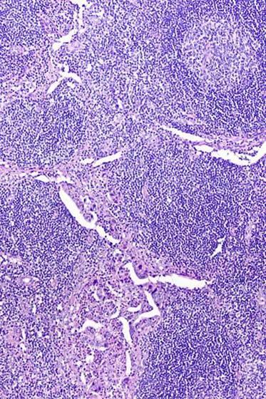
2 Tumours of unknown origin: Histology Brain tumour - biopsy


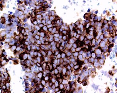
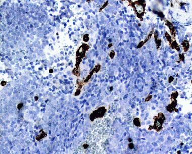
3 CD45 VIM Tumours of unknown origin: Immunohistochemistry Pan-CK S-100
4
5
6
7
8
9
10 ?????


11 IHC classification of the Unknown Primary Tumour Pathologist - knowledge, acceptance, skill Tumour material - diagnostic markers Antibodies available - applic. in diagnostic algorithms Methods - protocol: sensitivity, specificity, reliability - interpretation: cut-off level for positivity clinical relevance ~50% GIST: S-100B Protease ~50% GIST: S-100B MWO


12 IHC classification of the Unknown Primary Tumour Pathologist - knowledge, acceptance, skill Tumour material - diagnostic markers Antibodies available - applic. in diagnostic algorithms Methods - protocol: sensitivity, specificity, reliability - interpretation: cut-off level for positivity clinical relevance positive = any staining CK 20 negative = < 50% stained cells CK 20
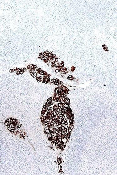
13 Primary panel for the unknown primary tumour Haematolymphoid CD45 Pan-CK S-100 VIM +/(-) -/(+) -/(+) +/(-) Epithelial - +/(-) -/+ -/+ Mesothelial Mesenchymal and neuronal Non-neuronal neuroepithelial - -/(+) -/ /(+) + + Germ cell - -/+ -/+ +
14 CD45 - Leucocyte common antigen (LCA) Transmembrane protein tyrosin phosphatase essential for haematopoietic signal transduction and cell activation Membrane associated component: 5 isotypes Intracellular component: one common type
15 CD45 - Leucocyte common antigen (LCA) Large majority of haematolymphoid cells and Lost in maturing erythocytes, megakaryocytes and plasmacells Never found in non-haematolymphoid cells and


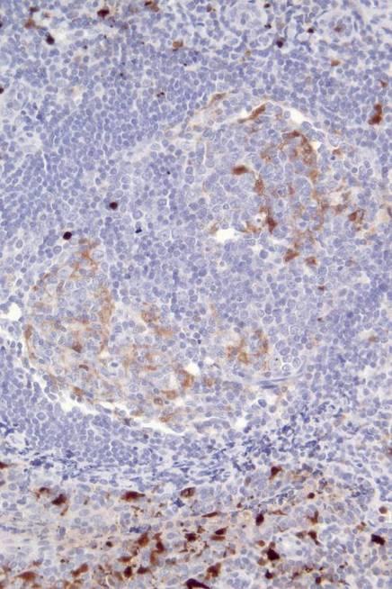
16 CD45 - Leucocyte common antigen (LCA) Normal lymph node Malignant lymphoma
 Liver")

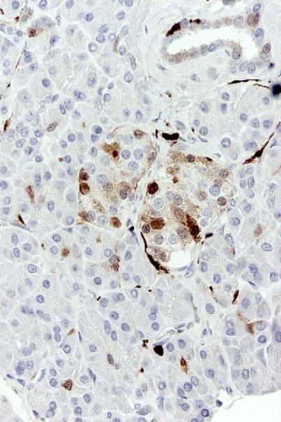
17 CD45 - Leucocyte common antigen (LCA) Liver Brain Kupffer cells: Critical assay performance control
18 CD45 NordiQC run assessment-cd45.htm Which is best?
19 CD45 NordiQC run assessment-cd45.htm Optimal Insufficient


20 Optimal Insufficient CD45 NordiQC run 37 B-CLL assessment-cd45.htm 56% of labs 18% of labs
21 CD45 NordiQC run % 82%
22 CD45 NordiQC run % 82% They followed the vendor s protocol recommendation: no HIER

 CD45 RO ~")

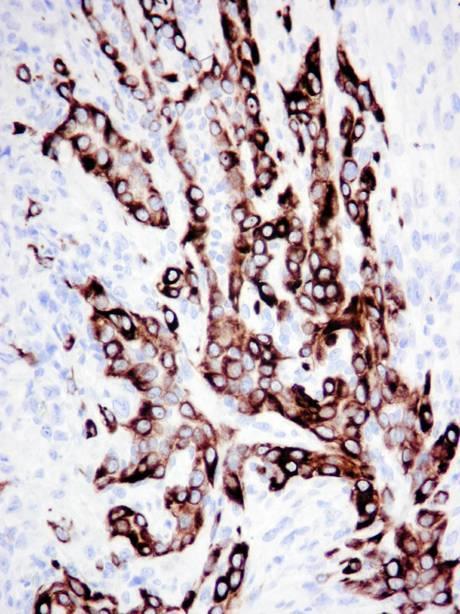
23 CD45 - Leucocyte common antigen (LCA) CD45 RO ~ T-cells Lymph node/tonsil CD45 RA ~ B-cells


24 CD45 - Leucocyte common antigen (LCA)


25 CD45 - Leucocyte common antigen (LCA)

26 CD45 - Leucocyte common antigen (LCA)
27 Primary panel for the unknown primary tumour Haematolymphoid CD45 Pan-CK S-100 VIM +/(-) -/(+) -/(+) +/(-) Epithelial - +/(-) -/+ -/+ Mesothelial Mesenchymal and neuronal Non-neuronal neuroepithelial - -/(+) -/ /(+) + + Germ cell - -/+ -/+ +
 # Microtubuli")
28 Cellular filaments # Microfilaments: (6 nm) # Intermediate filaments (7-11 nm) # Microtubuli (23 nm)
 and microtubules (23 nm) Important for mechanical strength and cellular")
29 Intermediate filaments Group of mainly cytoplasmic filaments 7 11 nm in diameter Part of the cytoskeleton in virtually all cells, creating as meshwork and connecting nuclear membrane with cell membrane Often associated with microfilaments (6 nm) and microtubules (23 nm) Important for mechanical strength and cellular functions

30 Intermediate filaments - 5 classes I acidic cytokeratins II basic-neutral cytokeratins III vimentin, desmin, glial fibrillary acidic protein, peripherin IV neurofilament protein, α-internexin, nestin V lamins

31 Cytokeratins as tonofilaments Cytokeratin intermediate filaments attached to desmosomes Drochmans et al. J Cell Biol. 1978, 79:427
32 Cytokeratins in diagnostic pathology Cytokeratins (CKs) belong to the most fundamental markers of epithelial differentiation CKs comprise a large family of subtypes. Different cell types express different patterns of CK subtypes Cancers generally express CK patterns that at least in part represent the pattern of the putative cell of origin Metastases express CK patterns fairly concordant with those of the primary tumours
33 HE CK Micrometastases identified by cytokeratin


34 HE-frys CK-frys Carcinoma in frosen section identified by cytokeratin


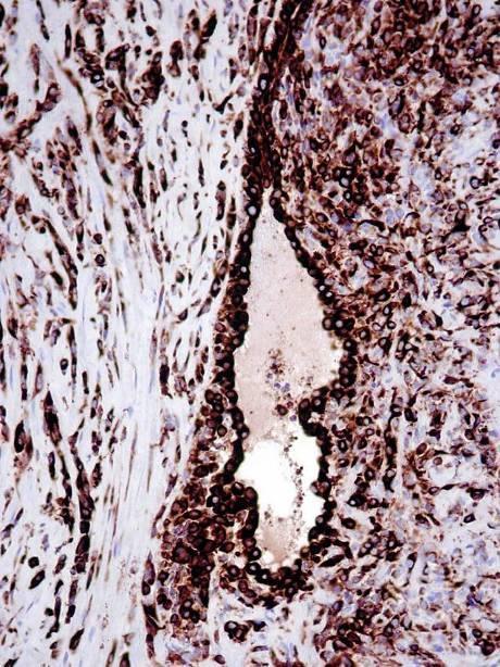
35 Low molecular weight cytokeratins in carcinomas Carcinomas always LMW-CK-positive, except some cases of Renal cell carcinoma Adrenal cortical carcinoma Small cell carcinoma CK8: Adrenal cortical carcinoma CK8: Renal cell carcinoma CK: renal cell carcinoma
36 Primary panel for the unknown primary tumour Haematolymphoid CD45 Pan-CK S-100 VIM +/(-) -/(+) -/(+) +/(-) Epithelial - +/(-) -/+ -/+ Mesothelial Mesenchymal and neuronal Non-neuronal neuroepithelial - -/(+) -/ /(+) + + Germ cell - -/+ -/+ +


37 Cytokeratins in non-epithelial tumours CK8 CD20 & CD3 42 y, tumour infiltrating retroperitoneum Malignant lymphoma! CD45 & CD79a
38 Primary panel for the unknown primary tumour Haematolymphoid CD45 Pan-CK S-100 VIM +/(-) -/(+) -/(+) +/(-) Epithelial - +/(-) -/+ -/+ Mesothelial Mesenchymal and neuronal Non-neuronal neuroepithelial - -/(+) -/ /(+) + + Germ cell - -/+ -/+ +
39 Cytokeratins in malignant mesothelioma CK8 CK5
40 Primary panel for the unknown primary tumour Haematolymphoid CD45 Pan-CK S-100 VIM +/(-) -/(+) -/(+) +/(-) Epithelial - +/(-) -/+ -/+ Mesothelial Mesenchymal and neuronal Non-neuronal neuroepithelial - -/(+) -/ /(+) + + Germ cell - -/+ -/+ +


41 Cytokeratins in sarcomas Synovial sarcoma angiosarcoma
42 Primary panel for the unknown primary tumour Haematolymphoid CD45 Pan-CK S-100 VIM +/(-) -/(+) -/(+) +/(-) Epithelial - +/(-) -/+ -/+ Mesothelial Mesenchymal and neuronal Non-neuronal neuroepithelial - -/(+) -/ /(+) + + Germ cell - -/+ -/+ +
43 Cytokeratins in malignant melanoma CK
44 Primary panel for the unknown primary tumour Haematolymphoid CD45 Pan-CK S-100 VIM +/(-) -/(+) -/(+) +/(-) Epithelial - +/(-) -/+ -/+ Mesothelial Mesenchymal and neuronal Non-neuronal neuroepithelial - -/(+) -/ /(+) + + Germ cell - -/+ -/+ +


45 Cytokeratins in germ cell tumours CK8: Seminoma CK8: Embr. carcinoma

46 Cytokeratins: proteolysis causes false negativity TP FN HIER Proteolysis PAN-CK - AE1/AE3 clone cocktail: AE1 detects CK8 after HIER only AE1 does not detect CK18 AE3 neither detects CK8 or CK18 SCLC Proteolysis CK: renal cell carcinoma
47 Cytokeratins: retrieval causing false negativity TP FN HIER Proteolysis RCC TP CK: renal cell carcinoma FN

48 Misleading datasheets Giving false negative results when only LMW-CKs are present 48
49 Misleading datasheets
50 IHC - NordiQC 2016 AE1/AE3 : Optimal results only obtained by HIER in NordiQC runs AE1/AE3/PCK26: Optimal results mainly obtained by HIER+protelysis Dako: RTU HIER Leica: RTU Proteolysis Thermo: Conc: Proteolysis or HIER Conc: HIER Conc: HIER Quanto Proteolysis UltraVision Misleading data sheets + Wrong control material used!

51 Improved datasheets By 17 th October 2014
52 NordiQC run Fra: Galloway, Mary Sendt: 13. november :14 Til: Søren Nielsen / Region Nordjylland Emne: RE: Changes Made to Package Inserts Sören, Thanks for identifying and alerting us to the issues with...anti-pan Keratin. The package inserts are now changed (see links below). I hope we can continue to learn of any future staining problems you may uncover. Much appreciated! False positive: EP700Y Mary RCC TP FN

53 IHC - NordiQC 2016

54 IHC Controls and CSQI for the primary panel Liver Leica: AE1 (CK19) + AE3 (CK8) 1:10? Dako: AE1 (CK19) + AE3 (CK8) 1:3? AE1AE3, Leica, Enzyme 1, 5 min - Bond AE1AE3, Leica ER 2, 20 min. - Bond AE1AE3, Dako, ER 2, 20 min - Bond 54 Ref.: AE1AE3, Dako BenchMark Ultra
55 Primary panel for the unknown primary tumour Haematolymphoid CD45 CK S-100 VIM +/(-) -/(+) -/(+) +/(-) Epithelial - +/(-) -/+ -/+ Mesothelial Mesenchymal and neuronal Non-neuronal neuroepithelial - -/(+) -/ /(+) + + Germ cell - -/+ -/+ +
56 S-100 protein Family of acid calcium binding proteins 9/13 kda Located in nuclei, cytoplasm and cell membranes at least 10 α-chains and one β-chain creating homo- and heterodimers S-100 β-chain mainly found in Melanocytes Glial cells Langerhans cells / interdigitating reticulum cells Fat cells Myoepithelial cells Polyclonal antibodies primarily detects the β-chain

57 S-100 protein Tonsil

58 S-100 protein pancreas


59 S-100 in malignant tumours >90% +++ mal. melanoma breast carcinoma

60 S100 run : 299 labs Conc.+RTU 117 labs
61 Primary panel for the unknown primary tumour Real CD45 CK S-100 VIM Haematolymphoid +/(-) -/(+) -/(+) +/(-) Epithelial - +/(-) -/+ -/+ Mesothelial Mesenchymal and neuronal Non-neuronal neuroepithelial - -/(+) -/ /(+) + + Germ cell - -/+ -/+ +

62 Vimentin Cytoplasmic intermediate filament, 57 kda Present in all mesenchymal cells Present in early stages of all cells, replaced by other intermediate filaments in most non-mesenchymal cells Coexpressed with cytokeratin in some epithelia Endometrium, renal tubules, thyroid gland Coexpressed with cytokeratin in some non-epithelial cells Mesothelium


63 Vimentin in normal tissue HE Normal brain VIM


64 Vimentin in carcinomas renal cell carcinoma endometrioid carcinoma


65 Vimentin in non-epithelial tumours mal. melanoma mal. mesothelioma

66 Vimentin run : 308 labs RTUs 159 labs
67 The unknown primary tumour: IHC classification part I, Mogens Vyberg Professor of Clinical Pathology Director of NordiQC Aalborg University Hospital, Aalborg, Denmark the primary panel - Antibody selection, protocol optimization, controls and EQA
The unkown primary tumour: IHC Classification, antibody selection, protocol optimization, controls and EQA (part I)
") The unkown primary tumour: IHC Classification, antibody selection, protocol optimization, Mogens Vyberg Professor of Clinical Pathology Director of NordiQC Aalborg University Hospital, Aalborg, Denmark
The unkown primary tumour: IHC Classification, antibody selection, protocol optimization, Mogens Vyberg Professor of Clinical Pathology Director of NordiQC Aalborg University Hospital, Aalborg, Denmark
Classification of the unknown primary tumour: the primary IHC panel
 CIQC/CAP-ACP SEMINAR 2013: DIAGNOSTIC IHC AND MOLECULAR PATHOLOGY Classification of the unknown primary tumour: the primary IHC panel Aalborg University Hospital Denmark Tumours of unknown origin: Histology
Immunohistochemical classification of the unknown primary tumour (UPT) Part I. Prof. Mogens Vyberg NordiQC Institute of Pathology Aalborg, Denmark
 Part I. Prof. Mogens Vyberg NordiQC Institute of Pathology Aalborg, Denmark") Immunohistochemical classification of the unknown primary tumour (UPT) Part I Prof. Mogens Vyberg NordiQC Institute of Pathology Aalborg, Denmark Tumours of unknown origin: Histology Brain tumour - biopsy
Immunohistochemical classification of the unknown primary tumour (UPT) Part I Prof. Mogens Vyberg NordiQC Institute of Pathology Aalborg, Denmark Tumours of unknown origin: Histology Brain tumour - biopsy
Quality Assurance in Immunohistochemistry: Experiences from NordiQC
 Nordic immunohistochemical Quality Control 2 Quality Assurance in Immunohistochemistry: Experiences from NordiQC Prof. Mogens Vyberg NordiQC Institute of Pathology Aalborg University Hospital Aalborg,
10 years of NordiQC Why are 30% of labs still getting it wrong?
 Mogens Vyberg & Søren Nielsen NordiQC Institute of Pathology Aalborg University Hospital Aalborg, Denmark May 29th 2015 10 years of NordiQC Why are 30% of labs still getting it wrong? Nothing to declare
Nordic Immunohistochemical Quality Control
 Nordic Immunohistochemical Quality Control Immunohistochemistry in the classifiation of neoplasias of the alimentary tract & External Quality Assurance of Immunohistochemistry for GI cancer markers Mogens
The impact of proficiency testing on lab immunoassays
 The impact of proficiency testing on lab immunoassays Mogens Vyberg Professor of Clinical Pathology Director of NordiQC Aalborg University Hospital, Aalborg, Denmark Nordic Immunohistochemical Quality
External Quality Assessment of melanocytic marker analyses NordiQC experience
 External Quality Assessment of melanocytic marker analyses NordiQC experience Jan Klos MD, Department of Pathology Stavanger University Hospital Norway 1 Content 18 Runs = 2112 submissions between 2001-2014
Diagnostic IHC in lung and pleura pathology
 Diagnostic IHC in lung and pleura pathology Mogens Vyberg Professor of Clinical Pathology Director of NordiQC Aalborg University Hospital, Aalborg, Denmark WHO 2004 and Web Malignant mesothelioma Epithelioid
Immunohistochemical classification of lung carcinomas and mesotheliomas. Prof. Mogens Vyberg NordiQC Institute of Pathology Aalborg, Denmark
 Immunohistochemical classification of lung carcinomas and mesotheliomas Prof. Mogens Vyberg NordiQC Institute of Pathology Aalborg, Denmark Endobronchial ultrasound guided transbronchial needle biopsy
NordiQC - update
 NordiQC - update 00-0 EQUALIS Uppsala 0 Tomas Seidal NordiQC participants NordiQC participants n:30 S DK N 6 F Ice Bel 54 NL 4 Ger 6 Aust USA 0 It 8 Argent 8.. 96% participation in S,DK & N ~ 60% in Finland
Breast cancer: Antibody selection, protocol optimzation controls and EQA
 Breast cancer: Antibody selection, protocol optimzation controls and EQA Workshop in Diagnostic Immunohistochemistry Oud St. Jan/ Old St. John Brugge (Bruges), Belgium June 13th 15nd 2018 Rasmus Røge,
The unkown primary tumour: IHC Classification, antibody selection, protocol optimization, controls and EQA (part II)
") The unkown primary tumour: IHC Classification, antibody selection, protocol optimization, Mogens Vyberg Professor of Clinical Pathology Director of NordiQC Aalborg University Hospital, Aalborg, Denmark
The unkown primary tumour: IHC Classification, antibody selection, protocol optimization, Mogens Vyberg Professor of Clinical Pathology Director of NordiQC Aalborg University Hospital, Aalborg, Denmark
Assessment Run GATA3
 Assessment Run 44 2015 GATA3 Material The slide to be stained for GATA3 comprised: 1. Tonsil 2. Kidney, 3. Urothelial carcinoma, 4. Breast ductal carcinoma, 5. Colon adenocarcinoma All tissues were fixed
Breast cancer: IHC classification. Mogens Vyberg Professor of Clinical Pathology Director of NordiQC Aalborg University Hospital, Aalborg, Denmark
 Breast cancer: IHC classification Mogens Vyberg Professor of Clinical Pathology Director of NordiQC Aalborg University Hospital, Aalborg, Denmark http://upload.wikimedia.org/wikipedia/commons/1/1a/breast.svg
Optimization of antibodies, selection, protocols and controls Breast tumours
 Optimization of antibodies, selection, protocols and controls Breast tumours Søren Nielsen Project coordinator & Scheme Manager NordiQC Aalborg University Hospital, Denmark Breast panel: GCDFP-15 Mammaglobin
Cytokeratin 19 (CK19)
") Assessment Run 34 202 Cytokeratin 9 (CK9) Material The slide to be stained for CK9 comprised:. Thyroid gland, 2. Appendix, 3. Esophagus, 4. Papillary thyroid carcinoma, 5 & 6. Pancreatic neuroendocrine
Assessment Run 34 202 Cytokeratin 9 (CK9) Material The slide to be stained for CK9 comprised:. Thyroid gland, 2. Appendix, 3. Esophagus, 4. Papillary thyroid carcinoma, 5 & 6. Pancreatic neuroendocrine
SMH (Myosin, smooth muscle heavy chain)
") Material The slide to be stained for SMH comprised: Assessment Run 50 2017 SMH (Myosin, smooth muscle heavy chain) 1.Tonsil, 2. Esophagus, 3. Breast hyperplasia, 4. Breast ductal carcinoma in situ (DCIS),
Material The slide to be stained for SMH comprised: Assessment Run 50 2017 SMH (Myosin, smooth muscle heavy chain) 1.Tonsil, 2. Esophagus, 3. Breast hyperplasia, 4. Breast ductal carcinoma in situ (DCIS),
Thyroid transcription factor-1 (TTF1) Assessment run
 Assessment run") Thyroid transcription factor- (TTF) Assessment run 39 203 The slide to be stained for TTF comprised:. Thyroid gland, 2. Liver, 3. Normal lung, 4. Lung adenocarcinoma 5. Colon adenocarcinoma, 6 & 7. Lung
Thyroid transcription factor- (TTF) Assessment run 39 203 The slide to be stained for TTF comprised:. Thyroid gland, 2. Liver, 3. Normal lung, 4. Lung adenocarcinoma 5. Colon adenocarcinoma, 6 & 7. Lung
Assessment Run CK19
 Assessment Run 29 200 CK9 The slide to be stained for CK9 comprised:. Appendix, 2. Thyroid gland, 3. Pancreas, 4. Ductal breast carcinoma, 5. Esophagus, 6. Papillary thyroid carcinoma. All tissues were
Cancers of unknown primary : Knowing the unknown. Prof. Ahmed Hossain Professor of Medicine SSMC
 Cancers of unknown primary : Knowing the unknown Prof. Ahmed Hossain Professor of Medicine SSMC Definition Cancers of unknown primary site (CUPs) Represent a heterogeneous group of metastatic tumours,
NordiQC External Quality Assurance in Immunohistochemistry
 NordiQC External Quality Assurance in Immunohistochemistry Mogens Vyberg Professor of Clinical Pathology Director of NordiQC Aalborg University Hospital, Aalborg, Denmark AALBORG (~ 200.000 inhabitants)
Sal-like protein 4 (SALL4)
") Assessment Run 43 205 Sal-like protein 4 (SALL4) The slide to be stained for SALL4 comprised:. Appendix, 2. Testis, 3. Renal clear cell carcinoma, 4. Seminoma, 5. Intratubular germ cell neoplasia (IGCN),
Assessment Run 43 205 Sal-like protein 4 (SALL4) The slide to be stained for SALL4 comprised:. Appendix, 2. Testis, 3. Renal clear cell carcinoma, 4. Seminoma, 5. Intratubular germ cell neoplasia (IGCN),
Estrogen receptor (ER)
") Assessment Run B7 204 Estrogen receptor (ER) Material The slide to be stained for ER comprised: No. Tissue ER-positivity* ER-intensity*. Uterine cervix 80-90% Moderate to strong 2. Breast carcinoma 0%
Assessment Run B7 204 Estrogen receptor (ER) Material The slide to be stained for ER comprised: No. Tissue ER-positivity* ER-intensity*. Uterine cervix 80-90% Moderate to strong 2. Breast carcinoma 0%
Image analysis in IHC overview, considerations and applications
 Image analysis in IHC overview, considerations and applications Rasmus Røge, MD, Institute of Pathology, Aalborg University Hospital NordiQC workshop September 2016 Aalborg, Denmark Outline Theory Image
Charles Halsey, DVM, PhD, DACVP Pfizer, Inc. IHC Resources
 Charles Halsey, DVM, PhD, DACVP Pfizer, Inc. IHC Resources 1 IHC Identification Targets Specimens Controls 2 Tissue controls Trouble Spots 3 The Key to Description IHC Description 4 Intermediate Filaments
Carcinoembryonic antigen (CEA)
") Assessment Run 37 2013 Carcinoembryonic antigen (CEA) Material The slide to be stained for CEA comprised: 1. Appendix, 2. Liver, 3-4. Colon adenocarcinoma, 5. Urothelial carcinoma All tissues were fixed
Assessment Run 37 2013 Carcinoembryonic antigen (CEA) Material The slide to be stained for CEA comprised: 1. Appendix, 2. Liver, 3-4. Colon adenocarcinoma, 5. Urothelial carcinoma All tissues were fixed
Assessment Run B HER-2 IHC. HER-2/chr17 ratio**
 Assessment Run B2 20 HER-2 IHC Material The slide to be stained for HER-2 comprised the following 5 tissues: IHC HER-2 Score* (0, +, 2+,3+) FISH HER-2/chr7 ratio**. Breast ductal carcinoma 0..3 2. Breast
C.L. Davis Foundation Descriptive Veterinary Pathology Course
 C.L. Davis Foundation 2015 Descriptive Veterinary Pathology Course IHC Resources IHC Identification Targets Antibodies Antibodies 1 Antibodies Specimens Antigen Retrieval Unmasks antigen epitopes Methods
Epithelial cell-cell adhesion molecule (Ep-CAM)
") Assessment Run 3 011 Epithelial cell-cell adhesion molecule (Ep-CAM) Material The slide to be stained for Ep-CAM comprised: 1. Appendix,. Kidney, 3. Adrenal gland, 4. Lung carcinoid, 5 & 6. Renal clear
Assessment Run 3 011 Epithelial cell-cell adhesion molecule (Ep-CAM) Material The slide to be stained for Ep-CAM comprised: 1. Appendix,. Kidney, 3. Adrenal gland, 4. Lung carcinoid, 5 & 6. Renal clear
Assessment Run C1 2017
 Assessment Run C1 2017 PD-L1 The first assessment in this new NordiQC Companion module C1 focused on the accuracy of the PD-L1 IHC assays performed by the participating laboratories to identify patients
Assessment Run
 Assessment Run 50 2017 S100 Material The slide to be stained for S100 comprised: 1. Appendix, 2. Tonsil, 3. Schwannoma, 4-5. Malignant melanoma, 6. Colon adenocarcinoma. All tissues were fixed in 10% neutral
Estrogen receptor (ER)
") Material The slide to be stained for ER comprised: Assessment B25 208 Estrogen receptor (ER) No. Tissue ER-positivity* ER-intensity*. Uterine cervix 80-90% Moderate to strong 2. Tonsil < 2-5% Weak to strong
Material The slide to be stained for ER comprised: Assessment B25 208 Estrogen receptor (ER) No. Tissue ER-positivity* ER-intensity*. Uterine cervix 80-90% Moderate to strong 2. Tonsil < 2-5% Weak to strong
Estrogen receptor (ER)
") Material The slide to be stained for ER comprised: Assessment Run B26 2018 Estrogen receptor (ER) No. Tissue ER-positivity* ER-intensity* 1. Uterine cervix 80-90% Moderate to strong 2. Tonsil 1-5% Weak
Material The slide to be stained for ER comprised: Assessment Run B26 2018 Estrogen receptor (ER) No. Tissue ER-positivity* ER-intensity* 1. Uterine cervix 80-90% Moderate to strong 2. Tonsil 1-5% Weak
Assessment Run B HER2 IHC
 Assessment Run B26 208 HER2 IHC Material The slide to be stained for HER2 comprised the following 5 materials: IHC: HER2 Score* (0, +, 2+, 3+) FISH: HER2 gene/chr 7 ratio**. Breast carcinoma, no. 2+..3
Assessment Run C3 2018
 Assessment Run C3 2018 PD-L1 Amended version May 14 th 2018 The third assessment in NordiQC Companion module C3 focused on the accuracy of the PD-L1 IHC assays performed by the participating laboratories
External Quality Assessment of Breast Marker Analysis. NordiQC data
 External Quality Assessment of Breast Marker Analysis NordiQC data Søren Nielsen Scheme Manager NordiQC Aalborg University Hospital, Denmark Aalborg 12.06 2015 Markers assessed in NordiQC Predictive markers
Assessment Run NKX3.1 (NKX3.1)
") Assessment Run 49 2017 NKX3.1 (NKX3.1) Material The slide to be stained for NKX3.1 comprised: 1. Testis 2. Appendix 3-4. Prostate adenocarcinoma 5. Prostate hyperplasia All tissues were fixed in 10% neutral
Assessment Run 49 2017 NKX3.1 (NKX3.1) Material The slide to be stained for NKX3.1 comprised: 1. Testis 2. Appendix 3-4. Prostate adenocarcinoma 5. Prostate hyperplasia All tissues were fixed in 10% neutral
Mesothelioma: diagnostic challenges from a pathological perspective. Naseema Vorajee August 2016
 Mesothelioma: diagnostic challenges from a pathological perspective Naseema Vorajee August 2016 Naseema.vorajee@nhls.ac.za Pleural diseases (whether neoplastic, reactive or infective) may have similar
Lung Anaplastic Lymphoma Kinase (lu-alk)
") Assessment Run 5 207 Lung Anaplastic Lymphoma Kinase (lu-alk) Material The slide to be stained for lu-alk comprised:. Appendix, 2. Tonsil, 3. Merkel cell carcinoma, 4. Anaplastic large cell lymphoma with
Assessment Run 5 207 Lung Anaplastic Lymphoma Kinase (lu-alk) Material The slide to be stained for lu-alk comprised:. Appendix, 2. Tonsil, 3. Merkel cell carcinoma, 4. Anaplastic large cell lymphoma with
Assessment Run B HER-2
 Assessment Run B1 2006 HER-2 The slide to be stained for HER-2 comprised: 1. Cell line JIMT-1 (Amplified)* 2. Cell line MDA-453 (Amplified) 3. Cell line MCF-7 (Not amplified) 4. Cell line BT474 (Amplified)
Assessment Run B HER2 IHC
 Assessment Run B24 2017 HER2 IHC Material The slide to be stained for HER2 comprised the following 5 materials: IHC: HER2 Score* (0, 1+, 2+, 3+) FISH: HER2 gene/chr 17 ratio** 1. Breast carcinoma, no.
Quality Control/Quality Assurance in Diagnostic Immunohistochemistry
 CIHRT Exhibit P- Page Quality Control/Quality Assurance in Diagnostic Immunohistochemistry Emina Torlakovic, MD, PhD College of Medicine University of Saskatchewan Emina Emilia Torlakovic, MD, PhD Associate
Applications of IHC. Determination of the primary site in metastatic tumors of unknown origin
 Applications of IHC Determination of the primary site in metastatic tumors of unknown origin Classification of tumors that appear 'undifferentiated' by standard light microscopy Precise classification
Immunohistochemistry. Potential and challenges To be or not to be
 Immunohistochemistry Potential and challenges To be or not to be Søren Nielsen Scheme Manager NordiQC Aalborg University Hospital, Denmark Vårmöte 19.05.2016 Karlstad Overview IHC project coordinator at
HER2 ISH (BRISH or FISH)
") Assessment Run H14 2018 HER2 ISH (BRISH or FISH) Material Table 1. Content of the multi-block used for the NordiQC HER2 ISH assessment, run H14 HER2 IHC* IHC score Dual - SISH** FISH*** FISH*** HER2/chr17
Assessment Run H14 2018 HER2 ISH (BRISH or FISH) Material Table 1. Content of the multi-block used for the NordiQC HER2 ISH assessment, run H14 HER2 IHC* IHC score Dual - SISH** FISH*** FISH*** HER2/chr17
Schedule of Accreditation issued by United Kingdom Accreditation Service 2 Pine Trees, Chertsey Lane, Staines-upon-Thames, TW18 3HR, UK
 Schedule of ccreditation United Kingdom ccreditation Service 2 Pine Trees, Chertsey Lane, Staines-upon-Thames, TW18 3HR, UK External Quality ssessment Services for Cancer Diagnostics CIC Issue No: 005
Differential diagnosis of HCC
 Hepatocellular Carcinoma Quest for an Ideal Immunohistochemical Panel Sanjay Kakar, MD UCSF Differential diagnosis of HCC Hepatocellular lesions Adenoma, FNH, HG dysplasia Adenocarcinoma CholangioCA, metastasis
IHC Panels as an Aid in Diagnostic Decision Making
 IHC Antibody Test Selection Using a Panel Approach Steven Westra B.S. Reagent Product Specialist Leica Biosystems IHC Panels as an Aid in Diagnostic Decision Making Diagnostic Use of Tumors Using Algorithms
IHC Stainer platforms. Overview, pros and cons
 IHC Stainer platforms Overview, pros and cons Bart De Wiest Quality manager IHC OLV Hospital, Aalst, Belgium Donald Van Hecke Lab & Quality manager AZ St-Lucas, Brugge, Belgium Goal of this lecture: to
Renáta Schipp Gergely Berta Department of Medical Biology
 The cell III. Renáta Schipp Gergely Berta Department of Medical Biology Size and Biology Biology is a visually rich subject many of the biological events and structures are smaller than the unaided human
Thermo Scientific UltraVision Quanto for Immunohistochemistry The New Generation Micro-Polymer Detection System
 Thermo Scientific for Immunohistochemistry The New Generation Micro-Polymer Detection System highest sensitivity sharp crisp clear shorter incubation times UltraVision Quanto the new Micro-Polymer System
Single and Multiplex Immunohistochemistry
 Single and Multiplex Immunohistochemistry Steve Westra, BS Reagent Product Specialist Leica Biosystems IHC Theory Polyclonal vs Monoclonal Polyclonal reagents Detect a multitude of epitopes Batch to batch
Immunohistochemistry on Fluid Specimens: Technical Considerations
 Immunohistochemistry on Fluid Specimens: Technical Considerations Blake Gilks Dept of Pathology University of British Columbia, Vancouver, BC, Canada Disclosures None Learning Objectives At the end of
Lecture 13 - Intermediate filaments
 02.12.10 Lecture 13 - Intermediate filaments Intermediate filaments Present in nearly all animals, but absent from plants and fungi Rope-like network of filaments in the cell Principle function is maintenance
DIAGNOSTIC SLIDE SEMINAR: PART 1 RENAL TUMOUR BIOPSY CASES
 DIAGNOSTIC SLIDE SEMINAR: PART 1 RENAL TUMOUR BIOPSY CASES Dr. Andrew J. Evans MD, PhD, FACP, FRCPC Consultant in Genitourinary Pathology University Health Network, Toronto, ON Case 1 43 year-old female,
Chapter 2 Cell. Zhou Li Prof. Dept. of Histology and Embryology
 Chapter 2 Cell Zhou Li Prof. Dept. of Histology and Embryology The inner life of the cell Ⅰ. Plasma membrane (Plasmalemma) 1.1 The structure Unit membrane: inner layer 3-layered structure outer layer mediat
Vim 3 antibodyuse of Vimentin 3 for the. diagnosis and differentiation of benign and malignant renal carcinoma
 Vim 3 antibodyuse of Vimentin 3 for the diagnosis and differentiation of benign and malignant renal carcinoma Prof. Dr. Jochen Fries, Dr. Melanie von Brandenstein Facts about kidney cancer ca. 338,000
The Panel Approach to Diagnostics. Lauren Hopson International Product Specialist Cell Marque Corporation
 The Panel Approach to Diagnostics Lauren Hopson International Product Specialist Cell Marque Corporation Cell Marque Rocklin, California About Cell Marque: IVD primary antibody manufacturer Distributors
Histopathological diagnosis of CUP
 Histopathological diagnosis of CUP Dr Karin Oien karin.oien@glasgow.ac.uk Disclosure slide Dr Karin Oien has no financial interests in any company mentioned in this presentation. Dr Karin Oien is conducting
57th Annual HSCP Spring Symposium 4/16/2016
 An Unusual Malignant Spindle Cell Lesion to Involve the Breast Erinn Downs-Kelly, D.O. Associate Professor of Pathology University of Utah & ARUP Laboratories No disclosures Case 39 y/o female with no
Technology from Abcam
 CD2 (EP222) CD2 is one of the earliest T-cell lineage restricted antigens to appear during T-cell differentiation and only rare CD2+ cells can be found in the bone marrow. Anti-CD2 is a pan-t-cell antigen
CODING TUMOUR MORPHOLOGY. Otto Visser
 CODING TUMOUR MORPHOLOGY Otto Visser INTRODUCTION The morphology describes the tissue of the tumour closest to normal tissue Well differentiated tumours are closest to normal Undifferentiated tumours show
Product Introduction
 Product Introduction Product Codes: HCL026, HCL027 and HCL028 Contents Introduction to HER2 2 HER2 immunohistochemistry 3 Cell lines as controls 5 HER2 Analyte Control DR IHC 7 HER2 Analyte Control DR
Immunohistochemical Evaluation of Necrotic Malignant Melanomas
 Anatomic Pathology / EVALUATION OF NECROTIC MALIGNANT MELANOMAS Immunohistochemical Evaluation of Necrotic Malignant Melanomas Daisuke Nonaka, MD, Jordan Laser, MD, Rachel Tucker, HTL(ASCP), and Jonathan
Reporting of carcinoma of unknown primary tumour (CUP)
") Reporting of carcinoma of unknown primary tumour (CUP) Prof John Schofield Kent Oncology Centre with grateful thanks to Dr Karin Oien University of Glasgow Royal College of Pathologists Cancer datasets
Reporting of carcinoma of unknown primary tumour (CUP) Prof John Schofield Kent Oncology Centre with grateful thanks to Dr Karin Oien University of Glasgow Royal College of Pathologists Cancer datasets
3/24/2017 DENDRITIC CELL NEOPLASMS: HISTOLOGY, IMMUNOHISTOCHEMISTRY, AND MOLECULAR GENETICS. Disclosure of Relevant Financial Relationships
 DENDRITIC CELL NEOPLASMS: HISTOLOGY, IMMUNOHISTOCHEMISTRY, AND MOLECULAR GENETICS Jason L. Hornick, M.D., Ph.D. Director of Surgical Pathology and Immunohistochemistry Brigham and Women s Hospital Professor
The role of immunohistochemistry in surgical pathology of the uterine corpus and cervix
 The role of immunohistochemistry in surgical pathology of the uterine corpus and cervix Prof. Ben Davidson, MD PhD Department of Pathology, Norwegian Radium Hospital, Oslo University Hospital, Oslo, Norway
Immunohistochemical Expression Of Cytokeratin 8 And 18 In Breast Carcinoma.
 ISPUB.COM The Internet Journal of Pathology Volume 13 Number 3 Immunohistochemical Expression Of Cytokeratin 8 And 18 In Breast Carcinoma. B Rattan, A Baghla, M Manjari, P Kakkar, S Kahlon, S Paul Citation
Aggressive Malignant Mesothelioma In A Patient Without Previous Asbestos Exposure
 ISPUB.COM The Internet Journal of Pathology Volume 19 Number 1 Aggressive Malignant Mesothelioma In A Patient Without Previous Asbestos Exposure A Julai, G Wathuge Citation A Julai, G Wathuge. Aggressive
Fig Intermediate Filaments
 Fig 16-20 Intermediate Filaments Overview: Intermediate filaments are easily bent and stretched but highly resistant to breaking. They provide mechanical strength to cells. Unlike actin filaments and microtubules,
What I Learned from 3 Cases and 3 Antibodies
 What I Learned from 3 Cases and 3 Antibodies Melinda Sanders, M.D Vanderbilt University Medical Center Professor of Pathology Consultant in Breast Pathology Disclosure of Relevant Financial Relationships
Immunohistochemistry in Bone and Soft Tissue Tumors. Sahar Rassi Zankoul, MD
 Immunohistochemistry in Bone and Soft Tissue Tumors Sahar Rassi Zankoul, MD Introduction Bone tumors represent a wide variety of tumors of various origins and malignant potentials. These different tumor
Expression of Cytokeratin 5/6 in Epithelial Neoplasms: An Immunohistochemical Study of 509 Cases
 Expression of Cytokeratin 5/6 in Epithelial Neoplasms: An Immunohistochemical Study of 509 Peiguo G. Chu, M.D., Ph.D., Lawrence M. Weiss, M.D. Department of Pathology, City of Hope National Medical Center,
EQA circulation 35 educational cases. Dr. A Graham Aberdeen Royal Infirmary
 EQA circulation 35 educational cases Dr. A Graham Aberdeen Royal Infirmary Case E1 Female 52 Polypoid mass right side of cervix, adjacent to os 70 Biphasic lesion 4 No answer 3 Prolapsed tube 2 Endometriosis
Immunohistochemical principles The technical test approach. Pre-analytical parametres
 Immunohistochemical principles The technical test approach Pre-analytical parametres Søren Nielsen Global Pathology Manager Agilent Technologies (Former Scheme Manager, NordiQC) 2 IHC project coordinator
Molecular Cell Biology. Intermediate Filaments Cooper
 Molecular Cell Biology Intermediate Filaments Cooper Introduc7on Filaments 10 nm wide => intermediate Present in Metazoa / Animals i.e. not Plants or Unicellular Organisms Complex Gene Superfamily 70 in
Product Introduction. Product Codes: HCL029, HCL030 and HCL031. Issue
 Product Introduction Product Codes: HCL029, HCL030 and HCL031 Issue 1. 180510 Contents Introduction to Estrogen Receptor 2 ER immunohistochemistry 3 Quality control 5 Cell lines as controls 6 Estrogen
Myoepithelial carcinoma (malignant myoepithelioma) of the parotid gland arising in a pleomorphic adenoma
 of the parotid gland arising in a pleomorphic adenoma") 552 Royal Group of Hospitals Trust, Belfast, UK Department of Pathology W G McCluggage P G Toner Department of Otorhinolaryngology W J Primrose Correspondence to: Dr W G McCluggage, Department of Pathology,
552 Royal Group of Hospitals Trust, Belfast, UK Department of Pathology W G McCluggage P G Toner Department of Otorhinolaryngology W J Primrose Correspondence to: Dr W G McCluggage, Department of Pathology,
Prof. Dr. med. Beata BODE-LESNIEWSKA Institute of Pathology and Molecular Pathology University Hospital; Zurich
 Prof. Dr. med. Beata BODE-LESNIEWSKA Institute of Pathology and Molecular Pathology University Hospital; Zurich 32 year old man 2 months history of growing left supraclavicular lymph nodes Antibiotic treatment
The Cytoplasm Li Shulei Department of Histology & Embryology
 The Cytoplasm Li Shulei lishulei@tom.com Department of Histology & Embryology Cell components Cytoplasm Plasma membrane Organelles Cytoplasmic deposits Cytoskeleton Cytosol ( Matrix ) Nucleus Plasma membrane
GUT-C 11/30/2017. Debasmita Das, M.D. PGY-1 Danbury Hospital
 GUT-C 11/30/2017 Debasmita Das, M.D. PGY-1 Danbury Hospital CLINICAL SUMMARY 8/2017 59 year old female Presented to the ED with 1 month history of general malaise, fever and weight loss PMH: Significant
Spindle Cell Lesions Of The Breast. Emad Rakha Professor of Breast Pathology and Consultant Pathologist
 Spindle Cell Lesions Of The Breast Emad Rakha Professor of Breast Pathology and Consultant Pathologist * SCLs comprise a wide spectrum of diseases, ranging from reactive processes to aggressive malignant
MEDICAL POLICY Gene Expression Profiling for Cancers of Unknown Primary Site
 POLICY: PG0364 ORIGINAL EFFECTIVE: 04/22/16 LAST REVIEW: 07/26/18 MEDICAL POLICY Gene Expression Profiling for Cancers of Unknown Primary Site GUIDELINES This policy does not certify benefits or authorization
The cytoskeleton and cell movement. (Actin microfilaments)
") The cytoskeleton and cell movement (Actin microfilaments) What is the cytoskeleton? A dynamic network of protein filaments extending throughout the cytoplasm Three types: microfilaments (actin), microtubules
The cytoskeleton and cell movement (Actin microfilaments) What is the cytoskeleton? A dynamic network of protein filaments extending throughout the cytoplasm Three types: microfilaments (actin), microtubules
Subcellular biochemistry
 Department of Medical Biochemistry Semmelweis University Subcellular biochemistry February-March 2017 Subcellular biochemistry (biochemical aspects of cell biology) Miklós Csala Semmelweis University Dept.
Financial disclosures
 Cutaneous Mesenchymal Neoplasms with EWSR1 Rearrangement By Konstantinos Linos MD, FCAP, FASDP Bone, Soft Tissue and Dermatopathology Assistant Professor of Pathology Dartmouth-Hitchc Geisel School of
I. Diagnosis of the cancer type in CUP
 Latest Research: USA I. Diagnosis of the cancer type in CUP II. Outcomes of site-specific therapy of the cancer type in CUP a. Prospective clinical trial b. Retrospective clinical trials 1 Latest Research:
MULTIPLE CHOICE. Choose the one alternative that best completes the statement or answers the question.
 Exam Name MULTIPLE CHOICE. Choose the one alternative that best completes the statement or answers the question. 1) All of the following are synthesized along various sites of the endoplasmic reticulum
Rare Cancers. Andrew J. Wagner, MD, PhD Center for Sarcoma and Bone Oncology Dana-Farber Cancer Institute Sarcoma Patient Symposium October 15, 2017
 Rare Cancers Andrew J. Wagner, MD, PhD Center for Sarcoma and Bone Oncology Dana-Farber Cancer Institute Sarcoma Patient Symposium October 15, 2017 Why should we care about Rare Cancers? Raise your hand
The Motile Machinery and Cytoskeleton of the Cell
 The Motile Machinery and Cytoskeleton of the Cell Professor Alfred Cuschieri Department of Anatomy, University of Malta Objectives By the end of this session the student should be able to: Distinguish
Objectives. Salivary Gland FNA: The Milan System. Role of Salivary Gland FNA 04/26/2018
 Salivary Gland FNA: The Milan System Dr. Jennifer Brainard Section Head Cytopathology Cleveland Clinic Objectives Introduce the Milan System for reporting salivary gland cytopathology Define cytologic
What s New in Adrenal Gland Pathology. Marina Scarpelli
 What s New in Adrenal Gland Pathology Marina Scarpelli Background Histological criteria for adrenocortical proliferative lesions Immunohistochemical markers Molecular markers Histological Criteria for
Lung Tumor Cases: Common Problems and Helpful Hints
 Lung Tumor Cases: Common Problems and Helpful Hints Brandon T. Larsen, MD, PhD Senior Associate Consultant Department of Laboratory Medicine and Pathology Mayo Clinic Arizona Arizona Society of Pathologists
Diagnostic Value of Immunohistochemistry in Soft Tissue Tumors
 Original Article DOI: 10.21276/APALM.1637 Diagnostic Value of Immunohistochemistry in Soft Tissue Tumors Sridevi. V*., Susruthan Muralitharan., and Thanka. J Dept of Pathology, SriMuthukumaran Medical
DOUBLE STAINS. Toll-Free: Direct:
 DOUBLE STAINS CD61 + CD71 DAB Brown: CD61 Alk. Phos. Red: CD71 Bone Marrow DAB Brown: Megakaryocytes Alk. Phos. Red: Erythroid Precursors 400x CD61 (2f2) 0.1 ml concentrate............. 161M-14 0.5 ml
performed to help sway the clinician in what the appropriate diagnosis is, which can substantially alter the treatment of management.
 Hello, I am Maura Polansky at the University of Texas MD Anderson Cancer Center. I am a Physician Assistant in the Department of Gastrointestinal Medical Oncology and the Program Director for Physician
Serous Effusions. Spasenija Savic Prince, MD Pathology, University Hospital Basel, Switzerland
 Serous Effusions Spasenija Savic Prince, MD Pathology, University Hospital Basel, Switzerland Serous membrane Body cavities: Pleural Pericardial Peritoneal Effusion = Excess of fluid 80% Benign 20% Malignant
Role of immunohistochemistry in the differential diagnosis of malignant small round cell tumor: a study of 38 cases
 International Journal of Research in Medical Sciences Patel A et al. Int J Res Med Sci. 2015 Dec;3(12):3833-3839 www.msjonline.org pissn 2320-6071 eissn 2320-6012 Research Article DOI: http://dx.doi.org/10.18203/2320-6012.ijrms20151452