Melanocytic Tumours. Molecular Biology 02/06/2015. Cutaneous Melanocytic Tumours Introduction. Thomas Brenn. Intermediate Malignancy
|
|
|
- Nathaniel Summers
- 5 years ago
- Views:
Transcription


1 Cutaneous Melanocytic Tumours Introduction Melanocytic Tumours: Update on Epidemiology and Molecular Biology Thomas Brenn Wide clinical and morphological spectrum Ranging from benign naevi to melanoma Majority benign Separating benign from malignant poses significant clinical and histological challenge Most important: identification of predictors of outcome Melanocytic Tumours Naevi Melanoma Common acquired naevi SSM Congenital naevi Nodular M Special site naevi Acral lentiginous M Dysplastic naevi Lentigo maligna M Sclerosing naevi Desmoplastic M Blue Naevi Mucosal M DPN Lentiginous M Blue naevus like M Uveal M Cutaneous Melanocytic Tumours Intermediate Malignancy Spitzoid melanoctic tumours Pigmented epithelioid melanocytoma (Pigment synthesising melanoma) 1


2 Melanoma Epidemiology The 20 Most Common Cancers in 2011 Number of New Cases, UK The 10 Most Commonly Diagnosed Cancers: 2012 Estimates Total Number and Percentage of New Cases Diagnosed per Year, Worldwide Malignant Melanoma (C43): Anatomical Distribution Average Number of New Cases Per Year and Age-Specific Incidence Rates per 100,000 Population, UK Malignant Melanoma (C43) Percentage Distribution of Cases Diagnosed on Parts of the Body, by Sex, UK,
3 Risk Factors Fair skin Sun exposure chronic sun exposure intermittent sun exposure, repeated burns Increasing age Large number of atypical naevi Giant congenital naevi Family and personal history of melnanoma Deficient immune system Xeroderma pigmentosun Malignant Melanoma (C43): Melanoma Incidence European Age-Standardised Incidence Rates per 100,000 Population, by Sex, Great Britain Melanoma Mortality Melanoma Survival Malignant Melanoma (C43): European Age-Standardised Mortality Rates per 100,000 Population, UK Malignant Melanoma (C43): Net Survival up to Ten Years after Diagnosis, Adults (Aged 15-99), England and Wales 3

: 2002-2006 Melanoma Survival Five-Year")


4 Melanoma Survival Malignant Melanoma (C43): Age-Standardised Five-Year Net Survival, England and Wales Malignant Melanoma (C43): Melanoma Survival Five-Year Relative Survival (%) by Stage, Adults Aged 15-99, Former Anglia Cancer Network Malignant Melanoma (C43): Proportion of Cases Diagnosed at Each Stage, Adults 15-99, Former Anglia Cancer Network Stage Men Women Adults Stage I 61.4% 71.3% 66.4% Stage II 21.0% 17.1% 19.1% Stage III 13.7% 8.5% 11.1% Stage IV 2.0% 0.7% 1.3% Stage not known 1.9% 2.4% 2.1% All stages 100.0% 100.0% 100.0% 4


5 Tumour thickness Anatomic level Mitotic activity Ulceration LN Metastasis Distant Metastasis Prognostic Factors Melanoma Diagnosis Clinical presentation Histological features Immunohistochemistry Molecular Biology Melanoma Clinical Worrying clinical features: Asymmetry Irregular Borders Colour variation Diamter >5mm Evolution (any change in appearance, including ulceration and bleeding) Superficial Spreading Melanoma Most frequent subtype of melanoma Prognostic information on melanoma is based on SSM Caucasians Adulthood, increasing incidence with advanced age Sites of intermittent sun exposure Extremities and trunk Enlarging and changing pigmented lesion 5


6 Melanoma Morphological Spectrum Superficial Spreading Melanoma Nodular Melanoma Lentigo Maligna Melanoma Acral Lentiginous Melanoma Lentiginous Mucosal Melanoma Desmoplastic Melanoma Naevoid Melanoma Spitzoid Melanoma Pigment Synthesizing Melanoma Malignant Blue Naevus Balloon Cell Melanoma Metaplastic Melanoma Myxoid Melanoma Signet Ring Cell Melanoma Small Cell Melanoma Melanoma Variants Clinical setting Histological features Diagnostic criteria Specific differential diagnosis Immunohistochemical phenotype Genetic profile Prognosis and behaviour 6


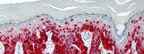
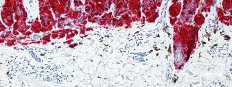
7 Melanoma Immunohistochemistry S100 Sox10 Melan A/Mart 1 HMB45 MITF MIB1 cyclin D1 p53 bcl2 p16 p21 BRAF, NRAS, CKIT, ALK, ROS, BAP1 S100 S100/SOX10 Melan A First line melanocytic marker: High Sensitivity for melanocytic tumours Expressed in Naevi and Melanoma Low specificity, also seen in neural and myoepithelial neoplasms Best marker in desmoplastic melanoma May be weak and focal in benign and malignant blue naevi S100 Second line melanocytic marker: Good sensitivity for epithelioid melanocytic tumours But often lost in spindle cell melanoctic neoplasms Expressed in Naevi and Melanoma Relatively high specificity, but also seen in PECOMA Useful for evaluation of junctional component Often strong and diffuse in benign and malignant blue naevi Melan A 7


8 HMB 45 Second line melanocytic marker: Low sensitivity but high specificity Highlights maturation with dpeth in naevi Expression in deep component seen in DPN/blue naevi, melanoma and metastasis Useful for evaluation of junctional component HMB45 HMB45 Micropthalmia Transcription Factor Third line melanocytic marker: High sensitivity but low specificity MITF MIB1/Ki67 Proliferation marker: Low expression in naevi High expression in deeply invasive and metastatic melanoma Many pitfalls: Interpretation in thin melanoma and intermediate thickness tumours difficult Low proliferation index in desmoplastic melanoma MIB1 8


:1215 24.")
9 Cyclin D1 Little diagnositc/prognostic value Highlights maturation in naevi Diffuse expression: concerning for melanoma but rare phenomenon limited to deeply invasive tumours also seen in Spitzoid neoplasms MIB1 CyclinD1 P16 Melanoma Genetics Diffuse expression in naevi, including Spitzoid tumours Loss of expression: in subset of melanoma in Spitzoid tumours concerning for more aggressive behaviour Rapid progress over past 15 years: genomic instability of melanoma development of diagnostic FISH assay p g y mutational analysis and targeted therapy P16 Garrido Ruiz MC, et al. Mod Pathol. 2010;23(9): Kapur P, et al. ModPathol. 2005;18(2):
:1765 70.")

10 Melanoma Genetics Comparative Genomic Hybridisation Frequent chromosomal aberrations in melanoma (>95%) in contrast to benign naevi 13 regions on 8 chromosomes Bastian BC, et al. Am J Pathol 2003;163(5): Melanoma Genetics Melanoma FISH Using CGH data a panel of 4 probes was found most informative: Centromere chromosome 6 Gain of CCND1 (11q13) Gain of RREB1 (6p25) Loss of MYB (6q23) Sensitivity of 87% and specificity of 96% Recent inclusion of probe for 9p21 as marker of prognosis Technically demanding and expensive Fluorescence In Situ Hybridization as an Ancillary Tool in the Diagnosis of Ambiguous Melanocytic Neoplasms: A Review of 804 Cases. North, Jeffrey; Garrido, Maria; Kolaitis, Nicholas; LeBoit, Philip; McCalmont, Timothy; Bastian, Boris American Journal of Surgical Pathology. 38(6): , June DOI: /PAS Mutational Spectrum in Melanoma Whole exome sequencing: numerous (>1,000) gene mutations Challenge: Driver vs bystander mutations Inactivating mutations in CDKN2a (40 80%) PTEN (30 70%) Promoter mutations in TERT gene FIGURE 9. Polyploidy in FISH. All 4 FISH probes showing >2 signals. Red 6p25, Green 11q13, Yellow 6q23, Aqua centr omere by Lippincott Williams & Wilkins. Published by Lippincott Williams & Wilkins, Inc. 2 High rate of mutations in BRAF in 50% (vemurafenib) NRAS in 20% (MEK inhibitor MEK162) CKIT in 10% (imatinib) GNAQ/GNA11 10

:2191 9.")

 Robles Espinoza CD, et al.")
11 Uveal Melanoma Lentigo Maligna Vulval Melanoma Mutations in GNAQ and GNA11 gene mutations Additional BAP1 mutations may predict more aggressive behaviour GANQ and GNA11 gene mutations also observed in blue naevi and blue naevuslike melanoma ( malignant blue naevus ) Acral Melanoma Van Raamsdonk CD, et al. Nature. 2009;457(7229): Van Raamsdonk CD, et al. N Engl J Med. 2010;363(23): Familial Melanoma Familial Melanoma CDKN2a Familial atypical multiple mole melanoma (FAMM)/Dysplastic nevus syndrome 40% of familial melanoma Autosomal dominant Multiple atypical naevi and melanoma Superficial spreading and nodular melanoma POT 1 Recently described Loss of function mutations in the Protection of telomers 1 (POT 1) gene on chromosome 7q31 4% of familial melanoma Also linked to chronic lymphocytic leukemia (CLL) Robles Espinoza CD, et al. Nat Genet. 2014;46(5): Shi J, et al. Nat Genet. 2014;46(5):


12 BAP1 Familial Melanoma BAPoma Tumour suppressor gene mutations in BRCA1 associated protein 1 (BAP1)on chromosome 3q21 Autosomal dominant t Multiple melanocytic tumours with epithelioid cell change Uveal and cutaneous melanoma Mesothelioma Renal cell carcinoma Wiesner T, et al. Nat Genet. 2011;43(10): Wiesner T, et al. Am J Surg Pathol 2012;36(6): courtesy Dr Zlatko Marusic, Zagreb, Croatia BAP1 BRAF Pigment Synthesising Melanoma Pigment Synthesizing Melanoma Pigmented Epithelioid Melanocytoma Animal/Equine type Melanoma Unusual distinctive melanocytic tumour Reminiscent of melanomas in grey horses Animal / Equine type Melanoma Significant morphological overlap with epithelioid blue naevus in Carney complex Pigmented Epithelioid Melanocytoma Loss of expression of Protein Kinase A Regulatory Subunit 1a (PRKAR1A) in sporadic PEM and EBN in patients with Carney complex {Zembowicz A et al Am J Surg Pathol 2007; 31: } 12


")
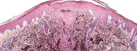
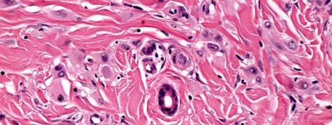
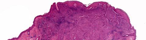
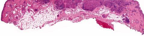
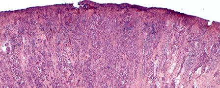
13 Pigment Synthesising Melanoma Clinical Presentation Deeply pigmented tumours Extremities, head & neck, trunk Mucosa, genital sites Young adults (20 30 yrs) Wide age range including congenital onset M=F 13


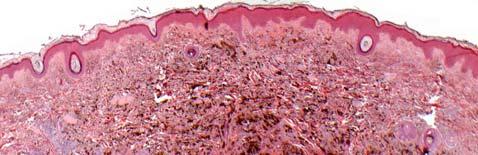
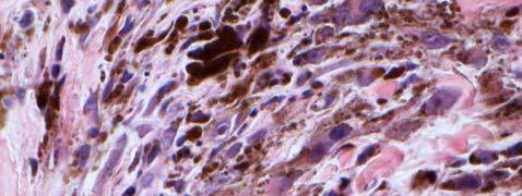
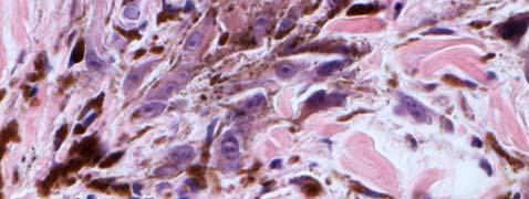
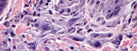
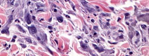
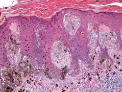
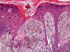
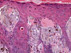
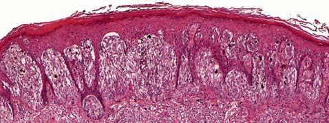
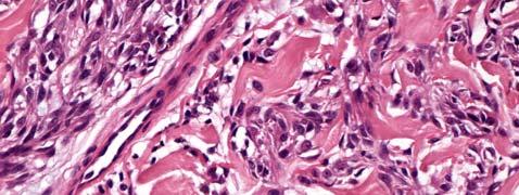
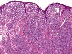
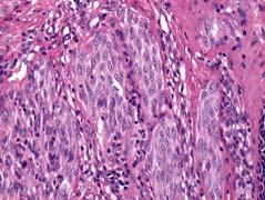
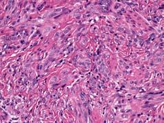
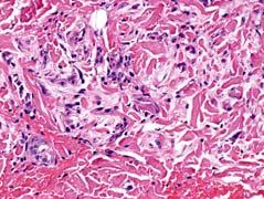
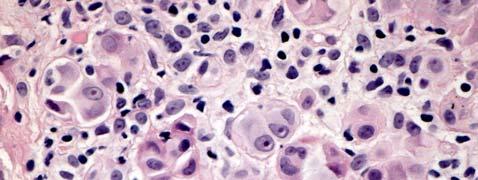
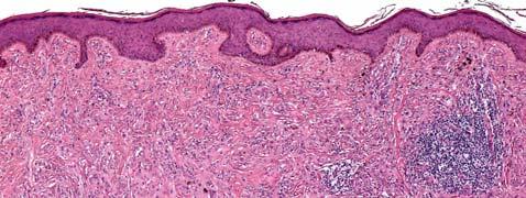
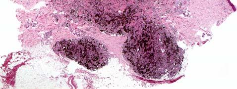
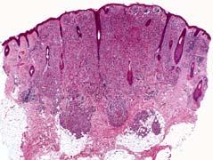
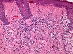
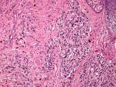
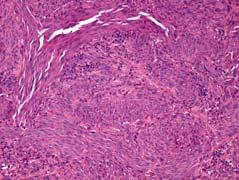
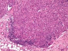
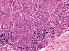
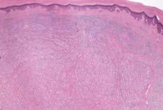
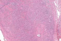
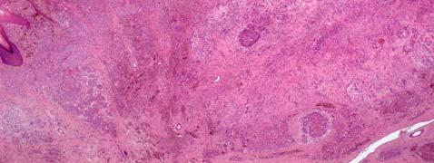
14 9 year old boy with a pigmented lesion on the left superior deltoid area 14
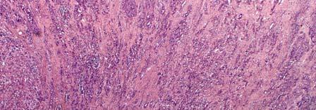
15 15
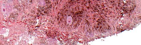
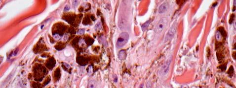
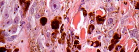
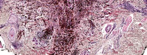
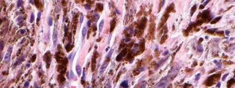
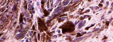
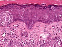
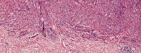
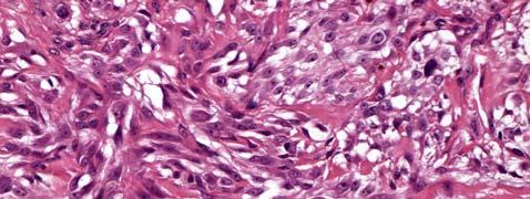
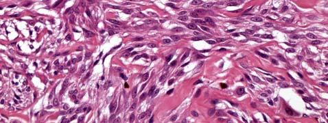
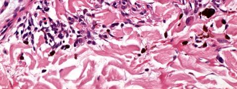
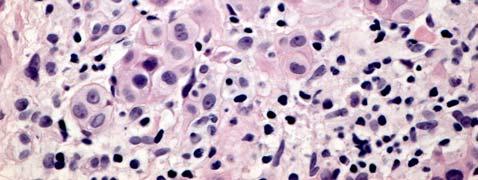
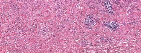
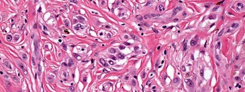
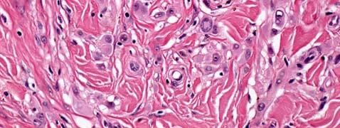
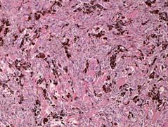
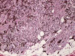
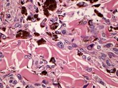
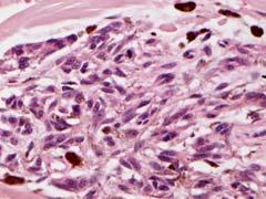
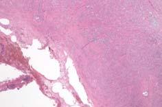
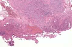
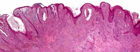
 Large ovoid to polygonal cells Heavily pigmented cytoplasm and vesicular nuclei with prominent nucleoli Low mitotic rate No or minimal junctional component Pigment Synthesising Melanoma")

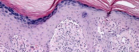
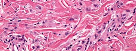
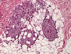
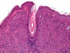
16 Pigment Synthesising Melanoma Histological Features Deeply pigmented tumours Invasion of Level IV or V Tumour thickness: 2 3 mm (range: mm) Large ovoid to polygonal cells Heavily pigmented cytoplasm and vesicular nuclei with prominent nucleoli Low mitotic rate No or minimal junctional component Pigment Synthesising Melanoma Clinical Behaviour Overall favourable prognosis High rate of LN metastasis (30 50%) Rare distant metastasis to liver Rare documented mortality (limited follow up) {Antony FC Histopathology 2006;48: } {Zembowicz A et al Am J Surg Pathol 2004;28:31 40} {Mandal RV Am J Surg Pathol 2009;33: } Spitz Naevus Introduction One of the most challenging and controversial aspects of melanoctic tumour pathology to date Described by Sophie Spitz in 1948 as juvenile melanoma Favourable outcome despite worrying histological features Histological features poor predictor of outcome (disease associated mortality in 1/13 patients) Spitz Naevus Clinical Presentation Wide age spectrum but most common in childhood and adolescence Decreasing frequency with age Predilection for Caucasians Slight female predominance Wide anatomic distribution Face and ear in childhood Trunk and extremities in adulthood Non pigmented, red to skin coloured papule <1cm in diameter Calonje et al. McKee s Pathology of the Skin, 4 th ed. Elsevier


17 Spitz Naevus Problems 65 years after Sophie Spitz publication Poor insight into the biology of Spitz tumours Difficult to predict behaviour Classification into Spitz Naevus Atypical Spitz Tumour/Spitz Tumour of Uncertain Malignant Potential Spitzoid Melanoma Spitz Naevus Genetics Lack of mutation in NRAS, KIT, GNAQ, GNA11 HRAS mutations in ~10% (desmoplastic SN) Homozygous loss of BAP1 and BRAF mutation tti Kinase fusions involving ROS1, NTRK1, ALK, BRAF, RET in ~50% Spitz Tumours Busam KJ, et al. Clinical and pathologic findings of Spitz nevi and atypical Spitz tumors with ALK fusions. Am J Surg Pathol. 2014;38(7): Wiesner T, et al. Kinase fusions are frequent in Spitz tumours and spitzoid melanomas. Nat Commun. 2014;5:3116. Wiesner T, et al. A distinct subset of atypical Spitz tumors is characterized by BRAF mutation and loss of BAP1 expression. Am J Surg Pathol. 2012;36: Wiesner T, et al. Germline mutations in BAP1 predispose to melanocytic tumors. Nat Genet. 2011;43: Bastian BC, LeBoit PE, Pinkel D. Mutations and copy number increase of HRAS in Spitz nevi with distinctive histopathological features. Am J Pathol. 2000;157:
18 18
19 19


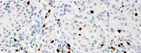
20 S100 Melan a cyclin D1 MIB1 p16 p21 20


21 Morphological Spectrum of Spitz Naevi Pagetoid Spitz Acral Spitz Desmoplastic Spitz Pigmented Spitz and Spindle Cell Naevus (Reed) Deep Penetrating Naevus Atypical Spitz Naevus/Tumour/Stump Spitzoid Melanoma Desmoplastic Spitz Naevus Brown/erythemato us papules <1cm Adulthood F>M Extremities HRAS mutations 21


22 Kiuru M, Patel RM, Busam KJ. J Cutan Pathol. 2012;39(10):
23 Deep-pentrating naevus Wide age range; adolescence and early adulthood Face, neck, extremities Darkly pigmented to grayish symmetrical papule/nodule <1cm Closely related to Spitz Naevi Mutations in HRAS, but not GNAQ, GNA11 23


24 BAPoma Characteristic epithelioid melanocytic tumours with Spitzoid features Loss of BAP1 expression Additional BRAF mutation courtesy Dr Zlatko Marusic, Zagreb, Croatia Familial or sporadic Also seen in AST and cutaneous and uveal melanoma courtesy Dr Zlatko Marusic, Zagreb, Croatia Wiesner T, et al. Am J Surg Pathol. 2012; 36: Wiesner T, et al. Nat Genet ; 43: M, 28 Jahre, Gesicht BRAF BAP1 24

 and the remaining intact ALK gene (1 juxtaposed green and orange signal).")
. Yeh I, et al. Am J Surg Pathol. 2015;39(5):581 91. Busam KJ, et al. Am J Surg Pathol. 2014;38(7):925 33.")

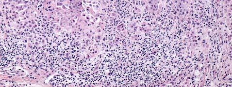
25 Spitz Naevus with Kinase Fusion Clinical, Histopathologic, and Genomic Features of Spitz Tumors With ALK Fusions. Yeh, Iwei; MD, PhD; de la Fouchardiere, Arnaud; MD, PhD; Pissaloux, Daniel; Mully, Thaddeus; Garrido, Maria; MD, PhD; Vemula, Swapna; Busam, Klaus; LeBoit, Philip; McCalmont, Timothy; Bastian, Boris; MD, PhD American Journal of Surgical Pathology. 39(5): , May DOI: /PAS ALK1 FIGURE 2. Disruption of ALK identified by FISH. A, Unbalanced rearrangement of the ALK gene, with preservation of the 3' signal (red) and the remaining intact ALK gene (1 juxtaposed green and orange signal). B, Balanced rearrangement of the ALK gene with separated orange and green signals compared with the remaining intact locus. C, Amplification of the 3' oncogenic signal (1 or 2 fusion signals + numerous clustered orange signals). Yeh I, et al. Am J Surg Pathol. 2015;39(5): Busam KJ, et al. Am J Surg Pathol. 2014;38(7): Copyright 2015 Wolters Kluwer Health, Inc. All rights reserved. Published by Lippincott Williams & Wilkins, Inc. 2 Atypical Spitz Tumour/ Spitzoid Melanoma Poorly defined criteria with poor interobserver agreement Constellation of features Atypical Spitz Tumour/Spitzoid Melanoma -Concerning Features- Architecture: Diameter in mm (>10 mm) Depth in mm (involvement of subcutaneous fat) Ulceration Poor circumscription Diffuse Pagetoid spread High cellular density Lack of zonation and maturation Asymmetry Few or no dull pink (Kamino) bodies Cytology: High nuclear to cytoplasmic ratios Loss of delicate or dispersed chromatin patterns Thickening of nuclear membranes Hyperchromatism Large nucleoli Proliferation: Significant mitotic rate Deep/marginal mitoses Increased mib-1 proliferation index Modified from: Barnhill RL. Modern Pathology. 2006; 19: S21-S33 25
26 26
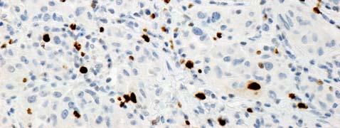
27 mib-1 27


28 mib-1 Melan a 2 year old girl; right ankle tumour 10 year old female with an enlarging lesion on the left calf present for 8 months 28


 4.")


29 Melanoma in Children and Adolescents Nodular M Sp M PSM SSM Nevoid M n age (yrs) site H&N>E>T E>H&N>T H&N E>T E>T thick (mm) ulceration 50% 24% nil 0.8% nil necrosis 25% nil nil nil nil mitoses 6.5/mm 2 5/mm 2 1/mm 2 1/mm 2 2.5/mm 2 LN met 57% 24% 100% nil 30% Dist met 50% nil nil nil nil mortality 40% nil nil nil nil 29

:377 8.")

:1387-94.")
30 AST/Spitzoid Melanoma -Prognose- In young children (<10 years): Favourable behaviour Risk for involvement of locoregional lymph nodes Rare disseminated disease and mortality Sentinel lymph node biopsy not helpful and should be avoided Spitzoid Melanoma No firm established diagnostic criteria Distinction from Atypical Spitz Tumour largely arbitrary Best avoided in young patients Lallas A, eta l. Lancet Oncol. 2014;15(4):e doi: /S (13) Duncan LM. Lancet Oncol. 2014;15(4): AST/Spitzoid Melanoma -Genetics- Homozygous but not heterozygous 9p21 deletion associated with more aggressive behaviour Yazdan P, et al. Comparative analysis of atypical spitz tumors with heterozygous versus homozygous 9p21 deletions for clinical outcomes, histomorphology, BRAF mutation, and p16 expression. Am J Surg Pathol May;38(5): Gerami P, Cooper C, Bajaj S, Wagner A, Fullen D, Busam K, Scolyer RA, Xu X, Elder DE, et al. Outcomes of atypical spitz tumors with chromosomal copy number aberrations and conventional melanomas in children. Am J Surg Pathol Sep;37(9): Gerami P, et al. Risk assessment for atypical spitzoid melanocytic neoplasms using FISH to identify chromosomal copy number aberrations. Am J Surg Pathol. 2013;37(5):
Case 26 Male 37. Right jawline 5mm nodule?keloid. The best diagnosis is:
 Case 26 Male 37. Right jawline 5mm nodule?keloid. The best diagnosis is: A. Desmoplastic Spitz naevus B. Atypical Spitz Tumour C. Spitzoid melanoma D. Deep penetrating naevus E. Spitz naevus Case 26: M
Molecular Aspects of Melanocytic Neoplasia. Iwei Yeh MD, PhD University of California, San Francisco
 Molecular Aspects of Melanocytic Neoplasia Iwei Yeh MD, PhD University of California, San Francisco Thanks to: Boris Bastian Timothy McCalmont Philip LeBoit Beth Ruben Jeff North Laura Pincus Thaddeus
Michael T. Tetzlaff MD, PhD
 Molecular alterations informing the diagnosis of melanocytic tumors Michael T. Tetzlaff MD, PhD Associate Professor Department of Pathology, Section of Dermatopathology Department of Translational and
Dermatopathology. Dr. Rafael Botella Estrada. Hospital La Fe de Valencia
 Dermatopathology Dr. Rafael Botella Estrada. Hospital La Fe de Valencia Melanoma and mimics Dr. Martin Mihm Malignant lesions result from the accumulation of mutations Class I lesions (benign) Class II
Update on Spitzoid and Blue nevus-like melanocytic lesions Emphasis on molecular studies informing diagnosis, prognosis and therapy
 Update on Spitzoid and Blue nevus-like melanocytic lesions Emphasis on molecular studies informing diagnosis, prognosis and therapy Michael T. Tetzlaff MD, PhD Associate Professor Department of Pathology,
Melanoma and the genes: Molecular alterations informing the diagnosis of melanocytic tumors
 Melanoma and the genes: Molecular alterations informing the diagnosis of melanocytic tumors Michael T. Tetzlaff MD, PhD Associate Professor Department of Pathology, Section of Dermatopathology Department
21/07/2017. The «gray zone» of diagnosis is visible. Nevus Atypical nevus Melanoma. Melanoma ex-blue nevus
 Update on the Clinico- Pathological and Molecular Diagnosis of Melanocytic Lesions None to declare Conflicts of interest Belfast pathology Arnaud de la Fouchardière MD, PhD Lyon, France What is new? Today
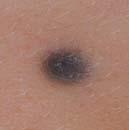
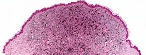
Female 18. Deeply pigmented lesion on trunk.?warty naevus?seborrhoeic keratosis?malignant melanoma. The best diagnosis is:
 Female 18. Deeply pigmented lesion on trunk.?warty naevus?seborrhoeic keratosis?malignant melanoma. The best diagnosis is: A. deep penetrating naevus B. naevoid malignant melanoma C. pigment synthesising
Melanocytic Lesions: Use of Immunohistochemistry and Special Studies Napa Valley 2018
 Melanocytic Lesions: Use of Immunohistochemistry and Special Studies Napa Valley 2018 Victor G. Prieto, MD, PhD Professor Depts. of Pathology and Dermatology University of Texas - MD Anderson Cancer Center
6/22/2015. Original Paradigm. Correlating Histology and Molecular Findings in Melanocytic Neoplasms
 6 Correlating Histology and Molecular Findings in Melanocytic Neoplasms Pedram Gerami MD, Associate Professor of Dermatology and Pediatrics at Northwestern University Disclosures: I have been a consultant
Vernon K. Sondak. Department of Cutaneous Oncology Moffitt Cancer Center Tampa, Florida
 Vernon K. Sondak Department of Cutaneous Oncology Moffitt Cancer Center Tampa, Florida Australasian Melanoma Conference 2016 Sydney, NSW, Australia October 29, 2016 Disclosures Dr. Sondak is a compensated
Ways to get into trouble, ideas on avoiding trouble, and diagnostic approaches to keep trouble at bay
 Pitfalls in the diagnosis of melanocytic tumors Timothy McCalmont, MD University of California, San Francisco Ways to get into trouble, ideas on avoiding trouble, and diagnostic approaches to keep trouble
The Enigmatic Spitz Lesion
 The Enigmatic Spitz Lesion The Dawn of Spitz S Spitz Sophie Spitz Melanomas of Childhood ; Am J Pathol 1948 1910-1956 13 children (18 mo - 12 yrs) 12/13 had a benign clinical course Sophie Spitz Born 1910
Diagnoses of Cases 1. Lentigo, other melanosis and the acquired nevus 2. Variations on the acquired nevus 3. Dermal melanocytosis
 Diagnoses of Cases 1. Lentigo, other melanosis and the acquired nevus 1 1A. Lentigo simplex 4 1B. Psoralens and ultraviolet A (PUVA) lentigo 6 1C. Solar lentigo 8 1D. Café au lait macule 10 1E. Ink-spot
Patricia Chevez-Barrrios AAOOP-USCAP /12/2016
 Biomarkers in Ocular Melanoma Patricia Chévez-Barrios, MD Pathology and Genomic Medicine, Houston Methodist Hospital Professor of Pathology and Laboratory Medicine and Ophthalmology, Weill Cornell Medical
Integrating Fluorescence in situ Hybridization and Genomic Array Results into the Diagnostic Workup of Melanoma
 Integrating Fluorescence in situ Hybridization and Genomic Array Results into the Diagnostic Workup of Melanoma Association for Molecular Pathology United States and Canadian Academy of Pathology Companion
Springer Healthcare. Staging and Diagnosing Cutaneous Melanoma. Concise Reference. Dirk Schadendorf, Corinna Kochs, Elisabeth Livingstone
 Concise Reference Staging and Diagnosing Cutaneous Melanoma Dirk Schadendorf, Corinna Kochs, Elisabeth Livingstone Extracted from Handbook of Cutaneous Melanoma: A Guide to Diagnosis and Treatment Published
10/2/17. MELTUMP, SAMPUS, AST.An Algorithmic Approach to Challenging (Often Borderline) Melanocytic Tumors. An Introduction to SNP Arrays
 Melanocytic Tumors. An Introduction to SNP Arrays") MELTUMP, SAMPUS, AST.An Algorithmic Approach to Challenging (Often ) Melanocytic Tumors An Introduction to SNP Arrays Rajiv M. Patel, M.D. RCPA NZ ASM 2017 (11:45-12:30pm, Saturday, 23-09-17) Why do we
MELTUMP, SAMPUS, AST.An Algorithmic Approach to Challenging (Often ) Melanocytic Tumors An Introduction to SNP Arrays Rajiv M. Patel, M.D. RCPA NZ ASM 2017 (11:45-12:30pm, Saturday, 23-09-17) Why do we
A PRACTICAL APPROACH TO ATYPICAL MELANOCYTIC LESIONS BIJAN HAGHIGHI M.D, DIRECTOR OF DERMATOPATHOLOGY, ST. JOSEPH HOSPITAL
 A PRACTICAL APPROACH TO ATYPICAL MELANOCYTIC LESIONS BIJAN HAGHIGHI M.D, DIRECTOR OF DERMATOPATHOLOGY, ST. JOSEPH HOSPITAL OBJECTIVES Discuss current trends and changing concepts in our understanding of
There is NO single Melanoma Stain. > 6000 Mutations in Melanoma. What else can be done to discriminate atypical nevi from melanoma?
 Las Vegas Fall Clinical 2016: The Assessment and Diagnosis of Melanoma Whitney A. High, MD, JD, MEng Associate Professor, Dermatology & Pathology Director of Dermatopathology (Dermatology) University of
Genetic Testing: When should it be ordered? Julie Schloemer, MD Dermatology
 Genetic Testing: When should it be ordered? Julie Schloemer, MD Dermatology Outline Germline testing CDKN2A BRCA2 BAP1 Somatic testing Gene expression profiling (GEP) BRAF Germline vs Somatic testing
MAPK Pathway. CGH Next Generation Sequencing. Molecular Tools in Care of Patients with Pigmented Lesions 7/20/2017
 Molecular Tools in Care of Patients with Pigmented Lesions Tammie Ferringer, MD Geisinger Medical Center, Danville, PA tferringer@geisinger.edu DISCLOSURE OF RELATIONSHIPS WITH INDUSTRY Tammie Ferringer,
Conflict of Interest 9/2/2014. Pathogenesis and Comparison of Atypical Spitz Nevi vs Benign Spitz, and Childhood Melanoma
 Pathogenesis and Comparison of Atypical Spitz Nevi vs Benign Spitz, and Childhood Melanoma Martin C. Mihm Jr., M.D., F.A.C.P. Harvard Medical School Brigham and Women s Hospital Dana Farber Cancer Center
The Pathology of Neoplasia Part II
 The Pathology of Neoplasia Part II February 2018 PAUL BOGNER, MD A S S O C I A T E P R O F E S S O R O F O N C O L O G Y P A T H O L O G Y A N D D E R M A T O L O G Y Clinical goals of cancer pathology
Desmoplastic Melanoma R/O BCC. Clinical Information. 74 y.o. man with lesion on left side of neck r/o BCC
 R/O BCC Sabine Kohler, M.D. Professor of Pathology and Dermatology Dermatopathology Service Stanford University School of Medicine Clinical Information 74 y.o. man with lesion on left side of neck r/o
Melanocytic proliferations in sundamaged
 Atypical Spitzoid Tumor: What Does It Mean And How Should It Be Managed? Melanocytic proliferations in sundamaged skin Jane L. Messina, Jane L. Messina MD International Melanoma Pathology Working Group
Update on Genetic Testing for Melanoma
 Update on Genetic Testing for Melanoma Emily Y. Chu, M.D., Ph.D. Assistant Professor of Dermatology & Pathology and Laboratory Medicine Hospital of the University of Pennsylvania February 18, 2018 AAD
1/10/2018. Soft Tissue Tumors Showing Melanocytic Differentiation. Overview. Desmoplastic/ Spindle Cell Melanoma
 2016 MFMER slide-1 2016 MFMER slide-2 2016 MFMER slide-3 Soft Tissue Tumors Showing Melanocytic Differentiation Andrew L. Folpe, M.D. Professor of Laboratory Medicine and Pathology Mayo Clinic, Rochester,
Case RAC7783. M46. Ear. Mole. r/o MM.?Blue naevus RAC7783
 Case RAC7783. M46. Ear. Mole. r/o MM.?Blue naevus RAC7783 Pie Chart Participants N=74 Benign: 48 N=74 Blue naevus: 38 Intradermal: 12 DPN: 10 Compound 3 Clonal: 3; Spitz 2; Special Site: 1; Congenital:
Financial disclosures
 Mesenchymal Neoplasms with Melanocytic Differentiation By Konstantinos Linos MD, FCAP, FASDP Bone, Soft Tissue and Dermatopathology Assistant Professor of Pathology Dartmouth-Hitchcock Medical Center Geisel
David B. Troxel, MD. Common Medicolegal Situations: Misdiagnosis of Melanoma
 Common Medicolegal Situations: Misdiagnosis of Melanoma David B. Troxel, MD Medical Director, The Doctors Company, Napa, California Clinical Professor Emeritus, University of California at Berkeley Past
Blue Melanocytic Proliferations
 Blue Melanocytic Proliferations Labib R. Zakka M.D., M.A. Research Fellow Melanoma Program Department of Dermatology Brigham and Women s Hospital Harvard Medical School Conflicts of Interest No conflicts
21/07/2017. Hobnail endothelial cells are not the same as epithelioid endothelial cells
 UPDATE IN CUTANEOUS VASCULAR S DERMATOPATHOLOGY SESSION BELFAST PATHOLOGY JUNE 21/2017 Dr E Calonje St John s Institute of Dermatology, London, United Kingdom THE FAMILY OF VASCULAR S WITH EPITHELIOID
Dermatologica Sinica
 DERMATOLOGICA SINICA 30 (2012) 57e61 Contents lists available at SciVerse ScienceDirect Dermatologica Sinica journal homepage: http://www.derm-sinica.com CASE REPORT Pigmented epithelioid melanocytoma:
The Dermal Melanocytoses. Conflicts of Interest 5/22/2018. The Nevi of Ota and Ito. Martin C. Mihm M.D.
 The Dermal Melanocytoses Martin C. Mihm M.D. Director Mihm Cutaneous Pathology Consultative Service (MCPCS) Brigham and Women s Hospital Director Melanoma Program Brigham and Women s Hospital and Harvard
Update on Cutaneous Mesenchymal Tumors. Thomas Brenn
 Update on Cutaneous Mesenchymal Tumors Thomas Brenn Cutaneous Mesenchymal Tumours Wide morphological and biological spectrum Myofibroblastic, smooth muscle, neural, vascular, apidocytic, undifferentiated;
Benign and malignant epithelial lesions: Seborrheic keratosis: A common benign pigmented epidermal tumor occur in middle-aged or older persons more
 Benign and malignant epithelial lesions: Seborrheic keratosis: A common benign pigmented epidermal tumor occur in middle-aged or older persons more common on the trunk; but extremities, head and neck are
Benign versus Cancerous Lesions How to tell the difference FMF 2014 Christie Freeman MD, CCFP, DipPDerm, MSc
 1 Benign versus Cancerous Lesions How to tell the difference FMF 2014 Christie Freeman MD, CCFP, DipPDerm, MSc Benign lesions Seborrheic Keratoses: Warty, stuck-on Genetics and birthdays Can start in late
Pathology of the skin. 2nd Department of Pathology, Semmelweis University
 Pathology of the skin 2nd Department of Pathology, Semmelweis University Histology of the skin Epidermis: Stratum corneum Stratum granulosum Stratum spinosum Stratum basale Dermis: papillary and reticular
Conflicts of Interest
 Challenging Melanocytic Lesions Carlos N. Prieto-Granada M.D. Assistant Professor University of Alabama at Birmingham (UAB) Department of Pathology 2017 AAD Annual Meeting 3/2/17 - Orlando, FL None Conflicts
I have no relevant conflicts of interest to disclose. John T. Seykora MD PhD Departments of Dermatology & Pathology and Laboratory Medicine
 Molecular Characterization of Stage 1-3 Melanoma: Are we close to accurate prognostication and prediction? I have no relevant conflicts of interest to disclose. John T. Seykora MD PhD Departments of Dermatology
Diagnosis of melanocytic proliferations remains one of
 Update on Fluorescence In Situ Hybridization in Melanoma State of the Art Pedram Gerami, MD; Artur Zembowicz, MD, PhD N Context. Recent advances in understanding the molecular basis of melanoma have resulted
An Overview of Melanoma. Harriet Kluger, M.D. Associate Professor Section of Medical Oncology Yale Cancer Center
 An Overview of Melanoma Harriet Kluger, M.D. Associate Professor Section of Medical Oncology Yale Cancer Center Melanoma Statistics Median age at presentation 45-55 55 years Incidence: 2003 54,200 cases
Which melanoma patients benefit from genetic testing?
 Which melanoma patients benefit from genetic testing? Michael A. Marchetti, MD Assistant Attending, Dermatology Service Memorial Sloan Kettering Cancer Center American Academy of Dermatology Annual Meeting
Melanocytic lesions on Genital Skin Melanoma vs. Melanocytic Nevus, Revisited. Timothy H. McCalmont, MD University of California, San Francisco
 Melanocytic lesions on Genital Skin Melanoma vs. Melanocytic Nevus, Revisited Timothy H. McCalmont, MD, San Francisco I. IS IT BENIGN OR IS IT MALIGNANT? One of the commonest determinations we make as
Case 231: F7. Exophytic naevus over left trapezious. Grown over a few weeks. Iniitally flat.?spitz naevus,?malignant
 Case 231: F7. Exophytic naevus over left trapezious. Grown over a few weeks. Iniitally flat.?spitz naevus,?malignant Dermoscopy: coarse vascular structures. c/o A, B, C RAC7750 Case 231: F7. Exophytic
Rare melanoma: Are the options improving? Dr Neil Steven Consultant in Medical Oncology University Hospital Birmingham University of Birmingham
 Rare melanoma: Are the options improving? Dr Neil Steven Consultant in Medical Oncology University Hospital Birmingham University of Birmingham Classifying melanoma Melanoma (site of origin, thickness,
Malignant tumors of melanocytes: Part 1. Deba P Sarma, MD., Omaha
 Malignant tumors of melanocytes: Part 1 Deba P Sarma, MD., Omaha The melanocytic tumor is one of the most difficult and confusing areas in Dematopathology. It is true that most (95%) of such lesions are
Pathological diagnosis of melanocytic tumours: clues and pitfalls # Richard A. Scolyer 1,2,3* and Stanley W. McCarthy 1,2,3
 Pathological diagnosis of melanocytic tumours: clues and pitfalls # Richard A. Scolyer 1,2,3* and Stanley W. McCarthy 1,2,3 1 Tissue Pathology and Diagnostic Oncology, Royal Prince Alfred Hospital, Sydney,
BAP-oma & BEYOND MICHAEL A NOWAK, MD
 BAP-oma & BEYOND MICHAEL A NOWAK, MD CONFLICTS No conflicts with the content of this lecture BAP-oma Wiesner 2011: Families with multiple tan dome-shaped papules of head, neck, trunk, and extremities.
Page 1 of 3. We suggest the following changes:
 Page 1 of 3 Loren E. Clarke, M.D. Myriad Genetic Laboratories, Inc. 320 Wakara Way, Salt Lake City, UT 84108 Phone: 801.883.3470 Email: lclarke@myriad.com Date of Request: June 2017 NCCN Guidelines Panel:
The Relevance of Cytologic Atypia in Cutaneous Neural Tumors
 The Relevance of Cytologic Atypia in Cutaneous Neural Tumors Recent Findings - New Developments New Problems Zsolt B. Argenyi, M.D. Professor of Pathology & Dermatology Director of Dermatopathology Department
A diagnostic algorithm for atypical spitzoid tumors: guidelines for immunohistochemical and molecular assessment
 Modern Pathology (2016), 1 15 2016 USCAP, Inc All rights reserved 0893-3952/16 $32.00 1 A diagnostic algorithm for atypical spitzoid tumors: guidelines for immunohistochemical and molecular assessment
المركب النموذج--- سبيتز وحمة = Type Spitz's Nevus, Compound SPITZ NEVUS 1 / 7
 SPITZ NEVUS 1 / 7 Epidemiology An annual incidence rate of 1.4 cases of Spitz nevus per 100,000 individuals has been estimated in Australia, compared with 25.4 per 100,000 individuals for cutaneous melanoma
2/6/2018. Original Paradigm. Clonal Chromosomal A berrations. Only 20% of Spitz Nevi 95% 6p, 7q, 17q, 20q, 4q,8q, 1q, 11q. Isolated Gain in 11p
 Molecular Diagnostics for Melanocytic Neoplasms: Moving towards a Revolution in the Management of Melanocytic Neoplasms Pedr am Gerami MD Associate Professor of Dermatology, Pathology and Pediatrics at
K Blessing, J J H Grant, D S A Sanders, M M Kennedy, A Husain, P Coburn
 J Clin Pathol 2000;53:591 595 591 Papers Pathology, Aberdeen University, Foresterhill, Aberdeen AB25 2ZD, K Blessing Pathology, Birmingham University, Birmingham B15 2TT, D S A Sanders Pathology, Heartlands
Cutaneous Mesenchymal Neoplasms with EWSR1 Rearrangement
 Cutaneous Mesenchymal Neoplasms with EWSR1 Rearrangement By Konstantinos Linos MD, FCAP, FASDP Bone, Soft Tissue and Dermatopathology Assistant Professor of Pathology Dartmouth-Hitchcock Medical Center
Melanoma. Kaushik Mukherjee MD A. Scott Pearson MD
 Melanoma Kaushik Mukherjee MD A. Scott Pearson MD Disclosures You still have to study Not all inclusive No Western blots Extensive use of Google Image Search and Sabiston Melanoma Basics 8 th most common
5/21/2018. Disclosures. Consulting: Myriad Genetics SciBase. Superficial Atypical Melanocytic Proliferations. SSM, LMM and (some of) their Simulants
 their Simulants") Disclosures Consulting: Myriad Genetics SciBase Superficial Atypical Melanocytic Proliferations SSM, LMM and (some of) their Simulants 1 Melanomas and Nevi. Nevi are important mainly in relation to melanoma
Disclosures Consulting: Myriad Genetics SciBase Superficial Atypical Melanocytic Proliferations SSM, LMM and (some of) their Simulants 1 Melanomas and Nevi. Nevi are important mainly in relation to melanoma
Clinical characteristics
 Skin Cancer Fernando Vega, MD Seattle Healing Arts Clinical characteristics Precancerous lesions Common skin cancers ACTINIC KERATOSIS Precancerous skin lesions Actinic keratoses Dysplastic melanocytic
Artur Zembowicz, MD, PhD; Sung-Eun Yang, MD; Antonios Kafanas, MD; Stephen R. Lyle, MD, PhD
 Correlation Between Histologic Assessment and Fluorescence In Situ Hybridization Using MelanoSITE in Evaluation of Histologically Ambiguous Melanocytic Lesions Artur Zembowicz, MD, PhD; Sung-Eun Yang,
Primary Cutaneous Melanoma Pathology Reporting Proforma DD MM YYYY. *Tumour site. *Specimen laterality. *Specimen type
 Primary Cutaneous Melanoma Pathology Reporting Proforma Includes the International Collaboration on Cancer reporting dataset denoted by * Family name Given name(s) Date of birth DD MM YYYY Sex Male Female
Protocol applies to melanoma of cutaneous surfaces only.
 Melanoma of the Skin Protocol applies to melanoma of cutaneous surfaces only. Procedures Biopsy (No Accompanying Checklist) Excision Re-excision Protocol revision date: January 2005 Based on AJCC/UICC
Guy Perrot (Ги Перро)
") НАУЧНО-ПРАКТИЧЕСКАЯ КОНФЕРЕНЦИЯ (МАСТЕР-КЛАСС) «ПРАКТИЧЕСКИЕ АСПЕКТЫ ДИАГНОСТИКИ И ЛЕЧЕНИЯ МЕЛАНОМЫ КОЖИ» DIAGNOSTIC AND PITFALLS IN MELANOMA Guy Perrot (Ги Перро) MD PHD pathologist, University Hospital
НАУЧНО-ПРАКТИЧЕСКАЯ КОНФЕРЕНЦИЯ (МАСТЕР-КЛАСС) «ПРАКТИЧЕСКИЕ АСПЕКТЫ ДИАГНОСТИКИ И ЛЕЧЕНИЯ МЕЛАНОМЫ КОЖИ» DIAGNOSTIC AND PITFALLS IN MELANOMA Guy Perrot (Ги Перро) MD PHD pathologist, University Hospital
ACCME/Disclosures ALK FUSION-POSITIVE MESENCHYMAL TUMORS. Tumor types with ALK rearrangements. Anaplastic Lymphoma Kinase. Jason L.
 Companion Meeting of the International Society of Bone and Soft Tissue Pathology The Evolving Concept of Mesenchymal Tumors ALK FUSION-POSITIVE MESENCHYMAL TUMORS Jason L. Hornick, MD, PhD March 13, 2016
Disclosure Information. Lecture Outline. Lecture Outline. Introduction. Molecular Pathology of Cutaneous Melanoma. Nothing to disclose
 Molecular Pathology of Cutaneous Melanoma Disclosure Information Nothing to disclose Jonathan L. Curry, MD Assistant Professor of Pathology and Dermatology University of Texas-MD Anderson Cancer Center
Management of pediatric melanocytic lesions
 Open Journal of Clinical & Medical Case Reports Management of pediatric melanocytic lesions Volume 3 (2017) Issue 8 ISSN 2379-1039 Jin Kim, BS; Emmanuel Gabriel MD, PhD; Weiguo Liu MD, PhD; Lin Lin MD,
Enterprise Interest Nothing to declare
 Enterprise Interest Nothing to declare Diagnoses one would not like to miss in soft tissue pathology early in your career Marta Sbaraglia, MD Department of Pathology Hospital of Treviso University of Padua
IT S FUNDAMENTAL MY DEAR WATSON! A SHERLOCKIAN APPROACH TO DERMATOLOGY
 IT S FUNDAMENTAL MY DEAR WATSON! A SHERLOCKIAN APPROACH TO DERMATOLOGY Skin, Bones, and other Private Parts Symposium Dermatology Lectures by Debra Shelby, PhD, DNP, FNP-BC, FADNP, FAANP Debra Shelby,
MECHANISMS OF HUMAN DISEASE: LABORATORY SESSION PATHOLOGY OF THE SKIN LAB. Friday, February 12, :30 am 11:00 am
 MECHANISMS OF HUMAN DISEASE: LABORATORY SESSION PATHOLOGY OF THE SKIN LAB Friday, February 12, 2012 9:30 am 11:00 am FACULTY COPY GOALS: Describe the basic clinical and morphologic features of various
Dermatopathology: The tumor is composed of keratinocytes which show atypia, increase mitoses and abnormal mitoses.
 Squamous cell carcinoma (SCC): A common malignant tumor of keratinocytes arising in the epidermis, usually from a precancerous condition: 1- UV induced actinic keratosis, usually of low grade malignancy.
Supplementary Figure 1. Spitzoid Melanoma with PPFIBP1-MET fusion. (a) Histopathology (4x) shows a domed papule with melanocytes extending into the
 Histopathology (4x) shows a domed papule with melanocytes extending into the") Supplementary Figure 1. Spitzoid Melanoma with PPFIBP1-MET fusion. (a) Histopathology (4x) shows a domed papule with melanocytes extending into the deep dermis. (b) The melanocytes demonstrate abundant
Supplementary Figure 1. Spitzoid Melanoma with PPFIBP1-MET fusion. (a) Histopathology (4x) shows a domed papule with melanocytes extending into the deep dermis. (b) The melanocytes demonstrate abundant
Cutaneous Melanoma: Epidemiology (USA) The Sentinel Node in Head and Neck Melanoma. Cutaneous Melanoma: Epidemiology (USA)
 The Sentinel Node in Head and Neck Melanoma. Cutaneous Melanoma: Epidemiology (USA)") The Sentinel Node in Head and Neck Melanoma Cutaneous Melanoma: Epidemiology (USA) 6 th leading cause of cancer among men and women 68,720 new cases of invasive melanoma in 2009 8,650 deaths from melanoma
The Sentinel Node in Head and Neck Melanoma Cutaneous Melanoma: Epidemiology (USA) 6 th leading cause of cancer among men and women 68,720 new cases of invasive melanoma in 2009 8,650 deaths from melanoma
Diploma Examination. Dermatopathology: First paper. Tuesday 20 March Candidates must answer FOUR questions. Time allowed: 3 hours
 Dermatopathology: First paper Tuesday 20 March 2018 Candidates must answer FOUR questions Time allowed: 3 hours 1. Give an account of the genetic aberrations encountered in Spitzoid neoplasms and how these
We are IntechOpen, the world s leading publisher of Open Access books Built by scientists, for scientists. International authors and editors
 We are IntechOpen, the world s leading publisher of Open Access books Built by scientists, for scientists 3,500 108,000 1.7 M Open access books available International authors and editors Downloads Our
Simulators of melanoma
 Simulators of melanoma Philip E. LeBoit, M.D. Depts. of Pathology and Dermatology University of California, San Francisco Simulators of melanoma Simulators of melanoma in situ Melanocytic Non-melanocytic
Histopathology of Melanoma
 THE YALE JOURNAL OF BIOLOGY AND MEDICINE 48, 409-416 (1975) Histopathology of Melanoma G. J. WALKER SMITH Department ofpathology, Yale University School ofmedicine, 333 Cedar Street, New Haven, Connecticut
Interesting Case Series. Desmoplastic Melanoma
 Interesting Case Series Desmoplastic Melanoma Anthony Maurice Kordahi, MD, Joshua B. Elston, MD, Ellen M. Robertson, MD, and C. Wayne Cruse, MD Division of Plastic Surgery, Department of Surgery, University
S everal morphological features are frequently used in the
 1194 ORIGINAL ARTICLE Interobserver reproducibility of histological features in cutaneous malignant melanoma C Urso, F Rongioletti, D Innocenzi, C Saieva, D Batolo, S Chimenti, R Filotico, R Gianotti,
Differential diagnosis of hematolymphoid tumors composed of medium-sized cells. Brian Skinnider B.C. Cancer Agency, Vancouver General Hospital
 Differential diagnosis of hematolymphoid tumors composed of medium-sized cells Brian Skinnider B.C. Cancer Agency, Vancouver General Hospital Lymphoma classification Lymphoma diagnosis starts with morphologic
Index. Springer-Verlag Berlin Heidelberg 2017 J.A. Plaza, V.G. Prieto, Pathology of Pigmented Skin Lesions, DOI /
 A Acral lentiginous (mucosal lentiginous) melanoma, 483 Acral lentiginous melanoma (ALM) asymmetric and irregular lentiginous junctional growth, 431 clinical features, 427 428 differential diagnosis, 428
3/24/2017 DENDRITIC CELL NEOPLASMS: HISTOLOGY, IMMUNOHISTOCHEMISTRY, AND MOLECULAR GENETICS. Disclosure of Relevant Financial Relationships
 DENDRITIC CELL NEOPLASMS: HISTOLOGY, IMMUNOHISTOCHEMISTRY, AND MOLECULAR GENETICS Jason L. Hornick, M.D., Ph.D. Director of Surgical Pathology and Immunohistochemistry Brigham and Women s Hospital Professor
SEBACEOUS NEOPLASMS. Dr. Prachi Saraogi Clinical Fellow in Dermatology
 SEBACEOUS NEOPLASMS Dr. Prachi Saraogi Clinical Fellow in Dermatology Sebaceous neoplasms Sebaceous adenoma (Benign) Sebaceous carcinoma (Malignant) SEBACEOUS ADENOMA Benign tumours composed of incompletely
Toby Maurer, MD University of California, San Francisco. Lifetime risk of an American developing melanoma
 Distinguishing Pigmented Skin Lesions and Melanoma Toby Maurer, MD University of California, San Francisco Epidemiology of Melanoma Lifetime risk of an American developing melanoma 1935: 1 in 1500 1980:
Melanoma Underwriting Presented at 2018 AHOU Conference. Hank George FALU
 Melanoma Underwriting Presented at 2018 AHOU Conference Hank George FALU MELANOMA EPIDEMIOLOGY 70-80,000 American cases annually Majority are in situ or thin > 20% are diagnosed age 45 8-9,000 melanoma
Maligna Melanoma and Atypical Fibroxanthoma: An Unusual Collision Tumour G Türkcü 1, A Keleş 1, U Alabalık 1, D Uçmak 2, H Büyükbayram 1 ABSTRACT
 Maligna Melanoma and Atypical Fibroxanthoma: An Unusual Collision Tumour G Türkcü 1, A Keleş 1, U Alabalık 1, D Uçmak 2, H Büyükbayram 1 ABSTRACT Two different neoplasia in the same biopsy material called
Time to reconsider Spitzoid neoplasms?
 DERMATOLOGY PRACTICAL & CONCEPTUAL www.derm101.com Time to reconsider Spitzoid neoplasms? Carmelo Urso 1 1 Department of Anatomic Pathology, Dermatopathology Section, SM Annunziata Hospital, AUSL Toscana
57th Annual HSCP Spring Symposium 4/16/2016
 An Unusual Malignant Spindle Cell Lesion to Involve the Breast Erinn Downs-Kelly, D.O. Associate Professor of Pathology University of Utah & ARUP Laboratories No disclosures Case 39 y/o female with no
Malignant tumors of melanocytes : Part 3. Deba P Sarma, MD., Omaha
 Malignant tumors of melanocytes : Part 3 Deba P Sarma, MD., Omaha Let s go over one case of melanoma using the following worksheet. Of the various essential information that needs to be included in the
ORAL MELANOMA Definition Epidemiology Clinical Presentation
 ORAL MELANOMA Definition Melanoma is a highly malignant neoplasia, arising from melanocytes, the cells that produce the brownish pigment melanin. Melanin is the determinant in skin colour and protects
STUDY. Sensitivity of Fluorescence In Situ Hybridization for Melanoma Diagnosis Using RREB1, MYB, Cep6, and 11q13 Probes in Melanoma Subtypes
 STUDY Sensitivity of Fluorescence In Situ Hybridization for Melanoma Diagnosis Using RREB1, MYB, Cep6, and 11q13 Probes in Melanoma Subtypes Pedram Gerami, MD; Mariam Mafee, BS; Teekay Lurtsbarapa, MD;
Living Beyond Cancer Skin Cancer Detection and Prevention
 Living Beyond Cancer Skin Cancer Detection and Prevention Cutaneous Skin Cancers Identification Diagnosis Treatment options Prevention What is the most common cancer in people? What is the most common
Reviewers' comments: Reviewer #1 (Remarks to the Author):
:") Reviewers' comments: Reviewer #1 (Remarks to the Author): In this study the authors analysed 18 deep penetrating nevi for oncogenic genomic changes (single nucleotide variations, insertions/deletions,
Reviewers' comments: Reviewer #1 (Remarks to the Author): In this study the authors analysed 18 deep penetrating nevi for oncogenic genomic changes (single nucleotide variations, insertions/deletions,
MECHANISMS OF HUMAN DISEASE: LABORATORY SESSION PATHOLOGY OF THE SKIN LAB. Friday, February 13, :30 am 11:00 am
 MECHANISMS OF HUMAN DISEASE: LABORATORY SESSION PATHOLOGY OF THE SKIN LAB Friday, February 13, 2009 9:30 am 11:00 am FACULTY COPY GOALS: Describe the basic clinical and morphologic features of various
Evening Specialty Conference Bone and Soft Tissue Pathology. Diagnostic pitfalls in bone and soft tissue pathology
 Evening Specialty Conference Bone and Soft Tissue Pathology. Case 1 Elizabeth G Demicco, MD, PhD Mount Sinai Hospital, New York Disclosure of Relevant Financial Relationships USCAP requires that all planners
Self assessment case. Dr Saleem Taibjee Dorset County Hospital, Dorchester
 Self assessment case Dr Saleem Taibjee saleemtaibjee@gmail.com Dorset County Hospital, Dorchester Clinical details 34-year-old man: Shave excision Skin tag / papilloma left thigh The best diagnosis is:
Metastatic Melanoma. Cynthia Kwong February 16, 2017 SUNY Downstate Medical Center Department of Surgery Grand Rounds
 Metastatic Melanoma Cynthia Kwong February 16, 2017 SUNY Downstate Medical Center Department of Surgery Grand Rounds Case Presentation 77 year old male with previous history of scalp melanoma and thyroid
MELANOMA IN ADOLESCENTS AND YOUNG ADULTS
 Cancer in Adolescents and Young Adults (AYA) Working Group MELANOMA IN ADOLESCENTS AND YOUNG ADULTS Emmanouil Saloustros MD, DSc General Hospital of Heraklion Venizelio Heraklion, Crete, Greece ESMO Preceptorship
ACCME/Disclosures. Diagnosing Mesothelioma in Limited Tissue Samples. Papanicolaou Society of Cytopathology Companion Meeting March 12 th, 2016
 Diagnosing Mesothelioma in Limited Tissue Samples Papanicolaou Society of Cytopathology Companion Meeting March 12 th, 2016 Sanja Dacic, MD, PhD University of Pittsburgh ACCME/Disclosures GENERAL RULES