Case Study: #3: Gallbladder Carcinoma?
|
|
|
- Domenic Dickerson
- 5 years ago
- Views:
Transcription


1 Case Study: #3: Gallbladder Carcinoma? By: Megan Wyatt K. SON Wyatt 225 2B1 RDMS, RVT

2 Patient: Male 85 YOA Caucasian Indication: Elevated Alkaline Phosphatase


3 History Annual physical showed elevated alkaline phosphatase Patient stated that he felt fine and denied having pain, N & V, or any other symptoms No personal or family history of biliary stones
4 Complete Abdominal Ultrasound was performed
5
6
7
8
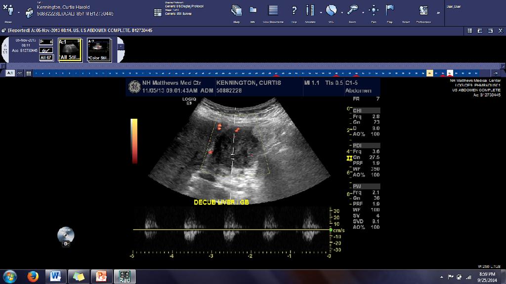
9
10
11
12
13
14
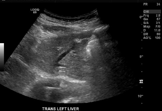
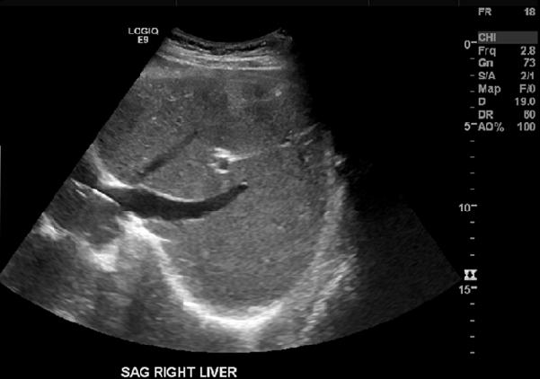
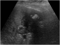
15 Interpretation: Liver: normal size and echotexture Gallbladder: Normal gallbladder not identified. 4 x 4 cm fairly well circumscribed mass-like focus in GB fossa. Periphery of mass is heterogeneous with intermediate echotexture, small central area with low echotexture. No calcification.
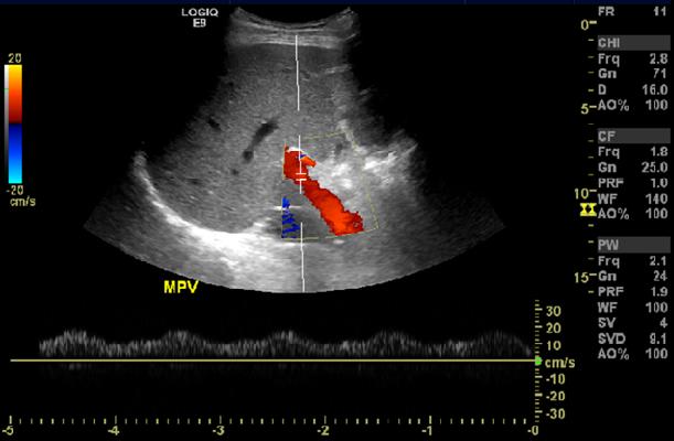
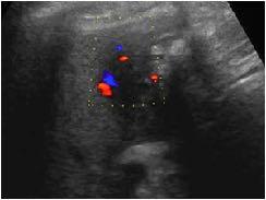
16 Mass demonstrates internal vascularity and blood flow with Doppler imaging. CBD: 6.7 mm, no stones seen MPV: patent with normal flow Pancreas: not visualized due to bowel gas Spleen: 7.3 cm, normal appearing IVC and Aorta are normal appearing No peritoneal fluid seen
17 Abnormal mass-like focus located in gallbladder fossa. No normal appearing gallbladder identified. Findings are worrisome for possible gallbladder carcinoma. A contrast enhance CT of the abdomen should be performed. Mildly dilated CBD
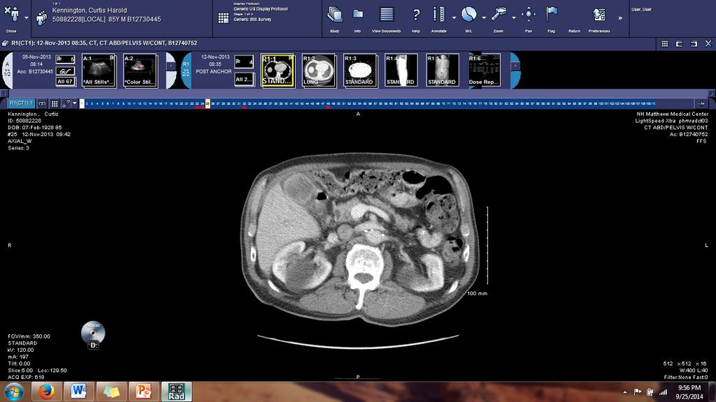
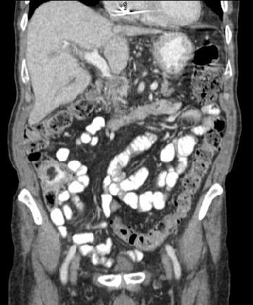
18 Contrast Enhanced CT of the Abdomen was performed one week later
19
20

21 Conclusion: 5 cm mass found in the fundus of the gallbladder. Possibly malignant. Although the gallbladder comes in contact with the hepatic parenchyma at the gallbladder fossa, there is no invasion identified. Mildly enlarged lymph nodes in the aortocaval space, not definitely malignant
22 Common Clinical Symptoms and Presentation of Gallbladder Carcinoma


23 Many patients are asymptomatic Early on symptoms are non-specific, usually mimicking benign gallbladder disease. As disease progresses patients may present with N&V, weight loss, anorexia, RUQ pain, hepatomegaly, and jaundice (Henningson & Kuntz, 2014)
24 Key Statistics About Gallbladder Cancer
25 Almost 6,000 new cases diagnosed every year 90% of GB cancers are adenocarcinomas 6% of GB cancers are papillary adenocarcinomas. Best prognosis because they do not spread. 4% adenosquamous carcinomas, squamous cell carcinomas, small cell carcinomas, and sarcomas. (Cancer Facts and Figures, 2014)
26 Risk Factors

 Diabetes Obesity (Cancer")
27 Older age Female (7 out of 10) Family HX Smoking Chemical exposure Prior history of biliary disease (GB stones, chronic cholecystitis, porcelain GB, choledochal cysts, primary sclerosing cholangitis, & GB polyps) Diabetes Obesity (Cancer Facts & Figures,2014)
28 Sonographic Findings
29 Inhomogeneous, polypoid lesion with irregular margins Localized wall thickening Mass that replaces gallbladder Calcification of gallbladder wall Polyps larger than 10 mm suspicious for malignancy Metastatic lesions in the liver Ascites Intraductal biliary dilation Internal blood flow (Henningson, 2014)
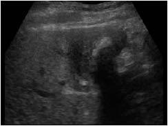
30 It is impossible to diagnose gallbladder carcinoma with ultrasound alone. GB carcinomas have the same sonographic appearance and symptoms as other diseases of the gallbladder. Let s see if you can determine what is cancer and what is not
31 (Abdomen and retroperitoneum, n.d.)


32 This was believed to be a gallbladder carcinoma infiltrating the liver. Actual diagnosis cholecystitis. (Abdomen and retroperitoneum, n.d.)
33 (Abdomen and retroperitoneum, n.d.)


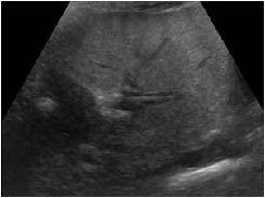
34 Described as cholecystitis, large stone, and sludge. Actual diagnosis: gallbladder carcinoma with invasion of bile ducts. (Abdomen and retroperitoneum, n.d.)
35 (Abdomen and retroperitoneum, n.d.)


36 (Abdomen and retroperitoneum, n.d.) Adenocarcinoma of the GB mistaken for polyps, cholecystitis, and stones.
37 (Abdomen and retroperitoneum, n.d.)
38 Cholecystitis with gallstones mimicking gallbladder carcinoma. (Abdomen and retroperitoneum, n.d.)
39 Abdomen and retroperitoneum 1.2 Gallbladder and bile ducts Case Gallbladder Carcinoma. (n.d.). Retrieved September 27, 2014, from Cancer Facts and Figures. (2014, February 6). Retrieved September 24, 2014, from gallbladder-cancer Henningsen, C., & Kuntz, K. (2014). Clinical guide to sonography: Exercises for critical thinking (Second ed., p. 7). St. Louis: Elsevier.
4/9/2018 OBJECTIVES PANCREAOTO BILIARY ULTRASOUND: BEYOND CHOLECYSTITIS
 PANCREAOTO BILIARY ULTRASOUND: BEYOND CHOLECYSTITIS Jean Yves Sewah Kaiser Permanente West Los Angeles 1 OBJECTIVES Discuss the role of ultrasound in the evaluation of the gallbladder, biliary tree and
Anatomy of the biliary tract
 Harvard-MIT Division of Health Sciences and Technology HST.121: Gastroenterology, Fall 2005 Instructors: Dr. Jonathan Glickman Anatomy of the biliary tract Figure removed due to copyright reasons. Biliary
Radiology of hepatobiliary diseases
 GI cycle - Lecture 14 436 Teams Radiology of hepatobiliary diseases Objectives 1. To Interpret plan x-ray radiograph of abdomen with common pathologies. 2. To know the common pathologies presentation.
Imaging of common diseases of hepatobiliary and GI system
 Imaging of common diseases of hepatobiliary and GI system Natthaporn Tanpowpong, M.D. Diagnostic radiology Faculty of Medicine, Chulalongkorn University Normal plain radiograph A = Common bile duct
Biliary Tree Ultrasound - In a nutshell. Pamela Parker Lead Sonographer
 Biliary Tree Ultrasound - In a nutshell Pamela Parker Lead Sonographer Aims Review what we know about the biliary system Common pathologies Pitfalls Reporting tips The Nutshell Background Biliary examinations
Abdominal ultrasound:
 Abdominal ultrasound: Non-traumatic acute abdomen Wittanee Na-ChiangMai, MD Department of Radiology ChiangMai University 26/04/2017 Contents Technique of examination Normal anatomy Emergency conditions
Dr Claire Smith, Consultant Radiologist St James University Hospital Leeds
 Dr Claire Smith, Consultant Radiologist St James University Hospital Leeds Imaging in jaundice and 2ww pathway Image protocol Staging Limitations Pancreatic cancer 1.2.4 Refer people using a suspected
Biliary Tree Ultrasound - In a nutshell. Pamela Parker Lead Sonographer
 Biliary Tree Ultrasound - In a nutshell Pamela Parker Lead Sonographer Aims Review what we know about the biliary system Common pathologies Pitfalls Reporting tips The Nutshell Background Biliary examinations
Imaging iconography of gallbladder cancer. Assessment by CT.
 1 REVISTA DE IMAGENOLOGIA- EII / Vol. XVI / Num. 2 Imaging iconography of gallbladder cancer. Assessment by CT. Doctors Crisci, Alejandro (1); Landó, Fernando.(2). CASMU CT Department Hospital of Tacuarembó
GASTROINTESTINAL IMAGING STUDY GUIDE
 GASTROINTESTINAL IMAGING STUDY GUIDE Pharynx Diverticula Foreign bodies Trauma o Motility Disorders Esophagus Diverticula Trauma Esophagitis Barrett esophagus Rings, webs, and strictures Varices Benign
Hepatobiliary Ultrasound Rimon Bengiamin, MD, RDMS Assistant Clinical Professor Director of Emergency Ultrasound UCSF Fresno. Objectives. Why?
 Hepatobiliary Ultrasound Rimon Bengiamin, MD, RDMS Assistant Clinical Professor Director of Emergency Ultrasound UCSF Fresno Objectives Discuss the goals of point-of-care biliary ultrasound Review the
Biliary tree dilation - and now what?
 Biliary tree dilation - and now what? Poster No.: C-1767 Congress: ECR 2012 Type: Educational Exhibit Authors: I. Ferreira, A. B. Ramos, S. Magalhães, M. Certo; Porto/PT Keywords: Pathology, Diagnostic
Case 1. Intro to Gallbladder & Pancreas Pathology. Case 1 DIAGNOSIS??? Acute Cholecystitis. Acute Cholecystitis. Helen Remotti M.D.
 Cholecystitis acute chronic Gallbladder tumors Adenomyoma (benign) Adenocarcinoma Pancreatitis acute chronic Pancreatic tumors Intro to Gallbladder & Pancreas Pathology Helen Remotti M.D. Case 1 70 year
Role of Ultrasonography and Computed Tomography in Gallbladder Masses and their Correlation with Fine-needle Aspiration Cytology
 Original Article Print ISSN: 2321-6379 Online ISSN: 2321-595X DOI: 10.17354/ijss/2018/119 Role of Ultrasonography and Computed Tomography in Gallbladder Masses and their Correlation with Avadhesh P S Kushwah
Intrahepatic Cholangiocarcinoma (ICC) Detected by Sonography
 Detected by Sonography") 661245JDMXXX10.1177/8756479316661245Journal of Diagnostic Medical SonographyHamer research-article2016 Case Study Intrahepatic Cholangiocarcinoma (ICC) Detected by Sonography Journal of Diagnostic Medical
661245JDMXXX10.1177/8756479316661245Journal of Diagnostic Medical SonographyHamer research-article2016 Case Study Intrahepatic Cholangiocarcinoma (ICC) Detected by Sonography Journal of Diagnostic Medical
Navigating the Biliary Tract with CT & MR: An Imaging Approach to Bile Duct Obstruction
 Navigating the Biliary Tract with CT & MR: An Imaging Approach to Bile Duct Obstruction Ann S. Fulcher, MD Medical College of Virginia Virginia Commonwealth University Richmond, Virginia Objectives To
Appendix 5. EFSUMB Newsletter. Gastroenterological Ultrasound
 EFSUMB Newsletter 87 Examinations should encompass the full range of pathological conditions listed below A log book listing the types of examinations undertaken should be kept Training should usually
Jaundice. Agnieszka Dobrowolska- Zachwieja, MD, PhD
 Jaundice Agnieszka Dobrowolska- Zachwieja, MD, PhD Jaundice definition Jaundice, as in the French jaune, refers to the yellow discoloration of the skin. It arises from the abnormal accumulation of bilirubin
Objectives. Hepatobiliary Ultrasound: Anatomy, Technique, Pathology. RUQ: Normal Anatomy. Emergency Ultrasound: Gallbladder Location
 Hepatobiliary Ultrasound: Anatomy, Technique, Pathology Laleh Gharahbaghian, MD FAAEM Associate Director, EM Ultrasound Co-Director, EM Ultrasound Fellowship Stanford University Medical Center Seric Cusick,
REFERRAL GUIDELINES: GALLSTONES
 REFERRAL GUIDELINES: GALLSTONES Document Purpose To ensure patients with gallstones disease are managed appropriately in primary/ secondary care Oxford Radcliffe Hospital Surgical Department Surgical Registrar
Abdomen and Retroperitoneum Ultrasound Protocols
 Abdomen and Retroperitoneum Ultrasound Protocols Reviewed By: Anna Ellermeier, MD Last Reviewed: March 2018 Contact: (866) 761-4200, Option 1 **NOTE for all examinations: 1. If documenting possible flow
Intraductal papillary neoplasms in the bile ducts
 Intraductal papillary neoplasms in the bile ducts Seok Hwa Youn Myunghee Yoon Dong Hoon Shin Kosin University Gospel Hospital Department of general surgery Hepato-biliary-pancreatic division Introduction
Sonography of Gall Bladder
 Sonography of Gall Bladder Vikram Dogra,MD Professor of Radiology, Urology and BME Director of Ultrasound Associate Chair of Education and Research University of Rochester, NY Objectives Describe the Congenital
Biliary Ultrasonography Kathleen O Brien MD MPH RDMS Kaiser Permanente South Sacramento
 Biliary Ultrasonography Kathleen O Brien MD MPH RDMS Kaiser Permanente South Sacramento https://www.google.com/search?sa=g&hl=en&q=public+disclosure&tbm=isch&tbs=simg:caqsigeahwelekju2aqaaawlelcmpwgaygpgcamskpib_1qnza7ai
Anatomy Jessica Ferguson Ashley Dobos May 31, 2006 LIVER
 Anatomy Jessica Ferguson Ashley Dobos May 31, 2006 LIVER 1) Other Names: Reidel s Lobe normal anatomic variant; projection of the right lobe that can extend as far as the iliac crest (Tempkin, p.54, Anatomy).
Case Scenario 1. Discharge Summary
 Case Scenario 1 Discharge Summary A 69-year-old woman was on vacation and noted that she was becoming jaundiced. Two months prior to leaving on that trip, she had had a workup that included an abdominal
Evaluation of Suspected Pancreatic Cancer
 Evaluation of Suspected Pancreatic Cancer October 15, 2015 If you experience technical difficulty during the presentation: Contact WebEx Technical Support directly at: US Toll Free: 1-866-779-3239 Toll
Intraductal papillary mucinous neoplasm of the bile ducts: a rare form of premalignant lesion of invasive cholangiocarcinoma
 Intraductal papillary mucinous neoplasm of the bile ducts: a rare form of premalignant lesion of invasive cholangiocarcinoma Authors: R. Revert Espí, Y. Fernandez Nuñez, I. Carbonell, D. P. Gómez valencia,
Newcastle HPB MDM updated radiology imaging protocol recommendations. Author Dr John Scott. Consultant Radiologist Freeman Hospital
 Newcastle HPB MDM updated radiology imaging protocol recommendations Author Dr John Scott. Consultant Radiologist Freeman Hospital This document is intended as a guide to aid radiologists and clinicians
Abdominal Ultrasound. Diane Hallinen, MD. Bloodroot
 Abdominal Ultrasound Diane Hallinen, MD Bloodroot Abdominal Ultrasound Vasculature Hepatobiliary Spleen Kidney Bladder Bowel Where to put the probe? Vasculature We are going to talk about Celiac Trunk
Imaging Guided Biopsy. Edited & Presented by ; Hussien A.B ALI DINAR. Msc Lecturer,Reporting Sonographer
 Imaging Guided Biopsy Edited & Presented by ; Hussien A.B ALI DINAR. Msc Lecturer,Reporting Sonographer Objective By the End of this lessons you should : Define what biopsy Justify Aim to perform biopsy
Personal Profile. Name: 劉 XX Gender: Female Age: 53-y/o Past history. Hepatitis B carrier
 Personal Profile Name: 劉 XX Gender: Female Age: 53-y/o Past history Hepatitis B carrier Chief complaint Fever on and off for 2 days Present illness 94.10.14 Sudden onset of epigastric pain 94.10.15 Fever
Hilar cholangiocarcinoma. Frank Wessels, Maarten van Leeuwen, UMCU utrecht
 Hilar cholangiocarcinoma Frank Wessels, Maarten van Leeuwen, UMCU utrecht Content Anatomy Biliary strictures (Hilar) Cholangiocarcinoom Staging Biliary tract 1 st order Ductus hepatica dextra Ductus hepaticus
Gallbladder & Pancreas Ultrasonography
 복부초음파 : 담낭과췌장 Gallbladder & Pancreas Ultrasonography 김정훈 Department of Radiology 1 Interaction of sound with matter (1) 반사 (Reflection) (2) 굴절 (Refraction) (3) 흡수 (Absorption) (4) 산란 (Scattering) 음향저항
Biliary cancers: imaging diagnosis. Study of 30 cases
 Biliary cancers: imaging diagnosis. Study of 30 cases N Hammoune, S Semlali, M Eddarai, T. Amil, M Zentar, S. El Kandri,, M Benameur,, S Chaouir. Radiology Department. Mohamed V Military Hospital. Rabat-
US in non-traumatic acute abdomen. Lalita, M.D. Radiologist Department of radiology Faculty of Medicine ChiangMai university
 US in non-traumatic acute abdomen Lalita, M.D. Radiologist Department of radiology Faculty of Medicine ChiangMai university Sagittal Orientation Transverse (Axial) Orientation Coronal Orientation Intercostal
Elastography in the. technically difficult patient. EPIQ ultrasound system. Ultrasound
 Ultrasound Elastography in the technically difficult patient EPIQ ultrasound system Chairman Department of Diagnostic Radiology Allegheny General Hospital Pittsburgh, PA, USA You can offer more information
MANAGEMENT OF INCIDENTALLY DETECTED GALLBLADDER CANCER
 MANAGEMENT OF INCIDENTALLY DETECTED GALLBLADDER CANCER Orlando Jorge M. Torres Full Professor and Chairman Department of Gastrointestinal Surgery Hepatopancreatobiliary Unit Federal University of Maranhão
GALLBLADDER CANCER. Lidie M. Lajoie MD Downstate Surgery M&M July 21, 2011
 GALLBLADDER CANCER Lidie M. Lajoie MD Downstate Surgery M&M July 21, 2011 Agenda Case Presentation Epidemiology Pathogenesis & Pathology Staging Presentation & Diagnosis Stage-wise Management Outcomes/Prognosis
My Patient Has Abdominal Pain PoCUS of the Biliary Tract and the Urinary Tract
 My Patient Has Abdominal Pain PoCUS of the Biliary Tract and the Urinary Tract Objectives PoCUS for Biliary Disease PoCUS for Renal Colic PoCUS for Urinary Retention Biliary Disease A patient presents
Cholangiocarcinoma: Radiologic evaluation and interventions
 November 2014 Cholangiocarcinoma: Radiologic evaluation and interventions Colin Nevins, Harvard Medical School Year III Agenda Initial course and work-up Endoscopic retrograde cholangiopancreatography
Malignant Focal Liver Lesions
 Malignant Focal Liver Lesions Other Than HCC Pablo R. Ros, MD, MPH, PhD Departments of Radiology and Pathology University Hospitals Cleveland Medical Center Case Western Reserve University Pablo.Ros@UHhospitals.org
Vesalius SCALpel : Biliary (see also: biliary/pancreatic folios) Physiology
 Physiology") Vesalius SCALpel : Biliary (see also: biliary/pancreatic folios) Physiology 95% of bile acids reabsorbed; colic and chenodeoxycolic primary bile acids cholecystokinin (CCK) major stimulus of gallbladder
Vesalius SCALpel : Biliary (see also: biliary/pancreatic folios) Physiology 95% of bile acids reabsorbed; colic and chenodeoxycolic primary bile acids cholecystokinin (CCK) major stimulus of gallbladder
Guidelines, Policies and Statements D5 Statement on Abdominal Scanning
 Guidelines, Policies and Statements D5 Statement on Abdominal Scanning Disclaimer and Copyright The ASUM Standards of Practice Board have made every effort to ensure that this Guideline/Policy/Statement
Liver Ultrasound - Beyond the Basics. Pamela Parker Lead Sonographer
 Liver Ultrasound - Beyond the Basics Pamela Parker Lead Sonographer Aims Review what we know about the liver Reasons for imaging Focal lesions Diffuse disease Can we do more? The Liver The Liver The Liver
Chief Complain. Liver lesion found in routine health check 41 days ago
 Chief Complain Liver lesion found in routine health check 41 days ago Present Illness On 2005-7-26 at 台北署立醫院 he underwent a health check for the first time. Abdominal US showed suspicious of a 6*5 cm hepatoma,
Appendix 9: Endoscopic Ultrasound in Gastroenterology
 Appendix 9: Endoscopic Ultrasound in Gastroenterology This curriculum is intended for clinicians who perform endoscopic ultrasonography (EUS) in gastroenterology. It includes standards for theoretical
Pediatric Hepatobiliary, Pancreatic & Splenic US
 Pediatric Hepatobiliary, Pancreatic & Splenic US Susan J. Back, MD Department of Radiology, The Children s Hospital of Philadelphia No Disclosures Objectives Normal Abnormal: cases and US advances Objectives
Congenital dilatation of the common bile duct and pancreaticobiliary maljunction clinical implications
 Langenbecks Arch Surg (2009) 394:209 213 DOI 10.1007/s00423-008-0330-6 CURRENT CONCEPT IN CLINICAL SURGERY Congenital dilatation of the common bile duct and pancreaticobiliary maljunction clinical implications
Alice Fung, MD Oregon Health and Science University
 Alice Fung, MD Oregon Health and Science University Disclosure Comments The speaker Alice Fung, MD Has relevant financial relationships to disclose. Received honorarium from (Guerbet). This individual
Cholangiocellular carcinoma. Dr. med. Henrik Csaba Horváth PhD
 Cholangiocellular carcinoma Dr. med. Henrik Csaba Horváth PhD Acalculous biliary diseases April 12, 2017 2 Cholangiocarcinoma A slow growing malignancy of the biliary tract which tend - to infiltrate locally
Icd-10 for cancer that spread to the lungs
 Icd-10 for cancer that spread to the lungs The Borg System is 100 % Icd-10 for cancer that spread to the lungs Breast cancer metastatic to lymph node; Cancer metastatic to lymph node, adenocarcinoma; Cancer
Management of Gallbladder Disease
 Management of Gallbladder Disease Steven B. Johnson, MD, FACS, FCCM Professor and Chairman, Department of Surgery Program Director, Phoenix Integrated Surgical Residency University of Arizona College of
Hepatocellular Dysfunction
 www.nuclearmd.com A Normal HIDA Scan Dynamic or Static images of the abdomen are acquired after the IV administration 6-8 mci of Tc99m disofenin or mebrofenin, for one hour. Patients have to be NPO for
Abdominal Imaging. Gallbladder perforation: color Doppler findings
 Abdom Imaging 27:47 50 (2002) DOI: 10.1007/s00261-001-0048-1 Abdominal Imaging Springer-Verlag New York Inc. 2002 Gallbladder perforation: color Doppler findings K. Konno, 1 H. Ishida, 1 M. Sato, 1 H.
MALIGNANT HEPATIC NEOPLASMS: USING ULTRASONOGRAPHY AS A MEANS OF DEFINING HEPATIC LESIONS. 1.5 Contact Hours. Presented by: CEU Professor 7
 MALIGNANT HEPATIC NEOPLASMS: USING ULTRASONOGRAPHY AS A MEANS OF DEFINING HEPATIC LESIONS 1.5 Contact Hours Presented by: CEU Professor 7 www.ceuprofessoronline.com Copyright 8 2007 The Magellan Group,
OF HEMATOMA INTRACRANIAL HEMORRHAGE
 SCOPE TUTORIAL RADIOLOGY CT BRIAN BREASTS US MRCP Natrada Rawdhetubhai, M.D. Radiology Department, Lerdsin General Hospital CT OF HEMATOMA INTRACRANIAL HEMORRHAGE Intra-axial Extra-axial Intraventricular
Policies, Standards, and Guidelines. Guidelines for Abdominal Ultrasound Examination
 Policies, Standards, and Guidelines Guidelines for Abdominal Ultrasound Examination Approved by Council Feb 2018 Disclaimer and Copyright The ASUM Standards of Practice Board have made every effort to
Cholangiocarcinoma (Bile Duct Cancer)
") Cholangiocarcinoma (Bile Duct Cancer) The Bile Duct System (Biliary Tract) A network of bile ducts (tubes) connects the liver and the gallbladder to the small intestine. This network begins in the liver
Cholangiocarcinoma (Bile Duct Cancer) The Bile Duct System (Biliary Tract) A network of bile ducts (tubes) connects the liver and the gallbladder to the small intestine. This network begins in the liver
Role of multidetector computed tomography (MDCT) in diagnosis and staging of gall bladder carcinoma
 in diagnosis and staging of gall bladder carcinoma") The Egyptian Journal of Radiology and Nuclear Medicine (2013) 44, 1 7 Egyptian Society of Radiology and Nuclear Medicine The Egyptian Journal of Radiology and Nuclear Medicine www.elsevier.com/locate/ejrnm
The Egyptian Journal of Radiology and Nuclear Medicine (2013) 44, 1 7 Egyptian Society of Radiology and Nuclear Medicine The Egyptian Journal of Radiology and Nuclear Medicine www.elsevier.com/locate/ejrnm
Management of Cholangiocarcinoma. Roseanna Lee, MD PGY-5 Kings County Hospital
 Management of Cholangiocarcinoma Roseanna Lee, MD PGY-5 Kings County Hospital Case Presentation 37 year old male from Yemen presented with 2 week history of epigastric pain, anorexia, jaundice and puritis.
Undifferentiated carcinoma of the gall bladder: a rare entity
 Case Report Undifferentiated carcinoma of the gall bladder: a rare entity Medhi Pranita 1,*, Dowerah Swagata 2, Dutta Utpal 3, Dutta Aparna 4 1 Associate Professor, 2 Demonstrator, 3 PGT, 4 Assistant Professor,
Contemporary Imaging of Biliary Malignancy and Preoperative Evaluation
 Contemporary Imaging of Biliary Malignancy and Preoperative Evaluation Linda Pantongrag-Brown, MD Advanced Diagnostic Imaging, Ramathibodi Hospital, Bangkok, Thailand Malignancy of biliary tract Cholangiocarcinoma
Spectrum of Cholangiocarcinoma
 Scholars Journal of Applied Medical Sciences (SJAMS) Sch. J. App. Med. Sci., 2013; 1(6):695-699 Scholars Academic and Scientific Publisher (An International Publisher for Academic and Scientific Resources)
IMAGING OF LIVER, BILIARY TREE, PANCREAS
 IMAGING OF LIVER, BILIARY TREE, PANCREAS Department of Radiology West China Hospital, Sichuan University Yao Jin Learning Points The methodology for imaging the LBP (liver, biliary tree, and pancreas )
Certificate in Clinician Performed Ultrasound (CCPU) Syllabus. Biliary
 Syllabus. Biliary") Certificate in Clinician Performed Ultrasound (CCPU) Syllabus Biliary Page 1 of 6 12/18 Biliary Syllabus Purpose: This unit is designed to cover the theoretical and practical curriculum for basic ultrasound
Certificate in Clinician Performed Ultrasound (CCPU) Syllabus Biliary Page 1 of 6 12/18 Biliary Syllabus Purpose: This unit is designed to cover the theoretical and practical curriculum for basic ultrasound
Hepato-Pancreatico-Biliary Surgery. Dr. Ankur J. Shah. MS, DNB, MNAMS, MRCSEd (UK), FRCS (UK)
, FRCS (UK)") Hepato-Pancreatico-Biliary Surgery Dr. Ankur J. Shah MS, DNB, MNAMS, MRCSEd (UK), FRCS (UK) Consultant Hepato-Pancreatico-Biliary and Liver Transplant Surgeon Ansh Liver Clinic Prevention to Cure Address
Hepato-Pancreatico-Biliary Surgery Dr. Ankur J. Shah MS, DNB, MNAMS, MRCSEd (UK), FRCS (UK) Consultant Hepato-Pancreatico-Biliary and Liver Transplant Surgeon Ansh Liver Clinic Prevention to Cure Address
The role for contrast-enhanced ultrasonography outside of focal liver lesions
 The role for contrast-enhanced ultrasonography outside of focal liver lesions Paul S. Sidhu King s College Hospital, London, UK Introduction Contrast-enhanced ultrasonography (US) of focal liver lesions
Clinical Spectrum of Presentation of Obstructive Jaundice in Inflammation, Stone Disease, and Malignancy
 Original Article Print ISSN: 2321-6379 Online ISSN: 2321-595X DOI: 10.17354/ijss/2017/326 Clinical Spectrum of Presentation of Obstructive Jaundice in Inflammation, Stone Disease, and Malignancy R Selvasekaran
Gall bladder cancer. Information for patients Hepatobiliary
 Gall bladder cancer Information for patients Hepatobiliary page 2 of 12 Who will provide my care? You will be cared for by a number of professionals who work together. These professionals will be specialist
EFSUMB EUROPEAN FEDERATION OF SOCIETIES FOR ULTRASOUND IN MEDICINE AND BIOLOGY Building a European Ultrasound Community
 MINIMUM TRAINING REQUIREMENTS FOR THE PRACTICE OF MEDICAL ULTRASOUND IN EUROPE Appendix 9: Endoscopic Ultrasound in Gastroenterology This curriculum is intended for clinicians who perform endoscopic ultrasonography
Pancreas Case Scenario #1
 Pancreas Case Scenario #1 An 85 year old white female presented to her primary care physician with increasing abdominal pain. On 8/19 she had a CT scan of the abdomen and pelvis. This showed a 4.6 cm mass
Case Discussion Splenic Abscess
 Case Discussion Splenic Abscess Personal Data Gender: male Birth Date: 1928/Mar/06th Allergy: Mefenamic Smoking: 0.5 PPD for 55 years Alcohol: negative (?) 4 Months Ago Abdominal pain: epigastric area
ULTRASOUND AND ABDOMINAL MASSES
 Med. J. Malaysia Vol. 37 No. I March 1982. ULTRASOUND AND ABDOMINAL MASSES AHMAD KAMAL BIN MD ALIF INTRODUCTION It is approximately 30 years since ultrasound was first introduced into the field of medicine,
Approach to the Patient with Liver Disease
 Approach to the Patient with Liver Disease Diagnosis of liver disease Careful history taking Physical examination Laboratory tests Radiologic examination and imaging studies Liver biopsy Liver diseases
IT 의료융합 1 차임상세미나 복부질환초음파 이재영
 IT 의료융합 1 차임상세미나 2013-4-3 복부질환초음파 이재영 나는오늘누구를위하여 종을울리나? 전통적의료 의사 공학설계자 의사 최첨단진단장비들 USG, CT, MRI 환자 환자 현대의료 사용자중심의사고 US in the Abdomen Detection DDx Look Behavior Response by external stimuli Guiding Tool
Imaging of liver and pancreas
 Imaging of liver and pancreas.. Disease of the liver Focal liver disease Diffusion liver disease Focal liver disease Benign Cyst Abscess Hemangioma FNH Hepatic adenoma HCC Malignant Fibrolamellar carcinoma
Case Reports. Intraductal Papillary Cholangiocarcinoma: Case Report and Review of the Literature INTRODUCTION CASE REPORT
 Case Reports Kongkam K, Rerknimitr R 45 Case Report and Review of the Literature Pradermchai Kongkam, M.D. Rungsun Rerknimitr, M.D. ABSTRACT A case of papillary cholangiocarcinoma is presented. A 64-year-old
Birthday: 1952/07/31 Date of admission:1999/12/30 Age:48 y/o Past medication:esrd under regular HD for 5+ years; denied DM and HTN
 Birthday: 1952/07/31 Date of admission:1999/12/30 Age:48 y/o Past medication:esrd under regular HD for 5+ years; denied DM and HTN Chief Complaint : 1)intermittent LLQ cramping pain for 2 months 2) LGI
Liver and Pancreatic Case discussion
 The Royal Marsden Liver and Pancreatic Case discussion Dr Ian Chau Consultant Medical Oncologist The Royal Marsden 77 year old gentleman with 2 months history of vague abdominal ache and clinically finding
MR imaging of primary sclerosing cholangitis (PSC) using the hepatobiliary specific contrast agent Gd-EOB-DTPA
 using the hepatobiliary specific contrast agent Gd-EOB-DTPA") MR imaging of primary sclerosing cholangitis (PSC) using the hepatobiliary specific contrast agent Gd-EOB-DTPA Poster No.: C-0019 Congress: ECR 2010 Type: Educational Exhibit Topic: Abdominal Viscera (Solid
MR imaging of primary sclerosing cholangitis (PSC) using the hepatobiliary specific contrast agent Gd-EOB-DTPA Poster No.: C-0019 Congress: ECR 2010 Type: Educational Exhibit Topic: Abdominal Viscera (Solid
Extrahepatic Bile Duct Ostruction (Blockage of the Extrahepatic or Common Bile Duct) Basics
 Basics") Extrahepatic Bile Duct Ostruction (Blockage of the Extrahepatic or Common Bile Duct) Basics OVERVIEW The liver is the largest gland in the body; it has many functions, including production of bile (a fluid
Extrahepatic Bile Duct Ostruction (Blockage of the Extrahepatic or Common Bile Duct) Basics OVERVIEW The liver is the largest gland in the body; it has many functions, including production of bile (a fluid
US Applications. Case Based Wrap-Up 1. Case 1 E-FAST. Case presentations E-FAST Abdominal. Pearls for each indication
 Case Based Wrap-Up 1 Stephanie J. Doniger MD RDMS FAAP FACEP Associate Director, Pediatric Emergency Ultrasound Stanford University Medical Center US Applications Case presentations E-FAST Abdominal Aorta
Biliary Tract Disease. Emmet Andrews Cork University Hospital 6 th September 2010
 Biliary Tract Disease Emmet Andrews Cork University Hospital 6 th September 2010 Overview Gallstones Biliary tract tumours Other conditions Acute acalculous cholecystitis Mirizzi s syndrome Primary Biliary
A patient with an unusual congenital anomaly of the pancreaticobiliary tree
 A patient with an unusual congenital anomaly of the pancreaticobiliary tree Thomas Hocker, HMS IV BIDMC Core Radiology Case Presentation September 17, 2007 Review of Normal Pancreaticobiliary Tract Anatomy
Ovarian Lesion Benign vs Malignant?
 Ovarian Lesion Benign vs Malignant? Michele Keenan 1,2 Bernice Dunne 2 Mary Moran 1 Therese Herlihy 1 1. Radiography and Diagnostic Imaging, School of Medicine, University College Dublin, Ireland 2. Midland
MRI Abdomen Protocol Pancreas/MRCP with Contrast
 MRI Abdomen Protocol Pancreas/MRCP with Contrast Reviewed By: Brett Mollard, MD; Anna Ellermeier, MD Last Reviewed: July 2018 Contact: (866) 761-4200 Standard uses: 1. Characterization of cystic and solid
Chronic pancreatitis mimicking intraductal papillary mucinous neoplasm of the pancreas; Report of tow cases
 Jichi Medical University Journal Chronic pancreatitis mimicking intraductal papillary mucinous neoplasm of the pancreas; Report of tow cases Noritoshi Mizuta, Hiroshi Noda, Nao Kakizawa, Nobuyuki Toyama,
Expected and unexpected gallstones in primary care
 Expected and unexpected gallstones in primary care 7 Speets AM, Van der Graaf Y, Hoes AW, Kalmijn S, De Wit NJ, Mali WPThM. Expected and unexpected gallstones in primary care. Submitted. CHAPTER 7 Abstract
Basic Abdominal Sonography
 24S Basic Abdominal Sonography Procedural Overview JOHN FATCHETT II, RDMS is provided. Patient preparation (i.e., fasting) scanning techniques, spleen, transducer. evaluation of abdominal anatomy in the
Original Contributions
 PII S0736-4679(01)00329-8 The Journal of Emergency Medicine, Vol. 21, No. 1, pp. 7 13, 2001 Copyright 2001 Elsevier Science Inc. Printed in the USA. All rights reserved 0736-4679/01 $ see front matter
Multi modality Imaging in Acute Pancreatitis. Marsha Lynch, HMS III Gillian Lieberman, MD BIDMC Core Clerkship in Radiology March 2009
 Multi modality Imaging in Acute Pancreatitis Marsha Lynch, HMS III Gillian Lieberman, MD BIDMC Core Clerkship in Radiology March 2009 Our Patient R: Introduction 52M with 10d history of nausea, vomiting
CT 101 :Pancreas and Spleen
 CT 101 :Pancreas and Spleen Shikha Khullar,, MD, MPH Division of Radiology University of South Alabama The Pancreas Normal Pancreas 3 Phase Pancreatic CT Non contrast Arterial phase : 30-35 35 second
Frank Burton Memorial Update on Pancreato-biliary Cancers
 Frank Burton Memorial Update on Pancreato-biliary Cancers Diagnosis and management of pancreatic cancer: common dilemmas Moderators: Banke Agarwal, MD Paul Buse, MD Evaluation of patients with obstructive
7/11/2017. We re gonna help a lot of people today. Biliary/Pancreatic Endoscopy. AGS July 1-2, Kenneth M. Sigman, MD
 Biliary/Pancreatic Endoscopy AGS July 1-2, 2017 Kenneth M. Sigman, MD We re gonna help a lot of people today 1 2 3 4 Cannulation It all starts with cannulation Double Wire Cannulation Difficult cannulations
Normal Sonographic Anatomy
 hapter 2:The Liver DUNSTAN ABRAHAM Normal Sonographic Anatomy Homogeneous, echogenic texture (Figure 2-1) Measures approximately 15 cm in length and 10 12.5 cm anterior to posterior; measurement taken
The campaign on laboratory: focus on Gallstone Disease and ERCP
 The campaign on laboratory: focus on Gallstone Disease and ERCP Mauro Giuliani, MD, Specialist in Visceral Surgery, Vice Head Physician, Surgical Ward, Ospedale Regionale di Locarno Alberto Fasoli, MD,
Resident Teaching Conference:
 Resident Teaching Conference: Evaluation of the Dreaded Liver Mass May 6, 2011 Sunil Geevarghese, MD, HS 00 Matt Landman, MD Anatomy Overview Couinaud anatomy Resection nomenclature Functional Assessment
Rokitansky-Aschoff sinuses are epithelial invaginations in the gallbladder wall that from as a result of increased gallbladder pressures.
 Anatomy The complexity of the biliary tree can be broken down into much simpler segments. The intrahepatic ducts converge to form the right and left hepatic ducts which exit the liver and join to become
X-ray Corner. Imaging of The Pancreas. Pantongrag-Brown L
 X-ray Corner 125 Imaging of The Pancreas Modern imaging modalities commonly used in pancreas include ultrasound (US), CT, and MRI. Pancreas is a retroperitoneal organ which makes it difficult to visualize