Managing Patient Dose in Computed Tomography (CT)
|
|
|
- Emmeline Miles
- 5 years ago
- Views:
Transcription


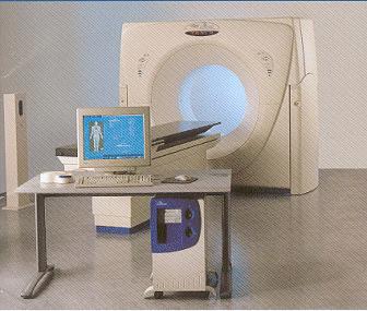
1 Managing Patient Dose in Computed Tomography (CT)

2 International Commission on Radiological Protection Information abstracted from ICRP Publication 87 Available at Task Group: M.M. Rehani, G. Bongartz, S.J. Golding, L.Gordon, W. Kalender, T. Murakami, P. Shrimpton, R. Albrecht, K. Wei

3 Use and Disclaimer This is a PowerPoint file It may be downloaded free of charge It is intended for teaching and not for commercial purposes This slide set is intended to be used with the complete text provided in ICRP Publication 87


4 Contents Situation analysis Why increased frequency? Why increased dose? Is the dose really high? How high? What can be done to manage patient dose? What can operator do? Action for manufacturer Action for physician & radiologist

5 Situation Analysis CT continues to evolve rapidly despite many advances in other imaging modalities It is one of the most important radiological examinations worldwide The frequency of CT examinations is increasing rapidly from 2% of all radiological examinations in some countries a decade ago to % now Patient doses in CT have not decreased in contrast to radiography where nearly 30% reduction has been documented in last decade

 The absorbed dose to tissues from CT can often approach or exceed the levels known to")
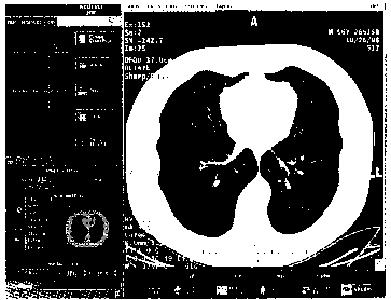
6 What is the dose from CT? How high? The effective dose in chest CT is in the order of 8 msv (around 400 times more than chest radiograph dose) and in some CT examinations like that of pelvic region, it may be around 20 msv (1000 CXRs) The absorbed dose to tissues from CT can often approach or exceed the levels known to increase the probability of cancer as shown in epidemiological studies
 CT Chest 8 msv (400) CT Abdomen or Pelvis 10 msv (500) Virtual Colonoscopy 36-50")
7 Radiation Exposure by Source/ Test Chest X-Ray 0.02 msv (1) Lumbar Spine 1.3 msv (65) CT Chest 8 msv (400) CT Abdomen or Pelvis 10 msv (500) Virtual Colonoscopy msv( ) Hiroshima Survivors mSv


8 A. Contributions to frequency B. Contributions to prescribed dose 5% 8% 41% 9% 15% 12% 2% 1% 6% 5% 29% 34% 10% 1% UNSCEAR 2000


9 Why Increased Frequency? 20 years ago, a standard CT of the thorax took several minutes while today similar information can be accumulated in a single breath hold making it attractive, patient & user friendly Advances in CT technology have made possible CT fluoroscopy and interventional procedures, in some cases replacing ultrasound guided interventions Repeat CT exams

10 Why increased dose Unlike radiography where over-exposure results in blackening of film, better image quality is obtained with higher exposures in CT There is a tendency to increase the volume covered in a particular examination Modern helical CT involves volume scanning with no inter-slice gap and with possibility of overlapping scans Repeat CT examinations

 as for abdomen (low contrast")
11 Why increased dose (cont d) Same exposure factors used for children as for adult Same exposure factors for pelvic (high contrast region) as for abdomen (low contrast region)

12 Organ doses in CT Breast dose in thorax CT may be as much as mgy, even though breasts are not the target of imaging procedure Eye lens dose in brain CT, thyroid in brain or in thorax CT and gonads in pelvic CT receive high doses


13 Tissues in the field although they are not the area of interest for the procedure Lens of the eye Breast tissue

14 Cataract in eye of interventionist after repeated use of over table x-ray tube
 Not all skin reactions are due to contrast medium")
15 Informed consent and records Patients are entitled to know the risks of radiation injury if likely to be high A written record should be kept if skin doses are estimated to be >3 Gy (1 Gy for repeated procedures) Not all skin reactions are due to contrast medium allergy


16 Atrophic indurated plaque Hyper & hypo pigmentation, with telangiectasia Chronic radiodermatitis in 17 year old female patient after x2 radiofrequency ablation procedures


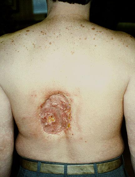
17 Follow-up Radiation skin injury may present late and the association not considered if no documentation All patients with estimated skin doses of 3 Gy should be followed up days after exposure A system to identify repeat procedures should be set up


18 Example of chronic skin injury due to cumulative skin dose of ~20,000 mgy (20 Gy) from coronary angiography and x2 angioplasties 21 months after first procedure, base of ulcer exposes spinous process
19

20 Leukemia and Cancer Most interventional procedures are performed on older patients where benefit almost always outweighs radiation risk The radiation risk increases progressively with younger age groups Radiation has been shown to increase the risk for leukemia and many types of cancer in adults and children
21 Does spiral CT give more or less radiation dose? It depends upon the choice of factors Even though it is possible to perform a spiral CT with lower radiation dose than slice-by-slice CT, in practice the patient gets higher dose due to the factors chosen (scan volume, mas, pitch, slice width

22 Does multi-slice CT impart more or less radiation dose? An increase by 10-30% may occur with multi-slice detector array


23 Some Observations Most doctors including many radiologists have a feeling that modern CT scanners which are very fast give lesser radiation dose Unfortunately time and radiation dose are not proportional in such a situation Over the years the x-ray tubes are becoming more and more powerful such that they can give high bursts of x-rays which can give satisfactory image in shorter exposure time


24 Actions for Physician & Radiologist Justification: Ensure that patients are not irradiated unjustifiably Request for CT examination should be generated only by properly qualified medical or dental practitioners depending upon national educational and qualification system. The physician is responsible for weighing the benefits against risks Clinical guidelines advising which examinations are appropriate and acceptable should be available to clinicians and radiologists

25 Actions for Physician & Radiologist (cont d) Consider whether the required information be obtained by MRI, ultrasonography Consider value of contrast medium enhancement prior to commencing examination CT scanning in pregnancy may not be contraindicated, particularly in emergency situations, although examinations of the abdomen or pelvis should be carefully justified

26 Actions for Physician & Radiologist (cont d) CT examination should not be repeated without clinical justification and should be limited to the area of interest Clinician has the responsibility to communicate to the radiologist about previous CT examination of the patient CT examination for research purposes that do not have clinical justification (immediate benefit to the person undergoing the examination) should be subject to critical evaluation by an ethics committee

27 Actions for Physician & Radiologist (cont d) CT examination of chest in young girls and young females needs to be justified in view of high breast dose Once the examination has been justified, radiologist has the primary responsibility for ensuring that the examination is carried out with good technique

28 Web sites for additional information on radiation sources and effects European Commission (radiological protection pages): europa.eu.int/comm/environment/radprot International Atomic Energy Agency: International Commission on Radiological Protection: United Nations Scientific Committee on the Effects of Atomic Radiation: World Health Organization:

29 Interventional Procedures Avoiding Radiation Injuries

30 Background Interventional techniques using radiation are now practiced by clinicians of many specialities Most clinicians are unaware of the potential for radiation injury

31 Background Patients are often not informed of radiation risks Staff may also be exposed to high doses Techniques are available to reduce doses to patients and staff

32 Introduction Many interventional procedures are performed by clinicians largely untrained in radiation effects and safety some patients & staff have suffered unnecessary injuries Most patients are not counselled on radiation risks nor followed up appropriately to detect injury Doses to patients and staff can often be reduced without compromising clinical outcome

33 Medical Radiation Procedures All procedures involving radiation should be justified (more benefit than risk) Medical exposures should also be justified on an individual basis before being performed Once justified, the actual procedure and dose should be tailored to the individual patient

34 Interventional Procedures Doses In some procedures, patient skin doses approach those used in radiotherapy fractions In young patients, organ doses may significantly increase the risk of radiation-induced cancer in later life


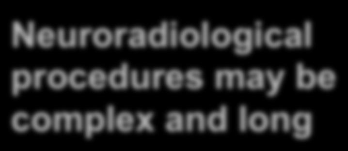
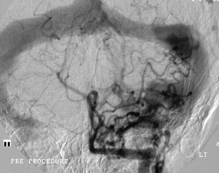
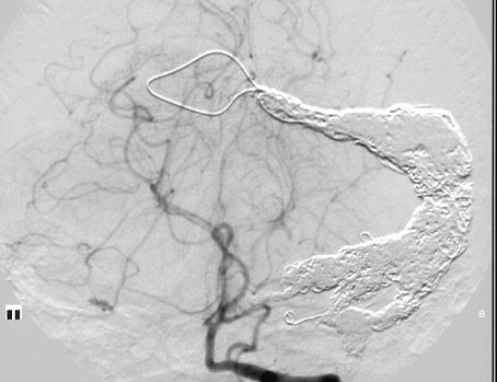
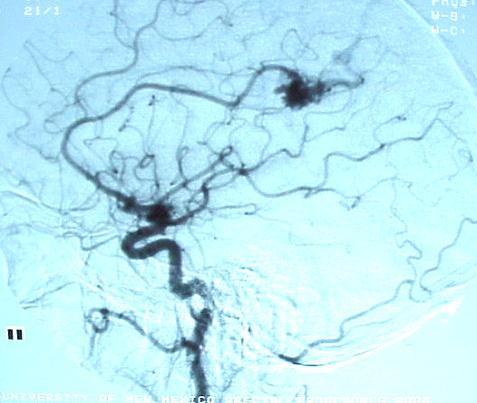
35 17 years female - large dural fistula of left lateral sinus Neuroradiological procedures may be complex and long Post embolisation Images courtesy of Dr JN Higgins

36 Interventional procedures doses Higher doses are often due to inappropriate equipment or poor technique Irradiation of the eye can cause cataract


37 Patient age 60. Tight stenosis of obtuse marginal artery on coronary angiography Technically difficult procedure lengthy screening Following angioplasty and stent insertion

38 Interventional procedures REMEMBER: Even a straightforward procedure can become high-dose with poor technique However, even with good technique adverse effects occur


39 Case 17 years female. Left dural fistula pre and post embolisation - multiple procedures Fluoroscopy time over 19 hours in one year 2 episodes hair loss - both recovered Lengthy and repeated procedures may be unavoidable Images courtesy of Dr JN Higgins


40 Controlling Dose to Staff REMEMBER: Controlling dose to patient will help control dose to staff


41 Summary The radiation risk is usually outweighed by the benefit of the procedure Both patients and staff are at risk of radiation injury Appropriate equipment and training are needed to minimise this risk Patient counselling should be undertaken routinely, and follow up when appropriate
Managing Patient Dose in Computed Tomography (CT) INTERNATIONAL COMMISSION ON RADIOLOGICAL PROTECTION
 INTERNATIONAL COMMISSION ON RADIOLOGICAL PROTECTION") Managing Patient Dose in Computed Tomography (CT) International Commission on Radiological Protection Information abstracted from ICRP Publication 87 Available at www.icrp.org Task Group: M.M. Rehani,
Managing Patient Dose in Computed Tomography (CT) International Commission on Radiological Protection Information abstracted from ICRP Publication 87 Available at www.icrp.org Task Group: M.M. Rehani,
CT Radiation Risks and Dose Reduction
 CT Radiation Risks and Dose Reduction Walter L. Robinson, M.S. D.A.B.S.N.M., D.A.B.M.P., D.A.B.R. Consultant Certified Medical Radiation Health & Diagnostic Imaging Physicist Medical Radiation and Children
ICRP Symposium on the International System of Radiological Protection
 ICRP Symposium on the International System of Radiological Protection October 24-26, 2011 Bethesda, MD, USA M. Balonov, ICRP Committee 2, IRH, Russia P. Shrimpton, HPA, UK To be used just for regulatory
Why radiation protection matters?
 Why radiation protection matters? Elias Brountzos Professor of Radiology 2 nd Department of Radiology Medical School, University of Athens Athens, Greece A definition for radiation protection Radiation
Radiology. General radiology department. X-ray
 The radiology directorate provides a diagnostic, interventional and therapeutic service for its local population, and a tertiary service for the region. It also provides support to some national work such
Managing Radiation Risk in Pediatric CT Imaging
 Managing Radiation Risk in Pediatric CT Imaging Mahadevappa Mahesh, MS, PhD, FAAPM, FACR, FACMP, FSCCT. Professor of Radiology and Cardiology Johns Hopkins University School of Medicine Chief Physicist
 Page 1 of 5 Patient Safety: Radiation Dose in X-Ray and CT Exams What are x-rays and what do they do? X-rays are forms of radiant energy, like light or radio waves. Unlike light, x-rays can penetrate the
Radiation Dose To Pediatric Patients in Computed Tomography in Sudan
 Radiation Dose To Pediatric Patients in Computed Tomography in Sudan Omer Osman,Saeed Medical Physics Department, ALNeelain University, Sudan Presentation outlines Introduction Objectives Materials and
Why is CT Dose of Interest?
 Why is CT Dose of Interest? CT usage has increased rapidly in the past decade Compared to other medical imaging CT produces a larger radiation dose. There is direct epidemiological evidence for a an increase
Radiation related cancer risk & benefit/risk assessment for screening procedures
 WHO Workshop on Justification of CT for IHA 15-17 Oct 2014 Radiation related cancer risk & benefit/risk assessment for screening procedures Elke A. Nekolla BfS Federal Office for Radiation Protection Radiation
Radiation Protection- Cath lab
 Radiation Protection- Cath lab Dr. Mawya A Khafaji Associate Prof. Medical Physics, Faculty of Medicine, KAU Head of Medical Physics Unit Dept. of Radiology -KAUH Head, Volunteer Office -KAUH Outline:
Cancer Risks from CT Scans: Now We Have Data What Next?
 Cancer Risks from CT Scans: Now We Have Data What Next? David J. Brenner, PhD, DSc Center for Radiological Research Columbia University Medical Center djb3@columbia.edu There is no question that CT has
Outline. NCRP Scientific Committee 6-2
 Magnitude of Medical Radiation Exposures to US population Mahadevappa Mahesh, MS, PhD, FAAPM. Assistant Professor of Radiology & Cardiology Chief Physicist - Johns Hopkins Hospital The Russell H. Morgan
Chief Radiographer TEI Clinical Associate 2016
 MDCT Principles i and Applications Ε ΑGADAKOS MSc Ε. ΑGADAKOS MSc Chief Radiographer TEI Clinical Associate 2016 Aim To understand d recent technological advances in MSCT and how they can be effectively
Evening session 1 Stakeholder platform opportunity 18:30-20:00 IAEA International Atomic Energy Agency
 Evening session 1 Stakeholder platform opportunity 18:30-20:00 IAEA International Atomic Energy Agency Evening session 1 1. Foster dialogue between various stakeholders to deepen discussion on points of
Facing the challenges: IAEA activities to protect the patients
 Facing the challenges: IAEA activities to protect the patients Debbie Gilley Radiation Protection of Patients Radiation Safety and Monitoring Section Division of Radiation, Transport and Waste Safety 18
Australian Per Caput Doses from Diagnostic Imaging and Nuclear Medicine
 Australian Per Caput Doses from Diagnostic Imaging and Nuclear Medicine A.J.M Hayton, P.N Johnston, J Baldas, P.A Marks, K Edmonds, A.B Wallace Australian Radiation Protection and Nuclear Safety Agency,
Practice and Risk at Medical Facilities in Agency Operations
 Practice and Risk at Medical Facilities in Agency Operations Igor Gusev Radiation Protection Unit IAEA International Atomic Energy Agency Outline What is medical radiation exposure? Radiation sources and
The Increasing Use of CT and Its Risks
 STUDENT SCOPE The Increasing Use of CT and Its Risks Matthew Voress is a radiography student at Owens Community College in Toledo, Ohio. This article was awarded first prize in the Ohio Society of Radiologic
RADIATION SAFETY. Junior Radiology Course
 RADIATION SAFETY Junior Radiology Course Expectations for the Junior Radiology Course Medical School wants students to learn basic principles, factual knowledge, safety info, etc. Medical Students want
Radiation Dose in X-Ray and CT Exams
 Scan for mobile link. Patient Safety: Radiation Dose in X-Ray and CT Exams What are x-rays and what do they do? X-rays are forms of radiant energy, like light or radio waves. Unlike light, x-rays can penetrate
X-ray (Radiography) - Bone
 - Bone") Scan for mobile link. X-ray (Radiography) - Bone Bone x-ray uses a very small dose of ionizing radiation to produce pictures of any bone in the body. It is commonly used to diagnose fractured bones or
Scan for mobile link. X-ray (Radiography) - Bone Bone x-ray uses a very small dose of ionizing radiation to produce pictures of any bone in the body. It is commonly used to diagnose fractured bones or
X-rays How safe are they?
 X-rays How safe are they? Patient information Thirty years ago, X-rays were the only way to see what was going on inside your body. Now other methods of medical imaging are available, some using different
Dose-equivalent equivalent = absorbed
 UCSF General Surgery 2010 Radiation Risks of Diagnostic Radiology in Trauma Robert A. Izenberg, M.D., FACS University of California, San Francisco San Francisco General Hospital Context Increasingly liberal
Debra Pennington, MD Director of Imaging Dell Children s Medical Center
 Debra Pennington, MD Director of Imaging Dell Children s Medical Center 1 Gray (Gy) is 1 J of radiation energy/ 1 kg matter (physical quantity absorbed dose) Diagnostic imaging doses in mgy (.001 Gy)
created by high-voltage devices Examples include medical and dental x-rays, light, microwaves and nuclear energy
 What is radiation? Radiation is energy emitted from a source, that travels through space and can penetrate matter. Listed below are two types that we are exposed to and contribute to our overall radiation
Survey of patients CT radiation dose in Jiangsu Province
 Original Article Page 1 of 6 Survey of patients CT radiation dose in Jiangsu Province Yuanyuan Zhou 1, Chunyong Yang 1, Xingjiang Cao 1, Xiang Du 1, Ningle Yu 1, Xianfeng Zhou 2, Baoli Zhu 1, Jin Wang
Computed Tomography (CT) - Sinuses
 - Sinuses") Scan for mobile link. Computed Tomography (CT) - Sinuses Computed tomography (CT) of the sinuses uses special x-ray equipment to evaluate the paranasal sinus cavities hollow, air-filled spaces within the
Scan for mobile link. Computed Tomography (CT) - Sinuses Computed tomography (CT) of the sinuses uses special x-ray equipment to evaluate the paranasal sinus cavities hollow, air-filled spaces within the
Computed Tomography (CT) - Body
 - Body") Computed Tomography (CT) - Body What is CT Scanning of the Body? CT scanning sometimes called CAT scanning is a noninvasive medical test that helps physicians diagnose and treat medical conditions. CT
Computed Tomography (CT) - Body What is CT Scanning of the Body? CT scanning sometimes called CAT scanning is a noninvasive medical test that helps physicians diagnose and treat medical conditions. CT
Justification, Optimization and Communication in Pediatric CT Imaging: Recent Improvements and Persistent Challenges Designated Emphasis in Nuclear
 Justification, Optimization and Communication in Pediatric CT Imaging: Recent Improvements and Persistent Challenges Designated Emphasis in Nuclear Sciences 2016 Seminar Jerrold Bushberg Ph.D., DABMP,
Estimating Risks from CT Scans - in the Context of CT Scan Benefits
 Estimating Risks from CT Scans - in the Context of CT Scan Benefits David J. Brenner Center for Radiological Research Columbia University Medical Center djb3@cumc.columbia.edu There is no question that
Ionizing Radiation Exposure of the Population of the United States. David A. Schauer Executive Director
 Ionizing Radiation Exposure of the Population of the United States David A. Schauer Executive Director Key Dates in NCRP s s History 1929: U.S. Advisory Committee on X-ray and Radium Protection 1946: U.S.
Ionizing Radiation Exposure from Radiologic Imaging: The Issue and What Can We Do?
 Ionizing Radiation Exposure from Radiologic Imaging: The Issue and What Can We Do? Background, The increased use of diagnostic imaging requiring the use of ionizing radiation, the rapidly expanding use
X-ray (Radiography) - Lower GI Tract
 - Lower GI Tract") Scan for mobile link. X-ray (Radiography) - Lower GI Tract Lower gastrointestinal tract radiography or lower GI uses a form of real-time x-ray called fluoroscopy and a barium-based contrast material to
Scan for mobile link. X-ray (Radiography) - Lower GI Tract Lower gastrointestinal tract radiography or lower GI uses a form of real-time x-ray called fluoroscopy and a barium-based contrast material to
Background Radiation in U.S. ~ msv/yr msv/yr ~0.02 ~0.02 msv msv/day /day (~2 m rem/day) mrem/day) NCRP 4
 mrem/day) NCRP 4") Patient Safety Concerns in Diagnostic Radiology? Lawrence T. Dauer, PhD, CHP Assistant Attending Health Physicist Department of Medical Physics RAMPS/GNYCHPS Spring Symposium April 30, 2010 Benefits?
Patient Safety Concerns in Diagnostic Radiology? Lawrence T. Dauer, PhD, CHP Assistant Attending Health Physicist Department of Medical Physics RAMPS/GNYCHPS Spring Symposium April 30, 2010 Benefits?
HONG KONG COLLEGE OF RADIOLOGISTS. Higher Training (Radiology) Subspecialty Training in Computed Tomography
 Subspecialty Training in Computed Tomography") HONG KONG COLLEGE OF RADIOLOGISTS Higher Training (Radiology) Subspecialty Training in Computed Tomography [The following guidelines should be read in conjunction with the General Guidelines on Higher
HONG KONG COLLEGE OF RADIOLOGISTS Higher Training (Radiology) Subspecialty Training in Computed Tomography [The following guidelines should be read in conjunction with the General Guidelines on Higher
Radiation exposure of the Yazd population from medical conventional X-ray examinations
 Iran. J. Radiat. Res., 2007; 4 (4): 195-200 Radiation exposure of the Yazd population from medical conventional X-ray examinations F. Bouzarjomehri 1*, M.H. Dashti 2, M.H. Zare 1 1 Department of Medical
Computed Tomography (CT) - Chest
 - Chest") Scan for mobile link. Computed Tomography (CT) - Chest Computed tomography (CT) of the chest uses special x-ray equipment to examine abnormalities found in other imaging tests and to help diagnose the
Scan for mobile link. Computed Tomography (CT) - Chest Computed tomography (CT) of the chest uses special x-ray equipment to examine abnormalities found in other imaging tests and to help diagnose the
Ask EuroSafe Imaging Tips & Tricks. CT Working Group. CT in Pregnancy
 Ask EuroSafe Imaging Tips & Tricks CT Working Group CT in Pregnancy Eileen Kelly (Galway University Hospitals, IE) Matthias Stefan May (University Hospital Erlangen, DE) Robert Bujila (Karolinska University
X-ray (Radiography) - Chest
 - Chest") Scan for mobile link. X-ray (Radiography) - Chest Chest x-ray uses a very small dose of ionizing radiation to produce pictures of the inside of the chest. It is used to evaluate the lungs, heart and chest
Scan for mobile link. X-ray (Radiography) - Chest Chest x-ray uses a very small dose of ionizing radiation to produce pictures of the inside of the chest. It is used to evaluate the lungs, heart and chest
Radiation Dose Reduction Strategies in Coronary CT Angiography
 Radiation Dose Reduction Strategies in Coronary CT Angiography Noor Diyana Osman, PhD noordiyana@usm.my Contents: Introduction Radiation dosimetry in CT Radiation risk associated with coronary CT angiography
Iranian physicians' knowledge about radiation dose, received by patients in diagnostic radiology
 Iran. J. Radiat. Res., 2009; 6 (4): 207-212 Iranian physicians' knowledge about radiation dose, received by patients in diagnostic radiology K. Ghazikhanlou Sani 1*, M. Jafari 2, M. Mohammadi 3, M. Mojiri
Ask EuroSafe Imaging Tips & Tricks. CT Working Group. On the use of Diagnostic Reference Levels in CT
 Ask EuroSafe Imaging Tips & Tricks CT Working Group On the use of Diagnostic Reference Levels in CT Robert Bujila (Karolinska University Hospital, SE) Eileen Kelly (Galway University Hospitals, IE) Matthias
Radiation Safety Training Module: Diagnostic Radiology Radiation Protection in Diagnostic Radiology
 Radiation Safety Training Module: Diagnostic Radiology Radiation Protection in Diagnostic Radiology Radiological Safety Division Atomic Energy Regulatory Board Content Mission of AERB ICRP-Principle for
Breast Tomosynthesis. What is breast tomosynthesis?
 Scan for mobile link. Breast Tomosynthesis Breast tomosynthesis is an advanced form of mammography, a specific type of breast imaging that uses low-dose x-rays to detect cancer early when it is most treatable.
Recent Progress in Radiation Dosimetry for Epidemiology and Radiological Protection. John Harrison ICRP Committee 2
 Recent Progress in Radiation Dosimetry for Epidemiology and Radiological Protection John Harrison ICRP Committee 2 Joint ICRP-RERF-JHPS Workshop: Tokyo, December 2017 Task Group 79 : Use of Effective Dose
Managing the imaging dose during Image-guided Radiotherapy. Martin J Murphy PhD Department of Radiation Oncology Virginia Commonwealth University
 Managing the imaging dose during Image-guided Radiotherapy Martin J Murphy PhD Department of Radiation Oncology Virginia Commonwealth University Radiographic image guidance has emerged as the new paradigm
IOWA RADIOLOGY 1. Guide to CT Scans Clive Downtown Des Moines
 IOWA RADIOLOGY 1 Guide to CT Scans 515-226-9810 Clive Downtown Des Moines Table of Contents IOWA RADIOLOGY 2 What is CT?... 3.Why have a CT scan?... 4 What are the risks of CT scanning?... 5 Ionizing Radiation...
Ionizing Radiation Exposure from Radiologic Imaging
 Ionizing Radiation Exposure from Radiologic Imaging Background The increased use of diagnostic imaging requiring the use of ionizing radiation, the rapidly expanding use of computed tomography in the emergency
THE SAAD CENTRE FOR RADIOGRAPHY OPEN FOR BUSINESS. The University for business and the professions
 THE SAAD CENTRE FOR RADIOGRAPHY OPEN FOR BUSINESS The University for business and the professions THE SAAD CENTRE FOR RADIOGRAPHY OPEN FOR BUSINESS The Department of Radiography at City University London
Educational Objectives
 Overview of global usage of x- rays for diagnostic purpose, issues and approaches for safety Madan M. Rehani, PhD mrehani@mgh.harvard.edu madan.rehani@gmail.com Educational Objectives 1 To understand the
Computed Tomography (CT) - Chest
 - Chest") Scan for mobile link. Computed Tomography (CT) - Chest What is CT Scanning of the Chest? Computed tomography, more commonly known as a CT or CAT scan, is a diagnostic medical test that, like traditional
Scan for mobile link. Computed Tomography (CT) - Chest What is CT Scanning of the Chest? Computed tomography, more commonly known as a CT or CAT scan, is a diagnostic medical test that, like traditional
Radiation Safety in the Catheterization Lab
 SCAI FALL FELLOWS COURSE - 2015 Radiation Safety in the Catheterization Lab V. Vivian Dimas, MD, FSCAI Associate Professor Pediatrics, Cardiology UT Southwestern Medical Center Dallas TX None Disclosures
Prof. Dr. Doğan BOR Ankara University Institute of Nuclear Science
 PATIENT DOSIMETRY IN DIAGNOSTIC RADIOLOGY MODALITIES Prof. Dr. Doğan BOR Ankara University Institute of Nuclear Science Ankara University Institute of Nuclear Science USE OF RADIATION! INCREASING? Natural
Radiation Exposure in Pregnancy. John R. Mayo UNIVERSITY OF BRITISH COLUMBIA
 Radiation Exposure in Pregnancy John R. Mayo UNIVERSITY OF BRITISH COLUMBIA Illustrative Clinical Scenario 32 year old female 34 weeks pregnant with recent onset shortness of breath and central chest pain
Medical Fluoroscopic Exposures and Incidents
 Medical Fluoroscopic Exposures and Incidents FCHPS Spring 2014 Meeting Orlando, FL. April 11, 2014 Kevin Nelson, Ph.D., CHP Radiation Safety Officer Mayo Clinic, Jacksonville, FL nelson.kevin2@mayo.edu
Radiology Rounds A Newsletter for Referring Physicians Massachusetts General Hospital Department of Radiology
 Radiology Rounds A Newsletter for Referring Physicians Massachusetts General Hospital Department of Radiology Minimizing CT Radiation Dose CT examinations improve health care and are an essential part
ROMANIA MEDICAL EXPOSURE IN 2016 IN ROMANIAN RADIOLOGICAL DEPARTMENTS. Olga GIRJOABA and Alexandra CUCU
 ROMANIA MEDICAL EXPOSURE IN 2016 IN ROMANIAN RADIOLOGICAL DEPARTMENTS Olga GIRJOABA and Alexandra CUCU National Institute of Public Health, Bucharest, Romania olga.garjoaba@insp.gov.ro PURPOSE Improvement
Computed Tomography (CT) - Abdomen and Pelvis
 - Abdomen and Pelvis") Scan for mobile link. Computed Tomography (CT) - Abdomen and Pelvis Computed tomography (CT) of the abdomen and pelvis is a diagnostic imaging test used to help detect diseases of the small bowel, colon
Scan for mobile link. Computed Tomography (CT) - Abdomen and Pelvis Computed tomography (CT) of the abdomen and pelvis is a diagnostic imaging test used to help detect diseases of the small bowel, colon
Measurement of organ dose in abdomen-pelvis CT exam as a function of ma, KV and scanner type by Monte Carlo method
 Iran. J. Radiat. Res., 2004; 1(4): 187-194 Measurement of organ dose in abdomen-pelvis CT exam as a function of ma, KV and scanner type by Monte Carlo method M.R. Ay 1, M. Shahriari 2, S. Sarkar 3, P.
Computed Tomography (CT) - Spine
 - Spine") Scan for mobile link. Computed Tomography (CT) - Spine Computed tomography (CT) of the spine is a diagnostic imaging test used to help diagnose or rule out spinal column damage in injured patients. CT
Scan for mobile link. Computed Tomography (CT) - Spine Computed tomography (CT) of the spine is a diagnostic imaging test used to help diagnose or rule out spinal column damage in injured patients. CT
The effects of computed tomography derived low doses on human peripheral blood cells
 The effects of computed tomography derived low doses on human peripheral blood cells Piroska Virág The Oncology Institute Prof. Dr. I. Chiricuta, Cluj-Napoca, Romania Low dose radiation effects on the
An Assessment of Organ and Effective Dose of Patients who Undertake CT Examinations in two Teaching Hospitals of Mashhad&Isfahan
 An Assessment of Organ and Effective Dose of Patients who Undertake CT Examinations in two Teaching Hospitals of Mashhad&Isfahan *M.T.Bahreyni Toossi, **S.Mohandes Dastgherdi Medical Physics Dep., Faculty
Mammography. What is Mammography?
 Scan for mobile link. Mammography Mammography is a specific type of breast imaging that uses low-dose x-rays to detect cancer early before women experience symptoms when it is most treatable. Tell your
CT Dose Estimation. John M. Boone, Ph.D., FAAPM, FSBI, FACR Professor and Vice Chair of Radiology. University of California Davis Medical Center
 CT Dose Estimation John M. Boone, Ph.D., FAAPM, FSBI, FACR Professor and Vice Chair of Radiology 1 University of California Davis Medical Center CT Dose Estimation Introduction The CTDI Family of Metrics
CT Dose Reduction in Pediatric Patients
 CT Dose Reduction in Pediatric Patients By Kelly Firestine, RT(R)(CT)(M) Executive Summary CT is an incredibly valuable imaging tool, but there are unique concerns with pediatric patients, including the
Calculation of Effective Doses for Radiotherapy Cone-Beam CT and Nuclear Medicine Hawkeye CT Laura Sawyer
 Calculation of Effective Doses for Radiotherapy Cone-Beam CT and Nuclear Medicine Hawkeye CT Laura Sawyer Department of Medical Physics and Bioengineering, Royal United Hospital, Bath Overview Varian Acuity
Regional diagnostic reference levels and collective effective doses from CT scanners in India
 Regional diagnostic reference levels and collective effective doses from CT scanners in India Roshan S Livingstone and Paul M Dinakaran Department of Radiology Christian Medical College, Vellore, S India
Skyscan 1076 in vivo scanning: X-ray dosimetry
 Skyscan 1076 in vivo scanning: X-ray dosimetry DOSIMETRY OF HIGH RESOLUTION IN VIVO RODENT MICRO-CT IMAGING WITH THE SKYSCAN 1076 An important distinction is drawn between local tissue absorbed dose in
3 rd International Symposium on the System of Radiological Protection Seoul, October John Harrison
 3 rd International Symposium on the System of Radiological Protection Seoul, October 2015 John Harrison UK Task Group 79 : Use of Effective Dose as a Risk-related Radiological Protection Quantity John
Radiation Dose in Pediatric Imaging
 Radiation Dose in Pediatric Imaging A Brief History of Radiology Dose: Why Does It Matter? Measuring Exposure and Dose Deterministic Effects Stochastic Effects Common Exams: What is the Risk? Reducing
Article Excerpts: Radiation protection concerns among staff performing Fluoroscopic procedures.
 Article Excerpts: Radiation protection concerns among staff performing Fluoroscopic procedures. E.P./Cath Lab Pain Management Radiology Contents: Interventional Radiology Carries Occupational Risk for
Chapter 23:Justification and Optimisation in Clinical Practice
 Chapter 23:Justification and Optimisation in Clinical Practice Slide set of 138 slides based on the chapter authored by M. Sandborg, M. Båth, H. Järvinen and K. Faulkner of the publication (ISBN 978-92-0-131010-1):
CT Colonography. What is CT Colonography?
 Scan for mobile link. CT Colonography Computed tomography (CT) colonography or virtual colonoscopy uses special x-ray equipment to examine the large intestine for cancer and growths called polyps. During
Pediatric Imaging Spine MRI and Spine CT Test Request Tip Sheet
 Pediatric Imaging Spine MRI and Spine CT MRI is almost always preferred over CT scan; if ordering CT, CLEARLY document why MRI is not appropriate. In cases of back pain without red flags, six weeks of
UNMH Radiology Clinical Privileges. Name: Effective Dates: From To
 All new applicants must meet the following requirements as approved by the UNMH Board of Trustees, effective April 28, 2017: Initial Privileges (initial appointment) Renewal of Privileges (reappointment)
Radiation Dose Risk and Diagnostic Benefit in Imaging Investigations
 American Journal of Bioscience and Bioengineering 2015; 3(3-1): 22-26 Published online April 10, 2015 (http://www.sciencepublishinggroup.com/j/bio) doi: 10.11648/j.bio.s.2015030301.14 ISSN: 2328-5885 (Print);
Krueger-Gilbert Health Physics, Inc.
 Krueger-Gilbert Health Physics, Inc. 1 Educational Objectives Radiation bioeffects Sources of radiation for the US population Typical radiation doses in diagnostic imaging Maryland, FDA and JCAHO guidelines
Computed Tomography (CT) - Head
 - Head") Scan for mobile link. Computed Tomography (CT) - Head Computed tomography (CT) of the head uses special x-ray equipment to help assess head injuries, severe headaches, dizziness, and other symptoms of
Scan for mobile link. Computed Tomography (CT) - Head Computed tomography (CT) of the head uses special x-ray equipment to help assess head injuries, severe headaches, dizziness, and other symptoms of
Children's (Pediatric) Voiding Cystourethrogram
 Voiding Cystourethrogram") Scan for mobile link. Children's (Pediatric) Voiding Cystourethrogram A children s (pediatric) voiding cystourethrogram uses fluoroscopy a form of real-time x-ray to examine a child s bladder and lower
Scan for mobile link. Children's (Pediatric) Voiding Cystourethrogram A children s (pediatric) voiding cystourethrogram uses fluoroscopy a form of real-time x-ray to examine a child s bladder and lower
A Standardised Approach to Optimisation
 The Radiographer vol. 51: 105-110 A Standardised Approach to Optimisation Helen M. Warren-Forward 1 & Ronald Beckhaus 2 ABSTRACT Purpose: Optimisation of radiographic images is said to have been obtained
Current status of diagnostic imaging in dental university hospitals in Japan
 Oral Radiol (2004) 20:15 21 Japanese Society for Oral and Maxillofacial Radiology and Springer-Verlag Tokyo 2004 DOI 10.1007/s11282-004-0010-3 ORIGINAL ARTICLE Takehito Sasaki Minoru Fujita Tsuguhisa Katoh
"The Good Side of Radiation: Medical Applications"
 "The Good Side of Radiation: Medical Applications" J. Battista, Ph.D. Medical Physicist London Regional Cancer Program LHSC http://www.macmillan.org.uk/images/cancerinfo Role of Medical Physicists Diagnostic
Doses from pediatric CT examinations in Norway Are pediatric scan protocols developed and in daily use?
 Doses from pediatric CT examinations in Norway Are pediatric scan protocols developed and in daily use? Eva Godske Friberg * Norwegian Radiation Protection Authority, P.O. Box, Østerås, Norway Abstract.
Spine MRI and Spine CT Test Request Tip Sheet
 Spine MRI and Spine CT With/Without Contrast CT, MRI Studies should NOT be ordered simultaneously as dual studies (i.e., with and without contrast). Radiation exposure is doubled and both views are rarely
TM on the New Dose Limits for the Lens of the Eye: Implications and Implementation
 TM on the New Dose Limits for the Lens of the Eye: Implications and Implementation Vienna October 2-4, 2012 Christopher Clement ICRP Scientific Secretary Publication 118 TG 63 report approved Initial plan
Therapeutic Enema for Intussusception
 Scan for mobile link. Therapeutic Enema for Intussusception Therapeutic enema is used to help identify and diagnose intussusception, a serious disorder in which one part of the intestine slides into another
Fistulogram/Sinogram. What is a Fistulogram/Sinogram? What are some common uses of the procedure?
 Scan for mobile link. Fistulogram/Sinogram A fistulogram uses a form of real-time x-ray called fluoroscopy and a barium-based contrast material to produce images of an abnormal passage within the body
X-Ray & CT Physics / Clinical CT
 Computed Tomography-Basic Principles and Good Practice X-Ray & CT Physics / Clinical CT INSTRUCTORS: Dane Franklin, MBA, RT (R) (CT) Office hours will be Tuesdays from 5pm to 6pm CLASSROOM: TIME: REQUIRED
Intravenous Pyelogram (IVP)
") Scan for mobile link. Intravenous Pyelogram (IVP) Intravenous pyelogram (IVP) is an x-ray exam that uses an injection of contrast material to evaluate your kidneys, ureters and bladder and help diagnose
Scan for mobile link. Intravenous Pyelogram (IVP) Intravenous pyelogram (IVP) is an x-ray exam that uses an injection of contrast material to evaluate your kidneys, ureters and bladder and help diagnose
Computed Tomography (CT) - Body
 - Body") Scan for mobile link. Computed Tomography (CT) - Body Computed tomography (CT) of the body uses special x-ray equipment to help detect a variety of diseases and conditions. CT scanning is fast, painless,
Scan for mobile link. Computed Tomography (CT) - Body Computed tomography (CT) of the body uses special x-ray equipment to help detect a variety of diseases and conditions. CT scanning is fast, painless,
STANDARD OF PRACTICE. Dental CT Scanners CONTENTS. April 2011
 April 2011 STANDARD OF PRACTICE Approved by Council April 18, 2011 Dental CT Scanners This document is the standard of practice in relation to the use of dental computed tomography (CT) scanners with respect
17. Imaging and interventional radiology
 17. Imaging and interventional radiology These guidelines have been adapted from the Leeds Major Trauma Centre Imaging in Paediatric Major Trauma guidelines Written by Dr Annmarie Jeanes (Consultant Paediatric
Radiation Safety in the Modern Radiology Department: A Growing Concern
 ISPUB.COM The Internet Journal of Radiology Volume 5 Number 2 Radiation Safety in the Modern Radiology Department: A Growing Concern G Marshall, S Keene Citation G Marshall, S Keene. Radiation Safety in
Madan M. Rehani, Pedro Ortiz López International Atomic Energy Agency Vienna, Austria
 Keynote Lecture IRPA12, TS II.2.4 Protection of Patients in Medical Exposures Madan M. Rehani, Pedro Ortiz López International Atomic Energy Agency Vienna, Austria M.Rehani@iaea.org Topics What is patient
Overview of Medical Diagnostic and Interventional Imaging: Usage Patterns
 Overview of Medical Diagnostic and Interventional Imaging: Usage Patterns Beebe Symposium Washington DC, December 9, 2009 Fred A. Mettler Jr. M.D., M.P.H. Radiology & Nuclear Medicine New Mexico Federal
Lung Cancer Screening
 Scan for mobile link. Lung Cancer Screening What is lung cancer screening? Screening examinations are tests performed to find disease before symptoms begin. The goal of screening is to detect disease at
Carolyn Costigan and Andrea Shemilt Nottingham University Hospitals NHS Trust
 Carolyn Costigan and Andrea Shemilt Nottingham University Hospitals NHS Trust Why we are vigilant about radiation About R&DISU Streamlining research processes involving imaging and radiation Supporting
Understanding radiation-induced cancer risks at radiological doses
 Understanding radiation-induced cancer risks at radiological doses David J. Brenner Center for Radiological Research Columbia University Medical Center New York, NY djb3@columbia.edu Let s distinguish
Author(s) : Title: HERCA WG Medical Applications / Sub WG Exposure of Asymptomatic Individuals in Health Care
 : Title: HERCA WG Medical Applications / Sub WG Exposure of Asymptomatic Individuals in Health Care") Author(s) : Juergen Griebel, (Bfs, DE) Steve Ebdon-Jackson (HPA, UK) Title: HERCA WG Medical Applications / Sub WG Exposure of Asymptomatic Individuals in Health Care Position Paper on Screening Summary:
Author(s) : Juergen Griebel, (Bfs, DE) Steve Ebdon-Jackson (HPA, UK) Title: HERCA WG Medical Applications / Sub WG Exposure of Asymptomatic Individuals in Health Care Position Paper on Screening Summary: