CONTRACTING ORGANIZATION: University of Michigan Ann Arbor, MI
|
|
|
- Horace Byrd
- 5 years ago
- Views:
Transcription
1 AD Award Number: W81XWH TITLE: Chemo Resistance of Breast Cancer Stem Cells PRINCIPAL INVESTIGATOR: Max S. Wicha, M.D. CONTRACTING ORGANIZATION: University of Michigan Ann Arbor, MI REPORT DATE: May 2007 TYPE OF REPORT: Final PREPARED FOR: U.S. Army Medical Research and Materiel Command Fort Detrick, Maryland DISTRIBUTION STATEMENT: Approved for Public Release; Distribution Unlimited The views, opinions and/or findings contained in this report are those of the author(s) and should not be construed as an official Department of the Army position, policy or decision unless so designated by other documentation.
2 REPORT DOCUMENTATION PAGE Form Approved OMB No Public reporting burden for this collection of information is estimated to average 1 hour per response, including the time for reviewing instructions, searching existing data sources, gathering and maintaining the data needed, and completing and reviewing this collection of information. Send comments regarding this burden estimate or any other aspect of this collection of information, including suggestions for reducing this burden to Department of Defense, Washington Headquarters Services, Directorate for Information Operations and Reports ( ), 1215 Jefferson Davis Highway, Suite 1204, Arlington, VA Respondents should be aware that notwithstanding any other provision of law, no person shall be subject to any penalty for failing to comply with a collection of information if it does not display a currently valid OMB control number. PLEASE DO NOT RETURN YOUR FORM TO THE ABOVE ADDRESS. 1. REPORT DATE (DD-MM-YYYY) 2. REPORT TYPE Final 3. DATES COVERED (From - To) 2 Apr Apr TITLE AND SUBTITLE 5a. CONTRACT NUMBER Chemo Resistance of Breast Cancer Stem Cells 5b. GRANT NUMBER W81XWH c. PROGRAM ELEMENT NUMBER 6. AUTHOR(S) 5d. PROJECT NUMBER Max S. Wicha, M.D. mwicha@umich.edu 5e. TASK NUMBER 5f. WORK UNIT NUMBER 7. PERFORMING ORGANIZATION NAME(S) AND ADDRESS(ES) 8. PERFORMING ORGANIZATION REPORT NUMBER University of Michigan Ann Arbor, MI SPONSORING / MONITORING AGENCY NAME(S) AND ADDRESS(ES) 10. SPONSOR/MONITOR S ACRONYM(S) U.S. Army Medical Research and Materiel Command Fort Detrick, Maryland SPONSOR/MONITOR S REPORT NUMBER(S) 12. DISTRIBUTION / AVAILABILITY STATEMENT Approved for Public Release; Distribution Unlimited 13. SUPPLEMENTARY NOTES Original contains colored plates: ALL DTIC reproductions will be in black and white. 14. ABSTRACT There is increasing evidence that breast cancers are driven by a small subcomponent that displays stem cell properties. We hypothesize that these breast cancer stem cells are resistant to chemotherapy and may contribute to tumor relapse. In order to provide evidence for this, we determined the effect of chemotherapeutic agents on breast cancer stem cell populations in primary mouse xenografts. Tumor regression induced by these chemotherapeutic agents is accompanied by an enrichment in cancer stem cells as determined by the stem cell marker CD44+ CD24- and Aldehyde dehydrogenase. In order to determine the clinical relevance of these studies, we have examined expression of these markers in patients receiving neoadjuvant therapy utilizing pre- and post-treatment biopsies. In two separate studies, one completed at the University of Michigan and one in collaboration with Baylor College of Medicine, we demonstrate that tumor shrinkage from neoadjuvant chemotherapy is associated with an increase in the percent of stem cells in residual tumors. These studies provide support for the cancer stem cell hypothesis and suggest that more effective therapies against breast cancer will require the development of strategies to target and eliminate the cancer stem cell population. 15. SUBJECT TERMS No subject terms provided. 16. SECURITY CLASSIFICATION OF: 17. LIMITATION OF ABSTRACT a. REPORT U b. ABSTRACT U c. THIS PAGE U UU NUMBER OF PAGES 19a. NAME OF RESPONSIBLE PERSON USAMRMC 19b. TELEPHONE NUMBER (include area code) Standard Form 298 (Rev. 8-98) Prescribed by ANSI Std. Z39.18
3 Table of Contents Introduction 4 Body 4-6 Key Research Accomplishments 7 Reportable Outcomes Conclusions 7-8 References 8 Appendices 8
4 Introduction The proposal BC030314, Chemoresistance of Breast Cancer Stem Cells, was based on our previous description of the isolation of tumor initiating cells from human breast cancers that have stem cell properties. These properties include the ability to self-renew as well as to differentiate into the non-tumorigenic cells which form the bulk of the tumor. The objectives of this study were to test the hypothesis that breast cancer stem cells are relatively resistant to chemotherapy compared to the differentiated cells which form the bulk of the tumor and thus may contribute to relapse following therapy. This was to be accomplished by utilizing mouse xenograft models as well as markers for stem cells in a clinical neoadjuvant chemotherapy studies. The neoadjuvant chemotherapy studies were done both at the University of Michigan and with our collaborator Dr. Jenny Chang at Baylor College of Medicine in Houston. Our results, both in xenografts models as well as in the neoadjuvant trials provide support for our hypothesis that breast cancer stem cells are relatively resistant to chemotherapy. These cells may contribute to treatment resistance and to relapse following chemotherapy treatment. Body Accomplishments 1. Creation of xenograft models-over the course of this grant we have developed nine human tumor xenografts of tumors directly transplanted from breast cancer patients. These xenografts comprise different molecular subtypes of breast cancer including: triple negative, ER PR positive and HER2 amplified breast cancers. All of these xenografts have been successfully passaged in NOD/scid mice. 2. Validation of new stem cell marker ALDH-1-The application of stem cell biology to breast cancer research has been limited by the lack of simple assays for the identification and isolation of normal and malignant mammary stem cells. In order to complete the specific aims of this proposal, it was necessary to develop more robust and simple methods to identify breast cancer stem cells in situ. Utilizing in vitro and in vivo assays, we have shown that normal and malignant cells with increased Aldehyde dehydrogenase activity (ALDH) have stem/progenitor cell properties. These cells are capable of selfrenewal, multilineage differentiation and are able to generate outgrowths in the humanized mammary fat pads of NOD/scid mice. In breast carcinomas, cells with high ALDH activity contain the cancer stem cell component capable of transplanting the tumor into NOD/scid mice as well as regenerating the heterogeneity of the initial tumor. In a series of 577 breast carcinomas, expression of ALDH-1 detection by immunochemistry correlated with poor clinical outcome. These findings offer a new important tool for the study of normal and malignant breast stem cells facilitating clinical application of stem cell contents [1]. ALDH is a marker of normal and malignant breast stem cells and a predictor of poor clinical outcome [1]. 4
5 3. Stem cells are resistant to chemotherapy in NOD/scid mice. In order to determine the relative sensitivities and resistance of breast cancer stem cells to adriamycin and taxol, two commonly used chemotherapeutic agents, we utilized the breast cancer xenografts generated during this proposal. Utilizing these xenograft models, we demonstrated that tumor shrinkage caused by chemotherapy administration resulted in increased expression of cells expressing the stem cell markers, CD44+ CD24-. Furthermore, we have found that the expression of ALDH-1 positive cells increases following chemotherapy. 4. In order to extend these xenografts studies into the clinical setting, we have examined the effect of neoadjuvant chemotherapy on the breast cancer stem cell number. This was done both utilizing a set of neoadjuvant chemotherapy patients treated at the University of Michigan and by our collaborator at Baylor College of Medicine in Houston, Dr. Jenny Chang, utilizing neoadjuvant patients at their institution. The University of Michigan data set comprisied 44 patients who received neoadjuvant chemotherapy. Breast biopsies from these patients assessed for the stem cell marker ALDH-1 expression before chemotherapy and following a course of neoadjuvant chemotherapy. Utilizing immunohistochemistry, ALDH-1 expression was detected in 14% of biopsies prechemotherapy biopsies but in 38% of biopsies post-chemotherapy (p=0.01, Table 1). In addition, the percent of cells expressing the stem cell marker ALDH-1 significantly increased following chemotherapy (Figure 1). These studies provided support for our hypothesis that breast cancer stem cells are relatively resistant to chemotherapy compared to cells which comprise the bulk of the tumor (manuscript in preparation). Table 1 5

6 Figure 1 In order to provide for further support for this hypothesis, we have collaborated with Dr. Jenny Chang at Baylor College of Medicine in Houston. Her group has treated 30 patients with neoadjuvant chemotherapy and measured the percent of cells expressing the stem cell marker CD44+ CD24- before and after chemotherapy. As shown in Figure 2, there was a significant increase in cells expressing stem cell markers following chemotherapy. Figure 2 Increase in Proportion of Stem Cells in Breast Cancer Patients Responding to Neoadjuvant Chemotherapy Pre- Chemo Post- Chemo CD44+/CD24+ CD44-/CD CD44+/CD24- CD44-/CD24- Pre- Chemo Patient 1 Patient 2 Post- Chemo CD44+/CD24+ Note: CD44-/CD24- percentages 80-99%. Axis is truncated at 20% CD44+/CD24- CD44-/CD24- CD44-/CD24+ Figure 3 J. Chang, Baylor University Thus, the results of two neoadjuvant trials utilizing two independent data sets and two different cancer stem cell marker combinations both support the hypothesis that breast cancer stem cells are relatively resistant to chemotherapy. 6
7 The relative resistance of breast cancer stem cells to chemotherapy highlights the importance of developing new approaches to target the key cell population. Although, not originally included within the statement of work for this grant, we have thus made substantial progress in elucidating pathways which regulate stem cell behavior. These pathways include both NOTCH and Hedgehog signaling [2-7]. The role of Hedgehog signaling as well as the polycomb gene BMI-1 in regulating the selfrenewal of both and normal and malignant mammary stem cells was published in Cancer Research. Together, the results of our studies regarding the resistance of breast cancer stem cells to chemotherapy and the reliance of these cells on Hedgehog and NOTCH signaling suggests that the use of a combination of chemotherapy to target the differentiated cells and either Hedgehog or NOTCH inhibitors to target the cancer stem cell population represents a rational therapeutic strategy. Based on these studies, we have submitted a new DOD grant in collaboration with Dr. Jeff Rosen and Jenny Change at Baylor College of Medicine, to utilize these approaches to develop strategies to target the cancer stem cell population. Research Accomplishments Generation of new xenografts obtained from breast cancer patients including ER+ and HER2/neu amplified Development and validation of ALDH as a marker for breast cancer stem cells. Use of ALDH expression to detect breast cancer stem cells in situ in fixed tissue specimens. Demonstration that taxotere and adriamyacin selectively killed differentiated cells and spared the stem cell components in xenografts models. Demonstration that the percent of ALDH+ cells increases following neoadjuvant chemotherapy treatments in a trial done at the University of Michigan. Collaboration with Dr. Jenny Change at Baylor College of Medicine to demonstrate that the percent of cells expressing the stem cell marker CD44+ CD24- increases following neoadjuvant chemotherapy. Demonstration of the importance of Hedgehog signaling and BMI-1 in the regulation of self-renewal are both normal and breast cancer stem cells [4]. Based on the above results, development of strategies combining chemotherapy and stem cell inhibitors for the treatment of breast cancer. Conclusions The study supported by this grant provides strong support for the cancer stem cell hypothesis. Furthermore, they support our hypothesis that breast cancer stem cells are relatively resistant to 7
8 chemotherapy. This was shown by demonstrating in human tumor xenografts in NOD/scid mice that tumor regression induced by chemotherapy was accompanied by enrichment of breast cancer stem cells. The clinical relevance of these studies were confirmed by demonstrating in two independent neoadjuvant chemotherapy studies that the percent of cells displaying stem cell markers was increased following chemotherapy. Together these studies support the hypothesis of chemoresistance of breast cancer stem cells and suggest that more effective strategies will require targeting of this cancer stem cell population. References 1. Ginestier C, Hur M, Charafe-Jauffret E, Monville F, Dutcher J, Brown M, Jacquemier J, Viens P, Kleer CG, Schott A et al: ALDH1 is a marker of normal and malignant breast stem cells and a predictor of poor clinical outcome. Cell: Stem Cell 2007, In press. 2. Liu S, Dontu G, Wicha MS: Mammary stem cells, self-renewal pathways, and carcinogenesis. Breast Cancer Res 2005, 7(3): Dontu G, Wicha MS: Survival of mammary stem cells in suspension culture: implications for stem cell biology and neoplasia. Journal of Mammary Gland Biology & Neoplasia 2005, 10(1): Liu S, Dontu G, Mantle ID, Patel S, Ahn NS, Jackson KW, Suri P, Wicha MS: Hedgehog signaling and Bmi-1 regulate self-renewal of normal and malignant human mammary stem cells. Cancer Res 2006, 66(12): Wicha MS: Identification of murine mammary stem cells: implications for studies of mammary development and carcinogenesis. Breast Cancer Research 2006, 8(5): Wicha MS: Cancer stem cells and metastasis: lethal seeds.[comment]. Clinical Cancer Research 2006, 12(19): Wicha MS, Liu S, Dontu G: Cancer stem cells: an old idea--a paradigm shift.[see comment]. Cancer Research 2006, 66(4): ; discussion Korkaya H, Wicha M: Selective Targeting of Cancer Stem Cells: A New Concept in Cancer Therapeutics. In press. Appendices: 1. Ginestier C, Hur M, Charafe-Jauffret E, Monville F, Dutcher J, Brown M, Jacquemier J, Viens P, Kleer CG, Schott A et al: ALDH1 is a marker of normal and malignant breast stem cells and a predictor of poor clinical outcome. Cell: Stem Cell 2007, In press. 2. Korkaya H, Wicha M: Selective Targeting of Cancer Stem Cells: A New Concept in Cancer Therapeutics. In press. 8
9 Editorial Manager(tm) for Cell Stem Cell Manuscript Draft Manuscript Number: Title: ALDH1 is a marker of normal and cancer breast stem cells and a predictor of poor clinical outcome Article Type: Research Article Section/Category: Keywords: Stem cell; Cancer stem cell; Breast; ALDH1; ALDEFLUOR; prognosis Corresponding Author: Dr Gabriela Dontu, M.D., Ph.D. Corresponding Author's Institution: University of Michigan, Comprehensive Cancer Center First Author: Christophe Ginestier, Ph.D. Order of Authors: Christophe Ginestier, Ph.D.; Min Hee Hur, M.D.; Emmanuelle Charafe-Jauffret, M.D., Ph.D.; Florence Monville; Julie Dutcher; Marty Brown; Jocelyne Jacquemier, M.D., Ph.D.; Patrice Viens, M.D.; Celina Kleer, M.D., Ph.D.; Daniel Birnbaum, M.D., Ph.D.; Max S Wicha, M.D.; Gabriela Dontu, M.D., Ph.D. Manuscript Region of Origin: Abstract: Application of stem cell biology to breast cancer research has been limited by the lack of simple assays for identification and isolation of normal and malignant stem cells. We show that stem/progenitor cells in normal breast epithelium and breast tumors have increased aldehyde dehydrogenase activity. Furthermore, immunostaining using ALDH1 antibody identifies normal and malignant stem/ progenitor cells in situ. In a series of 577 breast carcinomas on tissue microarrays, expression of ALDH1 was an independent predictor of poor prognosis. These findings provide support for the "cancer stem cell hypothesis" and offer an important new tool for the study of normal and malignant breast stem cells. Moreover, since ALDH1 immunodetection provides a simple method to identify cancer stem and progenitor cells in situ it should facilitate the clinical application of stem cell concept.
10 Cover Letter January 31, 2007 Dear Editors: We would like to submit two separate manuscripts for publication in Cell stem cell. The first manuscript ALDH1 is a marker of normal and breast cancer stem cells is a predictor of poor clinical outcome and the second Estrogen receptor expression in stem and progenitor cells defines a hierarchy of cellular differentiation in normal and malignant breast epithelium. Each of these manuscripts represents a distinct body of research; however, we are submitting them simultaneously because of the important interrelationship between these manuscripts. In particular, the second manuscript dealing with estrogen receptor expression in stem and progenitor cells relies on the data demonstrating that ALDH1 is indeed a marker of normal and malignant stem cells, which is shown in manuscript one. In the first manuscript, we demonstrate that aldehyde dehydrogenase may be used as a marker to isolate both normal and malignant mammary stem cells. This manuscript utilizes both in vitro culture systems as well as a mouse model developed by our laboratory and represents the first description of isolation of human mammary stem cells. Furthermore, by demonstrating that this same marker is able to isolate both normal stem cells as well as cancer stem cells from human breast carcinomas, it provides strong support for the cancer stem cell hypothesis. In a series of 577 breast carcinomas we demonstrate that the
11 expression of this stem cell marker is a significant independent predictor of poor prognosis. Thus, this manuscript not only provides support for the cancer stem cell hypothesis it offers an important new tool for the study of both normal and malignant breast stem cells. In the second manuscript which utilizes the technology described in the first one, we examine the cellular origin of estrogen receptor expression in normal human mammary stem and progenitor cells. We demonstrate, unequivocally, that human breast stem cells are estrogen receptor negative and give rise to estrogen receptor positive progenitor cells capable of proliferation. Furthermore, we demonstrate that this hierarchy of differentiation is maintained in a subset of human breast carcinomas. This subset of ER+ cancers which is driven by an ER- cancer stem cell has a significantly worse clinical outcome than the rest of ER+ breast cancers. These studies suggest that different subtypes of estrogen receptor positive breast cancers may have different cellular origins. These studies provide a new framework for understanding estrogen regulation of mammary development and carcinogenesis. Furthermore, these findings illustrate the important application of stem cell biology to clinical material. The ability to use simple immunochemical techniques to determine co-expression of estrogen receptor and stem cell markers may permit more tailored breast cancer treatments by defining molecular subcategories of estrogen receptor positive breast cancer.
12 We feel that the subjects of these two manuscripts are suitable for publication in Cell stem cell since they not only define important processes in stem cell biology but demonstrate the important clinical application of these concepts. We request that Dr Michael Clarke, Stanford University, a previous collaborator, not review these manuscripts due to potential conflicts of interest. Thank you for your consideration. Sincerely, Max Wicha, M.D. Distinguished Professor of Oncology Director, University of Michigan Comprehensive Cancer Center mwicha@umich.edu Gabriela Dontu, M.D., Ph.D. Research Assistant Professor University of Michigan Comprehensive Cancer Center gdontu@umich.edu
13 * Manuscript and tables ALDH1 is a marker of normal and cancer breast stem cells and a predictor of poor clinical outcome Christophe Ginestier 1, Min Hee Hur 2, Emmanuelle Charafe-Jauffret 3, Florence Monville 3, Julie Dutcher 1, Marty Brown 1, Jocelyne Jacquemier 3, Patrice Viens 3, Celina Kleer 1, Daniel Birnbaum 3, Max S. Wicha 1 and Gabriela Dontu 1 * 1 Comprehensive Cancer Center, Department of Internal Medicine, University of Michigan, Ann Arbor, Michigan 48109, USA, 2 Cheil General Hospital, Sungkyunkwan University, Seoul, SOUTH KOREA, 3 Centre de Recherche en Cancérologie de Marseille, Laboratoire d Oncologie Moléculaire, UMR599 Inserm/Institut Paoli- Calmettes, FRANCE * Corresponding author: Address: 1500 E Medical Center Dr., 6303 CCGC, Ann Arbor MI USA Telephone: Fax: gdontu@umich.edu Running title: ALDH1 in Human Breast Stem Cells 1
14 Abstract Application of stem cell biology to breast cancer research has been limited by the lack of simple assays for identification and isolation of normal and malignant stem cells. We show that stem/progenitor cells in normal breast epithelium and breast tumors have increased aldehyde dehydrogenase activity. Furthermore, immunostaining using ALDH1 antibody identifies normal and malignant stem/ progenitor cells in situ. In a series of 577 breast carcinomas on tissue microarrays, expression of ALDH1 was an independent predictor of poor prognosis. These findings provide support for the cancer stem cell hypothesis and offer an important new tool for the study of normal and malignant breast stem cells. Moreover, since ALDH1 immunodetection provides a simple method to identify cancer stem and progenitor cells in situ it should facilitate the clinical application of stem cell concept. 2
15 Introduction Although the concept that cancers arise from stem or germ cells was first proposed almost 150 years ago, it is only recently that advances in stem cell biology generated the experimental framework necessary to test this hypothesis (Reya et al., 2001; Sell et al., 2004). According to the cancer stem cell hypothesis, tumors originate in either tissue stem cells or progenitor cells through deregulation of the normally tightly regulated process of self-renewal (Molofsky et al., 2004; Passegue et al., 2003). Self-renewal is the process by which stem cells generate progeny identical to themselves. Stem cells also differentiate to generate multipotent progenitors that in turn give rise to committed progenitors and differentiated cells that ensure organ functionality. Cancer stem cells share these properties with their normal counterparts: they have self-renewal capacity driving tumorigenicity, recurrence and metastasis and they generate progeny able to differentiate, albeit aberrantly, generating a heterogeneous population of cancer cells. These differentiated cells constitute the bulk of the tumor, but they are not tumorigenic, due to their lack of self renewal capacity and limited proliferation potential. Experimental evidence supporting the cancer stem cell hypothesis was first generated in 1997 by Dicks group, who demonstrated that human leukemias are driven by a small population of leukemic stem cells capable of transferring the disease to NOD/scid mice (Bonnet and Dick, 1997). This concept was extended to solid tumors by Clarke and Wicha who demonstrated that human breast cancers contain a cell population with stem cell properties, characterized by the expression of the cell surface markers CD44+ CD24- lin- (Al-Hajj et al., 2003). Subsequently, cancer stem cells have been identified and prospectively isolated from a variety of malignancies, including brain cancers, prostate cancer, melanoma, multiple myeloma and colon cancer (Collins et al., 2005; Fang et al., 2005; Matsui et al., 2004; O brien et al., 2006; Ricci-Vitiani et al., 2006; Singh et al., 2004a; Singh et al., 2004b). It is likely that cancer stem cells have a phenotype defined by the cell of origin (stem cells or early progenitor cells) and by the oncogenic events that contributed to transformation. Recent studies have provided evidence that supports this concept (Jamieson et al., 2004; Kelly et al., 2002). One approach for finding shared stem cell markers is to focus on conserved stem and progenitor cell functions. These functional markers may be inherited by the malignant stem cell compartment, across multiple histologic subtypes of cancer from the same tissue of origin. A candidate marker which 3
16 fits this description is aldehyde dehydrogenase 1, a detoxifying enzyme responsible for the oxidation of intracellular aldehydes (Duester, 2000; Magni et al., 1996; Sophos and Vasiliou, 2003; Yoshida et al., 1998). ALDH may have a role in early differentiation of stem cells, through its role in oxidizing retinol to retinoic acid (Chute et al., 2006). It has been shown that murine and human hematopoietic and neural stem and progenitor cells have a high ALDH activity (Armstrong et al., 2004; Hess et al., 2004; Hess et al., 2006; Matsui et al., 2004). Increased ALDH activity has also been found in stem cell populations in multiple myeloma and acute myeloid leukemia (AML) (Matsui et al., 2004; Pearce et al., 2005). Aldehyde dehydrogenase activity may thus provide a common marker for both normal and malignant stem and progenitor cells. In the present study we demonstrate that cells with ALDH activity isolated from normal human breast have phenotypic and functional characteristics of mammary stem cells. Moreover, the ALDH+ cells isolated from human breast tumors contain the cancer stem cell population. We also demonstrate that both normal and malignant human mammary stem cells may be identified in situ in breast carcinoma specimens. Analyzing the expression of ALDH1 in 577 human breast carcinomas from two patient populations, we show that the expression of this stem/progenitor cell marker is a powerful predictor of poor clinical outcome. These findings provide support for the cancer stem cell hypothesis and open new possibilities for the study of mammary stem/progenitor cells and their role in mammary development and carcinogenesis. In addition, ALDH1 immunodetection is a simple method for identifying cancer stem/progenitor cells in situ, facilitating the clinical application of stem cell concepts. 4
17 Results The ALDEFLUOR-positive population isolated from normal mammary epithelium has stem cell properties. Single cell suspensions of normal mammary epithelial cells were obtained by mechanical and enzymatic digestion of breast reduction samples, as previously described (Dontu et al., 2003). We utilized the ALDEFLUOR assay (Stem Cell Technologies) to assess the presence and size of the population with ALDH enzymatic activity in normal human breast epithelium. Analysis of breast reduction samples from 14 different patients showed an average of 8% (8.18 ± 4.31, n=14) ALDEFLUOR-positive population in normal mammary epithelial cells (Figure 1, A and B). Using previously established in vitro and in vivo assays (Dontu et al., 2003; Kuperwasser et al., 2004; Stingl et al., 2006) we now show that functional characteristics associated with adult stem cells are displayed by the ALDEFLUORpositive but not the ALDEFLUOR-negative population. The ALDEFLUOR-positive population (Figure 1C), but not the ALDEFLUOR-negative population (Figure 1D) was capable of generating mammospheres in suspension culture. We have previously shown that mammary epithelial cells that survive and proliferate in anchorage-independent conditions are likely to be breast stem cells with self-renewal capacity (Dontu et al., 2003). In a clonogenic assay that assesses the lineage differentiation potential of single cells, the ALDEFLUOR-positive cells were enriched in bi-lineage progenitor cells that generated mixed ESA+CD10+ colonies (Figure 1E-H). These represented 67.2 ± 3.5% of the total number of colonies formed, whereas for the ALDEFLUOR-negative cells they represented only 9.1 ± 1.3%, (Figure 1E-H). Differentiation potential of ALDEFLUOR-positive and negative populations was also assessed by flow cytometry analysis of lineage-specific markers expressed in the progeny of these cells, generated in cultivation conditions that promote differentiation. The results confirmed the findings of the clonogenic assay (Figure 1I). The ALDEFLUOR-positive population is enriched in progenitors cells, which generate bipotent progeny (15.3 ± 3.2%, CD10-/ESA-; 21.2 ± 1.5%, CD10+/ESA+), myoepithelial (2.1 ± 0.3%, CD10+/ESA-) and luminal epithelial cells (63.2 ± 4.1%, CD10-/ESA+) 5
18 (Figure 1I, left panel). The ALDEFLUOR-negative population contains progenitors restricted to the luminal epithelial cell fate (93.5 ± 3,4%, CD10-/ESA+) (Figure 1I, right panel). We utilized the mouse model described by Kuperwasser et al. to evaluate the ability of sorted cells from normal breast epithelium to grow and differentiate in vivo (Kuperwasser et al., 2004). ALDEFLUOR-positive, ALDEFLUOR-negative and unsorted cells were transplanted into humanized cleared mammary fat pads of NOD/scid mice (25,000, 5,000, and 500 ALDEFLUOR-positive cells, 50,000, 5,000 and 500 ALDEFLUOR-negative cells, and 25,000, 5,000, and 500 unsorted cells). Only ALDEFLUOR-positive and unsorted cells had outgrowth potential (Supplementary Table1) as shown by ducts formation upon implantation of 25,000 cells (Figure 1J). As is the case in the human mammary tree, these small ducts were composed of a luminal epithelial layer, expressing CK18 (Figure 1L) and an outer myoepithelial cell layer, expressing smooth muscle actin (SMA) (Figure 1M). The ALDEFLUOR-negative population failed to repopulate the fat pads, even when 50,000 cells were injected (Figure 1K). Taken together, the results of the in vivo and in vitro assays indicate that the Aldefluorpositive cells represent the cell population with the broadest differentiation potential and highest ability to grow in vivo. In situ characterization of ALDH1-positive cells in normal breast epithelium and mammosphere sections. We next determined whether ALDH1 immunohistochemistry (IHC) could be utilized to detect mammary stem/progenitor cells in situ. We utilized flow cytometry analysis to determine the overlap between the cell population with a high ALDH enzymatic activity (ALDEFLUOR-positive) and the population immunostained by ALDH1. The ALDEFLUOR-positive and -negative populations from normal breast epithelium were isolated by FACS, fixed, and stained with an ALDH1 monoclonal antibody. The cells detected by immunostaining are contained in the ALDEFLUORpositive population, whereas the ALDEFLUOR-negative population contains no ALDH1- positive cells. (Supplementary Figure1) Immunostaining of paraffin-embedded sections of normal breast epithelium using the ALDH1 antibody identified a relatively rare population of ALDH1-positive cells located in 6
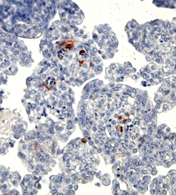
19 the terminal ductal lobular units (TDLUs). ALDH1-positive cells appeared to form a bridge in the lumen that could be located at the bifurcation point of side branches in the TDLUs (Figure 2A). A stem cell marker should not colocalize with markers of mature differentiated mammary epithelial cells. We performed double staining with ALDH1 and CK18, a marker of luminal epithelial cells and ALDH1 and SMA, a marker of myoepithelial cells. The ALDH1-positive cells did not co-localize with CK18, or SMA in sections through normal human breast epithelium (Figure 2C-D). This indicated that ALDH1-positive cells are not mature, differentiated luminal epithelial or myoepithelial cells. Although the phenotype of normal stem and/or progenitor cells from the human breast epithelium has not been identified, several markers including CK5/6 have been found expressed in undifferentiated mammary epithelial cells. We did not detect overlapping expression between CK5/6 and ALDH1 in sections through normal human breast epithelium (data not shown). To determine if this resulted from the scarcity of these two populations, we repeated the same analysis on mammosphere sections. We have shown previously that the mammospheres generated from normal mammary epithelium are enriched in stem/progenitor cells (Dontu et al., 2003). Immunostaining of mammosphere sections using ALDH1 antibody showed that the ALDH1-positive cells are present in small numbers in the mammospheres, comprising approx 5% of the total population (Figure 2B). A subset of these ALDH1-positive cells express CK5/6 (Figure 2E). These results are consistent with the hypothesis that ALDH1-positive cells represent the stem/progenitor population of the normal human breast epithelium. ALDEFLUOR-positive breast carcinoma cells display properties of cancer stem cells. To investigate the tumorigenicity of the ALDEFLUOR-positive population in breast cancers we established xenotransplants from four independent human breast cancers (MC1, UM1, UM2, UM3). Cells from these tumors were transplanted orthotopically in the humanized cleared fat-pad of NOD/scid mice, without cultivation in vitro. The tumors were human invasive ductal carcinomas, three ER-PR-ERBB2- (MC1, UM1, and UM3) and one ER+PR+ERBB2- (UM2). The tumorigenicity of the sorted ALDEFLUOR populations was assessed in early passages in the animals. In contrast to assays that test tumorigenicity of sorted populations directly from patient tumors, this experimental design minimizes the bias introduced by the variable ability of breast cancers to xenotransplant. We found that the ALDEFLUOR-positive population in these three 7
20 tumors constituted between 3% to 10% of the total cell population (Figure 3A-B and Supplementary Figure 2). We performed serial passages using, limiting dilutions of ALDEFLUOR-positive, -negative and unsorted cells (50,000 cells; 25,000 cells; 5,000 cells; 500 cells) in the humanized cleared fat pad of NOD/scid mice. For each of the three tumors and for each of the three passages performed, only the ALDEFLUORpositive population formed tumors, even when implanted in low numbers (Table 1 and Supplementary Figure 2). As shown in Figure 3D, tumor size and latency of tumor formation correlated with the number of ALDEFLUOR-positive cells injected. Remarkably, 500 ALDEFLUOR-positive cells generated a tumor in as few as 40 days. ALDEFLUOR-negative cells failed to reproducibly generate tumors although a limited growth was produced when 50,000 ALDEFLUOR-negative cells were injected. This is consistent with the presence of less than 0.01% contaminating ALDEFLUOR-positive cells, which is within the limits of FACS error (Figure 3E). H&E staining of the fat pad sections confirmed that tumors formed by ALDEFLUOR-positive cells contained malignant cells (Figure 3F) whereas only residual Matrigel, apoptotic cells and mouse tissue was seen at the sites of the ALDEFLUOR-negative cell injections (Figure 3G). No tumors were detected at these sites after weeks. Consistent with the ALDEFLUOR-positive population having stem cell characteristics, tumors generated by this population recapitulated the phenotypic heterogeneity of the initial tumor with a similar ratio of ALDEFLUOR-positive and negative cells (Figure 3C). This indicates that the ALDEFLUOR-positive cells were able to self-renew, generating ALDEFLUOR-positive cells and were able to differentiate, generating ALDEFLUORnegative cells. These results indicate that the cancer stem/progenitor cell population in these tumors has a high ALDH enzymatic activity and may be isolated by the ALDEFLUOR assay. Analysis of ALDH1 protein on tissue microarrays (TMA) and correlation with histoclinical parameters. To assess the potential use of ALDH1 expression as a diagnostic and prognostic marker in breast cancer, we analyzed expression of ALDH1 in two independent sets of breast tumors (U.M. set, I.P.C. set), by IHC on tissue microarrays (TMAs). Among these two sets, 481 tumors were available for ALDH1 staining, with 136 cases from the U.M. set and 345 cases from the I.P.C. set. In the U.M. set, 24 tumors (19%) expressed ALDH1 and 122 tumors (81%) did not. Similar results 8
21 were obtained in the I.P.C. set with 102 cases (30%) positive for ALDH1 staining and 243 cases negative (70%) (Figure 4A-D). Consistent with the idea that cancer stem cells represent a minority of the tumor population, ALDH1-positive cells represented an average of 5% of cells in tumors expressing ALDH1. Only two of the 481 tumors had ALDH1 staining in the vast majority of the cell population (Figure 4A). We next determined whether ALDH1 expression correlates with the histoclinical characteristics of the breast cancers. We found similar results in both sets (Table 2). ALDH1-positive tumors were associated with high histological grade (p<0.05 ; U.M. set, p<0.001 ; I.P.C. set, Fisher s exact test), ERBB2 overexpression (p<0.05 ; U.M. set, p<0.001 ; I.P.C. set, Fisher s exact test) and absence of estrogen and progesterone receptor expression (p<0.05 ; U.M. set, p< ; I.P.C. set, Fisher s exact test). No correlation was found with age, tumor size, and lymph node metastasis. ALDH1 protein expression and clinical outcome. Analysis of overall survival (OS) showed a strong association of ALDH1-positive tumors with poor clinical outcome for both populations (p= ; U.M. set, p= ; I.P.C. set, log-rank test) (Figure 4E-F). In the U.M. set, the 5-year OS was 19.8% [ ] for patients with an ALDH1-positive tumor and 58.7% [ ] for patients with an ALDH1-negative tumor. In the I.P.C. set, the 5-year OS was 69.59% [ ] for patients with an ALDH1-positive tumor and 84.55% [ ] for patients with an ALDH1-negative tumor. We performed a Cox multivariate analysis of OS in which the values for ALDH1, tumor size, age, lymph node metastasis, histological grade, ER, PR, Ki-67 and ERBB2 were considered as categorical variables. ALDH1 expression was an independent prognostic factor as was Ki-67 status, tumor size, and histological grade (Figure 4-G). The relative risk of death due to cancer was 1.76 for patients with ALDH1-positive tumors compared to patients with ALDH1-negative tumors (p<0.028). 9
22 Discussion The cancer stem cells hypothesis has fundamental implications for cancer biology in addition to clinical implications for cancer risk assessment, early detection, prognostication, and prevention. Furthermore, the development of cancer therapeutics based on tumor regression may have produced agents which kill differentiated tumor cells while sparing the small cancer stem cell population (Wicha et al., 2006). The development of more effective cancer therapies may thus require targeting this important cancer stem cell population. The success of these new approaches hinges on the identification, isolation and characterization of cancer stem cells. Recently, the phenotype of the mouse mammary stem cells was identified by several groups (Shackelton et al., 2006; Stingl et al., 2006). These studies showed that an entire, functional mammary gland can be regenerated in vivo in several serial passages, starting from a single cell (Shackelton et al., 2006). Also, considerable progress has been made recently towards identification of human mammary stem cells, although the phenotype of these cells has remained elusive (Clarke et al., 2006). Our study indicates that ALDH1 is a marker of stem/progenitor cells of the normal human breast and breast carcinomas. Moreover, identification of normal and malignant stem/progenitor cells by the same marker supports the concept that stem and progenitor cells are primary targets of transformation, and thus lends further support to the cancer stem cell hypothesis. In addition, the ability to identify stem/progenitor cells by this shared phenotypic trait, ALDH1 expression permits analysis of carcinogenesis from normal to pre-malignant and then malignant state. Unlike the previously described breast cancer stem cell phenotype, which requires the use of a combination of ten surface antigens (Al-Hajj et al., 2003), ALDH1 provides a simple tool to identify normal and cancer stem cells. In addition, the simplicity of this technique offers an important advantage for clinical applications. We show in the present study that ALDH1 detection by immunohistochemistry on paraffin-embedded sections is a reliable method for detecting cancer stem cells is situ. Moreover, ALDH1 expression is a powerful prognostic factor for breast cancer and it has direct or inverse correlation with known histoclinical parameters, such as tumor grade, ER/PR status and ERBB2 overexpression. In the vast majority of breast tumors analyzed in this study the ALDH1 positive cells represented a relatively small population, consistent with the notion that cancer stem 10
23 cells represent a minority of the tumor population. Remarkably, only two tumors out of 481 analyzed, had a predominant ALDH1 positive population. These tumors had a very aggressive clinical evolution and may have been driven by a stem cell population locked in self-renewal, undergoing little or no differentiation. We and others have proposed that cancer stem cells by virtue of resistance to current treatment modalities may contribute to tumor recurrence following therapy. Since ALDH has been shown to metabolize a number of chemotherapeutic agents such as cyclophosphamide (Moreb et al., 1992), expression of ALDH1 in breast cancer stem cells may contribute to resistance of these cells to cyclophosphamide, an agent frequently used in the treatment in breast cancer. We propose that ALDH 1 expression in a subset of tumors may reflect transformation of ALDH1-positive stem or early progenitor cells in these tumors. By contrast, ALDH1- negative tumors may be generated by the transformation of an ALDH1-negative progenitor cells. In the ALDH1-positive tumors, the cancer stem cell population may inherit properties of normal stem cells that confer aggressiveness: ability to self-renew, high proliferation potential, resistance to damaging agents, and chemoresistance. This hypothesis is consistent with the studies of AML (Bonnet et al., 1999). Alternatively, ALDH1-negative tumors may contain rare ALDH1-positive cells below the level of detection by immunostaining on TMAs. The detection of an ALDH1-positive population in TMAs cores may be due to an increased self-renewal activity in these tumors. A recent study has shown that a gene expression signature associated with increased self-renewal of normal stem cells is a predictor of poor prognosis (Glinsky et al., 2005; Lahad et al., 2005). In agreement with our findings, a previously described molecular signatures of breast cancer associated with a poor prognosis for breast cancer contain one or more ALDH isotypes (Alexe et al., 2006). Recently, a combinatorial analysis of gene expression data was used to re-analyze the van t Veer breast cancer gene expression data set (van t Veer et al., 2002). This analysis identified 17 genes associated with poor prognosis in breast cancer, two of which were ALDH isotypes. Moreover, a recent study showed that granulocyte macrophage progenitor cells, transformed by the MLL-AF9 fusion protein, retained the global expression profile of their normal cells of origin and had only a subset of genes re-programmed. This set included 363 genes which were associated with self-renewal in normal hematopoietic stem cells including an ALDH isotype (Krivtsov et al., 2006). 11
24 In conclusion, our study lends support to the cancer stem cell hypothesis, by showing that both normal and malignant mammary stem cells share a common functional marker, ALDH1. Identification of ALDH1 as a marker of normal and malignant human breast stem cells opens important new avenues of research in normal breast development of and carcinogenesis. Furthermore, we demonstrate that ALDH1 expression may be used to detect both normal and malignant mammary stem cells in situ, in fixed paraffinembedded sections. The clinical utility and relevance of this assay was demonstrated by a strong association of ALDH1 expression with clinical outcome in two independent tumor sets, totaling 577 patients. Since ALDH is also expressed in hematopoietic and neuronal stem cells, this marker may prove useful for the detection and isolation of cancer stem cells in other malignancies, thus facilitating the application of cancer stem cell biology to clinical practice. 12
25 Experimental Procedures Dissociation of normal breast epithelium Normal breast tissue from reduction mammoplasties was minced with scalpels and dissociated enzymatically as previously described. Fibroblasts, endothelial cells and blood cells were removed by differential centrifugation and by treatment with ammonium chloride solution. This method generates a suspension of cells highly enriched in epithelial cells (95-99% purity). To generate single cell suspension for the in vivo implantation, collagenase digestion time (6h) was shorter (Dontu et al., 2003). The mammoplasty samples were procured and utilized according to approved IRBMED protocols for research in human subjects. Mammosphere culture Single cells were plated in ultra-low attachment plates (Corning, Acton, MA, USA) or plates coated with 1% agarose in PBS, at a density of 100,000 viable cells/ml in primary culture and 5000 cells/ml in subsequent passages. For mammosphere culture, cells were grown in a serum-free mammary epithelial basal medium (MEBM) (Cambrex Bio Science Walkersville, Inc, Walkerville, MD, USA) supplemented with B27 (INVITROGEN, Carlsbad, CA, USA), 20 ng/ml EGF (BD Biosciences, San Jose, CA, USA), antibiotic-antimycotic (100 unit/ml penicillin G sodium, 100 ug/ml streptomycin sulfate and 0.25 μg/ml amphotericin B), 20 ug/ml Gentamycin, 1 ng/ml Hydrocortisone, 5 μg/ml Insulin and 100 μm beta-mercaptoethanol (GIBCO TM INVITROGEN) in a humidified incubator (10% CO 2 : 95% air, 37(C) for 7-10 days as previously described (Dontu et al., 2003). Differentiating culture conditions Single cell suspensions were plated on collagen-coated plates at a density of 2000 viable cells/10 cm diameter dish. Cells were grown in Ham's F-12 medium (GIBCOTM INVITROGEN) with 5% fetal bovine serum (FBS), 5 μg/ml insulin, 1 μg/ml hydrocortisone, 10 μg/ml cholera toxin (Sigma, St Louis, MO, USA), 10 ng/ml epidermal growth factor (BD Biosciences) and 1X Pen/Strep/Fungizone Mix (GIBCO). Cells were fixed or collected for immunostaining after 12 days. 13
26 Flow cytometry Cells were stained fresh or after fixation in methanol. Primary antibodies: ESA labeled FITC, CD10 labeled PE (dilution 1:25, Novocastra, Newcastle, UK) and ALDH1 (dilution 1/100, BD Biosciences) were used for immunostaining. Incubation was performed for 20 min. on ice in Hanks Balanced Salt Solution (HBSS, GIBCO) with 2% FBS, followed by washing in HBSS with 2% FBS. For ALDH1 staining, the same procedure was applied. Secondary antibody used was anti-mouse IgG, labeled with PE (1:250 dilution; Jackson Labs, MA, USA). After incubation, cells were washed once with HBSS and were resuspended in HBSS supplemented with 5% FBS. Fresh cells were stained with 1μg/ml propidium iodide (PI) (Sigma) for 5 min. for viability. Analysis was performed using FACStarPLUS (Becton Dickinson, Palo Alto, CA, USA) flow cytometer. Xenotransplants samples Human breast tumors were obtained as biopsy cores or pieces of tumors after surgery and implanted in humanized cleared fat pads of NOD/SCID mice for establishing xenotransplants. Four xenotransplants were used: an ER-PR-ERBB2- tumor at the 15th passage in the animals (MC1), an ER-PR-ERBB2- tumor at the 3rd passage (UM1), an ER+PR+ERBB2- tumor at the 4th passage in the animals (UM2), and an ER-PR- ERBB2- tumor at the 2nd passage (UM3). Two of the xenotransplants were generated from metastatic tumors (MC1, pleural effusion and UM2, ovarian metastasis) and two from primary tumors (UM1, UM3). Aldefluor assay and separation of the ALDH positive population by FACS The ALDEFLUOR kit (StemCell technologies, Durham, NC, USA) was used to isolate the population with a high ALDH enzymatic activity. Cells obtain from freshly dissociated normal breast epithelium or breast cancer xenografts were suspended in ALDEFLUOR assay buffer containing ALDH substrate (BAAA, 1 μmol/l per 1x10 6 cells) and incubated during 40 minutes at 37 C. In each experiment a sample of cells was stained under identical conditions with 50mmol/L of specific ALDH inhibitor diethylaminobenzaldehyde (DEAB) as negative control. Flow cytometry based sorting was conducted using a FACStarPLUS (Becton Dickinson). ALDEFLUOR fluorescence was excited at 488 nm and fluorescence emission was detected using a standard fluorescein isothiocyanate (FITC) 530/30 band pass filter. In addition, for the xenotransplanted tumors, incubation 14
27 with an anti-h2kd antibody (BD biosciences, 1/200, 20 min on ice) followed by a secondary antibody labeled with phycoerythrin (PE) (Jackson labs, 1/250, 20 min on ice) were used to eliminate cells of mouse origin. The sorting gates were established using as negative controls the PI stained cells for viability, the ALDEFLUOR-stained cells treated with DEAB and the staining with secondary antibody alone. Animal model NOD/SCID mice were used to assess the in vivo stem cell potential of the ALDEFLUOR-positive population, compared to the ALDEFLUOR-negative population and the unsorted population, from the normal breast epithelium and the four tumor xenografts. The animal model was described by Kuperwasser et al for xenotransplantation of normal mammary epithelial cells (Kuperwasser et al., 2004). The fat pads were cleared pre-puberty and humanized by injecting a mixture of irradiated and non-irradiated immortalized human fibroblasts (1:1 irradiated:non-irradiated, 50,000 cells/100μl Matrigel/fat pad). Irradiated fibroblasts (4Gy) support growth of normal and cancer epithelial cells by secreting a variety of growth factors, collagen and possibly directly interacting with the epithelial cells (Orimo et al., 2005; Tlsty et al., 2001). The immortalized fibroblasts were primary human mammary fibroblasts stably transfected with a retrovirus construct expressing telomerase. The fibroblast cell line is a generous gift from Dr. John Stingl and Dr. Connie Eaves (Terry Fox Laboratory, Vancouver, British Columbia, Canada). Estrogen pellets were implanted subcutaneously at the time of the clearing. The human normal breast or cancer cells were mixed with Matrigel (BD biosciences) (1:1) and implanted in the cleared humanized fat pads 2-4 weeks later. The animals injected with normal breast cells were euthanized after 10 weeks. The animals injected with cancer were euthanized when the tumors were approximately 1.2 cm in the largest diameter, to avoid tumor necrosis and in compliance with regulations for use of vertebrate animal in research. A portion of each fat pad injected was fixed in formalin and embedded in paraffin for histological analysis. The animal studies were approved by the ULAM committee for research invertebrate animals. Tissue Microarrays 15
28 The TMAs were provided by the Tissue Microarray Core laboratory at University of Michigan Medical School and by the Laboratoire d Oncologie Moleculaire, Institut Paoli- Calmettes de Marseille. The first TMA contained 154 breast cancer cores from a consecutive population of patients treated at the University of Michigan Hospital, MI, USA (U.M. set) between 1984 and 1991 and the second TMA contained 552 breast cancer cores from a consecutive population of patients treated at the Institut Paoli- Calmettes, Marseille, France (I.P.C. set) between 1987 and Clinical and histopathological data are available for these patients (Jacquemier et al., 2005; Kleer et al., 2003). Immunostaining To assess the lineage composition of the colonies, cells were fixed on plates for 20 min in methanol, at -20 C, and were then stained using Peroxidase Histostain-Plus and Alkaline-phosphatase Histostain-Plus kits (Zymed, South San Francisco, CA, USA), according to the manufacturer's protocol. The primary antibodies, epithelial-specific antigen (ESA) for luminal epithelial cells and CD10 for myoepithelial cells, were used at the dilutions indicated by the manufacturer. DAB (Zymed) was used for ESA staining as substrate for peroxidase, and NBT/BCIP (Gibco) was used for CD10 staining as substrates for alkaline phosphatase. For ALDH1 immunostaining, the paraffin-embedded sections through mammospheres, normal breast tissue and the TMA were deparaffinized in xylene and rehydrated in graded alcohol. Antigen enhancement was done by incubating the sections in citrate buffer ph6 (Dakocytomation, Copenhagen, Denmark) as recommended. Staining was done using Perixidase histostain-plus Kit (Zymed) according to the manufacturer s protocol. ALDH1 antibody (BD biosciences) were used at a 1/100 dilution and incubated for 1 hour. AEC (Zymed) was used as substrate for peroxidase. Slides were counterstained with hematoxylin, and coverslipped using glycerin. TMA results were expressed in terms of percentage (P) and intensity (I) of positive cells as described previously (Ginestier et al., 2002). Results were scored by the quick score (Q) (Q = P x I). For the TMA, the mean of the score of minimum 2 core biopsies was calculated for each case. For fluorescent double staining, the primary antibodies cytokeratin 18, smooth muscle actin (SMA), and cytokeratin 5/6 (Novocastra) were used at the dilutions indicated by 16
29 the manufacturer and incubated 1 hour at room temperature. Texas-red and FITC labeled secondary antibodies (Jackson Labs) were used at the dilution 1/250 and incubated for 20 minutes. Nuclei were counterstained with DAPI/antifade (INVITROGEN) and coverslipped. Sections were examined with a fluorescent microscope (Leica, Bannockborn, IL, USA). Statistical analysis Distributions of molecular markers and other categorical variables were compared using standard chi 2 tests or Fisher exact test. The overall survival interval was calculated from the date of diagnosis. For graphical presentation, follow-up was truncated at 100 months. Survival curves were derived from Kaplan-Meier estimates and the curves were compared by logrank tests. The influence of ALDH1 expression status was assessed in multivariate analysis by the Cox proportional hazard models with a stepwise selection. The model was adjusted for usual prognostic or predictive factors in breast cancer, including tumor size, age, lymph node metastasis, histological grade, ER, PR, Ki-67 and ERBB2 status. All statistical tests were 2-sided at the 5% level of significance, and were done using the R Version software. Survival rates and relative risks (RR) are presented with their 95% confidence intervals (CI). 17
30 Acknowledgments Thanks are due to Dr Thomas Giordano for tissue procurement, the University of Michigan Cancer Center Flow Cytometry core, Dr. Hayes, Dr Boman, Dr. Korkaya, Dr. Liu for advise and critical review of the paper. The fibroblast cell line is a generous gift from Dr. John Stingl and Dr. Connie Eaves, Terry Fox Laboratory, Vancouver, British Columbia, Canada. This work was supported by NIH grants CA66233 and CA101860, and in part by the University of Michigan Cancer Center NIH Support Grant 5 P 30 CA46592, and in part by Ligue Nationale Contre le Cancer (Label DB) and Institut National du Cancer. Note: Max Wicha has financial holdings and is a scientific advisor for OncoMed Pharmaceuticals. 18
31 REFERENCES Alexe,G. et al. (2006). Breast cancer prognosis by combinatorial analysis of gene expression data. Breast Cancer Res. 8, R41. Al-Hajj,M., Wicha,M.S., ito-hernandez,a., Morrison,S.J., & Clarke,M.F. (2003). Prospective identification of tumorigenic breast cancer cells. Proc. Natl. Acad. Sci. U. S. A 100, Armstrong,L. et al. (2004). Phenotypic characterization of murine primitive hematopoietic progenitor cells isolated on basis of aldehyde dehydrogenase activity. Stem Cells 22, Bonnet,D. and Dick,J.E. (1997). Human acute myeloid leukemia is organized as a hierarchy that originates from a primitive hematopoietic cell. Nat. Med. 3, Bonnet,D., Warren,E.H., Greenberg,P.D., Dick,J.E., & Riddell,S.R. (1999). CD8(+) minor histocompatibility antigen-specific cytotoxic T lymphocyte clones eliminate human acute myeloid leukemia stem cells. Proc. Natl. Acad. Sci. U. S. A 96, Chute,J.P., Muramoto,G.G., Whitesides,J., Colvin,M., Safi,R., Chao,N.J., McDonnell,D.P. (2006). Inhibition of aldehyde dehydrogenase and retinoid signaling induces the expansion of human hematopoietic stem cells. Proc. Natl. Acad. Sci. U. S. A. 31, Clarke,M.F. et al. (2006). Cancer Stem Cells--Perspectives on Current Status and Future Directions: AACR Workshop on Cancer Stem Cells. Cancer Res. 66, Collins,A.T., Berry,P.A., Hyde,C., Stower,M.J., & Maitland,N.J. (2005). Prospective identification of tumorigenic prostate cancer stem cells. Cancer Res. 65, Dontu,G. et al. (2003). In vitro propagation and transcriptional profiling of human mammary stem/progenitor cells. Genes Dev. 17, Duester,G. (2000). Families of retinoid dehydrogenases regulating vitamin A function: production of visual pigment and retinoic acid. Eur. J. Biochem. 267, Fang,D. et al. (2005). A tumorigenic subpopulation with stem cell properties in melanomas. Cancer Res. 65, Ginestier,C. et al. (2002). Distinct and complementary information provided by use of tissue and DNA microarrays in the study of breast tumor markers. Am. J. Pathol. 161, Glinsky,G.V., Berezovska,O., & Glinskii,A.B. (2005). Microarray analysis identifies a death-from-cancer signature predicting therapy failure in patients with multiple types of cancer. J. Clin. Invest 115,
32 Hess,D.A. et al. (2004). Functional characterization of highly purified human hematopoietic repopulating cells isolated according to aldehyde dehydrogenase activity. Blood 104, Hess,D.A. et al. (2006). Selection based on CD133 and high aldehyde dehydrogenase activity isolates long-term reconstituting human hematopoietic stem cells. Blood 107, Jacquemier,J. et al. (2005). Protein expression profiling identifies subclasses of breast cancer and predicts prognosis. Cancer Res. 65, Jamieson,C.H. et al. (2004). Granulocyte-macrophage progenitors as candidate leukemic stem cells in blast-crisis CML. N. Engl. J. Med. 351, Kelly,L.M. & Gilliland,D.G. (2002). Genetics of myeloid leukemias. Annu. Rev. Genomics Hum. Genet. 3, Kleer,C.G. et al. (2003). EZH2 is a marker of aggressive breast cancer and promotes neoplastic transformation of breast epithelial cells. Proc. Natl. Acad. Sci. U. S. A 100, Krivtsov,A.V. et al. (2006). Transformation from committed progenitor to leukaemia stem cell initiated by MLL-AF9. Nature 442, Kuperwasser,C. et al. (2004). Reconstruction of functionally normal and malignant human breast tissues in mice. Proc. Natl. Acad. Sci. U. S. A 101, Lahad,J.P., Mills,G.B., & Coombes,K.R. (2005). Stem cell-ness: a "magic marker" for cancer. J. Clin. Invest 115, Magni,M. et al. (1996). Induction of cyclophosphamide-resistance by aldehydedehydrogenase gene transfer. Blood 87, Matsui,W. et al. (2004). Characterization of clonogenic multiple myeloma cells. Blood 103, Molofsky,A.V., Pardal,R., & Morrison,S.J. (2004). Diverse mechanisms regulate stem cell self-renewal. Curr. Opin. Cell Biol. 16, Moreb,J., Zucali,J.R., Zhang,Y., Colvin,M.O., & Gross,M.A. (1992). Role of aldehyde dehydrogenase in the protection of hematopoietic progenitor cells from 4- hydroperoxycyclophosphamide by interleukin 1 beta and tumor necrosis factor. Cancer Res. 52, O'brien,C.A., Pollett,A., Gallinger,S., & Dick,J.E. (2006). A human colon cancer cell capable of initiating tumour growth in immunodeficient mice. Nature 445, Orimo,A. et al. (2005). Stromal fibroblasts present in invasive human breast carcinomas promote tumor growth and angiogenesis through elevated SDF-1/CXCL12 secretion. Cell 121,
33 Passegue,E., Jamieson,C.H., Ailles,L.E., & Weissman,I.L. (2003). Normal and leukemic hematopoiesis: are leukemias a stem cell disorder or a reacquisition of stem cell characteristics? Proc. Natl. Acad. Sci. U. S. A 100 Suppl 1, Pearce,D.J. et al. (2005). Characterization of cells with a high aldehyde dehydrogenase activity from cord blood and acute myeloid leukemia samples. Stem Cells 23, Reya,T., Morrison,S.J., Clarke,M.F., & Weissman,I.L. (2001). Stem cells, cancer, and cancer stem cells. Nature 414, Ricci-Vitiani,L. et al. (2006). Identification and expansion of human colon-cancerinitiating cells. Nature 445, Sell,S. (2004). Stem cell origin of cancer and differentiation therapy. Crit Rev. Oncol. Hematol. 51, Shackleton,M. et al. (2006). Generation of a functional mammary gland from a single stem cell. Nature 439, Singh,S.K. et al. (2004a). Identification of human brain tumour initiating cells. Nature 432, Singh,S.K., Clarke,I.D., Hide,T., & Dirks,P.B. (2004b). Cancer stem cells in nervous system tumors. Oncogene 23, Sophos,N.A. & Vasiliou,V. (2003). Aldehyde dehydrogenase gene superfamily: the 2002 update. Chem. Biol. Interact , Stingl,J. et al. (2006). Purification and unique properties of mammary epithelial stem cells. Nature 439, Tlsty,T.D. (2001). Stromal cells can contribute oncogenic signals. Semin. Cancer Biol. 11, van,'t Veer,V. et al. (2002). Gene expression profiling predicts clinical outcome of breast cancer. Nature 415, Wicha,M.S., Liu,S., & Dontu,G. (2006). Cancer stem cells: an old idea--a paradigm shift. Cancer Res. 66, Yoshida,A., Rzhetsky,A., Hsu,L.C., & Chang,C. (1998). Human aldehyde dehydrogenase gene family. Eur. J. Biochem. 251,
34 Tables Table 1. Tumorigenicity in the humanized fat pad of NOD/SCID mice. Tumors/injections 5x x10 4 5x Mouse Passage 1 ALDEFLUOR-negative 3/6 0/1 0/6 0/6 ALDEFLUOR-positive 5/5 1/1 4/4 6/6 Unsorted 5/5 1/1 4/4 3/4 Mouse Passage 2 ALDEFLUOR-negative 1/ /4 ALDEFLUOR-positive 4/ /4 Unsorted 4/ /4 Mouse Passage 3 ALDEFLUOR-negative 0/ /4 ALDEFLUOR-positive 4/ /4 Unsorted 4/ /4 22
35 Table 2. Correlation between ALDH1 protein expression and histoclinical characteristics Characteristics U.M. set I.P.C. set ALDH1 ALDH1 ALDH1 ALDH1 Negative Positive Negative Positive No. of patients (%) No. of patients (%) p-value No. of patients (%) No. of patients (%) p-value All cases 122 (81) 24 (19) 243 (70) 102 (30) Age (years) (27) 6 (25) NS 79 (35) 25 (25) NS >50 89 (73) 18 (75) 164 (65) 77 (75) Pathological tumor size PT1 61 (60) 10 (45) NS 103 (42) 37 (37) NS PT2 33 (33) 7 (32) 108 (45) 46 (47) PT3 7 (7) 5 (23) 30 (13) 16 (16) SBR grade I 25 (22) 2 (9) < (33) 24 (24) <0.001 II 55 (49) 7 (30) 120 (49) 38 (38) III 33 (29) 14 (61) 43 (18) 39 (38) Lymph node metastasis Negative 53 (56) 9 (41) NS 134 (56) 43 (44) NS Positive 42 (44) 13 (59) 107 (44) 55 (56) Estrogen receptor Negative 38 (34) 14 (61) < (17) 39 (39) < Positive 75 (66) 10 (39) 198 (83) 61 (61) Progesterone receptor Negative 50 (44) 15 (62) < (26) 51 (52) < Positive 64 (56) 9 (38) 170 (74) 47 (48) Ki-67 status Negative (<20) (91) 77 (82) <0.05 Positive ( 20) (9) 17 (18) ERBB2 receptor Negative (0/1+) 94 (83) 15 (62) < (94) 76 (79) <0.001 Positive (2+/3+) 19 (17) 9 (38) 14 (6) 20 (21) 23
36 Figure legends Figure 1. ALDEFLUOR positive cells from normal breast epithelium have stem cell properties. A-B. Representative FACS analysis of ALDH activity of normal breast epithelial cells analyzed by the ALDEFLUOR assay. Cells incubated with ALDEFLUOR substrate (BAAA) and the specific inhibitor of ALDH, DEAB, were used to establish the baseline fluorescence of these cells (R1) and to define the ALDEFLUOR-positive region (R2) (A). Incubation of cells with ALDEFLUOR substrate in the absence of DEAB induces a shift in BAAA fluorescence defining the ALDEFLUOR-positive population (B). C-D. ALDEFLUOR-positive cells sorted from fresh reduction mammoplasties generated mammospheres in suspension culture (C), whereas ALDEFLUOR-negative cells did not (D). E-H. Evaluation of the differentiation potential of ALDEFLUOR-positive and ALDEFLUOR-negative cells. Sorted cells were grown in differentiating conditions for 12 days and stained by IHC with lineage-specific markers (ESA, CD10). The ALDEFLUORpositive population generated 67.2 ± 3.5% bi-potent progeny (ESA+ cells stained in brown and CD10+ stained in purple) (F), 2.9 ± 0.5% myoepithelial colonies (CD10+) (G), and 30.6 ± 5.4% luminal colonies (ESA+) (H), whereas the ALDEFLUOR-negative population produced 90.8 ± 3.1% luminal epithelial colonies (ESA+) (H), and only 9.1 ± 1.3% bi-potent progeny (E). I. ALDEFLUOR-positive and ALDEFLUOR-negative cells grown in differentiating conditions were collected for flow cytometry analysis of lineage markers (ESA, CD10). ALDEFLUOR-positive cells generated bi-potent progeny (15.3 ± 3.2%, CD10-/ESA-; 21.2 ± 1.5%, CD10+/ESA+), luminal cells (63.2 ± 4.1%, CD10- /ESA+) and myoepithelial cells (2.1 ± 0.3%, CD10+/ESA-) whereas ALDEFLUORnegative cells generated predominantly luminal cells (93.5 ± 3.4%, CD10-/ESA+). J-M. Small ducts produced in the NOD/scid humanized cleared fat pads injected with ALDEFLUOR-positive cells from normal breast epithelium. Hematoxylin and eosin staining revealing the presence of several small ducts at the site of injected ALDEFLUOR-positive cells (J) and residual Matrigel and mouse tissue at the site injected with ALDEFLUOR-negative cells (K). Immunostaining of the ducts produced by ALDEFLUOR-positive cells showed the presence of a luminal layer (stained with anti- CK18 brown staining) (L) and a myoepithelial layer (stained with SMA, red staining) (M). 24
37 Figure 2. Characterization of ALDH1 positive-cells in the normal breast epithelium and mammosphere section. A. ALDH1 staining of normal breast epithelium. ALDH1- positive cells (red cytoplasmic staining) were in a luminal position, bridging across the lumen, probably at branching points of side-ducts (arrow). B. ALDH1 staining in mammospheres. Only 1-5 cells/mammosphere showed positive staining for ALDH1, (approximately 5% of the total population). C-D. Immunofluorescence of normal breast epithelium. C. Double staining with CK18 (red) and ALDH1 (green). Composite image (merge) showed absence of overlap between CK18 positive cells (mature luminal cells) and ALDH1-positive cells (arrow). D. Double staining with SMA (green) and ALDH1 (red). Composite image (merge) showed absence of overlap between SMA positive cells (mature myoepithelial cells) and ALDH1-positve cells (arrow). E. Immunofluorescence of mammosphere sections. Double staining with CK5/6 (green) and ALDH1 (red). Composite image (merge) showed that only few ALDH1-positive cells displayed an exclusive red signal (arrow) whereas all the CK5/6 positive cells (asterisk) displayed a hybrid signal (yellow) corresponding to cells positive for ALDH1 and CK5/6. All nuclei were counterstained in DAPI. Figure 3.The ALDEFLUOR positive cell population displays properties of cancer stem cells in NOD/SCID mice. A-B. Representative flow cytometry analysis of ALDH activity in cells derived from a human breast tumor, orthotopically xenotransplanted in NOD/scid mice. Cells incubated with ALDEFLUOR substrate (BAAA) and the specific inhibitor of ALDH, DEAB, were used to establish the baseline fluorescence of these cells (R1) and to define the ALDEFLUOR-positive region (R2) (A). Incubation of cells with ALDEFLUOR substrate in the absence of DEAB induced a shift in BAAA fluorescence defining the ALDEFLUOR-positive population (B). C-G Only the ALDEFLUOR-positive population was tumorigenic. C. The ALDEFLUOR-positive population was capable of regenerating the phenotypic heterogeneity of the initial tumor after a passage in NOD/scid mice. D. Tumor progression curves were plotted for the numbers of cells injected (50,000 cells; 5,000 cells; 500 cells) and for each population 25
38 (ALDEFLUOR-positive, ALDEFLUOR-negative, unsorted). Tumor growth kinetics correlated with the latency and size of tumor formation and the number of ALDEFLUOR-positive cells. E. Representative tumor grown in NOD/scid mouse at the ALDEFLUOR-positive cells injection site (5,000 cells injected). No tumor was detected at the ALDEFLUOR-negative cells injection site (5,000 cells injected). F-G. H & E staining of ALDEFLUOR-positive cells injection site revealing presence of tumor cells (F). The ALDEFLUOR-negative cells injection site contained only residual Matrigel, apoptotic cells and mouse tissue (G). All the data presented in this figure were generated by analysis of the MC1 tumor. Similar results were obtained for three other tumors, generated from different patients (UM1, UM2, and UM3) tested (supplementary figure 2). Figure 4. Expression of ALDH1 in breast carcinomas, as shown by immunohistochemistry on tissue microarrays (TMA), using two independent tumor sets. A-D. Example of ALDH1 staining in breast cancer. Only two of the 477 tumors analyzed were fully positive for ALDH1 (A). Representative examples of breast tumor cores positive for ALDH1 with 5-10% ALDH1-positive cells detected (B-C). Example of a tumor core with no detectable ALDH1 staining (D). E-F. Kaplan-Meier plot of patient overall survival: Survival differed significantly according to ALDH1 expression. Patients with tumors positive for ALDH1 staining (green curve) had a poor prognosis compared to patients with tumors negative for ALDH1 staining (blue curve). Similar results were observed in the U.M. set composed of 136 patients (p=0.0459) (E) and I.P.C. set composed of 341 patients (p= ) (F). G. Cox multivariate analysis of overall survival for patients from I.P.C. set. When compared with known prognostic factors, ALDH1 status was an independent factor of prognosis, as was Ki-67 status, tumor size, SBR grade. 26
39 Abstract Abstract Application of stem cell biology to breast cancer research has been limited by the lack of simple assays for identification and isolation of normal and malignant stem cells. We show that stem/progenitor cells in normal breast epithelium and breast tumors have increased aldehyde dehydrogenase activity. Furthermore, immunostaining using ALDH1 antibody identifies normal and malignant stem/ progenitor cells in situ. In a series of 577 breast carcinomas on tissue microarrays, expression of ALDH1 was an independent predictor of poor prognosis. These findings provide support for the cancer stem cell hypothesis and offer an important new tool for the study of normal and malignant breast stem cells. Moreover, since ALDH1 immunodetection provides a simple method to identify cancer stem and progenitor cells in situ it should facilitate the clinical application of stem cell concept.


 70 60")

40 With DEAB Without DEAB R1 R2 R1 R2 ALDEFLUOR + ALDEFLUOR - CD10-PE-CY5 Region Events %Gated R R Region Events %Gated R R ESA-FITC A B I E 100 C Luminal colonies D F J K Myoepithelial colonies colony ty pe ( % ) Mixed colonies G ALDEFLUOR positive ALDEFLUOR negative E H L M Figure 1


41 A A B DAPI CK18 ALDH1 Merge C DAPI SMA ALDH1 Merge * D DAPI CK5/6 ALDH1 Merge E Figure 2


42 Figure 3 With DEAB Without DEAB R1 R2 R1 R2 R1 R2 (passage) Region Events %Gated R R Region Events %Gated R R Region Events %Gated R R A B C Tumor size (cm) Days after injection F 50,000 cells ALDEFLUOR + 5,000 cells ALDEFLUOR cells ALDEFLUOR + 50,000 cells ALDEFLUOR - 5,000 cells ALDEFLUOR cells ALDEFLUOR - ALDEFLUOR - ALDEFLUOR + 50,000 Unsorted cells 500 Unsorted cells D E G

 0.")
 20 1 >20 2.21 (1.22-4.02) 0.")

43 Figure 4 A B C D U.M. set I.P.C. set Variable Hazard ratio (95% CI) p-value Ki-67 status Negative 1 Positive 2.47 ( ) Overall Survival Overall Survival Tumor size (mm) 20 1 > ( ) SBR Grade I and II 1 III 1.83 ( ) p= p= ALDH1 status Negative 1 Months after surgery E Months after surgery F Positive 1.76 ( ) G
44 Supplementary Figure 1 ALDEFLUOR - ALDEFLUOR + Overlay Counts Region Events %Gated M A Region Events %Gated M B C ALDH1-PE
45 Supplementary Figure 2 UM1 UM2 UM3 With DEAB Without DEAB With DEAB Without DEAB With DEAB Without DEAB R1 R2 R1 R2 R1 R2 R1 R2 R1 R2 R1 R2 Region Events %Gated R R Region Events %Gated R R Region Events %Gated R R Region Events %Gated R R Region Events %Gated R R Region Events %Gated R R Tumor size (cm) Tumor size (cm) Tumor size (cm) Days after injection 50,000 cells ALDEFLUOR + 5,000 cells ALDEFLUOR cells ALDEFLUOR + 50,000 cells ALDEFLUOR - 5,000 cells ALDEFLUOR cells ALDEFLUOR - 50,000 unsorted cells 500 unsorted cells Days after injection 50,000 cells ALDEFLUOR + 5,000 cells ALDEFLUOR cells ALDEFLUOR + 50,000 cells ALDEFLUOR - 5,000 cells ALDEFLUOR cells ALDEFLUOR - 50,000 unsorted cells 500 unsorted cells Days after injection 50,000 cells ALDEFLUOR + 5,000 cells ALDEFLUOR cells ALDEFLUOR + 50,000 cells ALDEFLUOR - 5,000 cells ALDEFLUOR cells ALDEFLUOR - 50,000 unsorted cells 500 unsorted cells
46 Legend Supplementary Figures Supplementary Figure 1. Overlap between the normal mammary epithelial cell populations with ALDH activity detected by the ALDEFLUOR assay and the cell population with cytoplasmic ALDH1 protein detected by immunostaining. ALDEFLUOR-negative and ALDEFLUOR-positive cells from normal breast epithelium were separated by FACS using the ALDEFLUOR assay. Cells were then fixed in RNA later, immunostained with an ALDH1 antibody and analyzed by flow cytometry. (A-C). ALDEFLUOR-negative cells did not have levels of ALDH1 protein detectable by immunostaining (M1=0.88%) (A) whereas ALDEFLUOR-positive cells contained all the cell population detected by ALDH1 antibody (B). Overlay presenting a direct comparison between ALDH1 immunostaining of ALDEFLUOR-negative and ALDEFLUOR-positive cells (C). Supplementary Figure 2.The ALDEFLUOR positive cell population displays properties of cancer stem cells in NOD/SCID mice. Representative flow cytometry analysis of ALDH activity in cells derived from a human breast tumor, orthotopically xenotransplanted in NOD/SCID mice (UM1, left panel; UM2, central panel; UM3 right panel). Cells incubated with ALDEFLUOR substrate (BAAA) and the specific inhibitor of ALDH, DEAB, were used to establish the baseline fluorescence of these cells (R1) and to define the ALDEFLUOR-positive region (R2). Incubation of cells with ALDEFLUOR substrate in the absence of DEAB induced a shift in BAAA fluorescence defining the ALDEFLUOR population. Tumor progression curves were plotted for the numbers of cells injected in NOD/SCID mice (50,000 cells; 5,000 cells; 500 cells) and for each population (ALDEFLUOR-positive, ALDEFLUOR-negative, unsorted).
47 Supplementary Table1 Supplementary Table 1. Outgrowth potential in the humanized fat pad of NOD/SCID mice Outgrowth/injections 5x x104 5x ALDEFLUOR-positive --- 4/4 3/4 0/4 ALDEFLUOR-negative 0/ /3 0/2 Unsorted --- 5/5 3/5 0/2
48 1 Selective targeting of cancer stem cells: A new concept in cancer therapeutics Hasan Korkaya and Max S. Wicha University of Michigan Comprehensive Cancer Center 1500 E Medical Center Dr CCGC Ann Arbor, MI Correspondence: Hasan Korkaya University of Michigan Comprehensive Cancer Center 1500 E Medical Center Dr 7110 CCGC Ann Arbor, MI hkorkaya@med.umich.edu Phone:
49 2 Abstract Although the concept of cancer stem cell had been proposed more then a century ago, it has attracted a great deal of attention recently due to advances in stem cell biology leading to the identification of these cells in a wide variety of human cancers. Furthermore, there is accumulating evidence that the resistance of cancer stem cells to many conventional therapies may account for the inability of these therapies to cure most metastatic cancers. The recent identification of stem cell markers and advances in stem cell biology has facilitated research in multiple aspects of cancer stem cell behavior. Stem cell subcomponents have now been identified in a number of human malignancies including haematologic malignancies and tumors of the breast, prostate, brain, pancreas, head and neck and colon. Furthermore, pathways that regulate selfrenewal and cell fate in these systems is beginning to be elucidated. In addition to pathways known to regulate self-renewal of normal stem cell such as Wnt, Notch and Hedgehog, tumor suppressor genes such as PTEN and p53 have also been implicated in the regulation of cancer stem cell self-renewal. In cancer stem cells, these pathways are believed to be deregulated leading to uncontrolled self-renewal of cancer stem cells which generate tumors that are resistant to conventional therapies. Current cancer therapeutics based on tumor regression may target and kill differentiated tumor cells which compose the bulk of the tumor while sparing the rare cancer stem cell population. The cancer stem cell model suggests that the design of new cancer therapeutics may require the targeting and elimination of cancer stem cells. Therefore, it is imperative to design new strategies based upon a better understanding of the signaling pathways that control aspects of self-renewal and survival in cancer stem cells in order to identify novel therapeutic targets in these cells. Introduction Decades of cancer research may need to be re-evaluated, because standard tumortargeting therapies may be off the mark, mounting research suggests. Quote from ABC news, November 2006, voicing the concerns over the failure of current cancer therapies to cure the most common human cancers and posing the question of
50 3 whether we are targeting the right cells in human cancers. Conventional therapies have largely been designed to target bulk and cycling populations in tumors. Evidence is accumulating from number of human malignancies that most, if not all, malignancies possess a subcomponent of cancer cells that have stem cell properties which have been cancer stem cells (CSC). These properties include self-renewal that drives tumorigenesis and differentiation that generates the bulk of tumor cells. The deregulation of stem cell self-renewal pathways through accumulation of both genetic and epigenetic changes may be essential for the malignant transformation of CSCs (1-3). During normal development, signals from the surrounding niche regulate stem cell self-renewal. The altered reorganization of these niches may result in aberrant signals that lead to deregulation of stem cell self-renewal (4). This concept is supported by a recent finding which demonstrated an increase in self-renewing CSC population by increasing the vasculature in the brain tumor microenvironment (5). On the contrary, the depletion of blood vessels from xenografts ablated self-renewing tumor stem cells and inhibited tumor growth (5). Thus the aberrant regulation of stem cell self-renewal due to both extrinsic and intrinsic signals may generate the malignant phenotype. Although the processes that control self-renewal are complex and are only beginning to be understood, the concept of CSCs has fundamental implications for understanding the tumor biology as well as developing new strategies to combat cancer. This review will discuss the evidence for existence of cancer stem cells in a variety of human malignancies and implications of the cancer stem cell model for the development of new cancer therapeutics. A model which illustrates the rationale for the development of stem cell targeted therapies summarized in Figure 2. Isolation of normal adult and cancer stem cells Stem cells are defined by their two distinct properties; (a) self-renewal characterized by the ability to go through numerous cycles of cell division while maintaining an undifferentiated state and (b) multipotency or the ability to generate progeny of distinct cell types (6, 7). Tissue specific (adult) stem cells are distinguished from embryonic stem cells in that their ability to differentiate is largely restricted to cell types within a particular organ. Although transdifferentiation (plasticity) of adult stem cells into different tissues such as the brain or blood cells into mature cells of different tissues
51 4 has been reported, this apparent plasticity is often the result of a rare fusion of stem/progenitor cells of different origin (8-10). HSCs identified by cell surface marker expression from human and mouse (11-13) were able reconstitute the haematopoietic system. The expansion and regeneration of mammary epithelium during puberty and pregnancy in reproductive cycles suggests the existance of stem/progenitor cells. Kordon et al, first described the repopulation of mouse mammary gland through serial transplantaion of retrovirally tagged epithelial fragments (14) demonstrating the clonal nature of this repopulation. More recently, generation of a functional mammary gland from a single stem cell has also been desribed (15). In normal tissues, homeostasis is tightly regulated to ensure the generation of mature cells throughout life without depletion of stem cell pools (16). Each tissue is comprised of a cellular hierarcy including stem cells able to generate all progeny, committed progenitors and terminally differentiated cells. While stem cell self-renewal is necessary for tissue repair and regeneration, it also carries the risk of genetic alteration in stem cells due to the error-prone nature of DNA replication. Thus the pathways that control stem cell self-renewal and the microenvironment in which the stem cells reside both may play a role in carcinogenesis (17). Deregulation of self-renewal and subsequent loss of homeostasis may result in malignant transformation of human tissues and this forms the basis of cancer stem cell hypothesis. The concept of cancer stem cell was first proposed by Virchov and Cohnheim almost 150 years ago based on the similarities between fetal developmet and certain types of tumors such as teratocarcinomas (1). John Dick and his colleagues were the first to isolate such cells from acute myeloid leukaemias where a small subset of CD34 + CD38 - cells that comprised <1 in 10,000 cells could transfer human leukemia into NOD-SCID mice whereas the remaining population that did not bear this phenotype failed to do so (18). Furthermore, this group demonstrated the heterogeneity of leukemia CSCs with hierarcial self-renewal potential reminiscent of their normal counterparts (19). Similar techniques have been used to demonstrate a similar cellular hierarcies in solid tumors including breast, prostate, brain, pancreas and colon (20-25). Implantation of small subsets of cells from these solid tumors revealed that only the cells with stem cell characteristics were able form tumors suggesting the existance of CSCs in these tumors. For example, in
52 5 collaboration with Michael Carke, we demonstrated that human breast cancers contain a stem cell population characterized by the expression of cell surface markers CD44 + CD24 low Lin -. As few as 200 of these cells, comprising 1 to 10% of total cell population were capable of forming tumors when implanted in NOD-SCID mice. In contrast, 20,000 cells that did not express these markers were unable to form tumors (22). Consistent with the cancer stem cell model, the stem cells were able to generate tumors that recapitulated the phenotypic heterogeneity of initial tumor. We and others confirmed that breast CSCs are not only tumorigenic but also formed mammospheres in vitro, a property that we described previously for normal mammary stem/progenitor cells (26, 27). Interestingly, these cells also expressed a stem cell marker, Oct-4 lending additional support to CSC hypothesis (26). Identification and subsequent use of a cell surface antigen, CD133, a five transmembrane glycoprotein (28) enabled Uchida et al, to isolate human central nervous system stem cells characterized by CD133 + CD34 - CD45 - expression (29). Through serial dilution, this group demonstrated that a single CD133 + CD34 - CD45 - cell was able to form a neurosphere in in vitro culture (29). In addition to the identification of normal neuronal stem cells, the existence of CSCs in brain tumors have also been reported (30-32). Through cell sorting for CD133 +, a functional cellular hierarcy in brain tumor cell population was demonstrated (33). Furthermore, CD133+ human brain tumor cells but not CD133 - were able to form tumors in NOD-SCID mouse brains and neurospheres in in vitro cultures (21). Xin et al, demonstrated that prostate regenerating cells are enriched in stem cell antigen-1 (Sca-1) expression (34). Further evidence for the existence of CSC population in human prostate tumors has been reported (35). Richardson et al, has identified a stem cell population in normal human prostate characterized by CD133 + expression and in human prostate tumors characterized as CD44 + /α 2 β hi 1 /CD133 - (35). As few as 500 cells with this phenotype that constituted 0.1% of total tumor cells formed tumors in NOD/SCID mice, whereas 5x10 5 CD44 - cells failed to form tumors (35). The existence of stem cells in normal lung and lung cancer has also been shown by the isolation of cells that exhibited self-renewal and multipotency (36). Most recently, the identification and characterization of cancer stem cell populations in colon tumors has been reported (23, 24). Ricci-Vitiani et al, and
53 6 O brien et al, isolated CD133 + and CD133 - cells from number of human colon cancers and injected either subcutaneously or under the renal capsule of NOD-SCID mice. Both groups independently demonstrated that CD133 + cells were not only capable of tumor formation but that they also re-established the original tumor heterogeneity (23, 24). This is consistent with the cancer stem cell hypothesis that suggest that tumors are generated and maintained by a small subset of undifferentiated cells that are able to self-renew and differentiate to generate cells which constitute the bulk of tumor (37). Although cancer stem cells and their differentiated progeny carry the same oncogenic mutations, these more differentiated cells are non-tumorigenic due to their inability to self-renew (17). Although progress has been made in identifying cancer stem cells from variety of human malignancies, the pathways which drive transformation of these cells are poorly understood. Since transformation appears to be caused by mutations that dysregulate normal stem cell self-renewal, it is critical to understand the pathways that regulate this process (38). Increased self renewal and decreased differentiation of stem cells would be expected to lead to an increase in stem cell pools. This has also been termed maturation arrest or blocked ontogeny as oppose to dedifferentiation of mature cells (39). As early as 1950 s, J. Furth proposed the acquired inability of immature leukocytes to respond to forces normally regulating their proliferation and maturation (40). It is now widely accepted that the idea of maturation arrest through arrested differentiation of tissue-based stem cells or their immediate progeny is closely linked to the development of human malignancies (41). Therefore a great deal of research to better understand the self-renewal and differentiation pathways of normal and cancer stem cells is underway. Self-renewal pathways that are dysregulated in cancer stem cells Stem cells in their microenvironment are maintained through infrequent and mainly asymmetric divisions by which a stem cell gives rise to two daughter cells with distinct fates: one is the exact copy of parent, another is programmed to differentiate. Stem cells in self-renewing mammalian epithelium are believed to exert an axis of polarity. Asymmetric cell division takes place perpendicular to this axis, regenerating the stem cells as well as generating a committed daughter cell (42). These processes are
54 7 relatively well studied in Drosophila and suggest interesting links between stem cell self-renewal and transformation (43-45). Disruption of asymmetric cell division in D. melanogaster impaired the polarity and induced neoplastic growth in epithelia and neurons (46, 47). Based on studies in flies and initial observations in mammalian stem cells, alteration of self-renewal pathways appears to be an important mechanism underlying the malignant transformation resulting in the generation of CSCs. Pathways involved in stem cell self-renewal Hedgehog signaling: One of the signaling pathways implicated in embryonic development is Hedgehog (Hh) first identified in drosophila screen for genes that were required for patterning of the early embryo (48). Subsequent identification of three Hh members including Sonic (Shh), Desert (Dhh) and Indian (Ihh) in mammals led to the demonstration of its role in the development of human malignancies (49, 50). Dahmane et al. demonstrated a layer-specific expression of Shh in the perinatal mouse neocortex and tectum while that the expression of Gli genes were limited to the proliferative zones (51). Thus Shh serves as a mitogen for neocortical and tectal precursors which mediates cellular proliferation in the dorsal brain (51). Moreover, Hh- Gli pathway regulates homeostasis in embryonic and adult mouse neocortical stem cells by co-operation with EGF signaling (52). Palma et al. reported similar findings that Hh-Gli pathways is required for proliferation of mouse forebrain's subventricular zone (SVZ) stem cell niche and for the production of new olfactory interneurons in vivo (53, 54). Abrogation of Shh signaling resulted in dramatic reduction of number of neural progenitors in both the postnatal subventricular zone and hippocampus (55). Deregulation of the Hedgehog (Hh) pathway has been reported for number of human malignancies including basal cell carcinoma (BCC), medulloblastoma, glioma, colon, prostate, small cell lung cancer, pancreatic and breast cancers (50, 56-61). Although rare, mutations of Shh were found in BCC, medulloblastomas and breast carcinomas (58). However, another study found no missense mutations of Ptch and Shh in 84 primary breast carcinomas (62). Hh ligands bind to Patched1 (Ptch1) and Patched2 (Ptch2) transmembrane receptors (63, 64). The ligation of Hh with Ptch receptors relieves the inhibitory effect of Ptch on another transmembrane protein, Smoothened (Smo) and subsequently induces the activation of Gli transcription factors (65, 66). We
55 8 have recently demonstrated that Hh signaling components, Ptch1, Gli1 and Gli2 are highly expressed in normal human mammary epithelial stem/progenitor cells while downregulated in differentiated cells (67). Activation of hedgehog signaling increases mammosphere-initiating cell number and mammosphere size, conversely inhibition of the pathway results in a reduction of these effects. These effects are mediated by the polycomb gene Bmi-1. Furthermore, overexpression of Gli2 in mammosphere-initiating cells results in the formation of ductal hyperplasia, and modulation of Bmi-1 expression in mammosphere-initiating cells alters mammary development in a humanized immunodeficient mouse model (67). Gli1 originally was identified as a gene which was amplified in human glioma (68). Ectopic expression of Gli1 or Gli2 in the skin of xenopus or mice results in tumor formation (69, 70). Hh signaling components were undetectable in normal human ductal epithelium but are strongly expressed in precursor cells and invasive lesions and recently an abnormal Hh expression has also been reported in pancreatic cancer stem cells (25, 71). Notch signaling: Notch signaling was first discovered in flies due to loss of function which resulted in notches at the wing margin. In flies, the Notch gene encodes a 300 kd transmembrane receptor with 36 tandem epidermal growth factor receptor-like repeats and three cysteine-rich Notch/LIN-12 repeats in its extracellular domain (72). Notch proteins, Notch1-4 encoded by four homologous genes and two Notch ligands, Delta and Jagged, have been identified in vertebrates (73, 74). Notch is known to promote the survival and proliferation of neural stem cells through inhibition of their differentiation (75, 76). Notch also plays role in brain development, a transient administration of Notch ligands to the brain of adult rats increases the number of newly generated precursor cells and improves motor skills after ischeamic injury (77). Binding of ligand to a notch receptor initiates three proteolytic cleavages, two cleavages take place at the extracellular domain of Notch followed by third cleavage by a γ-secretase complex in the plasma membrane that releases the intracellular domain of the receptor into cytoplasm (78). This intracellular domain of Notch then translocates into nucleus to transcribe a number of target genes. Inhibitors of the γ- secretase complex deplete stem cells and slow the growth of Notch-dependent tumors such as medulloblastoma and T-cell leukemia (79-82). We have previously
56 9 demonstrated that induction of Notch signaling promotes self-renewal of mammary stem cells via increasing cellular proliferation of stem and early progenitor cells. We observed a 10-fold increase in secondary mammosphere formation after treatment with Notch-activating DSL peptide. Activation of this pathway also increased branching morphogenesis in three-dimensional matrigel cultures. These effects were completely blocked by a Nocth blocking antibody or γ-secretase inhibitor suggesting a specific requirement of Notch in these signaling events (83). The vertebrate Notch4 gene has been shown to be involved in normal mammary development (84). In vitro, overexpression of a constitutively active form of Notch4 inhibits differentiation of normal breast epithelial cells (85). In vivo, transgenic mice expressing a constitutively active form of Notch4 fail to develop normal mammary glands and subsequently develop mammary tumors (86). In contrast, Notch1 may also function as tumor suppressor in a tissue specific fashion. Nicolas et al. demonstrated that Notch1 inactivation results in increased Gli2 expression and subsequently development of basal-cell carcinoma-like tumors (87). Wnt pathway: The Wnt (wingless gene) was originally identified as a segment polarity gene in Drosophila melanogaster that functions during embryogenesis (48). The canonical Wnt patway regulates a number of events in cells by binding to cell-surface receptors of the Frizzled family, resulting in activation of Dishevelled family of proteins and ultimately nucelar translocation of β-catenin (Figure 1). Dishevelled (DSH) is a key component of a membrane-associated Wnt receptor complex which inhibits the axin/gsk-3/apc protein complex. DICKKOPF-1 (DKK-1) encodes a secreted Wnt antagonist that binds to LRP5/6 and induces its endocytosis, leading to inhibition of the canonical pathway (88).The axin/gsk-3/apc complex normally promotes the proteolytic degradation of the β-catenin (Figure 1). However inhibition of the β-catenin destruction complex leads to stabilization and nuclear translocation of β-catenin where it interacts with TCF/LEF family of transcription factors to promote specific gene expression. Growing number of Wnt/β-catenin pathway target genes have been identified ( Among those cyclin D, c-myc, metalloproteinases, c-met, VEGF and Jagged1 are implicated in tumorigenesis. The Wnt pathway is absolutely required for embryonic development.
57 10 Mice deficient in any Wnt pathway components such as Wnt3, LRP5/6 or β-catenin fail to develop a primitive streak and lack mesoderm (89, 90). Li et al. demonstrated the expansion of an epithelial cell population expressing progenitor cell markers, keratin 6 and Sca-1 in MMTV-Wnt transgenic mice (91). However, this phenotype was lacking in MMTV transgenic mice expressing Neu, H-Ras or polyama middle T antigen (91). Furthermore, loss of Lrp5 significantly reduced early proliferation of progenitor cells and subsequent formation of mammary tumors in MMTV-Wnt1 transgenic mice indicating a role for Lrp5 in Wnt signaling (92). Intestinal stem cells have been identified by using a 5-bromodeoxyuridine (BrdU)-retaining assay (93). These cells are located at the bottom of each crypt (93). Nuclear β-catenin accumulates at the bottom of normal adult crypts in small intestine and colon where the stem/progenitor cells reside (94, 95). Moreover, transgenic expression of the Wnt specific inhibitor Dkk-1, in the intestine of adult mice reduces epithelial proliferation with subsequent loss of crypts as well as ablation of secretary cell lineages (96). This suggest a role for the Wnt pathway in the maintenance of intestinal stem cells. Bone morphogenetic protein, BMP signaling also plays key role in gastrointestinal development and maintenance of adult tissue homeostasis. He et al. demonstrated that the conditional deletion of Bmpr1a in mice results in expansion of stem/progenitor cells and development of intestinal polyposis resembling the human juvenile polyposis syndrome cuased by germline nonsense mutations of Bmpr1 (97). Studies to understand the pathways that regulate haematopoietic stem cell self-renewal revealed a requirement for Wnt signaling (98). Overexpression of activated beta-catenin not only expands the pool of HSCs in long term cultures but also activates the LEF-1/TCF reporter suggesting that HSCs respond to Wnt signaling in vivo (98). Altogether these results strongly suggest that the Wnt pathway plays a key role in self-renewal of adult stem cells and that deregulation of the pathway is involved in carcinogenesis. PTEN pathway: Since the discovery of the tumor suppressor gene, phosphatase and tension homolog on chromosome ten (PTEN), (99, 100) a number of studies placed the protein in the center of complex signaling networks. Mutations or allelic loss of PTEN has been found in a large number of human malignancies including brain, breast and prostate ( ). In addition, germ line mutations of PTEN cause rare
58 11 inherited diseases including Cowden syndrome which is associated with development of malignant tumors (104). PTEN acts as a lipid phosphatase to dephosphorylate phosphatidylinositol-triphosphate (PIP3) that antogonizes the PI3-K/Akt pathway (Figure 1). Inhibition or deletion of PTEN results in increased activation of the PI3- K/Akt pathway which in turn phosphorylates a number of substrate proteins. In addition to its role in cell-cycle regulation, Akt also phosphorylates and inactivates GSK3-β which is involved in the regulation of Wnt signaling (105) Akt has also been shown to directly phosphorylate β-catenin at Ser 552 which promotes its nuclear transport. Thus activation of Akt promotes the Wnt signaling resulting in nuclear accumulation of β-catenin as illustrated in Figure 1 (106). Furthermore increased Akt activation in breast cancer patients predicts poor prognosis (107). Deletion or reduced PTEN expression in wide range of human tumors predicts resistance to conventional therapies and a relapse following initial regression (108, 109). Shamon et al, have reported a strong correlation between downregulation of PTEN expression and failure to respond tamoxifen treatment in 100 ERα-positive tumors treated with tamoxifen (108). In prostate tumors, loss of PTEN expression also predicts progression towards invasive and metastatic disease (110). These results further support the concept of CSC since they suggest that current cancer therapies do not target CSCs, only differentiated cells will be eliminated and the residual tumors containing CSCs will reconstitute the tumor. Deletion of PTEN in murine model of prostate cancer resulted in expansion of the prostate stem/progenitor cell population and initiated prostate tumors resembling those in humans (111). In the haematopoietic system, Yilmaz et al. and Zhang et al. have recently reported that conditional deletion of the PTEN tumor suppressor gene resulted in excessive proliferation of HSCs and their subsequent depletion in bone marrow (112, 113). Thus PTEN deficiency results in myeloproliferative disorders and eventually leukemia (112, 113). The most recent study by He et al, using conditional deletion of PTEN demonstrated the expansion of intestinal stem cells and formation of intestinal polyposis in a mouse model (114).This further indicates that as a tumor suppressor, PTEN might play a key role in maintaining the homeostasis in variety of tissues through regulating stem cell selfrenewal.
59 12 p53 pathway: The tumor suppressor p53, its downstream target p21 and its regulator p19 ARF have all been implicated in the regulation of stem cell self-renewal ( ). The majority of human malignancies display either p53 mutations or dysregulation of the p53 pathway (119, 120). In response to stress signals, such as UV irradiation and DNA damaging agents, p53 becomes activated and promotes cell-cycle arrest or apoptosis depending on the signal. In embryonic stem cells (ESC), however, the p53 cascade appears to play a different role. Despite abundant accumulation of p53 in response to DNA damage, ESCs from wild type mice did not activate a p53- dependent stress responses (121). Lin et al, suggested that activated p53 binds to the promoter of Nanog, a gene required for ESC self-renewal (122, 123), and suppresses Nanog expression after DNA damage. The rapid donwregulation of Nanog expression during differentiation correlates with the induction of p53 transcriptional activity and Ser 315 phosphorylation (124). Meletis et al, recently reported that p53 suppresses self-renewal of adult neural stem cells as demonstrated by increased neural stem cell proliferation in vivo and increased neurosphere formation of cells in vitro from p53 null mice brain as compare to that of wild type mice (125). One of the p53-transcriptional target genes, p21 has been implicated in maintenance of HSC quiescence. In p21 null mice, baseline HSC self-renewal is increased. However, exposing animals to cell cycle specific myelotoxic injury resulted in premature death due to rapid depletion of HSCs (118). It is believed that p21 functions as a molecular switch regulating the cell cycle entry of stem cells. In its absence increased cell cycling causes extensive cellular proliferation leading exhaustion of HSCs. The mammalian INK4a/ARF locus encodes two tumor suppressor proteins: the cyclin-dependent kinase inhibitor p16 INK4a and p19 ARF, a potent regulator of p53 stability. Further examination of the role of these two genes revealed that expression of p16 INK4a and p19 ARF resulted in proliferative arrest and p53-dependent cell death (126). In Bmi-1null mice, a relationship between stem cell self-renewal and cellular ageing may also involve p16 INK4a (115). HSCs in older mice have decreased self-renewal and increased cell death in response to stress (127, 128). Janzen et al, have tested the levels of p16 INK4a in HSCs characterized by Lin - Kit + Sca1 + CD34 low FLK-2 low from two strains of young and old mice and demonstrated that p16 INK4a mrna was not detectable in young mice, whereas in
60 13 old animals increased p16 INK4a mrna levels were observed (129). Consistent with these findings, Molofsky et al, reported that ageing p16 INK4a wild type mice demonstrated significantly more decline in subventricular zone proliferation, olfactory bulb regeneration and self renewal when compared to p16 INK4a deficient mice (130). Animal models of tumor recurrence recently have provided some clues as to pathways that might be involved. Doxycycline-inducible Wnt1 transgenic mouse model (MTB/TWNT) of mammary adenocarcinomas depends on continued Wnt signaling and downregulation of Wnt pathway results in rapid disappearance of primary mammary tumors as well as pulmonary metastasis (131). However, a significant fraction of tumors progress to a Wnt independent state. Studies to further investigate molecular pathways involved in the re-growth of residual tumors showed that the majority of regressed tumors exhibited complete or partial LOH at the p53 locus implying a selective loss of the wild type p53 allele. Furthermore, almost all tumors with MTB/TWNT/p53 +/+ regressed to non-palpable state following doxycycline withdrawal, whereas 40% of tumors arising from MTB/TWNT/p53 +/- mice failed to regress suggesting a specific role for p53 (131). Most recently two different studies have demonstrated that in p53 deficient tumors the restoration of p53 results in tumor regression or arrest of tumor growth (132, 133). Therapeutic targeting of CSCs: The lack of substantial progress in treating a variety of common advanced human cancers suggests a change in approach is needed. In addition to drug resistance, the recurrence of tumors after initial tumor regression by conventional therapies is also very frequent. One potential reason for this is the failure of current therapies to target cancer stem cells. Design and development of new cancer treatments is therefore necessary to target stem cell properties; self-renewal and differentiation. If the malignancy results from a blocked ontogeny (39) then it should be possible to treat cancer by inducing differentiation (Figure 2). Over the years approaches to treat human cancers with differentiation therapy have been attempted. These strategies have had variable success (134, 135). Although number
61 14 of agents has been studied to target differentiation, FDA approved all-trans-retinoic acid (ATRA) and sodium phenylbutyrate (PB) have been widely used in treating haematologic malignancies which exhibit blocks in differentiation. In the haematopoeitic system, blocked differentiation occurs in acute myelogenous leukemia (AML) that is characterized by the accumulation of myeloblasts in the bone marrow. AML can be divided into 8 subclasses (AML-M0 to M7) based on the differentiation of malignant cells (136). AML-M3 has a dominant accumulation of promyelocytes and that is called acute promyelocytic leukemia (APL). APL is associated with reciprocal chromosomal translocations one of which is the fusion of retinoic acid receptor α (RARα) gene with PML gene (136). The PML-RARα fusion product inhibits the RARα and acts as transcriptional repressor blocking haematopoietic differentiation. Differentiation induction therapy with ATRA followed by chemotherapy has increased long-term APL-free survival of patients ( ). ATRA binds to PML-RARα fusion protein and displaces msin-3/n-cor/hdac deacetylation complex that causes transcriptional repression (139). However, point mutations of the RARα gene confer ATRA-resistance and this can be overcome by combining ATRA with histonedeacetylase (HDAC) inhibitors. The combination of PB with ATRA has been reported to be effective in inducing differentiation in an ATRA resistant patient (140). Retinoid resistance of breast tumors were also overcome by combination of retinoic acids with histone deacetylase inhibitors (141). All these differentiation therapies are aimed at inducing differentiation of cancer cells in general may also affect the differentiation of CSCc which would loose their ability to self-renewal. This is depicted in Figure 2. In addition to inducing differentiation, a number of stem cell self-renewal pathways have been targeted for treatment of varies human tumors. As indicated above the Hedgehog/Gli pathway activated in many human tumors and in CSCs (66). Cyclopamine is a natural steroidal alkaloid that inhibits the Hh pathway by directly binding to suppressing smoothened receptor (142). Recent studies demonstrated that cyclopamine inhibits the growth in cell lines and xenografts from number human malignancies including breast, prostate, pancreas, medulloblastoma, small cell lung cancer, glioma and digestive track tumors (57, 71, ). Clement et al,
62 15 demonstrated that the Hh/Gli1 pathway is required for self-renewal of CD133 + glioma cancer stem cells (144). This group has also compared the effect of current chemotherapeutic agent, temezomolide (TMZ) and cyclopamine in a glioma xenograft model for inhibiting tumor growth and stem cell self-renewal. Cyclopamine was shown to be effective in inhibiting self-renewal and tumor growth when compared to TMZ (144, 150). Taken together, these results suggest that successful in vivo blockage of the Hh pathway in tumors with increased Hh signaling may be an effective treatment which has the potential to target CSCs. Activation of Notch signaling depends on proteolytic activity of γ-secretase which cleaves the intracellular domain of Nocth. Inhibitors of γ-secretase (GSI) have been shown to inhibit Notch signaling. This pathway is activated in Ras-transformed human cells and this activation is required for the maintenance of tumorigenesis (151). Moreover, Pece et al, showed that inhibition of the Notch pathway in breast tumors with increased Notch activity can reduce the tumor growth (152). Furthermore the treatment of embryonal brain tumors with GSI-18, γ-secretase inhibitor not only slowed tumor growth but also blocked Notch signaling and resulted in a decreases in the stem cell population (79). Although the mechanism is not clear, initial studies have suggested that non-steroidal anti-inflammatory drugs (NSAID) are effective in prevention of intestinal tumorigenesis in the FAP animal model (153). NSAID sulindac was shown to reduce both size and number of colorectal tumors in human FAP patients (154). Furthermore, He et al, demonstrated that NSAID sulindac and indomethacin mimic the action of APC by downregulating the transcriptional activity of the PPAR family of nuclear receptor proteins (PPAR) (155) suggesting that it inhibits the downstrem targets of Wnt pathway. NSAID also inhibits the expression of COX-2 which is one of the Wnt target gene and elevated in human colorectal tumors (156). Several ongoing studies are aimed at directly targeting the Wnt/β-catenin complex utilizing the neutralizing antibodies or a small molecule inhibitors. A recent hightroughput screen identified a number of compounds that inhibit the TCF4/β-catenin complex in a reporter assay system (157). This may have potential implications for variety of human tumors with an activated Wnt/β-catenin pathway.
63 16 Bone morphogenic proteins (BMP) play important role in a variety of early developmental processes such as the induction of neurogenesis in neural crest stem cells and smooth muscle differentiation (158). Delivery of BMP4 in vivo produces a significant reduction in the stem-like, tumor-initiating precursors of human glioblastomas (GBMs). This effectively blocks tumour growth and associated mortality that occur in 100% of mice after intracerebral grafting of human GBM cells indicating a tumor suppressor activity of BMPs by mediating the stem cell self-renewal (159). Moreover the transient in vitro exposure to BMP4 abolishes the capacity of transplanted GBM cells to establish intracerebral GBMs (159). The need to design molecularly targeted therapeutics for tumors based on their molecular diversity has long been recognized. An example of such targeted therapies is the use of Herceptin (Trastuzumab) in HER-2 amplified breast tumors. Despite the success of this therapy, a fraction of patients with HER-2 amplification do not respond to Herceptin and studies suggest that mutation or allelic loss of PTEN may contribute to Herceptin resistance. A recent study by Nagata et al, reported that reconstruction of PTEN in ErbB2-amplified breast cancer cell lines sensitizes these cells to herceptin treatment (109). As discussed earlier PTEN is required for appropriate stem cell selfrenewal and deletion of PTEN leads to expansion of stem cell population in the haematopoietic system and prostate. Therefore the requirement of PTEN for proper action of Herceptin suggests that aberrant self-renewal due to lack of PTEN may contribute to Herceptin resistance. In the haematopoietic system, normal HSC maintenance depends on PTEN and this was mediated by mtor. Yilmaz et al, reported that conditional deletion of PTEN in HSCs generated transplantable leukemias within weeks (113). Treatment of these leukemias with Rapamycin not only depleted leukemia-initiating cells but also restored normal HSC function (113). This demonstrates that in this system Rapamycin can selectively target the generation and maintenance of leukemia initiating cells and moreover restoring the normal HSCs. This study has important clinical implications since it suggests the feasibility od designing therapeutic approach to selectively target CSC while sparing the normal stem cell counterpart. Derivatives of Rapamycin are also been used in a number of ongoing clinical trials following the promising in vitro results. The cell cycle inhibitor-
64 (CCI-779) is a rapamycin ester and was shown to be effective in a selective breast cancer cell lines with increased Akt activity (160). Frost et al, reported an antitumor responses of CCI-779 in a xenograft model of melanoma and that these antitumor responses were associated with induced apoptosis and decreased proliferation and angiogenesis (161). Data from currently ongoing clinical trials of endocrine therapies with mtor inhibitors, CCI-779 or RAD001 will be invlauable in designing molecularly targeted therapies that may target CSCs. The tumor suppressor gene TP53 appears to have critical role in tumorigenesis and stem cell self-renewal. Not surprisingly, TP53 mutations occur in approximately 50% of human solid tumors and inactivation of wild type protein by the components of upstream pathways are also frequent. Small molecule inhibitors (Nutlins, Spirooxindales) of mdm2-p53 interaction have been developped to restore p53 function in patients with wild type p53 (162, 163). The clinical use of Nutlins selectively enhanced the cytotoxicity of chemoptherapeutic agents in AML blasts but not in normal haematopoietic progenitors raising the hope for the design of tailored molecular therapies. Effective treatment of AML requires elmination of leukemic stem cells that have the ability to initiate and maintain the clonal hierarcy. Although these leukemic stem cells are homologous to normal HSCs, they show aberrant expression of cell surface proteins. Of such proteins, CD44 is a transmembrane glycoprotein with many variant isoforms due to alternative splicing. Increased expression of certain CD44 variants in AML has been well decumented (164). CD44 mediates interactions between cell-cell and cell-extracellular matrix through binding to its ligand hyaluronan. Jin et al, recently explored the possiblity of targeting the CD44 by activating monoclonal antibody (H90). Administration of H90 to immune-deficient mice transplanted with human AML significantly reduced leukemic repopulation (165). Furthermore, the absence of leukemia in serially transplanted mice and the dramatic decrease in the number of CD34+CD38- cells suggested that leukemia initiating stem cells were targeted (165).The fact that CD44 is also expressed on a variety of CSCs including breast, colon, prostate, head and neck and pancreas suggest the feasibility that a similar approach may prove effective in treating these malignancies.
65 18 Taken together, these studies support the feasibility of selectively targeting the cancer stem cell population. Although the signaling networks that control the fate of stem cells are complex and the underlying mechanisms of action for number of candidate inhibitors are elusive. As illustrated in Figure 2, recent evidence indicates the feasibility of selective targeting of these pathways. These therapeutic strategies aimed at molecular targets which induce differentiation or death of CSCs may lead to more effective cancer therapies (Figure2). Conclusion: This is increasing evidence that a variety of human cancers may be driven by a subset of cells termed cancer stem cells which retain properties of their normal stem cells counterparts. These properties include self-renewal which drives tumorogenesis and differentiation which generates the cellular heterogeneity constituting the bulk of tumors. In vitro and animal model studies have implicated a number of signaling pathways involved in the regulation of these cancer stem cells (Figure 1). This has facilitated the generation of therapeutic agents designed to target cancer stem cell specific pathways. Clearly, more research is needed to better identify stem cell markers and pathways responsible for maintaining this cell population. However, promising preliminary results from both in vitro and animal studies suggest the feasibility of selectively targeting cancer stem cells. A number of therapeutic trials based on these concepts are now entering the clinic. These therapies have the potential to significantly improve the effectiveness of cancer therapies. Figure Legends: Figure 1. Potential therapeutic interventions involving the PTEN and Wnt pathways. PTEN activates PI3-K/Akt pathway via dephosphorylation of PI(3,4,5)P3. While PTEN activated Akt phosphorylates and activates target proteins such as mtor, p70s6k
66 19 and S6. Activated Akt inhibits GSK3 that results in disruption of β-catenin-gsk3 complex leading to activation of β-catenin. A strong association of PTEN deletion and poor prognosis in many human tumors makes components of PTEN pathway attractive drug targets. Sites for potential therapeutic intervention are indicated. Figure 2. Comparative model of conventional and alternative models of cancer stem cell (CSC) therapies. Conventioanl therapies fail to effectively treat advanced and metastatic tumors. The CSC hypothesis offers an alternative model suggesting the development of therapies which can target the rare cancer stem cell population. Preliminary studies suggested that the CSCs can be targeted either by directly targeting self-renewal pathways or by inducing terminal differentiation which will result in depletion of CSCs with self-renewal. Acknowledgements We thank Dr. Christophe Ginestier for helpful comments. This study was supported by NIH grants RO1-CA101860, P30CA46592 and by the DOD. Note: Max S. Wicha has financial holdings and is a scientific adviser for OncoMed Pharmaceuticals.
67 20 References 1. Wicha MS, Liu S, Dontu G. Cancer stem cells: an old idea--a paradigm shift. Cancer Res Feb 15;66(4): ; discussion Al-Hajj M, Clarke MF. Self-renewal and solid tumor stem cells. Oncogene Sep 20;23(43): Reya T, Morrison SJ, Clarke MF, Weissman IL. Stem cells, cancer, and cancer stem cells. Nature Nov 1;414(6859): Scadden DT. The stem-cell niche as an entity of action. Nature Jun 29;441(7097): Calabrese C, Poppleton H, Kocak M, Hogg TL, Fuller C, Hamner B, et al. A perivascular niche for brain tumor stem cells. Cancer Cell Jan;11(1): Evans MJ, Kaufman MH. Establishment in culture of pluripotential cells from mouse embryos. Nature Jul 9;292(5819): Martin GR. Teratocarcinomas as a model system for the study of embryogenesis and neoplasia. Cell Jul;5(3): Wang X, Willenbring H, Akkari Y, Torimaru Y, Foster M, Al-Dhalimy M, et al. Cell fusion is the principal source of bone-marrow-derived hepatocytes. Nature Apr 24;422(6934):
68 21 9. Wagers AJ, Sherwood RI, Christensen JL, Weissman IL. Little evidence for developmental plasticity of adult hematopoietic stem cells. Science Sep 27;297(5590): Krause DS, Theise ND, Collector MI, Henegariu O, Hwang S, Gardner R, et al. Multiorgan, multi-lineage engraftment by a single bone marrow-derived stem cell. Cell May 4;105(3): Morrison SJ, Weissman IL. The long-term repopulating subset of hematopoietic stem cells is deterministic and isolatable by phenotype. Immunity Nov;1(8): Baum CM, Weissman IL, Tsukamoto AS, Buckle AM, Peault B. Isolation of a candidate human hematopoietic stem-cell population. Proc Natl Acad Sci U S A Apr 1;89(7): Spangrude GJ, Heimfeld S, Weissman IL. Purification and characterization of mouse hematopoietic stem cells. Science Jul 1;241(4861): Kordon EC, Smith GH. An entire functional mammary gland may comprise the progeny from a single cell. Development May;125(10): Shackleton M, Vaillant F, Simpson KJ, Stingl J, Smyth GK, Asselin-Labat ML, et al. Generation of a functional mammary gland from a single stem cell. Nature Jan 5;439(7072): Zon LI. Developmental biology of hematopoiesis. Blood Oct 15;86(8): Rossi DJ, Weissman IL. Pten, tumorigenesis, and stem cell self-renewal. Cell Apr 21;125(2): Bonnet D, Dick JE. Human acute myeloid leukemia is organized as a hierarchy that originates from a primitive hematopoietic cell. Nat Med Jul;3(7): Hope KJ, Jin L, Dick JE. Acute myeloid leukemia originates from a hierarchy of leukemic stem cell classes that differ in self-renewal capacity. Nat Immunol Jul;5(7): Lawson DA, Xin L, Lukacs R, Xu Q, Cheng D, Witte ON. Prostate stem cells and prostate cancer. Cold Spring Harb Symp Quant Biol. 2005;70: Singh SK, Hawkins C, Clarke ID, Squire JA, Bayani J, Hide T, et al. Identification of human brain tumour initiating cells. Nature Nov 18;432(7015): Al-Hajj M, Wicha MS, Benito-Hernandez A, Morrison SJ, Clarke MF. Prospective identification of tumorigenic breast cancer cells. Proc Natl Acad Sci U S A Apr 1;100(7): Ricci-Vitiani L, Lombardi DG, Pilozzi E, Biffoni M, Todaro M, Peschle C, et al. Identification and expansion of human colon-cancer-initiating cells. Nature Nov O'Brien C A, Pollett A, Gallinger S, Dick JE. A human colon cancer cell capable of initiating tumour growth in immunodeficient mice. Nature Nov Li C, Heidt DG, Dalerba P, Burant CF, Zhang L, Adsay V, et al. Identification of pancreatic cancer stem cells. Cancer Res Feb 1;67(3): Ponti D, Costa A, Zaffaroni N, Pratesi G, Petrangolini G, Coradini D, et al. Isolation and in vitro propagation of tumorigenic breast cancer cells with stem/progenitor cell properties. Cancer Res Jul 1;65(13): Dontu G, Abdallah WM, Foley JM, Jackson KW, Clarke MF, Kawamura MJ, et al. In vitro propagation and transcriptional profiling of human mammary stem/progenitor cells. Genes Dev May 15;17(10):
69 Miraglia S, Godfrey W, Yin AH, Atkins K, Warnke R, Holden JT, et al. A novel fivetransmembrane hematopoietic stem cell antigen: isolation, characterization, and molecular cloning. Blood Dec 15;90(12): Uchida N, Buck DW, He D, Reitsma MJ, Masek M, Phan TV, et al. Direct isolation of human central nervous system stem cells. Proc Natl Acad Sci U S A Dec 19;97(26): Kondo T, Setoguchi T, Taga T. Persistence of a small subpopulation of cancer stemlike cells in the C6 glioma cell line. Proc Natl Acad Sci U S A Jan 20;101(3): Hemmati HD, Nakano I, Lazareff JA, Masterman-Smith M, Geschwind DH, Bronner- Fraser M, et al. Cancerous stem cells can arise from pediatric brain tumors. Proc Natl Acad Sci U S A Dec 9;100(25): Ignatova TN, Kukekov VG, Laywell ED, Suslov ON, Vrionis FD, Steindler DA. Human cortical glial tumors contain neural stem-like cells expressing astroglial and neuronal markers in vitro. Glia Sep;39(3): Singh SK, Clarke ID, Terasaki M, Bonn VE, Hawkins C, Squire J, et al. Identification of a cancer stem cell in human brain tumors. Cancer Res Sep 15;63(18): Xin L, Lawson DA, Witte ON. The Sca-1 cell surface marker enriches for a prostateregenerating cell subpopulation that can initiate prostate tumorigenesis. Proc Natl Acad Sci U S A May 10;102(19): Richardson GD, Robson CN, Lang SH, Neal DE, Maitland NJ, Collins AT. CD133, a novel marker for human prostatic epithelial stem cells. J Cell Sci Jul 15;117(Pt 16): Kim CF, Jackson EL, Woolfenden AE, Lawrence S, Babar I, Vogel S, et al. Identification of bronchioalveolar stem cells in normal lung and lung cancer. Cell Jun 17;121(6): Wang JC, Dick JE. Cancer stem cells: lessons from leukemia. Trends Cell Biol Sep;15(9): Guo W, Lasky JL, 3rd, Wu H. Cancer stem cells. Pediatr Res Apr;59(4 Pt 2):59R-64R. 39. Sell S, Pierce GB. Maturation arrest of stem cell differentiation is a common pathway for the cellular origin of teratocarcinomas and epithelial cancers. Lab Invest Jan;70(1): Furth J. Recent studies on the etiology and nature of leukemia. Blood Nov;6(11): Burkert J, Wright NA, Alison MR. Stem cells and cancer: an intimate relationship. J Pathol Jul;209(3): Clevers H. Stem cells, asymmetric division and cancer. Nat Genet Oct;37(10): Bardin AJ, Le Borgne R, Schweisguth F. Asymmetric localization and function of cellfate determinants: a fly's view. Curr Opin Neurobiol Feb;14(1): Chia W, Yang X. Asymmetric division of Drosophila neural progenitors. Curr Opin Genet Dev Aug;12(4): Caussinus E, Gonzalez C. Induction of tumor growth by altered stem-cell asymmetric division in Drosophila melanogaster. Nat Genet Oct;37(10): Humbert P, Russell S, Richardson H. Dlg, Scribble and Lgl in cell polarity, cell proliferation and cancer. Bioessays Jun;25(6):
70 Gateff E. Malignant neoplasms of genetic origin in Drosophila melanogaster. Science Jun 30;200(4349): Nusslein-Volhard C, Wieschaus E. Mutations affecting segment number and polarity in Drosophila. Nature Oct 30;287(5785): Ingham PW, McMahon AP. Hedgehog signaling in animal development: paradigms and principles. Genes Dev Dec 1;15(23): Pasca di Magliano M, Hebrok M. Hedgehog signalling in cancer formation and maintenance. Nat Rev Cancer Dec;3(12): Dahmane N, Sanchez P, Gitton Y, Palma V, Sun T, Beyna M, et al. The Sonic Hedgehog-Gli pathway regulates dorsal brain growth and tumorigenesis. Development Dec;128(24): Palma V, Ruiz i Altaba A. Hedgehog-GLI signaling regulates the behavior of cells with stem cell properties in the developing neocortex. Development Jan;131(2): Palma V, Lim DA, Dahmane N, Sanchez P, Brionne TC, Herzberg CD, et al. Sonic hedgehog controls stem cell behavior in the postnatal and adult brain. Development Jan;132(2): Lai K, Kaspar BK, Gage FH, Schaffer DV. Sonic hedgehog regulates adult neural progenitor proliferation in vitro and in vivo. Nat Neurosci Jan;6(1): Machold R, Hayashi S, Rutlin M, Muzumdar MD, Nery S, Corbin JG, et al. Sonic hedgehog is required for progenitor cell maintenance in telencephalic stem cell niches. Neuron Sep 11;39(6): Olsen CL, Hsu PP, Glienke J, Rubanyi GM, Brooks AR. Hedgehog-interacting protein is highly expressed in endothelial cells but down-regulated during angiogenesis and in several human tumors. BMC Cancer Aug 4;4: Karhadkar SS, Bova GS, Abdallah N, Dhara S, Gardner D, Maitra A, et al. Hedgehog signalling in prostate regeneration, neoplasia and metastasis. Nature Oct 7;431(7009): Oro AE, Higgins KM, Hu Z, Bonifas JM, Epstein EH, Jr., Scott MP. Basal cell carcinomas in mice overexpressing sonic hedgehog. Science May 2;276(5313): Clement V, Sanchez P, de Tribolet N, Radovanovic I, Ruiz i Altaba A. HEDGEHOG- GLI1 signaling regulates human glioma growth, cancer stem cell self-renewal, and tumorigenicity. Curr Biol Jan 23;17(2): Vestergaard J, Pedersen MW, Pedersen N, Ensinger C, Tumer Z, Tommerup N, et al. Hedgehog signaling in small-cell lung cancer: frequent in vivo but a rare event in vitro. Lung Cancer Jun;52(3): Douard R, Moutereau S, Pernet P, Chimingqi M, Allory Y, Manivet P, et al. Sonic Hedgehog-dependent proliferation in a series of patients with colorectal cancer. Surgery May;139(5): Vorechovsky I, Benediktsson KP, Toftgard R. The patched/hedgehog/smoothened signalling pathway in human breast cancer: no evidence for H133Y SHH, PTCH and SMO mutations. Eur J Cancer May;35(5): Chuang PT, McMahon AP. Vertebrate Hedgehog signalling modulated by induction of a Hedgehog-binding protein. Nature Feb 18;397(6720): Carpenter D, Stone DM, Brush J, Ryan A, Armanini M, Frantz G, et al. Characterization of two patched receptors for the vertebrate hedgehog protein family. Proc Natl Acad Sci U S A Nov 10;95(23):
71 Ruiz i Altaba A, Sanchez P, Dahmane N. Gli and hedgehog in cancer: tumours, embryos and stem cells. Nat Rev Cancer May;2(5): Ruiz i Altaba A, Palma V, Dahmane N. Hedgehog-Gli signalling and the growth of the brain. Nat Rev Neurosci Jan;3(1): Liu S, Dontu G, Mantle ID, Patel S, Ahn NS, Jackson KW, et al. Hedgehog signaling and Bmi-1 regulate self-renewal of normal and malignant human mammary stem cells. Cancer Res Jun 15;66(12): Kinzler KW, Bigner SH, Bigner DD, Trent JM, Law ML, O'Brien SJ, et al. Identification of an amplified, highly expressed gene in a human glioma. Science Apr 3;236(4797): Nilsson M, Unden AB, Krause D, Malmqwist U, Raza K, Zaphiropoulos PG, et al. Induction of basal cell carcinomas and trichoepitheliomas in mice overexpressing GLI-1. Proc Natl Acad Sci U S A Mar 28;97(7): Dahmane N, Lee J, Robins P, Heller P, Ruiz i Altaba A. Activation of the transcription factor Gli1 and the Sonic hedgehog signalling pathway in skin tumours. Nature Oct 23;389(6653): Thayer SP, di Magliano MP, Heiser PW, Nielsen CM, Roberts DJ, Lauwers GY, et al. Hedgehog is an early and late mediator of pancreatic cancer tumorigenesis. Nature Oct 23;425(6960): Wharton KA, Johansen KM, Xu T, Artavanis-Tsakonas S. Nucleotide sequence from the neurogenic locus notch implies a gene product that shares homology with proteins containing EGF-like repeats. Cell Dec;43(3 Pt 2): Jen WC, Wettstein D, Turner D, Chitnis A, Kintner C. The Notch ligand, X-Delta-2, mediates segmentation of the paraxial mesoderm in Xenopus embryos. Development Mar;124(6): Lindsell CE, Shawber CJ, Boulter J, Weinmaster G. Jagged: a mammalian ligand that activates Notch1. Cell Mar 24;80(6): Gaiano N, Fishell G. The role of notch in promoting glial and neural stem cell fates. Annu Rev Neurosci. 2002;25: Solecki DJ, Liu XL, Tomoda T, Fang Y, Hatten ME. Activated Notch2 signaling inhibits differentiation of cerebellar granule neuron precursors by maintaining proliferation. Neuron Aug 30;31(4): Androutsellis-Theotokis A, Leker RR, Soldner F, Hoeppner DJ, Ravin R, Poser SW, et al. Notch signalling regulates stem cell numbers in vitro and in vivo. Nature Aug 17;442(7104): Frisen J, Lendahl U. Oh no, Notch again! Bioessays Jan;23(1): Fan X, Matsui W, Khaki L, Stearns D, Chun J, Li YM, et al. Notch pathway inhibition depletes stem-like cells and blocks engraftment in embryonal brain tumors. Cancer Res Aug 1;66(15): Weng AP, Ferrando AA, Lee W, Morris JPt, Silverman LB, Sanchez-Irizarry C, et al. Activating mutations of NOTCH1 in human T cell acute lymphoblastic leukemia. Science Oct 8;306(5694): Hallahan AR, Pritchard JI, Hansen S, Benson M, Stoeck J, Hatton BA, et al. The SmoA1 mouse model reveals that notch signaling is critical for the growth and survival of sonic hedgehog-induced medulloblastomas. Cancer Res Nov 1;64(21):
72 Fang TC, Alison MR, Wright NA, Poulsom R. Adult stem cell plasticity: will engineered tissues be rejected? Int J Exp Pathol Jun;85(3): Dontu G, Jackson KW, McNicholas E, Kawamura MJ, Abdallah WM, Wicha MS. Role of Notch signaling in cell-fate determination of human mammary stem/progenitor cells. Breast Cancer Res. 2004;6(6):R Smith GH, Gallahan D, Diella F, Jhappan C, Merlino G, Callahan R. Constitutive expression of a truncated INT3 gene in mouse mammary epithelium impairs differentiation and functional development. Cell Growth Differ May;6(5): Uyttendaele H, Soriano JV, Montesano R, Kitajewski J. Notch4 and Wnt-1 proteins function to regulate branching morphogenesis of mammary epithelial cells in an opposing fashion. Dev Biol Apr 15;196(2): Soriano JV, Uyttendaele H, Kitajewski J, Montesano R. Expression of an activated Notch4(int-3) oncoprotein disrupts morphogenesis and induces an invasive phenotype in mammary epithelial cells in vitro. Int J Cancer Jun 1;86(5): Nicolas M, Wolfer A, Raj K, Kummer JA, Mill P, van Noort M, et al. Notch1 functions as a tumor suppressor in mouse skin. Nat Genet Mar;33(3): Gonzalez-Sancho JM, Aguilera O, Garcia JM, Pendas-Franco N, Pena C, Cal S, et al. The Wnt antagonist DICKKOPF-1 gene is a downstream target of beta-catenin/tcf and is downregulated in human colon cancer. Oncogene Feb 3;24(6): Kelly OG, Pinson KI, Skarnes WC. The Wnt co-receptors Lrp5 and Lrp6 are essential for gastrulation in mice. Development Jun;131(12): Huelsken J, Vogel R, Brinkmann V, Erdmann B, Birchmeier C, Birchmeier W. Requirement for beta-catenin in anterior-posterior axis formation in mice. J Cell Biol Feb 7;148(3): Li Y, Welm B, Podsypanina K, Huang S, Chamorro M, Zhang X, et al. Evidence that transgenes encoding components of the Wnt signaling pathway preferentially induce mammary cancers from progenitor cells. Proc Natl Acad Sci U S A Dec 23;100(26): Lindvall C, Evans NC, Zylstra CR, Li Y, Alexander CM, Williams BO. The Wnt Signaling Receptor Lrp5 Is Required for Mammary Ductal Stem Cell Activity and Wnt1- induced Tumorigenesis. J Biol Chem Nov 17;281(46): Potten CS, Owen G, Booth D. Intestinal stem cells protect their genome by selective segregation of template DNA strands. J Cell Sci Jun 1;115(Pt 11): van de Wetering M, Sancho E, Verweij C, de Lau W, Oving I, Hurlstone A, et al. The beta-catenin/tcf-4 complex imposes a crypt progenitor phenotype on colorectal cancer cells. Cell Oct 18;111(2): Batlle E, Henderson JT, Beghtel H, van den Born MM, Sancho E, Huls G, et al. Betacatenin and TCF mediate cell positioning in the intestinal epithelium by controlling the expression of EphB/ephrinB. Cell Oct 18;111(2): Pinto D, Gregorieff A, Begthel H, Clevers H. Canonical Wnt signals are essential for homeostasis of the intestinal epithelium. Genes Dev Jul 15;17(14): Howe JR, Bair JL, Sayed MG, Anderson ME, Mitros FA, Petersen GM, et al. Germline mutations of the gene encoding bone morphogenetic protein receptor 1A in juvenile polyposis. Nat Genet Jun;28(2): Reya T, Duncan AW, Ailles L, Domen J, Scherer DC, Willert K, et al. A role for Wnt signalling in self-renewal of haematopoietic stem cells. Nature May 22;423(6938):
73 Steck PA, Pershouse MA, Jasser SA, Yung WK, Lin H, Ligon AH, et al. Identification of a candidate tumour suppressor gene, MMAC1, at chromosome 10q23.3 that is mutated in multiple advanced cancers. Nat Genet Apr;15(4): Li J, Yen C, Liaw D, Podsypanina K, Bose S, Wang SI, et al. PTEN, a putative protein tyrosine phosphatase gene mutated in human brain, breast, and prostate cancer. Science Mar 28;275(5308): Teng DH, Hu R, Lin H, Davis T, Iliev D, Frye C, et al. MMAC1/PTEN mutations in primary tumor specimens and tumor cell lines. Cancer Res Dec 1;57(23): Tashiro H, Blazes MS, Wu R, Cho KR, Bose S, Wang SI, et al. Mutations in PTEN are frequent in endometrial carcinoma but rare in other common gynecological malignancies. Cancer Res Sep 15;57(18): Garcia JM, Silva JM, Dominguez G, Gonzalez R, Navarro A, Carretero L, et al. Allelic loss of the PTEN region (10q23) in breast carcinomas of poor pathophenotype. Breast Cancer Res Treat Oct;57(3): Cantley LC, Neel BG. New insights into tumor suppression: PTEN suppresses tumor formation by restraining the phosphoinositide 3-kinase/AKT pathway. Proc Natl Acad Sci U S A Apr 13;96(8): Fang X, Yu SX, Lu Y, Bast RC, Jr., Woodgett JR, Mills GB. Phosphorylation and inactivation of glycogen synthase kinase 3 by protein kinase A. Proc Natl Acad Sci U S A Oct 24;97(22): Monick MM, Carter AB, Robeff PK, Flaherty DM, Peterson MW, Hunninghake GW. Lipopolysaccharide activates Akt in human alveolar macrophages resulting in nuclear accumulation and transcriptional activity of beta-catenin. J Immunol Apr 1;166(7): Perez-Tenorio G, Stal O. Activation of AKT/PKB in breast cancer predicts a worse outcome among endocrine treated patients. Br J Cancer Feb 12;86(4): Shoman N, Klassen S, McFadden A, Bickis MG, Torlakovic E, Chibbar R. Reduced PTEN expression predicts relapse in patients with breast carcinoma treated by tamoxifen. Mod Pathol Feb;18(2): Nagata Y, Lan KH, Zhou X, Tan M, Esteva FJ, Sahin AA, et al. PTEN activation contributes to tumor inhibition by trastuzumab, and loss of PTEN predicts trastuzumab resistance in patients. Cancer Cell Aug;6(2): Schmitz M, Grignard G, Margue C, Dippel W, Capesius C, Mossong J, et al. Complete loss of PTEN expression as a possible early prognostic marker for prostate cancer metastasis. Int J Cancer Dec Wang S, Garcia AJ, Wu M, Lawson DA, Witte ON, Wu H. Pten deletion leads to the expansion of a prostatic stem/progenitor cell subpopulation and tumor initiation. Proc Natl Acad Sci U S A Jan 31;103(5): Zhang J, Grindley JC, Yin T, Jayasinghe S, He XC, Ross JT, et al. PTEN maintains haematopoietic stem cells and acts in lineage choice and leukaemia prevention. Nature May 25;441(7092): Yilmaz OH, Valdez R, Theisen BK, Guo W, Ferguson DO, Wu H, et al. Pten dependence distinguishes haematopoietic stem cells from leukaemia-initiating cells. Nature May 25;441(7092): He XC, Yin T, Grindley JC, Tian Q, Sato T, Tao WA, et al. PTEN-deficient intestinal stem cells initiate intestinal polyposis. Nat Genet Jan 21.
74 Molofsky AV, He S, Bydon M, Morrison SJ, Pardal R. Bmi-1 promotes neural stem cell self-renewal and neural development but not mouse growth and survival by repressing the p16ink4a and p19arf senescence pathways. Genes Dev Jun 15;19(12): Molofsky AV, Pardal R, Morrison SJ. Diverse mechanisms regulate stem cell selfrenewal. Curr Opin Cell Biol Dec;16(6): Lowe SW, Sherr CJ. Tumor suppression by Ink4a-Arf: progress and puzzles. Curr Opin Genet Dev Feb;13(1): Scadden DTC, T., Rodrigues N, Shen H, Yang Y, Dombkowski D, Sykes M, et al. Hematopoietic stem cell quiescence maintained by p21cip1/waf1. Science Mar 10;287(5459): Soussi T, Kato S, Levy PP, Ishioka C. Reassessment of the TP53 mutation database in human disease by data mining with a library of TP53 missense mutations. Hum Mutat Jan;25(1): Soussi T. The p53 pathway and human cancer. Br J Surg Nov;92(11): Aladjem MI, Spike BT, Rodewald LW, Hope TJ, Klemm M, Jaenisch R, et al. ES cells do not activate p53-dependent stress responses and undergo p53-independent apoptosis in response to DNA damage. Curr Biol Jan 29;8(3): Mitsui K, Tokuzawa Y, Itoh H, Segawa K, Murakami M, Takahashi K, et al. The homeoprotein Nanog is required for maintenance of pluripotency in mouse epiblast and ES cells. Cell May 30;113(5): Chambers I, Colby D, Robertson M, Nichols J, Lee S, Tweedie S, et al. Functional expression cloning of Nanog, a pluripotency sustaining factor in embryonic stem cells. Cell May 30;113(5): Lin T, Chao C, Saito S, Mazur SJ, Murphy ME, Appella E, et al. p53 induces differentiation of mouse embryonic stem cells by suppressing Nanog expression. Nat Cell Biol Feb;7(2): Meletis K, Wirta V, Hede SM, Nister M, Lundeberg J, Frisen J. p53 suppresses the selfrenewal of adult neural stem cells. Development Jan;133(2): Park IK, Qian D, Kiel M, Becker MW, Pihalja M, Weissman IL, et al. Bmi-1 is required for maintenance of adult self-renewing haematopoietic stem cells. Nature May 15;423(6937): Krishnamurthy J, Torrice C, Ramsey MR, Kovalev GI, Al-Regaiey K, Su L, et al. Ink4a/Arf expression is a biomarker of aging. J Clin Invest Nov;114(9): Zindy F, Quelle DE, Roussel MF, Sherr CJ. Expression of the p16ink4a tumor suppressor versus other INK4 family members during mouse development and aging. Oncogene Jul 10;15(2): Janzen V, Forkert R, Fleming HE, Saito Y, Waring MT, Dombkowski DM, et al. Stemcell ageing modified by the cyclin-dependent kinase inhibitor p16ink4a. Nature Sep 28;443(7110): Molofsky AV, Slutsky SG, Joseph NM, He S, Pardal R, Krishnamurthy J, et al. Increasing p16ink4a expression decreases forebrain progenitors and neurogenesis during ageing. Nature Sep 28;443(7110): Gunther EJ, Moody SE, Belka GK, Hahn KT, Innocent N, Dugan KD, et al. Impact of p53 loss on reversal and recurrence of conditional Wnt-induced tumorigenesis. Genes Dev Feb 15;17(4):
75 Xue W, Zender L, Miething C, Dickins RA, Hernando E, Krizhanovsky V, et al. Senescence and tumour clearance is triggered by p53 restoration in murine liver carcinomas. Nature Feb 8;445(7128): Ventura A, Kirsch DG, McLaughlin ME, Tuveson DA, Grimm J, Lintault L, et al. Restoration of p53 function leads to tumour regression in vivo. Nature Feb 8;445(7128): Sell S. Stem cell origin of cancer and differentiation therapy. Crit Rev Oncol Hematol Jul;51(1): Spira AI, Carducci MA. Differentiation therapy. Curr Opin Pharmacol Aug;3(4): Bruserud O, Gjertsen BT. New strategies for the treatment of acute myelogenous leukemia: differentiation induction--present use and future possibilities. Stem Cells. 2000;18(3): Tallman MS, Andersen JW, Schiffer CA, Appelbaum FR, Feusner JH, Ogden A, et al. All-trans-retinoic acid in acute promyelocytic leukemia. N Engl J Med Oct 9;337(15): Fenaux P, Le Deley MC, Castaigne S, Archimbaud E, Chomienne C, Link H, et al. Effect of all transretinoic acid in newly diagnosed acute promyelocytic leukemia. Results of a multicenter randomized trial. European APL 91 Group. Blood Dec 1;82(11): Ding W, Li YP, Nobile LM, Grills G, Carrera I, Paietta E, et al. Leukemic cellular retinoic acid resistance and missense mutations in the PML-RARalpha fusion gene after relapse of acute promyelocytic leukemia from treatment with all-trans retinoic acid and intensive chemotherapy. Blood Aug 15;92(4): Zhou DC, Kim SH, Ding W, Schultz C, Warrell RP, Jr., Gallagher RE. Frequent mutations in the ligand-binding domain of PML-RARalpha after multiple relapses of acute promyelocytic leukemia: analysis for functional relationship to response to all-trans retinoic acid and histone deacetylase inhibitors in vitro and in vivo. Blood Feb 15;99(4): Emionite L, Galmozzi F, Grattarola M, Boccardo F, Vergani L, Toma S. Histone deacetylase inhibitors enhance retinoid response in human breast cancer cell lines. Anticancer Res Nov-Dec;24(6): Chen JK, Taipale J, Cooper MK, Beachy PA. Inhibition of Hedgehog signaling by direct binding of cyclopamine to Smoothened. Genes Dev Nov 1;16(21): Mimeault M, Moore E, Moniaux N, Henichart JP, Depreux P, Lin MF, et al. Cytotoxic effects induced by a combination of cyclopamine and gefitinib, the selective hedgehog and epidermal growth factor receptor signaling inhibitors, in prostate cancer cells. Int J Cancer Feb 15;118(4): Clement V, Sanchez P, de Tribolet N, Radovanovic I, Ruiz IAA. HEDGEHOG-GLI1 Signaling Regulates Human Glioma Growth, Cancer Stem Cell Self-Renewal, and Tumorigenicity. Curr Biol Dec Kubo M, Nakamura M, Tasaki A, Yamanaka N, Nakashima H, Nomura M, et al. Hedgehog signaling pathway is a new therapeutic target for patients with breast cancer. Cancer Res Sep 1;64(17): Berman DM, Karhadkar SS, Hallahan AR, Pritchard JI, Eberhart CG, Watkins DN, et al. Medulloblastoma growth inhibition by hedgehog pathway blockade. Science Aug 30;297(5586):
76 Sanchez P, Hernandez AM, Stecca B, Kahler AJ, DeGueme AM, Barrett A, et al. Inhibition of prostate cancer proliferation by interference with SONIC HEDGEHOG-GLI1 signaling. Proc Natl Acad Sci U S A Aug 24;101(34): Watkins DN, Berman DM, Baylin SB. Hedgehog signaling: progenitor phenotype in small-cell lung cancer. Cell Cycle May-Jun;2(3): Berman DM, Karhadkar SS, Maitra A, Montes De Oca R, Gerstenblith MR, Briggs K, et al. Widespread requirement for Hedgehog ligand stimulation in growth of digestive tract tumours. Nature Oct 23;425(6960): Sanchez P, Ruiz i Altaba A. In vivo inhibition of endogenous brain tumors through systemic interference of Hedgehog signaling in mice. Mech Dev Feb;122(2): Weijzen S, Rizzo P, Braid M, Vaishnav R, Jonkheer SM, Zlobin A, et al. Activation of Notch-1 signaling maintains the neoplastic phenotype in human Ras-transformed cells. Nat Med Sep;8(9): Pece S, Serresi M, Santolini E, Capra M, Hulleman E, Galimberti V, et al. Loss of negative regulation by Numb over Notch is relevant to human breast carcinogenesis. J Cell Biol Oct 25;167(2): Pollard M, Luckert PH. Indomethacin treatment of rats with dimethylhydrazine-induced intestinal tumors. Cancer Treat Rep. 1980;64(12): Meyskens FL, Jr. Chemoprevention of FAP with sulindac. Curr Oncol Rep Nov;4(6): He TC, Chan TA, Vogelstein B, Kinzler KW. PPARdelta is an APC-regulated target of nonsteroidal anti-inflammatory drugs. Cell Oct 29;99(3): Eisinger AL, Prescott SM, Jones DA, Stafforini DM. The role of cyclooxygenase-2 and prostaglandins in colon cancer. Prostaglandins Other Lipid Mediat Jan;82(1-4): Lepourcelet M, Chen YN, France DS, Wang H, Crews P, Petersen F, et al. Smallmolecule antagonists of the oncogenic Tcf/beta-catenin protein complex. Cancer Cell Jan;5(1): Shah NM, Groves AK, Anderson DJ. Alternative neural crest cell fates are instructively promoted by TGFbeta superfamily members. Cell May 3;85(3): Piccirillo SG, Reynolds BA, Zanetti N, Lamorte G, Binda E, Broggi G, et al. Bone morphogenetic proteins inhibit the tumorigenic potential of human brain tumour-initiating cells. Nature Dec 7;444(7120): Yu K, Toral-Barza L, Discafani C, Zhang WG, Skotnicki J, Frost P, et al. mtor, a novel target in breast cancer: the effect of CCI-779, an mtor inhibitor, in preclinical models of breast cancer. Endocr Relat Cancer Sep;8(3): Frost P, Moatamed F, Hoang B, Shi Y, Gera J, Yan H, et al. In vivo antitumor effects of the mtor inhibitor CCI-779 against human multiple myeloma cells in a xenograft model. Blood Dec 15;104(13): Vassilev LT. Small-molecule antagonists of p53-mdm2 binding: research tools and potential therapeutics. Cell Cycle Apr;3(4): Ding K, Lu Y, Nikolovska-Coleska Z, Wang G, Qiu S, Shangary S, et al. Structurebased design of spiro-oxindoles as potent, specific small-molecule inhibitors of the MDM2- p53 interaction. J Med Chem Jun 15;49(12): Bendall LJ, Bradstock KF, Gottlieb DJ. Expression of CD44 variant exons in acute myeloid leukemia is more common and more complex than that observed in normal blood, bone marrow or CD34+ cells. Leukemia Jul;14(7):
77 Jin L, Hope KJ, Zhai Q, Smadja-Joffe F, Dick JE. Targeting of CD44 eradicates human acute myeloid leukemic stem cells. Nat Med Oct;12(10):
78 Wnt Wnt Frizzled Wnt DSH LRP Dkk1 Src P RTKs P p85 P p110 LY PIP3 P PTEN P P P β-catenin P Degradation APC Axin GSK3 β-catenin P P β-catenin LEF TCF GSK3 Bio Akt P mtor P p70s6k P Akt inhibitor Rapamycin S6 P Translation


79 Conventional therapies Tumor shrinkage Following chemotherapy Tumor re-growth CSC targeted therapies Conventional therapies Differentiation induction Elimination of CSCs Elimination of tumor Conventional therapies Normal stem cell Cancer stem cell (CSC) Differentiation of CSCs Dead CSC Differentiated cell Dead cell Elimination of tumor
PREPARED FOR: U.S. Army Medical Research and Materiel Command Fort Detrick, Maryland
 AD Award Number: W81XWH-04-1-0618 TITLE: Are Breast Tumor Stem Cells Responsible for Metastasis and Angiogenesis PRINCIPAL INVESTIGATOR: Quintin Pan, Ph.D. CONTRACTING ORGANIZATION: University of Michigan
TITLE: Properties of Leukemia Stem Cells in a Novel Model of CML Progression to Lymphoid Blast Crisis
 AD Award Number: W81XWH-05-1-0608 TITLE: Properties of Leukemia Stem Cells in a Novel Model of CML Progression to Lymphoid Blast Crisis PRINCIPAL INVESTIGATOR: Craig T. Jordan, Ph.D. CONTRACTING ORGANIZATION:
TITLE: A PSCA Promoter Based Avian Retroviral Transgene Model of Normal and Malignant Prostate
 AD Award Number: DAMD17-03-1-0163 TITLE: A PSCA Promoter Based Avian Retroviral Transgene Model of Normal and Malignant Prostate PRINCIPAL INVESTIGATOR: Robert Reiter, M.D. CONTRACTING ORGANIZATION: The
Award Number: W81XWH TITLE: Characterizing an EMT Signature in Breast Cancer. PRINCIPAL INVESTIGATOR: Melanie C.
 AD Award Number: W81XWH-08-1-0306 TITLE: Characterizing an EMT Signature in Breast Cancer PRINCIPAL INVESTIGATOR: Melanie C. Bocanegra CONTRACTING ORGANIZATION: Leland Stanford Junior University Stanford,
Roles of microrna-mediated Drug Resistance in Tumor Stem Cells of Small Cell Lung Carcinoma
 AD Award Number: W81XWH-12-1-0479 TITLE: Roles of microrna-mediated Drug Resistance in Tumor Stem Cells of Small Cell Lung Carcinoma PRINCIPAL INVESTIGATOR: Kounosuke Watabe, Ph.D. CONTRACTING ORGANIZATION:
TITLE: Neural Protein Synuclein (SNCG) in Breast Cancer Progression
 in Breast Cancer Progression") AD AWARD NUMBER: DAMD17-01-1-0352 TITLE: Neural Protein Synuclein (SNCG) in Breast Cancer Progression PRINCIPAL INVESTIGATOR: Yangfu Jiang, M.D., Ph.D. CONTRACTING ORGANIZATION: Long Island Jewish Medical
AD AWARD NUMBER: DAMD17-01-1-0352 TITLE: Neural Protein Synuclein (SNCG) in Breast Cancer Progression PRINCIPAL INVESTIGATOR: Yangfu Jiang, M.D., Ph.D. CONTRACTING ORGANIZATION: Long Island Jewish Medical
TITLE: HER2/neu Antisense Therapeutics in Human Breast Cancer. CONTRACTING ORGANIZATION: Washington University School of Medicine St.
 AD Award Number: DAMD17-99-1-9436 TITLE: HER2/neu Antisense Therapeutics in Human Breast Cancer PRINCIPAL INVESTIGATOR: Jeffrey A. Drebin, M.D., Ph.D. CONTRACTING ORGANIZATION: Washington University School
TITLE: Evaluation of DNA Methylation as a Target for Intraductal Therapy for Ductal Carcinoma in Situ of the Breast
 AD AWARD NUMBER: DAMD17-02-1-0569 TITLE: Evaluation of DNA Methylation as a Target for Intraductal Therapy for Ductal Carcinoma in Situ of the Breast PRINCIPAL INVESTIGATOR: Kristin A. Skinner, M.D. CONTRACTING
CONTRACTING ORGANIZATION: Johns Hopkins Bloomberg School of Public Health
 AD Award Number: W81XWH-12-1-0170 TITLE: Prospective Evaluation of Intraprostatic Inflammation and Focal Atrophy as a Predictor of Risk of High-Grade Prostate Cancer and Recurrence after Prostatectomy
CONTRACTING ORGANIZATION: University of California Lawrence Berkeley National Laboratory Berkeley, CA 94720
 AD Award Number: W81XWH-05-1-0526 TITLE: Effects of Extracellular Matix on DNA Repair in Vivo PRINCIPAL INVESTIGATOR: Aylin Rizki, Ph.D. CONTRACTING ORGANIZATION: University of California Lawrence Berkeley
TITLE: 64Cu-DOTA-trastuzumab PET imaging in women with HER2 overexpressing breast cancer
 AD Award Number: W81XWH-10-1-0824 TITLE: 64Cu-DOTA-trastuzumab PET imaging in women with HER2 overexpressing breast cancer PRINCIPAL INVESTIGATOR: Joanne Mortimer, M.D. CONTRACTING ORGANIZATION: City of
TITLE: Investigation of the Akt/PKB Kinase in the Development of Hormone-Independent Prostate Cancer
 AD Award Number: TITLE: Investigation of the Akt/PKB Kinase in the Development of Hormone-Independent Prostate Cancer PRINCIPAL INVESTIGATOR: Linda A. degraffenried, Ph.D. CONTRACTING ORGANIZATION: The
TITLE: Role of ADAM15 in Tumor/Endothelial Interactions Prostate Cancer Regression
 AD Award Number: W81XWH-07-1-0030 TITLE: Role of ADAM15 in Tumor/Endothelial Interactions Prostate Cancer Regression PRINCIPAL INVESTIGATOR: Mark L. Day CONTRACTING ORGANIZATION: University of Michigan
CONTRACTING ORGANIZATION: Vanderbilt University Medical Center Nashville, TN
 AD Award Number: W81XWH-04-1-0867 TITLE: A Myc-Driven in Vivo Model of Human Prostate Cancer PRINCIPAL INVESTIGATOR: Simon W. Hayward, Ph.D. CONTRACTING ORGANIZATION: Vanderbilt University Medical Center
TITLE: Genes Associated with Food Allergy and Eosinophilic Esophagitis
 AD Award Number: W81XWH-1-1-75 TITLE: Genes Associated with Food Allergy and Eosinophilic Esophagitis PRINCIPAL INVESTIGATOR: Dr. David Broide CONTRACTING ORGANIZATION: University of California, San Diego
TITLE: Development of Antigen Presenting Cells for adoptive immunotherapy in prostate cancer
 AD Award Number: W8XWH-5-- TITLE: Development of Antigen Presenting Cells for adoptive immunotherapy in prostate cancer PRINCIPAL INVESTIGATOR: Mathias Oelke Ph.D. CONTRACTING ORGANIZATION: Johns Hopkins
TITLE: Dietary Genistein and Prostate Cancer Chemoprevention
 AD AWARD NUMBER: DAMD17-02-1-0662 TITLE: Dietary Genistein and Prostate Cancer Chemoprevention PRINCIPAL INVESTIGATOR: Coral A. Lamartiniere, Ph.D. CONTRACTING ORGANIZATION: The University of Alabama at
Award Number: W81XWH TITLE: Synthetic Lethal Gene for PTEN as a Therapeutic Target. PRINCIPAL INVESTIGATOR: Kounosuke Watabe, Ph.D.
 AD Award Number: W81XWH-12-1-0151 TITLE: Synthetic Lethal Gene for PTEN as a Therapeutic Target PRINCIPAL INVESTIGATOR: Kounosuke Watabe, Ph.D. CONTRACTING ORGANIZATION: University of Mississippi Medical
TITLE: "Impact of Obesity on Tamoxifen Chemoprevention in a Model of Ductal Carcinoma in Situ"
 AD Award Number: W81XWH-09-1-0720 TITLE: "Impact of Obesity on Tamoxifen Chemoprevention in a Model of Ductal Carcinoma in Situ" PRINCIPAL INVESTIGATOR: Sarah Dunlap-Smith, Ph.D. CONTRACTING ORGANIZATION:
TITLE: Identification of New Serum Biomarkers for Early Breast Cancer Diagnosis and Prognosis Using Lipid Microarrays.
 AD Award Number: W81XWH-06-1-0690 TITLE: Identification of New Serum Biomarkers for Early Breast Cancer Diagnosis and Prognosis Using Lipid Microarrays. PRINCIPAL INVESTIGATOR: Guangwei Du, Ph.D. CONTRACTING
TITLE: Increase in Breast Cancer Stem Cells in Response to Prostaglandin E2. CONTRACTING ORGANIZATION: University of Michigan Ann Arbor, MI
 AD Award Number: W81XWH-9-1-683 TITLE: Increase in Breast Cancer Stem Cells in Response to Prostaglandin E2 PRINCIPAL INVESTIGATOR: Monica Liebert, Ph.D. CONTRACTING ORGANIZATION: University of Michigan
CONTRACTING ORGANIZATION: Mount Sinai School of Medicine New York, New York
 AD AWARD NUMBER: W81XWH-05-1-0475 TITLE: Restoration of Epithelial Polarity in Metastatic Tumors PRINCIPAL INVESTIGATOR: Sergei Sokol, Ph.D. CONTRACTING ORGANIZATION: Mount Sinai School of Medicine New
PREPARED FOR: U.S. Army Medical Research and Materiel Command Fort Detrick, Maryland
 AD Award Number: W81XWH-05-1-0045 TITLE: MT 2A Phosphorylation by PKC Mu/PKD Influences Chemosensitivity to Cisplatin in Prostate Cancer PRINCIPAL INVESTIGATOR: Kethandapatti Balaji, M.D. CONTRACTING ORGANIZATION:
TITLE: Notch in Pathological Angiogenesis and Lymphangiogenesis
 Award Number: W81XWH-10-1-0304 TITLE: Notch in Pathological Angiogenesis and Lymphangiogenesis PRINCIPAL INVESTIGATOR: Minji Kim CONTRACTING ORGANIZATION: Columbia University New York, NY 10032 REPORT
TITLE: Prevention and Treatment of Neurofibromatosis Type 1-Associated Malignant Peripheral Nerve Sheath Tumors
 AWARD NUMBER: W81XWH-14-1-0073 TITLE: Prevention and Treatment of Neurofibromatosis Type 1-Associated Malignant Peripheral Nerve Sheath Tumors PRINCIPAL INVESTIGATOR: Kevin A. Roth, MD, PhD CONTRACTING
TITLE: Enhancing the Anti-Tumor Activity of ErbB Blockers with Histone Deaccetylase (HDAC) Inhibition in Prostate Cancer Cell Lines
 Inhibition in Prostate Cancer Cell Lines") AD Award Number: W81XWH-05-1-0040 TITLE: Enhancing the Anti-Tumor Activity of ErbB Blockers with Histone Deaccetylase (HDAC) Inhibition in Prostate Cancer Cell Lines PRINCIPAL INVESTIGATOR: Prakash Chinnaiyan,
AD Award Number: W81XWH-05-1-0040 TITLE: Enhancing the Anti-Tumor Activity of ErbB Blockers with Histone Deaccetylase (HDAC) Inhibition in Prostate Cancer Cell Lines PRINCIPAL INVESTIGATOR: Prakash Chinnaiyan,
PREPARED FOR: U.S. Army Medical Research and Materiel Command Fort Detrick, Maryland
 Page 1 09/10/2013 AD Award Number: W81XWH-12-1-0453 TITLE: The cytoplasm translocation of the androgen receptor cofactor p44 as a target for prostate cancer treatment PRINCIPAL INVESTIGATOR: Zhengxin Wang
TITLE: The Role of hcdc4 as a Tumor Suppressor Gene in Genomic Instability Underlying Prostate Cancer
 AD Award Number: TITLE: The Role of hcdc4 as a Tumor Suppressor Gene in Genomic Instability Underlying Prostate Cancer PRINCIPAL INVESTIGATOR: Audrey van Drogen, Ph.D. CONTRACTING ORGANIZATION: Sidney
Sphingosine-1-Phosphate Receptor Subtype 3: A Novel Therapeutic Target of K-Ras Mutant Driven Non-Small Cell Lung Carcinoma
 AWARD NUMBER: W81XWH-14-1-0346 TITLE: Sphingosine-1-Phosphate Receptor Subtype 3: A Novel Therapeutic Target of K-Ras Mutant Driven Non-Small Cell Lung Carcinoma PRINCIPAL INVESTIGATOR: Lee, Menq-Jer,
CONTRACTING ORGANIZATION: The Walter & Eliza Hall Institute Melbourne 3050 Australia
 AD Award Number: W81XWH-05-1-0506 TITLE: Breast Stem Cell Markers and Tumor Stem Cells in BRCA1, BRCA2 and Non- BRCA 1/2 Women PRINCIPAL INVESTIGATOR: Geoffrey J Lindeman, Ph.D. Jane E Visvade, Ph.D. Joseph
PREPARED FOR: U.S. Army Medical Research and Materiel Command Fort Detrick, Maryland
 AD Award Number: W81XWH- TITLE: PRINCIPAL INVESTIGATOR: CONTRACTING ORGANIZATION: REPORT DATE: TYPE OF REPORT: Annual PREPARED FOR: U.S. Army Medical Research and Materiel Command Fort Detrick, Maryland
TITLE: Inhibitors of Histone Deacetylases for Radiosensitization of Prostate Cancer
 AD Award Number: W81XWH-04-1-0170 TITLE: Inhibitors of Histone Deacetylases for Radiosensitization of Prostate Cancer PRINCIPAL INVESTIGATOR: Mira O. Jung, Ph.D. CONTRACTING ORGANIZATION: Georgetown University
PREPARED FOR: U.S. Army Medical Research and Materiel Command Fort Detrick, Maryland
 AD Award Number: W81XWH-09-1-0129 TITLE: Targeting Ovarian Carcinoma Stem Cells PRINCIPAL INVESTIGATOR: Richard Jones, M.D. CONTRACTING ORGANIZATION: Johns Hopkins University Baltimore, MD 21205 REPORT
PREPARED FOR: U.S. Army Medical Research and Materiel Command Fort Detrick, Maryland
 AD Award Number: W81XWH-12-1-0337 TITLE: Molecular Innovations Toward Theranostics of Aggressive Prostate Cancer PRINCIPAL INVESTIGATOR: Hsieh, Jer-Tsong CONTRACTING ORGANIZATION: University of Texas Southwestern
TITLE: New Advanced Technology to Improve Prediction and Prevention of Type 1 Diabetes
 AD Award Number: DAMD17-01-1-0009 TITLE: New Advanced Technology to Improve Prediction and Prevention of Type 1 Diabetes PRINCIPAL INVESTIGATOR: Robert A. Vigersky CONTRACTING ORGANIZATION: Children s
Award Number: W81XWH TITLE: Dietary Fish Oil in Reducing Bone Metastasis of Breast Cancer
 AD Award Number: W81XWH-04-1-0693 TITLE: Dietary Fish Oil in Reducing Bone Metastasis of Breast Cancer PRINCIPAL INVESTIGATOR: Nandini Ghosh-Choudhury, Ph.D. CONTRACTING ORGANIZATION: University of Texas
TITLE: Systemic Oncolytic Cytokine HSV Therapy of Prostate Cancer. CONTRACTING ORGANIZATION: Massachusetts General Hospital Boston, MA
 AD Award Number: W81XWH-05-1-0367 TITLE: Systemic Oncolytic Cytokine HSV Therapy of Prostate Cancer PRINCIPAL INVESTIGATOR: Susan Varghese, Ph.D. CONTRACTING ORGANIZATION: Massachusetts General Hospital
CONTRACTING ORGANIZATION: Children s Hospital Los Angeles Los Angeles, CA 90027
 AD Award Number: TITLE: Studies of the Tumor Microenvironment in Pathogenesis of Neuroblastoma PRINCIPAL INVESTIGATOR: Shahab Asgharzadeh, M D CONTRACTING ORGANIZATION: Children s Hospital Los Angeles
PREPARED FOR: U.S. Army Medical Research and Materiel Command Fort Detrick, Maryland
 AD Award Number: W81XWH-12-1-0225 TITLE: Epstein Barr virus and blood brain barrier in Multiple Sclerosis PRINCIPAL INVESTIGATOR: Luwen Zhang CONTRACTING ORGANIZATION: University of Nebraska Lincoln, NE
PREPARED FOR: U.S. Army Medical Research and Materiel Command Fort Detrick, Maryland
 AD Award Number: W81XWH-06-1-0524 TITLE: Elucidating and Modeling Irradiation Effects on Centrosomal and Chromosomal Stability within Breast Cancer PRINCIPAL INVESTIGATOR: Christopher A. Maxwell, Ph.D.
TITLE: Targeted Eradication of Prostate Cancer Mediated by Engineered Mesenchymal Stem Cells
 AD AWARD NUMBER: TITLE: Targeted Eradication of Prostate Cancer Mediated by Engineered Mesenchymal Stem Cells PRINCIPAL INVESTIGATOR: CONTRACTING ORGANIZATION: Louisiana State University New Orleans, Louisiana
TITLE: Crosstalk Between Cancer Cells and Bones Via the Hedgehog Pathway Determines Bone Metastasis of Breast Cancer
 AD Award Number: W81XWH-07-1-0400 TITLE: Crosstalk Between Cancer Cells and Bones Via the Hedgehog Pathway Determines Bone Metastasis of Breast Cancer PRINCIPAL INVESTIGATOR: Dr. Lalita Shevde-Samantrese
TITLE: Short-Term Exercise and Prostate Cancer Prevention in African American Men. CONTRACTING ORGANIZATION: Howard University Washington DC 20059
 AD Award Number: W81XWH-05-1-0366 TITLE: Short-Term Exercise and Prostate Cancer Prevention in African American Men PRINCIPAL INVESTIGATOR: Teletia R. Taylor, Ph.D. CONTRACTING ORGANIZATION: Howard University
TITLE: Investigation of the Akt/Pkb Kinase in the Development of Hormone- Independent Prostate Cancer
 AD Award Number: TITLE: Investigation of the Akt/Pkb Kinase in the Development of Hormone- Independent Prostate Cancer PRINCIPAL INVESTIGATOR: Linda A. degraffenried, PhD CONTRACTING ORGANIZATION: University
TITLE: The Role of HOX Proteins in Androgen-Independent Prostate Cancer
 AD Award Number: W81XWH-6-1-64 TITLE: The Role of HOX Proteins in Androgen-Independent Prostate Cancer PRINCIPAL INVESTIGATOR: Sunshine Daddario, B.A. CONTRACTING ORGANIZATION: University of Colorado Health
TITLE: Induction of Ephs/Ephrins-Mediated Tumor Cells-Endothelial Cells Repulsion as an Anti-Cancer Therapeutic Approach
 AD Award Number: W81XWH-04-1-0661 TITLE: Induction of Ephs/Ephrins-Mediated Tumor Cells-Endothelial Cells Repulsion as an Anti-Cancer Therapeutic Approach PRINCIPAL INVESTIGATOR: Gerald Batist, M.D. CONTRACTING
CONTRACTING ORGANIZATION: University of Texas M.D. Anderson Cancer Center Houston, TX 77030
 AD Award Number: W81XWH-7-1-345 TITLE: Second-Generation Therapeutic DNA Lymphoma Vaccines PRINCIPAL INVESTIGATOR: Larry W. Kwak, M.D., Ph.D. CONTRACTING ORGANIZATION: University of Texas M.D. Anderson
TITLE: Effect of COX-2 (PGE2) and IL-6 on Prostate Cancer Bone Mets
 and IL-6 on Prostate Cancer Bone Mets") AD Award Number: W81XWH-05-1-0166 TITLE: Effect of COX-2 (PGE2) and IL-6 on Prostate Cancer Bone Mets PRINCIPAL INVESTIGATOR: Alice C. Levine, M.D. CONTRACTING ORGANIZATION: Mount Sinai School of Medicine
AD Award Number: W81XWH-05-1-0166 TITLE: Effect of COX-2 (PGE2) and IL-6 on Prostate Cancer Bone Mets PRINCIPAL INVESTIGATOR: Alice C. Levine, M.D. CONTRACTING ORGANIZATION: Mount Sinai School of Medicine
TITLE: Role of CDK5 as a Tumor Suppressor Gene in Non-Small Cell Lung Cancer
 AWARD NUMBER: W81XWH-13-1-0157 TITLE: Role of CDK5 as a Tumor Suppressor Gene in Non-Small Cell Lung Cancer PRINCIPAL INVESTIGATOR: Barry D. Nelkin, Ph.D. CONTRACTING ORGANIZATION: Johns Hopkins University
TITLE: Development of Antigen Presenting Cells for adoptive immunotherapy in prostate cancer
 AD Award Number: W8-XWH-5-- TITLE: Development of Antigen Presenting Cells for adoptive immunotherapy in prostate cancer PRINCIPAL INVESTIGATOR: Mathias Oelke, Ph.D. CONTRACTING ORGANIZATION: Johns Hopkins
AD (Leave blank) TITLE: Proteomic analyses of nipple fluid for early detection of breast cancer
 TITLE: Proteomic analyses of nipple fluid for early detection of breast cancer") AD (Leave blank) Award Number: DAMD17-03-1-0575 TITLE: Proteomic analyses of nipple fluid for early detection of breast cancer PRINCIPAL INVESTIGATOR: Judy Garber CONTRACTING ORGANIZATION: Dana-Farber
AD (Leave blank) Award Number: DAMD17-03-1-0575 TITLE: Proteomic analyses of nipple fluid for early detection of breast cancer PRINCIPAL INVESTIGATOR: Judy Garber CONTRACTING ORGANIZATION: Dana-Farber
PREPARED FOR: U.S. Army Medical Research and Materiel Command Fort Detrick, Maryland
 AD Award Number: W81XWH-06-1-0093 TITLE: An Epigenetic Link to Prostate Cancer PRINCIPAL INVESTIGATOR: Raphael F Margueron, Ph.D. CONTRACTING ORGANIZATION: University of Medicine and Dentistry of New Jersey
TITLE: Selective Oncolytic Therapy for Hypoxic Breast Cancer Cells. CONTRACTING ORGANIZATION: Ordway Research Institute Albany, New York 12208
 AD Award Number: W81XWH-06-1-0586 TITLE: Selective Oncolytic Therapy for Hypoxic Breast Cancer Cells PRINCIPAL INVESTIGATOR: Michael T. Fasullo, Ph.D. CONTRACTING ORGANIZATION: Ordway Research Institute
Award Number: W81XWH
 Award Number: W81XWH-14-2-0193 TITLE: Prevention of Bone Loss after Acute SCI by Zoledronic Acid: Durability, Effect on Bone Strength, and Use of Biomarkers to Guide Therapy PRINCIPAL INVESTIGATOR: Thomas
PREPARED FOR: U.S. Army Medical Research and Materiel Command Fort Detrick, Maryland
 AD Award Number: TITLE: PRINCIPAL INVESTIGATOR: Jeremy Chien, PhD CONTRACTING ORGANIZATION: Mayo Clinic, REPORT DATE: September 2012 TYPE OF REPORT: Annual PREPARED FOR: U.S. Army Medical Research and
U.S. Army Medical Research and Materiel Command Fort Detrick, Maryland
 AWARD NUMBER: W81XWH-14-1-0503 TITLE: Effect of Diabetes and Obesity on Disparities in Prostate Cancer Outcomes PRINCIPAL INVESTIGATOR: Bettina F. Drake, MPH, PhD CONTRACTING ORGANIZATION: Washington University
TITLE: Investigating the Role of TBX2 in the Inhibition of Senescence in Prostate Cancer
 AD Award Number: W81XWH-07-1-0155 TITLE: Investigating the Role of TBX2 in the Inhibition of Senescence in Prostate Cancer PRINCIPAL INVESTIGATOR: Srinivas Nandana CONTRACTING ORGANIZATION: Vanderbilt
TITLE: Adipose Estrogen and Increased Breast Cancer Risk in Obesity: Regulation by Leptin and Insulin
 AD Award Number: W81XWH-05-1-0497 TITLE: Adipose Estrogen and Increased Breast Cancer Risk in Obesity: Regulation by Leptin and Insulin PRINCIPAL INVESTIGATOR: Fahumiya Samad CONTRACTING ORGANIZATION:
CONTRACTING ORGANIZATION: Oregon Health & Science University Portland OR 97239
 AWARD NUMBER: W81XWH-16-1-0300 TITLE: Metformin Therapy for Fanconis Anemia PRINCIPAL INVESTIGATOR: Markus Grompe CONTRACTING ORGANIZATION: Oregon Health & Science University Portland OR 97239 REPORT DATE:
TITLE: Targeting the Immune System s Natural Response to Cell Death to Improve Therapeutic Response in Breast Cancers
 AD Award Number: W81XWH-13-1-0158 TITLE: Targeting the Immune System s Natural Response to Cell Death to Improve Therapeutic Response in Breast Cancers PRINCIPAL INVESTIGATOR: Rebecca S. Cook CONTRACTING
Detection of Prostate Cancer Progression by Serum DNA Integrity
 AD Award Number: TITLE: Detection of Prostate Cancer Progression by Serum DNA Integrity PRINCIPAL INVESTIGATOR: Dave S.B. Hoon, Ph.D. CONTRACTING ORGANIZATION: John Wayne Cancer Institute Santa Monica,
TITLE: MicroRNA in Prostate Cancer Racial Disparities and Aggressiveness
 AWARD NUMBER: W81XWH-13-1-0477 TITLE: MicroRNA in Prostate Cancer Racial Disparities and Aggressiveness PRINCIPAL INVESTIGATOR: Cathryn Bock CONTRACTING ORGANIZATION: Wayne State University REPORT DATE:
CONTRACTING ORGANIZATION: University of Southern California Los Angeles, CA 90033
 AD Award Number: W81XWH-07-01-0264 TITLE: Function of Klotho and is MicroRNA in Prostate Cancer and Aging PRINCIPAL INVESTIGATOR: Shao-Yao Ying, Ph.D. CONTRACTING ORGANIZATION: University of Southern California
Fort Detrick, Maryland
 Award Number: W81XWH-15-1-0636 TITLE: Effect of Diet on Gulf War Illness: A Pilot Study PRINCIPAL INVESTIGATOR: Ashok Tuteja, M.D. M.P.H CONTRACTING ORGANIZATION: Western Institute for Biomedical Research
U.S. Army Medical Research and Materiel Command Fort Detrick, Maryland
 AWARD NUMBER: W81XWH-14-1-0503 TITLE: Effect of Diabetes and Obesity on Disparities in Prostate Cancer Outcomes PRINCIPAL INVESTIGATOR: Bettina F. Drake, MPH, PhD CONTRACTING ORGANIZATION: Washington University
TITLE: The Role of Constitutively Active Prolactin Receptors in the Natural History of Breast Cancer
 AD Award Number: W81XWH-06-1-0448 TITLE: The Role of Constitutively Active Prolactin Receptors in the Natural History of Breast Cancer PRINCIPAL INVESTIGATOR: Kuang-tzu Huang CONTRACTING ORGANIZATION:
CONTRACTING ORGANIZATION: Fred Hutchinson Cancer Research Center Seattle, WA 98109
 AWARD NUMBER: W81XWH-10-1-0711 TITLE: Transgenerational Radiation Epigenetics PRINCIPAL INVESTIGATOR: Christopher J. Kemp, Ph.D. CONTRACTING ORGANIZATION: Fred Hutchinson Cancer Research Center Seattle,
~ PRINCIPAL INVESTIGATOR: Yashaswi Shrestha. PREPARED FOR: U.S. Army Medical Research and Materiel Command Fort Detrick, Maryland
 AD (Leave blank) Award Number: W81XWH-08-1-0767 TITLE: Identifying Breast Cancer Oncogenes ~ PRINCIPAL INVESTIGATOR: Yashaswi Shrestha CONTRACTING ORGANIZATION: Dana-Farber Cancer Institute Boston, MA
CONTRACTING ORGANIZATION: North Eastern Ohio Universities Rootstown OH 44202
 AD Award Number: DAMD17-03-1-0082 TITLE: Prevalence and Outcomes of Restless Legs Syndrome among Veterans PRINCIPAL INVESTIGATOR: Claire C. Bourguet, Ph.D. CONTRACTING ORGANIZATION: North Eastern Ohio
TITLE: TREATMENT OF ENDOCRINE-RESISTANT BREAST CANCER WITH A SMALL MOLECULE C-MYC INHIBITOR
 AD Award Number: W81XWH-13-1-0159 TITLE: TREATMENT OF ENDOCRINE-RESISTANT BREAST CANCER WITH A SMALL MOLECULE C-MYC INHIBITOR PRINCIPAL INVESTIGATOR: Qin Feng CONTRACTING ORGANIZATION: Baylor College of
CONTRACTING ORGANIZATION: Rush University Medical Center Chicago, IL 60612
 AD Award Number: W81XWH-11-1-0302 TITLE: Yin and Yang of Heparanase in breast Tumor Initiation PRINCIPAL INVESTIGATOR: Xiulong Xu, Ph.D. CONTRACTING ORGANIZATION: Rush University Medical Center Chicago,
TITLE: Oxidative Stress, DNA Repair and Prostate Cancer Risk. PRINCIPAL INVESTIGATOR: Hua Zhao, Ph.D.
 AD Award Number: W81XWH-08-1-0416 TITLE: Oxidative Stress, DNA Repair and Prostate Cancer Risk PRINCIPAL INVESTIGATOR: Hua Zhao, Ph.D. CONTRACTING ORGANIZATION: Health Research Inc Buffalo, NY 14263 REPORT
La Jolla, CA Approved for Public Release; Distribution Unlimited
 AD Award Number: TITLE: Suppression of Breast Cancer Progression by Tissue Factor PRINCIPAL INVESTIGATOR: Wolfram Ruf, M.D. CONTRACTING ORGANIZATION: The Scripps Research Institute La Jolla, CA 92037 REPORT
TITLE: A Tissue Engineering Approach to Study the Progression of Breast Tumor Metastasis in Bone
 AD AWARD NUMBER: W81XWH-04-1-0749 TITLE: A Tissue Engineering Approach to Study the Progression of Breast Tumor Metastasis in Bone PRINCIPAL INVESTIGATOR: Mingxin Che, M.D., Ph.D. Daotai Nie, Ph.D. CONTRACTING
CONTRACTING ORGANIZATION: Regents of the University of Michigan Ann Arbor, MI 48109
 AWARD NUMBER: W81XWH-13-1-0463 TITLE: The Ketogenic Diet and Potassium Channel Function PRINCIPAL INVESTIGATOR: Dr. Geoffrey Murphy CONTRACTING ORGANIZATION: Regents of the University of Michigan Ann Arbor,
PREPARED FOR: U.S. Army Medical Research and Materiel Command Fort Detrick, Maryland
 AD Award Number: W81XWH-12-1-0212 TITLE: Wnt/beta-Catenin, Foxa2, and CXCR4 Axis Controls Prostate Cancer Progression PRINCIPAL INVESTIGATOR: Xiuping Yu CONTRACTING ORGANIZATION: Vanderbilt University
Approved for public release; distribution unlimited
 Award Number: W81XWH-16-1-0763 TITLE: Increasing Bone Mass and Bone Strength in Individuals with Chronic Spinal Cord Injury: Maximizing Response to Therapy PRINCIPAL INVESTIGATOR: Thomas J. Schnitzer,
TITLE: CYP1B1 Polymorphism as a Risk Factor for Race-Related Prostate Cancer
 AD Award Number: W81XWH-04-1-0579 TITLE: CYP1B1 Polymorphism as a Risk Factor for Race-Related Prostate Cancer PRINCIPAL INVESTIGATOR: Yuichiro Tanaka, Ph.D. CONTRACTING ORGANIZATION: Northern California
The Chancellor, Masters and Scholars of the University of Cambridge, Clara East, The Old Schools, Cambridge CB2 1TN
 AWARD NUMBER: W81XWH-14-1-0110 TITLE: A Molecular Framework for Understanding DCIS PRINCIPAL INVESTIGATOR: Gregory Hannon CONTRACTING ORGANIZATION: The Chancellor, Masters and Scholars of the University
TITLE: The Role Of Alternative Splicing In Breast Cancer Progression
 AD Award Number: W81XWH-06-1-0598 TITLE: The Role Of Alternative Splicing In Breast Cancer Progression PRINCIPAL INVESTIGATOR: Klemens J. Hertel, Ph.D. CONTRACTING ORGANIZATION: University of California,
CONTRACTING ORGANIZATION: Southern Illinois University School of Medicine, Springfield, IL
 Award Number: W81XWH-14-1-0019 TITLE: Identification and Reconstruction of Prostate Tumor-Suppressing Exosomes for Therapeutic Applications PRINCIPAL INVESTIGATOR: Daotai Nie CONTRACTING ORGANIZATION:
PREPARED FOR: U.S. Army Medical Research and Materiel Command Fort Detrick, Maryland
 Award Number: W81XWH-10-1-0593 TITLE: Probiotic (VSL#3) for Gulf War Illness PRINCIPAL INVESTIGATOR: Ashok Tuteja, M.D. M.P.H. CONTRACTING ORGANIZATION: Western Institute for Biomedical Research Salt Lake
PREPARED FOR: U.S. Army Medical Research and Materiel Command Fort Detrick, Maryland
 AWARD NUMBER: W81XWH-16-1-0737 TITLE: Developing a PTEN-ERG Signature to Improve Molecular Risk Stratification in Prostate Cancer PRINCIPAL INVESTIGATOR: Tamara Lotan CONTRACTING ORGANIZATION: Johns Hopkins
CONTRACTING ORGANIZATION: Sloan-Kettering Institute for Cancer Research New York, NY 10021
 AD Award Number: DAMD17-94-J-4376 TITLE: Position Emitter I124 Iododeoxyuridine as a Tracer to Follow DNA Metabolism on Scans and in Tumor Samples in Advanced Breast Cancer: Comparison of 18F 2-Fluror-2-Deoxy-(D)-Glucose,
CONTRACTING ORGANIZATION: Johns Hopkins University School of Medicine Baltimore, MD 21205
 AD Award Number: DAMD7---7 TITLE: Development of Artificial Antigen Presenting Cells for Prostate Cancer Immunotherapy PRINCIPAL INVESTIGATOR: Jonathan P. Schneck, M.D., Ph.D. Mathias Oelke, Ph.D. CONTRACTING
PREPARED FOR: U.S. Army Medical Research and Materiel Command Fort Detrick, Maryland
 AWARD NUMBER: W81XWH-14-1-0546 TITLE: Assessing EphA2 and Ephrin-A as Novel Diagnostic and Prognostic Biomarkers of Prostate Cancer PRINCIPAL INVESTIGATOR: Carvell Tran Nguyen, MD PhD CONTRACTING ORGANIZATION:
PREPARED FOR: U.S. Army Medical Research and Materiel Command Fort Detrick, MD
 AD Award Number: W81XWH-11-1-0126 TITLE: Chemical strategy to translate genetic/epigenetic mechanisms to breast cancer therapeutics PRINCIPAL INVESTIGATOR: Xiang-Dong Fu, PhD CONTRACTING ORGANIZATION:
TITLE: Development of Antigen Presenting Cells for adoptive immunotherapy in prostate cancer
 AD Award Number: W8-XWH-5-- TITLE: Development of Antigen Presenting Cells for adoptive immunotherapy in prostate cancer PRINCIPAL INVESTIGATOR: Mathias Oelke,. CONTRACTING ORGANIZATION: Johns Hopkins
Award Number: W81XWH TITLE: CYP1B1 Polymorphism as a Risk Factor for Race-Related Prostate Cancer
 AD Award Number: W81XWH-04-1-0579 TITLE: CYP1B1 Polymorphism as a Risk Factor for Race-Related Prostate Cancer PRINCIPAL INVESTIGATOR: Yuichiro Tanaka, Ph.D. Rajvir Dahiya, Ph.D. CONTRACTING ORGANIZATION:
TITLE: Prostate Expression Databases: Gene Expression Resources for Comparative Studies of Prostate Carcinogenesis
 AD Award Number: W81XWH-05-1-0110 TITLE: Prostate Expression Databases: Gene Expression Resources for Comparative Studies of Prostate Carcinogenesis PRINCIPAL INVESTIGATOR: Peter S. Nelson, Ph.D. CONTRACTING
PREPARED FOR: U.S. Army Medical Research and Materiel Command Fort Detrick, Maryland
 AD Award Number: TITLE: Suppression of Breast Cancer Progression by Tissue Factor PRINCIPAL INVESTIGATOR: Wolfram Ruf, M.D. CONTRACTING ORGANIZATION: The Scripps Research Institute La Jolla, CA 92037 REPORT
PREPARED FOR: U.S. Army Medical Research and Materiel Command Fort Detrick, Maryland
 AD Award Number: W81XWH-12-1-0212 TITLE: Wnt/Beta-Catenin, Foxa2, and CXCR4 Axis Controls Prostate Cancer Progression PRINCIPAL INVESTIGATOR: Xiuping Yu CONTRACTING ORGANIZATION: Vanderbilt University
CONTRACTING ORGANIZATION: University of Minnesota St Paul, MN 55108
 AD Award Number: W81XWH-06-1-0778 TITLE: Dietary Fat, Eicosanoids and Breast Cancer Risk PRINCIPAL INVESTIGATOR: Lindsay Orr CONTRACTING ORGANIZATION: University of Minnesota St Paul, MN 55108 REPORT DATE:
AWARD NUMBER: W81XWH TITLE: Androgen Deprivation Therapy and Cognitive Impairment. PRINCIPAL INVESTIGATOR: Robert N. Pechnick, Ph.D.
 AWARD NUMBER: W81XWH-16-1-0429 TITLE: Androgen Deprivation Therapy and Cognitive Impairment PRINCIPAL INVESTIGATOR: Robert N. Pechnick, Ph.D. CONTRACTING ORGANIZATION: Western University of Health Sciences
Expression and Promoter Methylation of P16INK4A During Estrogen-Induced Mammary Carcinogenesis in the ACI Rat. Omaha, NE
 AD Award Number: DAMD17-03-1-0466 TITLE: Expression and Promoter Methylation of P16INK4A During Estrogen-Induced Mammary Carcinogenesis in the ACI Rat PRINCIPAL INVESTIGATOR: Dr. Lois M. Bartsch CONTRACTING
TITLE: Prostate Derived Ets Factor, an Immunogenic Breast Cancer Antigen. CONTRACTING ORGANIZATION: Roswell Park Cancer Institute Buffalo, NY, 14263
 AD Award Number: W81XWH-05-1-0524 TITLE: Prostate Derived Ets Factor, an Immunogenic Breast Cancer Antigen PRINCIPAL INVESTIGATOR: Ashwani Sood, Ph.D. CONTRACTING ORGANIZATION: Roswell Park Cancer Institute
TITLE: Microenvironments and Signaling Pathways Regulating Early Dissemination, Dormancy, and Metastasis
 AWARD NUMBER: W81XWH-14-1-0296 TITLE: Microenvironments and Signaling Pathways Regulating Early Dissemination, Dormancy, and Metastasis PRINCIPAL INVESTIGATOR: John Condeelis CONTRACTING ORGANIZATION:
TUMOR INITIATING CELLS: THE STEM CELL THEORY OF CANCER
 Pr John DE VOS Département d Ingénierie Cellulaire et Tissulaire INSERM 1183 - IRMB Hôpital St Eloi - CHU de Montpellier john.devos@inserm.fr @_jdevos_ TUMOR INITIATING CELLS: THE STEM CELL THEORY OF CANCER
CONTRACTING ORGANIZATION: The Chancellor, Masters and Scholars of the University of Cambridge, Clara East, The Old Schools, Cambridge CB2 1TN
 AWARD NUMBER: W81XWH-14-1-0110 TITLE: A Molecular Framework for Understanding DCIS PRINCIPAL INVESTIGATOR: Gregory Hannon CONTRACTING ORGANIZATION: The Chancellor, Masters and Scholars of the University
Award Number: W81XWH TITLE: Regenerative Stem Cell Therapy for Breast Cancer Bone Metastasis
 AD Award Number: W81XWH-11-1-593 TITLE: Regenerative Stem Cell Therapy for Breast Cancer Bone Metastasis PRINCIPAL INVESTIGATOR: Selvarangan Ponnazhagan, Ph.D. CONTRACTING ORGANIZATION: University of Alabama