Cone Beam CT Protocol Optimisation for Prostate Imaging with the Varian Radiotherapy OBI imaging system. Dr Craig Moore & Dr Tim Wood
|
|
|
- Bartholomew Rodgers
- 5 years ago
- Views:
Transcription
1 Cone Beam CT Protocol Optimisation for Prostate Imaging with the Varian Radiotherapy OBI imaging system Dr Craig Moore & Dr Tim Wood
2 Background With the increasing use of CBCT imaging alongside complex radiotherapy treatment regimes, it is becoming more important to understand the implications of current practice On board CBCT daily imaging for verification of patient position is now common practice across the UK It is not acceptable to simply dismiss these concomitant exposures as negligible in comparison with the radiotherapy treatment dose Currently, all of our CBCT systems operate using Varian default settings A single set of exposure factors for all patients is clearly not optimised! Vital we have an idea of patient doses so that we can develop optimisation strategies

3 kv tube Treatment head MV beams generated here kv detector
4 Aims This talk will focus on: Development of a computational method to estimate dose and risk for CBCT prostate imaging Development of a strategy for patient sized protocol optimisation for CBCT prostate imaging
5 The first step The first phase of this project is to gain an understanding of the doses involved in CBCT imaging Given the context of these procedures (i.e. as part of a RT treatment), simple risk estimates based on the effective dose are probably not sufficient in isolation We need to start thinking about organ-at-risk tolerances and other healthy tissues that are not involved in the actual treatment Hence, we need to develop a broader understanding of where the dose is being deposited, i.e. organ doses What is the best way to do this? TLDs? A computational model? A bit of both?
6 Developing a CBCT dose model with PCXMC We have commercially available software (PCXMC) that is widely used for performing dose assessments for radiological examinations, etc Allows you to rotate around a reference point within a mathematical (Christy) phantom (ideal for modelling RT imaging) Only for simple uniform X-ray spectra

7 PCXMC Only uniform beams

8 Half-fan bow-tie filter = nonuniform beam Can we account this nonuniformity to make it fit with PCXMC
9
10 The PCXMC model To model the Varian CBCT system, 8 projections around the patient were used (at 45 intervals), with equal weighting for the final dosimetry Each projection was split into 4 slithers to account for nonuniformity of the x-ray beam Treat each slither independently for each projection PCXMC requires the correct air kerma and filtration for each slither to perform its calculation need some beam profiling!!!! 4 slithers used to correct for beam non-uniformity treat independently for each projection
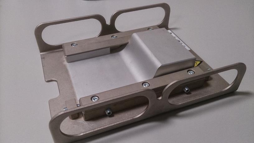
11 CBCT beam profiling Air-Kerma and tube filtration profiles were measured with the Unfors Xi chamber at the isocentre, and using the bed to step in 1 cm increments across the full width of the bow-tie profile Air kerma taken directly from the Unfors Xi, filtration a little more tricky!!
12 Total filtration (mm Al). Air Kerma (mgy/ kvp) S1 S2 S3 S Position relative to centre of field (cm) Use this info to plug into PCXMC to calculate patient dose per slither S1 S2 S3 S Position relative to centre of field (cm)
 Uterus & ovaries I know prostate patients don t have these, but it was useful for validation purposes!")
13 Performed TLD dosimetry on two linear accelerators (RT treatment machines), with Rando phantom loaded with 80 TLD-100H chips in the positions of the various important organs in and around the scan volume Liver & stomach were most superior organs measured (well outside the primary beam) Uterus & ovaries I know prostate patients don t have these, but it was useful for validation purposes! Bladder, prostate & testes these were all fully irradiated by the primary beam Small and large intestine partially irradiated by the primary beam Rando was positioned with the prostate at the isocentre, and three CBCT Pelvis scans performed Model validation
14 Model validation TLD dosimetry Mean Organ Dose* (mgy) Organ RT1 RT2 Mean Liver Stomach Uterus Ovaries Bladder Prostate Testicles Small Intestine Large Intestine * Measured Air Kerma corrected for ratio of (μ en /ρ) ICRU soft tissue /(μ en /ρ) air
15 Model validation The comparison So how do these compare with the PCXMC model? Mean Organ Dose (mgy) Organ Mean TLD PCXMC % diff. Liver Stomach Uterus Ovaries Bladder Prostate Testicles Small Intestine Large Intestine Large distance from beam Not bad given the inherent errors associated with TLD dosimetry
16 Effective dose? Using PCXMC to calculate the effective dose, taking out contribution to ovaries and uterus (not applicable to our prostate patients!), and the prostate (which is the target of the RT treatment, so probably should not be included in the calculation) Effective dose = 6.0 msv per scan Using TLD dosimetry with Rando Effective dose = 5.9 msv per scan Good agreement!! For daily prostate imaging we get up to 222 msv for a 37 fraction treatment regime, risk of fatal cancer: 1 in 150 for a healthy 60 year old male (using organ specific risk factors) 1 in 90 using generic 5% per Sv Not insignificant!!!
17 Organ doses? Total individual organ doses for daily imaging with 37 fractions (ignoring prostate); Bladder > 1.2 Gy Testicles > 1.4 Gy Large Intestine > 0.3 Gy These don t feel insignificant! Mean Organ Dose for 37# (Gy) Organ RT Treatment Pelvic CBCT CBCT as % of RT Gonads 0.8 >1.4 >175 Bladder 51.8 >1.2 >2.3 Colon 1.2 >0.3 >25 Rectum 40.0 >0.9 >2.3
18 Size specific CBCT Currently all Pelvis exposures use the same factors (125 kvp/80ma/13ms/650 projections ~ 680 total mas) No compensation for patient size means the organ/effective dose reduces as the patient gets bigger But, we should probably be increasing exposure factors for the biggest patients to ensure we get acceptable images We have it on good authority that these patients are difficult to image Equally, smaller patients should have a lower dose protocol


19 Protocol Optimisation Have started looking at patient size specific exposure protocols We have used the CT AEC phantom Tim discussed in his talk earlier today Scanned this at the default exposure setting 125 kvp, 80 ma, 13 ms per projection, 650 projections, Total of 680 mas Decreased the ma to assess the effect on image noise: Wanted to increase ma as well but 80 ma is its upper limit!!! Also scanned with increased/decreased ms:
20 Noise (SD) Protocol Optimisation Effect of ma (dose) Noise with ma mA, 13ms 60mA, 13ms 60 40mA, 13ms 20mA, 13ms Patient thickness CT slice
21 Protocol Optimisation Effect of ma (dose) As expected decrease in noise as the ma (dose) increases, for a given patient thickness Also, increase in noise as the patient gets thicker, for a given ma (dose) There is definitely scope to optimise the ma for average and thinner patients Possibly as low as 40 ma for the very thin ones?? Even scope to decrease ma for thicker patients 60 ma is not too different in terms of noise compared to 80 ma
22 Noise (SD) Protocol Optimisation Effect of ms Noise with ms mA, 26ms 50 80mA, 23ms 40 80mA, 20ms 80mA, 13ms 30 80mA, 7ms Small patients Large patients CT slice
23 Protocol Optimisation Effect of ms As patient size increases noise increases Less obvious with thinner patients As ms increases noise decreases May be able to decrease to 7ms for very thin patients (with 80 ma) Given that we have been told larger patient images can be poor, and that we can t increase the ma (max 80mA which is the default), it may be possible to increase the ms for larger patients to improve image quality. Probably go to 26ms for same noise as average patient


24 Hot off the press!!! Very large patient scanned with default settings led to images that were not usable We recommended they use 26 ms and image quality had improved such that images are now acceptable for the clinical intent
25 Summary Developed a PCXMC model that simulates CBCT organ doses for pelvic (prostate) imaging Organ doses are not insignificant for daily CBCT imaging!!! Developing size specific protocols should be possible Increase/decrease in ma Increase decrease in ms Future work will include Adopt size specific protocols into clinical practice Looking in more detail at the organ specific risks of cancer induction Create some written justification protocols for the use of CBCT with dose information for size specific scans Looking at other anatomical sites
Calculation of Effective Doses for Radiotherapy Cone-Beam CT and Nuclear Medicine Hawkeye CT Laura Sawyer
 Calculation of Effective Doses for Radiotherapy Cone-Beam CT and Nuclear Medicine Hawkeye CT Laura Sawyer Department of Medical Physics and Bioengineering, Royal United Hospital, Bath Overview Varian Acuity
Measurement of organ dose in abdomen-pelvis CT exam as a function of ma, KV and scanner type by Monte Carlo method
 Iran. J. Radiat. Res., 2004; 1(4): 187-194 Measurement of organ dose in abdomen-pelvis CT exam as a function of ma, KV and scanner type by Monte Carlo method M.R. Ay 1, M. Shahriari 2, S. Sarkar 3, P.
Introduction. Modalities used in imaging guidance. Flat panel detector. X-ray Imaging Dose to Patients in the Era of Image-Guided Radiation Therapy
 X-ray Imaging Dose to Patients in the Era of Image-Guided Radiation Therapy George Ding, Ron Price, Charles Coffey Vanderbilt-Ingram Cancer Center Vanderbilt University Medical Center, Nashville, TN Introduction
AAPM Task Group 180 Image Guidance Doses Delivered During Radiotherapy: Quantification, Management, and Reduction
 AAPM Task Group 180 Image Guidance Doses Delivered During Radiotherapy: Quantification, Management, and Reduction Parham Alaei, Ph.D. Department of Radiation Oncology University of Minnesota NCCAAPM Fall
Accounting for Imaging Dose
 Accounting for Imaging Dose High Profile Over-exposures Lead to Growing Concern FDA issues warning in October 2009-209 patients exposed to 8 times typical dose for CT brain perfusion scan (3-4 Gy) - Some
Skyscan 1076 in vivo scanning: X-ray dosimetry
 Skyscan 1076 in vivo scanning: X-ray dosimetry DOSIMETRY OF HIGH RESOLUTION IN VIVO RODENT MICRO-CT IMAGING WITH THE SKYSCAN 1076 An important distinction is drawn between local tissue absorbed dose in
Managing the imaging dose during Image-guided Radiotherapy. Martin J Murphy PhD Department of Radiation Oncology Virginia Commonwealth University
 Managing the imaging dose during Image-guided Radiotherapy Martin J Murphy PhD Department of Radiation Oncology Virginia Commonwealth University Radiographic image guidance has emerged as the new paradigm
Dosimetric Consideration in Diagnostic Radiology
 Dosimetric Consideration in Diagnostic Radiology Prof. Ng Kwan-Hoong Department of Biomedical Imaging University of Malaya ngkh@um.edu.my Radiation Dosimetry Workshop, 28-29 March 2014 2 Why do we measure
Managing the imaging dose during image-guided radiation therapy
 Managing the imaging dose during image-guided radiation therapy Martin J Murphy PhD Department of Radiation Oncology Virginia Commonwealth University Richmond VA Imaging during radiotherapy Radiographic
Quantitative and Qualitative Assessment of Thorax Cone Beam CT Image Quality across Multiple Imaging Systems
 Quantitative and Qualitative Assessment of Thorax Cone Beam CT Image Quality across Multiple Imaging Systems Matthew Williams Pre-registration Clinical Scientist Velindre NHS Trust, Cardiff Computed Tomography
IGRT1 technologies. Paweł Kukołowicz Warsaw, Poland
 IGRT1 technologies Paweł Kukołowicz Warsaw, Poland Minimal prerequisite for good, efficient radiotherapy ICTP 2015 Paweł Kukołowicz 2/29 Minimal prerequisite for good, efficient radiotherapy Well trained
Doses from pediatric CT examinations in Norway Are pediatric scan protocols developed and in daily use?
 Doses from pediatric CT examinations in Norway Are pediatric scan protocols developed and in daily use? Eva Godske Friberg * Norwegian Radiation Protection Authority, P.O. Box, Østerås, Norway Abstract.
Adopting DAP as a dose metric in CT
 Adopting DAP as a dose metric in CT Nick Weir Department of Medical Physics Royal Infirmary of Edinburgh NHS Lothian Dose Area Product (DAP) in CT Hypothesis: There are advantages to using DAP: (i) In
RADIATION PROTECTION IN DIAGNOSTIC AND INTERVENTIONAL RADIOLOGY. L19: Optimization of Protection in Mammography
 IAEA Training Material on Radiation Protection in Diagnostic and Interventional Radiology RADIATION PROTECTION IN DIAGNOSTIC AND INTERVENTIONAL RADIOLOGY L19: Optimization of Protection in Mammography
Varian Edge Experience. Jinkoo Kim, Ph.D Henry Ford Health System
 Varian Edge Experience Jinkoo Kim, Ph.D Henry Ford Health System Disclosures I participate in research funded by Varian Medical Systems. Outline of Presentation Review advanced imaging in Varian Edge Linear
Assessment of effective dose in paediatric CT examinations
 Assessment of effective dose in paediatric CT examinations E. Dougeni 1,2 CL. Chapple 1, J. Willis 1, G. Panayiotakis 2 1 Regional Medical Physics Department, Freeman Hospital, Freeman Road, Newcastle
Prof. Dr. Doğan BOR Ankara University Institute of Nuclear Science
 PATIENT DOSIMETRY IN DIAGNOSTIC RADIOLOGY MODALITIES Prof. Dr. Doğan BOR Ankara University Institute of Nuclear Science Ankara University Institute of Nuclear Science USE OF RADIATION! INCREASING? Natural
Collapsed Cone Convolution 2D illustration
 Collapsed Cone Convolution 2D illustration 8 cones Energy desposition decreases very quickly with distance Energy is absorber in blue pixels only. IGRT1 technologies Paweł Kukołowicz Warsaw, Poland IGRT
CT Dose Estimation. John M. Boone, Ph.D., FAAPM, FSBI, FACR Professor and Vice Chair of Radiology. University of California Davis Medical Center
 CT Dose Estimation John M. Boone, Ph.D., FAAPM, FSBI, FACR Professor and Vice Chair of Radiology 1 University of California Davis Medical Center CT Dose Estimation Introduction The CTDI Family of Metrics
Comparison of organ doses estimations in radiology with PCXMC application based on MIRD phantoms and CALDose-X application based on voxel phantoms
 Comparison of organ doses estimations in radiology with PCXMC application based on MIRD phantoms and CALDose-X application based on voxel phantoms G. Gialousis 1, Z. Pappouli 2, A. Dimitriadis 2, E. Karavassilis
Simple methods to reduce patient dose in a Varian cone beam CT system for delivery verification in pelvic radiotherapy
 The British Journal of Radiology, 82 (2009), 855 859 SHORT COMMUNICATION Simple methods to reduce patient dose in a Varian cone beam CT system for delivery verification in pelvic radiotherapy 1 P ROXBY,
8/18/2011. Acknowledgements. Managing Pediatric CT Patient Doses INTRODUCTION
 Managing Pediatric CT Patient Doses Keith J. Strauss, MSc, FAAPM, FACR President X-Ray Computations, Inc. Boston, Massachusetts Acknowledgements Marilyn Goske, MD John Boone, PhD Cynthia McCollough, PhD
Computed tomography Acceptance testing and dose measurements
 Computed tomography Acceptance testing and dose measurements Jonas Andersson Medical Physicist, Ph.D. Department of Radiation Sciences University Hospital of Norrland, Umeå Sweden Contents The Computed
CURRENT CT DOSE METRICS: MAKING CTDI SIZE-SPECIFIC
 CURRENT CT DOSE METRICS: MAKING CTDI SIZE-SPECIFIC Keith Strauss, MSc, FAAPM, FACR Cincinnati Children s Hospital University of Cincinnati College of Medicine Acknowledgments John Boone, PhD Michael McNitt-Grey,
A more accurate method to estimate patient dose during body CT examinations with tube current modulation
 A more accurate method to estimate patient dose during body CT examinations with tube current modulation Poster No.: C-0738 Congress: ECR 2014 Type: Scientific Exhibit Authors: A. Kawaguchi 1, Y. Matsunaga
Dosimetry of recently introduced CBCT Units for Oral and Maxillofacial Radiology
 Dosimetry of recently introduced CBCT Units for Oral and Maxillofacial Radiology John B Ludlow, Laura E Davies-Ludlow, André Mol University of North Carolina, Chapel Hill, NC Background CBCT is seeing
IMRT for Prostate Cancer
 IMRT for Cancer All patients are simulated in the supine position. Reproducibility is achieved using a custom alpha cradle cast that extends from the mid-back to mid-thigh. The feet are positioned in a
Quanitative and qualitative analysis of Image Quality and Imaging Dose of KV CBCT from XVI Elekta Linac.
 IOSR Journal of Applied Physics (IOSR-JAP) e-issn: 2278-4861.Volume 9, Issue 5 Ver. IV (Sep. - Oct. 2017), PP 46-52 www.iosrjournals.org Quanitative and qualitative analysis of Image Quality and Imaging
Assessment of radiation risk to pediatric patients undergoing conventional X-ray examinations
 Radioprotection 50(1), 19-25 (2015) c EDP Sciences 2015 DOI: 10.1051/radiopro/2014023 Available online at: www.radioprotection.org Article Assessment of radiation risk to pediatric patients undergoing
7/16/2009. Cost-benefit, QALYs and the Value of a Statistical Life
 7/16/29 Cost-benefit, QALYs and the Value of a Statistical Life Alan McKenzie, Bristol The benefits of many developments in radiotherapy may be obvious for instance, replacing hand planning by computer
Introduction and Background
 CT Lung Cancer Screening and the Medical Physicist: Background, Findings and Participant Dosimetry Summary of the National Lung Screening Trial (NLST) Randell Kruger, PhD, DABR Medical Physics Section
IMAGING DOSES IN RADIATION THERAPY FROM KILOVOLTAGE CONE-BEAM COMPUTED TOMOGRAPHY
 IMAGING DOSES IN RADIATION THERAPY FROM KILOVOLTAGE CONE-BEAM COMPUTED TOMOGRAPHY By DANIEL ELLIS HYER A DISSERTATION PRESENTED TO THE GRADUATE SCHOOL OF THE UNIVERSITY OF FLORIDA IN PARTIAL FULFILLMENT
CT Radiation Risks and Dose Reduction
 CT Radiation Risks and Dose Reduction Walter L. Robinson, M.S. D.A.B.S.N.M., D.A.B.M.P., D.A.B.R. Consultant Certified Medical Radiation Health & Diagnostic Imaging Physicist Medical Radiation and Children
CT Dosimetry in the Clinical Environment: Methods and Analysis
 CT Dosimetry in the Clinical Environment: Methods and Analysis Manuel Arreola, Ph.D. DABR Associate Chair of Radiology Director, Medical Physics Graduate Program Department of Radiology University of Florida
PHY138Y Nuclear and Radiation
 PHY38Y Nuclear and Radiation Professor Tony Key MP40 key@physics.utoronto.ca Announcements MP problems set #4 due Sunday at midnight PS#5 WRITTEN now posted! - do in teams, no Lone Wolves!! NB correction
Radiation Dose Reduction Strategies in Coronary CT Angiography
 Radiation Dose Reduction Strategies in Coronary CT Angiography Noor Diyana Osman, PhD noordiyana@usm.my Contents: Introduction Radiation dosimetry in CT Radiation risk associated with coronary CT angiography
Recent Progress in Radiation Dosimetry for Epidemiology and Radiological Protection. John Harrison ICRP Committee 2
 Recent Progress in Radiation Dosimetry for Epidemiology and Radiological Protection John Harrison ICRP Committee 2 Joint ICRP-RERF-JHPS Workshop: Tokyo, December 2017 Task Group 79 : Use of Effective Dose
Equivalent dose to organs and tissues in Hysterosalpingography calculated with the FAX (Female Adult voxel) phantom
 phantom") Equivalent dose to organs and tissues in Hysterosalpingography calculated with the FAX (Female Adult voxel) phantom 1 R Kramer, PhD, 1 H J Khoury, PhD, 1 C. Lopes and 2 J W Vieira, PhD 1 Departamento de
Equivalent dose to organs and tissues in Hysterosalpingography calculated with the FAX (Female Adult voxel) phantom 1 R Kramer, PhD, 1 H J Khoury, PhD, 1 C. Lopes and 2 J W Vieira, PhD 1 Departamento de
Non-target dose from radiotherapy: Magnitude, Evaluation, and Impact. Stephen F. Kry, Ph.D., D.ABR.
 Non-target dose from radiotherapy: Magnitude, Evaluation, and Impact Stephen F. Kry, Ph.D., D.ABR. Goals Compare out-of-field doses from various techniques Methods to reduce out-of-field doses Impact of
ESTABLISHING DRLs in PEDIATRIC CT. Keith Strauss, MSc, FAAPM, FACR Cincinnati Children s Hospital University of Cincinnati College of Medicine
 ESTABLISHING DRLs in PEDIATRIC CT Keith Strauss, MSc, FAAPM, FACR Cincinnati Children s Hospital University of Cincinnati College of Medicine CT Dose Indices CTDI INTRODUCTION CTDI 100, CTDI w, CTDI vol
Toshiba Aquillion 64 CT Scanner. Phantom Center Periphery Center Periphery Center Periphery
 Comparison of radiation dose and imaging performance for the standard Varian x-ray tube and the Richardson Healthcare ALTA750 replacement tube for the Toshiba Aquillion CT scanners. by Robert L. Dixon,
Radiology Review Course Hotel del Coronado Coronado, California
 37 th Annual Radiology Review Course Hotel del Coronado Coronado, California Saturday, April 22, 2017 - AM TABLE OF CONTENTS Saturday, April 22, 2017 - AM 7:00 AM 7:30 AM Coffee and Pastries for Registrants
DOSE DISTRIBUTION ANALYZE OF THE BODY STI USED MONTE CARLO METHOD
 Proceedings of the Tenth EGS4 Users' Meeting in Japan, KEK Proceedings 2002-18, p.65-73 DOSE DISTRIBUTION ANALYZE OF THE BODY STI USED MONTE CARLO METHOD N. Tohyama, H. Saitoh, T. Fujisaki 1, S. Abe 1
Learning objective. Outline. Acknowledgements. KV CBCT Imaging Part I. R Hammoud AAPM 2008 CE-Therapy (SAM) 1
 1") 1 2 KV CBCT Imaging Part I Rabih Hammoud, MS, DABR Henry Ford Health System Detroit, Michigan Acknowledgements Indrin Chetty, PhD Teamour Nurushev, PhD Harrison Guan, PhD Jinkoo Kim, PhD JianYue Jin, PhD
1 2 KV CBCT Imaging Part I Rabih Hammoud, MS, DABR Henry Ford Health System Detroit, Michigan Acknowledgements Indrin Chetty, PhD Teamour Nurushev, PhD Harrison Guan, PhD Jinkoo Kim, PhD JianYue Jin, PhD
EQUIVALENT DOSE FROM SECONDARY NEUTRONS AND SCATTER PHOTONS IN ADVANCE RADIATION THERAPY TECHNIQUES WITH 15 MV PHOTON BEAMS
 PAPER EQUIVALENT DOSE FROM SECONDARY NEUTRONS AND SCATTER PHOTONS IN ADVANCE RADIATION THERAPY TECHNIQUES WITH 15 MV PHOTON BEAMS Isra Israngkul Na Ayuthaya *, Sivalee Suriyapee, Phongpheath Pengvanich
Comparison of high and low energy treatment plans by evaluating the dose on the surrounding normal structures in conventional radiotherapy
 Turkish Journal of Cancer Volume 37, No. 2, 2007 59 Comparison of high and low energy treatment plans by evaluating the dose on the surrounding normal structures in conventional radiotherapy MUHAMMAD BASIM
Dosimetry in digital mammography
 Dosimetry in digital mammography Professor David Dance NCCPM, Royal Surrey County Hospital, Guildford, United kingdom Outline Why do dosimetry? History Essentials of European breast dosimetry protocol
Managing Radiation Risk in Pediatric CT Imaging
 Managing Radiation Risk in Pediatric CT Imaging Mahadevappa Mahesh, MS, PhD, FAAPM, FACR, FACMP, FSCCT. Professor of Radiology and Cardiology Johns Hopkins University School of Medicine Chief Physicist
Bone Densitometry Radiation dose: what you need to know
 Bone Densitometry Radiation dose: what you need to know John Damilakis, PhD Associate Professor and Chairman University of Crete, Iraklion, Crete, GREECE Estimation of bone status using X-rays Assessment
Translating Protocols Across Patient Size: Babies to Bariatric
 Translating Protocols Across Patient Size: Babies to Bariatric Cynthia H. McCollough, PhD, FACR, FAAPM Professor of Radiologic Physics Director, CT Clinical Innovation Center Department of Radiology Mayo
Radiation exposure of the Yazd population from medical conventional X-ray examinations
 Iran. J. Radiat. Res., 2007; 4 (4): 195-200 Radiation exposure of the Yazd population from medical conventional X-ray examinations F. Bouzarjomehri 1*, M.H. Dashti 2, M.H. Zare 1 1 Department of Medical
Patient Dose Estimates. from CT Examinations. Patient Dose Estimates
 Patient Dose Estimates from CT Examinations Patient Dose Estimates from CT Examinations John M. Boone, Ph.D. Professor and Vice Chairman of Radiology Professor of Biomedical Engineering University of California,
Imaging doses from the Elekta Synergy X-ray cone beam CT system
 Imaging doses from the Elekta Synergy X-ray cone beam CT system A. Amer, PhD, T. Marchant, PhD, J. Sykes *, MSc, J Czajka M.Sc, C. Moore, PhD North Western Medical Physics, Christie Hospital NHS Trust,
IMAGE GENTLY HOW CAN YOU HELP?
 IMAGE GENTLY HOW CAN YOU HELP? Keith J. Strauss, MSc, FAAPM, FACR Director, Radiology Physics & Engineering Children s s Hospital Boston Harvard Medical School Acknowledgment Marilyn J. Goske,, MD Robert
GATE MONTE CARLO DOSIMETRY SIMULATION OF MARS SPECTRAL CT
 GATE MONTE CARLO DOSIMETRY SIMULATION OF MARS SPECTRAL CT R Aamir, C Lowe, J Damet, P Carbonez, A P H Butler, N Schleich, N G Anderson MARFO, Emmanuel Geant4 User Workshop 2017, Wollongong Overview MARS
Estimating Patient Radiation Dose from Computed Tomography
 Estimating Patient Radiation Dose from Computed Tomography C. Cagnon, J. DeMarco, E. Angel, M. McNitt-Gray UCLA David Geffen School of Medicine 1 Patient Dose from CT Advances in Technology... Helical
Implementation of full/half bowtie filter models in a commercial treatment planning system for kilovoltage cone-beam CT dose estimations
 JOURNAL OF APPLIED CLINICAL MEDICAL PHYSICS, VOLUME 17, NUMBER 2, 2016 Implementation of full/half bowtie filter models in a commercial treatment planning system for kilovoltage cone-beam CT dose estimations
Assessment of variation of wedge factor with depth, field size and SSD for Neptun 10PC Linac in Mashhad Imam Reza Hospital
 Iran. J. Radiat. Res., 2004; 2 (2): 53-58 Assessment of variation of wedge factor with depth, field size and SSD for Neptun 10PC Linac in Mashhad Imam Reza Hospital M. Hajizadeh Saffar 1*, M.R. Ghavamnasiri
Helical Tomotherapy Experience. TomoTherapy Whole Brain Head & Neck Prostate Lung Summary. HI-ART TomoTherapy System. HI-ART TomoTherapy System
 The Challenges Associated with Differential Dose Delivery using IMRT Chester Ramsey, Ph.D. Director of Medical Physics Thompson Cancer Center Knoxville, Tennessee, U.S.A Collaborators Chester Ramsey, Ph.D.
Outline. Chapter 12 Treatment Planning Combination of Beams. Opposing pairs of beams. Combination of beams. Opposing pairs of beams
 Chapter 12 Treatment Planning Combination of Beams Radiation Dosimetry I Text: H.E Johns and J.R. Cunningham, The physics of radiology, 4 th ed. http://www.utoledo.edu/med/depts/radther Outline Combination
Use of Monte Carlo Simulation Software for Calculating Effective dose in Cone Beam Computed Tomography
 Use of Monte Carlo Simulation Software for Calculating Effective dose in Cone Beam Computed Tomography Wilson Otto Gomes Batista Instituto Federal da Bahia Rua Emidio dos Santos s/n. Barbalho 40301-015
Concept for quantifying the dose from image guided radiotherapy
 Schneider et al. Radiation Oncology (2015) 10:188 DOI 10.1186/s13014-015-0492-7 RESEARCH Open Access Concept for quantifying the dose from image guided radiotherapy Uwe Schneider 1,2*, Roger Hälg 1,2 and
Performance of several active personal dosemeters in interventional radiology and cardiology
 Performance of several active personal dosemeters in interventional radiology and cardiology S. Chiriotti 1,*, M. Ginjaume 1, E. Vano 2,3, R. Sanchez 3, J.M. Fernandez 2,3, M.A. Duch 1, J. Sempau 1 1.
Measurement of Dose to Critical Structures Surrounding the Prostate from. Intensity-Modulated Radiation Therapy (IMRT) and Three Dimensional
 and Three Dimensional") Measurement of Dose to Critical Structures Surrounding the Prostate from Intensity-Modulated Radiation Therapy (IMRT) and Three Dimensional Conformal Radiation Therapy (3D-CRT); A Comparative Study Erik
Measurement of Dose to Critical Structures Surrounding the Prostate from Intensity-Modulated Radiation Therapy (IMRT) and Three Dimensional Conformal Radiation Therapy (3D-CRT); A Comparative Study Erik
S. Derreumaux (IRSN) Accidents in radiation therapy in France: causes, consequences and lessons learned
 Accidents in radiation therapy in France: causes, consequences and lessons learned") S. Derreumaux (IRSN) Accidents in radiation therapy in France: causes, consequences and lessons learned MEDICAL LINEAR ACCELERATORS Electron beam (MeV) Photon beam (MV) PRECISION REQUIRED IN RADIOTHERAPY
S. Derreumaux (IRSN) Accidents in radiation therapy in France: causes, consequences and lessons learned MEDICAL LINEAR ACCELERATORS Electron beam (MeV) Photon beam (MV) PRECISION REQUIRED IN RADIOTHERAPY
Investigation of the clinical performance of a novel solid-state diagnostic dosimeter
 JOURNAL OF APPLIED CLINICAL MEDICAL PHYSICS, VOLUME 16, NUMBER 4, 2015 Investigation of the clinical performance of a novel solid-state diagnostic dosimeter Jason Tse, a Donald McLean Medical Physics and
A Comparison of IMRT and VMAT Technique for the Treatment of Rectal Cancer
 A Comparison of IMRT and VMAT Technique for the Treatment of Rectal Cancer Tony Kin Ming Lam Radiation Planner Dr Patricia Lindsay, Radiation Physicist Dr John Kim, Radiation Oncologist Dr Kim Ann Ung,
ADVANCES IN RADIATION TECHNOLOGIES IN THE TREATMENT OF CANCER
 ADVANCES IN RADIATION TECHNOLOGIES IN THE TREATMENT OF CANCER Bro. Dr. Collie Miller IARC/WHO Based on trends in the incidence of cancer, the International Agency for Research on Cancer (IARC) and WHO
Out-of-field dosimetry in radiotherapy for input to epidemiological studies. Roger Harrison
 MELODI 7th Workshop, 9 11 November 2015 Helmholtz Zentrum München Next Generation Radiation Protection Research Out-of-field dosimetry in radiotherapy for input to epidemiological studies Roger Harrison
The Physics of Oesophageal Cancer Radiotherapy
 The Physics of Oesophageal Cancer Radiotherapy Dr. Philip Wai Radiotherapy Physics Royal Marsden Hospital 1 Contents Brief clinical introduction Imaging and Target definition Dose prescription & patient
Neutron dose evaluation in radiotherapy
 Neutron dose evaluation in radiotherapy Francesco d Errico University of Pisa, Italy Yale University, USA Radiation therapy with a linear accelerator (LINAC) Photoneutron production in accelerator head
CALCULATION OF BACKSCATTER FACTORS FOR LOW ENERGY X-RAYS USING THE TOPAS MONTE CARLO CODE
 CALCULATION OF BACKSCATTER FACTORS FOR LOW ENERGY X-RAYS USING THE TOPAS MONTE CARLO CODE Emily Hewson 1 Martin Butson 1,2 Robin Hill 1,2 1 Institute of Medical Physics, School of Physics, University of
TESTICULAR DOSES IN IMAGE-GUIDED RADIOTHERAPY OF PROSTATE CANCER
 doi:10.1016/j.ijrobp.2011.01.071 Int. J. Radiation Oncology Biol. Phys., Vol. 82, No. 1, pp. e39 e47, 2012 Copyright Ó 2012 Elsevier Inc. Printed in the USA. All rights reserved 0360-3016/$ - see front
Managing Patient Dose in Computed Tomography (CT) INTERNATIONAL COMMISSION ON RADIOLOGICAL PROTECTION
 INTERNATIONAL COMMISSION ON RADIOLOGICAL PROTECTION") Managing Patient Dose in Computed Tomography (CT) International Commission on Radiological Protection Information abstracted from ICRP Publication 87 Available at www.icrp.org Task Group: M.M. Rehani,
Managing Patient Dose in Computed Tomography (CT) International Commission on Radiological Protection Information abstracted from ICRP Publication 87 Available at www.icrp.org Task Group: M.M. Rehani,
Verification of treatment planning system parameters in tomotherapy using EBT Radiochromic Film
 Verification of treatment planning system parameters in tomotherapy using EBT Radiochromic Film E.B.Rajmohan¹, Pratik Kumar¹, Bhudatt Paliwal,² David Westerly², N.Gopishankar³, R.K.Bisht³, D.Tewatia²,
A comparison of radiation doses from modern multi-slice Computed Tomography angiography and conventional diagnostic Angiography:
 A comparison of radiation doses from modern multi-slice Computed Tomography angiography and conventional diagnostic Angiography: Rob Loader Oliver Gosling Introduction Approached by Dr Oliver Gosling (Research
Why is CT Dose of Interest?
 Why is CT Dose of Interest? CT usage has increased rapidly in the past decade Compared to other medical imaging CT produces a larger radiation dose. There is direct epidemiological evidence for a an increase
ABSTRACT. Objectives: To investigate the radiation dose received by patients undergoing routine plain x-ray
 Radiation Dose Distribution for Patients Undergoing Routine Radiological Scans for Kidney Stone Diagnosis at the University Hospital of the West Indies DC Walker 1, WD Aiken 2, S Shah 3, MK Voutchkov 1,
Imaging of Scattered Radiation for Real Time Tracking of Tumor Motion During Lung SBRT
 Imaging of Scattered Radiation for Real Time Tracking of Tumor Motion During Lung SBRT April 25 nd, 2015 Lung Cancer Lung cancer is the most lethal cancer: Over 224,000 new diagnoses in the U.S. predicted
Imaging Rotation. University of Michigan Department of Radiation Oncology Division of Radiation Physics. Resident:
 University of Michigan Department of Radiation Oncology Division of Radiation Physics Imaging Rotation Resident: Rotation staff mentor/ advisor: James Balter, supplemental mentors: Dale Litzenberg, Don
Dosimetric Analysis of 3DCRT or IMRT with Vaginal-cuff Brachytherapy (VCB) for Gynaecological Cancer
 for Gynaecological Cancer") Dosimetric Analysis of 3DCRT or IMRT with Vaginal-cuff Brachytherapy (VCB) for Gynaecological Cancer Tan Chek Wee 15 06 2016 National University Cancer Institute, Singapore Clinical Care Education Research
Dosimetric Analysis of 3DCRT or IMRT with Vaginal-cuff Brachytherapy (VCB) for Gynaecological Cancer Tan Chek Wee 15 06 2016 National University Cancer Institute, Singapore Clinical Care Education Research
An Assessment of Organ and Effective Dose of Patients who Undertake CT Examinations in two Teaching Hospitals of Mashhad&Isfahan
 An Assessment of Organ and Effective Dose of Patients who Undertake CT Examinations in two Teaching Hospitals of Mashhad&Isfahan *M.T.Bahreyni Toossi, **S.Mohandes Dastgherdi Medical Physics Dep., Faculty
Practical Reference Dosimetry Course April 2015 PRDC Program, at a glance. Version 1.0. Day 1 Day 2 Day 3 Day 4
 Practical Reference Dosimetry Course 21-24 April 2015 PRDC 2015 Program, at a glance Version 1.0 Day 1 Day 2 Day 3 Day 4 Quantities and Units Free air chambers Uncertainties Brachytherapy traceability
CT NUMBER ACCURACY ANALYSIS FOR RADIOTHERAPY TREATMENT PLANNING IMAGING
 CT NUMBER ACCURACY ANALYSIS FOR RADIOTHERAPY TREATMENT PLANNING IMAGING Julian Liu a, Keisha Robinson a, DhanaJayan Kothandan a and Joshua Luis b (a) Cancer Centre London (b) University College London
CT Optimisation for Paediatric SPECT/CT Examinations. Sarah Bell
 CT Optimisation for Paediatric SPECT/CT Examinations Sarah Bell Sarah.bell14@nhs.net Outline 1. Introduction 2. Aims and Objectives 3. Methods 4. Results 5. Discussion 6. Conclusions 7. References Introduction
Radiation qualities in carbon-ion radiotherapy at NIRS/HIMAC
 Radiation qualities in carbon-ion radiotherapy at NIRS/ Shunsuke YONAI Radiological Protection Section Research Center for Charged Particle Therapy National Institute of Radiological Sciences (NIRS) E-mail:
7/31/2018. Research Activities: Imaging for RT Image Guidance for Pediatric Radiotherapy. Chia-ho Hua, PhD. Outline
 Research Activities: Imaging for RT Image Guidance for Pediatric Radiotherapy Chia-ho Hua, PhD Chia-ho Associate Hua, Member PhD St. Jude Children s Research Hospital AAPM-COG Joint Session, 60 th Annual
An organ and effective dose study of XVI and OBI cone-beam CT systems
 JOURNAL OF APPLIED CLINICAL MEDICAL PHYSICS, VOLUME 11, NUMBER 2, SPRING 2010 An organ and effective dose study of XVI and OBI cone-beam CT systems Daniel E. Hyer, 1a Christopher F. Serago, 2 Siyong Kim,
Application of asi-kvcbct for Volume Assessment and Dose Estimation: An Offline Adaptive Study for Prostate Radiotherapy
 DOI:10.31557/APJCP.2019.20.1.229 Volume and Dose Assessment on Adapted CT RESEARCH ARTICLE Editorial Process: Submission:08/10/2018 Acceptance:01/05/2019 Application of asi-kvcbct for Volume Assessment
Radiation Related Second Cancers. Stephen F. Kry, Ph.D., D.ABR.
 Radiation Related Second Cancers Stephen F. Kry, Ph.D., D.ABR. Objectives Radiation is a well known carcinogen Atomic bomb survivors Accidental exposure Occupational exposure Medically exposed Radiotherapy
Automatic Patient Centering for MDCT: Effect on Radiation Dose
 Patient Centering for MDCT CT Imaging Original Research Jianhai Li 1 Unni K. Udayasankar 1 Thomas L. Toth 2 John Seamans 2 William C. Small 1 Mannudeep K. Kalra 1,3 Li J, Udayasankar UK, Toth TL, Seamans
Investigation of the radiation dose from cone-beam CT for image-guided radiotherapy: A comparison of methodologies
 Received: 21 September 217 Revised: 31 October 217 Accepted: 3 November 217 DOI: 1.12/acm2.12239 RADIATION ONCOLOGY PHYSICS Investigation of the radiation dose from cone-beam CT for image-guided radiotherapy:
Application(s) of Alanine
 of Alanine") Application(s) of Alanine Simon Duane Radiotherapy Standards User Group, 5 June 2007 Outline Alanine/EPR dosimetry characteristics, usage (dis)advantages for reference dosimetry Traceable dosimetry for
Application(s) of Alanine Simon Duane Radiotherapy Standards User Group, 5 June 2007 Outline Alanine/EPR dosimetry characteristics, usage (dis)advantages for reference dosimetry Traceable dosimetry for
MEASUREMENT OF THE EQUIVALENT INDIVIDUAL DOSES FOR PATIENTS IN ANGIOGRAPHY PROCEDURE AND INTERVENTIONAL RADIOLOGY WITH THERMOLUMINESCENT SYSTEMS
 RADIOPROTECTION AND DOSIMETRY MEASUREMENT OF THE EQUIVALENT INDIVIDUAL DOSES FOR PATIENTS IN ANGIOGRAPHY PROCEDURE AND INTERVENTIONAL RADIOLOGY WITH THERMOLUMINESCENT SYSTEMS DANIELA ADAM 1, ANA STOCHIOIU
Joint ICTP/IAEA Advanced School on Dosimetry in Diagnostic Radiology and its Clinical Implementation May 2009
 2033-4 Joint ICTP/ Advanced School on Dosimetry in Diagnostic Radiology and its Clinical Implementation 11-15 May 2009 Dosimetry for General Radiology and Clinical Uncertainty Peter Homolka EFOMP Training
Estimation of Effective Doses for Radiation Cancer Risks on ISS, Lunar and Mars Missions with Space Radiation Measurements
 Estimation of Effective Doses for Radiation Cancer Risks on ISS, Lunar and Mars Missions with Space Radiation Measurements Myung-Hee Y. Kim Wyle Laboratories, Houston, Texas, 77058 and Francis A. Cucinotta
Kerma Area Product Method for Effective Dose Estimation During Lumbar Epidural Steroid Injection Procedures: Phantom Study
 Vascular and Interventional Radiology Original Research Kim et al. Dose for Lumbar Steroid Injection Vascular and Interventional Radiology Original Research Sangroh Kim 1 Greta Toncheva 2,3 Colin Anderson-Evans
CT Radiation Dosimetry Study Using Monte Carlo Simulation and. Computational Anthropomorphic Phantoms
 CT Radiation Dosimetry Study Using Monte Carlo Simulation and Computational Anthropomorphic Phantoms by Yakun Zhang Graduate Program in Medical Physics Duke University Date: Approved: Ehsan Samei, Supervisor
Introduction. Measurement of Secondary Radiation for Electron and Proton Accelerators. Introduction - Photons. Introduction - Neutrons.
 Measurement of Secondary Radiation for Electron and Proton Accelerators D. Followill, Ph.D. Radiological Physics Center U. T. M. D. Anderson Cancer Center Introduction Patients undergoing radiation therapy
Estimation of Organ and Effective Doses for Neonate and Infant Diagnostic Cardiac Catheterizations
 Medical Physics and Informatics Original Research Kawasaki et al. Dose Estimates for Neonate and Infant Diagnostic Cardiac Catheterization Medical Physics and Informatics Original Research Toshio Kawasaki