Liver Cancer (Hepatocellular Carcinoma or HCC) Overview
|
|
|
- Jennifer Cole
- 5 years ago
- Views:
Transcription
 and metastatic liver lesions.")

1 Liver Cancer (Hepatocellular Carcinoma or HCC) Overview Recent advances in liver cancer care seek to address the rising incidence of liver cancer, which has steadily increased over the past three decades. At the UMC Center for Transplantation, our team sees individuals with both hepatocellular carcinoma (primary liver cancer) and metastatic liver lesions. Current treatment options include surgical removal of tumors, the application of chemotherapy and radiation into the liver, and systemic chemotherapy treatments. Tumor Detection Liver cancer may be hard to detect. Most patients are asymptomatic. For this reason, physicians advise screening high-risk patients periodically. Surveillance tools for HCC include ultrasound and the alphafetoprotein (AFP) blood test. AFP is a protein produced by the liver. An elevated level can indicate tumor growth, though some patients with liver cancer have normal AFP levels. For patients with abnormalities in these tests, an evaluation with a contrast-enhanced CT or MRI is recommended to determine if cancer is present. If patients develop symptoms, the first is usually pain in the right side of the abdomen. Weight loss is common and sometimes patients have episodes of severe pain, fever, and nausea. Rapidly deteriorating health, weakness, swelling, and jaundice may also imply HCC. Determining the Size, Stage and Scope of a Liver Lesion Once blood tests reveal an elevated AFP level or an ultrasound shows a lesion in the liver, patients should undergo further evaluation to determine if liver cancer is present and to assess for size and number of tumors. This preoperative evaluation includes the diagnosis and localization of the lesion, staging, and a determination of treatment options. UMC uses a variety of techniques to help determine the location and stage of HCC. Diagnostic imaging procedures are the most accurate methods. In some cases, a liver biopsy is required to determine the type of liver tumor. Diagnostic Imaging At UMC, liver imaging may include a four-phase computed tomography (CT), including spiral CT scans obtained during hepatic arterial and portal venous phases following intravenous contrast administration. Imaging also involves state-of-the-art magnetic resonance imaging (MRI). These techniques can accurately demonstrate the number of primary tumors within the liver and their relationship to vascular structures. Doppler ultrasound can be used as a complementary imaging technique. FDG positron emission tomography (PET) is occasionally helpful in characterizing atypical hepatic masses as well as in detecting extrahepatic malignancy. Metastatic Work-Up Patients with HCC require a metastatic work-up to determine if cancer has spread to surrounding tissue. This work-up includes a chest CT scan and a nuclear medicine bone scan. After treatment, patients should continue to be staged every three to six months with chest/abdomen CT and bone scans. Surgical Interventions for Liver Cancer When determining treatment options for hepatocellular carcinoma (HCC), our team evaluates the lesion imaging and lab results, along with the patient s age, liver function, and overall health to recommend appropriate treatment options. Because choosing a treatment plan is an important decision, we review all options with patients and their families, explaining the benefits and disadvantages of each. Some patients may require liver biopsy to determine surgical candidacy, since advanced liver scarring or cirrhosis limit the ability to safely perform surgery to remove the tumor.

2 Ablation Some patients are not candidates for resection due to inadequate liver reserve, large or multiple lesions in multiple lobes, or cirrhosis. These patients may benefit from minimally invasive ablation therapy. This therapy uses extreme heat to destroy liver tumors. The ideal patient for ablation generally has no more than three lesions that are no greater than 5 cm in size. Ablation delivers radiofrequency energy to the tumor, heating it to temperatures above 113º F, thereby destroying the lesion. This technique can be performed either by an interventional radiologist or by a surgeon, depending on the lesion s size and location. Systemic Chemotherapy Systemic chemotherapy uses a mixture of anti-cancer drugs injected into a vein or taken by mouth. Typically, this alternative is reserved for patients with metastatic disease or those who are not candidates for other procedures. Depending on any underlying disease, different drugs are applied. Patients are always encouraged to seek out and participate in clinical trials, which offer hope and help us learn how to better care for patients with this disease. Metastatic Liver Lesions Diagnosis and Treatment Overview of Metastatic Liver Lesions Many recent advances in medicine and surgery have made it possible to successfully treat cancers that were previously considered incurable. At UMC Center for Transplantation, these advances have been incorporated into a multidisciplinary approach that can offer treatment for most patients who have developed metastatic liver lesions from malignant tumors, such as colon cancer or breast cancer. Our team of specialists will work with you to provide a comprehensive treatment plan that includes the latest technology in diagnosis and treatment. What are Metastatic Liver Lesions? Cancer can develop in any organ and each cancer acts differently in its tendency to spread to other organs. When a cancer has spread to the liver, it is referred to as a metastatic liver lesion. Sometimes the metastatic lesion is identified at the same time as the original cancer (synchronous) and sometimes the metastatic lesion is discovered later, after the original cancer has been treated or surgically removed (metachronous).


3 The liver is the most common site for metastasis from gastrointestinal cancers, such as colon cancer or pancreatic cancer. Detecting and Staging Metastases When cancer has spread to the liver, it typically does not cause any symptoms. As a result, most patients do not notice any change in their health. Blood tests and imaging, such as CT scan or ultrasound, are important tools to detect metastatic liver lesions at an early stage, when they are most effectively treated. These tests can also be helpful in determining if the cancer has spread to areas in addition to the liver. In some patients additional testing, such as a positron emission tomography (PET) and MRI scan may be required to determine the extent of the liver metastases. Treatment Options for Metastatic Liver Lesions Once a metastatic liver lesion is detected, patients should undergo a rapid medical, oncologic and surgical evaluation to determine the most appropriate treatment. The treatment may vary between patients, and will be tailored to best fit each individual's specific needs. Frequently, combinations of treatments, rather than a single type of treatment will be required, and you will meet with a team of specialists to discuss these different options during your evaluation. Chemotherapy Systemic Chemotherapy The most common treatment used for metastatic liver lesions is systemic chemotherapy. In this treatment, anti-cancer medications may be delivered intravenously or by ingestion of an oral preparation containing the anti-cancer drug. Cancer cells vary widely in their response to systemic chemotherapy. Some cancers respond well to chemotherapy, while others may be unaffected. This type of treatment is commonly offered for patients who have large malignant tumors that cannot be removed surgically, or patients who have cancer spread to other organs in addition to the liver. Surgery Surgical resection involves removing a portion of the liver that contains the metastatic lesion. This treatment is reserved for patients with no underlying liver disease (such as cirrhosis), and who are otherwise healthy enough to withstand a major operation. Surgeons can remove as much as 70 percent of the liver in attempting to remove cancerous lesions, since the liver possesses the ability to regenerate after surgical resection. The liver will typically replace the removed liver volume within several weeks after surgical resection. In patients with metastatic liver lesions localized to one anatomic region of the liver, surgical resection offers the best chance for cure. Only a physician or surgeon experienced with the treatment of metastatic liver lesions can determine if surgical resection is right for you.
 in patients without underlying liver disease and usually need no specific treatment.")
4 Overview of Non-Cancerous Liver Lesions Masses within the liver are increasingly being detected inadvertently when patients are evaluated for unrelated reasons. These liver masses are usually benign (non-cancerous) in patients without underlying liver disease and usually need no specific treatment. We recommend that the work-up and management of benign lesions be overseen by our multidisciplinary team including radiologists, gastroenterologists, oncologists and surgeons, to ensure a patient receives the best possible care. Benign masses can be categorized into two groups: solid or cystic (fluid filled). Solid Masses Among the most common solid masses include: Hemangioma Focal nodular hyperplasia Adenoma Focal fatty change Nodular regenerative hyperplasia Hemangiomas are the most common of all benign liver masses. They are more prevalent in women and may be affected by hormonal changes. Symptoms such as pain are mostly noted in lesions less than 6 cm and are related to compression of adjacent structures. Bleeding is rare. Diagnosis of these lesions is usually made radiologically with magnetic resonance imaging (MRI) offering the most definitive means of diagnosis. No specific treatment is required for asymptomatic lesions whatever the size. Surgical resection is the treatment of choice for symptomatic lesions. Focal Nodular Hyperplasia (FNH) is the second most common benign lesion of the liver. It is usually asymptomatic and has no malignant potential or risk of rupture. Symptomatic lesions are usually larger and cause compression of adjacent structures. Laboratory studies are usually normal and diagnosis is made radiologically. At times a biopsy may be needed. Surgical resection is indicated only if the diagnosis is of question or the patient is symptomatic.
 and have a potential to become cancerous (10%).")


5 Adenomas are a rare entity and have a strong association with oral contraceptive use. Larger adenomas (less than 5 cm) may present with abdominal discomfort or a feeling of fullness. Other symptoms include nausea, vomiting and fevers. Larger lesions have a tendency to bleed (40%) and have a potential to become cancerous (10%). Diagnosis of these lesions is made by a combination of radiographic examinations and sometimes biopsy. Treatment consists of first discontinuing oral contraception use, to see if the mass regresses. Additionally, all lesions greater than 4-5 cm or where malignancy cannot be excluded should be surgically resected. Focal Fatty Change occurs when fat distribution within the liver is not evenly spread. Areas of increased fat accumulation are referred to as focal fatty change. Patients who have a history of diabetes, obesity, hepatitis C or malnutrition may be predisposed to this condition. Individuals are usually asymptomatic. These lesions are diagnosed by radiographic examination (MRI) and at times require a biopsy. No specific treatment is required.
 Cysts may be present from birth (congenital) or may arise later in life. There appears to be a higher incidence of this process in females.")
6 Cystic Masses Two major categories of cystic masses exist and are related to either an infectious or a non-infectious cause. Non-Infectious Cystic Masses Bile Duct (Choledochal) Cysts may be present from birth (congenital) or may arise later in life. There appears to be a higher incidence of this process in females. Adult onset choledochal cysts are usually an incidental finding. If symptomatic, the patient may present with pain under the right rib cage, nausea, vomiting, fever and/or jaundice. In extreme cases, a patient may present with back pain. Patients may rarely present with inflammation of the liver and sometimes cirrhosis of the liver due to chronic obstruction of the bile duct. In addition to laboratory studies, a variety of imaging modalities may be needed. More invasive studies by a gastroenterologist or an interventional radiologist are required to fully delineate the extent of the disease process. Biopsy of the bile duct may be needed to rule out bile duct cancer. The presence of cancer may be known either before or at the time of the operation. The operation consists of resecting the diseased bile duct and reconnecting the remnant to the small intestine. A transplant evaluation is needed if liver cirrhosis is noted on the preoperative workup.


7 A simple liver cyst is usually a single cyst located within the liver, which is present from birth. Most cysts are asymptomatic and are uncommonly diagnosed before age 40. If symptomatic, patients complain of abdominal fullness and pain. Diagnosis is made radiographically. No specific treatment is needed in cases where the cyst is less than 8 cm in diameter or is within the confines of liver tissue. Infectious or cancerous causes as well as possible communication with the bile duct system must be ruled out prior to performing an operation. Laparoscopic marsupialization (widely opening the cyst to drain into the abdominal cavity) is the approach of choice. In certain cases, the location of the cyst may preclude marsupialization and may require a partial liver resection. Polycystic Liver Disease (PCLD) is an inherited condition and may be associated with cystic lesions of the kidneys. Most patients are asymptomatic with normal laboratory studies. The liver cysts are multiple and tend to enlarge slowly. Symptoms are similar to that of simple cysts. Ultrasound and CT scans are reliable in detecting the lesions. These cysts must be differentiated from multiple simple cysts given that PCLD is an inherited disease. There are genetic tests available to help counsel afflicted patients and families. A liver, kidney or combined liver-kidney transplant may be necessary depending on disease severity of the organs afflicted.
, trauma to the liver, or ablative therapies (TACE, RFA) used to treat liver cancer.")
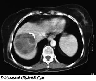
8 Infectious Cystic Masses Pyogenic Liver Abscesses (bacterial cause) - There are numerous causes of bacterial infections that bring about abscess formation in the liver. Presently, disease processes within the bile duct that cause bile flow obstruction are the most common cause of pyogenic abscesses. Other causes include intra-abdominal infections (i.e. appendicitis or diverticulitis), trauma to the liver, or ablative therapies (TACE, RFA) used to treat liver cancer. Finally, distant infectious processes such as dental abscesses and endocarditis may cause liver abscess formation. A specific source is not identified in up to 55% of cases. Patients can present with fevers, chills, nausea, vomiting, abdominal pain and loss of appetite. Some may present with a severe illness if rupture of the abscess into the abdominal cavity has occurred. The diagnosis can be made with a combination of history and physical examination, laboratory studies and radiologic examination. Treatment depends on the clinical condition of the patient and radiologic findings. Typically, antibiotic therapy is initiated and the abscess is drained using a catheter placed directly into the abscess by the radiologist (90% successful). Surgical intervention is needed in more severe cases. Amebic Liver Abscess - Amebic infection or amebiasis is a common infection in the tropics. In the United States individuals at risk for amebiasis are those who have immigrated from or traveled to endemic areas. The organism responsible for the disease process is Entamoeba Histolytica. Transmission usually occurs via ingestion of infected water. Liver abscess formation occurs when the ameba penetrates through the intestines and into local veins that drain into the liver. Liver abscesses are more common in patients who are immunocompromised, malnourished or have a malignancy. Less than one-third of the patients have intestinal symptoms prior to the diagnosis of liver abscess. Patients usually present with acute abdominal pain and fevers. Up to 8% of patients present with mild jaundice. Tests to detect antibodies in the blood (positive in up to 95% of patients) are available and should be performed. Various radiologic studies can be used to help in the diagnosis. Treatment is primarily with antibiotics. Aspiration of the abscess is rarely indicated. An operation is indicated if worsening infection is noted despite adequate medical therapy. Hydatid Cysts - These liver cysts are caused by a parasitic organism found in dogs. Echinococcus granulosus or Echinococcus multilocularis are parasites (tapeworms) that infect dogs. Tapeworm embryos are present in the feces of dogs. After inadvertent ingestion, the tapeworm embryo will penetrate the intestine and usually find its way to the liver. It may also migrate to other structures such as the lung, spleen, brain, bone or kidney after entering the bloodstream. Cysts are usually visible three weeks after ingestion and continue to secrete fluid causing compression of the liver. Cysts less than 5 cm are usually asymptomatic and no specific treatment is required. Patients usually have symptoms of abdominal fullness. Pain usually is noted when cysts get infected or rupture. The most common site of rupture is into the bile ducts within the liver causing symptoms of bile duct obstruction and infection. Some patients may present with an allergic reaction after cyst rupture. Radiologic studies used to diagnose hydatid cysts are ultrasound and CT scans. Antibody tests are available to detect hydatid cysts and should be completed. Treatment options range from chemotherapy (mebendazole and albendazole) to surgery. Surgery can entail a conservative approach (various drainage type procedures) or a radical operation that removes the entire cyst with a rim of normal liver.
9
Liver Cancer And Tumours
 Liver Cancer And Tumours What causes liver cancer? Many factors may play a role in the development of cancer. Because the liver filters blood from all parts of the body, cancer cells from elsewhere can
Liver Tumors. Prof. Dr. Ahmed El - Samongy
 Liver Tumors Prof. Dr. Ahmed El - Samongy Objective 1. Identify the most important features of common benign liver tumors 2. Know the risk factors, diagnosis, and management of hepatocellular carcinoma
Liver Tumors. Patient Education. Treatment options 8 4A. About the Liver. Surgical Specialties
 Patient Education Treatment options This handout describes different kinds of tumors that form in the liver and how they are treated. About the Liver Your liver is the largest organ in your abdomen. It
HEPATIC METASTASES. We can state 3 types of metastases depending on their treatment options:
 HEPATIC METASTASES 1. Definition Metastasis means the spread of cancer. Cancerous cells can separate from the primary tumor and enter the bloodstream or the lymphatic system (the one that produces, stores,
Evaluation of Liver Mass Lesions. American College of Gastroenterology 2013 Regional Postgraduate Course
 Evaluation of Liver Mass Lesions American College of Gastroenterology 2013 Regional Postgraduate Course Lewis R. Roberts, MB ChB, PhD Division of Gastroenterology and Hepatology Mayo Clinic College of
Workup of a Solid Liver Lesion
 Workup of a Solid Liver Lesion Joseph B. Cofer MD FACS Chief Quality Officer Erlanger Health System Affiliate Professor of Surgery UTHSC-Chattanooga I have no financial or other relationships with any
Hepato-Pancreatico-Biliary Surgery. Dr. Ankur J. Shah. MS, DNB, MNAMS, MRCSEd (UK), FRCS (UK)
, FRCS (UK)") Hepato-Pancreatico-Biliary Surgery Dr. Ankur J. Shah MS, DNB, MNAMS, MRCSEd (UK), FRCS (UK) Consultant Hepato-Pancreatico-Biliary and Liver Transplant Surgeon Ansh Liver Clinic Prevention to Cure Address
Hepato-Pancreatico-Biliary Surgery Dr. Ankur J. Shah MS, DNB, MNAMS, MRCSEd (UK), FRCS (UK) Consultant Hepato-Pancreatico-Biliary and Liver Transplant Surgeon Ansh Liver Clinic Prevention to Cure Address
Transarterial Chemoembolisation (TACE) with Drug-Eluting Beads
 with Drug-Eluting Beads") Transarterial Chemoembolisation (TACE) with Drug-Eluting Beads A minimally invasive treatment for liver cancer Provided as an educational service by Biocompatibles UK Ltd, a BTG International group company
Transarterial Chemoembolisation (TACE) with Drug-Eluting Beads A minimally invasive treatment for liver cancer Provided as an educational service by Biocompatibles UK Ltd, a BTG International group company
HEPATO-BILIARY IMAGING
 HEPATO-BILIARY IMAGING BY MAMDOUH MAHFOUZ MD PROF.OF RADIOLOGY CAIRO UNIVERSITY mamdouh.m5@gmail.com www.ssregypt.com CT ABDOMEN Indications Patient preparation Patient position Scanogram Fasting 4-6 hours
ACG Clinical Guideline: Diagnosis and Management of Focal Liver Lesions
 ACG Clinical Guideline: Diagnosis and Management of Focal Liver Lesions Jorge A. Marrero, MD, 1 Joseph Ahn, MD, FACG, 2 K. Rajender Reddy, MD, FACG 3 1 University of Texas at Southwestern, Dallas, Texas,
Hepatobiliary and Pancreatic Malignancies
 Hepatobiliary and Pancreatic Malignancies Gareth Eeson MD MSc FRCSC Surgical Oncologist and General Surgeon Kelowna General Hospital Interior Health Consultant, Surgical Oncology BC Cancer Agency Centre
LIVER CIRRHOSIS. The liver extracts nutrients from the blood and processes them for later use.
 LIVER CIRRHOSIS William Sanchez, M.D. & Jayant A. Talwalkar, M.D., M.P.H. Advanced Liver Disease Study Group Miles and Shirley Fiterman Center for Digestive Diseases Mayo College of Medicine Rochester,
Chief Complain. Liver lesion found in routine health check 41 days ago
 Chief Complain Liver lesion found in routine health check 41 days ago Present Illness On 2005-7-26 at 台北署立醫院 he underwent a health check for the first time. Abdominal US showed suspicious of a 6*5 cm hepatoma,
X-Plain Pancreatic Cancer Reference Summary
 X-Plain Pancreatic Cancer Reference Summary Introduction Pancreatic cancer is the 4th leading cause of cancer deaths in the U.S. About 37,000 new cases of pancreatic cancer are diagnosed each year in the
performed to help sway the clinician in what the appropriate diagnosis is, which can substantially alter the treatment of management.
 Hello, I am Maura Polansky at the University of Texas MD Anderson Cancer Center. I am a Physician Assistant in the Department of Gastrointestinal Medical Oncology and the Program Director for Physician
What is Liver Cancer? About the Liver
 Your liver is important and it has many functions. The top three are that it cleans your blood of toxins, gives you energy and produces bile for digestion. What is Liver Cancer? Cancer starts when cells
Liver surgery, acute GI tract bleeding
 Semmelweis University, Faculty of Medicine, 1 st Department of Surgery Liver surgery, acute GI tract bleeding Oszkár HAHN M.D. LIVER CYST US, CT, MRI Parasite (ELISA, eosinophil, anaphylaxy) Echinococcus
Essentials of Clinical MR, 2 nd edition. 65. Benign Hepatic Masses
 65. Benign Hepatic Masses Pulse sequences acquired for abdominal MRI typically consist of fast acquisition schemes such as single-shot turbo spin echo (i.e. HASTE) and gradient echo schemes such as FLASH
Appendix 5. EFSUMB Newsletter. Gastroenterological Ultrasound
 EFSUMB Newsletter 87 Examinations should encompass the full range of pathological conditions listed below A log book listing the types of examinations undertaken should be kept Training should usually
Imaging of liver and pancreas
 Imaging of liver and pancreas.. Disease of the liver Focal liver disease Diffusion liver disease Focal liver disease Benign Cyst Abscess Hemangioma FNH Hepatic adenoma HCC Malignant Fibrolamellar carcinoma
LIVER IMAGING TIPS IN VARIOUS MODALITIES. M.Vlychou, MD, PhD Assoc. Professor of Radiology University of Thessaly
 LIVER IMAGING TIPS IN VARIOUS MODALITIES M.Vlychou, MD, PhD Assoc. Professor of Radiology University of Thessaly Hepatocellular carcinoma is a common malignancy for which prevention, screening, diagnosis,
Colorectal Cancer Treatment
 Scan for mobile link. Colorectal Cancer Treatment Colorectal cancer overview Colorectal cancer, also called large bowel cancer, is the term used to describe malignant tumors found in the colon and rectum.
Breast Cancer Diagnosis, Treatment and Follow-up
 Breast Cancer Diagnosis, Treatment and Follow-up What is breast cancer? Each of the body s organs, including the breast, is made up of many types of cells. Normally, healthy cells grow and divide to produce
Cholangiocarcinoma (Bile Duct Cancer)
") Cholangiocarcinoma (Bile Duct Cancer) The Bile Duct System (Biliary Tract) A network of bile ducts (tubes) connects the liver and the gallbladder to the small intestine. This network begins in the liver
Cholangiocarcinoma (Bile Duct Cancer) The Bile Duct System (Biliary Tract) A network of bile ducts (tubes) connects the liver and the gallbladder to the small intestine. This network begins in the liver
Radiology of hepatobiliary diseases
 GI cycle - Lecture 14 436 Teams Radiology of hepatobiliary diseases Objectives 1. To Interpret plan x-ray radiograph of abdomen with common pathologies. 2. To know the common pathologies presentation.
Newcastle HPB MDM updated radiology imaging protocol recommendations. Author Dr John Scott. Consultant Radiologist Freeman Hospital
 Newcastle HPB MDM updated radiology imaging protocol recommendations Author Dr John Scott. Consultant Radiologist Freeman Hospital This document is intended as a guide to aid radiologists and clinicians
Cystic Disease of the Liver Work Up and Management. Louis Ferrari MD, PGY 3 6/9/16 SUNY Downstate Medical Center
 Cystic Disease of the Liver Work Up and Management Louis Ferrari MD, PGY 3 6/9/16 SUNY Downstate Medical Center The Case 73F presents to clinic after diagnostic laparoscopy at OSH. Known liver mass for
Interventional Radiology in Liver Cancer. Nakarin Inmutto MD
 Interventional Radiology in Liver Cancer Nakarin Inmutto MD Liver cancer Primary liver cancer Hepatocellular carcinoma Cholangiocarcinoma Metastasis Interventional Radiologist Diagnosis Imaging US / CT
PANCREAS DUCTAL ADENOCARCINOMA PDAC
 CONTENTS PANCREAS DUCTAL ADENOCARCINOMA PDAC I. What is the pancreas? II. III. IV. What is pancreas cancer? What is the epidemiology of Pancreatic Ductal Adenocarcinoma (PDAC)? What are the risk factors
Imaging in gastric cancer
 Imaging in gastric cancer Gastric cancer remains a deadly disease because of late diagnosis. Adenocarcinoma represents 90% of malignant tumors. Diagnosis is based on endoscopic examination with biopsies.
Financial Disclosure
 Benign Liver Masses Adil Abdalla, MBBS Creighton University-CHI Health August 25, 2018 Financial Disclosure Nothing to disclose Financial Disclosure 1 Objectives To assess patients with benign liver tumors
Gall bladder cancer. Information for patients Hepatobiliary
 Gall bladder cancer Information for patients Hepatobiliary page 2 of 12 Who will provide my care? You will be cared for by a number of professionals who work together. These professionals will be specialist
RICCARDO LENCIONI,CLOTILDE DELLA PINA, LAURA CROCETTI,DANIA CIONI. Chapter 1
 RICCARDO LENCIONI,CLOTILDE DELLA PINA, LAURA CROCETTI,DANIA CIONI Chapter 1 Impact of European Federation of Societies for Ultrasound in Medicine and Biology (EFSUMB) Guidelines on the Use of Contrast
Brain Tumors. What is a brain tumor?
 Scan for mobile link. Brain Tumors A brain tumor is a collection of abnormal cells that grows in or around the brain. It poses a risk to the healthy brain by either invading or destroying normal brain
General summary GENERAL SUMMARY
 General summary GENERAL SUMMARY In Chapter 2.1 the long-term results and prognostic factors of radiofrequency ablation (RFA) for unresectable colorectal liver metastases (CRLM) in a single center with
The Focal Hepatic Lesion: Radiologic Assessment
 The Focal Hepatic Lesion: Radiologic Assessment Kevin Kuo, Harvard Medical School Year III Our Patient: PS 67 y/o female w/ long history of alcohol use Drinking since age 18, up to one bottle of wine/day
General Information Key Points
 The content of this booklet was adapted from content originally published by the National Cancer Institute. Male Breast Cancer Treatment (PDQ ) Patient Version. Updated September 29,2017. https://www.cancer.gov/types/breast/patient/male-breast-treatment-pdq
Hepatocellular Carcinoma: Diagnosis and Management
 Hepatocellular Carcinoma: Diagnosis and Management Nizar A. Mukhtar, MD Co-director, SMC Liver Tumor Board April 30, 2016 1 Objectives Review screening/surveillance guidelines Discuss diagnostic algorithm
Dr Claire Smith, Consultant Radiologist St James University Hospital Leeds
 Dr Claire Smith, Consultant Radiologist St James University Hospital Leeds Imaging in jaundice and 2ww pathway Image protocol Staging Limitations Pancreatic cancer 1.2.4 Refer people using a suspected
Pancreatic Cancer. What is pancreatic cancer?
 Scan for mobile link. Pancreatic Cancer Pancreatic cancer is a tumor of the pancreas, an organ that is located behind the stomach in the abdomen. Pancreatic cancer does not always cause symptoms until
Cirrhosis. A Chronic Liver Problem
 Cirrhosis A Chronic Liver Problem What Is Cirrhosis? Cirrhosis is a chronic (long-lasting) liver problem. It results from damaged and scarred liver tissue. Cirrhosis can t be cured, but it can be treated.
GASTROINTESTINAL IMAGING STUDY GUIDE
 GASTROINTESTINAL IMAGING STUDY GUIDE Pharynx Diverticula Foreign bodies Trauma o Motility Disorders Esophagus Diverticula Trauma Esophagitis Barrett esophagus Rings, webs, and strictures Varices Benign
Breast Cancer. What is breast cancer?
 Scan for mobile link. Breast Cancer Breast cancer is a malignant tumor in or around breast tissue. It usually begins as a lump or calcium deposit that develops from abnormal cell growth. Most breast lumps
Upper GI Malignancies Imaging Guidelines for the Management of Gastric, Oesophageal & Pancreatic Cancers 2012
 Upper GI Malignancies Imaging Guidelines for the Management of Gastric, Oesophageal & Pancreatic Cancers 2012 Version Control This is a controlled document please destroy all previous versions on receipt
IT 의료융합 1 차임상세미나 복부질환초음파 이재영
 IT 의료융합 1 차임상세미나 2013-4-3 복부질환초음파 이재영 나는오늘누구를위하여 종을울리나? 전통적의료 의사 공학설계자 의사 최첨단진단장비들 USG, CT, MRI 환자 환자 현대의료 사용자중심의사고 US in the Abdomen Detection DDx Look Behavior Response by external stimuli Guiding Tool
Multiple Primary Quiz
 Multiple Primary Quiz Case 1 A 72 year old man was found to have a 12 mm solid lesion in the pancreatic tail by computed tomography carried out during a routine follow up study of this patient with adult
Alice Fung, MD Oregon Health and Science University
 Alice Fung, MD Oregon Health and Science University Disclosure Comments The speaker Alice Fung, MD Has relevant financial relationships to disclose. Received honorarium from (Guerbet). This individual
Malignant Focal Liver Lesions
 Malignant Focal Liver Lesions Other Than HCC Pablo R. Ros, MD, MPH, PhD Departments of Radiology and Pathology University Hospitals Cleveland Medical Center Case Western Reserve University Pablo.Ros@UHhospitals.org
Recently role of non-invasive diagnostics methods
 CERTAIN ASPECTS OF NLS-DIAGNOSTICS OF LIVER FOCAL PATHOLOGY A.Y. Shvack, V.I. Nesterov, N.L. Ogluzdina This article contains information about NLS-graphy application in diagnostics of liver focal affections:
Indications for Surgical Removal of Adrenal Glands
 The adrenal glands are orange-colored endocrine glands which are located on the top of both kidneys. The adrenal glands are triangular shaped and measure about one-half inch in height and 3 inches in length.
Case Scenario 1. Discharge Summary
 Case Scenario 1 Discharge Summary A 69-year-old woman was on vacation and noted that she was becoming jaundiced. Two months prior to leaving on that trip, she had had a workup that included an abdominal
Jesse Civan, M.D. Medical Director, Jefferson Liver Tumor Center
 Liver Tumors Jesse Civan, M.D. Medical Director, Jefferson Liver Tumor Center Differential Diagnosis Malignant Metastatic from non-hepatic primary Hepatocellular carcinoma Cholangiocarcinoma Biliary cystcarcinoma
Liver Cancer: Diagnosis and Treatment Options
 Liver Cancer: Diagnosis and Treatment Options Fred Poordad, MD Chief, Hepatology University Transplant Center Professor of Medicine UT Health, San Antonio VP, Academic and Clinical Affairs, Texas Liver
Biliary tree dilation - and now what?
 Biliary tree dilation - and now what? Poster No.: C-1767 Congress: ECR 2012 Type: Educational Exhibit Authors: I. Ferreira, A. B. Ramos, S. Magalhães, M. Certo; Porto/PT Keywords: Pathology, Diagnostic
BOWEL CANCER. Causes of bowel cancer
 A cancer is an abnormality in an organ that grows without control. The growth is often quite slow, but will continue unabated until it is detected. It can cause symptoms by its presence in the organ or
TRANSEARTERIAL CHEMO- EMBOLIZATION FOR HEPATIC METASTASES FROM NEURO-ENDOCINE NEOPLASIA AND HEPATOMA DR SAMIA AHMAD
 UNIVERSITY OF PRETORIA STEVE BIKO ACADEMIC HOSPITAL SOUTH AFRICA TRANSEARTERIAL CHEMO- EMBOLIZATION FOR HEPATIC METASTASES FROM NEURO-ENDOCINE NEOPLASIA AND HEPATOMA DR SAMIA AHMAD 1 INTRODUCTION Hepatic
Complete Summary GUIDELINE TITLE. Liver lesion characterization. BIBLIOGRAPHIC SOURCE(S)
") Complete Summary GUIDELINE TITLE Liver lesion characterization. BIBLIOGRAPHIC SOURCE(S) Foley WD, Bree RL, Gay SB, Glick SN, Heiken JP, Huprich JE, Levine MS, Ros PR, Rosen MP, Shuman WP, Greene FL, Rockey
Complete Summary GUIDELINE TITLE Liver lesion characterization. BIBLIOGRAPHIC SOURCE(S) Foley WD, Bree RL, Gay SB, Glick SN, Heiken JP, Huprich JE, Levine MS, Ros PR, Rosen MP, Shuman WP, Greene FL, Rockey
6 th August 2018 Day 1 - Gallbladder & Bile duct Topic
 Venue: Sterling Hospital Auditorium, Sterling Hospitals, Gurukul Road Ahmedabad, Gujarat 6 th August 2018 Day 1 - Gallbladder & Bile duct Registration(8:00am-8:15am) Inauguration(8:15am-8:30am) Welcome
Esophageal Cancer. Source: National Cancer Institute
 Esophageal Cancer Esophageal cancer forms in the tissues that line the esophagus, or the long, hollow tube that connects the mouth and stomach. Food and drink pass through the esophagus to be digested.
Liver Ultrasound - Beyond the Basics. Pamela Parker Lead Sonographer
 Liver Ultrasound - Beyond the Basics Pamela Parker Lead Sonographer Aims Review what we know about the liver Reasons for imaging Focal lesions Diffuse disease Can we do more? The Liver The Liver The Liver
Breast Cancer. What is breast cancer?
 Scan for mobile link. Breast Cancer Breast cancer is a malignant tumor in or around breast tissue. It usually begins as a lump or calcium deposit that develops from abnormal cell growth. Most breast lumps
Index. Surg Oncol Clin N Am 16 (2007) Note: Page numbers of article titles are in boldface type.
 Note: Page numbers of article titles are in boldface type.") Surg Oncol Clin N Am 16 (2007) 465 469 Index Note: Page numbers of article titles are in boldface type. A Adjuvant therapy, preoperative for gastric cancer, staging and, 339 B Breast cancer, metabolic
Surg Oncol Clin N Am 16 (2007) 465 469 Index Note: Page numbers of article titles are in boldface type. A Adjuvant therapy, preoperative for gastric cancer, staging and, 339 B Breast cancer, metabolic
State of the Art Imaging for Hepatic Malignancy: My Assignment
 State of the Art Imaging for Hepatic Malignancy: My Assignment CT vs MR vs MRCP Which one to choose for HCC vs Cholangiocarcinoma What special protocols to use for liver tumors Role of PET and Duplex US
Early Klebsiella pneumoniae Liver Abscesses associated with Pylephlebitis Mimic
 Early Klebsiella pneumoniae Liver Abscesses associated with Pylephlebitis Mimic Hepatocellular Carcinoma Chih-Hao Shen, MD 3, Jung-Chung Lin, MD, PhD 2, Hsuan-Hwai Lin, MD 1, You-Chen Chao, MD 1, and Tsai-Yuan
Hepatobiliary Malignancies Retrospective Study at Truman Medical Center
 Hepatobiliary Malignancies 206-207 Retrospective Study at Truman Medical Center Brandon Weckbaugh MD, Prarthana Patel & Sheshadri Madhusudhana MD Introduction: Hepatobiliary malignancies are cancers which
LAPAROSCOPIC APPENDICECTOMY
 LAPAROSCOPIC APPENDICECTOMY WHAT IS THE APPENDIX? The appendix is a small, fingerlike pouch of the intestinal tract located where the small and large join. It has no known use. It is postulated that the
Approach to Liver Lesions. Anjana A. Pillai, MD Associate Professor of Medicine Director, Liver Tumor Clinic The University of Chicago Medical Center
 Approach to Liver Lesions Anjana A. Pillai, MD Associate Professor of Medicine Director, Liver Tumor Clinic The University of Chicago Medical Center Objectives Identify common clinical features and imaging
Management of Rare Liver Tumours
 Gian Luca Grazi Hepato-Biliary-Pancreatic Surgery National Cancer Institute Regina Elena Rome Fibrolamellar Carcinoma Mixed Hepato Cholangiocellular Carcinoma Hepatoblastoma Carcinosarcoma Primary Hepatic
Interesting Cases from Liver Tumor Board. Jeffrey C. Weinreb, M.D.,FACR Yale University School of Medicine
 Interesting Cases from Liver Tumor Board Jeffrey C. Weinreb, M.D.,FACR Yale University School of Medicine jeffrey.weinreb@yale.edu Common Liver Diseases Hemangioma Cyst FNH Focal Fat/Sparing THID Non-Cirrhotic
What is cancer? l Cancer is a group of several diseases caused by the abnormal growth of cells.
 Cancer Biology What is cancer? l Cancer is a group of several diseases caused by the abnormal growth of cells. What is cancer? l Cancer is a group of several diseases caused by the abnormal growth of cells.
X-Ray Corner. Imaging Approach to Cystic Liver Lesions. Pantongrag-Brown L. Solitary cystic liver lesions. Hepatic simple cyst (Figure 1)
") THAI J 136 Imaging Approach to Cystic Liver Lesions GASTROENTEROL 2013 X-Ray Corner Imaging Approach to Cystic Liver Lesions Pantongrag-Brown L Cystic liver lesions are common findings in daily practice
THAI J 136 Imaging Approach to Cystic Liver Lesions GASTROENTEROL 2013 X-Ray Corner Imaging Approach to Cystic Liver Lesions Pantongrag-Brown L Cystic liver lesions are common findings in daily practice
Small Bowel and Colon Surgery
 Small Bowel and Colon Surgery Why Do I Need a Small Bowel Resection? A variety of conditions can damage your small bowel. In severe cases, your doctor may recommend removing part of your small bowel. Conditions
Surgical indications: Non-malignant pulmonary diseases. Punnarerk Thongcharoen
 Surgical indications: Non-malignant pulmonary diseases Punnarerk Thongcharoen Non-malignant Malignant as a pathological term: Cancer Non-malignant = not cancer Malignant as an adjective: Disposed to cause
Health Bites Breast Cancer. Breast Cancer. Normal breast
 Health Bites Breast Cancer Breast Cancer Normal breast The normal breast tissue varies in size and shape. The breasts rest in front of the rib cage. The breasts are made up of fatty tissue, milk ducts
Update on RECIST and Staging of Common Pediatric Tumors Ethan A. Smith, MD
 Update on RECIST and Staging of Common Pediatric Tumors Ethan A. Smith, MD Section of Pediatric Radiology C.S. Mott Children s Hospital University of Michigan ethans@med.umich.edu Disclosures No relevant
Alpha-1 Antitrypsin Deficiency: Liver Disease
 Alpha-1 Antitrypsin Deficiency: Liver Disease Who is at risk to develop Alpha-1 liver disease? Alpha-1 liver disease may affect children and adults who have abnormal Alpha-1 antitrypsin genes. Keys to
Management of Colorectal Liver Metastases
 Management of Colorectal Liver Metastases MM Bernon, JEJ Krige HPB Surgical Unit, Groote Schuur Hospital Department of Surgery, University of Cape Town 50% of patients with colorectal cancer develop liver
Esophageal Cancer. What is esophageal cancer?
 Scan for mobile link. Esophageal Cancer Esophageal cancer occurs when cancer cells develop in the esophagus. The two main types are squamous cell carcinoma and adenocarcinoma. Esophageal cancer may not
Wilms' tumor most often occurs in just one kidney, though it can sometimes be found in both kidneys at the same time.
 Wilms' Tumor Wilms' tumor is a rare kidney cancer that primarily affects children. Also known as nephroblastoma, Wilms' tumor is the most common cancer of the kidneys in children. Wilms' tumor most often
Surveillance for Hepatocellular Carcinoma
 Surveillance for Hepatocellular Carcinoma Marion G. Peters, MD John V. Carbone, MD, Endowed Chair Professor of Medicine Chief of Hepatology Research University of California San Francisco Recorded on April
Liver imaging takes a step forward with Ingenia
 Publication for the Philips MRI Community ISSUE 49 2013 / 2 Liver imaging takes a step forward with Ingenia Lyon South Hospital strives to move from several studies first CT, then MR or PET to using just
Case Discussion Splenic Abscess
 Case Discussion Splenic Abscess Personal Data Gender: male Birth Date: 1928/Mar/06th Allergy: Mefenamic Smoking: 0.5 PPD for 55 years Alcohol: negative (?) 4 Months Ago Abdominal pain: epigastric area
Information about liver tumours and surgery. - a guide for patients and relatives
 Information about liver tumours and surgery - a guide for patients and relatives About the liver The liver is a large organ, which lies in the upper part of the abdominal cavity, directly below the diaphragm.
Transarterial Chemoembolization (TACE)
") Transarterial Chemoembolization (TACE) Princess Margaret Know what to expect Read this booklet to learn: What TACE is...1 How TACE works...2 The benefits of TACE...3 The risks and side effects of TACE...3
Transarterial Chemoembolization (TACE) Princess Margaret Know what to expect Read this booklet to learn: What TACE is...1 How TACE works...2 The benefits of TACE...3 The risks and side effects of TACE...3
is time consuming and expensive. An intra-operative assessment is not going to be helpful if there is no more tissue that can be taken to improve the
 My name is Barry Feig. I am a Professor of Surgical Oncology at The University of Texas MD Anderson Cancer Center in Houston, Texas. I am going to talk to you today about the role for surgery in the treatment
Hepatocellular Carcinoma HCC Updated November 2015 by: Dr. Mohammed Alghamdi (Medical Oncology Fellow, University of Calgary)
") Hepatocellular Carcinoma HCC Updated November 2015 by: Dr. Mohammed Alghamdi (Medical Oncology Fellow, University of Calgary) Staff Reviewers: Dr. Yoo Joung Ko (Medical Oncologist, Sunnybrook Odette Cancer
Hepatocellular Carcinoma HCC Updated November 2015 by: Dr. Mohammed Alghamdi (Medical Oncology Fellow, University of Calgary) Staff Reviewers: Dr. Yoo Joung Ko (Medical Oncologist, Sunnybrook Odette Cancer
Prostate Disease in Dogs
 Prostate Disease in Dogs An essential component of a complete physical examination for every male dog is an evaluation of the prostate, a walnut-sized gland located between the urinary bladder and the
ACG Clinical Guideline: Diagnosis and Management of Pancreatic Cysts
 ACG Clinical Guideline: Diagnosis and Management of Pancreatic Cysts Grace H. Elta, MD, FACG 1, Brintha K. Enestvedt, MD, MBA 2, Bryan G. Sauer, MD, MSc, FACG (GRADE Methodologist) 3 and Anne Marie Lennon,
General Surgery Curriculum Royal Australasian College of Surgeons, General Surgeons Australia & New Zealand Association of General Surgeons
 General Surgery Curriculum Royal Australasian College of Surgeons, General Surgeons Australia & New Zealand Association of General Surgeons MODULE TITLE: UPPER GI & HPB - HEPATIC, PANCREATIC & BILIARY
Lung cancer forms in tissues of the lung, usually in the cells lining air passages.
 Scan for mobile link. Lung Cancer Lung cancer usually forms in the tissue cells lining the air passages within the lungs. The two main types are small-cell lung cancer (usually found in cigarette smokers)
Diagnostic Studies Then. It s important to be able to distinguish. Diagnostic Studies Now
 Jonathan S. Fisher, MD, FACS It s important to be able to distinguish Diagnostic Studies Then Diagnostic Studies Then History Biopsy Diagnostic Studies Now History Biopsy Serum markers (AFP, CA19 9, CEA)
Simplifying liver assessment in internal medicine
 Ultrasound Customer story Simplifying liver assessment in internal medicine Philips Affiniti ultrasound for elastography and contrast-enhanced ultrasound (CEUS) Where Sonography Institute, Uster, Switzerland
How to integrate surgery in the treatment of patients with liver-only metastatic disease
 How to integrate surgery in the treatment of patients with liver-only metastatic disease Luis Sabater Ortí MD, PhD Associate Professor University of Valencia European Board Surgical Qualification HBP (EBSQ-HPB)
Gastrointestinal tract
 Gastrointestinal tract Colloidal liver-spleen imaging Presented by: Jehad Felemban Introduction: To obtain better anatomic display of liver and spleen architecture, we use (CT Ultrasound). (Radionuclide
General Surgery PURPLE SERVICE MUHC-RVH Site
 Preamble HPB is a clinical teaching unit with several different vocations: It regroups all solid organ Transplantation as well as most advanced Hepatobiliary and Pancreatic clinical activities performed
MALIGNANT HEPATIC NEOPLASMS: USING ULTRASONOGRAPHY AS A MEANS OF DEFINING HEPATIC LESIONS. 1.5 Contact Hours. Presented by: CEU Professor 7
 MALIGNANT HEPATIC NEOPLASMS: USING ULTRASONOGRAPHY AS A MEANS OF DEFINING HEPATIC LESIONS 1.5 Contact Hours Presented by: CEU Professor 7 www.ceuprofessoronline.com Copyright 8 2007 The Magellan Group,
Lung Cancer Treatment
 Scan for mobile link. Lung Cancer Treatment Lung cancer overview More than one in four of all diagnosed cancers involve the lung, and lung cancer remains the most common cancer-related cause of death among
Innovations in HCC Imaging: MDCT/MRI
 Innovations in HCC Imaging: MDCT/MRI Anthony E. Cheng, M.D. Cardinal MRI Center Cardinal Santos Medical Center, Wilson Street, San Juan Innovations in HCC Imaging: Goals/Objectives MDCT/MRI Learn the diagnostic
What Are Gallstones? GALLSTONES. Gallstones are pieces of hard, solid matter that form over time in. the gallbladder of some people.
 What Are Gallstones? Gallstones are pieces of hard, solid matter that form over time in the gallbladder of some people. The gallbladder sits under the liver and stores bile (a key digestive juice ). Gallstones