Chapter 7 Membrane Structure and Function
|
|
|
- Marcia Brooks
- 5 years ago
- Views:
Transcription
1 Chapter 7 Membrane Structure and Function
2 CELL MEMBRANE: Basics: Regulates flow in and out of the cell Composed of phospholipids and proteins - some carbohydrates and lipids SELECTIVELY PERMEABLE - allows some things across more easily than others Structure varies from one cell type to another determines function of the membrane
3
4 Membrane Structure Cellular membranes are fluid mosaics of lipids and proteins Phospholipids Are the most abundant lipid in the plasma membrane Are amphipathic, containing both hydrophobic and hydrophilic regions Organized in a bilayer

5 WATER Hydrophilic head Hydrophobic tail WATER
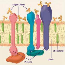
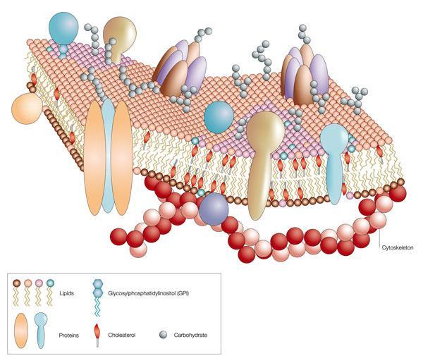
6 Membrane Structure States that a membrane is a fluid structure with a mosaic of various proteins embedded in it Proteins are either integral or peripheral Integral embedded in the membrane and traverse the phospholipid bilayer - transmembranal Peripheral on the surface - on both the inner and outer surfaces - surfaces differ membrane is bifacial or asymmetric
7

8 Singer and Nicolson Model Proposed that membrane proteins are dispersed and individually inserted into the phospholipid bilayer Hydrophobic region of protein Phospholipid bilayer Figure 7.3 Hydrophobic region of protein

9 Freeze-fracture studies of the plasma membrane Supported the fluid mosaic model of membrane structure A cell is frozen and fractured with a knife. The fracture plane often follows the hydrophobic interior of a membrane, splitting the phospholipid bilayer into two separated layers. The membrane proteins go wholly with one of the layers. Coat the split layers with a metal, usually gold or silver, and examine it under an electron scanning microscope
 in the two layers, demonstrating")
10 APPLICATION A cell membrane can be split into its two layers, revealing the ultrastructure of the membrane s interior. TECHNIQUE Extracellular layer Knife Proteins Plasma membrane Cytoplasmic layer RESULTS These SEMs show membrane proteins (the bumps ) in the two layers, demonstrating that proteins are embedded in the phospholipid bilayer.
11 Chemical Nature of Bilayer Amphipathic Parts Match Up - non-polar portions of proteins embed in the hydrophobic tails of the bilayer - polar portions of proteins embed in the hydrophilic heads or jut into the interior or exterior of the cell
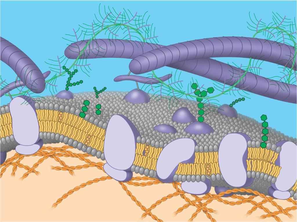
12 The Fluidity of Membranes Membrane parts are held together by hydrophobic interactions - weak bonds so phospholipids move around laterally (Side to side) very rare for a phospholipid to flip-flop from one side of the membrane to the other
 (a) Movement of phospholipids Figure 7.")
13 The Fluidity of Membranes Lateral movement (~10 7 times per second) Flip-flop (~ once per month) (a) Movement of phospholipids Figure 7.5 A
14 Movement of Molecules phospholipids move quickly Slows with temperature decrease until freezes Unsaturated tails - more flowing Saturated tails - more viscous
 Membrane fluidity Figure")
15 Fluid Viscous Unsaturated hydrocarbon tails with kinks Saturated hydro- Carbon tails (b) Membrane fluidity Figure 7.5 B

16 Role of cholesterol wedges between phospholipids and stabilizes structure hinders close packing decreases freezing point of membrane Figure 7.5 (c) Cholesterol within the animal cell membrane Cholesterol
17 Proteins move slowly larger anchored to the cytoskeleton some travel along the cytoskeleton some immobile


18 Movement of Proteins in the Plasma Membrane EXPERIMENT Researchers labeled the plasma mambrane proteins of a mouse cell and a human cell with two different markers and fused the cells. Using a microscope, they observed the markers on the hybrid cell. RESULTS Membrane proteins + Mouse cell Human cell Hybrid cell Mixed proteins after 1 hour Figure 7.6 CONCLUSION The mixing of the mouse and human membrane proteins indicates that at least some membrane proteins move sideways within the plane of the plasma membrane.
 Carbohydrates glycocalyx glycoproteins glycolipids FUNCTIONS: cell stickyness cell communication and")
19 TYPES OF STRUCTURES ATTACHED TO MEMBRANES 1) Proteins: Integral - transmembranal - interior and exterior end Peripheral - surface - on outside attached to membrane but not imbedded - attached to extracellular matrix and cytoskelton 2) Carbohydrates glycocalyx glycoproteins glycolipids FUNCTIONS: cell stickyness cell communication and recognition

20 Membrane Proteins and Their Functions Fibers of extracellular matrix (ECM) Glycoprotein Carbohydrate Glycolipid EXTRACELLULAR SIDE OF MEMBRANE Figure 7.7 Microfilaments of cytoskeleton Cholesterol Peripheral protein Integral protein CYTOPLASMIC SIDE OF MEMBRANE

21 Integral proteins Penetrate the hydrophobic core of the lipid bilayer Are often transmembrane proteins, completely spanning the membrane EXTRACELLULAR SIDE N-terminus Figure 7.8 C-terminus a Helix CYTOPLASMIC SIDE

22 Peripheral proteins Are appendages loosely bound to the surface of the membrane
 A protein that spans the membrane may provide a hydrophilic channel across the membrane that is selective for a particular solute.")
23 An overview of six major functions of membrane proteins (a) Transport. (left) A protein that spans the membrane may provide a hydrophilic channel across the membrane that is selective for a particular solute. (right) Other transport proteins shuttle a substance from one side to the other by changing shape. Some of these proteins hydrolyze ATP as an energy ssource to actively pump substances across the membrane. ATP (b) Enzymatic activity. A protein built into the membrane may be an enzyme with its active site exposed to substances in the adjacent solution. In some cases, several enzymes in a membrane are organized as a team that carries out sequential steps of a metabolic pathway. Enzymes (c) Signal transduction. A membrane protein may have a binding site with a specific shape that fits the shape of a chemical messenger, such as a hormone. The external messenger (signal) may cause a conformational change in the protein (receptor) that relays the message to the inside of the cell. Signal Figure 7.9 Receptor
.")
24 (d) Cell-cell recognition. Some glyco-proteins serve as identification tags that are specifically recognized by other cells. Glycoprotein (e) Intercellular joining. Membrane proteins of adjacent cells may hook together in various kinds of junctions, such as gap junctions or tight junctions (see Figure 6.31). (f) Attachment to the cytoskeleton and extracellular matrix (ECM). Microfilaments or other elements of the cytoskeleton may be bonded to membrane proteins, a function that helps maintain cell shape and stabilizes the location of certain membrane proteins. Proteins that adhere to the ECM can coordinate extracellular and intracellular changes (see Figure 6.29). Figure 7.9
25 Synthesis and Sidedness of Membranes Secreted proteins are on the inside of secretory vesicles Proteins on the outside of the cell membrane are on the inside of a secretory vesicle. Proteins on the inside of the cell membrane are on the outside of the secretory vesicle.

26 1 Transmembrane glycoproteins Secretory protein Glycolipid ER Golgi apparatus 2 Vesicle 3 4 Plasma membrane: Cytoplasmic face Extracellular face Secreted protein Transmembrane glycoprotein Figure 7.10 Membrane glycolipid
27 Traffic Across Membranes movement of materials is dependent upon the make up of the membrane certain substances move more easily certain substances move more quickly
28 The Permeability of the Lipid Bilayer Hydrophobic molecules Are lipid soluble and can pass through the membrane rapidly Polar molecules Do not cross the membrane rapidly Transports: hydrocarbons carbon dioxide oxygen small polar molecules with a neutral charge (H 2 O) Impedes: ions polar molecule large, uncharged polar molecules - glucose

29 Transport Proteins: Create channels for ions and polar molecules to pass through the lipid bilayer Selectively transport like enzymes - substrate specific each protein transports one specific molecule Methods: hydrophilic channel bind substance and move it across the bilayer
30 Types of Transport Across Membrane Passive vs. Active Passive: does not require energy downhill process moves from high concentration to low concentration with the concentration gradient (comparison of concentration levels across a membrane) Active: Requires energy uphill process moves from low concentration to high concentration against the concentration gradient requires transport proteins
31 Types of Passive Transport Diffusion Osmosis Facilitated Diffusion
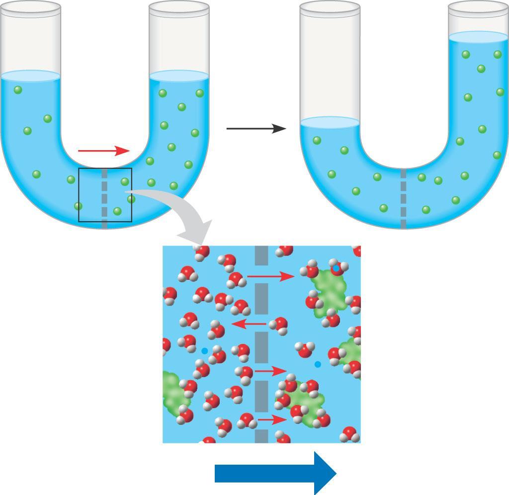
32 Diffusion based on thermal kinetics particles move throughout a space until they are distributed evenly movement of particles from high concentration to low concentration until net flow is zero NET FLOW: change in concentrations due to flow across membrane - each substance moves independently of the others continues until equilibrium is reached

33 Diffusion (a) Diffusion of one solute. The membrane has pores large enough for molecules of dye to pass through. Random movement of dye molecules will cause some to pass through the pores; this will happen more often on the side with more molecules. The dye diffuses from where it is more concentrated to where it is less concentrated (called diffusing down a concentration gradient). This leads to a dynamic equilibrium: The solute molecules continue to cross the membrane, but at equal rates in both directions. Molecules of dye Membrane (cross section) Net diffusion Net diffusion Equilibrium Figure 7.11 A


34 (b) Diffusion of two solutes. Solutions of two different dyes are separated by a membrane that is permeable to both. Each dye diffuses down its own concentration gradient. There will be a net diffusion of the purple dye toward the left, even though the total solute concentration was initially greater on the left side. Net diffusion Net diffusion Equilibrium Figure 7.11 B Net diffusion Net diffusion Equilibrium
35 Osmosis - special case of diffusion - APPLIES TO WATER ONLY movement of water across the membrane to balance solute concentration movement of water continues directionally until solute concentrations are equal Based on relative comparisons of solute concentration (tonicity) hypertonic: solution with more solute hypotonic: solution with less solute isotonic: solutions with equal concentrations Movement of water: 2 ways to say the same thing: from least solute to higher solute from high water concentration to low water concentration
36 Net Flow
37 Water Balance of Cells Without Walls Isotonic The concentration of solutes is the same as it is inside the cell There will be no net movement of water
38 Hypertonic The concentration of solutes is greater than it is inside the cell The cell will lose water
39 Hypotonic The concentration of solutes is less than it is inside the cell The cell will gain water
40 Water balance in cells without walls Identify the Tonicity H 2 O H 2 O H 2 O H 2 O Lysed Normal Shriveled 13
41 Water balance in cells without walls Hypotonic solution Isotonic solution Hypertonic solution H 2 O H 2 O H 2 O H 2 O Lysed Normal Shriveled 13
42 Water Balance of Cells with Walls Hypertonic environment - More solute outside of cell - Cell loses water - Becomes plasmolyzed
43 Water Balance of Cells with Walls Isotonic Same level of solute Net flow of water is zero No internal pressure plant cells have no support flaccid Plants wilt
44 Water Balance of Cells with Walls Hypotonic Environment More solute inside than out Positive flow of water into the cell plant cell is turgid healthy state in most plants Cells don t rupture because of strength of the cell wall Combination of osmotic pressure and the pressure of the walls is called the cell s water potential

45 Water balance in cells with walls Identify the Tonicity H 2 O H 2 O H 2 O H 2 O 13
46 Water balance in cells with walls Hypotonic solution Isotonic solution Hypertonic solution H 2 O H 2 O H 2 O H 2 O Turgid (normal) Flaccid Plasmolyzed 13
47 Water Potential determines the flow of water Water flows from high water potential to low water potential High pressure from the walls raises the water potential (tendency to lose water) High levels of solute in the cell lower water potential (tendency to gain water) Water flows into/out of the cell until the water potential reaches zero pressure from the cell walls prevents any more water from entering the cell.
48 Water Potential Equation Ψ = Ψ P + Ψ π Ψ P = Pressure Potential Ψ π = Osmotic Potential Ψ π = MiRT or Ψ π = CiRT M or C = osmotic molar concentration R = pressure constant (Gas Laws) T = temperature (K) i = ionization constant (with plants the solute is mainly sucrose which has an ionization constant of 1 because sucrose does not ionize)
 has a channel through which water molecules or a specific solute can pass.")
49 Facilitated Diffusion EXTRACELLULAR FLUID Channel protein Solute CYTOPLASM (a) A channel protein (purple) has a channel through which water molecules or a specific solute can pass. Figure 7.15
50 Facilitated Diffusion: Passive Transport Aided by Proteins Transmembranal proteins speed the movement of molecules across the plasma membrane passive - no energy required - moves with the concentration gradient Regulation of Facilitated Diffusion: protein saturation can only transport so much at once proteins are specialized for a specific substance protein inhibition similar molecules to normal transport substance bind to protein and block path of transport THESE ARE ONLY PHYSICAL CHANGES NOT CATALYZED REACTIONS
51 Mechanism of Transport Conformational changes - binding and release of substance causes change in protein - moves substance across membrane gated channels - stimulus causes protein to open and allow the flow of the substance - the stimulus is not the substance transported
 A carrier protein alternates between two conformations, moving a solute across the membrane as the shape of the protein changes.")
52 Carrier protein Solute Figure 7.15 (b) A carrier protein alternates between two conformations, moving a solute across the membrane as the shape of the protein changes. The protein can transport the solute in either direction, with the net movement being down the concentration gradient of the solute.
53 Active Transport pumping of solutes against the concentration gradient moving uphill - requires ATP or a membrane potential -membrane potential: electrical charge built up across the cell membrane due to unequal balance of anions and/or cations -
54 Types of Active Transport Use of ATP: phosphorylation reaction - adds phosphate to transport molecule and causes a conformational change EX: sodium-potassium pump
55 Na/K Ion Pump Generates an ELECTROCHEMICAL GRADIENT Unbalanced concentration of ions creates a new dynamic for diffusion - ions now move from high concentration and high positive charge important for Nerve Impulses
56 Maintenance of Membrane Potential by Ion Pumps Types of Pumps: electrogenic pump - pumping of various ions - animal cells proton pump - pumping H+ - plants, fungi, bacteria

57 Electrogenic pump ATP + + EXTRACELLULAR FLUID H + H + Proton pump H + + H + Figure 7.18 CYTOPLASM + + H + H +

58 COTRANSPORT movement of one type of ion out allows for the movement of another substance in when the ion diffuses back in through a cotransport protein + ATP H + + H + H + Proton pump + + Sucrose-H + cotransporter + + H + H + Sucrose Diffusion of H + H + H +
59 Other Types of Active Transport Use of Energized Electrons To Generate Chemical Gradients Ex: Kreb s Cycle and Light Dependent Reaction of Photosynthesis Drives the production of ATP


60 Electron Transport Chain In the Kreb s Cycle and Photosynthesis

61 Passive and active transport compared Passive transport. Substances diffuse spontaneously down their concentration gradients, crossing a membrane with no expenditure of energy by the cell. The rate of diffusion can be greatly increased by transport proteins in the membrane. Active transport. Some transport proteins act as pumps, moving substances across a membrane against their concentration gradients. Energy for this work is usually supplied by ATP. Figure 7.17 Diffusion. Hydrophobic molecules and (at a slow rate) very small uncharged polar molecules can diffuse through the lipid bilayer. Facilitated diffusion. Many hydrophilic substances diffuse through membranes with the assistance of transport proteins, either channel or carrier proteins. ATP
62 BULK MOVEMENT active transport that requires change in the cell membrane movement of large molecules (proteins and polysaccharides)
63 Exocytosis - movement of materials out of the cell transport vesicle fuses with cell membrane dumping contents into extracellular fluid Examples: insulin neurotransmitters glycocalyx
64 Endocytosis - movement of materials into the cell results in the formation of a vacuole or vesicle Three types Phagocytosis Pinocytosis Receptor Mediated Endocytosis
65 1) Phagocytosis - cellular eating cell membrane surrounds and engulfs large particles of food - becomes a food vacuole to be fused with lysosome
66 2) Pinocytosis - cellular drinking cell membrane draws in extra cellular fluid and forms a small vesicle Non-discriminatory process

67 Three types of endocytosis Figure 7.20 In phagocytosis, a cell engulfs a particle by Wrapping pseudopodia around it and packaging it within a membraneenclosed sac large enough to be classified as a vacuole. The particle is digested after the vacuole fuses with a lysosome containing hydrolytic enzymes. In pinocytosis, the cell gulps droplets of extracellular fluid into tiny vesicles. It is not the fluid itself that is needed by the cell, but the molecules dissolved in the droplet. Because any and all included solutes are taken into the cell, pinocytosis is nonspecific in the substances it transports. PHAGOCYTOSIS EXTRACELLULAR CYTOPLASM FLUID Pseudopodium Food or other particle Plasma membrane Food vacuole PINOCYTOSIS Vesicle Bacterium Food vacuole 1 µm Pseudopodium of amoeba An amoeba engulfing a bacterium via phagocytosis (TEM). 0.5 µm Pinocytosis vesicles forming (arrows) in a cell lining a small blood vessel (TEM).
68 3) Receptor Mediated Endocytosis - specific contents are taken into cell - regulated by surface proteins that recognize specific substances called LIGANDS receptor proteins usually gathered in one area of the cell membrane (coated pits) - concentrates the gathering effort so cell can absorb concentrated amounts of a specific substance
69 Receptor Mediated Endocytosis Animation

70 RECEPTOR-MEDIATED ENDOCYTOSIS Receptor-mediated endocytosis enables the cell to acquire bulk quantities of specific substances, even though those substances may not be very concentrated in the extracellular fluid. Embedded in the membrane are proteins with specific receptor sites exposed to the extracellular fluid. The receptor proteins are usually already clustered in regions of the membrane called coated pits, which are lined on their cytoplasmic side by a fuzzy layer of coat proteins. Extracellular substances (ligands) bind to these receptors. When binding occurs, the coated pit forms a vesicle containing the ligand molecules. Notice that there are relatively more bound molecules (purple) inside the vesicle, other molecules (green) are also present. After this ingested material is liberated from the vesicle, the receptors are recycled to the plasma membrane by the same vesicle. Receptor Coat protein Plasma membrane Ligand Coat protein Coated pit 0.25 µm Coated vesicle A coated pit and a coated vesicle formed during receptormediated endocytosis (TEMs).
71 CRASH COURSE
MEMBRANE STRUCTURE AND FUNCTION
 MEMBRANE STRUCTURE AND FUNCTION selective permeability permits some substances to cross it more easily than others Figure 7.1 Scientists studying the plasma Reasoned that it must be a phospholipid bilayer
Chapter 7: Membrane Structure & Function
 Chapter 7: Membrane Structure & Function 1. Membrane Structure 2. Transport Across Membranes 1. Membrane Structure Chapter Reading pp. 125-129 What are Biological Membranes? Hydrophilic head WATER They
Chapter 7: Membrane Structure & Function. 1. Membrane Structure. What are Biological Membranes? 10/21/2015. Why phospholipids? 1. Membrane Structure
 Chapter 7: Membrane Structure & Function 1. Membrane Structure 2. Transport Across Membranes 1. Membrane Structure Chapter Reading pp. 125-129 What are Biological Membranes? Hydrophilic head WATER They
BSC Exam I Lectures and Text Pages
 BSC 2010 - Exam I Lectures and Text Pages I. Intro to Biology (2-29) II. Chemistry of Life Chemistry review (30-46) Water (47-57) Carbon (58-67) Macromolecules (68-91) III. Cells and Membranes Cell structure
Membrane Structure and Function
 LECTURE PRESENTATIONS For CAMPBELL BIOLOGY, NINTH EDITION Jane B. Reece, Lisa A. Urry, Michael L. Cain, Steven A. Wasserman, Peter V. Minorsky, Robert B. Jackson Chapter 7 Membrane Structure and Function
Membrane Structure and Function
 Chapter 7 Membrane Structure and Function PowerPoint Lecture Presentations for Biology Eighth Edition Neil Campbell and Jane Reece Lectures by Chris Romero, updated by Erin Barley with contributions from
BIOLOGY. Membrane Structure and Function CAMPBELL. Reece Urry Cain Wasserman Minorsky Jackson
 CAMPBELL BIOLOGY TENTH EDITION Reece Urry Cain Wasserman Minorsky Jackson 7 Membrane Structure and Function Lecture Presentation by Nicole Tunbridge and Kathleen Fitzpatrick Life at the Edge The plasma
MEMBRANE STRUCTURE & FUNCTION
 MEMBRANE STRUCTURE & FUNCTION Chapter 8 KEY CONCEPTS Cellular s are fluid mosaics of lipids and proteins Membrane structure results in selective permeability Passive transport is diffusion of a substance
MEMBRANE STRUCTURE AND FUNCTION
 MEMBRANE STRUCTURE AND FUNCTION 2.4.2 Membranes organize the chemical activities of cells Membranes provide structural order for metabolism Form most of the cell's organelles Compartmentalize chemical
Ch. 7 Cell Membrane BIOL 222
 Ch. 7 Cell Membrane BIOL 222 Overview: Plasma Membrane Plasma boundary that separates the living cell from its surroundings Selec4ve permeability Allowance of some substances to cross more easily than
Chapter 7: Membrane Structure and Function
 Chapter 7: Membrane Structure and Function Concept 7.1 Cellular membranes are fluid mosaics of lipids and proteins 1. Phospholipids are amphipathic. Explain what this means. Name Period Amphipathic means
Membrane Structure and Function
 LECTURE PRESENTATIONS For CAMPBELL BIOLOGY, NINTH EDITION Jane B. Reece, Lisa A. Urry, Michael L. Cain, Steven A. Wasserman, Peter V. Minorsky, Robert B. Jackson Chapter 7 Membrane Structure and Function
Membrane Structure and Function
 Chapter 7 Membrane Structure and Function PowerPoint Lecture Presentations for Biology Eighth Edition Neil Campbell and Jane Reece Lectures by Chris Romero, updated by Erin Barley with contributions from
Chapter 7: Membrane Structure and Function. Key Terms:
 Key Terms: Selectively permeable Fluid mosaic model Amphipathic Phospholipid Bilayer Hydrophilic Hydrophobic Phosphate head Fatty acid tail Davson-Danielli Singer-Nicolson Freeze-Fracture EM Unsaturated
Membrane Structure and Function
 LECTURE PRESENTATIONS For CAMPBELL BIOLOGY, NINTH EDITION Jane B. Reece, Lisa A. Urry, Michael L. Cain, Steven A. Wasserman, Peter V. Minorsky, Robert B. Jackson Chapter 7 Membrane Structure and Function
Lecture Series 5 Cellular Membranes
 Lecture Series 5 Cellular Membranes Cellular Membranes A. Membrane Composition and Structure B. Animal Cell Adhesion C. Passive Processes of Membrane Transport D. Active Transport E. Endocytosis and Exocytosis
A. Membrane Composition and Structure. B. Animal Cell Adhesion. C. Passive Processes of Membrane Transport. D. Active Transport
 Cellular Membranes A. Membrane Composition and Structure Lecture Series 5 Cellular Membranes B. Animal Cell Adhesion E. Endocytosis and Exocytosis A. Membrane Composition and Structure The Fluid Mosaic
CHAPTER 8 MEMBRANE STUCTURE AND FUNCTION
 CHAPTER 8 MEMBRANE STUCTURE AND FUNCTION Plasma Membrane Plasma membrane is selectively permeable, (allowing some substances to cross more easily than others) PM is flexible bends and changes shape
Membrane Structure and Function
 Membrane Structure and Function Chapter 7 Objectives Define the following terms: amphipathic molecules, aquaporins, diffusion Distinguish between the following pairs or sets of terms: peripheral and integral
Concept 7.1: Cellular membranes are fluid mosaics of lipids and proteins
 Concept 7.1: Cellular membranes are fluid mosaics of lipids and proteins Lipids: Non-polar substances such as fat that contain C, H, O. Phospholipids: Lipid with phosphate group, very abundant in plasma
Lecture Series 4 Cellular Membranes
 Lecture Series 4 Cellular Membranes Reading Assignments Read Chapter 11 Membrane Structure Review Chapter 21 pages 709-717 717 (Animal( Cell Adhesion) Review Chapter 12 Membrane Transport Review Chapter
Cell Transport & the Cell Membrane
 Cell Transport & the Cell Membrane I. Cell Membrane A. Structure Structure of the cell membrane is referred to as the Fluid Mosaic Model. It is made up of lipids, proteins and carbohydrates. The membrane
Ch7: Membrane Structure & Function
 Ch7: Membrane Structure & Function History 1915 RBC membranes studied found proteins and lipids 1935 membrane mostly phospholipids 2 layers 1950 electron microscopes supported bilayer idea (Sandwich model)
Cell Membrane Structure and Function. What is the importance of having a cell membrane?
 Cell Membrane Structure and Function What is the importance of having a cell membrane? I. Membrane Structure a. Membranes contain proteins, lipids, and carbohydrates (which are all types of macromolecules)
Cell Membranes and Signaling
 5 Cell Membranes and Signaling Concept 5.1 Biological Membranes Have a Common Structure and Are Fluid A membrane s structure and functions are determined by its constituents: lipids, proteins, and carbohydrates.
Chapter 7 Membrane Structure and Function. The plasma membrane surrounds the living cells from their surroundings.
 Chapter 7 Membrane Structure and Function The plasma membrane surrounds the living cells from their surroundings. Only 8 nm thick (8,000 to equal the thickness of a sheet of paper) Controls passage of
Membrane Structure and Function. Cell Membranes and Cell Transport
 Membrane Structure and Function Cell Membranes and Cell Transport 1895 1917 1925 Membrane models Membranes are made of lipids Phospholipids can form membranes Its actually 2 layers - there are proteins
Membrane Structure and Function
 Chapter 7 LECTURE RESENTATIONS For CAMBELL BIOLOGY, NINTH EDITION Jane B. Reece, Lisa A. Urry, Michael L. Cain, Steven A. Wasserman, eter V. Minorsky, Robert B. Jackson Membrane Structure and Function
Membrane Structure and Function
 Chapter 7 Membrane Structure and Function PowerPoint Lecture Presentations for Biology Eighth Edition Neil Campbell and Jane Reece Lectures by Chris Romero, updated by Erin Barley with contributions from
Lecture Series 4 Cellular Membranes. Reading Assignments. Selective and Semi-permeable Barriers
 Lecture Series 4 Cellular Membranes Reading Assignments Read Chapter 11 Membrane Structure Review Chapter 12 Membrane Transport Review Chapter 15 regarding Endocytosis and Exocytosis Read Chapter 20 (Cell
CHAPTER 8 MEMBRANE STRUCTURE AND FUNCTION
 CHAPTER 8 MEMBRANE STRUCTURE AND FUNCTION Section B: Traffic Across Membranes 1. A membrane s molecular organization results in selective permeability 2. Passive transport is diffusion across a membrane
CONCEPT 5.1: Cellular membranes are fluid mosaics of lipids and proteins
 Ch 5 Membrane Transport and Signaling Overview The plasma separates the living cell from its surroundings The plasma exhibits selective permeability, allowing some substances to cross it more easily than
Membrane Structure & Function (Learning Objectives)
") Membrane Structure & Function (Learning Objectives) Review the basic function and biochemical composition of the plasma membrane. Learn the fluid state of membranes and the movement of its lipids and proteins.
Membrane Structure & Function (Learning Objectives) Review the basic function and biochemical composition of the plasma membrane. Learn the fluid state of membranes and the movement of its lipids and proteins.
Membrane Structure and Function
 Membrane Structure and Function What You Must Know: Why membranes are selectively permeable. The role of phospholipids, proteins, and carbohydrates in membranes. How water will move if a cell is placed
Membrane Structure and Function
 Membrane Structure and Function Check Your Gummy Bears Ø Take Day One measurements l Same measurements you took yesterday Ø What type solution was the gummy bear in? Hyper, Hypo, or Isotonic? Ø Put your
Chapter 5. The Working Cell. Lecture by Richard L. Myers
 Chapter 5 The Working Cell PowerPoint Lectures for Biology: Concepts & Connections, Sixth Edition Campbell, Reece, Taylor, Simon, and Dickey Lecture by Richard L. Myers MEMBRANE STRUCTURE AND FUNCTION
Lecture Series 4 Cellular Membranes
 Lecture Series 4 Cellular Membranes Reading Assignments Read Chapter 11 Membrane Structure Review Chapter 12 Membrane Transport Review Chapter 15 regarding Endocytosis and Exocytosis Read Chapter 20 (Cell
Membrane Structure and Membrane Transport of Small Molecules. Assist. Prof. Pinar Tulay Faculty of Medicine
 Membrane Structure and Membrane Transport of Small Molecules Assist. Prof. Pinar Tulay Faculty of Medicine Introduction Cell membranes define compartments of different compositions. Membranes are composed
Boundary Lipid bilayer Selectively Permeable Fluid mosaic of lipids and proteins Contains embedded proteins
 1 Boundary Lipid bilayer Selectively Permeable Fluid mosaic of lipids and proteins Contains embedded proteins 2 Phosphate head hydrophilic Fatty acid tails hydrophobic Amphipathic Phosphate attracted to
Membrane Structure and Function
 Chapter 7 LECTURE RESENTATIONS For CAMBELL BIOLOGY, NINTH EDITION Jane B. Reece, Lisa A. Urry, Michael L. Cain, Steven A. Wasserman, eter V. Minorsky, Robert B. Jackson Membrane Structure and Function
Chapter 7: Membranes
 Chapter 7: Membranes Roles of Biological Membranes The Lipid Bilayer and the Fluid Mosaic Model Transport and Transfer Across Cell Membranes Specialized contacts (junctions) between cells What are the
Phospholipids. Extracellular fluid. Polar hydrophilic heads. Nonpolar hydrophobic tails. Polar hydrophilic heads. Intracellular fluid (cytosol)
") Module 2C Membranes and Cell Transport All cells are surrounded by a plasma membrane. Eukaryotic cells also contain internal membranes and membrane- bound organelles. In this module, we will examine the
Module 2C Membranes and Cell Transport All cells are surrounded by a plasma membrane. Eukaryotic cells also contain internal membranes and membrane- bound organelles. In this module, we will examine the
Membrane Structure and Function. Selectively permeable membranes are key to the cell's ability to function
 Membrane Structure and Function Selectively permeable membranes are key to the cell's ability to function Amphipathic Molecules Have both hydrophilic and hydrophobic regions Phospholipids have hydrophilic
Biology Kevin Dees. Chapter 7 Membrane Structure and Function
 Chapter 7 Membrane Structure and Function The plasma membrane surrounds the living cells from their surroundings. Only 8 nm thick (8,000 to equal the thickness of a sheet of paper) Controls passage of
Concept 7.5: Bulk transport across the plasma membrane occurs by exocytosis and endocytosis
 Concept 7.5: Bulk transport across the plasma membrane occurs by exocytosis and endocytosis Small molecules and water enter or leave the cell through the lipid bilayer or by transport proteins Large molecules,
Membrane Structure and Function
 Chapter 7 Membrane Structure and Function PowerPoint Lecture Presentations for Biology Eighth Edition Neil Campbell and Jane Reece Lectures by Chris Romero, updated by Erin Barley with contributions from
Ch. 7 Cell Membrane BIOL 222
 Ch. 7 Cell Membrane BIOL 222 Overview: Plasma Membrane Plasma membrane boundary that separates the living cell from its surroundings Selec4ve permeability Allowance of some substances to cross more easily
Plasma Membrane Structure and Function
 Plasma Membrane Structure and Function The plasma membrane separates the internal environment of the cell from its surroundings. The plasma membrane is a phospholipid bilayer with embedded proteins. The
5 Membrane Transport and Cell Signaling
 CAMPBELL BIOLOGY IN FOCUS Urry Cain Wasserman Minorsky Jackson Reece 5 Membrane Transport and Cell Signaling Lecture Presentations by Kathleen Fitzpatrick and Nicole Tunbridge Overview: Life at the Edge
Cell Membranes Valencia college
 6 Cell Membranes Valencia college 6 Cell Membranes Chapter objectives: The Structure of a Biological Membrane The Plasma Membrane Involved in Cell Adhesion and Recognition Passive Processes of Membrane
Membrane Structure and Function
 BIOL1040 Page 1 Membrane Structure and Function Friday, 6 March 2015 2:58 PM Cellular Membranes Fluid mosaics of lipids and proteins Phospholipids - abundant Phospholipids are amphipathic molecules (has
Cell Membrane: a Phospholipid Bilayer. Membrane Structure and Function. Fluid Mosaic Model. Chapter 5
 Membrane Structure and Function Chapter 5 Cell Membrane: a Phospholipid Bilayer Phospholipid Hydrophilic Head Hydrophobic Tail Lipid Bilayer Fluid Mosaic Model Mixture of saturated and unsaturated fatty
I. Fluid Mosaic Model A. Biological membranes are lipid bilayers with associated proteins
 Lecture 6: Membranes and Cell Transport Biological Membranes I. Fluid Mosaic Model A. Biological membranes are lipid bilayers with associated proteins 1. Characteristics a. Phospholipids form bilayers
Phospholipid Bilayer Hydrophilic head Hydrophobic tail Molecules with hydrophilic and hydrophobic parts are called Ampipathic molecules
 Plasma Membrane The membrane at the boundary of every cell Functions as a selective barrier for the passage of materials in and out of cells Membrane Composition Phospholipids Proteins Carbohydrates Cholesterol
Membranes. Chapter 5. Membrane Structure
 Membranes Chapter 5 Membrane Structure Lipid Bilayer model: - double phospholipid layer - Gorter & Grendel: 1925 Fluid Mosaic model: consist of -phospholipids arranged in a bilayer -globular proteins inserted
Bio 111 Study Guide Chapter 5 Membrane Transport and Cell Signaling
 Bio 111 Study Guide Chapter 5 Membrane Transport and Cell Signaling BEFORE CLASS: Reading: Read the whole chapter from pp. 100-119. There are many great figures in this chapter. Make sure you study all
Membrane Transport and Cell Signaling
 CAMPBELL BIOLOGY IN FOCUS URRY CAIN WASSERMAN MINORSKY REECE 5 Membrane Transport and Cell Signaling Lecture Presentations by Kathleen Fitzpatrick and Nicole Tunbridge, Simon Fraser University SECOND EDITION
CH 7.2 & 7.4 Biology
 CH 7.2 & 7.4 Biology LABEL THE MEMBRANE Phospholipids Cholesterol Peripheral proteins Integral proteins Cytoskeleton Cytoplasm Extracellular fluid Most of the membrane A phospholipid bi-layer makes up
Membrane Structure and Function - 1
 Membrane Structure and Function - 1 The Cell Membrane and Interactions with the Environment Cells interact with their environment in a number of ways. Each cell needs to obtain oxygen and other nutrients
Chapter 7: Membrane Structure and Function
 Chapter 7: Membrane Structure and Function Name Period Concept 7.1 Cellular membranes are fluid mosaics of lipids and proteins 1. The large molecules of all living things fall into just four main classes.
The Plasma Membrane. 5.1 The Nature of the Plasma Membrane. Phospholipid Bilayer. The Plasma Membrane
 5.1 The Nature of the Plasma Membrane The Plasma Membrane Four principal components in animals Phospholipid bilayer Molecules of cholesterol interspersed within the bilayer. Membrane proteins embedded
Membranes. Chapter 5
 Membranes Chapter 5 Membrane Structure The fluid mosaic model of membrane structure contends that membranes consist of: -phospholipids arranged in a bilayer -globular proteins inserted in the lipid bilayer
Membrane Structure. Membrane Structure. Membrane Structure. Membranes
 Membrane Structure Membranes Chapter 5 The fluid mosaic model of membrane structure contends that membranes consist of: -phospholipids arranged in a bilayer -globular proteins inserted in the lipid bilayer
Chapter 7-3 Cell Boundaries
 Chapter 7-3 Cell Boundaries The Plasma Membrane: Cell Membrane Regulates what enters and leaves the cell. Provides protection and support. Highly selective barrier!!!! What the plasma membrane is made
The Cell Membrane & Movement of Materials In & Out of Cells PACKET #11
 1 February 26, The Cell Membrane & Movement of Materials In & Out of Cells PACKET #11 Introduction I 2 Biological membranes are phospholipid bilayers with associated proteins. Current data support a fluid
Plasma Membrane Structure and Function
 Plasma Membrane Structure and Function Chapter 7 Image from: http://www.biologie.uni-hamburg.de/b-online/ge22/03.gif Slide show modified from: http://www.explorebiology.com/pptap/2005/ http://facstaff.bloomu.edu/gdavis/links%20100.htm
Comprehensive and Easy Course Notes for BIOL1040 Exams and Assessment
 Comprehensive and Easy Course Notes for BIOL1040 Exams and Assessment MODULE 1: PRINCIPLES OF CELL FUNCTION Membrane Structure & Function Cellular membranes are fluid mosaics of lipids and proteins Phospholipids
10/28/2013. Double bilayer of lipids with imbedded, dispersed proteins Bilayer consists of phospholipids, cholesterol, and glycolipids
 Structure of a Generalized Cell MEMBRANES Figure 3.1 Plasma Membrane Fluid Mosaic Model Separates intracellular fluids from extracellular fluids Plays a dynamic role in cellular activity Glycocalyx is
Membrane Structure and Function
 Chapter 7 Membrane Structure and Function Lecture Outline Overview: Life at the Edge The plasma membrane separates the living cell from its nonliving surroundings. This thin barrier, 8 nm thick, controls
Unit 1 Matter & Energy for Life
 Unit 1 Matter & Energy for Life Chapter 2 Interaction of Cell Structure Biology 2201 Sept. 2011 Primary Membrane Function: Homeostasis Section 2.2 Conditions in the cell must remain more or less constant
Maintained by plasma membrane controlling what enters & leaves the cell
 CELL TRANSPORT AND HOMEOSTASIS Homeostasis Balanced internal condition of cells Also called equilibrium Maintained by plasma membrane controlling what enters & leaves the cell Functions of Plasma Membrane
Unit 1 Matter & Energy for Life
 Unit 1 Matter & Energy for Life Chapter 2 Interaction of Cell Structures Biology 2201 Primary Membrane Function: Homeostasis Section 2.2 Conditions in the cell must remain more or less constant under many
Chapter 4: Cell Membrane Structure and Function
 Chapter 4: Cell Membrane Structure and Function Plasma Membrane: Thin barrier separating inside of cell (cytoplasm) from outside environment Function: 1) Isolate cell s contents from outside environment
Transport: Cell Membrane Structure and Function. Biology 12 Chapter 4
 Transport: Cell Membrane Structure and Function Biology 12 Chapter 4 FLUID-MOSAIC MODEL OF MEMBRANE STRUCTURE The cell membrane (plasma membrane) is made of two layers of phospholipid molecules (bilayer)
Chapter 5. The Working Cell. PowerPoint Lectures for Campbell Biology: Concepts & Connections, Seventh Edition Reece, Taylor, Simon, and Dickey
 Chapter 5 The Working Cell PowerPoint Lectures for Campbell Biology: Concepts & Connections, Seventh Edition Reece, Taylor, Simon, and Dickey Lecture by Edward J. Zalisko Lesson Plans Flipped Classroom
Transport. Slide 1 of 47. Copyright Pearson Prentice Hall
 & Transport 1 of 47 Learning Targets TN Standard CLE 3216.1.3 Explain how materials move into and out of cells. CLE 3216.1.5 Investigate how proteins regulate the internal environment of a cell through
Chapter 8. Cell Membranes
 Chapter 8 Cell Membranes Composition of cell membrane: the fluid mosaic model 流體鑲嵌模型 ---structural element (lipid脂質) Page 197 ---carry out the specific functions (protein蛋白質) Phospholipids 磷脂質 Are the
The Cell Membrane & Movement of Materials In & Out of Cells PACKET #11
 1 The Cell Membrane & Movement of Materials In & Out of Cells PACKET #11 Introduction I 2 Biological membranes are phospholipid bilayers with associated proteins. Current data support a fluid mosaic model
The Cell Membrane. Phospholipids. Chapter 7: Arranged as a Phospholipid bilayer. Cell membrane defines cell! Cell membrane separates living cell from
 Chapter 7: The Cell Membrane Phospholipids! Amphipathic Molecules: " Phosphate head! hydrophilic " Fatty acid tails! Hydrophobic! Arranged as a bilayer Phosphate attracted to water Fatty acid repelled
What kind of things must pass into and out of cells?? Be careful not to go too fast.
 1. A membrane s molecular organization results in selective permeability What kind of things must pass into and out of cells?? Be careful not to go too fast. Permeability of a molecule through a membrane
Cytoskeleton. Provide shape and support for the cell. Other functions of the cytoskeleton. Nucleolus. Nucleus
 Chapter 4: Cell Structure and Function Cytoskeleton The cytoskeleton is a network of fibers that organizes structures and activities in the cell. Microtubules (the largest) Intermediate fibers Microfilaments
COR 011 Lecture 9: ell membrane structure ept 19, 2005
 COR 011 Lecture 9: ell membrane structure ept 19, 2005 Cell membranes 1. What are the functions of cell membranes? 2. What is the current model of membrane structure? 3. Evidence supporting the fluid mosaic
Unit 1 Matter & Energy for Life
 Unit 1 Matter & Energy for Life Chapter 2 Interaction of Cell Structure Biology 2201 Primary Membrane Function: Homeostasis Conditions in the cell must remain more or less constant under many different
The Plasma Membrane - Gateway to the Cell
 The Plasma Membrane - Gateway to the Cell 1 Photograph of a Cell Membrane 2 Cell Membrane The cell membrane is flexible and allows a unicellular organism to move 3 Homeostasis Balanced internal condition
Chapter 4 Skeleton Notes: Membrane Structure & Function
 Chapter 4 Skeleton Notes: Membrane Structure & Function Overview/Objectives 4.1 Plasma Membrane Structure & Function o Structure and Function of the PM o Major functions of proteins 4.2- Permeability of
The Cell Membrane. Lecture 3a. Overview: Membranes. What is a membrane? Structure of the cell membrane. Fluid Mosaic Model. Membranes and Transport
 Lecture 3a. The Cell Membrane Membranes and Transport Overview: Membranes Structure of cell membranes Functions of cell membranes How things get in and out of cells What is a membrane? Basically, a covering
Cytology I Study of Cells
 Cytology I Study of Cells Biology 20 Which cell type has organelles such as mitochondria, nuclues, Golgi bodies, etc? A) prokaryotic B) eukaryotic C) bacterial D) viral E) none of these Cellular Basis
What kind of things must pass into and out of cells?? Be careful not to go too fast.
 1. A membrane s molecular organization results in selective permeability What kind of things must pass into and out of cells?? Be careful not to go too fast. Permeability of a molecule through a membrane
The Working Cell: G: Membrane Transport & H: Enzymes. Chapter 5
 The Working Cell: G: Membrane Transport & H: Enzymes Chapter 5 Standards Unit G: Membrane Transport I can recognize the fluid mosaic model and accurately identify and describe the function of the components.
Delve AP Biology Lecture 4: 10/9/11 Melissa Ko and Anne Huang
 Today s Agenda: I. Review of organelles II. More important organelles III. Plasma membrane structure IV. Diffusion and transport Delve AP Biology Lecture 4: 10/9/11 Melissa Ko and Anne Huang I. Review
Diffusion across cell membrane
 The Cell Membrane and Cellular Transport Diffusion across cell membrane Cell membrane is the boundary between inside & outside separates cell from its environment Can it be an impenetrable boundary? NO!
TRANSPORT ACROSS MEMBRANES
 Unit 2: Cells, Membranes and Signaling TRANSPORT ACROSS MEMBRANES Chapter 5 Hillis Textbook TYPES OF TRANSPORT ACROSS THE CELL (PLASMA) MEMBRANE: What do you remember? Complete the chart with what you
Chapter 5. The Working Cell. Lecture by Richard L. Myers
 Chapter 5 The Working Cell PowerPoint Lectures for Biology: Concepts & Connections, Sixth Edition Campbell, Reece, Taylor, Simon, and Dickey Copyright 2009 Pearson Education, Inc. Lecture by Richard L.
Diffusion, Osmosis and Active Transport
 Diffusion, Osmosis and Active Transport Particles like atoms, molecules and ions are always moving Movement increases with temperature (affects phases of matter - solid, liquid, gas) Solids - atoms, molecules
Homeostasis, Transport & The Cell Membrane. Chapter 4-2 (pg 73 75) Chapter 5
 Chapter 5") Homeostasis, Transport & The Cell Membrane Chapter 4-2 (pg 73 75) Chapter 5 Unit 5: Lecture 1 Topic: The Cell Membrane Covers: Chapter 5, pages 95-96 Chapter 4, pages 73-75 The Cell Membrane The chemistry
Homeostasis, Transport & The Cell Membrane Chapter 4-2 (pg 73 75) Chapter 5 Unit 5: Lecture 1 Topic: The Cell Membrane Covers: Chapter 5, pages 95-96 Chapter 4, pages 73-75 The Cell Membrane The chemistry
Outline. Membrane Structure and Function. Membrane Models Fluid-Mosaic. Chapter 5
 Membrane Structure and Function Chapter 5 Membrane Models Fluid-Mosaic Outline Plasma Membrane Structure and Function Protein Functions Plasma Membrane Permeability! Diffusion! Osmosis! Transport Via Carrier
Plasma Membranes. Plasma Membranes WJEC GCE BIOLOGY 4.6
 4.6 Repeat Fig 3.20A here Fluid Mosaic Model of the Plasma Membrane Carbohydrate chain Glycoprotein Intrinsic Protein Non-polar hydrophobic fatty acid Phospholipids Appearance of the Cell Membrane Seen
The Plasma Membrane - Gateway to the Cell
 The Plasma Membrane - Gateway to the Cell 1 Photograph of a Cell Membrane 2 Cell Membrane The cell membrane is flexible and allows a unicellular organism to move 3 Homeostasis Balanced internal condition
Cells: The Living Units
 Cells: The Living Units Introduction Life in general occurs in an aqueous environment All chemical processes essential to life occur within the aqueous environment of the cell and surrounding fluids contained