Intracellular Insights into α-syn Pathology Transmission
|
|
|
- Stephen Parker
- 6 years ago
- Views:
Transcription
1 JBC Papers in Press. Published on June 13, 2017 as Manuscript M The latest version is at Intracellular Insights into α-syn Pathology Transmission Selective imaging of internalized proteopathic α-synuclein seeds in primary neurons reveals mechanistic insight into transmission of synucleinopathies Richard J. Karpowicz, Jr, 1 Conor M. Haney, 2 Tiberiu S. Mihaila, 2 Raizel M. Sandler, 1 E. James Petersson, 2 Virginia M.-Y. Lee 1 1 Center for Neurodegenerative Disease Research, Department of Pathology and Laboratory Medicine, Perelman School of Medicine, University of Pennsylvania, 3600 Spruce Street, Philadelphia, PA Department of Chemistry, University of Pennsylvania, 213 South 34 th Street, Philadelphia, PA Running Title: Intracellular Insights into α-syn Pathology Transmission To whom correspondence should be addressed: Virginia M.-Y. Lee, 3600 Spruce Street, 3 Maloney Building, Philadelphia, PA 19104, vmylee@upenn.edu Keywords: Synucleinopathies, alpha-synuclein, amyloid, fibril, lysosome, neurodegenerative disease, transmission, confocal microscopy. ABSTRACT Direct cell-to-cell transmission of proteopathic alpha-synuclein (α-syn) aggregates is thought to underlie the progression of neurodegenerative synucleinopathies. However, the specific intracellular processes governing this transmission remain unclear because currentlyavailable model systems are limited. For example, in cell culture models of α-syn seeded aggregation, it is difficult to discern intracellular from extracellular exogenously applied α-syn seed species. Herein, we employed fluorescently labeled α-syn preformed fibrils (pffs) in conjunction with the membrane-impermeable fluorescence quencher trypan blue to selectively image internalized α-syn seeds in cultured primary neurons and to quantitatively characterize the concentration dependence, time course, and inhibition of pff uptake. To study the long-term fates of exogenous α-syn pffs in neurons, we developed a pff species labeled at amino acid residue 114 with the environmentally insensitive fluorophore BODIPY or the ph-sensitive dye phrodo red. We found that pffs are rapidly trafficked along the endolysosomal pathway where most of the material remains for days. We also found that brief pharmacological perturbation of lysosomes shortly after the pff treatment causes aberrations in intracellular processing of pff seeds concomitant with an increased rate of inclusion formation via recruitment of endogenous α-syn to a relatively small number of exogenous seeds. Our results validate a quantitative assay for pff uptake in primary neurons, implicate lysosomal processing as the major fate of internalized proteopathic seeds, and suggest lysosomal integrity as a significant rate-determining step in the transmission of α-syn pathology. Further, lysosomal processing of transmitted seeds may represent a new therapeutic target to combat the spread of synucleinopathies. INTRODUCTION Mounting evidence implicates direct cellto-cell transmission of misfolded amyloidogenic protein species as a central component underlying the spatiotemporal progression of pathophysiology in numerous proteinopathies (1,2). In synucleionpathies including Parkinson s disease (PD), Parkinson s disease with dementia (PDD), Dementia with Lewy bodies (DLB), and Multiple System Atrophy (MSA), the amyloidogenic protein alpha-synuclein (α-syn) aggregates into Lewy bodies, Lewy neurites, and glial cytoplasmic inclusions (3,4). These intracellular proteinaceous inclusions have been widely recapitulated in a number of in vivo and cell-based model systems via introduction of pathological, insoluble α-syn species from either brain extracts of diseased postmortem tissue or pre-formed fibrils of 1 Copyright 2017 by The American Society for Biochemistry and Molecular Biology, Inc.
2 recombinant origin (5-10). Despite strong evidence gleaned from these models implicating a causal relationship between exposure of neurons to insoluble α-syn aggregates and the subsequent development of inclusions bearing pathological hallmarks of synucleinopathies, the specific processes underlying cell-to-cell transmission of proteopathic seeds and resulting intracellular events leading to the development of pathological aggregates remain poorly understood. While recent advances using flow cytometry or microfluidic culture chambers have been employed to study internalization and intracellular trafficking of amyloidogenic species, respectively, there exists a dearth of quantitative methods capable of unambiguous and spatially-resolved discernment of intracellular from extracellular misfolded proteopathic species in a primary neuronal culture (11-15). As such, the study of the uptake and intracellular processing of proteopathic α-syn seeds, as well as their subsequent interaction with endogenous α-syn in the seeding of intracellular aggregates, has been impeded. To overcome these limitations and to quantitatively characterize α-syn uptake and processing in cultured primary neurons, we adapted well-precedented fluorescence quenching techniques to the study of fluorophore-labeled α- syn pff uptake by primary neurons. Trypan blue is widely used to quench fluorescence of extracellular green and red fluorophores, and it does not appreciably cross the plasma membrane over short time courses (<30 min) (16-18). Herein, we develop and validate an assay using fluorescently-labeled α-syn pffs in conjunction with the membrane-impermeable fluorescence quencher trypan blue to selectively image internalized α-syn fibrils under a number of conditions, allowing for the quantitative characterization of concentration dependence, time course, and inhibition of uptake by cultured primary neurons. Using pffs labeled sitespecifically with differentially responsive environmental fluorophores, we present evidence that internalized α-syn seeds are rapidly acidified along the endolysosomal pathway where the majority of material remains for days after uptake. Further, we employ the lysosomotropic weak base chloroquine (CHQ) as a simple model of lysosomal dysfunction. We observe perturbations in endocytic processing of intercellular α-syn seeds after uptake, leading to an acceleration of recruitment of endogenous α-syn as determined by total pathological α-syn and further visualized by high resolution imaging of pff seeds in direct interaction with endogenous α-syn. Taken together, these results highlight the endolysosomal system as a major fate of transmitted proteopathic α-syn in primary neurons and suggest that only a distinct minority of internalized seeds escape the endocytic pathway to template recruitment of endogenous α-syn into pathological aggregates. The endolysosomal pathway is likely protective against transmission of proteopathic α-syn seeds, and may represent a general therapeutic target for modifying the progression of neurodegenerative diseases. RESULTS Trypan blue completely quenches the fluorescence of Syn-GFP pre-formed fibrils Trypan blue is widely used in cell culture applications as a vital stain due to its polarity, and as a fluorescence quencher capable of accepting energy from the excited state of fluorophores through dipole-dipole interactions (16-18). Additionally, trypan blue is a structural analog of the reported amyloid-binding dye Congo red (Figure 1A), and is reported to have an affinity for the amyloid fold (19). We set out to examine the ability of trypan blue to quench fluorescence of pre-formed fibrils (pffs) generated from recombinant mouse α-syn-(6x)his-gfp (msyn- GFP). msyn-gfp pffs were originally chosen for our initial studies as recombinant msyn-gfp is readily prepared using established protocols (20), and the C-terminal GFP tag is known not to interfere with aggregation (21,22). In order to determine the optimal trypan blue concentration for quenching of msyn-gfp pffs, we treated a constant concentration of sonicated msyn-gfp pffs with increasing concentration of trypan blue, and demonstrated >99.95% total fluorescence quenching at 500 µm trypan blue, confirming that the vast majority of GFP moieties are quenched at this concentration (Figure 1B). We next set out to confirm quenching by microscopy. Poly-D-lysine (PDL)-coated MatTek dishes were treated for 1 h with sonicated msyn- GFP pffs and were imaged as serial confocal z- sections before and immediately after (< 15 min) application of 500 M trypan blue to establish 2
3 unquenched and quenched fluorescence levels. Similar dishes containing primary hippocampal neurons were treated with PBS (vehicle control) and imaged to establish autofluorescence levels. Mean fluorescence intensities of pffs in contact with trypan blue were significantly reduced; indeed the fluorescence intensity was similar to autofluorescence of untreated neurons. (Figure 1C) As such, it was determined that independent thresholds for quantifying intracellular objects within each uptake experiment should be determined by relationship to the autofluorescence from PBS treated neurons, such that no significant signal contribution from unquenched fibrils or cellular autofluorescence would be identified in each image frame. It was empirically determined that a threshold defined at 10X mean autofluorescence excludes signal contribution from both cellular autofluorescence and trypan blue quenched extracellular pffs. Brief treatment with trypan blue is not significantly toxic to primary neurons In order to assess potential toxicity of trypan blue at concentrations necessary for quenching and treatment times required for imaging, we undertook a pulse-chase experiment utilizing a brief exposure of primary hippocampal neurons to trypan blue followed by a complete wash and medium exchange, and measured toxicity 24 h post-treatment. Measurement of lactate dehydrogenase (LDH) release (Promega) at 24 h following a brief 30 min exposure to trypan blue (500 µm) indicates no significantly higher toxicity affected by trypan blue compared to a PBS wash control (Figure 1D). Furthermore, bulk metabolic activity of the culture as assayed by alamarblue (Thermo Fisher) reduction shows no significant difference between trypan blue and PBS treated controls (Figure 1D). Taken together, these results indicate that a brief treatment with trypan blue is not acutely toxic to primary neurons. Transduction of neurons with msyn-gfp pffs and quantification of uptake by quenching extracellular pffs Treatment of primary mouse hippocampal neurons with 1.5 µg/dish msyn-gfp pffs for 1 h results in a layer of insoluble msyn- GFP pffs accumulating on surfaces of the dish as well as on the plasma membrane of neurons (Figure 1E). The amount of msyn-gfp pffs added to each culture dish was chosen to be consistent with equivalents of α-syn previously optimized in neuronal transductions using unlabeled α-syn pffs (23). In order to distinguish intracellular from extracellular labeled puncta, trypan blue (500 µm) was applied to the culture dish. Serial confocal Z-sections reveal the presence of msyn-gfp puncta contained both in processes and cell bodies (Figure 1E, left) indicating rapid uptake by the neuron. Trypan blue fluorescence in solution is suppressed through twisted-intramolecular charge transfer quenching, although when bound to protein, trypan blue fluoresces with ex ~ 620, em ~ 650 nm. As such, trypan blue provides a fluorescent label of the plasma membrane when interacting with extracellular membrane-bound proteins, affording visual confirmation of the boundaries of each cell when serially-acquired GFP and trypan blue frames are overlaid (Figure 1E, right). These results represent the first time that intracellular msyn-gfp pff puncta have been quantitatively imaged in both neuronal somata and processes with confocal resolution and no signal contribution from extracellular pffs. Validation of trypan blue quenching for quantitative measurement of α-syn pff uptake In order to validate the method of fluorescence quenching for determining pff uptake and to better understand the mechanisms by which cultured primary neurons take up proteopathic seeds, inhibition of pff uptake was measured under different conditions. First, neurons were transduced with msyn-gfp pffs at 4 C for 60 min and fluorescence signal was compared to neurons transduced at 4 C for 30 min followed by a 30 min incubation at 37 C. After 60 min at 4 C, a layer of pffs had coated neuronal membranes and surfaces of the dish. Upon addition of trypan blue, the majority of these puncta were quenched, indicating a reduction of uptake (Figure 2A). Inhibition of uptake by continuous incubation at 4 C relative to the warmed control was found to be 98.5 ± 0.5% (Figure 2A,B; mean ± SD, n = 3), strongly implicating an energy-dependent mechanism of translocation across the membrane, consistent with endocytosis and inconsistent with direct membrane permeation. It has been suggested that pff uptake can be mediated by Heparan Sulfate Proteoglycans (HSPGs) and that preincubation of pffs with heparin inhibits pff binding to cell surface HSPGs via competitive inhibition, resulting in decreased pff uptake (11). 3
4 Indeed, pretreatment of pffs with 200 µm heparin for 12 h prior to transduction inhibited both cell surface binding as well as uptake. Pff uptake was reduced by 83.2 ± 1.5% (mean ± SD) relative to the uninhibited control (Figure 2C,D). Additionally, sonication of pffs was found to be required for robust uptake. Treatment of neurons with an unsonicated pff suspensions showed a 68.6 ± 15.4% (mean ± SD) reduced uptake at 1 h compared to a sonicated control (Figure 2E,F), suggesting size gating at the membrane consistent with previous reports of tau pff systems (12,13). Thus, it was found that uptake is contingent on a number of factors, including endocytic activity, membrane surface binding, and fibril size. These inhibition results additionally validate trypan blue quenching of extracellular fluorescent pffs as an appropriate method to determine pff uptake under a number of conditions. To further characterize pff uptake into primary neurons, we established a dose-response relationship at 1 h post transduction (ptd). Transduction of neurons with increasing concentrations of msyn-gfp pffs resulted in a dose-dependent increase of detectable intracellular msyn-gfp positive puncta (EC 50 = 1.81 ± 0.36 µg/coverslip (cs), mean ± SD; Figure 2G,H). Interestingly, uptake appears saturable, suggesting a facilitated rather than nonspecific mechanism of uptake. Trypan blue facilitates the measurement of pff uptake time course We next set out to characterize the time course of msyn-gfp pff uptake into primary hippocampal neurons by using trypan blue to unambiguously distinguish intracellular from extracellular msyn-gfp signal. Transducing primary neurons on day 7 in vitro (DIV7) with 1.5 µg msyn-gfp pffs/14 mm dish and imaging in the presence of trypan blue at different times ptd provides a biphasic curve indicating uptake and loss of signal (Figure 3A,B). Neurons were first imaged immediately after transduction with a trypan blue treatment approximately 3 min ptd. By this time, a small amount of intracellular objects are visible (4.4 ± 1.4 objects per frame, mean ± SD). Uptake increases thereafter, with ± 87.6 objects detectable after 15 min, followed by an apparent plateau at 60 min ptd (677.6 ± objects, mean ± SD). By 24 h ptd, there is a significant loss of signal, as only ± 38.2 objects are detected (mean ± SD), indicating either photophysical quenching of GFP fluorescence (at ph < 6.0)(24) in progressively acidifying compartments or the beginning of proteolytic lysosomal degradation of msyn-gfp. This observation is consistent with previous studies using traditional biochemical approaches or microfluidic chamber imaging systems, (15,25) with additional advantages of quantitative wholeculture imaging with both spatial and temporal resolution of intracellular puncta. By 2d ptd, few objects are detectable, indicating either robust degradation or ph-dependent fluorescence quenching of intracellular msyn-gfp seeds. The half-life of signal loss after batch transduction as defined by decrease of detectable GFP puncta was calculated to be 15.9 ± 2.1 h (mean ± SD, Figure 3C). To confirm lysosomal processing of msyn- GFP puncta upon endocytosis, we transfected primary neurons with LAMP1-RFP and examined colocalization. Indeed, msyn-gfp puncta localize to lysosomes within the first 12 h of pff treatment, and this colocalization is lost as msyn-gfp signal is reduced, strongly suggesting that msyn-gfp pffs are processed in lysosomes (Figure S12). Sequential extraction of cellular lysate collected at 5 d ptd into Triton-X and SDS-containing buffers followed by western blot analysis indicates little detectable truncation of msyn-gfp pffs, although the quantity is reduced compared to a pff control equal to the relative amount of material added to each well. It is unclear whether the measured population reflects mostly intracellular or extracellular msyn-gfp, as both would be present in the lysate (Figure S13). Additionally, dynamics of intracellular puncta can be recorded for brief time periods in living cells shortly after pff treatment (Supplemental Data, Video S1). Pffs persist within the cell for days after initial uptake The sensitivity of the GFP fluorophore to both ph and proteolytic degradation provides utility as a loss of signal probe for determining initial stages of endocytic or autophagic clearance of msyn-gfp pffs, although the ultimate fate of intracellular pffs in neurons cannot be determined using this label, as seeding events generally begin days after transduction (23). To extend the window through which intracellular pffs can be detected, we have generated mutant constructs of both mouse and 4
5 human α-syn E114C labeled with the environmentally-insensitive fluorophore BODIPY (αs-c Bdp 114, Figure 4A) (26-28). This labeling scheme is attractive given the proximity of amino acid residue 114 to the NAC, the lack of steric interference with recruitment in vitro or in primary neurons, and the internal position relative to C- terminal truncation sites (28). α-syn E114C was purified from bacterial culture and labeled with BODIPY maleimide at position 114 (Figure S1- S4, Table S1, S2). We found that the presence of the label does not interfere with membrane association as determined by circular dichroism spectroscopy of α-syn monomer in the presence of sodium dodecylsulfate (SDS) micelles (Figure S5). Additionally, the label does not perturb fibrillization as determined by Congo red analysis of aggregation kinetics (Figure S6, Table S3), sedimentation analysis (Figure S7, S8) and transmission electron microscopy (TEM; Figure S9, S10, Table S4). Importantly, pffs generated from 100% αs-c Bdp 114 monomer have similar intracellular seeding capacity to wt pffs, as evidenced by concentration-dependence comparison to hwt α-syn pffs (Figure S11), and are similarly quenched by trypan blue in uptake experiments (Figure 4B,C). Detailed information regarding synthesis, purification, characterization, and validation of E114C-labeled pffs can be found in the Supplemental Data. We set out to compare the time course of uptake and intracellular persistence of αs-c Bdp 114 pffs to msyn-gfp pffs. Both mouse and human α-syn E114C-labeled pffs were tested in our uptake assay to characterize any potential differences in behavior between species. Indeed, detectable mouse and human αs-c Bdp 114 puncta persist at least 1 week ptd (Figure 4B-E). In fact, αs-c Bdp 114signal persists for days longer than GFP signal, due to the ph-insensitivity of the appended BODIPY-FL fluorophore. Interestingly, no significant differences in uptake and intracellular persistence were observed between mouse and human pffs. Pffs are mostly trafficked to lysosomes after initial uptake In order to characterize the environment around intracellular α-syn puncta at different times ptd, we generated dual-labeled pffs from 85% αs-c Bdp 114 and 15% monomer labeled with the ph-sensitive rhodamine analog phrodo red (αs-c phr 114). phrodo red is widely used to study endocytosis due to its favorable phsensitive photophysical properties, displaying little fluorescence at neutral ph and fully unquenched fluorescence in acidic environments (29). Using the same quenching-based imaging techniques as above, we can observe the relative proportion of internalized pffs in the late endocytic pathway via quantitative comparison of puncta positive for BODIPY alone with those positive for both BODIPY and phrodo; acidified pffs in late endosomes or lysosomes would fluoresce both green and red, whereas pffs in early endosomes or outside the endocytic pathway entirely should only fluoresce green. By imaging a time course of these dual-labeled pffs, it is evident that pffs are acidified rapidly after internalization by neurons (Figure 5A,B). Indeed, the percentage of dualfluorescent puncta increases from 26.6 ± 8.3% at 2 h ptd to 54.9 ± 9.6% at 4 h ptd, with 84.9 ± 7.9% of all puncta displaying dual-fluorescence by 24 h ptd, indicating that the vast majority of pffs are in acidified late endosomes or lysosomes (mean ± SD) as determined by an object-based colocalization analysis (Fiji). At 7 d ptd, that proportion of co-labeled objects increases to 94.6 ± 3.5%. Thus, it appears that the vast majority of pffs taken up by the neuron pass through the endocytic pathway, remaining therein. Additionally, using pffs generated from 100% αs- C Bdp 114, which remain detectable within the cells 24 h ptd (Figure 4), we performed uptake imaging experiments on Lysosomal-associated membrane protein 1-RFP (LAMP1-RFP) transfected neurons. At both 24 h and 72 h posttransduction, it is evident that the majority of intracellular punctate signal in LAMP1-expressing neurons is contained within LAMP1-RFP-positive structures (Figure 5C). Finally, to confirm the intracellular persistence of pffs for days post transduction, we examined hwt α-syn pff uptake at the EM level at 7 d ptd through immunostaining with the h-α-syn-specific antibody Syn 204 (Figure 5D). We observe considerable h-α-syn immunoreactivity within endocytic organelles as detected by areas of 3,3 - diaminobenzidine (DAB) deposition. Thus, using a different detection method with ultrastructural resolution, we demonstrate that α-syn is retained in the endolysosomal system. These results are consistent with the observation that punctate BODIPY and phrodo red signal indeed reflecting 5
6 the persistence of α-syn within the endocytic pathway for days after uptake. Additionally, we observe the presence of αs-c Bdp 114 pffs 4h ptd by DAB (Figure S15A,B) confirming that the pffs exist within the endocytic pathway at early time points. Nanogold detection further reveals the fibrillar nature of endocytosed species (Figure S15C-E). Brief perturbation of lysosomes leads to an increase in detectable msyn-gfp puncta 24 h post-transduction α-syn pffs have been shown to at least partially localize to lysosomes in the hours following transduction in model cell lines and neurons (14,15,30). In this context, our results indicate that the majority of the intracellular pffs are localized in the endocytic pathway shortly after transduction (Figure 5). As lysosomal dysfunction has been implicated in neurodegenerative synucleinopathies, we asked whether brief disruption of lysosomal activity would result in an increased proportion of exogenous pffs resisting endolysosomal sequestration or degradation. The lipophilic weak base CHQ is a widely used as a nonspecific inhibitor of lysosomal hydrolases by collapsing the ph gradient across the lysosomal membrane; the resulting alkalinization inhibits ph-dependent hydrolases. CHQ has previously been used to enhance efficiency of DNA transfections by suppressing lysosomal degradation of exogenously applied nucleic acids, (31-34) and has been suggested to inhibit degradation of α-syn pff species (35). Furthermore, CHQ has been shown to cause dilation of lysosomes (36,37) as well as disruption of lysosomal membranes (38,39). In light of our results suggesting the endocytic location of exogenously-applied α-syn fibrils (Figures 3&5), we asked whether brief treatment with CHQ shortly after transduction with pffs would result in an increase in msyn-gfp puncta at 24 h compared to a vehicle-treated control. In order to measure either retardation in endocytic processing or outright escape from the endocytic pathway, we exploited the ph and proteolytic sensitivity of GFP, which we hypothesized would afford an increased detectable population of intracellular msyn-gfp puncta at 24 h ptd, either through escape from the endocytic pathway or decreased acidification and/or proteolytic degradation of the fluorophore in damaged lysosomes. Indeed, when neurons are transduced for 3 h with msyn-gfp pffs followed by a brief treatment with the lysosomotropic CHQ (100 µm, 30 min) and extensive washing, the number of msyn-gfp puncta increased from 56.0 ± 24.6 to ± 60.1 (Figure 6A,B; untreated and CHQ treated, respectively; mean ± SD, n = 3 independent transductions of 2 plates each, p = , student s t-test). The observed increase in msyn-gfp puncta at 24 h in response to CHQ treatment confirms the endocytic pathway as a major fate of exogenously-applied proteopathic seeds after uptake by neurons. This is supported by a similar experiment employing pffs generated from 85% αs-c Bdp 114 and 15% as-c phr 114. It was found that signal colocalization between phrodo red and BODIPY was significantly decreased in CHQ-treated cultures 24 h after transduction, indicating pffs are exposed to less acidic environments (Figure S14A,B). Importantly, CHQ treatment did not seem to have an effect on 85% αs-c Bdp 114/15% as-c phr 114 pff uptake, as the total number of BODIPY-positive puncta was unchanged compared to vehicle control (Figure S14C). Whether the prolonged signal of msyn- GFP and the decreased colocalization of as- C phr 114 with αs-c Bdp 114 result from residual chemical alkalinization of acidic compartments, perturbation of those organelles (ex: lysosomal swelling or rupture), or direct escape, CHQ affects lysosomal processing of exogenous seeds. These experiments demonstrate the ability to directly observe and interrogate efficiency and impairment of lysosomal processing of fluorescently-labeled α-syn seeds after uptake without background contribution from extracellular exogenous protein. Brief perturbation of lysosomes leads to an acceleration of intracellular inclusion formation and recruitment We then asked whether the increase in detectable intracellular msyn-gfp seeds after CHQ treatment leads to an acceleration in recruitment of endogenous α-syn relative to pff treated control. Treatment of neurons with pffs for 3 h followed by a brief CHQ pulse (100 µm, 30 min) and subsequent incubation for 72 h resulted in a significant increase in the amount of p-syn (p-ser129; 81A)-positive inclusions as determined by quantitative immunofluorescence. The extent of aggregation was quantified by both signal area and fluorescence intensity on whole coverslips using a slide scanner (Perkin Elmer). Pff treatment alone 6
7 followed by extensive washing and incubation with PBS vehicle control resulted in a small amount of pathology (Figure 6C,D; 2.63 ± 1.19, mean ± SD fold/pbs control, n = 4) by day 3, consistent with previous reports (20,23). Treatment with a brief CHQ pulse shortly after pff transduction resulted in a significant increase in pathology, to ± (Figure 6C,D; mean fold/ PBS control ± SD, n = 4; p < 0.01 compared to PBS/PBS control, p < 0.05 compared to pff/pbs control). Brief CHQ treatment had no deleterious effect on morphology of the somatodendritic compartment as determined by total microtubuleassociated protein 2 (MAP2) staining on the same coverslips 3d after treatment. (Figure 6C,E) Additionally, CHQ treatment alone had no effect on p-syn aggregation or MAP2 morphology in parallel cultures not treated with pffs (Figure S16). We next examined colocalization between αs-c Bdp 114 pffs and p-syn inclusions (antibody 81A, p-ser129) at 24 h ptd with or without CHQ. Colocalization between p-syn and αs-c Bdp 114 is present in a subset of 81A-positive structures at 24 h ptd in CHQ-treated cells (Figure 7A,D), indicating that compromised lysosomal integrity can lead to accelerated seeding directly by pff seeds. Little to no p-syn positive structures are visible at the same time in pff-treated control neurons (Figure 7A,D). By 4 d ptd, p-syn aggregates are detected in pff treated neurons, and robust p-syn signal is visible in neurons exposed to a CHQ pulse (Figure 7B,C). Colocalization between p-syn and exogenous αs-c Bdp 114 seeds is evident in a subpopulation of inclusions only in pff and CHQ treated neurons at 24 h and both pffvehicle and pff-chq treated neurons by 4 d ptd (Figure 7A,B). Colocalization events are relatively sparse in each (Figure 7D). Seeding and recruitment by αs-c Bdp 114 pffs was observed by super-resolution stimulated emission depletion (STED) microscopy (Figure 7E,F), suggesting that colocalization events represent seeded aggregation resulting from exogenous pff species. These data suggest that once recruitment is initiated by a small number of seeds relative to the total intracellular population, the resultant aggregates are likely dynamic in nature and the recruited endogenous α-syn can fragment from pathological aggregates to further seed additional discrete inclusions. It is entirely possible that the development of intracellular α-syn inclusions in neurons is a result of a relatively small number of seeds escaping endocytic degradation to initiate recruitment. DISCUSSION Intercellular transmission of α-syn pathology is now widely considered to be a mediator of the spatiotemporal spread of synucleinopathies in the brain, although a lack of model systems capable of unambiguous visualization of internalized proteopathic seeds has obscured the fate of these seeds in the initial hours and days after uptake. This critical window must be further explored to better understand the biological and chemical processes underlying the development of intracellular pathological aggregates in response to a proteopathic seed. Our studies introduce an experimental system for the quantitative characterization of α-syn pff uptake and intracellular processing in primary neurons under various conditions. We found that the vast majority of internalized α-syn seeds persist in lysosomes for days, suggesting the essential role of endolysosomal integrity in determining the rate of cell-to-cell transmission of α-syn pathology. By modeling lysosomal malfunction during pff treatment, recruitment of pathological aggregates is accelerated and colocalization with individual BODIPY-labeled pffs can be observed at as little as one day ptd. Together, these results suggest initiation of recruitment is predicated on few individual seeding events even in the presence of large amounts of proteopathic seeds inside the cell. Determination of neuronal uptake and the immediate fate of seeds has been impeded by a dearth of robust, selective, and quantitative methods. In our hands, the majority of sonicated α-syn pffs remain extracellular following addition to the neuronal culture medium, likely becoming sequestered by the culture dish itself or remaining too large after sonication for efficient endocytosis. The result is a confounding amount of extracellular signal that is impossible to practically distinguish from intracellular signal. In order to quantitatively characterize neuronal uptake of pffs and to observe intracellular distribution of proteopathic α-syn seeds, we set out to adapt our pff-induced neuronal synucleinopathy model in conjunction with an uptake assay whereby extracellular fluorophores are quenched via 7
8 dipole-dipole interaction with a cell-impermeable fluorescence quencher. We were encouraged to observe that a relatively low concentration of trypan blue (500 µm) quenches fluorescence of msyn-gfp pffs to background cellular autofluorescence levels without inducing significant toxicity to neurons over treatment periods less than 30 min, although we were careful to limit imaging of any single culture dish to < 10 min. Trypan blue provides a red fluorescent label of the plasma membrane, allowing visual confirmation of intracellular location of remaining α-syn pff fluorescence. Most importantly, quantification of 3D puncta is possible using automated object identification algorithms. Our results both validate the trypan blue method for measuring uptake and confirm endocytosis as the principal mechanism of pff uptake in primary neurons. First, uptake is significantly inhibited by low temperature, competitive binding of fibrils to heparin, and lack of sonication. Second, saturation of uptake at high concentrations of fibrils also indicates limited pathway(s) of uptake consistent with endocytosis. Using this assay system, we found that lysosomal processing is the major fate of internalized α-syn pffs. After a rapid burst of uptake of msyn-gfp pffs followed by a plateau period of saturation with respect to number of intracellular objects, the neurons are largely cleared of fluorescent msyn-gfp puncta within 24 h. Recognizing the ph and proteolytic sensitivity of EGFP fluorescence, we hypothesized that by 24 h post treatment, the msyn-gfp pffs have entered the late stages of the endocytic pathway where loss of signal is likely due to a complex interplay between proteolytic cleavage and protonation of the GFP fluorophore. By employing the E114C α- syn mutant such that the ph-insensitive fluorophore BODIPY can be labeled specifically at Cys114, we have shown that α-syn pffs are long-lived within the cells and can be imaged at least 1 week post-treatment. Treatment of neurons with αs-c Bdp 114co-fibrillized with a small amount of E114C α-syn labeled with the ph-sensitive endocytic probe phrodo red (29) indicates that the vast majority of these intracellular puncta are rapidly acidified within hours after initial treatment and largely remain in a low ph environment for up to 7 d. This is consistent with trafficking along and sequestration in the endocytic pathway. Thus, we have two separate lines of evidence from ph-sensitive fluorophores suggesting acidification of nearly all internalized pffs 24-48h after pff treatment. Additionally, we observed a high degree of colocalization between αs-c Bdp 114 puncta with LAMP1-RFP in transfected neurons. Finally, we observe exogenous α-syn pff seeds encapsulated within endocytic compartments from 4h to 7d ptd at the EM level. We thus conclude that the bulk of internalized species remain in and are processed by lysosomes for days after internalization. Along with previous immunofluorescence studies showing a subset of α-syn pffs in lysosomes within 24 h after pff treatment, further evidence from cultured immortalized cell lines has recently surfaced suggesting some extent of vesicular location of pffs shortly after uptake (35,40). In order for seeding of endogenous α-syn to occur after uptake by endocytosis, pathogenic species must somehow escape endocytic trafficking and degradation such that they can interact with soluble species and initiate recruitment. While it has been suggested that large quantities of α-syn fibrils can permeabilize and escape from lysosomes directly, the processes governing this putative escape are still poorly understood (41,42). Other pathogenic species, particularly oligomers, have also been postulated to directly permeabilize membranes or form membrane-spanning pores (43-45). It has recently been demonstrated that phosphorylation of exogenous α-syn species leads to an increase in lysosomal rupture following endocytosis, which may be a relevant mechanism of pathological α- syn transmission (46). Using sonicated pffs generated from unmodified recombinant α-syn, we did not observe endocytosis-independent mechanisms of uptake in our studies. Under our standard treatment conditions we could not detect large-scale escape from the endocytic pathway or alkalinization of endocytic organelles, although intracellular seeded aggregation occurs readily as a result of pff treatment. It has been well established in human postmortem studies and model systems that genetic defects in endocytic trafficking, lysosomal integrity, and lysosomal hydrolase activity are risk factors for synucleinopathies (25,47-55). Lysosomal defects are thought to contribute to de novo aggregation of α-syn and impaired 8
9 autophagic degradation of mature cytosolic aggregates (56,57). Using CHQ to model lysosomal impairment, we measured an increase in rate of inclusion generation in a seeded aggregation system, suggesting that defects in lysosomal activity and integrity may further accelerate the rate of synucleinopathy transmission. Our seeded aggregation results using αs-c Bdp 114 show that it is possible to image direct seeding of endogenous α-syn to exogenous pffs, although relatively few intracellular p-syn inclusions contain αs-c Bdp 114 signal. These results further suggest that recruitment may be initiated by a relatively small number of insoluble species compared to the population sequestered in the endocytic pathway. Multiple cell-based models have been converging recently on the likely centrality of cellto-cell transfer of proteopathic seeds in the progression of synucleinopathies, although mechanistic questions remain. It is still unclear whether specific α-syn strains are internalized via distinct receptors or endocytic mechanisms, and different mechanisms of seed uptake could be biased towards distinct fates for the cargo (i.e. proteolytic degradation, endocytic escape, trafficking to recycling endosomes, etc). The recent report of LAG3 as a putative receptor for α-syn uptake is compelling, although multiple receptors or transmission mechanisms for α-syn aggregate internalization are possible (58). For example, it has been demonstrated via flow cytometry that macropinocytotic uptake of both α- syn and tau by immortalized cells and primary neurons is mediated by heparan sulfate proteoglycans (HSPGs)(11). Tunneling nanotubes have been implicated in transfer of exogenous α- syn pffs as lysosomal cargo between cells in culture (59). Our studies indicate that proteopathic seeds remain in lysosomes for days, providing an extended window for direct cell-to-cell transfer if such mechanisms occur in tissue. Direct release of exogenous pffs from neurons has been demonstrated following treatment in microfluidic culture systems (15). Anterograde and retrograde axonal trafficking of endocytosed α-syn pffs has been observed with kinetics of slow component b of axonal transport (14). Whether internalized seeds are transported in endocytic organelles or as naked assemblies on microtubules remains an important unanswered question (60). Based on our results, we propose that the majority of intracellular pff-derived seeds remain in the endocytic pathway during axoplasmic transport. In summary, our studies introduce an experimental system for quantitative characterization of pff uptake and intracellular processing. The selective imaging of internalized α-syn pff species has been validated by inhibition experiments and represents an assay for quantitative analysis of pff uptake into neurons under various conditions. Using pffs labeled with different environmentally-responsive fluorophores, we found that α-syn seeds are sequestered by lysosomes for days post-treatment. By modeling lysosomal impairment for a brief period following pff treatment, recruitment of endogenous α-syn is accelerated and can be observed on a distinct minority of individual BODIPY-labeled pffs in as little as one day ptd. If continuous endocytic sequestration and eventual lysosomal degradation of internalized proteopathic seeds are central for staving off the transmission of pathology, disease-associated defects in endolysosomal processing could result in increased seeding of pathology in predisposed patients. Our results suggest that lysosomal integrity may be paramount to protection from transmission of proteopathic synucleinopathies and targeting lysosomal degradation of transmitted proteopathic seeds could represent a new therapeutic strategy for modifying progression of synucleinopathies or transmissible neurodegenerative diseases in general. EXPERIMENTAL PROCEDURES Preparation and characterization of fluorophore-labeled pffs Recombinant fulllength mwt α-syn linked to EGFP at the C- terminus by a 6x His tag (msyn-gfp) was generated and purified as previously described for hwt a-syn (20,61). msyn-gfp pffs were generated by incubating purified α-syn (15 mg/ml in DPBS) at 37 C with constant agitation for 7 days. Formation of pffs was assayed by sedimentation and EM analysis as previously described (20). E114C mutant α-syn monomer was prepared as previously described and labeled with BODIPY or phrodo Red maleimide dyes, and labeled E114C α-syn was further characterized by matrix-assisted laser desorption ionization mass spectrometry (MALDI MS), 9
10 circular dichroism (CD), and aggregation kinetic analysis as previously described (28). For detailed synthetic protocols and complete characterization of E114C-labeled pffs, see the accompanying Supplemental Materials. Primary Neuronal Culture Primary neuronal cultures were prepared from E15-E17 embryos of CD1 mice (Charles River) as previously described (23). All procedures were performed according to the NIH Guide for the Care and Use of Experimental Animals and were approved by the University of Pennsylvania Institutional Animal Care and Use Committee (IACUC). Dissociated hippocampal neurons were plated onto sterile, poly-d-lysine coated 13mm German glass coverslips (Karl Hecht GmbH & Co. KG) in a 24 well plate at 100,000 cells/coverslip for immunocytochemistry experiments or 100,000 cells in the glass center chamber of a MatTek dish (35 mm dish,14 mm well, #1.5 coverglass) for live cell imaging, and were allowed to mature for 7-10 d in complete neuronal medium (Neurobasal without phenol red (Thermo Fisher), 5% B27 supplement (Thermo Fisher)). Medium was partially exchanged every 3-4 days. Toxicity of trypan blue was determined by a pulse chase experiment. Briefly, neurons were treated for increasing time up to 30 min with 500 M trypan blue in DPBS, washed thoroughly with complete neuronal medium, and incubated for 24h. LDH release into the conditioned medium was assayed following the manufacturer s protocol of a commercial colorimetric kit (CytoTox 96, Promega). Bulk metabolic activity in the culture was determined by the alamarblue reduction assay (Thermo Fisher) per the manufacturer s protocol. General Protocol for msyn-gfp pff Transduction and Trypan Blue Quenching Trypan blue diphosphate (Sigma-Aldrich) solutions were freshly prepared on the day of experiment as a 2x solution (1 mm in DPBS without Ca 2+, Mg 2+, Gibco) and sterilized by filtration. Trypan blue solution and fresh neurobasal without phenol red, B27, or antibiotic supplementation were equilibrated at 37 C, 5% CO 2. msyn-gfp pffs were diluted in DPBS to 300 µg/ml for msyn-gfp or 100 µg/ml for E114Clabeled mutants and sonicated (Diagenode Biorupter UCD-300 bath sonicator set to high, 30s sonication followed by a delay period of 30s, 10 min total). For dose-response experiments, serial dilutions were prepared after sonication. Sonicated pffs were then diluted in neurobasal to working concentrations. The neuronal culture dish was then washed gently 2x with 2 ml equilibrated neurobasal to remove B27. After washing, all neurobasal was removed from the dish and 100 µl of transduction suspensions were directly added to the center chamber. Transductions were allowed to proceed for the indicated time. Transductions longer than 4 h were conducted in equilibrated neuronal medium, which was gently washed and replaced with neurobasal prior to imaging. Immediately before imaging, the volume of transduction medium in the MT dish was increased to 1 ml by adding 900 µl neurobasal. Images were acquired on a spinning disk confocal microscope equipped with a Yokogawa CSU X1 scan head combined with an Olympus IX 81 microscope. Acquisition and hardware were controlled by MetaMorph, version 7.7 (Molecular Devices, Downingtown PA). An Andor ixon3 897 EMCCD camera (Andor Technology, South Windsor CT) was used for image capture. Solidstate lasers for excitation (488 nm, 561 nm) were housed in a launch constructed by Spectral Applied Research (Richmond Hill, Ontario, Canada). An Olympus 100X, 1.4 NA UPlanSApo oil immersion objective was used for image acquisition. Serial Z-stacks (0.25 µm distance) were acquired before the application of quencher for each dish imaged to indicate presence of fibrils. Trypan blue solution (1 ml, 1 mm in DBPS) was added dropwise to the dish and mixed by gentle pipetting for a final concentration of 500 µm trypan blue. At least 5 serial Z-stacks were acquired for each plate ( ex = 488, em = 525/50 for GFP; ex = 561, em = 630/75 for trypan blue. X-Y dimensions were defined by live bright field imaging, and Z-dimensions were defined for each Z-stack by using trypan blue fluorescence to identify the bottom and top of the culture in each frame. For trypan blue quenched conditions, fibril fluorescence was not observed prior to image acquisition. Time course experiments utilized separate dishes which were quenched at the time indicated. No evidence of increased detectable intracellular puncta was observed over the course of imaging quenched cultures (<10 min), likely due to uptake of quencher along with pffs during this time period. Uptake data were quantified 10
11 using the ImageJ 3D object identification algorithm, using intensity thresholds established from measurements of cellular autofluorescence of PBS (vehicle)-treated neurons during each experiment. Inhibition of msyn-gfp uptake To validate trypan blue quenching approach for the measurement of intracellular fibrils, uptake of msyn-gfp pffs was inhibited thermally, by heparin treatment, and shown to be largely sonication-dependent. For low temperature experiments, neurons were briefly chilled to 4 C in a refrigerator for 10 min prior to transduction as above with a pff suspension pre-equilibrated to 4 C. Neurons were incubated for 30 min at 4 C followed by an additional 30 min incubation at 4 C for experimental dishes or 37 C for control dishes. All cultures were immediately imaged at room temperature (rt) after the timed incubation. For inhibition by heparin, pffs were diluted to sonication concentrations in DPBS containing 200 µm heparin (Sigma-Aldrich) or acetate buffer (vehicle control, 2% v/v) and incubated overnight. α-syn pffs were sonicated and diluted in neuronal medium containing 100 µm heparin or vehicle. Neurons were transduced with pffs (1.5 µg/dish) in the presence of 100 µm heparin or vehicle control as above. For examination of effects of sonication on α-syn pff uptake, pffs were diluted for delivery to neurons in separate Eppendorf tubes, one of which was subjected to sonication. Otherwise, dilution and pff transduction was performed as above. Neurons were imaged 1 h ptd. All experiments were conducted in duplicate plates on three separate days. Pff treatments for ICC analysis Primary neurons growing on 13 mm German coverglass in 24 well plates were treated as previously described (20,23). Briefly, pffs were diluted to 100 µm and sonicated as above. Pffs were diluted 20x in neuronal medium and added to neuronal cultures. Medium was exchanged every 3-4 days ptd before fixation and staining. Perturbation of lysosomal activity and integrity by chloroquine (CHQ) pulse Neurons growing on 13 mm German glass coverslips were transduced as above for 3 h in a total volume of 0.5 ml neuronal medium + α-syn pff suspension or DPBS vehicle. 0.5 ml of a 200 µm solution of chloroquine (CHQ) or DPBS vehicle (0.1% v/v) in neuronal medium was added to each well for a final concentration of 100 µm CHQ. Cultures were incubated for 30 min. Coverslips were physically removed from treatment wells with forceps, dipped 3x in separate containers of DBPS wash buffer, and added to wells of a separate plate containing 0.5 ml of 3:1 mixture of filtered conditioned medium:complete neuronal medium. Cells were incubated for the indicated times prior to fixation and immunocytochemical analysis. Immunocytochemistry Neurons were fixed at indicated times with solutions of 4% paraformaldehyde (v/v, Electron Microscopy Systems) and 4% sucrose (wt/v, Sigma-Aldrich) in Dulbecco s Phosphate Buffered Saline without Magnesium and Calcium (DPBS, Corning). Cells were incubated in fixative for min, washed 3x with DPBS before permeabilization with 0.1% Triton-X100 in DPBS and an additional 3x wash with DPBS. Cells were blocked for 1 h at rt with a solution of 3% bovine serum albumin (BSA, Corning) and 5% fetal bovine serum (FBS, Gibco) in DPBS (ICC block buffer). Samples were incubated with primary antibodies for either 2h at rt or o/n at 4 C. Primary antibodies were washed 3x with DPBS before treatment with fluorophoreconjugated secondary antibodies for 1h at rt. Samples were washed 2x in DPBS and 2x in deionized H 2 O. Coverslips were mounted on Fisher glass slides in DAPI-Fluoromount and allowed to cure overnight before imaging. Automated Digital Image Acquisition and Analysis of Fixed Neurons Digital images of slide-mounted coverslips were acquired at 20x magnification using a Lamina (Perkin Elmer, Waltham, MA) slide scanner with a pixel size of 6.5 µm 2 (i.e., pixel resolution of µm), camera resolution of 2560 x 2160, and a bit depth of 16. HALO digital image software v1.90 (Indica Labs; Albuquerque, NM) was utilized to develop detection algorithms to quantify α-syn pathology, MAP2 coverage, and nuclei. Objects of interest were identified using intensity thresholding and size restrictions, confirmed with visual inspection across all conditions. Single sized regions of interest were stamped on all coverslips, with areas of folded or torn cell monolayers excluded manually. Stimulated Emission Depletion (STED) Imaging of Fixed Neurons For Stimulated Emission Depletion Imaging experiments, neurons were treated with αs-c Bdp 114with and without CHQ 11
12 and fixed as above. Cells were stained with 81A (Covance) followed by goat anti-mouse- AlexaFluor532 (Thermo Fisher) and mounted using ProLong Diamond Antifade (Thermo Fisher). Images were acquired on a Leica TCS SP8 gsted 3X microscope (Leica, Buffalo Grove, IL). Fluorescence excitation was provided by a WLL (white-light laser) set to 492 nm for BODIPY and 532 nm for AlexaFluor 532, and depletion lasers used were 594 nm (for BODIPY) and 660 nm (for AlexaFluor538). Coverslips were imaged using a HC PL APO 100X/1.40 oil objective, and serial Z-stacks were acquired in 0.25 µm increments. Western Blot Analysis of msyn-gfp Pffs at 5 d ptd Sequential extraction and Western blot analysis of cellular lysate from primary hippocampal neurons 5 d after pff treatment was conducted as previously described.(23) For detailed information, see the Supporting Data. Immuno-Electron Microscopy of α-syn Seed Uptake and Intracellular Persistence Immuno-EM was conducted as previously described, (23) using anti-bodipy (Fischer Scientific) or Syn 204 antibodies as indicated. Cells for anti-bodipy treatment were fixed 20 minutes in 2% paraformaldehyde % glutaraldehyde in 0.1M cacodylate buffer, ph 7.4. All other cultures were fixed for 2 h in PLP (periodate/lysine/paraformaldehyde). Cells for nanogold stain were permeablized for 10 min in 50% ethanol before immunostaining. The nanogold-labeled cells were post-fixed for 20 min in 1% osmium tetroxide in 0.1M cacodylate buffer, ph 7.4, gold toned for 5 min in 0.05% gold chloride, postfixed for 20 min in 1% OsO 4 and 1.5% potassium ferrocyanide in 0.05 M cacodylate buffer, and dehydrated and embedded in Epon. Transmission EM images were collected using a Jeol 1010 electron microscope at the University of Pennsylvania s Biomedical Imaging Core. ACKNOWLEDGMENTS This work was supported by funding from the National Institutes of Health (NS to VMYL and NIH NS to EJP), the Ofer Nemirovsky Family Fund, as well as the University of Pennsylvania. Instruments supported by the National Science Foundation include: CD spectrometer (NSF DMR ) and MALDI MS (NSF MRI ). CMH is supported by an Age Related Neurodegenerative Disease Training Grant fellowship (NIH T32AG000255). The authors would like to thank Dawn M. Riddle for providing primary neurons, Matthew D. Byrne and Kyle J. McKee for technical assistance, Anna Steiber for the immuno-em experiments, and Dr. Andrea Stout for helpful discussions regarding confocal microscopy experiments. All confocal images were acquired with instruments from the University of Pennsylvania Cell and Developmental Biology Microscopy Core. CONFLICT OF INTEREST The authors declare that they have no conflicts of interest with the contents of this article. AUTHOR CONTRIBUTIONS RJK and VMYL conceived and coordinated the study and wrote the paper. EJP conceived and coordinated the production, characterization, and application of the E114C-labeled alpha-synuclein used in this study. RJK designed, performed, and analyzed the neuronal pff uptake experiments. CMH and TSM designed, performed, and analyzed experiments regarding production and characterization of E114C-labeled alpha-synuclein species. RMS provided technical assistance, replicated key results, and analyzed data. All authors reviewed the results, edited the manuscript, and approved the final version for submission. REFERENCES 1. Jucker, M., and Walker, L. C. (2013) Self-propagation of pathogenic protein aggregates in neurodegenerative diseases. Nature 501,
13 2. Guo, J. L., and Lee, V. M. (2014) Cell-to-cell transmission of pathogenic proteins in neurodegenerative diseases. Nature medicine 20, Spillantini, M. G., Crowther, R. A., Jakes, R., Hasegawa, M., and Goedert, M. (1998) alpha- Synuclein in filamentous inclusions of Lewy bodies from Parkinson's disease and dementia with lewy bodies. Proceedings of the National Academy of Sciences of the United States of America 95, Goedert, M., and Spillantini, M. G. (1998) Lewy body diseases and multiple system atrophy as alpha-synucleinopathies. Molecular psychiatry 3, Hansen, C., Angot, E., Bergstrom, A. L., Steiner, J. A., Pieri, L., Paul, G., Outeiro, T. F., Melki, R., Kallunki, P., Fog, K., Li, J. Y., and Brundin, P. (2011) alpha-synuclein propagates from mouse brain to grafted dopaminergic neurons and seeds aggregation in cultured human cells. The Journal of clinical investigation 121, Luk, K. C., Kehm, V., Carroll, J., Zhang, B., O'Brien, P., Trojanowski, J. Q., and Lee, V. M. (2012) Pathological alpha-synuclein transmission initiates Parkinson-like neurodegeneration in nontransgenic mice. Science 338, Luk, K. C., Kehm, V. M., Zhang, B., O'Brien, P., Trojanowski, J. Q., and Lee, V. M. (2012) Intracerebral inoculation of pathological alpha-synuclein initiates a rapidly progressive neurodegenerative alpha-synucleinopathy in mice. The Journal of experimental medicine 209, Paumier, K. L., Luk, K. C., Manfredsson, F. P., Kanaan, N. M., Lipton, J. W., Collier, T. J., Steece-Collier, K., Kemp, C. J., Celano, S., Schulz, E., Sandoval, I. M., Fleming, S., Dirr, E., Polinski, N. K., Trojanowski, J. Q., Lee, V. M., and Sortwell, C. E. (2015) Intrastriatal injection of pre-formed mouse alpha-synuclein fibrils into rats triggers alpha-synuclein pathology and bilateral nigrostriatal degeneration. Neurobiology of disease 82, Peelaerts, W., Bousset, L., Van der Perren, A., Moskalyuk, A., Pulizzi, R., Giugliano, M., Van den Haute, C., Melki, R., and Baekelandt, V. (2015) alpha-synuclein strains cause distinct synucleinopathies after local and systemic administration. Nature 522, Recasens, A., Dehay, B., Bove, J., Carballo-Carbajal, I., Dovero, S., Perez-Villalba, A., Fernagut, P. O., Blesa, J., Parent, A., Perier, C., Farinas, I., Obeso, J. A., Bezard, E., and Vila, M. (2014) Lewy body extracts from Parkinson disease brains trigger alpha-synuclein pathology and neurodegeneration in mice and monkeys. Annals of neurology 75, Holmes, B. B., DeVos, S. L., Kfoury, N., Li, M., Jacks, R., Yanamandra, K., Ouidja, M. O., Brodsky, F. M., Marasa, J., Bagchi, D. P., Kotzbauer, P. T., Miller, T. M., Papy-Garcia, D., and Diamond, M. I. (2013) Heparan sulfate proteoglycans mediate internalization and propagation of specific proteopathic seeds. Proceedings of the National Academy of Sciences of the United States of America 110, E Mirbaha, H., Holmes, B. B., Sanders, D. W., Bieschke, J., and Diamond, M. I. (2015) Tau Trimers Are the Minimal Propagation Unit Spontaneously Internalized to Seed Intracellular Aggregation. The Journal of biological chemistry 290, Wu, J. W., Herman, M., Liu, L., Simoes, S., Acker, C. M., Figueroa, H., Steinberg, J. I., Margittai, M., Kayed, R., Zurzolo, C., Di Paolo, G., and Duff, K. E. (2013) Small misfolded Tau species are internalized via bulk endocytosis and anterogradely and retrogradely transported in neurons. The Journal of biological chemistry 288, Freundt, E. C., Maynard, N., Clancy, E. K., Roy, S., Bousset, L., Sourigues, Y., Covert, M., Melki, R., Kirkegaard, K., and Brahic, M. (2012) Neuron-to-neuron transmission of alphasynuclein fibrils through axonal transport. Annals of neurology 72, Brahic, M., Bousset, L., Bieri, G., Melki, R., and Gitler, A. D. (2016) Axonal transport and secretion of fibrillar forms of alpha-synuclein, Abeta42 peptide and HTTExon 1. Acta neuropathologica 131,
14 16. Loike, J. D., and Silverstein, S. C. (1983) A fluorescence quenching technique using trypan blue to differentiate between attached and ingested glutaraldehyde-fixed red blood cells in phagocytosing murine macrophages. Journal of immunological methods 57, Hed, J., Hallden, G., Johansson, S. G., and Larsson, P. (1987) The use of fluorescence quenching in flow cytofluorometry to measure the attachment and ingestion phases in phagocytosis in peripheral blood without prior cell separation. Journal of immunological methods 101, Wan, C. P., Park, C. S., and Lau, B. H. (1993) A rapid and simple microfluorometric phagocytosis assay. Journal of immunological methods 162, Stopa, B., Piekarska, B., Konieczny, L., Rybarska, J., Spolnik, P., Zemanek, G., Roterman, I., and Krol, M. (2003) The structure and protein binding of amyloid-specific dye reagents. Acta biochimica Polonica 50, Volpicelli-Daley, L. A., Luk, K. C., and Lee, V. M. (2014) Addition of exogenous alphasynuclein preformed fibrils to primary neuronal cultures to seed recruitment of endogenous alphasynuclein to Lewy body and Lewy neurite-like aggregates. Nature protocols 9, Hansen, C., Bjorklund, T., Petit, G. H., Lundblad, M., Murmu, R. P., Brundin, P., and Li, J. Y. (2013) A novel alpha-synuclein-gfp mouse model displays progressive motor impairment, olfactory dysfunction and accumulation of alpha-synuclein-gfp. Neurobiology of disease 56, Osterberg, V. R., Spinelli, K. J., Weston, L. J., Luk, K. C., Woltjer, R. L., and Unni, V. K. (2015) Progressive aggregation of alpha-synuclein and selective degeneration of lewy inclusion-bearing neurons in a mouse model of parkinsonism. Cell reports 10, Volpicelli-Daley, L. A., Luk, K. C., Patel, T. P., Tanik, S. A., Riddle, D. M., Stieber, A., Meaney, D. F., Trojanowski, J. Q., and Lee, V. M. (2011) Exogenous alpha-synuclein fibrils induce Lewy body pathology leading to synaptic dysfunction and neuron death. Neuron 72, Kneen, M., Farinas, J., Li, Y., and Verkman, A. S. (1998) Green fluorescent protein as a noninvasive intracellular ph indicator. Biophysical journal 74, Bae, E. J., Yang, N. Y., Lee, C., Kim, S., Lee, H. J., and Lee, S. J. (2015) Haploinsufficiency of cathepsin D leads to lysosomal dysfunction and promotes cell-to-cell transmission of alphasynuclein aggregates. Cell death & disease 6, e Karolin, J., Johansson, L. B. A., Strandberg, L., and Ny, T. (1994) Fluorescence and Absorption Spectroscopic Properties of Dipyrrometheneboron Difluoride (Bodipy) Derivatives in Liquids, Lipid-Membranes, and Proteins. Journal of the American Chemical Society 116, Loudet, A., and Burgess, K. (2007) BODIPY dyes and their derivatives: syntheses and spectroscopic properties. Chemical reviews 107, Haney, C. M., Wissner, R. F., Warner, J. B., Wang, Y. J., Ferrie, J. J., D, J. C., Karpowicz, R. J., Lee, V. M., and James Petersson, E. (2016) Comparison of strategies for non-perturbing labeling of alpha-synuclein to study amyloidogenesis. Organic & biomolecular chemistry 14, Miksa, M., Komura, H., Wu, R., Shah, K. G., and Wang, P. (2009) A novel method to determine the engulfment of apoptotic cells by macrophages using phrodo succinimidyl ester. Journal of immunological methods 342, Tran, H. T., Chung, C. H., Iba, M., Zhang, B., Trojanowski, J. Q., Luk, K. C., and Lee, V. M. (2014) Alpha-synuclein immunotherapy blocks uptake and templated propagation of misfolded alpha-synuclein and neurodegeneration. Cell reports 7, Luthman, H., and Magnusson, G. (1983) High efficiency polyoma DNA transfection of chloroquine treated cells. Nucleic acids research 11, Erbacher, P., Roche, A. C., Monsigny, M., and Midoux, P. (1996) Putative role of chloroquine in gene transfer into a human hepatoma cell line by DNA/lactosylated polylysine complexes. Experimental cell research 225, Schenborn, E. T., and Goiffon, V. (2000) DEAE-dextran transfection of mammalian cultured cells. Methods Mol Biol 130,
15 34. Ciftci, K., and Levy, R. J. (2001) Enhanced plasmid DNA transfection with lysosomotropic agents in cultured fibroblasts. International journal of pharmaceutics 218, Sacino, A. N., Brooks, M. M., Chakrabarty, P., Saha, K., Khoshbouei, H., Golde, T. E., and Giasson, B. I. (2016) Proteolysis of alpha-synuclein Fibrils in the Lysosomal Pathway Limits Induction of Inclusion Pathology. Journal of neurochemistry 36. Wibo, M., and Poole, B. (1974) Protein degradation in cultured cells. II. The uptake of chloroquine by rat fibroblasts and the inhibition of cellular protein degradation and cathepsin B1. The Journal of cell biology 63, Yoon, Y. H., Cho, K. S., Hwang, J. J., Lee, S. J., Choi, J. A., and Koh, J. Y. (2010) Induction of lysosomal dilatation, arrested autophagy, and cell death by chloroquine in cultured ARPE-19 cells. Investigative ophthalmology & visual science 51, Michihara, A., Toda, K., Kubo, T., Fujiwara, Y., Akasaki, K., and Tsuji, H. (2005) Disruptive effect of chloroquine on lysosomes in cultured rat hepatocytes. Biological & pharmaceutical bulletin 28, Frohlich, E., Meindl, C., Roblegg, E., Ebner, B., Absenger, M., and Pieber, T. R. (2012) Action of polystyrene nanoparticles of different sizes on lysosomal function and integrity. Particle and fibre toxicology 9, Apetri, M. M., Harkes, R., Subramaniam, V., Canters, G. W., Schmidt, T., and Aartsma, T. J. (2016) Direct Observation of alpha-synuclein Amyloid Aggregates in Endocytic Vesicles of Neuroblastoma Cells. PloS one 11, e Freeman, D., Cedillos, R., Choyke, S., Lukic, Z., McGuire, K., Marvin, S., Burrage, A. M., Sudholt, S., Rana, A., O'Connor, C., Wiethoff, C. M., and Campbell, E. M. (2013) Alphasynuclein induces lysosomal rupture and cathepsin dependent reactive oxygen species following endocytosis. PloS one 8, e Flavin, W. P., Bousset, L., Green, Z. C., Chu, Y., Skarpathiotis, S., Chaney, M. J., Kordower, J. H., Melki, R., and Campbell, E. M. (2017) Endocytic vesicle rupture is a conserved mechanism of cellular invasion by amyloid proteins. Acta neuropathologica 43. Volles, M. J., Lee, S. J., Rochet, J. C., Shtilerman, M. D., Ding, T. T., Kessler, J. C., and Lansbury, P. T., Jr. (2001) Vesicle permeabilization by protofibrillar alpha-synuclein: implications for the pathogenesis and treatment of Parkinson's disease. Biochemistry 40, Pacheco, C., Aguayo, L. G., and Opazo, C. (2012) An extracellular mechanism that can explain the neurotoxic effects of alpha-synuclein aggregates in the brain. Frontiers in physiology 3, Flach, K., Hilbrich, I., Schiffmann, A., Gartner, U., Kruger, M., Leonhardt, M., Waschipky, H., Wick, L., Arendt, T., and Holzer, M. (2012) Tau oligomers impair artificial membrane integrity and cellular viability. The Journal of biological chemistry 287, Samuel, F., Flavin, W. P., Iqbal, S., Pacelli, C., Sri Renganathan, S. D., Trudeau, L. E., Campbell, E. M., Fraser, P. E., and Tandon, A. (2016) Effects of Serine 129 Phosphorylation on alpha- Synuclein Aggregation, Membrane Association, and Internalization. The Journal of biological chemistry 291, Dehay, B., Martinez-Vicente, M., Ramirez, A., Perier, C., Klein, C., Vila, M., and Bezard, E. (2012) Lysosomal dysfunction in Parkinson disease: ATP13A2 gets into the groove. Autophagy 8, Ramirez, A., Heimbach, A., Grundemann, J., Stiller, B., Hampshire, D., Cid, L. P., Goebel, I., Mubaidin, A. F., Wriekat, A. L., Roeper, J., Al-Din, A., Hillmer, A. M., Karsak, M., Liss, B., Woods, C. G., Behrens, M. I., and Kubisch, C. (2006) Hereditary parkinsonism with dementia is caused by mutations in ATP13A2, encoding a lysosomal type 5 P-type ATPase. Nature genetics 38, Rosenbloom, B., Balwani, M., Bronstein, J. M., Kolodny, E., Sathe, S., Gwosdow, A. R., Taylor, J. S., Cole, J. A., Zimran, A., and Weinreb, N. J. (2011) The incidence of Parkinsonism in 15
16 patients with type 1 Gaucher disease: data from the ICGG Gaucher Registry. Blood cells, molecules & diseases 46, Goker-Alpan, O., Stubblefield, B. K., Giasson, B. I., and Sidransky, E. (2010) Glucocerebrosidase is present in alpha-synuclein inclusions in Lewy body disorders. Acta neuropathologica 120, Vilarino-Guell, C., Wider, C., Ross, O. A., Dachsel, J. C., Kachergus, J. M., Lincoln, S. J., Soto- Ortolaza, A. I., Cobb, S. A., Wilhoite, G. J., Bacon, J. A., Behrouz, B., Melrose, H. L., Hentati, E., Puschmann, A., Evans, D. M., Conibear, E., Wasserman, W. W., Aasly, J. O., Burkhard, P. R., Djaldetti, R., Ghika, J., Hentati, F., Krygowska-Wajs, A., Lynch, T., Melamed, E., Rajput, A., Rajput, A. H., Solida, A., Wu, R. M., Uitti, R. J., Wszolek, Z. K., Vingerhoets, F., and Farrer, M. J. (2011) VPS35 mutations in Parkinson disease. American journal of human genetics 89, Zimprich, A., Benet-Pages, A., Struhal, W., Graf, E., Eck, S. H., Offman, M. N., Haubenberger, D., Spielberger, S., Schulte, E. C., Lichtner, P., Rossle, S. C., Klopp, N., Wolf, E., Seppi, K., Pirker, W., Presslauer, S., Mollenhauer, B., Katzenschlager, R., Foki, T., Hotzy, C., Reinthaler, E., Harutyunyan, A., Kralovics, R., Peters, A., Zimprich, F., Brucke, T., Poewe, W., Auff, E., Trenkwalder, C., Rost, B., Ransmayr, G., Winkelmann, J., Meitinger, T., and Strom, T. M. (2011) A mutation in VPS35, encoding a subunit of the retromer complex, causes late-onset Parkinson disease. American journal of human genetics 89, Tang, F. L., Erion, J. R., Tian, Y., Liu, W., Yin, D. M., Ye, J., Tang, B., Mei, L., and Xiong, W. C. (2015) VPS35 in Dopamine Neurons Is Required for Endosome-to-Golgi Retrieval of Lamp2a, a Receptor of Chaperone-Mediated Autophagy That Is Critical for alpha-synuclein Degradation and Prevention of Pathogenesis of Parkinson's Disease. The Journal of neuroscience : the official journal of the Society for Neuroscience 35, Qiao, L., Hamamichi, S., Caldwell, K. A., Caldwell, G. A., Yacoubian, T. A., Wilson, S., Xie, Z. L., Speake, L. D., Parks, R., Crabtree, D., Liang, Q., Crimmins, S., Schneider, L., Uchiyama, Y., Iwatsubo, T., Zhou, Y., Peng, L., Lu, Y., Standaert, D. G., Walls, K. C., Shacka, J. J., Roth, K. A., and Zhang, J. (2008) Lysosomal enzyme cathepsin D protects against alpha-synuclein aggregation and toxicity. Molecular brain 1, Cullen, V., Lindfors, M., Ng, J., Paetau, A., Swinton, E., Kolodziej, P., Boston, H., Saftig, P., Woulfe, J., Feany, M. B., Myllykangas, L., Schlossmacher, M. G., and Tyynela, J. (2009) Cathepsin D expression level affects alpha-synuclein processing, aggregation, and toxicity in vivo. Molecular brain 2, Bendor, J. T., Logan, T. P., and Edwards, R. H. (2013) The function of alpha-synuclein. Neuron 79, Bourdenx, M., Bezard, E., and Dehay, B. (2014) Lysosomes and alpha-synuclein form a dangerous duet leading to neuronal cell death. Frontiers in neuroanatomy 8, Mao, X., et al. (2016) Pathological a-synuclein transmission initiated by binding lymphocyteactivation gene 3. Science Abounit, S., Bousset, L., Loria, F., Zhu, S., de Chaumont, F., Pieri, L., Olivo-Marin, J. C., Melki, R., and Zurzolo, C. (2016) Tunneling nanotubes spread fibrillar alpha-synuclein by intercellular trafficking of lysosomes. The EMBO journal 35, Bieri, G., Gitler, A. D., and Brahic, M. (2017) Internalization, axonal transport and release of fibrillar forms of alpha-synuclein. Neurobiology of disease 61. Giasson, B. I., Murray, I. V., Trojanowski, J. Q., and Lee, V. M. (2001) A hydrophobic stretch of 12 amino acid residues in the middle of alpha-synuclein is essential for filament assembly. The Journal of biological chemistry 276,
17 LEGENDS Figure 1: Trypan blue effectively quenches fluorescence of extracellular exogenously-applied msyn-gfp pffs, allowing for quantitative assessment of pff uptake in primary neurons. A) Trypan blue is a structural analog of the amyloid binding dye Congo red. B) Concentration dependence of trypan blue quenching of msyn-gfp pff fluorescence (25 µg/ml msyn-gfp). C) Histograms from representative confocal images of unquenched msyn-gfp pffs, quenched msyn-gfp pffs, and cellular autofluorescence of PBS vehicle-treated primary hippocampal neurons (DIV7). For each imaging experiment in this study, a threshold was generated form a multiple of the mean cellular autofluorescence of control cells. D) Trypan blue is not acutely toxic to primary hippocampal neurons 24 h following brief treatments. (Left) A pulse-chase experiment measuring LDH release into conditioned medium 24h after trypan blue (500 µm) or PBS (vehicle) treatments shows no significant toxicity of trypan blue at any time compared to PBS (vehicle), HBSS (100%) or cells subjected to a simple medium exchange. (Right) Similarly, metabolic activity assessed by alamarblue (resazurin, Thermo Fisher) reduction in the same cultures shows negligible toxicity at 30 min of treatment. Error bars = mean ± SD of triplicate treatments. E) Upon transduction of primary hippocampal neurons with msyn-gfp pffs, the insoluble pffs gradually settle to the bottom of the dish, coating the surface and neurons (left, spinning disc confocal image, ex = 488 nm, em = nm), where it is impossible to distinguish intracellular from extracellular populations. Application of 500 µm trypan blue (right) immediately and quantitatively quenches fluorescence of extracellular pffs, allowing for specific and quantitative identification of intracellular msyn-gfp puncta. Overlaid together with the 488 nm image is an image of TB fluorescence as marker of cellular morphology ( ex = 561, em = nm). The dotted line has been drawn in to guide the eye in distinguishing cell bodies (interior) from processes and extracellular space (exterior). Orthogonal projections of the confocal Z-stacks are presented to the bottom and left of each image. Figure 2: Inhibition of msyn-gfp pff uptake is quantifiable by trypan blue quenching. A,B) Brief transduction of neurons with msyn-gfp pffs at 4 C (30 min) allowed insoluble pffs to settle and interact with neurons. Cultures were then warmed to 37 C (left, uptake control) or maintained at 4 C for an additional 30 min (center), before intracellular puncta were imaged. By 60 min at 4 C, pffs have settled onto the neurons and surfaces of the dish (right). Inhibition of uptake by maintenance at 4 C was determined to be 98.5 ± 0.5% (mean ± SD, p < , n = 3) relative to the warmed control. Pre-quench refers to images of the same field taken immediately before application of trypan blue. C,D) Pretreatment of pffs with heparin (200 µm) overnight followed by co-treatment with 100 µm heparin during transduction resulted in inhibition of uptake by 83.2 ± 1.5% (mean ± SD, p = , n = 2) relative to vehicle control. E,F) Sonication of msyn-gfp pffs is required for robust uptake. Uptake of unsonicated pffs was reduced by 68.6 ± 15.4% (mean ± SD, p = , n = 3) relative to sonicated control. G,H) Pffs are taken up by neurons via a concentration-dependent, saturable mechanism. Trypan blue facilitates quantitation of uptake of increasing concentrations of msyn-gfp pffs at 1 h posttransduction (ptd) in primary hippocampal neurons (DIV9). Uptake becomes saturable at high [pffs], suggesting a limited capacity for uptake consistent with endocytosis. The EC 50 of uptake was determined to be 1.8 µg/coverslip (n = 4 independent transductions). Results were fit to the four-parameter logistic equation in GraphPad Prism 4: Y=Bottom + (Top-Bottom)/(1+10^((LogEC50-X)*HillSlope)) where X = the logarithm of pff amount, and Y= the measured response. Figure 3: Temporal dynamics of intracellular msyn-gfp pffs: time course of uptake and loss of fluorescence. A) Representative images of msyn-gfp puncta detected after TB quench at increasing times ptd. B) Quantification of msyn-gfp puncta in Z-stacks reveals an initial burst of uptake rapidly after pff treatment with the quantity of puncta per frame plateauing by 60 min ptd. By 24 h, detectable 17
18 intracellular puncta are reduced by 72.2% compared to 60 min. C) The half-life of degradation was determined to be 15.9 h calculated from the initial plateau 60 min ptd. All quantifications represent a mean of 5 independent experiments ± SD. Figure 4: A ph-insensitive label close to the NAC reveals persistence of pffs within cells. A) A schematic illustrating the label site on E114C α-syn, internal to the c-terminal caspase cleavage site between aa120-aa121 and spatially separated from the NAC (αα 61-95). Representative images of B) m- αs-c Bdp 114 pffs and C) h-αs-c Bdp 114 pffs immediately after TB treatment indicate days-long intracellular persistence of internalized pffs. Scale bar = 20 µm. D,E) Quantification of m-αs-c BODIPY 114 (n=3) and h- αs-c Bdp 114 (n=5) uptake and continued intracellular presence. Error bars represent SD of the means of independent experiments as indicated. Figure 5: Pffs are rapidly localized to the endocytic pathway after uptake. A) Dual-labeled α-syn pffs generated from 85% as-c Bdp 114 monomer and 15% as-c phr 114 monomer indicate the progressive acidification of pffs after uptake, consistent with endocytic trafficking. Scale bar = 10 µm. Images are representative of 4 replicates. B) Quantification of the percentage BODIPY-positive internalized puncta displaying red fluorescence from phrodo-red acidification in the late endocytic pathway, as determined by ImageJ JACoP object-based colocalization. C) Expression of LAMP1-RFP in primary mouse hippocampal neurons followed by transduction with αs- C Bdp 114 pffs indicate highly lysosomal localization of internalized pffs at 24 h (top) and 72 h (bottom) ptd. Left = representative images of LAMP1-RFP and trypan blue fluorescence. Center = intracellular αs-c Bdp 114 signal in the same field. Right = overlay, indicating that intracellular αs-c Bdp 114 signal is largely localized to lysosomes. D) Immuno-EM of hwt α-syn pffs on day 7 ptd confirms the prolonged endocytic localization of exogenous synuclein species. Detection was carried out with the human α-syn-specific antibody Syn 204 (epitope: residues ) followed by treatment with horseradish peroxidase-conjugated secondary and subsequent DAB oxidation. Images are a representative sampling of cells from 6 coverslips transduced in parallel. The highlighted square above represents the region of interest imaged at higher resolution below. Figure 6: Brief inhibition of lysosomal activity with chloroquine (CHQ) interferes with lysosomal processing of pffs and enhances the recruitment of endogenous α-syn to intracellular inclusions. A) Representative images of intracellular msyn-gfp puncta in live neurons 24 h ptd without (left) and with (right) a brief pulse of CHQ to inhibit lysosomal proteases (30 min, 100 µm). Images were acquired immediately after treatment with 500 M TB to quench fluorescence of extracellular msyn-gfp signal. B) Quantification of detectable intracellular msyn-gfp puncta after pff transduction (55.9 ± 24.7, mean ± SD) compared to pff transduction followed by CHQ pulse (202.5 ± 60.1, mean ± SD). Error bars represent the standard deviation of 3 independent experiments, each consisting of transductions performed in duplicate, at least 5 images per transduction. Statistical significance was determined by a student s t-test (*, p = ). C) Immunofluorescence staining of fixed cells 3 days post transduction with WT msyn pffs (0.5 g/coverslip) reveals a substantial increase in the amount of p-syn-positive inclusions (81A, p-ser129) resulting from a 3 h transduction period followed by 30 min CHQ pulse (+CHQ) relative to neurons transduced with pffs followed by a pulse with PBS vehicle (-CHQ). No difference in the appearance of neuronal morphology was indicated by staining for microtubule associated protein 2 (MAP2). D) Greater than 70% of total coverslip surface area was quantified using HALO analysis of scanned coverslips, revealing a / (mean ± SD, n = 4) fold increase in signal relative to PBS control (81A background) as a function of pff transduction. A 30 min CHQ pulse applied 3 h ptd increased pathology on day 3 ptd to / fold over PBS control (mean ± SD, n = 4, p <0.01, (ANOVA)). E) Coverslips quantified using a similar protocol to determine somatodendritic morphology showed no significant changes in MAP2 coverage as a function of any treatment (n = 4, ANOVA). 18
19 Figure 7: Recruitment and seeding by BODIPY114 pffs can be directly imaged after CHQ treatment. A) IF analysis of p-ser129 inclusions recruited to αs- C Bdp 114 seeds (green) shows recruitment 24 h ptd after a 30 min CHQ pulse (left). p-syn inclusions are detected (red), as are individual recruitment events as indicated by colocalization of p-syn and αs- C Bdp 114 signal. Control cells transduced at the same but treated with PBS vehicle show no detectable p-syn inclusions (right). B) Pff and CHQ-treated cells fixed 4 d ptd show a vast increase in the amount of p-syn pathology (left). By 4 d ptd, pff-treated cells show a slight increase in the amount of p-syn pathology detected (right). Although recruitment events are evident in both conditions, comparably few are detectable compared to the number of p-ser129 positive inclusions. Top panels show a single spinning disk confocal z-plane overlaid with a corresponding bright field image. Boxes denote zoomed regions of interest presented below. Orthogonal projections of the confocal Z-stacks are presented to the bottom and left of each image. C) Quantification of individual p- syn aggregates normalized to nuclei shows the effects of CHQ on acceleration of recruitment. D) Quantification of individual colocalization events per nucleus indicates that CHQ increases the amount of detectable recruitment events, although the relative proportion per cell is small regardless of condition. E) Super resolution stimulated emission depletion (STED) microscopy indicates that colocalization events indeed represent recruitment of endogenous p-syn-positive aggregates by exogenous αs- C Bdp 114 pffs at 4 d post treatment with pffs and CHQ. Scale bar = 5 m. F) Line profile analysis reveals αs- C Bdp 114 signal within the same p-ser129-positive inclusion (Inset image: a zoomed representation of the same inclusion showing the region of interest (ROI) used for line profile analysis). 19

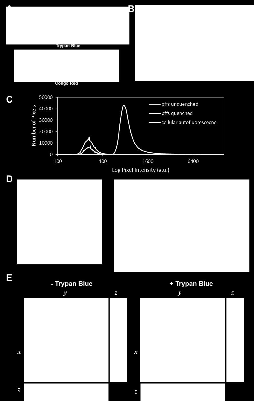
20 FIGURES Figure 1 20

21 Figure 2 21

22 Figure 3 22

23 Figure 4 23


24 Figure 5 24

25 Figure 6 25


26 Figure 7 26
BMDCs were generated in vitro from bone marrow cells cultured in 10 % RPMI supplemented
 Supplemental Materials Figure S1. Cultured BMDCs express CD11c BMDCs were generated in vitro from bone marrow cells cultured in 10 % RPMI supplemented with 15 ng/ml GM-CSF. Media was changed and fresh
T H E J O U R N A L O F C E L L B I O L O G Y
 Supplemental material Edens and Levy, http://www.jcb.org/cgi/content/full/jcb.201406004/dc1 T H E J O U R N A L O F C E L L B I O L O G Y Figure S1. Nuclear shrinking does not depend on the cytoskeleton
TFEB-mediated increase in peripheral lysosomes regulates. Store Operated Calcium Entry
 TFEB-mediated increase in peripheral lysosomes regulates Store Operated Calcium Entry Luigi Sbano, Massimo Bonora, Saverio Marchi, Federica Baldassari, Diego L. Medina, Andrea Ballabio, Carlotta Giorgi
Instructions for Use. APO-AB Annexin V-Biotin Apoptosis Detection Kit 100 tests
 3URGXFW,QIRUPDWLRQ Sigma TACS Annexin V Apoptosis Detection Kits Instructions for Use APO-AB Annexin V-Biotin Apoptosis Detection Kit 100 tests For Research Use Only. Not for use in diagnostic procedures.
THE ROLE OF ALTERED CALCIUM AND mtor SIGNALING IN THE PATHOGENESIS OF CYSTINOSIS
 Research Foundation, 18 month progress report THE ROLE OF ALTERED CALCIUM AND mtor SIGNALING IN THE PATHOGENESIS OF CYSTINOSIS Ekaterina Ivanova, doctoral student Elena Levtchenko, MD, PhD, PI Antonella
ab CytoPainter Golgi/ER Staining Kit
 ab139485 CytoPainter Golgi/ER Staining Kit Instructions for Use Designed to detect Golgi bodies and endoplasmic reticulum by microscopy This product is for research use only and is not intended for diagnostic
Optimization of the Fuse-It-mRNA Protocol for L929 Cells in the µ-plate 24 Well
 Optimization of the Fuse-It-mRNA Protocol for L929 Cells in the µ-plate 24 Well 1. General Information... 1 2. Background... 1 3. Material and Equipment Required... 2 4. Experimental Procedure and Results...
of an untreated HS-stained BAEC monolayer viewed using a laser confocal microscope; Bar = 10 µm.
 Supplemental Figure 1: EC monolayer heparan sulfate fluorescence intensity. (A) Enface perspective of an untreated HS-stained BAEC monolayer viewed using a laser confocal microscope; Bar = 10 µm. (B) Enface
ab Lysosome/Cytotoxicity Dual Staining Kit
 ab133078 Lysosome/Cytotoxicity Dual Staining Kit Instructions for Use For studying lysosome function at the cellular level. This product is for research use only and is not intended for diagnostic use.
genome edited transient transfection, CMV promoter
 Supplementary Figure 1. In the absence of new protein translation, overexpressed caveolin-1-gfp is degraded faster than caveolin-1-gfp expressed from the endogenous caveolin 1 locus % loss of total caveolin-1-gfp
SUPPLEMENTARY INFORMATION
 Supplementary Figures Supplementary Figure S1. Binding of full-length OGT and deletion mutants to PIP strips (Echelon Biosciences). Supplementary Figure S2. Binding of the OGT (919-1036) fragments with
B16-F10 (Mus musculus skin melanoma), NCI-H460 (human non-small cell lung cancer
, NCI-H460 (human non-small cell lung cancer") Electronic Supplementary Material (ESI) for ChemComm. This journal is The Royal Society of Chemistry 2017 Experimental Methods Cell culture B16-F10 (Mus musculus skin melanoma), NCI-H460 (human non-small
Electronic Supplementary Material (ESI) for ChemComm. This journal is The Royal Society of Chemistry 2017 Experimental Methods Cell culture B16-F10 (Mus musculus skin melanoma), NCI-H460 (human non-small
Annexin V-PE Apoptosis Detection Kit
 Annexin V-PE Apoptosis Detection Kit Catalog Number KA0716 100 assays Version: 02 Intended for research use only www.abnova.com Table of Contents Introduction... 3 Background... 3 General Information...
Influenza virus exploits tunneling nanotubes for cell-to-cell spread
 Supplementary Information Influenza virus exploits tunneling nanotubes for cell-to-cell spread Amrita Kumar 1, Jin Hyang Kim 1, Priya Ranjan 1, Maureen G. Metcalfe 2, Weiping Cao 1, Margarita Mishina 1,
Supplementary Figure 1
 Supplementary Figure 1 AAV-GFP injection in the MEC of the mouse brain C57Bl/6 mice at 4 months of age were injected with AAV-GFP into the MEC and sacrificed at 7 days post injection (dpi). (a) Brains
Modulating Glucose Uptake in Skeletal Myotubes: Insulin Induction with Bioluminescent Glucose Uptake Analysis
 icell Skeletal Myoblasts Application Protocol Modulating Glucose Uptake in Skeletal Myotubes: Insulin Induction with Bioluminescent Glucose Uptake Analysis Introduction The skeletal muscle is one of the
T H E J O U R N A L O F C E L L B I O L O G Y
 Supplemental material Beck et al., http://www.jcb.org/cgi/content/full/jcb.201011027/dc1 T H E J O U R N A L O F C E L L B I O L O G Y Figure S1. Membrane binding of His-tagged proteins to Ni-liposomes.
For the rapid, sensitive and accurate measurement of apoptosis in various samples.
 ab14082 500X Annexin V-FITC Apoptosis Detection Reagent Instructions for Use For the rapid, sensitive and accurate measurement of apoptosis in various samples. This product is for research use only and
RayBio Annexin V-FITC Apoptosis Detection Kit
 RayBio Annexin V-FITC Apoptosis Detection Kit User Manual Version 1.0 May 25, 2014 (Cat#: 68FT-AnnV-S) RayBiotech, Inc. We Provide You With Excellent Support And Service Tel:(Toll Free)1-888-494-8555 or
RayBio Annexin V-Cy5 Apoptosis Detection Kit
 RayBio Annexin V-Cy5 Apoptosis Detection Kit User Manual Version 1.0 Mar 20, 2014 (Cat#: 68C5-AnnV-S) RayBiotech, Inc. We Provide You With Excellent Support And Service Tel:(Toll Free)1-888-494-8555 or
DEVELOPMENTAL VALIDATION OF SPERM HY-LITER EXPRESS Jennifer Old, Chris Martersteck, Anna Kalinina, Independent Forensics, Lombard IL
 DEVELOPMENTAL VALIDATION OF SPERM HY-LITER EXPRESS Jennifer Old, Chris Martersteck, Anna Kalinina, Independent Forensics, Lombard IL Background and Introduction SPERM HY-LITER is the first immunofluorescent
LDL Uptake Cell-Based Assay Kit
 LDL Uptake Cell-Based Assay Kit Catalog Number KA1327 100 assays Version: 07 Intended for research use only www.abnova.com Table of Contents Introduction... 3 Background... 3 Principle of the Assay...
PROTOCOL: OPTIMIZATION OF LENTIVIRAL TRANSDUCTION USING SPINFECTION
 Last Modified: April 2018 Last Review: October 2018 PROTOCOL: OPTIMIZATION OF LENTIVIRAL TRANSDUCTION USING SPINFECTION Table of Contents 1. Brief Description 1 2. Materials and Reagents.1 3. Optimization
Annexin V-Cy3 Apoptosis Detection Reagent
 ab14143 Annexin V-Cy3 Apoptosis Detection Reagent Instructions for Use For the rapid, sensitive and accurate measurement of apoptosis in various samples This product is for research use only and is not
ab SREBP-2 Translocation Assay Kit (Cell-Based)
") ab133114 SREBP-2 Translocation Assay Kit (Cell-Based) Instructions for Use For analysis of translocation of SREBP-2 into nuclei. This product is for research use only and is not intended for diagnostic
ab133114 SREBP-2 Translocation Assay Kit (Cell-Based) Instructions for Use For analysis of translocation of SREBP-2 into nuclei. This product is for research use only and is not intended for diagnostic
Supplementary Material and Methods
 Online Supplement Kockx et al, Secretion of Apolipoprotein E from Macrophages 1 Supplementary Material and Methods Cloning of ApoE-GFP Full-length human apoe3 cdna (pcdna3.1/zeo + -apoe) was kindly provided
Supporting Information
 Supporting Information The Effects of Spacer Length and Composition on Aptamer-Mediated Cell-Specific Targeting with Nanoscale PEGylated Liposomal Doxorubicin Hang Xing +, [a] Ji Li +, [a] Weidong Xu,
SUPPLEMENTAL INFORMATION
 SUPPLEMENTAL INFORMATION EXPERIMENTAL PROCEDURES Tryptic digestion protection experiments - PCSK9 with Ab-3D5 (1:1 molar ratio) in 50 mm Tris, ph 8.0, 150 mm NaCl was incubated overnight at 4 o C. The
ab LDL Uptake Assay Kit (Cell-Based)
") ab133127 LDL Uptake Assay Kit (Cell-Based) Instructions for Use For the detection of LDL uptake into cultured cells. This product is for research use only and is not intended for diagnostic use. Version
ab133127 LDL Uptake Assay Kit (Cell-Based) Instructions for Use For the detection of LDL uptake into cultured cells. This product is for research use only and is not intended for diagnostic use. Version
Superior Fluorescent Labeling Dyes Spanning the Full Visible Spectrum...1. Trademarks: HiLyte Fluor (AnaSpec, Inc.)
") Table of Contents Fluor TM Labeling Dyes Superior Fluorescent Labeling Dyes Spanning the Full Visible Spectrum....1 Fluor TM 405 Dye, an Excellent Alternative to Alexa Fluor 405 & DyLight 405....2 Fluor
Table of Contents Fluor TM Labeling Dyes Superior Fluorescent Labeling Dyes Spanning the Full Visible Spectrum....1 Fluor TM 405 Dye, an Excellent Alternative to Alexa Fluor 405 & DyLight 405....2 Fluor
Gladstone Institutes, University of California (UCSF), San Francisco, USA
, San Francisco, USA") Fluorescence-linked Antigen Quantification (FLAQ) Assay for Fast Quantification of HIV-1 p24 Gag Marianne Gesner, Mekhala Maiti, Robert Grant and Marielle Cavrois * Gladstone Institutes, University of
Fluorescence-linked Antigen Quantification (FLAQ) Assay for Fast Quantification of HIV-1 p24 Gag Marianne Gesner, Mekhala Maiti, Robert Grant and Marielle Cavrois * Gladstone Institutes, University of
Multi-Parameter Apoptosis Assay Kit
 Multi-Parameter Apoptosis Assay Kit Catalog Number KA1335 5 x 96 assays Version: 05 Intended for research use only www.abnova.com Table of Contents Introduction... 3 Background... 3 Principle of the Assay...
Supplemental Information. Autophagy in Oncogenic K-Ras. Promotes Basal Extrusion. of Epithelial Cells by Degrading S1P. Current Biology, Volume 24
 Current Biology, Volume 24 Supplemental Information Autophagy in Oncogenic K-Ras Promotes Basal Extrusion of Epithelial Cells by Degrading S1P Gloria Slattum, Yapeng Gu, Roger Sabbadini, and Jody Rosenblatt
Chapter 3. Expression of α5-megfp in Mouse Cortical Neurons. on the β subunit. Signal sequences in the M3-M4 loop of β nachrs bind protein factors to
 22 Chapter 3 Expression of α5-megfp in Mouse Cortical Neurons Subcellular localization of the neuronal nachr subtypes α4β2 and α4β4 depends on the β subunit. Signal sequences in the M3-M4 loop of β nachrs
LDL Uptake Cell-Based Assay Kit
 LDL Uptake Cell-Based Assay Kit Item No. 10011125 www.caymanchem.com Customer Service 800.364.9897 Technical Support 888.526.5351 1180 E. Ellsworth Rd Ann Arbor, MI USA TABLE OF CONTENTS GENERAL INFORMATION
A ph-dependent Charge Reversal Peptide for Cancer Targeting
 Supporting Information A ph-dependent Charge Reversal Peptide for Cancer Targeting Naoko Wakabayashi 1, Yoshiaki Yano 1, Kenichi Kawano 1, and Katsumi Matsuzaki 1 1 Graduate School of Pharmaceutical Sciences,
NF-κB p65 (Phospho-Thr254)
") Assay Biotechnology Company www.assaybiotech.com Tel: 1-877-883-7988 Fax: 1-877-610-9758 NF-κB p65 (Phospho-Thr254) Colorimetric Cell-Based ELISA Kit Catalog #: OKAG02015 Please read the provided manual
Assay Biotechnology Company www.assaybiotech.com Tel: 1-877-883-7988 Fax: 1-877-610-9758 NF-κB p65 (Phospho-Thr254) Colorimetric Cell-Based ELISA Kit Catalog #: OKAG02015 Please read the provided manual
Instructions. Fuse-It-mRNA easy. Shipping and Storage. Overview. Kit Contents. Specifications. Note: Important Guidelines
 Membrane fusion is a highly efficient method for transfecting various molecules and particles into mammalian cells, even into sensitive and primary cells. The Fuse-It reagents are cargo-specific liposomal
Instructions. Fuse-It-Color. Overview. Specifications
 Membrane fusion is a novel and highly superior method for incorporating various molecules and particles into mammalian cells. Cargo-specific liposomal carriers are able to attach and rapidly fuse with
Note: During 30 minute incubation; proceed thru appropriate sections below (e.g. sections II, III and V).
.") LEGEND MAX β Amyloid x 40 LEGEND MAX β Amyloid x 40 ELISA Kit Components and Protocol Kit Components Capture Antibody Coated Plate 1 stripwell plate 1 40 Standard (2) 20μg vial 5X Wash Buffer 125mL Standard
LEGEND MAX β Amyloid x 40 LEGEND MAX β Amyloid x 40 ELISA Kit Components and Protocol Kit Components Capture Antibody Coated Plate 1 stripwell plate 1 40 Standard (2) 20μg vial 5X Wash Buffer 125mL Standard
Annexin V-Cy3 Apoptosis Detection Kit
 ab14142 Annexin V-Cy3 Apoptosis Detection Kit Instructions for Use For the rapid, sensitive and accurate measurement of apoptosis in various samples. This product is for research use only and is not intended
Protein Trafficking in the Secretory and Endocytic Pathways
 Protein Trafficking in the Secretory and Endocytic Pathways The compartmentalization of eukaryotic cells has considerable functional advantages for the cell, but requires elaborate mechanisms to ensure
Essential Medium, containing 10% fetal bovine serum, 100 U/ml penicillin and 100 µg/ml streptomycin. Huvec were cultured in
 Supplemental data Methods Cell culture media formulations A-431 and U-87 MG cells were maintained in Dulbecco s Modified Eagle s Medium. FaDu cells were cultured in Eagle's Minimum Essential Medium, containing
04_polarity. The formation of synaptic vesicles
 Brefeldin prevents assembly of the coats required for budding Nocodazole disrupts microtubules Constitutive: coatomer-coated Selected: clathrin-coated The formation of synaptic vesicles Nerve cells (and
ab Membrane fluidity kit Instructions for Use For the detection of membrane fluidity in cells
 ab189819 Membrane fluidity kit Instructions for Use For the detection of membrane fluidity in cells This product is for research use only and is not intended for diagnostic use. Version 1 Last Updated
Pre-made Lentiviral Particles for intracelular labeling: (LocLight TM Living cell imaging lentivirus for sub-cellular localization)
") Pre-made Lentiviral Particles for intracelular labeling: (LocLight TM Living cell imaging lentivirus for sub-cellular localization) LocLight TM cell organelle labeling lentivirus is provided as 200ul/per
Pre-made Lentiviral Particles for intracelular labeling: (LocLight TM Living cell imaging lentivirus for sub-cellular localization) LocLight TM cell organelle labeling lentivirus is provided as 200ul/per
Annexin V-FITC Apoptosis Detection Kit
 ab14085 Annexin V-FITC Apoptosis Detection Kit Instructions for Use For the rapid, sensitive and accurate measurement of Apoptosis in living cells (adherent and suspension). View kit datasheet: www.abcam.com/ab14085
SensoLyte Rh110 Cathepsin K Assay Kit *Fluorimetric* Revision#1.2 Last Updated: May 2017 Catalog # Kit Size
 SensoLyte Rh110 Cathepsin K Assay Kit *Fluorimetric* Revision#1.2 Last Updated: May 2017 Catalog # 72152 Kit Size 100 Assays (96-well plate) Optimized Performance: This kit detects Cathepsin K activity.
Procaspase-3. Cleaved caspase-3. actin. Cytochrome C (10 M) Z-VAD-fmk. Procaspase-3. Cleaved caspase-3. actin. Z-VAD-fmk
 Z-VAD-fmk. Procaspase-3. Cleaved caspase-3. actin. Z-VAD-fmk") A HeLa actin - + + - - + Cytochrome C (1 M) Z-VAD-fmk PMN - + + - - + actin Cytochrome C (1 M) Z-VAD-fmk Figure S1. (A) Pan-caspase inhibitor z-vad-fmk inhibits cytochrome c- mediated procaspase-3 cleavage.
A HeLa actin - + + - - + Cytochrome C (1 M) Z-VAD-fmk PMN - + + - - + actin Cytochrome C (1 M) Z-VAD-fmk Figure S1. (A) Pan-caspase inhibitor z-vad-fmk inhibits cytochrome c- mediated procaspase-3 cleavage.
Protocol for A-549 VIM RFP (ATCC CCL-185EMT) TGFβ1 EMT Induction and Drug Screening
 TGFβ1 EMT Induction and Drug Screening") Protocol for A-549 VIM RFP (ATCC CCL-185EMT) TGFβ1 EMT Induction and Drug Screening Introduction: Vimentin (VIM) intermediate filament (IF) proteins are associated with EMT in lung cancer and its metastatic
Protocol for A-549 VIM RFP (ATCC CCL-185EMT) TGFβ1 EMT Induction and Drug Screening Introduction: Vimentin (VIM) intermediate filament (IF) proteins are associated with EMT in lung cancer and its metastatic
Europium Labeling Kit
 Europium Labeling Kit Catalog Number KA2096 100ug *1 Version: 03 Intended for research use only www.abnova.com Table of Contents Introduction... 3 Intended Use... 3 Background... 3 Principle of the Assay...
Supporting Information
 Electronic Supplementary Material (ESI) for ChemComm. This journal is The Royal Society of Chemistry 2015 Supporting Information Enzyme-activatable Probe with a Self-immolative Linker for Rapid and Sensitive
Protocol for Gene Transfection & Western Blotting
 The schedule and the manual of basic techniques for cell culture Advanced Protocol for Gene Transfection & Western Blotting Schedule Day 1 26/07/2008 Transfection Day 3 28/07/2008 Cell lysis Immunoprecipitation
CytoPainter Lysosomal Staining Kit - Blue Fluorescence
 ab112135 CytoPainter Lysosomal Staining Kit - Blue Fluorescence Instructions for Use For staining Lysosomes in suspension and adherent cells by using our proprietary blue fluorescence probe This product
J. Cell Sci. 129: doi: /jcs : Supplementary information
 Movie 1. AgLDL is contained in small sub-regions of the lysosomal synapse that are acidic. J774 cells were incubated with agldl dual labeled with a ph sensitive and a ph insensitive fluorophore for 1 hr.
Supporting Information File S2
 Pulli et al. Measuring Myeloperoxidase Activity in Biological Samples Page 1 of 6 Supporting Information File S2 Step-by-Step Protocol Reagents: Sucrose (Sigma, S3089) CaCl 2 (Sigma, C5770) Heparin Sodium
Chapter 6. Antigen Presentation to T lymphocytes
 Chapter 6 Antigen Presentation to T lymphocytes Generation of T-cell Receptor Ligands T cells only recognize Ags displayed on cell surfaces These Ags may be derived from pathogens that replicate within
Production of Exosomes in a Hollow Fiber Bioreactor
 Production of Exosomes in a Hollow Fiber Bioreactor John J S Cadwell, President and CEO, FiberCell Systems Inc INTRODUCTION Exosomes are small lipid membrane vesicles (80-120 nm) of endocytic origin generated
Human SH-SY5Y neuroblastoma cells (A.T.C.C., Manassas, VA) were cultured in DMEM, F-12
 were cultured in DMEM, F-12") SUPPLEMENTARY METHODS Cell cultures Human SH-SY5Y neuroblastoma cells (A.T.C.C., Manassas, VA) were cultured in DMEM, F-12 Ham with 25 mm HEPES and NaHCO 3 (1:1) and supplemented with 10% (v/v) FBS, 1.0
SUPPLEMENTARY METHODS Cell cultures Human SH-SY5Y neuroblastoma cells (A.T.C.C., Manassas, VA) were cultured in DMEM, F-12 Ham with 25 mm HEPES and NaHCO 3 (1:1) and supplemented with 10% (v/v) FBS, 1.0
Supporting Information for:
 Supporting Information for: Methylerythritol Cyclodiphosphate (MEcPP) in Deoxyxylulose Phosphate Pathway: Synthesis from an Epoxide and Mechanisms Youli Xiao, a Rodney L. Nyland II, b Caren L. Freel Meyers
HIV-1 Virus-like Particle Budding Assay Nathan H Vande Burgt, Luis J Cocka * and Paul Bates
 HIV-1 Virus-like Particle Budding Assay Nathan H Vande Burgt, Luis J Cocka * and Paul Bates Department of Microbiology, Perelman School of Medicine at the University of Pennsylvania, Philadelphia, USA
Glucose Uptake Colorimetric Assay Kit
 ab136955 Glucose Uptake Colorimetric Assay Kit Instructions for Use For the sensitive and accurate measurement of Glucose uptake in various samples This product is for research use only and is not intended
SensoLyte 520 HIV-1 Protease Assay Kit *Fluorimetric*
 SensoLyte 520 HIV-1 Protease Assay Kit *Fluorimetric* Catalog # 71147 Kit Size 100 assays (96-well) or 500 assays (384-well) Convenient Format: Complete kit including all the assay components. Optimized
Rescue of mutant rhodopsin traffic by metformin-induced AMPK activation accelerates photoreceptor degeneration Athanasiou et al
 Supplementary Material Rescue of mutant rhodopsin traffic by metformin-induced AMPK activation accelerates photoreceptor degeneration Athanasiou et al Supplementary Figure 1. AICAR improves P23H rod opsin
High resolution structural evidence suggests the Sarcoplasmic Reticulum forms microdomains with Acidic Stores (lyososomes) in the heart.
 in the heart.") High resolution structural evidence suggests the Sarcoplasmic Reticulum forms microdomains with Acidic Stores (lyososomes) in the heart. Daniel Aston, Rebecca A. Capel, Kerrie L. Ford, Helen C. Christian,
High resolution structural evidence suggests the Sarcoplasmic Reticulum forms microdomains with Acidic Stores (lyososomes) in the heart. Daniel Aston, Rebecca A. Capel, Kerrie L. Ford, Helen C. Christian,
Product Use HPSC-CC are for research use only. It is not approved for human or animal use, or for application in in vitro diagnostic procedures.
 HPSC-derived Cardiomyocyte Cells (HPSC-CC) Catalog #6240 Cell Specification Human primary cardiomyocytes and cardiac tissue are superior modeling systems for heart disease studies, drug discovery and toxicity
Lab module 6a Receptor-mediated endocytosis
 Goal for the module Lab module 6a Receptor-mediated endocytosis To follow cell surface receptors as they are internalized. Pre-lab homework Read about receptor-mediated endocytosis and other forms of internalization
Human TRPC6 Ion Channel Cell Line
 TECHNICAL DATA SHEET ValiScreen Ion Channel Cell Line Caution: For Laboratory Use. A research product for research purposes only Human TRPC6 Ion Channel Cell Line Product No.: AX-012-C Lot No.: 512-548-A
TECHNICAL BULLETIN. Catalog Number RAB0447 Storage Temperature 20 C
 Phospho-Stat3 (ptyr 705 ) and pan-stat3 ELISA Kit for detection of human, mouse, or rat phospho-stat3 (ptyr 705 ) and pan-stat3 in cell and tissue lysates Catalog Number RAB0447 Storage Temperature 20
In vitro bactericidal assay Fig. S8 Gentamicin protection assay Phagocytosis assay
 In vitro bactericidal assay Mouse bone marrow was isolated from the femur and the tibia. Cells were suspended in phosphate buffered saline containing.5% BSA and 2 mm EDTA and filtered through a cell strainer.
Supplementary information Common molecular mechanism of amyloid pore formation by Alzheimer s -amyloid peptide and -synuclein
 1 Supplementary information Common molecular mechanism of amyloid pore formation by Alzheimer s -amyloid peptide and -synuclein by Coralie Di Scala, Nouara Yahi, Sonia Boutemeur, Alessandra Flores, Léa
Green Cathepsin B Kit. For Research Use Only
 Green Cathepsin B Kit For Research Use Only ICT9151-25 Tests Component Storage Conditions Quantity 1 vial Rhodamine 110-(RR) 2-20 o C 25 tests 1 vial Hoechst 33342 (200 µg) 2-8 o C 1 ml 1 bottle 10x Cellular
TSH Receptor Monoclonal Antibody (49) Catalog Number MA3-218 Product data sheet
 Catalog Number MA3-218 Product data sheet") Website: thermofisher.com Customer Service (US): 1 800 955 6288 ext. 1 Technical Support (US): 1 800 955 6288 ext. 441 TSH Receptor Monoclonal Antibody (49) Catalog Number MA3-218 Product data sheet Details
Website: thermofisher.com Customer Service (US): 1 800 955 6288 ext. 1 Technical Support (US): 1 800 955 6288 ext. 441 TSH Receptor Monoclonal Antibody (49) Catalog Number MA3-218 Product data sheet Details
Effect of Rhodamine-taxol conjugate on Caveolin dynamics
 Chapter 6 4 Effect of Rhodamine-taxol conjugate on Caveolin dynamics 1. Summary In the present study, Rhodamine- taxol conjugate treatment resulted in a transient recruitment of Caveolin-1 to the cell
Product Information Sheet (M ) MarkerGene TM LysoLive TM Lysosomal Sulfatase Assay Kit (Product M1377)
 MarkerGene TM LysoLive TM Lysosomal Sulfatase Assay Kit (Product M1377)") 1 Product Information Sheet (M1377-004) MarkerGene TM LysoLive TM Lysosomal Sulfatase Assay Kit (Product M1377) Marker Gene Technologies, Inc. University of Oregon Riverfront Research Park 1850 Millrace
1 Product Information Sheet (M1377-004) MarkerGene TM LysoLive TM Lysosomal Sulfatase Assay Kit (Product M1377) Marker Gene Technologies, Inc. University of Oregon Riverfront Research Park 1850 Millrace
Nature Protocols: doi: /nprot Supplementary Figure 1. Fluorescent titration of probe CPDSA.
 Supplementary Figure 1 Fluorescent titration of probe CPDSA. Fluorescent titration of probe CPDSA (10 um) upon addition of GSH in HEPES (10 mm, ph = 7.4) containing 10% DMSO. Each spectrum was recorded
Project report October 2012 March 2013 CRF fellow: Principal Investigator: Project title:
 Project report October 2012 March 2013 CRF fellow: Gennaro Napolitano Principal Investigator: Sergio Daniel Catz Project title: Small molecule regulators of vesicular trafficking to enhance lysosomal exocytosis
SUPPLEMENTARY INFORMATION
 SUPPLEMENTARY INFORMATION DOI: 10.1038/NNANO.2012.80 Protein-Inorganic Hybrid Nanoflowers Jun Ge, Jiandu Lei, and Richard N. Zare Supporting Online Material Materials Proteins including albumin from bovine
SUPPLEMENTARY INFORMATION
 SUPPLEMENTARY INFORMATION Advances in pancreatic islet monolayer culture on glass surfaces enable superresolution microscopy and insights into beta cell ciliogenesis and proliferation Edward A. Phelps,
Constitutive Reporter Lentiviral Vectors Expressing Fluorescent Proteins
 Constitutive Reporter Lentiviral Vectors Expressing Fluorescent Proteins www.vectalys.com/products/ Constitutive Reporter Lentiviral Vectors Catalog Number referring to this User Manual: 0008VCT; 0009VCT;
Development of a near-infrared fluorescent probe for monitoring hydrazine in serum and living cells
 Supporting Information for Development of a near-infrared fluorescent probe for monitoring hydrazine in serum and living cells Sasa Zhu, Weiying Lin,* Lin Yuan State Key Laboratory of Chemo/Biosensing
Mammalian Membrane Protein Extraction Kit
 Mammalian Membrane Protein Extraction Kit Catalog number: AR0155 Boster s Mammalian Membrane Protein Extraction Kit is a simple, rapid and reproducible method to prepare cellular protein fractions highly
INSTRUCTIONS Pierce Primary Cardiomyocyte Isolation Kit
 INSTRUCTIONS Pierce Primary Cardiomyocyte Isolation Kit 88281 Number Description 88281 Pierce Primary Cardiomyocyte Isolation Kit, contains sufficient reagents to isolate cardiomyocytes from 50 neonatal
Measuring Cardiac Activity:
 icell Cardiomyocytes 2 Application Protocol Measuring Cardiac Activity: Impedance and Extracellular Field Potential Detection with CardioExcyte 96 System Introduction icell Cardiomyocytes 2, human cardiomyocytes
Figure S1. (A) SDS-PAGE separation of GST-fusion proteins purified from E.coli BL21 strain is shown. An equal amount of GST-tag control, LRRK2 LRR
 SDS-PAGE separation of GST-fusion proteins purified from E.coli BL21 strain is shown. An equal amount of GST-tag control, LRRK2 LRR") Figure S1. (A) SDS-PAGE separation of GST-fusion proteins purified from E.coli BL21 strain is shown. An equal amount of GST-tag control, LRRK2 LRR and LRRK2 WD40 GST fusion proteins (5 µg) were loaded
Figure S1. (A) SDS-PAGE separation of GST-fusion proteins purified from E.coli BL21 strain is shown. An equal amount of GST-tag control, LRRK2 LRR and LRRK2 WD40 GST fusion proteins (5 µg) were loaded
TECHNICAL BULLETIN. Phospho-Akt (pser 473 ) ELISA Kit for detection of human, mouse, or rat phospho-akt (pser 473 ) in cell and tissue lysates
 ELISA Kit for detection of human, mouse, or rat phospho-akt (pser 473 ) in cell and tissue lysates") Phospho-Akt (pser 473 ) ELISA Kit for detection of human, mouse, or rat phospho-akt (pser 473 ) in cell and tissue lysates Catalog Number RAB0011 Storage Temperature 20 C TECHNICAL BULLETIN Product Description
Phospho-Akt (pser 473 ) ELISA Kit for detection of human, mouse, or rat phospho-akt (pser 473 ) in cell and tissue lysates Catalog Number RAB0011 Storage Temperature 20 C TECHNICAL BULLETIN Product Description
ab Lipid Peroxidation (MDA) Assay kit (Colorimetric/ Fluorometric)
 Assay kit (Colorimetric/ Fluorometric)") Version 10b Last updated 19 December 2018 ab118970 Lipid Peroxidation (MDA) Assay kit (Colorimetric/ Fluorometric) For the measurement of Lipid Peroxidation in plasma, cell culture and tissue extracts.
Version 10b Last updated 19 December 2018 ab118970 Lipid Peroxidation (MDA) Assay kit (Colorimetric/ Fluorometric) For the measurement of Lipid Peroxidation in plasma, cell culture and tissue extracts.
ab Exosome Isolation and Analysis Kit - Flow Cytometry, Cell Culture (CD63 / CD81)
") Version 1 Last updated 26 September 2018 ab239682 Exosome Isolation and Analysis Kit - Flow Cytometry, Cell Culture (CD63 / For the isolation and analysis of exosome from cell culture. This product is
Version 1 Last updated 26 September 2018 ab239682 Exosome Isolation and Analysis Kit - Flow Cytometry, Cell Culture (CD63 / For the isolation and analysis of exosome from cell culture. This product is
Institute for Biomaterials and Biomedical Engineering, University of Toronto, 164 College St, Toronto, ON, M5s 3G9, Canada
 for A Microfluidic Platform for Continuous Monitoring of Dopamine Homeostasis in Dopaminergic Cells Yue Yu, 1, 2 Richard P.S. de Campos, 2,3 Seolim Hong, 4 Dimitar L. Krastev, 2 Siddharth Sadanand, 2 Yen
The clathrin adaptor Numb regulates intestinal cholesterol. absorption through dynamic interaction with NPC1L1
 The clathrin adaptor Numb regulates intestinal cholesterol absorption through dynamic interaction with NPC1L1 Pei-Shan Li 1, Zhen-Yan Fu 1,2, Ying-Yu Zhang 1, Jin-Hui Zhang 1, Chen-Qi Xu 1, Yi-Tong Ma
The Schedule and the Manual of Basic Techniques for Cell Culture
 The Schedule and the Manual of Basic Techniques for Cell Culture 1 Materials Calcium Phosphate Transfection Kit: Invitrogen Cat.No.K2780-01 Falcon tube (Cat No.35-2054:12 x 75 mm, 5 ml tube) Cell: 293
Elucidation of Viral Replication Mechanisms in an Animal Model for Multiple Sclerosis
 Elucidation of Viral Replication Mechanisms in an Animal Model for Multiple Sclerosis Introduction It is often difficult to research human diseases due to obvious ethical implications. However, when an
Mitochondrial Trifunctional Protein (TFP) Protein Quantity Microplate Assay Kit
 Protein Quantity Microplate Assay Kit") PROTOCOL Mitochondrial Trifunctional Protein (TFP) Protein Quantity Microplate Assay Kit DESCRIPTION Mitochondrial Trifunctional Protein (TFP) Protein Quantity Microplate Assay Kit Sufficient materials
PROTOCOL Mitochondrial Trifunctional Protein (TFP) Protein Quantity Microplate Assay Kit DESCRIPTION Mitochondrial Trifunctional Protein (TFP) Protein Quantity Microplate Assay Kit Sufficient materials
Exo-Glow TM Exosome Labeling Kits
 Exo-Glow TM Exosome Labeling Kits Cat# EXOR100A-1 Cat# EXOG200A-1 Cat# EXOC300A-1 User Manual Store kit at -20 o C on receipt Version 8 3/10/2017 A limited-use label license covers this product. By use
SensoLyte 520 Cathepsin K Assay Kit *Fluorimetric*
 SensoLyte 520 Cathepsin K Assay Kit *Fluorimetric* Catalog # 72171 Kit Size 100 Assays (96-well plate) Optimized Performance: This kit is optimized to detect Cathepsin K activity. Enhanced Value: Ample
SensoLyte 490 HIV-1 Protease Assay Kit *Fluorimetric*
 SensoLyte 490 HIV-1 Protease Assay Kit *Fluorimetric* Catalog # 71127 Unit Size Kit Size 1 kit 500 assays (96-well) or 1250 assays (384-well) This kit is optimized to detect the activity of human immunodeficiency
ab Membrane Fractionation Kit Instructions for Use For the rapid and simple separation of membrane, cytosolic and nuclear cellular fractions.
 ab139409 Membrane Fractionation Kit Instructions for Use For the rapid and simple separation of membrane, cytosolic and nuclear cellular fractions. This product is for research use only and is not intended
Evaluation of directed and random motility in microslides Assessment of leukocyte adhesion in flow chambers
 Evaluation of directed and random motility in microslides Motility experiments in IBIDI microslides, image acquisition and processing were performed as described. PMN, which ended up in an angle < 180