Membrane Transport and Cell Signaling
|
|
|
- Shanon Burke
- 6 years ago
- Views:
Transcription

1 CAMPBELL BIOLOGY IN FOCUS URRY CAIN WASSERMAN MINORSKY REECE 5 Membrane Transport and Cell Signaling Lecture Presentations by Kathleen Fitzpatrick and Nicole Tunbridge, Simon Fraser University SECOND EDITION
2 Overview: Life at the Edge The plasma membrane separates the living cell from its surroundings The plasma membrane exhibits selective permeability, allowing some substances to cross it more easily than others
3 Video: Membrane and Aquaporin

4 Figure 5.1
5 Concept 5.1: Cellular membranes are fluid mosaics of lipids and proteins Phospholipids are the most abundant lipid in most membranes Phospholipids are amphipathic molecules, containing hydrophobic and hydrophilic regions A phospholipid bilayer can exist as a stable boundary between two aqueous compartments
 Glycoprotein Carbohydrate Glycolipid EXTRA- CELLULAR SIDE OF MEMBRANE")
6 Figure 5.2 Fibers of extracellular matrix (ECM) Glycoprotein Carbohydrate Glycolipid EXTRA- CELLULAR SIDE OF MEMBRANE Phospholipid Cholesterol Microfilaments of cytoskeleton Peripheral proteins Integral protein CYTOPLASMIC SIDE OF MEMBRANE
7 Most membrane proteins are also amphipathic and reside in the bilayer with their hydrophilic portions protruding The fluid mosaic model states that the membrane is a mosaic of protein molecules bobbing in a fluid bilayer of phospholipids Groups of certain proteins or certain lipids may associate in long-lasting, specialized patches

8 Figure 5.3 Two phospholipids Hydrophilic head WATER Hydrophobic tail WATER
9 The Fluidity of Membranes Most of the lipids and some proteins in a membrane can shift about laterally The lateral movement of phospholipids is rapid; proteins move more slowly Some proteins move in a directed manner; others seem to be anchored in place

10 Figure 5.4-s1 Results Membrane proteins Mouse cell Human cell

11 Figure 5.4-s2 Results Membrane proteins Mouse cell Human cell Hybrid cell
12 Figure 5.4-s3 Results Membrane proteins Mouse cell Human cell Hybrid cell Mixed proteins after 1 hour
13 As temperatures cool, membranes switch from a fluid state to a solid state The temperature at which a membrane solidifies depends on the types of lipids A membrane remains fluid to a lower temperature if it is rich in phospholipids with unsaturated hydrocarbon tails Membranes must be fluid to work properly; they are usually about as fluid as salad oil
14 The steroid cholesterol has different effects on membrane fluidity at different temperatures At warm temperatures (such as 37 C), cholesterol restrains movement of phospholipids At cool temperatures, it maintains fluidity by preventing tight packing

15 Figure 5.5 (a) Unsaturated versus saturated hydrocarbon tails. Fluid Viscous Unsaturated tails prevent packing. Saturated tails pack together. (b) Cholesterol reduces membrane fluidity at moderate temperatures, but at low temperatures hinders solidification. Cholesterol
16 Evolution of Differences in Membrane Lipid Composition Variations in lipid composition of cell membranes of many species appear to be adaptations to specific environmental conditions Ability to change the lipid compositions in response to temperature changes has evolved in organisms that live where temperatures vary
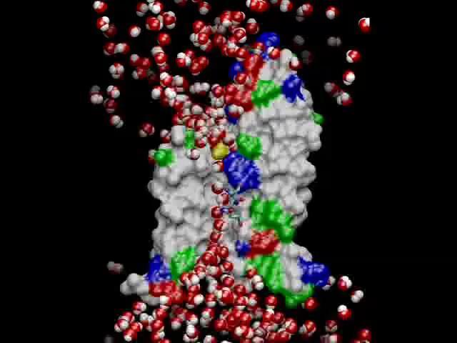
17 Membrane Proteins and Their Functions A membrane is a collage of different proteins embedded in the fluid matrix of the lipid bilayer Proteins determine most of the membrane s specific functions
18 Integral proteins penetrate the hydrophobic interior of the lipid bilayer Integral proteins that span the membrane are called transmembrane proteins The hydrophobic regions of an integral protein consist of one or more stretches of nonpolar amino acids, often coiled into helices Peripheral proteins are loosely bound to the surface of the membrane

19 Figure 5.6 N-terminus EXTRACELLULAR SIDE helix C-terminus CYTOPLASMIC SIDE
20 Six major functions of membrane proteins Transport Enzymatic activity Signal transduction Cell-cell recognition Intercellular joining Attachment to the cytoskeleton and extracellular matrix (ECM)
 Transport (b) Enzymatic activity (c) Signal transduction")
21 Figure 5.7 Enzymes Signaling molecule Receptor ATP Signal transduction (a) Transport (b) Enzymatic activity (c) Signal transduction Glycoprotein (d) Cell-cell recognition (e) Intercellular joining (f) Attachment to the cytoskeleton and extracellular matrix (ECM)

22 Figure Enzymes Signaling molecule Receptor ATP Signal transduction (a) Transport (b) Enzymatic activity (c) Signal transduction
 Cell-cell recognition (e) Intercellular joining")
23 Figure Glycoprotein (d) Cell-cell recognition (e) Intercellular joining (f) Attachment to the cytoskeleton and extracellular matrix (ECM)
24 The Role of Membrane Carbohydrates in Cell-Cell Recognition Cells recognize each other by binding to surface molecules, often containing carbohydrates, on the extracellular surface of the plasma membrane Membrane carbohydrates may be covalently bonded to lipids (forming glycolipids) or, more commonly, to proteins (forming glycoproteins) Carbohydrates on the external side of the plasma membrane vary among species, individuals, and even cell types in an individual
25 Synthesis and Sidedness of Membranes Membranes have distinct inside and outside faces The asymmetrical arrangement of proteins, lipids, and associated carbohydrates in the plasma membrane is determined as the membrane is built by the ER and Golgi apparatus

26 Figure 5.8 Transmembrane glycoproteins Secretory protein Golgi apparatus Attached carbohydrate Vesicle ER Glycolipid ER lumen Plasma membrane: Cytoplasmic face Extracellular face Transmembrane glycoprotein Secreted protein Membrane glycolipid
27 Concept 5.2: Membrane structure results in selective permeability A cell must regulate transport of substances across cellular boundaries Plasma membranes are selectively permeable, regulating the cell s molecular traffic
28 The Permeability of the Lipid Bilayer Hydrophobic (nonpolar) molecules, such as hydrocarbons, can dissolve in the lipid bilayer of the membrane and cross it easily Polar molecules, such as sugars, do not cross the membrane easily
29 Transport Proteins Transport proteins allow passage of hydrophilic substances across the membrane Some transport proteins, called channel proteins, have a hydrophilic channel that certain molecules or ions can use as a tunnel Channel proteins called aquaporins facilitate the passage of water
30 Other transport proteins, called carrier proteins, bind to molecules and change shape to shuttle them across the membrane A transport protein is specific for the substance it moves
31 Concept 5.3: Passive transport is diffusion of a substance across a membrane with no energy investment Diffusion is the tendency for molecules to spread out evenly into the available space Although each molecule moves randomly, diffusion of a population of molecules may be directional At dynamic equilibrium, as many molecules cross the membrane in one direction as in the other
32 Animation: Diffusion
 WATER Net diffusion Net diffusion Equilibrium (a) Diffusion of one solute")
33 Figure 5.9 Molecules of dye Membrane (cross section) WATER Net diffusion Net diffusion Equilibrium (a) Diffusion of one solute Net diffusion Net diffusion Equilibrium Net diffusion Net diffusion Equilibrium (b) Diffusion of two solutes
34 Substances diffuse down their concentration gradient, from where it is more concentrated to where it is less concentrated No work must be done to move substances down the concentration gradient The diffusion of a substance across a biological membrane is passive transport because no energy is expended by the cell to make it happen
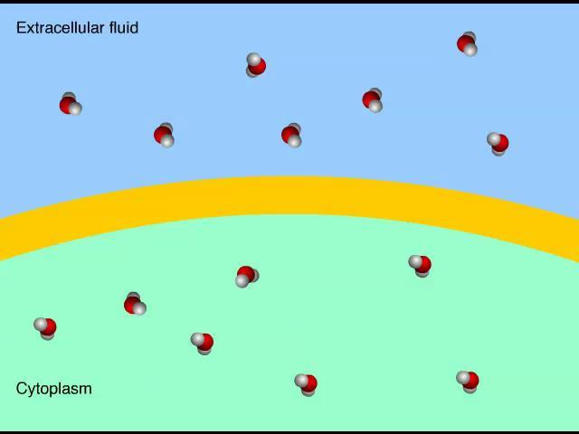
35 Effects of Osmosis on Water Balance Osmosis is the diffusion of free water across a selectively permeable membrane Water diffuses across a membrane from the region of lower solute concentration to the region of higher solute concentration until the solute concentration is equal on both sides

36 Animation: Membrane Selectivity
37 Animation: Osmosis
 Higher concentration of solute More similar")
38 Figure 5.10 Lower concentration of solute (sugar) Higher concentration of solute More similar concentrations of solute Sugar molecule H 2 O Selectively permeable membrane Osmosis
 Higher concentration of")
39 Figure Lower concentration of solute (sugar) Higher concentration of solute More similar concentrations of solute Sugar molecule H 2 O

40 Figure Selectively permeable membrane Water molecules can pass through pores, but sugar molecules cannot. Water molecules cluster around sugar molecules. This side has fewer solute molecules and more free water molecules. Osmosis This side has more solute molecules and fewer free water molecules. Water moves from an area of higher to lower free water concentration (lower to higher solute concentration).
41 Water Balance of Cells Without Walls Tonicity is the ability of a surrounding solution to cause a cell to gain or lose water Isotonic solution: Solute concentration is the same as inside the cell; no net water movement across the plasma membrane Hypertonic solution: Solute concentration is greater than that inside the cell; cell loses water Hypotonic solution: Solute concentration is less than that inside the cell; cell gains water

42 Video: Turgid Elodea
 Animal cell Hypotonic solution Isotonic solution")
43 Figure 5.11a (a) Animal cell Hypotonic solution Isotonic solution Hypertonic solution H 2 O H 2 O H 2 O H 2 O Lysed Normal Shriveled
44 Hypertonic or hypotonic environments create osmotic problems for organisms Osmoregulation, the control of solute concentrations and water balance, is a necessary adaptation for life in such environments The protist Paramecium caudatum, which is hypertonic to its pondwater environment, has a contractile vacuole that can pump excess water out of the cell
45 Video: Chlamydomonas

46 Video: Paramecium Vacuole

47 Figure 5.12 Contractile vacuole 50 μm
48 Water Balance of Cells with Walls Cell walls help maintain water balance A plant cell in a hypotonic solution swells until the wall opposes uptake; the cell is now turgid (very firm) If a plant cell and its surroundings are isotonic, there is no net movement of water into the cell; the cell becomes flaccid (limp), and the plant may wilt In a hypertonic environment, plant cells lose water; eventually, the membrane pulls away from the wall, a usually lethal effect called plasmolysis
 Plant cell Plasma Cell wall membrane H 2 O H 2 O H 2 O")
49 Figure 5.11b (b) Plant cell Plasma Cell wall membrane H 2 O H 2 O H 2 O Plasma membrane H 2 O Turgid (normal) Flaccid Plasmolyzed
50 Facilitated Diffusion: Passive Transport Aided by Proteins In facilitated diffusion, transport proteins speed the passive movement of molecules across the plasma membrane Channel proteins provide corridors that allow a specific molecule or ion to cross the membrane Channel proteins include Aquaporins, for facilitated diffusion of water Ion channels that open or close in response to a stimulus (gated channels)
51 Video: Aquaporins

52 Video: Membrane and Aquaporin
53 Carrier proteins undergo a subtle change in shape that translocates the solute-binding site across the membrane The shape change may be triggered by binding and release of the transported molecule No net energy input is required
 A channel protein Channel protein")
54 Figure 5.13 EXTRA- CELLULAR FLUID (a) A channel protein Channel protein CYTOPLASM Solute Carrier protein Solute (b) A carrier protein
55 Concept 5.4: Active transport uses energy to move solutes against their gradients Facilitated diffusion speeds transport of a solute by providing efficient passage through the membrane but does not alter the direction of transport Some transport proteins, however, can move solutes against their concentration gradients
56 The Need for Energy in Active Transport Active transport moves substances against their concentration gradients Active transport requires energy, usually in the form of ATP
57 Active transport allows cells to maintain concentration gradients that differ from their surroundings The sodium-potassium pump is one type of active transport system

58 Animation: Active Transport
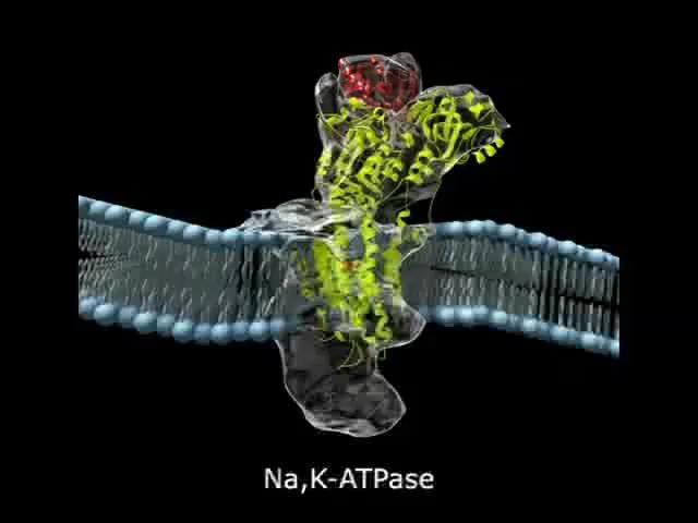
59 Video: Sodium-Potassium Pump

60 Video: Membrane Transport

61 Figure 5.14 EXTRA- CELLULAR FLUID Na + [Na + ] high [K + ] low Na + Na + Na + Na + CYTO- PLASM Na + [Na+ ] low [K + ] high P ADP ATP Na + Na + Na + P P P i

62 Figure EXTRACELLULAR FLUID [Na + ] high [K + ] low Na + Na + Na + Na + Na + CYTOPLASM Na + [Na + ] low [K + ] high P ADP ATP Cytoplasmic Na + binds to the sodium-potassium pump. The affinity for Na + is high when the protein has this shape. Na + binding stimulates phosphorylation by ATP.

63 Figure Na + Na+ Na + Phosphorylation leads to a change in protein shape, reducing its affinity for Na +, which is released outside. P P P i The new shape has a high affinity for K +, which binds on the extracellular side and triggers release of the phosphate group.

64 Figure Loss of the phosphate group restores the protein s original shape, which has a lower affinity for K +. K + is released; affinity for Na + is high again, and the cycle repeats.

65 Figure 5.15 Passive transport Active transport Diffusion Facilitated diffusion ATP
66 How Ion Pumps Maintain Membrane Potential Membrane potential is the voltage across a membrane Voltage is created by differences in the distribution of positive and negative ions across a membrane
67 Two combined forces, collectively called the electrochemical gradient, drive the diffusion of ions across a membrane A chemical force (the ion s concentration gradient) An electrical force (the effect of the membrane potential on the ion s movement)
68 An electrogenic pump is a transport protein that generates voltage across a membrane The sodium-potassium pump is the major electrogenic pump of animal cells The main electrogenic pump of plants, fungi, and bacteria is a proton pump Electrogenic pumps help store energy that can be used for cellular work

69 Figure 5.16 ATP H + Proton pump H + EXTRACELLULAR FLUID H + H + H + CYTOPLASM H +
70 Cotransport: Coupled Transport by a Membrane Protein Cotransport occurs when active transport of a solute indirectly drives transport of other solutes Plant cells use the gradient of hydrogen ions generated by proton pumps to drive active transport of nutrients into the cell

71 Figure 5.17 Sucrose + Sucrose Sucrose-H + cotransporter Diffusion of H + H H + H + H + H + H + H + Proton pump H + ATP + H +
72 Concept 5.5: Bulk transport across the plasma membrane occurs by exocytosis and endocytosis Water and small solutes enter or leave the cell through the lipid bilayer or by means of transport proteins Large molecules, such as polysaccharides and proteins, cross the membrane in bulk by means of vesicles Bulk transport requires energy
73 Exocytosis In exocytosis, transport vesicles migrate to the membrane, fuse with it, and release their contents Many secretory cells use exocytosis to export products
74 Endocytosis In endocytosis, the cell takes in molecules and particulate matter by forming new vesicles from the plasma membrane Endocytosis is a reversal of exocytosis, involving different proteins There are three types of endocytosis Phagocytosis ( cellular eating ) Pinocytosis ( cellular drinking ) Receptor-mediated endocytosis
75 Animation: Exocytosis Endocytosis Introduction
76 Animation: Exocytosis
77 Animation: Phagocytosis
78 Video: Phagocytosis
79 Animation: Pinocytosis

80 Animation: Receptor-Mediated Endocytosis

81 Figure 5.18 Phagocytosis EXTRACELLULAR FLUID Solutes Pinocytosis Receptor-Mediated Endocytosis Pseudopodium Plasma membrane Receptor Food or other particle Coated pit Coat protein Food vacuole Coated vesicle CYTOPLASM

82 Figure Phagocytosis 5 mm Green algal cell EXTRACELLULAR FLUID Solutes Pseudopodium Pseudopodium of amoeba An amoeba engulfing a green algal cell via phagocytosis (TEM) Food or other particle Food vacuole CYTOPLASM

83 0.25 mm Figure Pinocytosis Plasma membrane Coat protein Pinocytotic vesicles forming (TEMs) Coated pit Coated vesicle

84 Figure mm Receptor-Mediated Endocytosis Plasma membrane Coat protein Receptor Top: A coated pit Bottom: A coated vesicle forming during receptor-mediated endocytosis (TEMs)

85 Figure mm Green algal cell Pseudopodium of amoeba An amoeba engulfing a green algal cell via phagocytosis (TEM)

86 Figure mm Pinocytotic vesicles forming (TEMs)

87 Figure mm Plasma membrane Coat protein Top: A coated pit Bottom: A coated vesicle forming during receptor-mediated endocytosis (TEMs)
88 Concept 5.6: The plasma membrane plays a key role in most cell signaling In multicellular organisms, cell-to-cell communication allows the cells of the body to coordinate their activities Communication between cells is also essential for many unicellular organisms
89 Local and Long-Distance Signaling Eukaryotic cells may communicate by direct contact Animal and plant cells have junctions that directly connect the cytoplasm of adjacent cells These are called gap junctions (animal cells) and plasmodesmata (plant cells) The free passage of substances in the cytosol from one cell to another is a type of local signaling
90 In many other cases of local signaling, messenger molecules are secreted by a signaling cell These messenger molecules, called local regulators, travel only short distances One class of these, growth factors, stimulates nearby cells to grow and divide This type of local signaling in animal cells is called paracrine signaling

91 Figure Local signaling Target cells Secreting cell Secretory vesicles Local regulator (a) Paracrine signaling
92 Another more specialized type of local signaling occurs in the animal nervous system This synaptic signaling consists of an electrical signal moving along a nerve cell that triggers secretion of neurotransmitter molecules These diffuse across the space between the nerve cell and its target, triggering a response in the target cell

93 Figure Local signaling Electrical signal triggers release of neurotransmitter. Neurotransmitter diffuses across synapse. Target cell (b) Synaptic signaling
94 In long-distance signaling, plants and animals use chemicals called hormones In hormonal signaling in animals (called endocrine signaling), specialized cells release hormone molecules that travel via the circulatory system Hormones vary widely in size and shape

95 Figure Long-distance signaling Endocrine cell Target cell specifically binds hormone. Hormone travels in bloodstream. Blood vessel (c) Endocrine (hormonal) signaling
96 The Three Stages of Cell Signaling: A Preview Earl W. Sutherland discovered how the hormone epinephrine acts on cells Sutherland suggested that cells receiving signals undergo three processes Reception Transduction Response

97 Animation: Signaling Overview

98 Figure 5.20-s1 EXTRA- CELLULAR FLUID Reception Receptor Plasma membrane CYTOPLASM Signaling molecule
99 Figure 5.20-s2 EXTRA- CELLULAR FLUID Reception Receptor Plasma membrane Transduction CYTOPLASM Relay molecules Signaling molecule

100 Figure 5.20-s3 EXTRA- CELLULAR FLUID Reception Plasma membrane Transduction CYTOPLASM Response Receptor Activation Relay molecules Signaling molecule
101 Reception, the Binding of a Signaling Molecule to a Receptor Protein The binding between a signal molecule (ligand) and receptor is highly specific Ligand binding generally causes a shape change in the receptor Many receptors are directly activated by this shape change Most signal receptors are plasma membrane proteins
102 Receptors in the Plasma Membrane Most water-soluble signal molecules bind to specific sites on receptor proteins that span the plasma membrane There are two main types of membrane receptors G protein-coupled receptors Ligand-gated ion channels
103 G protein-coupled receptors (GPCRs) are plasma membrane receptors that work with the help of a G protein G proteins bind to the energy-rich molecule GTP Many G proteins are very similar in structure GPCR pathways are extremely diverse in function

104 Figure 5.21-s1 Activated GPCR Signaling molecule Inactive enzyme CYTOPLASM GTP Activated G protein Plasma membrane

105 Figure 5.21-s2 Activated GPCR Signaling molecule Inactive enzyme CYTOPLASM GTP Activated G protein Plasma membrane Activated enzyme GTP Cellular response
106 A ligand-gated ion channel receptor acts as a gate for ions when the receptor changes shape When a signal molecule binds as a ligand to the receptor, the gate allows specific ions, such as Na + or Ca 2+, through a channel in the receptor Ligand-gated ion channels are very important in the nervous system The diffusion of ions through open channels may trigger an electric signal
 Gate closed Ions")
107 Figure 5.22-s1 Signaling molecule (ligand) Gate closed Ions Ligand-gated ion channel receptor Plasma membrane
 Gate closed Ions Gate open")
108 Figure 5.22-s2 Signaling molecule (ligand) Gate closed Ions Gate open Ligand-gated ion channel receptor Plasma membrane Cellular response
 Gate closed Ions Gate open Ligand-gated")
109 Figure 5.22-s3 Signaling molecule (ligand) Gate closed Ions Gate open Ligand-gated ion channel receptor Plasma membrane Cellular response Gate closed
110 Intracellular Receptors Intracellular receptor proteins are found in the cytosol or nucleus of target cells Small or hydrophobic chemical messengers can readily cross the membrane and activate receptors Examples of hydrophobic messengers are the steroid and thyroid hormones of animals and nitric oxide (NO) in both plants and animals
111 Aldosterone behaves similarly to other steroid hormones It is secreted by cells of the adrenal gland and enters cells all over the body, but only kidney cells contain receptor cells for aldosterone The hormone binds the receptor protein and activates it The active form of the receptor enters the nucleus, acts as a transcription factor, and activates genes that control water and sodium flow
 EXTRA- CELLULAR FLUID Receptor protein Plasma")
112 Figure 5.23 Hormone (aldosterone) EXTRA- CELLULAR FLUID Receptor protein Plasma membrane Hormonereceptor complex DNA mrna NUCLEUS New protein CYTOPLASM
113 Transduction by Cascades of Molecular Interactions Signal transduction usually involves multiple steps Multistep pathways can amplify a signal: A few molecules can produce a large cellular response Multistep pathways provide more opportunities for coordination and regulation of the cellular response than simpler systems do
114 The molecules that relay a signal from receptor to response are often proteins Like falling dominoes, the activated receptor activates another protein, which activates another, and so on, until the protein producing the response is activated At each step, the signal is transduced into a different form, commonly a shape change in a protein
115 Protein Phosphorylation and Dephosphorylation Phosphorylation and dephosphorylation are a widespread cellular mechanism for regulating protein activity Protein kinases transfer phosphates from ATP to protein, a process called phosphorylation A signaling pathway involving phosphorylation and dephosphorylation can be referred to as a phosphorylation cascade The addition of phosphate groups often changes the form of a protein from inactive to active

116 Figure 5.24 Signaling molecule Receptor Activated relay molecule Inactive protein kinase 1 Inactive protein kinase 2 P i ATP PP Active protein kinase 1 ADP Active protein kinase 2 P Inactive protein P i ATP PP ADP Active protein P Cellular response

117 Figure Signaling molecule Receptor Activated relay molecule Inactive protein kinase 1 Active protein kinase 1

118 Figure Active protein kinase 1 Inactive protein kinase 2 ATP PP ADP Active protein kinase 2 P P i

119 Figure P i PP Active protein kinase 2 P Inactive protein ATP ADP P P i PP Active protein Cellular response
120 Protein phosphatases remove the phosphates from proteins, a process called dephosphorylation Phosphatases provide a mechanism for turning off the signal transduction pathway They also make protein kinases available for reuse, enabling the cell to respond to the signal again
121 Small Molecules and Ions as Second Messengers The extracellular signal molecule (ligand) that binds to the receptor is a pathway s first messenger Second messengers are small, nonprotein, watersoluble molecules or ions that spread throughout a cell by diffusion Cyclic AMP and calcium ions are common second messengers
122 Cyclic AMP (camp) is one of the most widely used second messengers Adenylyl cyclase, an enzyme in the plasma membrane, rapidly converts ATP to camp in response to a number of extracellular signals The immediate effect of camp is usually the activation of protein kinase A, which then phosphorylates a variety of other proteins
 G protein Adenylyl cyclase GTP G")
123 Figure 5.25 First messenger (signaling molecule such as epinephrine) G protein Adenylyl cyclase GTP G protein-coupled receptor ATP camp Second messenger Protein kinase A Cellular responses
124 Response: Regulation of Transcription or Cytoplasmic Activities Ultimately, a signal transduction pathway leads to regulation of one or more cellular activities The response may occur in the cytoplasm or in the nucleus Many signaling pathways regulate the synthesis of enzymes or other proteins, usually by turning genes on or off in the nucleus The final activated molecule in the signaling pathway may function as a transcription factor

125 Figure 5.26 Growth factor Receptor Reception Phosphorylation cascade Transduction CYTOPLASM DNA Inactive transcription factor Active transcription factor P Gene Response NUCLEUS mrna

126 Figure Growth factor Receptor Reception Phosphorylation cascade Transduction CYTOPLASM Inactive transcription factor NUCLEUS

127 Figure Phosphorylation cascade Transduction CYTOPLASM DNA Inactive transcription factor Active transcription factor P Gene Response NUCLEUS mrna
128 Other pathways regulate the activity of enzymes rather than their synthesis, such as the opening of an ion channel or a change in cell metabolism
 Data from T. Kondo and E.")
129 Concentration of radioactive glucose (mm) Figure 5.UN01-1 Glucose Uptake over Time in Guinea Pig Red Blood Cells day-old guinea pig 1-month-old guinea pig Incubation time (min) Data from T. Kondo and E. Beutler, Developmental changes in glucose transport of guinea pig erythrocytes, Journal of Clinical Investigation 65:1 4 (1980)

130 Figure 5.UN day-old and 1-month-old guinea pigs

131 Figure 5.UN02 LDL LDL receptor Normal cell Mild disease Severe disease

132 Figure 5.UN03
133 Figure 5.UN04 Passive transport: Facilitated diffusion Channel protein Carrier protein

134 Figure 5.UN05 Active transport ATP

135 Figure 5.UN06 Reception Transduction Response Receptor Relay molecules Activation of cellular response Signaling molecule

136 Figure 5.UN07
CONCEPT 5.1: Cellular membranes are fluid mosaics of lipids and proteins
 Ch 5 Membrane Transport and Signaling Overview The plasma separates the living cell from its surroundings The plasma exhibits selective permeability, allowing some substances to cross it more easily than
BIOLOGY. Membrane Structure and Function CAMPBELL. Reece Urry Cain Wasserman Minorsky Jackson
 CAMPBELL BIOLOGY TENTH EDITION Reece Urry Cain Wasserman Minorsky Jackson 7 Membrane Structure and Function Lecture Presentation by Nicole Tunbridge and Kathleen Fitzpatrick Life at the Edge The plasma
5 Membrane Transport and Cell Signaling
 CAMPBELL BIOLOGY IN FOCUS Urry Cain Wasserman Minorsky Jackson Reece 5 Membrane Transport and Cell Signaling Lecture Presentations by Kathleen Fitzpatrick and Nicole Tunbridge Overview: Life at the Edge
Membrane Structure and Function
 LECTURE PRESENTATIONS For CAMPBELL BIOLOGY, NINTH EDITION Jane B. Reece, Lisa A. Urry, Michael L. Cain, Steven A. Wasserman, Peter V. Minorsky, Robert B. Jackson Chapter 7 Membrane Structure and Function
Membrane Structure and Function
 LECTURE PRESENTATIONS For CAMPBELL BIOLOGY, NINTH EDITION Jane B. Reece, Lisa A. Urry, Michael L. Cain, Steven A. Wasserman, Peter V. Minorsky, Robert B. Jackson Chapter 7 Membrane Structure and Function
Membrane Structure and Function
 Chapter 7 Membrane Structure and Function PowerPoint Lecture Presentations for Biology Eighth Edition Neil Campbell and Jane Reece Lectures by Chris Romero, updated by Erin Barley with contributions from
Membrane Structure and Function
 LECTURE PRESENTATIONS For CAMPBELL BIOLOGY, NINTH EDITION Jane B. Reece, Lisa A. Urry, Michael L. Cain, Steven A. Wasserman, Peter V. Minorsky, Robert B. Jackson Chapter 7 Membrane Structure and Function
Membrane Structure and Function
 Chapter 7 Membrane Structure and Function PowerPoint Lecture Presentations for Biology Eighth Edition Neil Campbell and Jane Reece Lectures by Chris Romero, updated by Erin Barley with contributions from
Ch. 7 Cell Membrane BIOL 222
 Ch. 7 Cell Membrane BIOL 222 Overview: Plasma Membrane Plasma boundary that separates the living cell from its surroundings Selec4ve permeability Allowance of some substances to cross more easily than
Chapter 7: Membrane Structure & Function
 Chapter 7: Membrane Structure & Function 1. Membrane Structure 2. Transport Across Membranes 1. Membrane Structure Chapter Reading pp. 125-129 What are Biological Membranes? Hydrophilic head WATER They
Chapter 7: Membrane Structure & Function. 1. Membrane Structure. What are Biological Membranes? 10/21/2015. Why phospholipids? 1. Membrane Structure
 Chapter 7: Membrane Structure & Function 1. Membrane Structure 2. Transport Across Membranes 1. Membrane Structure Chapter Reading pp. 125-129 What are Biological Membranes? Hydrophilic head WATER They
Membrane Structure and Function
 Chapter 7 LECTURE RESENTATIONS For CAMBELL BIOLOGY, NINTH EDITION Jane B. Reece, Lisa A. Urry, Michael L. Cain, Steven A. Wasserman, eter V. Minorsky, Robert B. Jackson Membrane Structure and Function
Membrane Structure and Function
 Chapter 7 LECTURE RESENTATIONS For CAMBELL BIOLOGY, NINTH EDITION Jane B. Reece, Lisa A. Urry, Michael L. Cain, Steven A. Wasserman, eter V. Minorsky, Robert B. Jackson Membrane Structure and Function
Chapter 7: Membrane Structure and Function
 Chapter 7: Membrane Structure and Function Concept 7.1 Cellular membranes are fluid mosaics of lipids and proteins 1. Phospholipids are amphipathic. Explain what this means. Name Period Amphipathic means
Concept 7.1: Cellular membranes are fluid mosaics of lipids and proteins
 Concept 7.1: Cellular membranes are fluid mosaics of lipids and proteins Lipids: Non-polar substances such as fat that contain C, H, O. Phospholipids: Lipid with phosphate group, very abundant in plasma
MEMBRANE STRUCTURE AND FUNCTION
 MEMBRANE STRUCTURE AND FUNCTION selective permeability permits some substances to cross it more easily than others Figure 7.1 Scientists studying the plasma Reasoned that it must be a phospholipid bilayer
MEMBRANE STRUCTURE & FUNCTION
 MEMBRANE STRUCTURE & FUNCTION Chapter 8 KEY CONCEPTS Cellular s are fluid mosaics of lipids and proteins Membrane structure results in selective permeability Passive transport is diffusion of a substance
MEMBRANE STRUCTURE AND FUNCTION
 MEMBRANE STRUCTURE AND FUNCTION 2.4.2 Membranes organize the chemical activities of cells Membranes provide structural order for metabolism Form most of the cell's organelles Compartmentalize chemical
Bio 111 Study Guide Chapter 5 Membrane Transport and Cell Signaling
 Bio 111 Study Guide Chapter 5 Membrane Transport and Cell Signaling BEFORE CLASS: Reading: Read the whole chapter from pp. 100-119. There are many great figures in this chapter. Make sure you study all
Membrane Structure and Function
 Membrane Structure and Function Chapter 7 Objectives Define the following terms: amphipathic molecules, aquaporins, diffusion Distinguish between the following pairs or sets of terms: peripheral and integral
Membrane Structure and Function
 Chapter 7 Membrane Structure and Function PowerPoint Lecture Presentations for Biology Eighth Edition Neil Campbell and Jane Reece Lectures by Chris Romero, updated by Erin Barley with contributions from
Plasma membranes. Plasmodesmata between plant cells. Gap junctions between animal cells Cell junctions. Cell-cell recognition
 Cell Communication Cell Signaling Cell-to-cell communication is essential for multicellular organisms Communicate by chemical messengers Animal and plant cells have cell junctions that directly connect
BIOLOGY. Cell Communication CAMPBELL. Reece Urry Cain Wasserman Minorsky Jackson. Lecture Presentation by Nicole Tunbridge and Kathleen Fitzpatrick
 CAMPBELL BIOLOGY TENTH EDITION Reece Urry Cain Wasserman Minorsky Jackson 11 Cell Communication Lecture Presentation by Nicole Tunbridge and Kathleen Fitzpatrick Cellular Messaging Cells can signal to
Chapter 7: Membrane Structure and Function. Key Terms:
 Key Terms: Selectively permeable Fluid mosaic model Amphipathic Phospholipid Bilayer Hydrophilic Hydrophobic Phosphate head Fatty acid tail Davson-Danielli Singer-Nicolson Freeze-Fracture EM Unsaturated
BSC Exam I Lectures and Text Pages
 BSC 2010 - Exam I Lectures and Text Pages I. Intro to Biology (2-29) II. Chemistry of Life Chemistry review (30-46) Water (47-57) Carbon (58-67) Macromolecules (68-91) III. Cells and Membranes Cell structure
Membrane Structure and Function
 BIOL1040 Page 1 Membrane Structure and Function Friday, 6 March 2015 2:58 PM Cellular Membranes Fluid mosaics of lipids and proteins Phospholipids - abundant Phospholipids are amphipathic molecules (has
Comprehensive and Easy Course Notes for BIOL1040 Exams and Assessment
 Comprehensive and Easy Course Notes for BIOL1040 Exams and Assessment MODULE 1: PRINCIPLES OF CELL FUNCTION Membrane Structure & Function Cellular membranes are fluid mosaics of lipids and proteins Phospholipids
CHAPTER 8 MEMBRANE STRUCTURE AND FUNCTION
 CHAPTER 8 MEMBRANE STRUCTURE AND FUNCTION Section B: Traffic Across Membranes 1. A membrane s molecular organization results in selective permeability 2. Passive transport is diffusion across a membrane
Lecture Series 5 Cellular Membranes
 Lecture Series 5 Cellular Membranes Cellular Membranes A. Membrane Composition and Structure B. Animal Cell Adhesion C. Passive Processes of Membrane Transport D. Active Transport E. Endocytosis and Exocytosis
A. Membrane Composition and Structure. B. Animal Cell Adhesion. C. Passive Processes of Membrane Transport. D. Active Transport
 Cellular Membranes A. Membrane Composition and Structure Lecture Series 5 Cellular Membranes B. Animal Cell Adhesion E. Endocytosis and Exocytosis A. Membrane Composition and Structure The Fluid Mosaic
Lecture Series 4 Cellular Membranes
 Lecture Series 4 Cellular Membranes Reading Assignments Read Chapter 11 Membrane Structure Review Chapter 21 pages 709-717 717 (Animal( Cell Adhesion) Review Chapter 12 Membrane Transport Review Chapter
Membrane Structure & Function (Learning Objectives)
") Membrane Structure & Function (Learning Objectives) Review the basic function and biochemical composition of the plasma membrane. Learn the fluid state of membranes and the movement of its lipids and proteins.
Membrane Structure & Function (Learning Objectives) Review the basic function and biochemical composition of the plasma membrane. Learn the fluid state of membranes and the movement of its lipids and proteins.
What kind of things must pass into and out of cells?? Be careful not to go too fast.
 1. A membrane s molecular organization results in selective permeability What kind of things must pass into and out of cells?? Be careful not to go too fast. Permeability of a molecule through a membrane
Membrane Structure and Function. Selectively permeable membranes are key to the cell's ability to function
 Membrane Structure and Function Selectively permeable membranes are key to the cell's ability to function Amphipathic Molecules Have both hydrophilic and hydrophobic regions Phospholipids have hydrophilic
Ch. 7 Cell Membrane BIOL 222
 Ch. 7 Cell Membrane BIOL 222 Overview: Plasma Membrane Plasma membrane boundary that separates the living cell from its surroundings Selec4ve permeability Allowance of some substances to cross more easily
Cell Membrane Structure and Function. What is the importance of having a cell membrane?
 Cell Membrane Structure and Function What is the importance of having a cell membrane? I. Membrane Structure a. Membranes contain proteins, lipids, and carbohydrates (which are all types of macromolecules)
Membrane Structure and Function
 Chapter 7 Membrane Structure and Function PowerPoint Lecture Presentations for Biology Eighth Edition Neil Campbell and Jane Reece Lectures by Chris Romero, updated by Erin Barley with contributions from
CHAPTER 8 MEMBRANE STUCTURE AND FUNCTION
 CHAPTER 8 MEMBRANE STUCTURE AND FUNCTION Plasma Membrane Plasma membrane is selectively permeable, (allowing some substances to cross more easily than others) PM is flexible bends and changes shape
Membrane Structure and Function
 Membrane Structure and Function What You Must Know: Why membranes are selectively permeable. The role of phospholipids, proteins, and carbohydrates in membranes. How water will move if a cell is placed
Membrane Structure and Function
 Membrane Structure and Function Check Your Gummy Bears Ø Take Day One measurements l Same measurements you took yesterday Ø What type solution was the gummy bear in? Hyper, Hypo, or Isotonic? Ø Put your
Cell Membranes and Signaling
 5 Cell Membranes and Signaling Concept 5.1 Biological Membranes Have a Common Structure and Are Fluid A membrane s structure and functions are determined by its constituents: lipids, proteins, and carbohydrates.
What kind of things must pass into and out of cells?? Be careful not to go too fast.
 1. A membrane s molecular organization results in selective permeability What kind of things must pass into and out of cells?? Be careful not to go too fast. Permeability of a molecule through a membrane
Cell Membranes Valencia college
 6 Cell Membranes Valencia college 6 Cell Membranes Chapter objectives: The Structure of a Biological Membrane The Plasma Membrane Involved in Cell Adhesion and Recognition Passive Processes of Membrane
Chapter 7 Membrane Structure and Function
 Chapter 7 Membrane Structure and Function CELL MEMBRANE: Basics: Regulates flow in and out of the cell Composed of phospholipids and proteins - some carbohydrates and lipids SELECTIVELY PERMEABLE - allows
Chapter 5. The Working Cell. Lecture by Richard L. Myers
 Chapter 5 The Working Cell PowerPoint Lectures for Biology: Concepts & Connections, Sixth Edition Campbell, Reece, Taylor, Simon, and Dickey Lecture by Richard L. Myers MEMBRANE STRUCTURE AND FUNCTION
Membrane Structure and Function - 1
 Membrane Structure and Function - 1 The Cell Membrane and Interactions with the Environment Cells interact with their environment in a number of ways. Each cell needs to obtain oxygen and other nutrients
Cell Communication. Chapter 11. PowerPoint Lectures for Biology, Seventh Edition. Lectures by Chris Romero. Neil Campbell and Jane Reece
 Chapter 11 Cell Communication PowerPoint Lectures for Biology, Seventh Edition Neil Campbell and Jane Reece Lectures by Chris Romero Overview: The Cellular Internet Cell-to-cell communication Is absolutely
Cell Transport & the Cell Membrane
 Cell Transport & the Cell Membrane I. Cell Membrane A. Structure Structure of the cell membrane is referred to as the Fluid Mosaic Model. It is made up of lipids, proteins and carbohydrates. The membrane
What do you remember about the cell membrane?
 Cell Membrane What do you remember about the cell membrane? Cell (Plasma) Membrane Separates the internal environment of the cell from the external environment All cells have a cell membrane Selectively
Phospholipids. Extracellular fluid. Polar hydrophilic heads. Nonpolar hydrophobic tails. Polar hydrophilic heads. Intracellular fluid (cytosol)
") Module 2C Membranes and Cell Transport All cells are surrounded by a plasma membrane. Eukaryotic cells also contain internal membranes and membrane- bound organelles. In this module, we will examine the
Module 2C Membranes and Cell Transport All cells are surrounded by a plasma membrane. Eukaryotic cells also contain internal membranes and membrane- bound organelles. In this module, we will examine the
Membrane Structure and Function. Cell Membranes and Cell Transport
 Membrane Structure and Function Cell Membranes and Cell Transport 1895 1917 1925 Membrane models Membranes are made of lipids Phospholipids can form membranes Its actually 2 layers - there are proteins
Chapter 7-3 Cell Boundaries
 Chapter 7-3 Cell Boundaries The Plasma Membrane: Cell Membrane Regulates what enters and leaves the cell. Provides protection and support. Highly selective barrier!!!! What the plasma membrane is made
Chapter 7 Membrane Structure and Function. The plasma membrane surrounds the living cells from their surroundings.
 Chapter 7 Membrane Structure and Function The plasma membrane surrounds the living cells from their surroundings. Only 8 nm thick (8,000 to equal the thickness of a sheet of paper) Controls passage of
I. Fluid Mosaic Model A. Biological membranes are lipid bilayers with associated proteins
 Lecture 6: Membranes and Cell Transport Biological Membranes I. Fluid Mosaic Model A. Biological membranes are lipid bilayers with associated proteins 1. Characteristics a. Phospholipids form bilayers
10/28/2013. Double bilayer of lipids with imbedded, dispersed proteins Bilayer consists of phospholipids, cholesterol, and glycolipids
 Structure of a Generalized Cell MEMBRANES Figure 3.1 Plasma Membrane Fluid Mosaic Model Separates intracellular fluids from extracellular fluids Plays a dynamic role in cellular activity Glycocalyx is
Membrane Structure and Function
 Chapter 7 Membrane Structure and Function Lecture Outline Overview: Life at the Edge The plasma membrane separates the living cell from its nonliving surroundings. This thin barrier, 8 nm thick, controls
Biology Kevin Dees. Chapter 7 Membrane Structure and Function
 Chapter 7 Membrane Structure and Function The plasma membrane surrounds the living cells from their surroundings. Only 8 nm thick (8,000 to equal the thickness of a sheet of paper) Controls passage of
Lecture Series 4 Cellular Membranes. Reading Assignments. Selective and Semi-permeable Barriers
 Lecture Series 4 Cellular Membranes Reading Assignments Read Chapter 11 Membrane Structure Review Chapter 12 Membrane Transport Review Chapter 15 regarding Endocytosis and Exocytosis Read Chapter 20 (Cell
Membrane Structure and Membrane Transport of Small Molecules. Assist. Prof. Pinar Tulay Faculty of Medicine
 Membrane Structure and Membrane Transport of Small Molecules Assist. Prof. Pinar Tulay Faculty of Medicine Introduction Cell membranes define compartments of different compositions. Membranes are composed
Chapter 5: Cell Membranes and Signaling
 Chapter Review 1. For the diagram below, explain what information you would use to determine which side of the membrane faces the inside of the cell and which side faces the extracellular environment.
The plasma membrane plays a key role in most cell signaling
 CONCEPT 5.6 The plasma membrane plays a key role in most cell signaling In a multicellular organism, whether a human being or an oak tree, it is cell-to-cell communication that allows the trillions of
Plasma Membrane Structure and Function
 Plasma Membrane Structure and Function Chapter 7 Image from: http://www.biologie.uni-hamburg.de/b-online/ge22/03.gif Slide show modified from: http://www.explorebiology.com/pptap/2005/ http://facstaff.bloomu.edu/gdavis/links%20100.htm
BIOL1040 Study Guide Sample
 BIOL1040 Study Guide Sample Introduction: BIOL1040 is perhaps one of the hardest first year subjects due to both the 85% final exam and the amount of content involved. However it is conquerable and these
Chapter 7: Membranes
 Chapter 7: Membranes Roles of Biological Membranes The Lipid Bilayer and the Fluid Mosaic Model Transport and Transfer Across Cell Membranes Specialized contacts (junctions) between cells What are the
Chapter 11. Cell Communication
 Chapter 11 Cell Communication Overview: The Cellular Internet Cell-to-cell communication Is absolutely essential for multicellular organisms Concept 11.1: External signals are converted into responses
The Plasma Membrane. 5.1 The Nature of the Plasma Membrane. Phospholipid Bilayer. The Plasma Membrane
 5.1 The Nature of the Plasma Membrane The Plasma Membrane Four principal components in animals Phospholipid bilayer Molecules of cholesterol interspersed within the bilayer. Membrane proteins embedded
Chapter 8. Cell Membranes
 Chapter 8 Cell Membranes Composition of cell membrane: the fluid mosaic model 流體鑲嵌模型 ---structural element (lipid脂質) Page 197 ---carry out the specific functions (protein蛋白質) Phospholipids 磷脂質 Are the
The Cell Membrane. Phospholipids. Chapter 7: Arranged as a Phospholipid bilayer. Cell membrane defines cell! Cell membrane separates living cell from
 Chapter 7: The Cell Membrane Phospholipids! Amphipathic Molecules: " Phosphate head! hydrophilic " Fatty acid tails! Hydrophobic! Arranged as a bilayer Phosphate attracted to water Fatty acid repelled
The Cell Membrane. Usman Sumo Friend Tambunan Arli Aditya Parikesit. Bioinformatics Group Faculty of Mathematics and Science University of Indonesia
 The Cell Membrane Usman Sumo Friend Tambunan Arli Aditya Parikesit Bioinformatics Group Faculty of Mathematics and Science University of Indonesia Overview Cell membrane separates living cell from nonliving
Cell Communication. Chapter 11. Biology Eighth Edition Neil Campbell and Jane Reece. PowerPoint Lecture Presentations for
 Chapter 11 Cell Communication PowerPoint Lecture Presentations for Biology Eighth Edition Neil Campbell and Jane Reece Lectures by Chris Romero, updated by Erin Barley with contributions from Joan Sharp
Phospholipid Bilayer Hydrophilic head Hydrophobic tail Molecules with hydrophilic and hydrophobic parts are called Ampipathic molecules
 Plasma Membrane The membrane at the boundary of every cell Functions as a selective barrier for the passage of materials in and out of cells Membrane Composition Phospholipids Proteins Carbohydrates Cholesterol
Membranes. Chapter 5
 Membranes Chapter 5 Membrane Structure The fluid mosaic model of membrane structure contends that membranes consist of: -phospholipids arranged in a bilayer -globular proteins inserted in the lipid bilayer
The Cell Membrane & Movement of Materials In & Out of Cells PACKET #11
 1 February 26, The Cell Membrane & Movement of Materials In & Out of Cells PACKET #11 Introduction I 2 Biological membranes are phospholipid bilayers with associated proteins. Current data support a fluid
Cell Communication. Chapter 11. Biology Eighth Edition Neil Campbell and Jane Reece. PowerPoint Lecture Presentations for
 Chapter 11 Cell Communication PowerPoint Lecture Presentations for Biology Eighth Edition Neil Campbell and Jane Reece Lectures by Chris Romero, updated by Erin Barley with contributions from Joan Sharp
Membrane Structure. Membrane Structure. Membrane Structure. Membranes
 Membrane Structure Membranes Chapter 5 The fluid mosaic model of membrane structure contends that membranes consist of: -phospholipids arranged in a bilayer -globular proteins inserted in the lipid bilayer
Diffusion across cell membrane
 The Cell Membrane and Cellular Transport Diffusion across cell membrane Cell membrane is the boundary between inside & outside separates cell from its environment Can it be an impenetrable boundary? NO!
Cell Membrane: a Phospholipid Bilayer. Membrane Structure and Function. Fluid Mosaic Model. Chapter 5
 Membrane Structure and Function Chapter 5 Cell Membrane: a Phospholipid Bilayer Phospholipid Hydrophilic Head Hydrophobic Tail Lipid Bilayer Fluid Mosaic Model Mixture of saturated and unsaturated fatty
Chapter 5. The Working Cell. PowerPoint Lectures for Campbell Biology: Concepts & Connections, Seventh Edition Reece, Taylor, Simon, and Dickey
 Chapter 5 The Working Cell PowerPoint Lectures for Campbell Biology: Concepts & Connections, Seventh Edition Reece, Taylor, Simon, and Dickey Lecture by Edward J. Zalisko Lesson Plans Flipped Classroom
Cytoskeleton. Provide shape and support for the cell. Other functions of the cytoskeleton. Nucleolus. Nucleus
 Chapter 4: Cell Structure and Function Cytoskeleton The cytoskeleton is a network of fibers that organizes structures and activities in the cell. Microtubules (the largest) Intermediate fibers Microfilaments
The Cell Membrane. Cell membrane separates living cell from nonliving surroundings. Controls traffic in & out of the cell
 The Cell Membrane 1 Overview Cell membrane separates living cell from nonliving surroundings thin barrier = 8nm thick Controls traffic in & out of the cell selectively permeable allows some substances
Chapter 7: Membrane Structure and Function
 Chapter 7: Membrane Structure and Function Name Period Concept 7.1 Cellular membranes are fluid mosaics of lipids and proteins 1. The large molecules of all living things fall into just four main classes.
Lecture Series 4 Cellular Membranes
 Lecture Series 4 Cellular Membranes Reading Assignments Read Chapter 11 Membrane Structure Review Chapter 12 Membrane Transport Review Chapter 15 regarding Endocytosis and Exocytosis Read Chapter 20 (Cell
Membranes. Chapter 5. Membrane Structure
 Membranes Chapter 5 Membrane Structure Lipid Bilayer model: - double phospholipid layer - Gorter & Grendel: 1925 Fluid Mosaic model: consist of -phospholipids arranged in a bilayer -globular proteins inserted
Chapter 4: Cell Membrane Structure and Function
 Chapter 4: Cell Membrane Structure and Function Plasma Membrane: Thin barrier separating inside of cell (cytoplasm) from outside environment Function: 1) Isolate cell s contents from outside environment
The Cell Membrane AP Biology
 The Cell Membrane AP Biology! 2007-2008 Overview! Cell membrane separates living cell from nonliving surroundings " thin barrier = 8nm thick! Controls traffic in & out of the cell " selectively permeable
Cell Communication. Biology Eighth Edition Neil Campbell and Jane Reece. PowerPoint Lecture Presentations for
 Chapter 11 Cell Communication PowerPoint Lecture Presentations for Biology Eighth Edition Neil Campbell and Jane Reece Lectures by Chris Romero, updated by Erin Barley with contributions from Joan Sharp
Concept 7.5: Bulk transport across the plasma membrane occurs by exocytosis and endocytosis
 Concept 7.5: Bulk transport across the plasma membrane occurs by exocytosis and endocytosis Small molecules and water enter or leave the cell through the lipid bilayer or by transport proteins Large molecules,
Chapter 9. Cellular Signaling
 Chapter 9 Cellular Signaling Cellular Messaging Page 215 Cells can signal to each other and interpret the signals they receive from other cells and the environment Signals are most often chemicals The
Chapter 8 Cells and Their Environment
 Chapter Outline Chapter 8 Cells and Their Environment Section 1: Cell Membrane KEY IDEAS > How does the cell membrane help a cell maintain homeostasis? > How does the cell membrane restrict the exchange
The Cell Membrane & Movement of Materials In & Out of Cells PACKET #11
 1 The Cell Membrane & Movement of Materials In & Out of Cells PACKET #11 Introduction I 2 Biological membranes are phospholipid bilayers with associated proteins. Current data support a fluid mosaic model
BIOLOGY. Cell Communication CAMPBELL. Reece Urry Cain Wasserman Minorsky Jackson. Lecture Presentation by Nicole Tunbridge and Kathleen Fitzpatrick
 CAMPBELL BIOLOGY TENTH EDITION Reece Urry Cain Wasserman Minorsky Jackson 11 Cell Communication Lecture Presentation by Nicole Tunbridge and Kathleen Fitzpatrick Cellular Messaging Cells can signal to
Movement across the Membrane
 Chapter 8. Movement across the Membrane 2003-2004 1 Cell membrane Cells have an inside & an outside Cell membrane is the boundary Can it be an impenetrable boundary? NO! Why not? The cell needs materials
How Things Get In and Out of Cells, or gummy bears, zip lock bags or whatever!
 How Things Get In and Out of Cells, or gummy bears, zip lock bags or whatever! SC.912.L.14.3 Benchmark Clarifications: Students will compare and/or contrast the structures found in plant cells and in animal
Monday, September 30 th :
 Monday, September 30 th : QUESTION TO PONDER: Differentiate between a pro- and eukaryotic organism. List 4 organelles that each type of organism has in common. The Cell Membrane Modified from Kim Foglia
AP Biology. Overview. The Cell Membrane. Phospholipids. Phospholipid bilayer. More than lipids. Fatty acid tails. Phosphate group head
 Overview The Cell Membrane Cell separates living cell from nonliving surroundings thin barrier = 8nm thick Controls traffic in & out of the cell selectively permeable allows some substances to cross more
Outline. Membrane Structure and Function. Membrane Models Fluid-Mosaic. Chapter 5
 Membrane Structure and Function Chapter 5 Membrane Models Fluid-Mosaic Outline Plasma Membrane Structure and Function Protein Functions Plasma Membrane Permeability! Diffusion! Osmosis! Transport Via Carrier
Plasma Membrane Structure and Function
 Plasma Membrane Structure and Function The plasma membrane separates the internal environment of the cell from its surroundings. The plasma membrane is a phospholipid bilayer with embedded proteins. The
Membrane Structure and Function
 Chapter 7 Membrane Structure and Function Lecture Outline Overview: Life at the Edge The plasma membrane separates the living cell from its surroundings. This thin barrier, 8 nm thick, controls traffic
Chapter 5. The Working Cell. Lecture by Richard L. Myers
 Chapter 5 The Working Cell PowerPoint Lectures for Biology: Concepts & Connections, Sixth Edition Campbell, Reece, Taylor, Simon, and Dickey Copyright 2009 Pearson Education, Inc. Lecture by Richard L.