Rapid Induction of lipids and harvesting of microalgae for biofuel and high value products.
|
|
|
- Janel Rich
- 6 years ago
- Views:
Transcription
1 Rapid Induction of lipids and harvesting of microalgae for biofuel and high value products. Kalpesh K Sharma Masters of Biotechnology A thesis submitted for the degree of Doctor of Philosophy at The University of Queensland in 2014 School of Agriculture and Food Sciences i
2 Abstract Microalgae have been considered as a potential feedstock for sustainable production of biofuel, animal and aquaculture feed, antioxidants and bioactive compounds. These organisms are the primary producers of organic matter in aquatic environments due to their photosynthetic activities; they possess several advantages when compared to terrestrial plants. However, commercial scale microalgae application is rarely economical due to slow-growing high-lipid producing microalgal strains and high harvesting costs. The first problem is essentially that microalgae cannot grow rapidly and produce large amounts of lipids simultaneously. The second problem is that microalgae are microscopic organisms that are currently mostly harvested by centrifugation, a very expensive and energy-intensive step that is not easily scalable. Here we present a new approach that addresses both problems. UV-C radiation was used as an easily-applied external stimulant to rapidly induce lipids and also as a new method to induce overnight settling of microalgae for harvesting (a step that would normally require expensive flocculants or centrifuges). In the first study conducted on Chlorella sp. BR2 with different dosages of UV-C radiation, maximum lipid fluorescence was measured after cultures were radiated with 500 mj/cm 2. Moreover, the lipid induction was also reflected by an increase in total fatty acids after exposing to 100 and 250 mj/cm 2 of UV-C radiation. One of the major findings in this study was that there was a significant increase in total unsaturated fatty acid when compared to total saturated fatty acids in the microalgae. In the second phase of this study, to test whether nutrient starvation and UV-C treatment can lead to further lipid biosynthesis and facilitate settling in flagellate microalgae, combined sequential stress treatments were carried out on Tetraselmis sp. (M8). Maximum lipid induction was displayed by cultures radiated with 100 mj/cm 2 and 250 mj/cm 2 under laboratory conditions, and a dose of 48 J/cm 2 was found suitable for 12 cm-deep Tetraselmis 1000-L outdoor raceway pond cultures containing1.5x106 cells/ml. UV-C-induced settling occurred overnight within 10 h. The procedure essentially separates biomass growth from lipid accumulation and harvesting, and was optimized to be completed within 48 h for Tetraselmis sp (M8) using pilot-scale outdoor cultivation. Interestingly, the decrease of saturated fatty acids (SFAs) like C18 and C20 corresponded to the increase of unsaturated fatty acids (USFAs) like C16:2, C16:4, C18:1, C18:2, C18:3 and C20:4; which also coincided with the results obtain from Chlorella sp. BR2. Moreover, ii
3 an increase in reactive oxygen species (ROS) was also reported followed by an upregulation of enzymatic activity of superoxide dismutase and glutathione reductase as well as lipid peroxidation, indicating high oxidative damage. Experiments using UV-C radiation on Chlorella sp. BR2 and Tetraselmis sp. M8 showed that this external stress can stimulate lipid accumulation, especially PUFAs. Hence, it was tested weather UV-C radiation could be used to induce high-value fatty acids in other algae, including the highly productive oleaginous microalga Nannochloropsis sp. BR2. In particular the production of eicosacenoid pentanoic acid (EPA), an important omega-3 fatty acid with proven health benefits, was targeted. Optimised combined sequential stress treatments were carried out to achieve this aim. Comparing different USFAs, cultures treated with 100 and 250 mj/cm 2 UV-C showed significant increases for all detected USFAs, most notably C20:5 (EPA) which accounted to be nearly 50 % of the total fatty acid, and was increased highly significantly after exposure to 100 mj/cm 2 (P=0.0075) followed by 250 mj/cm 2 (P= ). From the above studies, it was discovered that short exposure of UV-C radiation could significantly induce lipid biosynthesis, most importantly polyunsaturated fatty acids (PUFAs) and cause oxidative damage. As carotenoids have been proven to have high anti-oxidative activity and scavenge free radicals the hypothesis was to use a small dosage of UV-C radiation to induce carotenoids in Dunaliella salina and Haematococcus sp., two microalgae that were found to contain high levels of beta-carotene and astaxanthin, respectively. A significant increase in total carotenoids and beta-carotene was observed after exposure to 50 mj/cm 2 for D.salina and two-fold increase in total astaxanthin production was observed at 30 mj/cm 2 for Haematococcus sp. Whereas, radiation of 50 mj/cm 2 was found sufficient to detach flagellae in both species tested and approximately 95% of settling was observed after 15 h and 3 h for D.salina and Haematococcus sp., respectively. This was the first study to report UV-C radiation not only induces lipids biosynthesis, but also induces carotenoids biosynthesis in D.salina and Haematococcus sp. Future studies are well-positioned to take the findings of this thesis to commercial levels of large-scale production of microalgae, where the use of UV-C could assist in lipid biosynthesis, in particular high-value fatty acids, such as EPA, carotenoid biosynthesis and settling of flagellate microalgae. During the present study, a 250,000-L algae demonstration farm was constructed in Pinjarra Hills adjacent to the Brisbane River to iii
4 develop large-scale production. Preliminary tests showed that UV-C exposure in pipes maybe the best option for cost-effective induction of lipids and/or carotenoids and settling for microalgal biomass harvesting. iv
5 Declaration by author This thesis is composed of my original work, and contains no material previously published or written by another person except where due reference has been made in the text. I have clearly stated the contribution by others to jointly-authored works that I have included in my thesis. I have clearly stated the contribution of others to my thesis as a whole, including statistical assistance, survey design, data analysis, significant technical procedures, professional editorial advice, and any other original research work used or reported in my thesis. The content of my thesis is the result of work I have carried out since the commencement of my research higher degree candidature and does not include a substantial part of work that has been submitted to qualify for the award of any other degree or diploma in any university or other tertiary institution. I have clearly stated which parts of my thesis, if any, have been submitted to qualify for another award. I acknowledge that an electronic copy of my thesis must be lodged with the University Library and, subject to the General Award Rules of The University of Queensland, immediately made available for research and study in accordance with the Copyright Act I acknowledge that copyright of all material contained in my thesis resides with the copyright holder(s) of that material. Where appropriate I have obtained copyright permission from the copyright holder to reproduce material in this thesis. v
6 Publications during candidature Peer-reviewed papers 1. Sharma KK, Schuhmann H, Schenk PM (2012) "High Lipid Induction in Microalgae for Biodiesel Production." Energies 5, no. 5: (Cited 90 times). 2. Sharma KK, Grag S, Yan Li, Schenk PM (2013) Critical analysis of microalgae harvesting technology. Biofuels 4, (Cited 6 times). 3. Sharma KK, Li Y, Schenk PM (2014) UV-C-mediated lipid induction and settling, a step change towards economical microalgal biodiesel production Green Chemistry 16: Lim DK, Schuhmann H, Sharma KK, Schenk PM (2014) Isolation of highlipidtetraselmis suecica strains following repeated UV-C mutagenesis, FACS and high-throughput growth selection. (Accepted in Bioenergy Research). 5. Sharma KK, Li Y, Schenk PM (2014) Rapid Induction of lipids in Chlorella sp. by UV-C treatment (Submitted to Bioenergy Research). 6. Sharma KK, Li Y, Schenk PM (2014) Induction of EPA in Nannochloropsis sp by UV-C treatment (Submitted to Biotechnology and Bioengineering). 7. Sharma KK, Ahmed F, Li Y, Schenk PM (2014) UV-C mediated rapid carotenoid induction and enhanced harvesting performance of Dunaliella salina and Haematococcus pluvialis (Submitted to Biotechnology and Bioengineering). 8. Garg S, Narala R.R, Thomas-Hall, Sharma KK, Deme M, Li Y, Schenk, P.M.:(2014) Comparison of microalgae cultivation in photobioreactor, open raceway pond and a two-stage hybrid system. (Submitted to Bioenergy Research). vi
7 Conferences abstracts 1. Sharma KK, Narala R, Garg S, Li Y, Schenk PM (2011) Rapid induction of lipid accumulation and flocculation in microalgae. Bioenergy Australia, Sunshine Coast, November (Poster). 2. Narala R, Garg S, Sharma KK, Li Y, Tannock S, Malekizadeh A, Pratt S, Schenk PM (2011) Pilot-scale production of triacylglycerides (TAG s) from microalgal cultivation using low-cost split system. Bioenergy Australia, Sunshine Coast, November (Poster). 3. Malekizadeh A, Sharma KK, Garg S, Narala R, Tannock S, Malekizadeh A, Pratt S, Schenk PM, (2014) Development of Low or No-Energy-Input Microalgae Harvesting Techniques. Algae Biomass conference, SanDiego, 29 Sep- OCT 2. (Oral presentation) 4. Forough Ghazemi, Skye R Thomas-Hall, Sharma KK, Xue Bai, Steven Pratt, Simon Tannock, Peer M Schenk, (2014) High Efficiency Wet Extraction of Microalgal Lipids for Biodiesel Production. Algae Biomass conference, SanDiego, 29 Sep- OCT 2 (Poster) 5. Narala R, Garg S, Sharma KK, Li Y, Tannock S, Malekizadeh A, Pratt S, Schenk PM (2014) Pilot-scale production of triacylglycerides (TAG s) from microalgal cultivation using low-cost split system. Algae Biomass conference, SanDiego, 29 Sep- OCT 2 (Poster) 6. Sharma KK, Ahmed F, Schenk PM (2014) Have you got your anti-oxidants from microalgae? 8th International algae conference, Florence, Italy, 1-3 December (Oral presentation) vii
8 Publications included in this thesis Publication citation incorporated as Chapter 1. Sharma, KK.,Schuhmann H, Schenk PM (2012). "High lipid induction in microalgae for biodiesel production." Energies 5: Contribution Author: Sharma KK (Candidate) Author: Schuhmann H Author : Schenk PM Statement of Contribution Wrote and edited paper 75% Wrote and edited paper 5% Wrote and edited paper 20% Publication citation incorporated as Chapter 2. Sharma KK, Garg S, Li Y, Schenk PM (2013) Critical analysis of microalgae harvesting technology. Biofuels 4: Contribution Author: Sharma KK (Candidate) Author: Garg S Author: Li Y Author: Malekizadeh A Author: Schenk PM Statement of Contribution Wrote and edited paper 30% Wrote and edited paper 30% Wrote and edited paper 5% Wrote and edited paper 5% Wrote and edited paper 30% viii
9 Submitted paper incorporated as Chapter 3. Sharma KK, Li Y, Schenk PM (2014) Rapid Induction of lipids in Chlorella sp. by UV-C treatment. (Submitted to Bioenergy Research) Contribution Author: Sharma KK (Candidate) Author: Li Y Author : Schenk PM Statement of Contribution Designed experiments 80% Performed experiment 100% Data analysis and interpretation 80% Wrote and edited paper 75% Designed experiments 10% Data analysis and interpretation 10% Wrote and edited paper 10% Designed experiments 10% Data analysis and interpretation 10% Wrote and edited paper 15% Publication citation incorporated as Chapter 4. Sharma KK, Li Y, Schenk PM (2014) UV-C-mediated lipid induction and settling, a step change towards economical microalgal biodiesel production Green Chemistry 16: Contribution Author: Sharma KK (Candidate) Author: Li Y Author : Schenk PM Statement of Contribution Designed experiments 80% Performed experiment 100% Data analysis and interpretation 80% Wrote and edited paper 75% Designed experiments 5% Data analysis and interpretation 5% Wrote and edited paper 5% Designed experiments 15% Data analysis and interpretation 15% Wrote and edited paper 20% ix
10 Submitted paper incorporated as Chapter 5. Sharma KK, Li Y, Schenk PM UV-C mediated rapid induction of Omega-3 in Nannochloropsis sp (accepted in Biotechnology and Bioengineering) Contribution Author: Sharma KK (Candidate) Author : Schenk PM Statement of Contribution Designed experiments 90% Performed experiment 100% Data analysis and interpretation 90% Wrote and edited paper 75% Designed experiments 10% Data analysis and interpretation 10% Wrote and edited paper 25% Submitted paper incorporated as Chapter 6. Sharma KK, Faruq A, Li Y, Schenk PM UV-C mediated: Rapid carotenoids induction and enhanced harvesting performance of Dunaliella salina and Haematococcus pluvialis. (Submitted to Biotechnology and Bioengineering) Contribution Author: Sharma KK (Candidate) Author: Ahmed F Author: Li Y Author : Schenk PM Statement of Contribution Designed experiments 80% Performed experiment 70% Data analysis and interpretation 60% Wrote and edited paper 70% Designed experiments 10% Performed experiment 30% Data analysis and interpretation 20% Wrote and edited the paper 10% Designed experiments 5% Data analysis and interpretation 10% Wrote and edited the paper 10% Designed experiments 5% Data analysis and interpretation 10% Wrote and edited the paper 10% x
11 Contributions by others to the thesis Critical conceptions, experimental design, interpretation of research data, drafting significant parts of the work, or critically revising it so as to contribute to the interpretation, were assisted by Prof Peer Schenk and Dr Yan Li. To carry out non-routine technical work, help was obtained from Sourabh Garg, Rakesh Reddy Narala, Dr Simon Tannock and Dr Skye Thomas-Hall. Statement of parts of the thesis submitted to qualify for the award of another degree None xi
12 Acknowledgements I would like to express my heartfelt gratitude to Professor Peer Schenk for being my mentor and providing me an opportunity to grow as a scientist and an inquisitive researcher. I appreciate all his contributions of time, ideas and funding to make my PhD experience productive and stimulating. I am also thankful for him to provide me with resources, expert advice, guidance and support through my studies in the Algae Biotechnology Laboratory at the University of Queensland. I would like to thank my co-supervisor Dr Yan Li for helping me in designing experiments and reviewing my work and providing his expert advice and guidance to achieve results. I would also like to thank Dr Simon Tannock and Dr Skye Thomas-Hall for their valuable contribution during my studies. I believe that at the Algae Biotechnology Lab we are blessed with some of best skilled people in the world who work well as a team. In particular, I would like to thank Sourabh Garg, Faruq Ahmed, Rakesh Narala, Ali Malekizadeh, Forough Naghdi Ghazemi, David Lim, T. Catalina Adarme-Vega for being the best buddies and lab mates to work with and for their immense support and help during my studies. Moreover, I would like to thank all other team member of the Schenk Lab who has helped me a lot during the course of my degree. Finally, a special thanks to my family for their undying love, support, and care a very special thank you to my wife Urvi, for her endless patience, care, support throughout my PhD. xii
13 Key words marine and freshwater microalgae, uv-c lipid induction, epa, pufa, sfa, carotenoids, lipids, harvesting, settling, dewatering, astaxanthin. Australian and New Zealand Standard Research Classifications (ANZSRC) ANZSRC code:,060701, Phycology, 50% ANZSRC code: , Environmental Sciences not elsewhere classified, 40% ANZSRC code: , Membrane and Separation Technologies, 10% Fields of Research (FoR) Classification FoR code: 0607, Phycology, 50% FoR code: 0904, Chemical engineering, 40% FoR code: 0605, Microbiology, 10% xiii
14 Table of Contents Acknowledgements... xii List of Abbreviations... xxii Project background and Research questions... 1 Aims of the study... 3 References... 4 Chapter 1:High Lipid Induction in Microalgae for Biodiesel Production Introduction Lipids in Microalgae Methods of Lipid Induction Nutrient Starvation Temperature Stress Salinity-Induced Lipid Production The Effect of ph and Heavy Metals Stress Light Irradiation Stress UV Irradiance for Lipid Induction Conclusions and Future Directions References Chapter 2:Critical analysis of current microalgae dewatering techniques Introduction Primary harvesting Secondary dewatering Techno-economic assessment Classification of current harvesting processes Bio-chemically-based methods Emerging technologies Conclusion xiv
15 Future perspective Executive summary References Chapter 3 - Rapid lipid induction in Chlorella sp. by UV-C radiation Overview Key Findings Chapter 3 - Rapid lipid induction in Chlorella sp. by UV-C radiation (Submitted).. 62 Abstract Introduction Materials and methods Microalgae cultivation and UV-C treatment Lipid fluorescence analysis and GC-MS analysis Microscopic analyses Analytical methods Results UV-C irradiation stimulates microalgal lipid accumulation but increases cell mortality UV-C induces lipid fluorescence in Chlorella populations Error! Bookmark not defined. UV-C treatment induces production of polyunsaturated fatty acids (PUFA) Discussion References Chapter Overview Key Findings Chapter 4:UV-C-mediated lipid induction and settling, a step change towards economical microalgal biodiesel production xv
16 Abstract Introduction Methods Enzymatic assays Results Discussion Acknowledgements References Chapter 5: Overview Key Findings Chapter 5: Rapid induction of omega-3 fatty acids (EPA) in Nannochloropsis sp. by UV-C radiation (Submitted) Abstract Introduction Materials and methods Lipid fluorescence analysis and GC-MS analyses Results Fatty acid profiling in Nannochloropsis following UV-C radiation shows lipid induction and a shift towards LC-PUFAs Discussion References Chapter 6: Overview Key Findings Chapter 6: UV-C mediated rapid carotenoid induction and enhanced harvesting performance of Dunaliella salina and Haematococcus pluvialis (Submitted) xvi
17 Abstract Introduction Materials and methods Results Discussion References Conclusion and Future direction Future directions Large-scale optimisation of LIS treatment at demonstration facility, Pinjarra Hills UQ Gene expression analysis and transformation Optimisation of UV-C settling technique UV-C for extraction of oil from microalgae Optimisation of UV-C method for other microalgal species Appendices xvii
18 List of Figures Chapter 1: High Lipid Induction in Microalgae for Biodiesel Production Figure 1. Lipid induction in microalgae under stress condition Chapter 2 - Critical analysis of current microalgae dewatering techniques Figure 1- Overview of microalgae harvesting techniques Figure 2- Interaction between different harvesting techniques Chapter 3 - Rapid lipid induction in Chlorella sp. by UV-C radiation Figure 1 Nile red-stained Chlorella sp. BR2 cells observed at 40x magnification with a scale size of 50 µm Figure 2: FACS analysis of Chlorella sp. BR2 at 24 h after receiving different UV-C dosages Figure 3: Total fatty acids produced by different UV-C-treated Chlorella sp. BR2 cultures. Values are mean ± SE from three separately grown cultures (n = 3); bars with different letters indicate significant differences (P<0.05) Figure 4: Effect of low dosage of UV-C radiation on the fatty acid synthesis pathway. Energy obtained from UV-C radiation might help in conversion of saturated fatty acids that serve as a storage function in plastids to unsaturated fatty acids that serve as antioxidants and repair damage to membrane lipids Online Resource Figure 1: Analysis of the non-polar fraction of TAGs in Chlorella sp. BR2 treated with different UV-C radiation doses ( mj/cm 2 ) Online Resource Figure 10: FACS analysis of Chlorella sp. BR2. Shown are cells without Nile red staining (Unstained Cells) and Nile red-stained with different UV-C dosages ranging from 0 mj/cm 2 (Control) to 1000 mj/cm 2 showing P1 and P2 populations. The Y- axis shows fluorescence intensity at the phycoerythrin excitation wavelength of 575 nm and the X-axis shows the forward scatter based on cell size Online Resource Figure 3: Comparison of different saturated fatty acids present in Chlorella sp. BR2 cultures treated with different doses of UV-C radiation. Values are mean ± SE from three separately-grown cultures (n = 3); bars with different letters indicate significant differences (P<0.05) xviii
19 Online Resource Figure 4: Comparison of different unsaturated fatty acids present in Chlorella sp. BR2 cultures treated with different doses of UV-C radiation. Values are mean ± SE from three separately-grown cultures (n = 3); bars with different letters indicate significant differences (P<0.05) Chapter 4 - UV C mediated lipid induction and settling, a step change towards economical microalgal biodiesel production Figure 1. Nile red-stained cells of Tetraselmis sp. M8 that received different doses of UV-C exposure Figure 2. Lipid induction in Tetraselmis sp. M8 cultures at 24 h after receiving different UV- C dosages Figure 3. Settling of control and UV-C-treated algal culture in 1000-L airlift-mixed raceway ponds Figure 4. Lipid fluorescence of Nile red-stained cells in control and UV-C-treated raceway ponds Figure 5. Biomass dry weights and fatty acid profiles in harvested culture of control and UV-C-treated raceway-grown algal cultures Chapter 5: Rapid induction of omega-3 fatty acids (EPA) in Nannochloropsis sp. by UV-C radiation Figure 1: Nile red-stained cells of Nannochloropsis sp. BR2 that received different doses of UV-C exposure after nutrient starvation Figure 2: FACS analysis of Nannochloropsis sp. BR2 unstained cells (a), Nile red-stained mock-treated control cells (b), UV-C treated cells (c-f) at 100, 250, 500 and 1000 mj/cm 2, respectively Figure 3: Lipid induction in Nannochloropsis sp. BR2 at 24 h after receiving different UV-C dosages.facs analysis of Nile red-stained cells showing distribution (a) and lipid fluorescence (b) of low (P1) and high (P2) fluorescence cell populations and of the total population (c) Figure 4: Fatty acid profiling and quantification by GC-MS showing (a-c) total fatty acids as well as saturated (SFA) and unsaturated (USFA) fatty acids, respectively, produced by different UV-C-treated Nannochloropsis sp. BR2 cultures xix
20 Figure 5: Dry weight and EPA proportion changes in Nannochloropsis sp. BR2, following nutrient starvation and different UV-C exposures Supplementary Fig. 1: Comparison of different saturated fatty acids present in Nannochloropsis sp. BR2 following different treatments of UV-C radiation Supplementary Fig. 2: Comparison of different unsaturated fatty acids present in Nannochloropsis sp. BR2 following different treatments of UV-C radiation Chapter 6: UV-C mediated rapid carotenoid induction and enhanced harvesting performance of Dunaliella salina and Haematococcus pluviali. Figure 1: a) Kill curve of D. salina and H. pluvialis cells, showing the number of survival cells after different UV-C radiation dosages; b) Settling curve of D. salina and H. pluvialis Figure 2: a-c) Nile red-stained cells of D. salina cultures exposed to UV-C radiation at 0 (control), 75 and 100 mj/cm 2, respectively. d-f) Nile red-stained cells of H. pluvialis cultures exposed to UV-C radiation at 0 (control), 30 and 100 mj/cm 2, respectively. g,h) Lipid fluorescence analysis of different UV-C treated samples of D. salina and H. pluvialis Figure 3:a-c) Total carotenoids, β-carotenoids and lutein measured in D. salina cells following different UV-C treatments Figure 4: a-c) Visible carotenoid induction in UV-C-treated H. pluvialis cultures at 0 (control), 30 and 100 mj/cm 2, respectively. d-g) Total astaxanthin, cis-astaxanthin, monoastaxanthin and di-astaxanthin contents in H. pluvialis cells upon different UV-C radiations Figure 5:a-c) Total fatty acids, total unsaturated fatty acids and total saturated fatty acids induced in D. salina cultures at different dosages of UV-C radiation..153 Figure 6: Decomposition of unstable peroxides (TBRAS) assay of UV-C-stressed and mock-treated H. pluvialis cells at different dosages xx
21 List of Tables Chapter 1: High Lipid Induction in Microalgae for Biodiesel Production Table 1: Examples of different types of nutrient starvation stress which have been studied to induce lipids in microalgae Table 2: Lipid induction in microalgae with different temperatures Table 3: Examples of lipid induction in microalgae due to salinity and ph stress Table 4: Lipid induction due to different light irradiation stress in microalgae Chapter 2 - Critical analysis of current microalgae dewatering techniques Table 1:Examples of various flocculation studies that have been used to harvest microalgae Table 2 :Examples of various flotation studies that have been used to harvest microalgae Table 3:Examples of various filtration studies that have been used to harvest microalgae...46 Table 4:Cost of harvesting 10,000 L of Chlorella sp. with different harvesting techniques...47 Chapter 4 - UV C mediated lipid induction and settling, a step change towards economical microalgal biodiesel production. Table 1. Costs of harvesting 10,000 L of Tetraselmis sp. culture with different harvesting techniques Conclusion and Future direction Table 1- Summary of different microalgae tested by UV-C lipid induction and settling (LIS) method. Red colour text indicates that the method has been optimised for pilot scale xxi
22 List of Abbreviations FAME CO 2 UV-C FA PUFA's SFA's NADPH TAG HRP PBR GHG NaNO 3 NaOH NaCl HCl TSS ECF M mm Kg g mg Fatty acid methyl ester Carbon dioxide Ultraviolet-C Fatty Acid Ploy Unsaturated Fatty acids Saturated Fatty Acids Nicotinamide adenine dinucleotide phosphate Triacylglycerol High rate pond Photobioreactor Greenhouse gas Sodium nitrate Sodium hydroxide Sodium chloride Hydrochloric acid Total suspended solids Electrocoagulation flocculation Molar Mill molar Kilogram Gram Milligram µg Microgram L Litre µl Microliter kwh Kilowatts hour xxii
23 nm Nanometre µm Micrometre cm m 3 K' ppm ppt rpm min s Centimetre Cubic metre Growth rate Parts per million Parts per thousand Rounds per minute Minutes Seconds xxiii
24 Project background and Research questions Sustainable production of renewable energy is being debated globally since it is increasingly understood that first generation biofuels, primarily produced from food crops and mostly oil seeds, compete for arable land, freshwater or bio-diverse natural landscapes and are limited in their ability to achieve targets for biofuel production. These concerns have increased the interest in developing second and third generation biofuels such as lignocellulosics and microalgae, respectively, which potentially offer great opportunities in the longer term and do not need to compete for arable land and precious freshwater [1,2]. Microalgae have thus become increasingly important feedstocks for different industrial processing applications. Triacylglycerides (TAGs) generally serve as energy storage in microalgae that, once extracted, can be easily converted into biodiesel through transesterification reactions [3,4]. These neutral lipids bear a common structure of triple esters where usually three long-chain fatty acids (FAs) are coupled to a glycerol molecule. Transesterification displaces glycerol with small alcohols (e.g., methanol). Recently, the rise in petroleum prices and the need to reduce greenhouse gas emission has seen a renewed interest in large-scale biodiesel production [5]. Within the last few decades the concept of lipid induction in microalgae has been intensively studied to increase TAG production in microalgae, but at present, different lipid induction techniques have not been compared to each other. Here we provide a review of different lipid inducing techniques in microalgae and their potential to be used for biodiesel production (Chapter 1: Sharma, K.K.; Schuhmann, H.; Schenk, P.M. High Lipid Induction in Microalgae for Biodiesel Production. Energies 2012, 5, ). It is clear that amongst different lipid induction techniques, nitrate starvation is most widely applied and studied in almost all the microalgae species that can be used in the commercial production of bio-fuel. The possible reason for this would be it is easy to apply nitrate stress on microalgae by just subtracting the nitrate source in the growth media or letting the culture starve and thus trigger nitrate stress. However, it still takes 3-5 days until significant amounts of lipids are induced which is complemented with slow growth rates and low cell counts and thus finally affects the total biomass (Widjaja et al. 2009). Change in temperature, ph, salinity and heavy metals can also induce lipids, but it is difficult to regulate these on large scale hence is inappropriate in commercial production of biofuel. Genetically modified strains of microalgae have the potential to produce more lipids, but there are regulatory issues to use these strains in outdoors cultivation, thus increasing cost. 1
25 Microalgae are typically 2-50 microns in size with a negative charge on the cell surface [6-8], but some microalgae, under certain conditions, have a larger cell size. In most cases they are motile, i.e. swimming or gliding such as dinoflagellates or raphid diatoms and form stable suspensions. Unfortunately, microalgal biomass is fairly dilute in cultures (up to g dry biomass per liter), resulting in difficulties to harvest and dewater algae costeffectively [6]. Microalgae harvesting can typically make up to 20 30% of the total biomass production cost [8-11]. This makes the harvesting process a major bottleneck, hindering the development of the microalgae industry. To date, there are a multitude of techniques being used for microalgae dewatering, but with low economical feasibility and are based on their large biodiversity, microalgae harvesting processes are to a large extent speciesspecific, (Chapter 2 : Sharma KK, Grag S, Li Y, Schenk PM Critical analysis of microalgae harvesting technology. Biofuels, 2013). Thus there is need of a superior technique that can not only induce lipids in exponential phase within hours, but also bridge the gap between the time taken in microalgae biomass production to harvesting the biomass. After considering different lipid induction (Chapter 1) and harvesting (Chapter 2) techniques it was concluded that, lipid induction with light irradiation is a potential method to serve this purpose. Light irradiation from the sun can be divided into visible light (740 nm-380 nm) and carries energy of 3.10 ev/photon. Ultraviolet light consists of UV-A (400 nm-315 nm) with an energy range of ev/photon, UV-B (315 nm-280 nm) with an energy range of ev/photon, and UV-C (280 nm-100 nm) with an energy range of ev/photon. UV-C light never reaches the Earth s surface as it is deflected back from the ozone layer due to its shorter wavelength. UV-A and UV-B have already been tested and proven to be successful to induce lipids in microalgae, however the time taken for lipid induction was more than 2-3 days. As UV-C light has the maximum amount of energy per photon, our hypothesis was to use UV-C (253 nm) radiation as a stress induction technique after reaching the maximum biomass (cell count), so as to minimize the biomass loss and obtain high lipid productivity within 24 h. 2
26 Aims of the study I. Develop a method for rapid induction of lipids in microalgae. Evaluate and optimise rapid induction of lipids in freshwater Chlorella sp. BR2 microalgae by UV-C radiation. Evaluate and optimise rapid induction of lipids EPA in the marine alga Nannochloropsis sp. BR2. Evaluate and optimise rapid induction of lipids in the marine flagellate alga Tetraselmis sp. M8 by UV-C radiation. Increase our understanding of UV-C-mediated stress for lipid biosynthesis in microalgae II. To develop a new technique for effective harvesting of microalgae method of Lipid induction and Settling (LIS) in microalgae (Pilot scale). Develop a method to simultaneously induce lipids and settling in Tetraselmis sp. M8. Apply Lipid induction and Settling (LIS) in microalgae at pilot scale. III. To develop and optimise methods of carotenoid/astaxanthin induction and harvesting by settling in D. salina and Haematoccocus sp. Develop and optimise carotenoid and fatty acid induction, as well as settling in D. salina by UV-C radiation. Develop and optimise astaxanthin and harvesting by settling in Haematococcus sp. by UV-C radiation. Increase our understanding of UV-C-mediated stress for carotenoid biosynthesis in microalgae 3
27 References 1. Schenk, P.M.; Thomas-Hall, S.R.; Stephens, E.; Marx, U.; Mussgnug, J.; Posten, C.; Kruse, O.; Hankamer, B. Second generation biofuels: High-efficiency microalgae for biodiesel production. BioEnerg. Res. 2008, 1, Chisti, Y. Biodiesel from microalgae. Biotechnol. Adv. 2007, 25, Chisti, Y. Biodiesel from microalgae beats bioethanol. Trends Biotech. 2008, 26, Fukuda, H.; Kondo, A.; Noda, H. Biodiesel fuel production by transesterification of oils. J. Biosci. Bioeng. 2001, 92, Chen, Y.F. Production of Biodiesel from Algal Biomass: Current Perspectives and Future; Academic Press: Waltham, MA, USA, 2011; p Garg S, Li Y, Wang L, Schenk PM. Flotation of marine microalgae: effect of algal hydrophobicity. Bioresour. Technol. 121, Pittman JK, Dean AP, Osundeko O. The potential of sustainable algal biofuel production using wastewater resources. Bioresour. Technol. 102(1), (2011). 8. Wang B, Li Y, Wu N, Lan CQ. CO 2 bio-mitigation using microalgae. Appl. Microbiol. Biotechnol. 79(5), Johnson M, Wen Z. Development of an attached microalgal growth system for biofuel production. Appl. Microbiol. Biotechnol. 85(3), Molina Grima E, Belarbi EH, Acién Fernández FG, Robles Medina A, Chisti Y. Recovery of microalgal biomass and metabolites: process options and economics. Biotechnol. Adv. 20, Gudin C, Thepenier C. Bioconversion of solar energy into organic chemicals by microalgae. Adv. Biotechnol. Process. 6,
28 Energies 2012, 5, 1-x manuscripts; doi: /en50x000x OPEN ACCESS energies ISSN Chapter 1: High Lipid Induction in Microalgae for Biodiesel Production Kalpesh K. Sharma, Holger Schuhmann and Peer M. Schenk * Algae Biotechnology Laboratory, School of Agriculture and Food Sciences, The University of Queensland, Brisbane, Queensland 4072, Australia; s: kalpesh.sharma@uqconnect.edu.au (K.K.S.); h.schuhmann@uq.edu.au (H.S.) * Author to whom correspondence should be addressed; p.schenk@uq.edu.au; Tel.: ; Fax: Received: in revised form: / Accepted: / Published: Abstract: Oil-accumulating microalgae have the potential to enable large-scale biodiesel production without competing for arable land or biodiverse natural landscapes. High lipid productivity of dominant, fast-growing algae is a major prerequisite for commercial production of microalgal oil-derived biodiesel. However, under optimal growth conditions, large amounts of algal biomass are produced, but with relatively low lipid contents, while species with high lipid contents are typically slow growing. Major advances in this area can be made through the induction of lipid biosynthesis, e.g., by environmental stresses. Lipids, in the form of triacylglycerides typically provide a storage function in the cell that enables microalgae to endure adverse environmental conditions. Essentially algal biomass and triacylglycerides compete for photosynthetic assimilate and a reprogramming of physiological pathways is required to stimulate lipid biosynthesis. There has been a wide range of studies carried out to identify and develop efficient lipid induction techniques in microalgae such as nutrients stress (e.g., nitrogen and/or phosphorus starvation), osmotic stress, radiation, ph, temperature, heavy metals and other chemicals. In addition, 5
29 several genetic strategies for increased triacylglycerides production and inducibility are currently being developed. In this review, we discuss the potential of lipid induction techniques in microalgae and also their application at commercial scale for the production of biodiesel. Keywords: algaculture; biofuels; biodiesel; induction; lipids; microalgae; oil production; triacylglycerides 1. Introduction Sustainable production of renewable energy is being debated globally since it is increasingly understood that first generation biofuels, primarily produced from food crops and mostly oil seeds, compete for arable land, freshwater or biodiverse natural landscapes and are limited in their ability to achieve targets for biofuel production. These concerns have increased the interest in developing second and third generation biofuels such as lignocellulosics and microalgae, respectively, which potentially offer great opportunities in the longer term and do not need to compete for arable land and precious freshwater [1,2]. Due to continuous and increasing combustion of fossil carbon, the amount of greenhouse gas CO 2 has increased. As a result global warming and climate change are threatening ecological stability, food security and social welfare [3,4]. The transportation and energy sector are the two major sources, responsible for the generation of 20% and 60% of greenhouse gases (GHG) emissions, respectively, and it is expected that with the development of emerging economies the global consumption of energy will rise considerably and this will lead to more environmental damage [5]. Photosynthesis is the only process that can convert CO 2 into organic compounds with high energy content, and thus can provide a source for sustainable transport fuel production. There is an urgent need to develop technologies which are able to produce an additional five to six billion tons of organic carbon apart from the current harvest from agricultural crops [3]. Large-scale cultivation of microalgae may be times more productive on a per hectare basis than other biofuel crops, are able to use a wide variety of water sources, and have a strong potential to produce biofuels without the competition for food production [2]. Algae can be produced either as macrophytes via marine aquaculture [6] or in large-scale microalgae cultivation systems in open ponds or in photobioreactors [1]. Microalgae are currently considered the most promising types of 6
30 algae for biofuel production, based on their high lipid contents. Recent progress in the production of microalgae has been intensively reviewed [7], and future perspectives have been presented by Stephens et al. [5]. Microalgae can also be cultivated as an integrated concept with wastewater treatment to optimize the energetic and financial input for the production process [8]. Triacylglycerides (TAGs) generally serve as energy storage in microalgae that, once extracted, can be easily converted into biodiesel through transesterification reactions [3,9]. These neutral lipids bear a common structure of triple esters where usually three longchain fatty acids (FAs) are coupled to a glycerol molecule. Transesterification displaces glycerol with small alcohols (e.g., methanol). Recently, the rise in petroleum prices and the need to reduce greenhouse gas emission has seen a renewed interest in large-scale biodiesel production [10]. Within the last few decades the concept of lipid induction in microalgae has been intensively studied to increase TAG production in microalgae, but at present different lipid induction techniques have not been compared to each other. Here we provide a review of different lipid inducing techniques in microalgae and their potential to be used for biodiesel production. 2. Lipids in Microalgae Lipids produced by microalgae generally include neutral lipids, polar lipids, wax esters, sterols and hydrocarbons, as well as prenyl derivatives such as tocopherols, carotenoids, terpenes, quinines and pyrrole derivatives such as the chlorophylls. Lipids produced by microalgae can be grouped into two categories, storage lipids (non-polar lipids) and structural lipids (polar lipids). Storage lipids are mainly in the form of TAG made of predominately saturated FAs and some unsaturated FAs which can be transesterified to produce biodiesel. Structural lipids typically have a high content of polyunsaturated fatty acids (PUFAs), which are also essential nutrients for aquatic animals and humans. Polar lipids (phospholipids) and sterols are important structural components of cell membranes which act as a selective permeable barrier for cells and organelles. These lipids maintain specific membrane functions, providing the matrix for a wide variety of metabolic processes and participate directly in membrane fusion events. In addition to a structural function, some polar lipids may act as key intermediates (or precursors of intermediates) in 7
31 cell signaling pathways (e.g., inositol lipids, sphingolipids, oxidative products) and play a role in responding to changes in the environment. Of the non-polar lipids, TAGs are abundant storage products, which can be easily catabolized to provide metabolic energy [11]. In general, TAGs are mostly synthesized in the light, stored in cytosolic lipid bodies, and then reutilized for polar lipid synthesis in the dark [12]. Microalgal TAGs are generally characterized by both, saturated and monounsaturated FAs. However, some oil-rich species have demonstrated a capacity to accumulate high levels of long-chain polyunsaturated fatty acids (PUFA) as TAG [13,14]. A detailed study on both accumulation of TAG in the green microalga Parietochloris incisa and storage into chloroplastic lipids (following recovery from nitrogen starvation) led to the conclusion that TAGs may play an additional role beyond being an energy storage product in this alga [13,15]. Hence, PUFA-rich TAGs are metabolically active and are suggested to act as a reservoir for specific fatty acids. In response to a sudden change in the environmental condition, when the de novo synthesis of PUFA may be slower, PUFA-rich TAG may donate specific acyl groups to monogalactosyldiacylglycerol (MGDG) and other polar lipids to enable rapid adaptive membrane reorganization [15,16]. 3. Methods of Lipid Induction The ability of microalgae to survive in diverse and extreme conditions is reflected in the tremendous diversity and sometimes unusual pattern of cellular lipids obtained from these microalgae [17]. Moreover, some of these microalgae can also modify lipid metabolism efficiently in response to changes in environmental conditions [12,18]. A review of microalgal lipid metabolism has recently been published [19]. Under optimal growth conditions, large amounts of algal biomass are produced but with relatively low lipid contents (Figure 1), which constitute about 5 20% of their dry cell weight (DCW), including glycerol-based membrane lipids. Essentially, microalgae biomass and TAGs compete for photosynthetic assimilate and a reprogramming of physiological pathways is required to stimulate lipid biosynthesis. Under unfavorable environmental or stress conditions many microalgae alter their lipid biosynthetic pathways towards the formation and accumulation of neutral lipids (20 50% DCW), mainly in the form of TAG, enabling microalgae to endure these adverse conditions (Figure 1). 8
 as a stress response Figure 1. Lipid induction in microalgae under stress condition.")

32 Lipid inducing stress conditions: Nutrients starvation Temperature (low, high) shift Change in salinity and ph Light or UV irradiation Microalgae cell division phase Accumulation of lipid bodies (yellow droplets) as a stress response Figure 1. Lipid induction in microalgae under stress condition. High capital costs due to low lipid productivity of FA-synthesizing microalgae are a major bottleneck, hindering the commercial production of microalgal oil-derived biodiesel. One who has grown microalgae under laboratory or outdoor condition is well aware of the fact that to obtain high lipid content, external stress or lipid induction techniques need to be applied. Many microalgae produce saturated and unsaturated FAs naturally under ideal growth conditions, which have high nutritional value, but are less ideal for biofuels. However, the synthesis of neutral lipids in the form of TAG can be induced in many species under stress conditions, and these lipids are suitable precursors for biodiesel production [20,21]. The occurrence and the extent to which TAGs are produced is species/strain-specific, and are ultimately controlled by the genetic make-up of individual organisms. Synthesis and accumulation of large amounts of TAGs accompanied by considerable alterations in lipid and FA composition can occur in microalgae when placed under stress conditions imposed by chemical or physical environmental stimuli, either acting individually or in combination [21]. There has been a wide range of studies carried out on lipid induction techniques in microalgae such as the use of nutrients stress, including nitrogen and/or phosphorus starvation, light irradiation, ph, temperature, heavy metals and other chemicals. The following paragraphs review the different TAG induction techniques and discuss their potential in different microalgae species. 9
33 3.1. Nutrient Starvation Nutrient availability has a significant impact on growth and propagation of microalgae and broad effects on their lipid and FA composition. Environmental stress condition when nutrients are limited, invariably cause a steadily declining cell division rate. Surprisingly, active biosynthesis of fatty acids is maintained in some algae species under such conditions, provided there is enough light and CO 2 available for photosynthesis [12]. When algal growth (as measured by cell divisions) slows down and there is no requirement for the synthesis of new membrane compounds, the cells instead divert and deposit fatty acids into TAG. Under these conditions, TAG production might serve as a protective mechanism. Under normal growth conditions, ATP and NADPH produced by photosynthesis are consumed by generating biomass, with ADP and NADP + eventually being available again as acceptor molecules in photosynthesis. When cell growth and proliferation is impaired due to the lack of nutrients, the pool of the major electron acceptor for photosynthesis, NADP +, can become depleted. Since photosynthesis is mainly controlled by the abundance of light, and cannot be shut down completely, this can lead to a potentially dangerous situation for the cell, damaging cell components. NADPH is consumed in FA biosynthesis, therefore, increased FAs production (which in turn are stored in TAGs) replenishes the pool of NADP + under growth-limiting conditions [12,21]. Nutrient starvation is one of the most widely used and applied lipid induction techniques in microalgal TAG production and has been reported for many species (Table 1). For example, when the diatom Stephanodiscus minutulus was grown under silicon, nitrogen or phosphorus limitation, an increase in TAG accumulation and a decrease of polar lipids (as percentage of total lipids) was noticed in all of the nutrient-limited cultures [22]. In the green alga Chlamydomonas moewusii, nutrient limitation resulted in decreased PUFA C16:3, C16:4, and C18:3 contents whereas overall levels of C16:1 and C18:1 FA were increased [23]. Nitrogen is the single most critical nutrient affecting lipid metabolism in algae. A general trend towards accumulation of lipids, particularly TAG, in response to nitrogen deficiency has been observed in numerous species or strains of various microalgae [24 26]. Hu et al. [27] conducted a study on nitrogen stress responses of several green microalgae, diatoms and cyanobacteria and all tested species showed a significant rise in lipid production. A detailed and large-scale model of lipid induction by nutrient starvation (nitrogen, phosphorus) on several diatoms, green algae, red algae, prymnesiophytes and 10
34 eustimatophytes is presented in a study carried out by Rodolfi et al. [28]. In the diatom Cyclotella cryptica, higher levels of neutral lipids (primarily TAG) and higher proportions of saturated and mono-unsaturated FAs were produced due to silicon deficiency [20]. However, only a small increase in TAG levels (from 69 to 75% from total lipids) together with phospholipids (from 6 to 8%) was reported for the microalga Phaeodactylum tricornutum as a result of reduced nitrogen concentrations [29]. Scenedesmus sp. subjected to nitrogen or phosphorus limitation showed an increase in lipids as high as 30% and 53%, respectively [30]. Lipid content of freshwater green alga Chlorella vulgaris could be significantly increased by 40% in low nitrogen-containing medium [31]. With manipulated culture conditions of 1 mm KNO 3, 1.0% CO 2, 60 µmol photon m 2 s 1 and 25 C, lipid production of C. vulgaris was increased by 2.5-fold [32]. In addition, lipid stimulation in Chlorella was also achieved via silicon deficiency [33] and iron supplementation [34]. Moreover, it was found for C. vulgaris that changing from normal nutrient to nitrogen depletion media gradually changed the lipid composition from free FArich lipids to lipid mostly contained as TAG [35]. Nitrogen starvation in microalgae not only affects the fatty acid metabolism, but also affects pigment composition. For Parietochloris incise grown in nitrogen-replete medium a considerable increase in the ratio of carotenoid and chlorophyll contents was recorded [36]. Phosphorus limitation resulted in increased lipid content, mainly as TAG, in P. tricornutum, Chaetoceros sp., Isochrysis galbana and Pavlova lutheri, but decreased lipid content in Nannochloris atomus and Tetraselmis sp. [37]. Due to phosphorus deprivation, production of C16:0 and C18:1 was increased and production of C18:4ω3, C20:5ω3 and C22:6ω3 was decreased [37]. In contrast, for phosphorus-starved cells of the green alga Chlorella kessleri, an elevated level of unsaturated fatty acids in all identified individual lipids, namely phosphatidylcholine (PC), phosphatidylglycerol (PG), digalactosyldiacylglycerol (DGDG), monogalactosyldiacylglycerol (MGDG), andsulfoquinovosyl diacylglycerols (SQDG) were found [38]. Phosphorus limitation was also found to increase the overall TAG production from 6.5% up to 39.3% with a gradual decrease in eicosapentaenoic acid (EPA) concentration. The cellular total lipid content increased, mainly due to TAG accumulation in Monodus subterraneus [15]. In studies carried out on other organisms, including higher plants, the authors have also acknowledged replacement of membrane phospholipids by non-phosphorus containing glycolipids and betain lipids under phosphate limitation [39,40]. 11
35 A study by Sato et al. [17] on sulphur and phosphorus depletion in green alga C. reinhardtii showed that sulphur depletion leads to decrease in SQDG but on the other hand PG was increased by 2-fold, representing a compensatory mechanism where lipids containing sulphur are substituted by lipids containing phosphate. When C. reinhardtii was grown in media with limited phosphorus it showed a 40% decrease in PG and also stimulated increase in the SQDG content. Thus, mechanisms that keep the total sum of SQDG and PG concentrations constant under both phosphorus and sulphur-limiting conditions appear to occur [17]. Other studies have also shown that sulfur deprivation led to increased total lipid content in the green algae Chlorella sp. and C. reinhardtii [41]. Based on the literature reviewed, it is clear that amongst all nutrient starvation approaches, nitrogen starvation technique is most widely applied and studied in almost all the microalgae species that can be considered for the production of biofuel (Table 1). Nitrogen is the most growth-limiting factor for eukaryotic microalgae and would be one of the first nutrients to be depleted during algae cultivation. It is relatively easy to apply controlled nitrogen stress on microalgae by subtracting the nitrogen source in the growth media. Moreover all the microalgae species studied so far (Table 1), seem to increase TAG production under nitrogen stress. Hence, nitrogen starvation is the most successful lipid inducing technique at present. However, high lipid production due to nitrogen stress may take 2 5 days and is complemented with slow growth rates and low cell counts and thus finally effecting the total biomass and lipid productivity as detailed by Widjaja et al. [35]. Table 1. Examples of different types of nutrient starvation stress which have been studied to induce lipids in microalgae. Microalgae Nutrient Referen Changes in lipid profile after induction species or strain stress ce Chlamydomonas N limitation Increase in total lipids [42] reinhardtii, Scenedesmus subspicatus (lipid: amide ratio) Nannochloropsis N limitation Total lipid increased by 15.31% [43] oculata Chlorella vulgaris N limitation Total lipid increased by 16.41% [43] Chlorella vulgaris N limitation Lipid productivity of 78 mg/l d [24] Chlorella sp. N limitation Lipid productivity of ± 0.63 mg/l d [25] Phaeodactylum tricornutum N limitation TAG levels increased from 69 to 75% [29] 12
36 Dunaliella tertiolecta N limitation Table 1. Cont. Five times increase in lipid fluorescence Chlorella vulgaris Nitrogen medium Lipids increased by 40% [31] Chlorella vulgaris Nlimitation Increase in TAG [35] Chlorella sp. Nutrient-deprived conditions (nitrogen, phosphatepotassium, iron, and all three combined) Total lipid production of ± 1.36 mg/l d Chlorella sp. Urea limitation Total lipid productivity of g/ L d [26] Neochloris oleoabundans Scenedesmus sp., Coelastrella sp. Phaeodactylum tricornutum, Chaetoceros sp., Isochrysis galbana Monodus subterraneus Scenedesmus sp Ammonium nitrate Combined effect of Ph and N- limitation Phosphorus limitation Phosphorus limitation Nitrogen and phosphorus starvation [44] [25] Lipid productivity of g /L d [45] Increase in TAG [46] Increase in total lipids with higher relative content of 16:0 and 18:1 [37] Increase in TAG [15] Lipids increased 30% and 53%, respectively Chlorella sp. Silicon deficiency - [33] Chlorella kessleri Chlamydomonas reinhardtii Chlamydomonas reinhardtii Phosphorus limitation [30] Increase in unsaturated FAs [38] Sulphur limitation PG was increased by 2-fold [17] Sulphur limition Increase in TAG [41] Cyclotella cryptica Silicon starvation Increased in total lipids from 27.6% to 54.1% [47] 13
37 3.2. Temperature Stress Temperature has been found to have a major effect on the fatty acid composition of microalgae (Table 2) [18,48]. A general trend towards increasing FA unsaturation with decreasing temperature and increasing saturated FA with increasing temperature has been observed in many microalgae and cyanobacteria (Table 2) [49 52]. It is generally accepted that many of the lipid profile changes alter the physical properties of membranes so that normal functions (e.g., ion permeability, photosynthetic and respiratory processes) can continue unimpaired [53]. The most commonly observed change in membrane lipids following a temperature shift is an alteration in FA unsaturation [54]. Due to their geometry, FAs with carbon-carbon double bonds cannot be as densely packed as saturated FA, therefore the fluidity of membranes containing unsaturated FA is increased. As membrane fluidity is decreased at lower temperatures, increased FA unsaturation provides an adaptation to the changing environment. Dunaliella salina has been extensively analyzed for low temperature modification of lipid composition [12]. A temperature shift from 30 C to 12 C increased the level of unsaturated lipids significantly by 20% [12]. In Ochromonas danica, as the incubation temperature rose from 15 to 30 C, the cell number per unit volume of medium was increased thus increasing total lipid content [55]. In Chlorella vulgaris and Botryococcus braunii, increased temperature resulted in a decrease of the relative content of unsaturated intracellular fatty FAs [56]. Increases in growth rate and total lipid production were obtained in Nannochloropsis salina with an increase in temperature [57]. Whereas, a decrease in culture temperature from 25 to 10 C led to an elevation in the relative proportion of oleate but a decrease in linoleate and stearidonic acid (C18:4n-3) in the green alga Selenastrum capricornutum [58]. In a culture of I. galbana grown at 30 C, total lipids accumulated at a higher rate with a slight decrease in the proportion of nonpolar lipids [59]. On the other hand, higher levels of omega-3 PUFA α-linolenic acid (ALA) and docosahexaenoic acid (DHA) with a corresponding decrease in saturated, monounsaturated, and linoleic fatty acids were found in the cells grown at 15 C [59]. Moreover, in the diatom P. tricornutum the highest yields of PUFA and EPA per unit dry mass were 4.9 and 2.6%, respectively, when temperature was shifted from 25 C to 10 C for 12 h, with both being raised by 120% compared with the control [60]. Study on the effects of low temperatures in some higher plants have also been shown to increase the amount of unsaturated FAs [61]. Similar results were also obtained in 14
38 Chlorella ellipsoidea where the content of unsaturated FA was increased by 2-fold. Moreover, a low temperature-adapted strain of this species also showed increased ALA and, therefore, more unsaturation in its PG [62]. In the marine microalga Pavlova lutheri, significant changes in acidic lipid and fatty acid composition have been reported for cultures grown at 15 C compared with 25 C [63]. The culture grown at 15 C resulted in an increased relative amount of EPA and DHA [63]. Variations of FA composition with growth temperature were also studied by Fork et al. on the thermophilic cyanobacterium Synechococcus lividus [64]. When the growth temperature was lowered from 55 C to 38 C, the amount of saturated FA C18:0 decreased while the unsaturated FAs C18:1 and C16:1 increased [64]. In general, there was an increase in the more fluid lipids in all of the lipid classes when the cells were grown at the lower temperature [64]. The cyanobacterium Spirulina platensis and eukaryotic microalgae Chlorella vulgaris and Botryococcus braunii were studied for the effect of ambient temperature on the composition of intracellular FAs and the release of free fatty acids (FFA) into the medium [56]. It was found that all of the above species studied, regardless of their taxonomic status, responded to the temperature regime by similar changes in their intracellular FA composition: the relative content of more unsaturated FAs decreased and saturated FAs increased with the elevation of temperature [56]. In contrast, no significant change in the lipid content was observed in Chlorella sorokiniana grown at various temperatures [65]. There was no effect of temperature shift on the content of the acidic thylakoid lipids, SQDG and PG, in C. reinhardtii [17]. It should be noted that only a limited amount of information is available on this subject and that all studies were carried at laboratory scale where it is very easy to maintain the desired temperature. Thus maintaining, decreasing or increasing temperature is feasible only in closed system photobioreactors which are costly when compared to open systems. At present, we are not aware of any study that has highlighted the effect of temperature to induce lipids on large-scale cultivation; but as lipid profiles clearly change at different temperatures, properties of algal-derived biodiesel would also change for different climates and seasons. Different strains or species may be used for different seasons (e.g., summer or winter strains) and efforts are underway to use flue gases and other heat sources to increase algae growth in colder climates. 15
39 Table 2. Lipid induction in microalgae with different temperatures. Microalgae species or strain Stressing agent Chaetoceros sp. Grown at 25 C Rhodomonas sp., Cryptomonas sp., Isochrysis sp. Nannochloropsis oculata Isochrysis galbana Chlorella ellipsoidea Nannochloropsis salina Dunaliella salina Ochromonas danica Selenastrum capricornutum Range of 27 C to 30 C Increase from 20 C to 25 C Increase from 15 C to 30 C Lowering temperature Increase in temperature Shift from 30 C to 12 C Increase from 15 C to 30 C Temperature from 25 C to 10 C Lipid profile change after induction Total lipid increased by 16.8% Lipid production increased by 15.5, 12.7, and 21.7% respectively Lipid production increased by 14.92% Reference [49] [49] [43] Increase in neutral lipids [59] Unsaturated FA was increased by 2-fold [62] Increase in total lipids [57] Increase in unsaturated lipids [12] Increase in total lipids [55] Increase in oleate fatty acid Isochrysis galbana Grown at 30 C Increase in total lipids [59] Phaeodactylum tricornutum Shifted from 25 C to 10 C for 12 h Pavlova lutheri Grown at 15 C Spirulina platensis, Chlorella vulgaris, Botryococcus braunii Increase in temperature Highest yields of PUFA and EPA Increased relative amount of EPA [58] [60] [63] Saturated FAs increased [56] 3.3. Salinity-Induced Lipid Production Dunaliella species provide the best examples of microalgae that can tolerate high salt concentrations. The ability of Dunaliella species to proliferate over practically the saturation range of salinities makes them one of the favorite candidates to study salinity effects on microalgae [66 68]. In a study carried out by Azachi et al. [66] cells of D. salina were transferred from 0.5 to 3.5 M (29 to 205 g/l) NaCl, and there was a significantly higher ratio of C18 (mostly unsaturated) to C16 (mostly saturated) FAs in the cells grown 16
40 in 3.5 M (205 g/l) NaCl compared with those grown at 0.5 M (29 g/l) NaCl [66]. An increase of the initial NaCl concentration from 0.5 M (29 g/l) to 1.0 M (58 g/l) followed by further addition of NaCl to 2.0 M (117 g/l) during cultivation of Dunaliella tertiolecta resulted in an increase in intracellular lipid content and a higher percentage of TAG [67]. An even stronger increase in salinity from 0.4 M to 4 M (23 to 234 g/l) in Dunaliella sp. increased the proportion of total saturated fatty and monounsaturated fatty acids, whereas the proportion of PUFA was decreased [68]. The diatom Nitzschia laevis is known to produce high amounts of EPA [69]. When these cells were subjected to high salt concentrations, the degree of FA unsaturation of both neutral and polar lipid fractions increased sharply when salt concentrations increased from 10 to 20 g/l, but decreased at salt concentrations of 30 g/l [69]. Highest contents of total fatty acids, EPA and polar lipids were all obtained at NaCl concentration of 20 g/l, under which 71.3% of total EPA existed in polar lipid fractions [69]. The amount of total free sterols was also increased with an increase in salt concentration. In three marine heterotrophic microalgae strains, Crythecodinium cohnii ATCC 30556, C. cohnii ATCC and C. cohnii RJH grown at different salinities, the FA composition was also affected [70]. At 9 g/l NaCl, C. cohnii ATCC had the highest total FA content and DHA (C22:6) proportion. In contrast, C. cohnii ATCC and C. cohnii RJH had the highest DHA content at 5 g/l NaCl. C. cohnii ATCC and ATCC had the highest DHA yield (132 and 68 mg/l respectively) at 9 g/l NaCl while C. cohnii RJH had the highest DHA yield (129 mg/l) at 5 g/l NaCl [70]. Growth, lipid content and FA composition of heterotrophic microalga Schizochytrium limacinum OUC88 at different temperatures (16 C, 23 C, 30 C and 37 C) and salinities (0, 9, 18, 27 and 36 g/l) were analyzed [71]. Highest lipid content was obtained at salinities of 9 36 g/l at a temperature range of C and the content of saturated fatty acids C15:0 and C17:0 was increased greatly [71]. In addition, the ratio of DHA to DPA changed at different temperatures and salinities [71] The Effect of ph and Heavy Metals Stress Fluctuations of the ph in the medium also have been found to alter the lipid composition of microalgae (Table 3). For example, alkaline ph stress led to TAG accumulation in Chlorella CHLOR1 and was not dependent on nitrogen or carbon limitation levels, and led to a decrease in membrane lipids [72]. Based on morphological observations, alkaline ph inhibited the growth of microalgae, thus diverting the energy to form TAG [72]. The effects 17
41 of ph on the lipid and FA composition of a Chlamydomonas sp. isolated from a volcanic acidic lake, and C. reinhardtii have been studied and compared [73]. In the unidentified Chlamydomonas sp., FAs of polar lipids were more saturated than those in C. reinhardtii. The relative proportion of TAG (as percentage of total lipids) was higher in Chlamydomonas sp. grown at ph 1 than that in the cells cultivated at higher ph. The increase in saturation of fatty acids in membrane lipids of Chlamydomonas has been suggested to represent an adaptive reaction at low ph to decrease membrane lipid fluidity [73]. Heavy metals like cadmium, iron, copper and zinc have also been found to increase the lipid content in some microalgae [74]. The effect of high levels of cadmium was studied in Euglena gracilis grown as autotrophic, heterotrophic (in the dark) and mixotrophic (in the light with an organic carbon source) cultures [74]. Cadmium caused an increase in the total lipid content per cell in all three culture systems [74]. Among the membrane lipids, sterol content was lower in cadmium-treated cells cultivated under illumination. There were no changes in the total phospholipid content, although there was an increase in PG. E. gracilis has also been shown to display somewhat different sensitivities to copper and zinc [74]. The effect of iron on growth and lipid accumulation in Chlorella vulgaris was investigated by Liu et al. [34]. The culture in the late exponential growth phase when supplemented with Fe 3+ at different concentrations, showed increased total lipid content of up to 56.6% biomass by dry weight [34]. 18
42 Table 3. Examples of lipid induction in microalgae due to salinity and ph stress. Microalgae sp Salinity change Lipid profile change after induction Reference Dunaliella salina Transferred from 29 to Increased concentration of 205 g/l NaCl C18 FA [66] Dunaliella Transferred from 29 Increase in lipid content and tertiolecta g/l to 58 g/l NaCl TAG [67] Dunaliella sp. Increased salinity from Increase in total FA and 23 to 234 g/l NaCl monounsaturated FA [68] NaCl concentration Nitzschia laevis increased from 10 to 20 g/l Increase in unsaturated FA [69] Crythecodinium. cohnii ATCC Schizochytrium limacinum Unidentified Chlamydomonas sp. At 9 g/l NaCl Salinity at 9 36 g/l at temperature range of C Increase in total FA content and DHA Saturated FA C15:0 and C17:0 was greatly increased [70] [71] Low ph Increase in saturated FAs [73] Chlorella sp. alkaline ph Increase in TAG [72] Euglenia gracilis Cadmium, copper, zinc Increase in total lipids [74] Chlorella vulgaris Fe 3+ Increase in total lipids to 56.6% of biomass [34] 3.5. Light Irradiation Stress Light is the most important element for photosynthesis, without which no autotrophic life can sustain or flourish. Microalgae have been reported to grow on various light intensities exhibiting remarkable changes in their gross chemical composition, pigment content and photosynthetic activity [75] (Table 4). Moreover, different light intensities and wavelengths have been reported to change the lipid metabolism in microalgae altering the lipid profile [76] (Table 4). High light intensity leads to oxidative damage of PUFA [76], and is also required for the synthesis of C16:1 (3 trans) and alters the level of this fatty acid in microalgae. Typically, low light intensity induces the formation of polar lipids, particularly the membrane polar lipids associated with the chloroplast, whereas high light intensity decreases total polar lipid content with a simultaneous increase in the amount of neutral storage lipids, mainly TAGs [77 80]. High light exposure decreased the total phospholipid content and increased the level of nonpolar lipid (namely TAG) in the filamentous green alga Cladophora sp. [79]. 19
43 In the red microalga Tichocarpus crinitus exposure to low light intensity resulted in increased levels of some cell membrane lipids, especially SQDG, PG and PC, whereas higher light intensities increased the level of TAG [78]. In the haptophyte Pavlova lutheri higher light intensities act as a catalyst to increase the lipid content and were associated with lower dilution rate-promoted increases in both cell population and weight per cell [81]. TAG production under high light conditions might serve as a protective mechanism for the cell. As outlined above, electron acceptors needed by the photosynthetic machinery might be depleted under high light conditions as well. Increased FA synthesis which in turn are stored as TAG, potentially helps the cell to re-generate its electron acceptor pool. Light intensity not only affects the fatty acid composition in microalgae, but also the pigment composition. In the green microalga Parietochloris incise under low irradiance photosynthetically active radiation, cultures displayed slow growth and a relatively low carotenoid-to-chlorophyll ratio [36]. At higher irradiances on complete medium, the alga displayed a higher growth rate and an increase in the carotenoid content, especially that of β-carotene and lutein [36]. Light/dark cycles at different growth phases also have a significant effect on algal lipid composition, as was successfully demonstrated in a detailed study on various light regimes on lipids of the diatom Thalassiosira pseudonana [77]. A culture grown to stationary phase under strong continuous light or under 12:12 h strong light/dark conditions had a higher amount of TAG with saturated and monounsaturated fatty acids compared to cultures grown with less light. At the exponential growth phase, however, the proportion of PUFA was highest under high light conditions [77]. This demonstrates the important role of growth phase in the accumulation of certain fatty acids. With the onset of stationary phase, algae typically show increased proportions of saturated and monounsaturated fatty acids and decreased amounts of PUFA [77]. The lipid and fatty acid compositions of three species of sea ice diatoms grown in chemostats have been analyzed and compared when cultivated at light-limiting conditions of 2 and 15 µmol photons m 2 s 1 [82]. Growing cultures at 2 µmol photons m 2 s 1 resulted in 50% more MGDG containing EPA than those grown at 15 µmol photons m 2 s 1. Growing cultures at 2 µmol photons m 2 s 1 resulted in higher amounts of non-polar lipid bilayer-forming MGDG in relation to total bilayer-forming lipids, especially DGDG (the ratio of MGDG:DGDG increased from 3.4 to 5.7) than in cultures grown at 15 µmol photons m 2 s 1. A shorter light period seemed to increase the production of 20
44 PUFA in Isochrysis galbana [83]. Sitosterol and stigmasterol were the two main sterols detected at and mg/100 g, respectively. A continuous increase in the level of total sterols was recorded during the life cycle at 24 h lighting [83]. The reduction of the photoperiod led to a decrease in the total sterols produced in the decay phase. A gradual increase in α-tocopherol production during the life cycle was also recorded [83]. Dark treatment caused a decrease in the relative proportion of oleate fatty acid and an increase in linoleate fatty acid in the green alga Selenastrum capricornutum [58]. In the dinoflagellate Prorocentrum minimum dark exposure led to a reduced content of TAG and galactosylglycerides, while the total content of phospholipids changed little [58]. Light irradiation can only be controlled in a closed system bioreactors or in laboratoryscale cultures, as shown by the examples above. Moreover, operational costs for controlled light add up to the production cost of biofuels from microalgae, although there are several commercial approaches of using LEDs or diverted sunlight in large-scale photobioreactors. Light is essential for TAG production, but if high light irradiation is used as a stimulant for increased TAG production, based on the examples above and in Table 4, it can be expected that this will differ for different species. In addition, TAG FA composition is different for different species in response to different light exposures. For example, in Nannochloropsis sp. the degree of unsaturation of FAs was lower with increasing irradiance with a significant decrease in omega-3 fatty acids (29% to 8% of total FA), caused mainly by a decrease of EPA (20:5n 3) [84]. In conclusion, light will normally stimulate fatty acid synthesis, growth and the formation of (particularly chloroplast) membranes. Therefore, the overall lipid content of algae will reflect such morphological changes UV Irradiance for Lipid Induction Current research on the effect of UV irradiance in microalgae is mainly focused on the impact of UV-A and UV-B radiation on algal growth, morphology, physiology and oxidative stress [85 89], with a special emphasis on pigments and photosynthesis [90]. Examples of studies on UV radiation on lipid profiles in microalgae are shown in Table 4. In a study carried out by Srinivas and Ochs [91] on Nannochloropsis oculata the effect of UV-A at different levels of exposure on total lipid accumulation was investigated. UV-A treatments significantly increased the PUFA (chlorophyll-specific lipid concentration) of N. oculata cells, and UV-A and decreased nutrients had a synergistic effect on lipid 21
45 accumulation. The effects of UV-B radiation on the total lipid, FA and sterol composition and content of three Antarctic marine phytoplankton species Odontella weissflogii, Chaetoceros simplex and the haptophyte Phaeocystis antarctica were examined in a preliminary culture experiment [92]. The cultures were exposed to constant UV-A and low or high UV-B radiation. The sterol, fatty acid and total lipid content for Odontella weissflogii changed little under low UV-B when compared with control conditions. In contrast, when P. antarctica was exposed to low UV-B irradiance, storage lipids were reduced and structural lipids increased [92]. P. antarctica also contained a higher proportion of polyunsaturated fatty acids under low UV-B exposure. Exposure of P. antarctica to high UV-B irradiance increased total lipid, TAG and FFA concentrations. Lipid concentrations per cell also increased when C. simplex was exposed to high UV-B irradiance [92]. This resulted from increases in FFA concentration principally saturated FA and may indicate degradation of complex lipids during high UV-B treatment [92]. Effect of UV-B radiation on lipid productivity was studied in detail in Tetraselmis sp. [93]. A 4 hour-exposure to UV-B radiation resulted in an overall increase in saturated FA and monounsaturated FA, whereas the PUFA content was decreased by 50% [93]. In addition, UV irradiance caused a decline in the overall rate of carbon incorporated into amino acids and a reduction in the pool size of total cellular amino acids [93]. In contrast, intracellular dissolved free amino acid increased [93]. The effect of UV radiation on growth and fatty acid composition of two diatoms, P. tricornutum and Chaetoceros muelleri, were examined in batch cultures [94]. UV radiation induced significant differences in all the major fatty acids of P. tricornutum. The percentages of EPA and PUFA increased while monounsaturated FA decreased in the UV-A treatment in comparison with no UV irradiance or combined UV-A+UV-B treatments [94]. On the other hand, all the major fatty acids of C. muelleri varied with harvest stage and UV irradiance. The percentage of monounsaturated FA in C. muelleri increased, while EPA and PUFA decreased under combined UV-A + UV-B treatment [94]. The study indicated that UV-A exposure may promote EPA and PUFA formation in P. tricornutum, whereas combined UV-A + UV-B exposure enhanced short FA and monounsaturated FA content, but suppressed PUFA formation in C. muelleri [94]. PUFAs, especially EPA and DHA, are abundantly synthesized by some phytoplankton species and play a key role in the marine food chain. However, they are generally considered to be sensitive to oxidation by UV radiation. P. lutheri and Odontella aurita were exposed to a combination of UV-A and UV-B radiation with a total daily dose of
46 kj/m 2 and lipid composition was then determined on days 3, 5, and 8 of UV exposure [95]. In P. lutheri exposure to UV led to a decrease in the proportion of PUFAs, especially those in structural lipids (glycolipids and phospholipids) and a reduction of 20% in EPA levels and 16% in DHA levels, after 8 days; whereas for O. aurita, exposure to UV did not change the fatty acid composition of the total lipids and lipid fractions of the cells [95]. Interestingly, UV radiation has been suggested for microalgal lipid induction in largescale cultivation systems. As UV radiation has genetically and physiologically deleterious effects on many life forms including microalgae [95], the impact is conceivably related to the radiation intensity. A recent study showed that the modulated use of UV-A radiation for seven days could lead to an increased production of fatty acids in Nannocloropsis sp. [96]. However, there is concern that constant use of UV-A light may not be viable for industrialscale cultivation, while shorter, but stronger UV radiation could also affect microalgal lipid composition and production as a result of inhibiting nutrient uptake, carbon assimilation mechanism and damaging DNA [97]. Table 4. Lipid induction due to different light irradiation stress in microalgae. Microalgae sp Tichocarpus crinitus Irradiation type 23 Lipid profile change after induction Referen ce Low light intensity Increased TAG [78] Pavlova lutheri High light intensities Increased total lipid content [81] Thalassiosira pseudonana Thalassiosira pseudonana Unidentified diatoms Selenastrum capricornutum Prorocentrum minimum Continuous or light/dark cycled strong light at exponential growth Continuous or light/dark cycled strong light at stationary phase Low light (2 µmol photons m 2 s 1 ) Increased PUFA [77] Increased TAG [77] 50% more MGDG [82] Dark treatment Increase in linoleate FA [58] Dark treatment Marginal increase in phospholipids Isochrysis galbana Shorter light period Increase of PUFA [83] Nannochloropsis oculata UV-A Increase of PUFA, structural lipids [58] [91]
47 P. antarctica Low UV-B Increase in PUFA, structural lipids C. simplex High UV-B Increase in total lipids [70] Tetraselmis sp. Phaeodactylum tricornutum Chaetoceros muelleri Nannochloropsis sp. UV-B radiation Increase in saturated and monounsaturated FA [70] [93] UV radiation Increased EPA and PUFA [98] UV-A UV-A Increased monounsaturated FA Increase in saturated FA to PUFA ratio 4. Genetic Engineering of Microalgae to Increase Lipid Production Apart from inducing lipid biosynthesis in microalgae by external cues, some progress is emerging towards metabolic engineering towards higher TAG or omega-3 accumulation capacities. The model species C. reinhardtii has been the focus of most molecular, genetic and physiological research [ ]. Significant advances in microalgae genomics have been achieved during the last decade [103,104,106]. Expressed sequence tag (EST) databases have been established; transcriptomes as well as nuclear, mitochondrial and chloroplastidial genomes from several microalgae have been sequenced; and several more are in progress of being sequenced [102]. Of particular relevance in relation to fatty acid biosynthesis, is acetyl-coa carboxylase (ACC) which was first isolated from the microalga Cyclotella cryptica in 1990 by Roessler [107] and was later successfully transformed into the diatoms C. cryptica and Navicula saprophila [108]. ACC1 was overexpressed leading to 2-3-fold enhanced enzyme activity. These experiments demonstrated that ACC could be transformed efficiently into microalgae although no significant increase of lipid accumulation was observed in the transgenic diatoms [108]. It also suggests that over-expression of ACC enzyme alone might not be sufficient to significantly enhance the lipid biosynthesis pathway. For a recent review on the potential of metabolic engineering and other genetic targets to enhance lipid accumulation in microalgae, see Schuhmann et al. [19]. However, it should be pointed out that at present, GM strains of microalgae can only be used in small-scale closed bioreactors and are very strictly regulated. This may increase the total cost of production when compared to non-gm algae in open pond systems. [98] [96] 24
48 5. Conclusions and Future Directions We have discussed the different lipid induction techniques that can be used to stimulate lipid biosynthesis, in microalgae, in particular TAG. It is clear that different microalgae species react to different stresses by producing different fatty acids or by altering their composition of fatty acids. Thus which techniques to apply for the lipid induction in particular microalgae might depend on the environmental conditions and cultivation systems. Perhaps the most obvious way to advance our understanding of how the environment can alter lipid metabolism in microalgae is to study one species under controlled laboratory conditions. Based on the literature reviewed, it is clear that amongst different lipid induction techniques, nitrogen starvation is most widely applied and studied in almost all the microalgae species that can be considered for the commercial production of biodiesel (Table 1). Change in temperature, ph, salinity and heavy metals can also induce lipids effectively, but may be difficult to regulate on a large-scale cultivation system (Tables 2 4). Rapid induction of lipid biosynthesis maybe achieved through hybrid systems where a nutrient-rich flow culture in exponential phase continuously produces algal biomass of which batches (e.g., 50%) can be transferred to low nutrient conditions but with sufficient light irradiance for photosynthesis. The lipid accumulation phase maybe further shortened by applying a combination of different induction types. For example, this may include a sudden induction, as possible in a hybrid system, combined with nutrient depletion, a salinity/ph change as well as exposure to UV irradiance. The exact combination of induction stresses that provides optimum lipid productivity in a large-scale commercial cultivation system for biodiesel production, will differ for every microalgae strain and depends on nutrient supply, environmental and climatic conditions. References 1. Schenk, P.M.; Thomas-Hall, S.R.; Stephens, E.; Marx, U.; Mussgnug, J.; Posten, C.; Kruse, O.; Hankamer, B. Second generation biofuels: High-efficiency microalgae for biodiesel production. BioEnerg. Res. 2008, 1, Chisti, Y. Biodiesel from microalgae. Biotechnol. Adv. 2007, 25, Chisti, Y. Biodiesel from microalgae beats bioethanol. Trends Biotech. 2008, 26, Christenson, L.; Sims, R. Production and harvesting of microalgae for wastewater treatment, biofuels, and bioproducts. Biotechnol. Adv. 2011, 29,
49 5. Stephens, E.; Ross, I.L.; Mussgnug, J.H.; Wagner, L.D.; Borowitzka, M.A.; Posten, C.; Kruse, O.; Hankamer, B. Future prospects of microalgal biofuel production systems. Trends Plant Sci. 2010, 15, Bruton, T.; Lyons, H.; Lerat, Y.; Stanley, M.; Rasmussen, M.B. A Review of the Potential of Marine Algae as a Source of Biofuel in Ireland; Technical Report; Sustainable Energy Ireland:, Dublin, Ireland, February Brennan, L.; Owende, P. Biofuels from microalgae A review of technologies for production, processing, and extractions of biofuels and co-products. Renew. Sustain. Energy Rev. 2010, 14, McGinn, P.; Dickinson, K.; Bhatti, S.; Frigon, J.-C.; Guiot, S.; O Leary, S. Integration of microalgae cultivation with industrial waste remediation for biofuel and bioenergy production: Opportunities and limitations. Photosynth. Res. 2011, 109, Fukuda, H.; Kondo, A.; Noda, H. Biodiesel fuel production by transesterification of oils. J. Biosci. Bioeng. 2001, 92, Chen, Y.F. Production of Biodiesel from Algal Biomass: Current Perspectives and Future; Academic Press: Waltham, MA, USA, 2011; p Gurr, M.I.; Harwood, J.L.; Frayn, K.N. Lipid Biochemistry: An introduction, 5th ed.; Blackwell: Oxford, UK, 2002; pp Thompson, G.A. Lipids and membrane function in green algae. Biochim. Biophys. Acta 1996, 1302, Bigogno, C.; Khozin-Goldberg, I.; Cohen, Z. Accumulation of arachidonic acid-rich triacylglycerols in the microalga Parietochloris incisa (trebuxiophyceae, chlorophyta). Phytochemistry 2002, 60, Alonso, D.L.; Belarbi, E.-H.; Rodríguez-Ruiz, J.; Segura, C.I.; Giménez, A. Acyl lipids of three microalgae. Phytochemistry 1998, 47, Khozin-Goldberg, I.; Cohen, Z. The effect of phosphate starvation on the lipid and fatty acid composition of the fresh water eustigmatophyte Monodus subterraneus. Phytochemistry 2006, 67, Makewicz, A.; Gribi, C.; Eichenberger, W. Lipids of Ectocarpus fasciculatus (phaeophyceae). Incorporation of [l- 14 C]oleate and the role of TAG and MGDG in lipid metabolism. Plant Cell Physiol. 1997, 38, Sato, N.; Hagio, M.; Wada, H.; Tsuzuki, A.M. Environmental effects on acidic lipids of thylakoid membranes. Biochem. Soc. Trans. 2000, 28, Guschina, I.A.; Harwood, J.L. Lipids and lipid metabolism in eukaryotic algae. Prog. Lipid Res. 2006, 45,
50 19. Schuhmann, H.; Lim, D.K.Y.; Schenk, P.M. Perspectives on metabolic engineering for increased lipid contents in microalgae. Biofuels 2011, 3, Miao, X.; Wu, Q. Biodiesel production from heterotrophic microalgal oil. Bioresource Technol. 2006, 97, Hu, Q.; Sommerfeld, M.; Jarvis, E.; Ghirardi, M.; Posewitz, M.; Seibert, M.; Darzins, A. Microalgal triacylglycerols as feedstocks for biofuel production: Perspectives and advances. Plant J. 2008, 54, Lynn, S.G.; Kilham, S.S.; Kreeger, D.A.; Interlandi, S.J. Effect of nutrient availability on the biochemical and elemental stoichiometry in the freshwater diatom Stephanodiscus minutulus (bacillariophyceae). J. Phycol. 2000, 36, Arisz, S.A.; van Himbergen, J.A.J.; Musgrave, A.; van den Ende, H.; Munnik, T. Polar glycerolipids of Chlamydomonas moewusii. Phytochemistry 2000, 53, Yeh, K.L.; Chang, J.S. Nitrogen starvation strategies and photobioreactor design for enhancing lipid production of a newly isolated microalga Chlorella vulgaris esp-31: Implications for biofuels. Biotechnol. J. 2011, 6, Praveenkumar, R.; Shameera, K.; Mahalakshmi, G.; Akbarsha, M.A.; Thajuddin, N. Influence of nutrient deprivations on lipid accumulation in a dominant indigenous microalga Chlorella sp., bum11008: Evaluation for biodiesel production. Biomass Bioenerg. 2012, 37, Hsieh, C.-H.; Wu, W.-T. Cultivation of microalgae for oil production with a cultivation strategy of urea limitation. Bioresour. Technol. 2009, 100, Hu, Q. PSA abstracts. J. Phycol. 2006, 42, Rodolfi, L.; Chini Zittelli, G.; Bassi, N.; Padovani, G.; Biondi, N.; Bonini, G.; Tredici, M.R. Microalgae for oil: Strain selection, induction of lipid synthesis and outdoor mass cultivation in a low-cost photobioreactor. Biotechnol. Bioeng. 2009, 102, Alonso, D.L.; Belarbi, E.-H.; Fernández-Sevilla, J.M.; Rodríguez-Ruiz, J.; Grima, E.M. Acyl lipid composition variation related to culture age and nitrogen concentration in continuous culture of the microalga Phaeodactylum tricornutum. Phytochemistry 2000, 54, Xin, L.; Hong-ying, H.; Ke, G.; Ying-xue, S. Effects of different nitrogen and phosphorus concentrations on the growth, nutrient uptake, and lipid accumulation of a freshwater microalga Scenedesmus sp. Bioresour. Technol. 2010, 101, Illman, A.M.; Scragg, A.H.; Shales, S.W. Increase in Chlorella strains calorific values when grown in low nitrogen medium. Enzyme Microb. Technol. 2000, 27, Lv, J.-M.; Cheng, L.-H.; Xu, X.-H.; Zhang, L.; Chen, H.-L. Enhanced lipid production of Chlorella vulgaris by adjustment of cultivation conditions. Bioresource Technol. 2010, 101,
51 33. Griffiths, M.; Harrison, S. Lipid productivity as a key characteristic for choosing algal species for biodiesel production. J. Appl. Phycol. 2009, 21, Liu, Z.-Y.; Wang, G.-C.; Zhou, B.-C. Effect of iron on growth and lipid accumulation in Chlorella vulgaris. Bioresource Technol. 2008, 99, Widjaja, A.; Chien, C.-C.; Ju, Y.-H. Study of increasing lipid production from fresh water microalgae Chlorella vulgaris. J. Taiwan Inst. Chem. Eng. 2009, 40, Solovchenko, A.; Khozin-Goldberg, I.; Didi-Cohen, S.; Cohen, Z.; Merzlyak, M. Effects of light and nitrogen starvation on the content and composition of carotenoids of the green microalga Parietochloris incisa. Russ. J. Plant Physiol. 2008, 55, Reitan, K.I.; Rainuzzo, J.R.; Olsen, Y. Effect of nutrient limitation on fatty acid and lipid content of marine microalgae. J. Phycol. 1994, 30, El-Sheek, M.M.; Rady, A.A. Effect of phosphorus starvation on growth, photosynthesis and some metabolic processes in the unicellular green alga Chlorella kessleri. Phyton 1995, 35, Härtel, H.; Dörmann, P.; Benning, C. DGD1-independent biosynthesis of extraplastidic galactolipids after phosphate deprivation in Arabidopsis. Proc. Nat. Acad. Sci. USA 2000, 97, Andersson, M.X.; Stridh, M.H.; Larsson, K.E.; Liljenberg, C.; Sandelius, A.S. Phosphate-deficient oat replaces a major portion of the plasma membrane phospholipids with the galactolipid digalactosyldiacylglycerol. FEBS Lett. 2003, 537, Matthew, T.; Zhou, W.; Rupprecht, J.; Lim, L.; Thomas-Hall, S.R.; Doebbe, A.; Kruse, O.; Hankamer, B.; Marx, U.C.; Smith, S.M.; et al. The metabolome of Chlamydomonas reinhardtii following induction of anaerobic H 2 production by sulfur depletion. J. Biol. Chem. 2009, 284, Dean, A.P.; Sigee, D.C.; Estrada, B.; Pittman, J.K. Using FTIR spectroscopy for rapid determination of lipid accumulation in response to nitrogen limitation in freshwater microalgae. Bioresource Technol. 2010, 101, Converti, A.; Casazza, A.A.; Ortiz, E.Y.; Perego, P.; Del Borghi, M. Effect of temperature and nitrogen concentration on the growth and lipid content of Nannochloropsis oculata and Chlorella vulgaris for biodiesel production. Chem. Eng. Process. 2009, 48, Chen, M.; Tang, H.; Ma, H.; Holland, T.C.; Ng, K.Y.S.; Salley, S.O. Effect of nutrients on growth and lipid accumulation in the green algae Dunaliella tertiolecta. Bioresour. Technol. 2011, 102,
52 45. Li, Y.; Horsman, M.; Wang, B.; Wu, N.; Lan, C. Effects of nitrogen sources on cell growth and lipid accumulation of green alga Neochloris oleoabundans. Appl. Microbiol. Biotechnol. 2008, 81, Gardner, R.; Peters, P.; Peyton, B.; Cooksey, K.E. Medium ph and nitrate concentration effects on accumulation of triacylglycerol in two members of the chlorophyta. J. Appl. Phycol. 2011, 23, Roessler, P.G. Effects of silicon deficiency on lipid composition and metabolism in the diatom Cyclotella cryptica. J. Phycol.1988, 24, Morgan-Kiss, R.M.; Priscu, J.C.; Pocock, T.; Gudynaite-Savitch, L.; Huner, N.P.A. Adaptation and acclimation of photosynthetic microorganisms to permanently cold environments. Microbiol. Mol. Biol. Rev. 2006, 70, Renaud, S.M.; Thinh, L.-V.; Lambrinidis, G.; Parry, D.L. Effect of temperature on growth, chemical composition and fatty acid composition of tropical Australian microalgae grown in batch cultures. Aquaculture 2002, 211, Lynch, D.V.; Thompson, T.G., Jr. Low temperature-induced alterations in the chloroplast and microsomal membranes of Dunaliella salina. Plant Physiol. 1982, 69, Murata, N.; Troughton, J.H.; Fork, D.C. Relationships between the transition of the physical phase of membrane lipids and photosynthetic parameters in Anacystis nidulans and lettuce and spinach chloroplasts. Plant Physiol. 1975, 56, Sato, N.; Murata, N. Temperature shift-induced responses in lipids in the blue-green alga, Anabaena variabilis: The central role of diacylmonogalactosylglycerol in thermoadaptation. BBA-Lipid Lipid Metab. 1980, 619, Somerville, C. Direct tests of the role of membrane lipid composition in lowtemperature-induced photoinhibition and chilling sensitivity in plants and cyanobacteria. Proc. Nat. Acad. Sci. USA 1995, 92, Harwood, J.L.; Jones, A.L. Lipid Metabolism in Algae. In Advances in Botanical Research; Callow, J.A., Ed.; Academic Press: Waltham, MA, USA, 1989; Volume 16, pp Aaronson, S. Effect of incubation temperature on the macromolecular and lipid content of the phytoflagellate Ochromonas danica. J. Phycol. 1973, 9, Sushchik, N.N.; Kalacheva, G.S.; Zhila, N.O.; Gladyshev, M.I.; Volova, T.G. A temperature dependence of the intra- and extracellular fatty-acid composition of green algae and cyanobacterium. Russ. J. Plant Physiol.2003, 50, Boussiba, S.; Vonshak, A.; Cohen, Z.; Avissar, Y.; Richmond, A. Lipid and biomass production by the halotolerant microalga Nannochloropsis salina. Biomass 1987, 12,
53 58. McLarnon-Riches, C.J.; Rolph, C.E.; Greenway, D.L.A.; Robinson, P.K. Effects of environmental factors and metals on Selenastrum capricornutum lipids. Phytochemistry 1998, 49, Zhu, C.; Lee, Y.; Chao, T. Effects of temperature and growth phase on lipid and biochemical composition of Isochrysis galbana tk1. J. Appl. Phycol. 1997, 9, Jiang, H.; Gao, K. Effects of lowering temperature during culture on the production of polyunsaturated fatty acids in the marine diatom Phaeodactylum tricornutum (bacillariophyceae). J. Phycol. 2004, 40, Murata, N. Molecular species composition of phosphatidylglycerols from chillingsensitive and chilling-resistant plants. Plant Cell Physiol. 1983, 24, Joh, T.; Yoshida, T.; Yoshimoto, M.; Miyamoto, T.; Hatano, S. Composition and positional distribution of fatty acids in polar lipids from Chlorella ellipsoidea differing in chilling susceptibility and frost hardiness. Physiol. Plant. 1993, 89, Tatsuzawa, H.; Takizawa, E. Changes in lipid and fatty acid composition of Pavlova lutheri. Phytochemistry 1995, 40, Fork, D.C.; Murata, N.; Sato, N. Effect of growth temperature on the lipid and fatty acid composition, and the dependence on temperature of light-induced redox reactions of cytochrome f and of light energy redistribution in the thermophilic bluegreen alga Synechococcus lividus. Plant Physiol. 1979, 63, Patterson, G. Effect of culture temperature on fatty acid composition of Chlorella sorokiniana. Lipids 1970, 5, Azachi, M.; Sadka, A.; Fisher, M.; Goldshlag, P.; Gokhman, I.; Zamir, A. Salt induction of fatty acid elongase and membrane lipid modifications in the extreme halotolerant alga Dunaliella salina. Plant Physiol. 2002, 129, Takagi, M.; Yoshida, T. Effect of salt concentration on intracellular accumulation of lipids and triacylglyceride in marine microalgae Dunaliella cells. J. Biosci. Bioeng. 2006, 101, Xu, X.-Q.; Beardall, J. Effect of salinity on fatty acid composition of a green microalga from an antarctic hypersaline lake. Phytochemistry 1997, 45, Chen, G.-Q.; Jiang, Y.; Chen, F. Salt-induced alterations in lipid composition of diatom Nitzschia laevis (bacillariophyceae) under heterotrophic culture condition1. J. Phycol. 2008, 44, Jiang, Y.; Chen, F. Effects of salinity on cell growth and docosahexaenoic acid content of the heterotrophic marine microalga Crypthecodinium cohnii. J. Ind. Microbiol. Biotechnol. 1999, 23,
54 71. Zhu, L.Y.; Zhang, X.C.; Ji, L.; Song, X.J.; Kuang, C.H. Changes of lipid content and fatty acid composition of Schizochytrium limacinum in response to different temperatures and salinities. Process Biochem. 2007, 42, Guckert, J.B.; Cooksey, K.E. Triglyceride accumulation and fatty acid profile changes in Chlorella (chlorophyta) during high ph-induced cell cycle inhibition. J. Phycol. 1990, 26, Tatsuzawa, H.; Takizawa, E.; Wada, M.; Yamamoto, Y. Fatty acid and lipid composition of the acidophilic green alga Chlamydomonas sp. J. Phycol. 1996, 32, Einicker-Lamas, M.; Mezian, G.A.; Fernandes, T.B.; Silva, F.L.S.; Guerra, F.; Miranda, K.; Attias, M.; Oliveira, M.M. Euglena gracilis as a model for the study of Cu 2+ and Zn 2+ toxicity and accumulation in eukaryotic cells. Environ. Pollut. 2002, 120, Richardson, K.; Beardall, J.; Raven, J.A. Adaptation of unicellular algae to irradiance: An analysis of strategies. New Phytol. 1983, 93, Harwood, J.L. Membrane Lipids in Algae. In Lipids in Photosynthesis: Structure, Fuction and Genetics; Siegenthaler, P.-A., Murata, N., Eds.; Kluwer Academic Publishers: Kluwer, The Netherlands, 1998; pp Brown, M.R.; Dunstan, G.A.; Norwood, S.J.; Miller, K.A. Effects of harvest stage and light on the biochemical composition of the diatom Thalassiosira pseudonana. J. Phycol. 1996, 32, Khotimchenko, S.V.; Yakovleva, I.M. Lipid composition of the red alga Tichocarpus crinitus exposed to different levels of photon irradiance. Phytochemistry 2005, 66, Napolitano, G.E. The relationship of lipids with light and chlorophyll measurements in freshwater algae and periphyton. J. Phycol. 1994, 30, Orcutt, D.; Patterson, G. Effect of light intensity upon lipid composition Nitzschia closterium (cylindrotheca fusiformis). Lipids 1974, 9, Carvalho, A.P.; Malcata, F.X. Optimization of ω-3 fatty acid production by microalgae: Crossover effects of CO 2 and light intensity under batch and continuous cultivation modes. Mar. Biotech. 2005, 7, Mock, T.; Kroon, B.M.A. Photosynthetic energy conversion under extreme conditions ii: The significance of lipids under light limited growth in antarctic sea ice diatoms. Phytochemistry 2002, 61, Bandarra, N.M.; Pereira, P.A.; Batista, I.; Vilela, M.H. Fatty acids, sterols and α- tocopherol in isochrysis galbana. J. Food Lipids 2003, 10,
55 84. Fábregas, J.; Maseda, A.; Domínguez, A.; Otero, A. The cell composition of nannochloropsis sp. changes under different irradiances in semicontinuous culture. World J. Microbiol. Biotechnol. 2004, 20, Holzinger, A.; Lütz, C. Algae and UV irradiation: Effects on ultrastructure and related metabolic functions. Micron 2006, 37, Xue, L.; Zhang, Y.; Zhang, T.; An, L.; Wang, X. Effects of enhanced ultraviolet-b radiation on algae and cyanobacteria. Crit. Rev. Microbiol. 2005, 31, He, Y.-Y.; Häder, D.-P. UV-B-induced formation of reactive oxygen species and oxidative damage of the cyanobacterium Anabaena sp.: Protective effects of ascorbic acid and n-acetyl-cysteine. J. Photochem. Photobiol. B-Biol. 2002, 66, Bhandari, R.; Sharma, P. Photosynthetic and biochemical characterization of pigments and UV-absorbing compounds in Phormidium tenue due to UV-B radiation. J. Appl. Phycol. 2011, 23, Wiley, P.S. Photosynthetic and Oxidative Stress in the Green Alga Dunaliella tertiolecta: The Effects of UV-B and UV-A Radiation. Ph.D. Thesis, University of New Hampshire, Durham, NH, USA, Fouqueray, M.; Mouget, J.-L.; Morant-Manceau, A.; Tremblin, G. Dynamics of shortterm acclimation to UV radiation in marine diatoms. J. Photochem. Photobiol. B-Biol. 2007, 89, Srinivas, R.; Ochs, C. Effect of UV-A irradiance on lipid accumulation in Nannochloropsis oculata. Photochem. Photobiol. 2012, 88, Skerratt, J.H.; Davidson, A.D.; Nichols, P.D.; McMeekin, T.A. Effect of uv-b on lipid content of three antarctic marine phytoplankton. Phytochemistry 1998, 49, Goes, J.I.; Handa, N.; Taguchi, S.; Hama, T.; Saito, H. Impact of uv radiation on the production patterns and composition of dissolved free and combined amino acids in marine phytoplankton.j. Plankton Res. 1995, 17, Liang, Y.; Beardall, J.; Heraud, P. Effect of uv radiation on growth, chlorophyll fluorescence and fatty acid composition of Phaeodactylum tricornutum and Chaetoceros muelleri (bacillariophyceae). Phycologia 2006, 45, Guihéneuf, F.; Fouqueray, M.; Mimouni, V.; Ulmann, L.; Jacquette, B.; Tremblin, G. Effect of UV stress on the fatty acid and lipid class composition in two marine microalgae Pavlova lutheri (pavlovophyceae) and Odontella aurita (bacillariophyceae). J. Appl. Phycol. 2010, 22, Forján, E.; Garbayo, I.; Henriques, M.; Rocha, J.; Vega, J.; Vílchez, C. UV-A mediated modulation of photosynthetic efficiency, xanthophyll cycle and fatty acid production of Nannochloropsis. Mar. Biotechnol. 2011, 13,
56 97. Guihéneuf, F.; Mimouni, V.; Ulmann, L.; Tremblin, G. Combined effects of irradiance level and carbon source on fatty acid and lipid class composition in the microalga Pavlova lutheri commonly used in mariculture. J. Exp. Mar. Biol. Ecol. 2009, 369, Liang, Y.; Beardall, J.; Heraud, P. Effects of nitrogen source and uv radiation on the growth, chlorophyll fluorescence and fatty acid composition of Phaeodactylum tricornutum and Chaetoceros muelleri (bacillariophyceae). J. Photochem. Photobiol. B-Biol. 2006, 82, Shrager, J.; Hauser, C.; Chang, C.-W.; Harris, E.H.; Davies, J.; McDermott, J.; Tamse, R.;Zhang, Z.; Grossman, A.R. Chlamydomonas reinhardtii genome project. A guide to the generation and use of the cdna information. Plant Physiol. 2003, 131, Jain, M.; Shrager, J.; Harris, E.H.; Halbrook, R.; Grossman, A.R.; Hauser, C.; Vallon, O. Est assembly supported by a draft genome sequence: An analysis of the Chlamydomonas reinhardtii transcriptome. Nucl. Acid. Res. 2007, 35, Molnar, A.; Schwach, F.; Studholme, D.J.; Thuenemann, E.C.; Baulcombe, D.C. Mirnas control gene expression in the single-cell alga Chlamydomonas reinhardtii. Nature 2007, 447, Courchesne, N.M.D.; Parisien, A.; Wang, B.; Lan, C.Q. Enhancement of lipid production using biochemical, genetic and transcription factor engineering approaches. J. Biotechnol. 2009, 141, Merchant, S.S.; Kropat, J.; Liu, B.; Shaw, J.; Warakanont, J. TAG, You re it! Chlamydomonas as a reference organism for understanding algal triacylglycerol accumulation. Curr. Opin. Biotechnol. 2011, 23, Boyle, N.R.; Page, M.D.; Liu, B.; Blaby, I.K.; Casero, D.; Kropat, J.; Cokus, S.; Hong-Hermesdorf, A.; Shaw, J.; Karpowicz, S.J.; et al. Three acyltransferases and a nitrogen responsive regulator are implicated in nitrogen starvation-induced triacylglycerol accumulation in Chlamydomonas. J. Biol. Chem. 2012, 287, ; doi: /jbc.M Yohn, C.; Mendez, M.; Behnke, C.; Brand, A. Stress-Induced Lipid Trigger. Patent No. WO/2011/097261, 11 August Siaut, M.; Cuine, S.; Cagnon, C.; Fessler, B.; Nguyen, M.; Carrier, P.; Beyly, A.; Beisson, F.; Triantaphylides, C.; Li-Beisson, Y.; et al. Oil accumulation in the model green alga Chlamydomonas reinhardtii: Characterization, variability between common laboratory strains and relationship with starch reserves. BMC Biotechnol. 2011, 11, Roessler, P.G. Purification and characterization of acetyl-coa carboxylase from the diatom Cyclotella cryptica. Plant Physiol. 1990, 92,
57 108. Dunahay, T.G.; Jarvis, E.E.; Roessler, P.G. Genetic transformation of the diatoms Cyclotella cryptica and Navicula saprophila. J. Phycol. 1995, 31, by the authors; licensee MDPI, Basel, Switzerland. This article is an open access article distributed under the terms and conditions of the Creative Commons Attribution license ( 34
58 Chapter 2: Critical analysis of current microalgae dewatering techniques Kalpesh K Sharma 1^, Sourabh Garg 1^, Yan Li 1,2, Ali Malekizadeh 1 and Peer M Schenk 1 * 1 Algae Biotechnology Laboratory, School of Agriculture and Food Sciences, The University of Queensland, Brisbane, Queensland 4072, Australia 2 School of Tropical and Marine Sciences, James Cook University, Douglas, 4811, Queensland, Australia *Corresponding author p.schenk@uq.edu.au ^These authors contributed equally Abstract Oil-accumulating microalgae have the potential to enable large-scale biodiesel production without competing for arable land or biodiverse natural landscapes. However, microalgae harvesting/dewatering is a major obstruction to industrial-scale processing of microalgae for biofuel production. The dilute nature of microalgae in cultivation creates high operational costs for harvesting, thus making microalgal fuel less economical. Within the last decade significant advances have been made to develop new technologies for dewatering or harvesting of microalgae. The choice of which harvesting technique to apply, depends on the microalgae cell size and the desired product. Microalgae dewatering process can broadly be classified as primary and secondary dewatering. This article provides an overview of current dewatering techniques along with a critical analysis of costs and efficiencies and provides recommendations towards cost-effective dewatering. Keywords: biofuels, dewatering, harvesting, microalgae 35
59 Microalgae- Generally unicellular photosynthetic microorganisms some of which can also form a chain or colony ranging from a few micrometers (µm) to a few hundreds of micrometers. Primary dewatering- This step aims to concentrate an algae culture from the initial biomass concentration. Secondary dewatering (including drying) - Aims to further concentrate the slurry, increasing the total solid matter up to 90-95%. Flocculation- Process to form algae flocs that is often performed as a pretreatment to destabilize algae cells from water and to increase the particle size before using another method such as settling or flotation. Flotation- In this process, microalgae cells are trapped on micro-air bubbles and float at the surface of water. Costing- For the cost analysis, harvesting of 10,000 L of Chlorella sp. culture was considered and was calculated by using the information from previously carried out studies and information provided by companies that supply the required chemicals and equipment to Brisbane, Australia. Introduction Microalgae have a robust photosynthetic capability for fixing CO 2 and converting solar energy into chemical energy. Moreover they do not need to compete with arable land and freshwater and have been considered as one of the most promising feedstocks for biofuels [1, 2]. Microalgae are typically 2-50 microns in size with a negative charge on the cell surface [3-5], but some microalgae, under certain conditions, have a larger cell size. In most cases they are motile, i.e. swimming or gliding such as dinoflagellates or raphid diatoms and form stable suspensions. Unfortunately, microalgal biomass is fairly dilute in cultures (up to g dry biomass per litre), resulting in difficulties to harvest and dewater algae cost-effectively [6]. Microalgae harvesting can typically make up to 20 30% of the total biomass production cost [7-9]. This makes the harvesting process a major bottleneck, hindering the development of the microalgae industry. To date, there are a multitude of techniques being used for 36
60 microalgae dewatering, but with low economical feasibility. Based on their large biodiversity, microalgae harvesting processes are to a large extent species-specific [10, 11]. They are also closely linked to cell density and cultivation conditions [12]. The production of biofuel, such as biodiesel, from microalgae is a multi-step process involving cultivation, biomass harvest, lipid extraction and oil conversion. Compared to the other processes, harvesting is arguably still the most critical and challenging stage in microalgae biomass production [4, 8, 12-15]. When considering commercial-scale processes for dewatering and recovering algal biomass for further downstream processes, a traditional harvesting method may involve up to two steps; the first is known as primary harvesting/bulk harvesting and the second is known as secondary dewatering/thickening (Fig. 1) [8-10, 16]. During the primary harvesting process, the microalgae mass ratio to water volume is increased [17]. This step aims to achieve a concentration containing 2 7% total solid matter, from the initial biomass concentration [16]. Secondary dewatering concentrates the biomass up to 15-25% which when followed by drying, aims to further concentrate the slurry, increasing the total solid matter up to 90-95%. This step is generally a more energy-intensive step than primary harvesting. Several techniques for dewatering of microalgae cultures have been developed [16]. This paper attempts to provide an overview of these techniques, to estimate their efficiencies, and then classify these techniques based on their properties. It also highlights the need for developing hybrid technology. It is desired to optimize microalgae dewatering processes by combining the strengths of several different harvesting techniques. Primary harvesting Primary harvesting methods reviewed here include flocculation, flotation, sedimentation, and electro-flocculation (Fig. 1) [1, 6, 18]. Flocculation Flocculation is often performed as a pretreatment to increase the particle size before using another method (Table 1). Hence, flocculation is commonly used before secondary dewatering processes to facilitate further steps such as centrifugation or filtration [4, 10, 19]. In some cases negative charges of microalgae cells inhibit aggregation, therefore cationic 37
61 flocculants, cationic polymers and metal salts (e.g., ferric chloride, alum, aluminum sulfate, and ferric sulfate) are used to neutralize charges and facilitate aggregation [4, 13, 16, 19-22]. The efficiency of electrolytes to induce coagulation is measured by the critical coagulation concentration, or the concentration required causing rapid coagulation. Coagulation efficiency of metal ions increases with increasing ionic charge. Multivalent metal salts such as alum have been widely used to flocculate algal biomass in wastewater treatment processes [23, 24]. Alum is an effective flocculant for freshwater species such as Scenedesmus and Chlorella [25]; however, for maximizing the economic value derived from the feedstock, there is a need to produce various co-products such as pigments, protein, omega-3 fatty acids, and animal feed along with biofuel production [26]. Hence flocculation by metal salts may be unacceptable if biomass is to be used in certain aquaculture applications or to be used as food or feed. Polyferric sulphates (PFS) are reported to be a better flocculant compared to the more traditional non-polymerized metal salt flocculants as shown by Jiang et al. (1993) [27]. Pre-polymerized metal salts are effective over a wider ph range than non-polymerized salts [27]. Moreover, flocculation was carried out by adjustment of ph using sodium hydroxide and addition of the non-anionic polymer Magnafloc LT-25 to a final concentration of 0.5 mg L -1 by Knuckey et al. (2006) [28]. Ultrasound has also been used to induce aggregation in microalgae [29]. Microbial flocculation under nutrient depletion stress has been investigated by Lee et al. (2008) [30]. Flocculation happens naturally in some microalgae, for example by high light, nitrogen stress, changes in ph, salinity or the level of dissolved oxygen [22]. This typically leads to flocculation and settling and probably presents a protective survival mechanism for algae in their natural environment. Electrolytes and synthetic polymers are typically added to coagulate (neutralize charge) and flocculate the cells, respectively [31]. Smith and Davis (2012) recently investigated autoflocculation using magnesium-based flocculants naturally available in brackish water [32]. Moreover, magnesium-based flocculants can be obtained from wastewater treatment plants. A recent study carried out by Taylor et al. (2012) on Nannochloropsis oculata observed that by artificially treating the algae with algal extracts can not only effectively flocculate microalgae but also can increase overall lipid content [33]. Interrupting carbon dioxide to algae culture may also cause auto-flocculation [34, 35]. However, auto-flocculation may not 38
62 be as reliable as chemical flocculation [22]. Electro-coagulation flocculation (ECF) has been evaluated as a method for harvesting a freshwater (Chlorella vulgaris) and a marine (Phaeodactylum tricornutum) microalgal species by Vandamme et al. (2011) [36]. In this study, ECF was shown to be more efficient using an aluminium anode than using an iron anode. Moreover the efficiency of the ECF process could be substantially improved by reducing the initial ph and by increasing the turbulence in the microalgal suspension. In another study conducted by Xu et al. (2010) a rapid and efficient electroflocculation method integrated with dispersed-air flotation was developed for harvesting Botryococcus braunii with a recovery of 98.6% within 14 min [37]. When considering downstream processes to produce bio-products from algae, the use of metal salts for coagulation and flocculation poses many challenges. In wastewater sludge treatment, aluminum and sulfate have been shown to affect the specific methanogenic activity of methanogenic and acetogenic bacteria and to reduce their anaerobic digestion ability [38]. A similar problem may be faced when using algal biomass for anaerobic digestion. Land application of aluminum-treated sludge can increase heavy metal uptake and cause phosphorus deficiency in plants [39]. Natural polymers that do not raise environmental concerns may also be used as flocculants, although these are less studied. One of the most widely-used and studied natural polymers for flocculation is chitosan (at a ph around 7) that is typically derived from crab shell. Divakaran et al. (2002) reported successful flocculation and settling of algae by adding chitosan [40], which is considered an environmentally friendly option that has also been used in various other studies [41-43]. Other nonconventional flocculants like Moringa oleifera seed flour has been used by Teixeira et al. (2012) as another non-toxic microalgae flocculant [44]. Cationic starch is also mentioned as another potential effective flocculant for freshwater microalgae by Vandamme et al. (2009) [45]. Gravity-assisted sedimentation This process is commonly used in wastewater treatment. However, this process is also appropriate for microalgae larger than 70 µm in size [16, 46], but is typically fairly slow due to the low specific gravity of algae cells [4]. 39
63 Primary Dewatering Secondary Harvesting Flotation Flocculation Gravityassisted Settling Centrifugation Filtration Direct Drying DAF Chemical Belt Filtration Freeze Drying DiAF Bio-Chemical Micro Filtration Spray Drying Electro- Flotation Electro- Flocculation Ultra Filtration Rotary Filtration Cross-Flow Filtration Vacuum Drum Filtration Figure 1- Overview of microalgae harvesting techniques 40
64 Table 1: Examples of various flocculation studies that have been used to harvest microalgae ALGAE FLOCCULANT SOURCE ph DOSAGE RESULT REFERENCE Tetraselmis Acrylamide (Non- Praestol mg L -1 70% in 30 suecica Biological) mins [47] Spirulina Acrylamide (Non- Praestol mg L -1 70% in 30 platensis Biological) mins [47] Rhodopseudomo Acrylamide (Non- Praestol mg L -1 86% in 30 nas palustris Biological) mins [47] Chaetoceros Magnafloc Polyacrylamide (Non >1 mg L -1 93% in 4 calcitrans LT25 Biological) hours [48] Chaetoceros Inorganic polymer Chitosan mg L -1 83% in 4 calcitrans (Biological) hours [48] Cholrella aluminium Inorganic salt (Non mg L -1 90% in 5 minutissima chloride Biological) hours [49] Scenedesmus Greenfloc 120 Cationic starch - >10 mg L -1 <90% in 30 + [45] Phaeodactylum tricornutum Phaeodactylum tricornutum Chlorella vulgaris sodium hydroxide Chitosan magnesium + sodium hydroxide Alkaline agent Inorganic polymer (Biological) mins 90 97% in 1 hour mg L -1 90% in 30 mins mm <90% in mins [50] [50] [51] 41
65 Flotation In this process, microalgae cells are trapped on micro-air bubbles and float to the surface [16]. Efficient flotation relies on successful collision and attachment of bubbles and particles and works best when algae cells are hydrophobic [3, 52]. Dissolved air flotation (DAF) has been successfully used in water treatment plants [53, 54] and is also widely used for microalgae harvesting (Table 2). It involves the release of pressurized water (saturated with air) into the tank containing microalgae. Due to the difference of pressure, many fine bubbles form, carrying algae cells as a froth which can be skimmed off. The effectiveness of this process depends on air bubble size, solubility and the pressure difference of air, the hydraulic retention time and the floated particle size [55]. Before algae can be removed using DAF they need to be flocculated. The flocculation increases the efficiency of removal. A study carried out by Edzwald et al. (1993) found DAF to be more effective than sedimentation [55]. Suspended air flotation is an alternative method that could potentially harvest microalgae with a lower air:solids ratio, lower energy requirements, and higher loading rates compared to DAF [56]. In dispersed air flotation (DiAF) (or foam flotation), algae cells are floated in a mechanical cell with a high speed agitator through which a constant stream of air is passed [3]. Fine bubbles of about 1 mm diameter are generated by either agitation combined with air injection or bubbling air through porous media [57]. Hydrophobic interaction plays an important role for attachment particles, such as microalgae, to the bubbles [3]. Bubbles then rise to the surface and constantly accumulate as foam as a result of solid-liquid separation [3]. Foam fractionation is considered as an alternative to the use of expensive centrifugation for microalgae harvesting [58]. 42
66 Table 2- Examples of various flotation studies that have been used to harvest microalgae. ALGAE SURFACTANT SURFACTANT TYPE Scenedesmus quadricauda SDS + Chitosan Anionic surfactant ph DOSAGE RESULT REFERENCE mg 95% in 20 L -1 min [59] Chlorella sp. CTAB* Cationic surfactant mg L -1 86% in 20 min [60] Chlorella sp. SDS + Chitosan Anionic surfactant mg L % in 20 min [60] Scenedesmus quadricauda CTAB Cationic surfactant mg L -1 >90% in 20 min [61] Chlorella sp. CTAB Cationic surfactant mg L % in 11 min [3] Chlorella sp. CTAB Cationic surfactant 10 mg L min [62] Dunaliella salina Aluminium sulphate Ferric sulphate Ferric Chloride Inorganic metallic coagulants mg L mg L mg L -1 95% in 30 min 98% in 30 min 98.7% in 30 min [63] *Cetyl trimethyl ammonium bromide 43
67 Secondary dewatering In secondary dewatering or thickening, the algae slurry is concentrated about 10 to 30 times and consequently the water content of the produced algae paste can be as low as %, (Fig. 1) [13]. Energy intensive processes such as centrifugation and ultrasonic aggregation are commonly used at this stage [16]. This step requires more energy input than primary dewatering [46], and therefore needs more capital and operating costs. Centrifugation Centrifugation is the ideal method for rapid harvesting of algae containing high value products. Generally centrifuges can be of various types and sizes depending on the uses. A disc stack centrifuge consists of a relatively shallow cylindrical bowl containing a number (stack) of closely-spaced metal cones (discs) that rotate, it is mostly used in commercial plants for high value algal products and in algal biofuel pilot plants. Decanter centrifuges have been found to be as effective as solid bowl centrifuges for separating microalgae, but the energy consumption of decanter centrifuges is higher than that of disc bowl centrifuges at 8 kwh m -3 [8]. A hydrocyclone is a relatively low energy (0.3 kwh m -3 ) particle sorting device compared to other centrifuge methods, but on the other hand it was reported to be an unreliable means of concentrating microalgae as only a maximum concentration factor of 4 could be achieved [8]. Spiral plate centrifuges are considered a relatively new generation of centrifuges, manufactured by Evodus, the suspension flows outwards in thin films over vertical plates with the solid sediment or microalgae being forced by centrifugal force to collect on the outer bottom edge of the vanes. Table 4 provides more analyses and details about the harvesting of 10,000 litres of Chlorella sp. with an Evodos centrifuge. More detailed studies on centrifuge harvesting have been carried out by Molina Grima et al. (2003) [8]. However, centrifugation is energy-intensive, not easily scalable and requires high maintenance due to fast-moving mechanical parts [8, 16, 22]. Therefore, centrifugation has high capital and operating costs and is considered too expensive for low value products such as biofuel [8, 64]. Furthermore, high speed spinning can disrupt algae cells [19, 65]. 44
68 Filtration Filtration methods such as micro-strainers, vibrating screen filters, micro-filtration and ultrafiltration have been widely studied, and have proven to be efficient (Table 3) [66-68]. One of the major disadvantages of these techniques is the high capital and operating costs to avoid filter blinding and disruptive pressure changes (high pressure or vacuum). Membrane- and ultra-filtration are costly for large-scale operations due to high operating costs for membrane replacement, clogging and pumping [4, 8, 10, 13, 16, 22, 46, 67]. Although the filtration process may be considered to be slower than centrifugation for some applications [10], it is still a simpler and lower cost alternative when compared to centrifugation, if implemented properly. Fast formation of thick filter cake which dramatically decreases flow rate, is another disadvantage of conventional filtration processes [22]. Cross-flow filtration (tangential flow filtration) has been shown to solve these problems as the filter cake is washed away during the cross-flow filtration process which increases the operation time of the filtration system [69]. However this technology is still very expensive for low value products and is not easily scalable. In addition, most studies consider the conventional filtration process as unsuitable for harvesting of small microalgae (smaller than 30 micron) [4, 10, 13, 16, 22, 70]. Drying The water content of algal paste after secondary dewatering should not exceed 50% before oil extraction [12]. Because the cost of thermal drying is high (even higher than mechanical drying), a harvesting method with a high solid content is preferable before drying [10]. Common methods for drying microalgae after secondary dewatering are: spray-drying, drum drying, freeze-drying, and sun-drying [10]. Spray-drying is considered too expensive for lowvalue products such as biofuel [10]. The influence of short-term storage and spray- and freeze-drying of fresh microalgal paste on the stability of lipids and carotenoids of Phaeodactylum tricornutum was investigated by Ryckebosch et al. (2011) [76].. Solar drying is considered the most economical drying process; however it requires large land areas for large-scale operations [12, 77]. 45
69 Table 3-Examples of various filtration studies that have been used to harvest microalgae Species Type of filtration Effective Reference Coelastrum sp. Coelastrum sp., Scenedesmus sp. Non-precoated vacuum drum filter Potato starch vacuum drum filter 18% TSS [6] 37% TSS [6] Coelastrum sp. Belt filter 9.5% TSS [6] Chlorella and Micro filtration - [55] Cyclotella Scenedesmus Ultra-filtration - [71] quadricauda membranes Spirulina sp. Ultra-filtration 20%TSS [72] membranes Spirulina Rotary 1-2% TSS [73] micractinium vacuum filter Spirulina Belt filters 18 % TSS [8, 74] Haslea ostrearia, Skeletonema costatum Cross-flow microfiltration ultra-filtration and - [75] Techno-economic assessment Using the information from previously carried out studies and specifications provided by companies that supply the equipment and chemicals, a theoretical calculation was carried out to determine the techno-economic feasibility of overall biomass recovery in a one-step as well as a two-steps method. For the costing purpose, harvesting of 10,000 L of Chlorella sp. culture was considered (Table 4). Assessments were independently developed in accordance with Australian conditions and where possible, were compared to equivalent costing from previous economic analyses of microalgae biofuel systems. 46
70 Table 4 - Cost of harvesting 10,000 L of Chlorella sp. with different harvesting techniques Total Energy Consumed Single step Primary Secondary Centrifugat ion Sedimentati on Flotation (1) Flotation (2) Filtratio n Centrifugat ion kwh/10 m 3^ - kwh/10 kwh/10 kwh/m³ m 3+ m 3! [4] 5.5 kwh/m³^ Energy Cost (AUD) $ $12.10* - Dosage required Chemical Cost (AUD) ph adjustment Dosage ph Adjustment Cost Total Cost (AUD) mg L -1 [1] $2.50 $25/kg) 1.5 to 2 L acetic acid~ $1.20 $800/ ton $12.10 $ $1.62 $1.84* 30 3 mg L -1 [3] $0.24 $8/kg) $0.033 $0.22 $0.66* $1.21* mg L $ $8/kg) $1.86 $2.08 $ Australian Dollar (= approx. US $1.04) * Electricity prices were calculated based on $0.22 per kwh ^ An Evodos centrifuge was used for this study + Flotation cell considered is Jameson cell and energy consumption was determined using various published studies and our own published [3} and unpublished data.! Flotation cell considered is Column flotation cell and energy consumption was determined using work done by Coward et al. (2013) [62] ~ The volume was estimated by doing an experiment with 1 L of algae culture and mathematical calculation. $0.833 $0.22 $0.66 $
71 The above table compares some of the traditionally used harvesting methods in microalgae bioprocessing. From the table it can be summarized that due to its high energy consumption, single step centrifugation is the most expensive method when compared to other techniques. Flotation appears to be the most cost-effective method for primary dewatering; however if used with centrifugation the overall setup costs will increase and would result in higher capital costs. On the other hand for flotation, if used in conjunction with filtration, the overall process may become more feasible, but there is still room for improvement. Moreover, the CTAB chemical used for flotation is not only toxic to the environment but also makes the biomass unfit for human and animal consumption. Flocculation coupled with filtration may be more cost-effective, but chitosan used for flocculation is biodegradable, as it is derived from a biological source (crustacean). However, large-scale use of chitosan may not be possible as it is expensive as well as this would put pressure on crustacean populations. Commercial chitosan is derived from the shells of shrimp and other sea crustaceans, including Pandalus borealis. Hence some harvesting techniques are more feasible than others, when considering costs only, but some of these may not be environmentally friendly. Thus there is a need to optimize current methods or to develop improved methods which are not only cost-effective but also environmentally friendly. Classification of current harvesting processes Current harvesting methods mentioned above can be divided into chemically-based, mechanically-based and biologically-based categories (Fig. 2). Various combinations or sequences of these methods can be used for cost-effective harvesting. Currently biologically-based methods are being investigated as a cost-reducing and environmentally friendly means of harvesting [78]. In any case, it needs to be checked if any desirable valuable compounds are lost during the process. To develop a cost-effective harvesting technique, apart from the costs, one has to consider the following three main aspects: (1) species-specific requirements of microalgae that need to be harvested, (2) recovery/yield of desired product and (3) environmental impact. 48
72 Chemical Flocculation Chemmechanical Flotation Filtration Electro- Flocculation Bio-Chemical Bio-Flocculation Mechanical Centrifugation Bio- Mechanical Biological Zooplankton Figure 2- Interaction between different harvesting techniques. Chemically-based method can be termed as a harvesting method that involves addition of chemicals to the microalgae culture to induce flocculation, which is used in various solid liquid separation processes as a pre-treatment stage [79]. The chemical reactions are highly sensitive to ph, and high doses of flocculants are required to produce large amounts of sludge which may leave a residue in the treated effluent. Though being costeffective a major disadvantage could be the presence of harmful salts and chemicals in the extracted biomass which can possibly pose health and environmental risks. For example use of aluminium oxide to flocculate microalgae can lead to accumulation of aluminium salt precipitates in the biomass. Mechanical harvesting as the name suggests is the method that involves the use of a mechanical machine to harvest microalgae which generally includes centrifugation, filtration and flotation. Molina Grima et al. (2003) [8] concluded that centrifugation is a preferred method for harvesting of microalgal biomass, especially for producing extended shelf-life concentrates for aquaculture, pharmaceuticals and other high value products like omega-3. However, Knuckey et al. (2006) state that exposure of microalgae cells to high gravitational and shear forces can damage the cell structure [28]. In addition, processing a large amount of culture using centrifugation is time-consuming and increases the overall 49
73 costs of microalgae biomass production (Table 4). Filtration and gravitational sedimentation are widely applied in wastewater treatment facilities to harvest relatively large (>70 µm) microalgae such as Coelastrum and Spirulina. However, they cannot be used to harvest algae species approaching bacterial dimensions (<30 µm) like Scenedesmus, Dunaliella and Chlorella which can rapidly and easily blind the filter [16]. This may result in higher operating costs and frequent replacement of filters. In summary, most technologies including chemical and mechanical methods, greatly increase operational costs for algal production and are only economically feasible for production of high-value products [80]. Biological harvesting is the method in which bioproducts or other microorganisms are used for harvesting of microalgae. When cultivating microalgae, some cultures tend to aggregate and grow as fluffy pellets, tightly packed, compact or dense granules. These fluffy pellets are caused by filamentous microorganisms, including some species of molds and bacteria [81-83], and may assist in trapping additional microalgal cells, one of the major advantages of cell pelletization [7, 78, 83]. Fungal cell growth can be induced by changing operational conditions during cell cultivation, rather than using CaCO 3 powder or other nuclei to induce the fungal pelletization [81] which are costly and cause solid waste disposal issues. A preliminary study was recently conducted by Zhou et al. (2012) [83] to inoculate filamentous fungal spores when culturing mixotrophic green algae, Chlorella vulgaris, with the result that pellets clearly formed within two days of culture. Microalgae cells, aggregated together with fungal cells, were immobilized in the pellets [82]. Bioflocculation using flocculating microalgae has also been investigated by others [78, 83]. The advantage of this method is that neither addition of chemical flocculants is required nor the cultivation conditions have to be changed. This method is as simple and effective as chemical flocculation; however it is potentially more sustainable and cost-effective. No additional costs are involved for pre-treatment of the biomass before oil extraction and for the medium before it can be re-used [27]. An interesting method is the use of zooplankton to harvest microalgae [84]. Biological harvesting could be a cost-effective method to harvest microalgae, but it is time-consuming and has limitations in large-scale cultivation as enough bio-product must be co-produced. Also chances of cross-contamination are very high. Based upon the current study conducted on microalgae harvesting technologies, it is evident that harvesting techniques should not only be cost-effective and rapid, but also 50
74 have to be environmentally safe and easily scalable for a microalgae-based bio-refinery industry. Thus there is a need to think outside the box and develop new hybrid methods that may combine the best aspects of several techniques (Fig. 2). Bio-chemically-based methods As described above flocculation assisted by chitosan (biologically-derived) has been used in many studies on different microalgae and has proven to be very promising [41, 85, 86]. Another example of biologically-derived flocculation is the use of Moringa oleifera seeds, which have also been used for water treatment due to their high flocculation potential, low cost and low toxicity. Recently, Teixeira et al. (2012) demonstrated M. oleifera as a successful flocculating agent for Chlorella vulgaris [44]. In addition, a range of new bioflocculants are proposed to address the cost and environmental concerns for current flocculation methods [14]. Microalgae flocculation was also achieved by using naturallyavailable ions in brackish water, and a variety of precipitating ions, including Mg 2+, Ca 2+ and CO 2 3 can lead to auto-flocculation of microalgae [32]. A combination of bioflocculants together with a low dose of chemicals may lead to the best flocculation outcome. Emerging technologies When considering chemical, mechanical and biological harvesting methods, each method has its advantages and disadvantages. Bio-mechanical and chemical-mechanical methods for flocculation are less explored when compared to other methods. Developing hybrid techniques which make use of all three harvesting categories may be a viable option that is worth exploring. The conceptual photobioreactor shown by Chen et al. (2011) has the potential to be developed into a commercially viable microalgae cultivation system with zero electricity consumption [66]. This was made possible by combining sunlight and multi-led light sources with solar panels and a wind power generator. Similarly when considering harvesting, electricity cost is the key factor that makes the process costly, but renewable energy sources such as solar and wind can be used to generate green electricity [87]. The main disadvantage of these systems is the high construction costs. Another option to reduce the cost of harvesting could be by combining two or more stages of microalgal biodiesel production with a harvesting method into one step, for example the 51
75 study carried out by Taylor et al. (2012) [33]. By doing so, not only the cost can be reduced but also the overall time required for a full production cycle can be reduced. For example, developing a process that can help in rapid induction of lipids as well as flocculation could accelerate the harvesting process. Similarly, a method was developed by Hejazi et al. (2004) for milking β-carotene from Dunaliella salina in a two-phase bioreactor [88]. In this technique, cells were first grown under normal growth conditions and then stressed by excess light to produce larger amounts of β-carotene and later a biocompatible organic phase was added and the β-carotene was extracted selectively via continuous recirculation of a biocompatible organic solvent through the aqueous phase containing the cells. Because the cells continue to produce β-carotene, the extracted product was continuously replaced by newly produced molecules. Therefore, the cells are continuously reused and do not need to be grown again. Thus, in contrast to existing commercial processes, this method does not require harvesting, concentrating and disruption of cells for extraction of the desired product [88, 89]. Matrix-attached algae culture systems have been developed for growing microalgae on the surface of polystyrene foam to simplify the cell harvest [7, 90]. These methods are innovative and will decrease the harvesting costs to some extent if developed successfully, but require heavy investments on equipment and chemical supplies with various combinations or sequences of these methods. Xu et al. (2011) developed a simple and rapid in situ magnetic harvesting method by using Fe 3 O 4 nanoparticles on Botryococcus braunii and Chlorella ellipsoidea. Magnetic particles were added to the microalgal culture broth and then separated by an external magnetic field [91]. Recently a GM approach has also been used for harvesting microalgae of genera Chlamydomonas, Dunaliella, Scenedesmus and Hematococcus sp. [91]. Conclusion When considering the research carried out in the field of harvesting microalgae over the past few decades, much progress has been made. Researchers have optimized various techniques; machines have become more energy-efficient. There is a need to optimize current methods or to develop improved techniques which are not only cost-effective but also environmentally friendly. Moreover, there is a need to develop hybrid harvesting technology that can use the best of all current harvesting methods. The costing calculation in this review suggests that flotation for primary dewatering coupled to filtration maybe the 52
76 most cost-effective method for microalgae harvesting, but this maybe different for different microalgae strains. The comparison also highlights the fact that none of the harvesting methods is cost-effective when considering cultivation of microalgae solely for biodiesel production. Hence it is a necessity to derive a secondary product which has a higher market value when compared to biodiesel. In the past, the majority of studies have focused on freshwater microalgae species and not much work has been done on marine species. With limited availability of freshwater, further research should be focused more on the processing of marine microalgae. Future perspective Rapid depletion of fossil fuels and rising greenhouse gas emissions have made the case of microalgae as a biofuel source even more compelling. Moreover, microalgae grown on non-arable land have great potential for provision of animal feed, and microalgae can also be used for wastewater purification. At present, harvesting technologies are costly and labour-intensive but recent studies indicate that major efforts are underway to develop new, more efficient and cheaper harvesting technologies, many of which will be microalgae strain-specific. Microalgae are being grown in outdoor ponds, greenhouses, photo-bioreactors, fermenters and hybrid systems combining bioreactors and ponds. As more and larger microalgae pilot plants will be in operation within 5-10 years, more accurate economic assessments of different harvesting methods will be possible that will feed into the life cycle analyses of future algal biorefineries. With the availability of new and more efficient harvesting systems, microalgae harvesting will be less costly, easier to manage and more accessible for farmers, rural communities and industry around the world. Microalgal biorefineries are expected to be first established at large-scale in countries with (1) high irradiation, (2) flat, non-arable, desert, saline or low biodiversity land and (3) access to water unsuitable for human consumption or irrigation (brackish, marine or polluted). Executive summary Background Microalgae harvesting/dewatering is a major obstruction to industrial-scale processing of microalgae for biofuel production. 53
77 When considering commercial-scale processes in order to dewater and prepare algal biomass for further downstream processing, a traditional harvesting method may involve up to two steps; the first is known as primary harvesting/bulk harvesting and second is known as secondary dewatering/thickening. Classification of harvesting techniques Primary harvesting includes flocculation, flotation and sedimentation. Whereas, secondary harvesting includes centrifugation and filtration. Current harvesting methods can be classified into chemically-based, mechanicallybased and biologically-based categories. Costing and analyses of current harvesting techniques Single step centrifugation is the most expensive method when compared to other techniques. Flocculation coupled with filtration may be cost-effective, but chemicals used in flocculation of microalgae may lead to environmental damage. Flotation could be the most cost-effective method for primary dewatering; however if used with centrifugation the overall setup costs increase and result in higher capital costs. Future perspective There is a need to think outside the box and develop new hybrid methods that use best-of-all techniques and are not only cost-effective, but also have a low environmental impact. Acknowledgements We wish to thank the Australian Research Council and Meat & Livestock Australia for financial support. References 1. Li Y, Moheimani NR, Schenk PM. Current research and perspectives of microalgal biofuels in Australia. Biofuels 3(4), (2012). 2. Sharma KK, Schuhmann H, Schenk PM. High lipid induction in microalgae for biodiesel production. Energies 5(5), (2012). 3. Garg S, Li Y, Wang L, Schenk PM. Flotation of marine microalgae: effect of algal hydrophobicity. Bioresour. Technol. 121, (2012). 4. Pittman JK, Dean AP, Osundeko O. The potential of sustainable algal biofuel production using wastewater resources. Bioresour. Technol. 102(1), (2011). 5. Wang B, Li Y, Wu N, Lan CQ. CO 2 bio-mitigation using microalgae. Appl. Microbiol. Biotechnol. 79(5), (2008). 6. Shelef G, Sukenik A, Green M. Microalgae harvesting and processing: a literature review. (1984). 54
78 ** In-depth review about nearly all harvesting technologies that are still relevant today. The review is based on the US Department of Energy s Aquatic Species Program and focuses on filtration and centrifugation. 7. Johnson M, Wen Z. Development of an attached microalgal growth system for biofuel production. Appl. Microbiol. Biotechnol. 85(3), (2010). 8. Molina Grima E, Belarbi EH, Acién Fernández FG, Robles Medina A, Chisti Y. Recovery of microalgal biomass and metabolites: process options and economics. Biotechnol. Adv. 20, (2003). * Comprehensive review about microalgae cultivation and harvesting methods. The paper includes economic analyses considering also the downstream recovery of metabolites and EPA. 9. Gudin C, Thepenier C. Bioconversion of solar energy into organic chemicals by microalgae. Adv. Biotechnol. Process. 6, (1986). 10. Mata TM, Martins AA, Caetano NS. Microalgae for biodiesel production and other applications: a review. Renewable and Sustainable Energy Reviews 14(1), (2010). 11. Singh A, Nigam PS, Murphy JD. Mechanism and challenges in commercialisation of algal biofuels. Bioresour. Technol. 102(1), (2011). 12. Kumar A, Ergas S, Yuan X et al. Enhanced CO 2 fixation and biofuel production via microalgae: recent developments and future directions. Trends Biotechnol. 28(7), (2010). 13. Wang B, Li Y, Wu N, Lan CQ. CO 2 bio-mitigation using microalgae. Appl. Microbiol. Biotechnol.79(5), (2008). 14. Khan SA, Rashmi, Hussain MZ, Prasad S, Banerjee UC. Prospects of biodiesel production from microalgae in India. Renewable and Sustainable Energy Reviews 13(9), (2009). 15. Zeng X, Danquah MK, Chen XD, Lu Y. Microalgae bioengineering: from CO 2 fixation to biofuel production. Renewable and Sustainable Energy Reviews 15(6), (2011). 16. Brennan L, Owende P. Biofuels from microalgae a review of technologies for production, processing, and extractions of biofuels and co-products. Renewable and Sustainable Energy Reviews 14(2), (2010). 17. Yang J, Xu M, Zhang X, Hu Q, Sommerfeld M, Chen Y. Life-cycle analysis on biodiesel production from microalgae: water footprint and nutrients balance. Bioresour. Technol. 102(1), (2011). 18. Danquah MK, Gladman B, Moheimani N, Forde GM. Microalgal growth characteristics and subsequent influence on dewatering efficiency. Chem. Eng. J. 151(1 3), (2009). 19. Harun R, Singh M, Forde GM, Danquah MK. Bioprocess engineering of microalgae to produce a variety of consumer products. Renewable and Sustainable Energy Reviews 14(3), (2010). 20. Granados MR, Acien FG, Gomez C, et al. Evaluation of flocculants for the recovery of freshwater microalgae. Bioresour. Technol. 118, (2012). 55
79 21. Şirin S, Trobajo R, Ibanez C, Salvadó J. Harvesting the microalgae Phaeodactylum tricornutum with polyaluminum chloride, aluminium sulphate, chitosan and alkalinityinduced flocculation. J. Appl. Phycol. 24(5), (2011). 22. Schenk PM, Thomas-Hall SR, Stephens E et al. Second generation biofuels: highefficiency microalgae for biodiesel production. BioEnergy Research 1(1), (2008). 23. McGarry MG. Algal flocculation with aluminum sulfate and polyelectrolytes. Journal (Water Pollution Control Federation) 42(5), R191-R201 (1970). 24. Dodd JC. Algae production and harvesting from animal wastewaters. Agricult. Wastes 1(1), (1979). 25. Golueke CG, Oswald WJ. Harvesting and processing sewage-grown planktonic algae. Journal (Water Pollution Control Federation) 37(4), (1965). 26. Subhadra B, Edwards M. An integrated renewable energy park approach for algal biofuel production in United States. Energy Policy 38(9), (2010). 27. Jiang JQ, Graham NJ, Harward C. Comparison of polyferric sulphate with other coagulants for the removal of algae and algae-derived organic matter. Water Sci. Technol. 27(11), (1993). 28. Knuckey RM, Brown MR, Robert R, Frampton DMF. Production of microalgal concentrates by flocculation and their assessment as aquaculture feeds. Aquacult. Eng. 35(3), (2006). 29. Bosma R, van Spronsen WA, Tramper J, Wijffels RH. Ultrasound, a new separation technique to harvest microalgae. J. Appl. Phycol. 15, (2003). 30. Lee AK, Lewis DM, Ashman PJ. Microbial flocculation, a potentially low-cost harvesting technique for marine microalgae for the production of biodiesel. J. Appl. Phycol. 21(5), (2008). 31. Bernhardt H, Clasen J. Flocculation of micro-organisms. Aqua- Journal of Water Supply: Research and Technology 40(2), (1991). 32. Smith BT, Davis RH. Sedimentation of algae flocculated using naturally-available, magnesium-based flocculants. Algal Research 1(1), (2012). 33. Taylor R, Rand J, Caldwell GS. Treatment with algae extracts promotes flocculation, and enhances growth and neutral lipid content in Nannochloropsis oculata a aandidate for biofuel production. Marine Biotechnol. 14(6), (2012). 34. Amin S. Review on biofuel oil and gas production processes from microalgae. Energy Conversion and Management 50(7), (2009). 35. Demirbas A. Use of algae as biofuel sources. Energy Conversion and Management 51(12), (2010). 36. Vandamme D, Pontes SC, Goiris K, Foubert I, Pinoy LJ, Muylaert K. Evaluation of electro-coagulation-flocculation for harvesting marine and freshwater microalgae. Biotechnol. Bioeng. 108(10), (2011). 37. Xu L, Wang F, Li H-Z, et al. Development of an efficient electroflocculation technology integrated with dispersed-air flotation for harvesting microalgae. J. Chem. Technol. Biotechnol. 85(11), (2010). 56
80 38. Cabiro N, Barragán EJ, Durán A, Noyola A. Effect of aluminium and sulphate on anaerobic digestion of sludge from wastewater enhanced primary treatment. Water Science and Technology 48(6), 235 (2003). 39. Bugbee GJ, Frink CR, Alum sludge as a soil amendment: Effects on soil properties and plant growth 1985: Connecticut Agricultural Experiment Station New Haven, Conn. (1985). 40. Divakaran R, Pillai VS Flocculation of algae using chitosan. J. Appl. Phycol. 14(5), (2002). 41. Morales J, De La Noüe J, Picard G. Harvesting marine microalgae species by chitosan flocculation. Aquacult. Eng. 4(4), (1985). 42. Ahmad AL, Mat Yasin NH, Derek CJC, Lim JK. Optimization of microalgae coagulation process using chitosan. Chem. Eng. J. 173(3), (2011). 43. Riaño B, Molinuevo B, García-González MC. Optimization of chitosan flocculation for microalgal-bacterial biomass harvesting via response surface methodology. Ecolog. Eng. 38(1), (2012). 44. Teixeira CMLL, Kirsten FV, Teixeira PCN. Evaluation of Moringa oleifera seed flour as a flocculating agent for potential biodiesel producer microalgae. J. Appl. Phycol. 24(3), (2012). 45. Vandamme D, Foubert I, Meesschaert B, Muylaert K. Flocculation of microalgae using cationic starch. J. Appl. Phycol. 22(4), (2010). 46. Amaro HM, Guedes AC, Malcata FX. Advances and perspectives in using microalgae to produce biodiesel. Applied Energy 88(10), (2011). 47. Pushparaj B, Pelosi E, Torzillo G, Materassi R. Microbial biomass recovery using a synthetic cationic polymer. Bioresour. Technol. 43(1), (1993). 48. Harith ZT. Effect of different flocculants on the flocculation performance of microalgae, Chaetoceros calcitrans, cells. African J. Biotechnol. 8(21), 5971 (2009). 49. Papazi A, Makridis P, Divanach P. Harvesting Chlorella minutissima using cell coagulants. J. Appl. Phycol. 22(3), (2010). 50. Şirin S, Trobajo R, Ibanez C, Salvadó J. Harvesting the microalgae Phaeodactylum tricornutum with polyaluminum chloride, aluminium sulphate, chitosan and alkalinityinduced flocculation. J. Appl. Phycol. 24(5), (2012). 51. Vandamme D, Foubert I, Fraeye I, et al. Flocculation of Chlorella vulgaris induced by high ph: Role of magnesium and calcium and practical implications. Bioresour. Technol. 105, (2012). 52. Henderson RK, Parsons SA, Jefferson B. The potential for using bubble modification chemicals in dissolved air flotation for algae removal. Separation Science and Technology 44(9), (2009). 53. Matis KA, Gallios GP, Kydros KA. Separation of fines by flotation techniques. Separations Technology 3(2), (1993). 54. Rubio J, Souza ML, Smith RW. Overview of flotation as a wastewater treatment technique. Minerals Engineering 15(3), (2002). 55. Edzwald JK. Algae, bubbles, coagulants, and dissolved air flotation. Water Sci. Technol. 27(10), (1993). 57
81 * Explains well how bubbles and co-agulants affect the overall dynamics of flotation. The authors demonstrate that by varying the bubble size and the co-agulant concentration the overall algal biomass recovery can be significantly improved. 56. Wiley PE, Brenneman KJ, Jacobson AE. Improved algal harvesting using suspended air flotation. Water Environ. Res. 81(7), (2009). 57. Hill AM, Feinberg DA. Fuel from microalgae lipid products (No. SERI/TP ; CONF ). Solar Energy Research Inst., Golden, CO (USA), (1984). 58. Csordas A, Wang J-K. An integrated photobioreactor and foam fractionation unit for the growth and harvest of Chaetoceros spp. in open systems. Aquacult. Eng. 30(1-2), (2004). 59. Chen YM, Liu JC, Ju Y-H. Flotation removal of algae from water. Colloids and Surfaces B: Biointerfaces 12(1), (1998). 60. Liu JC, Chen YM, Ju Y-H. Separation of algal cells from water by column flotation. Separation Science and Technology 34(11), (1999). 61. Phoochinda W, White DA. Removal of algae using froth flotation. Environmental Technology 24(1), (2003). 62. Coward T, Lee JGM, Caldwell GS. Development of a foam flotation system for harvesting microalgae biomass. Algal Res. 2(2), (2013). * This study reports the use of dispersed air column flotation for harvesting Chlorella species, achieving a very high (230-fold) concentration. In addition, the authors have used only a very small amount (0.015 kwh/m 3 algae culture) of electricity and only 10 mg/l of collector chemical (CTAB). 63. Hanotu J, Bandulasena HCH, Zimmerman WB. Microflotation performance for algal separation. Biotechnol. Bioeng. 109(7), (2012). 64. Packer M. Algal capture of carbon dioxide; biomass generation as a tool for greenhouse gas mitigation with reference to New Zealand energy strategy and policy. Energy Policy 37(9), (2009). 65. Mohn FH. Experiences and strategies in the recovery of biomass from mass cultures of microalgae. Algae Biomass, Elsevier Biomedical Press, (1980). 66. Chen CY, Yeh KL, Aisyah R, Lee DJ, Chang JS. Cultivation, photobioreactor design and harvesting of microalgae for biodiesel production: a critical review. Bioresour. Technol. 102(1), (2011). 67. Rawat I, Ranjith Kumar R, Mutanda T, Bux F. Dual role of microalgae: Phycoremediation of domestic wastewater and biomass production for sustainable biofuels production. Applied Energy 88(10), (2011). 68. Gonzalez Lopez CV, Acien Fernandez FG, Fernandez Sevilla JM, et al. Utilization of the cyanobacteria Anabaena sp. ATCC in CO2 removal processes. Bioresour. Technol. 100(23), (2009). 69. Danquah MK, Gladman B, Moheimani N, Forde GM. Microalgal growth characteristics and subsequent influence on dewatering efficiency. Chem. Eng. J. 151(1 3), (2009). 70. Gong Y, Jiang M. Biodiesel production with microalgae as feedstock: from strains to biodiesel. Biotechnol. Lett. 33(7), (2011). 58
82 71. Zhang X, Hu Q, Sommerfeld M, Puruhito E, Chen Y. Harvesting algal biomass for biofuels using ultrafiltration membranes. Bioresour. Technol. 101(14), (2010). 72. Rossi N, Petit I, Jaouen P, et al. Harvesting of cyanobacterium Arthrospira platensis using inorganic filtration membranes. Separation Science and Technology 40(15), (2005). 73. Goh A. Production of microalgae using pig waste as a substrate. J Cramer, Colorado. (1984). 74. Mohn FH. Harvesting of micro-algal biomass. Micro-algal biotechnology. Cambridge University Press, Cambridge, (1988). 75. Rossignol N, Vandanjon L, Jaouen P, Quéméneur F. Membrane technology for the continuous separation microalgae/culture medium: compared performances of cross-flow microfiltration and ultrafiltration. Aquacult. Eng. 20(3), (1999). 76. Ryckebosch E, Muylaert K, Eeckhout M, et al. Influence of drying and storage on lipid and carotenoid stability of the microalga Phaeodactylum tricornutum. J. Agric..Food Chem. 59(20), (2011). 77. Li Y. Biofuels from microalgae. Biotechnol. Prog. 24, (2008). 78. Salim S, Bosma R, Vermuë M, Wijffels R. Harvesting of microalgae by bioflocculation. J. Appl. Phycol. 23(5), (2011). 79. Lee SJ, Kim SB, Kim JE, et al. Effects of harvesting method and growth stage on the flocculation of the green alga Botryococcus braunii. Lett. Appl. Microbiol. 27(1), (1998). 80. Park JBK, Craggs RJ, Shilton AN. Wastewater treatment high rate algal ponds for biofuel production. Bioresour. Technol. 102(1), (2011). 81. Liao W, Liu Y, Chen S. Studying pellet formation of a filamentous fungus Rhizopus oryzae to enhance organic acid production. Appl. Biochem. Biotechnol (1-12), (2007). 82. Zhou W, Cheng Y, Li Y, et al. Novel fungal pelletization-assisted technology for algae harvesting and wastewater treatment. Appl. Biochem. Biotechnol. 167(2), (2012). 83. Salim S, Vermue MH, Wijffels RH. Ratio between autoflocculating and target microalgae affects the energy-efficient harvesting by bio-flocculation. Bioresour. Technol. 118, (2012). 84. Li Y, Mangott A, Grierson S, Schenk PM. Thinking outside of the box: potential of zooplankton for microalgae harvesting. Biofuels 4(3), (2013). 85. Lavoie A, De La Noüe J. Harveting microalgae with chitosan. Journal of the World Mariculture Society 14(1-4), (1983). 86. Lubián LM. Concentrating cultured marine microalgae with chitosan. Aquacult. Eng. 8(4), (1989). 87. Ramachandra TV, Mahapatra DM, Gordon R. Milking diatoms for sustainable energy: biochemical engineering versus gasoline-secreting diatom solar panels. Industrial & Engineering Chemistry Research, 48(19), (2009). 59
83 88. Hejazi MA, Holwerda E, Wijffels RH. Milking microalga Dunaliella salina for β- carotene production in two-phase bioreactors. Biotechnol. Bioeng. 85(5), (2004). 89. Hejazi MA, Wijffels RH. Milking of microalgae. Trends Biotechnol. 22(4), (2004). 90. Wilkie AC, Mulbry WW. Recovery of dairy manure nutrients by benthic freshwater algae. Bioresour. Technol. 84(1), (2002). 91. Xu L, Guo C, Wang F, et al. A simple and rapid harvesting method for microalgae by in situ magnetic separation. Bioresour. Technol. 102(21), (2011). 92. Mendez M, Behnke C, Poon Y, Lee P. Induction of flocculation in photosynthetic organisms, WO Patent 2009/158,658 (2009). 60
84 Chapter 3 - Rapid lipid induction in Chlorella sp. by UV-C radiation Overview Previous UV radiation research in microalgae is mainly focused on the impact of UV-A and UV-B radiation on algal growth, morphological and physiological changes and triggered oxidative stress. Compared to UV-A and UV-B, UV-C is more energetic and it was anticipated that UV-C radiation could be more efficient for lipid stimulation in microalgae. In this chapter, we used Chlorella sp. BR2 exposed to instant UV-C radiation as a module to study the efficiency of UV-C radiation on algal lipid stimulation. Key Findings Short dosage of UV-C radiation (100 and 250 mj/cm 2 ) can be used to induce lipids in Chlorella sp. BR2 within 24 h. Nearly double the amount of total lipids were present in the culture radiated at 100 mj/cm 2 and 250 mj/cm 2 with significant increases in total unsaturated fatty acids. UV-C lipid induction technique could be used to induce polyunsaturated fatty acids (PUFA s) biosynthesis. 61
85 Chapter 3 - Rapid lipid induction in Chlorella sp. by UV-C radiation (Submitted) Kalpesh K. Sharma, Yan Li, and Peer M. Schenk* Algae Biotechnology Laboratory, School of Agriculture and Food Sciences, The University of Queensland, Brisbane, Queensland 4072, Australia *Corresponding author p.schenk@uq.edu.au Abstract In this study we present a novel approach to induce lipids in Chlorella sp. within 24 h by short term UV-C radiation (UVR) stress at different energy intensities ranging from mj/cm 2. Increase in the lipid fluorescence was measured by Nile red staining and fluorescence-activated cell sorting analysis followed by gas chromatography-mass spectrometry. Lipid fluorescence was significantly increased in cultures radiated at or above 250 mj/cm 2 compared to the mock-treated control cultures. Lower dosages at 100 mj/cm 2 and 250 mj/cm 2 led to a near doubling of total fatty acids, with a significant increase in total unsaturated fatty acids. This study provides a protocol for rapid lipid induction of microalgal cells by UV-C and the possible impact of UV-C radiation on fatty acid metabolism. Keywords Chlorella, Lipids, Microalgae, PUFA, UV-C Introduction Microalgae have become increasingly important feedstocks for different industrial processing applications like biofuel, high valve compounds and carbon sequestration [1,2]. The interest in microalgae for these potentials predominantly focuses on their high growth rates [3] and high lipid content, especially non-polar triacylglycerides (TAG) suitable for biodiesel production [4]. Being secondary metabolites, however, the production of TAGs is normally not compatible with the high growth rates in microalgae. Therefore, finding suitable methods to increase the lipid production without challenging algal growth rates is one of the major obstacles hindering the development of microalgae industrial application. As microalgae have immense diversity, screening for optimal strains is the most important criteria for their potential utilization, particularly for uses in valuable oil or biofuel applications strains should display relatively high lipid contents, fast growth and easy 62
86 cultivation and harvesting properties. Chlorella sp. is successfully cultivated at large-scale for commercial purposes and is considered a good feedstock for algal biodiesel production [5]. Hence, there are many attempts to increase lipid production/productivity in these species. For example, lipid content of C. vulgaris could be significantly increased by 40% in low nitrogen medium [6]. With manipulated culture conditions of 1.0 mm KNO 3, 1.0% CO 2, 60 µmol photon m -2 s -1 and 25 o C, the lipid production of C. vulgaris was further increased 2.5-fold [7]. In addition, the lipid stimulation in Chlorella sp. was also achieved via silicon deficiency [8] and iron supplementation [9]. Similar to these autotrophic culture conditions, the yield of bio-oil from heterotrophically-grown C. protothecoides increased 3.4 times and made up to 58% of dry weight [10-12]. When co-immobilized in alginate beads with the microalgae growth-promoting bacterium Azospirillum brasilense, about 4- fold lipid production was obtained in C. vulgaris [13]. Although these protocols highlight the possibility to increase lipid production based on laboratory research, there are still many concerns for their promotion and application, such as the difficulty of maintenance, elongated cultivation periods leading to low lipid productivity, high cost for large-scale cultivation and potential environmental impacts. In most cases, it is also uncertain whether the induced lipid production during cultivation was derived from sacrificing of algal biomass growth or not. Therefore, there is a need to develop an effective technique to rapidly stimulate lipid production, while sustaining high cell growth. Different from the conventional cultivation protocols that aim at biomass, accumulation of lipids in microalgae has been achieved mainly under unfavorable and stressful conditions [14-16]. Due to the disturbance on normal metabolism of membrane phospholipids during rapid cell division, stress can induce cessation of cell division and diversion of photosynthetic energy into TAG production [15]. In addition to nutrient starvations, stresses also include temperature and ph changes as well as high irradiance (e.g. UVlight) [17,15]. In comparison, UV radiation (UVR) is a sound protocol for microalgae lipid induction in large-scale cultivation. As UV radiation has genetic and physiological deleterious effects on many life forms including microalgae [18], the impact is conceivably related to the radiation intensity. A study conducted by Forján et al. showed that the modulated use of UV-A radiation for seven days could lead to increase in production of fatty acids in Nannochloropsis sp. [19]. Previous UVR research in microalgae was mainly focused on the impact of UV-A and UV- B radiations on algal growth, morphological and physiological changes and triggered oxidative stress [19-25]. Compared to UV-A and UV-B, UV-C has more energetic radiation 63
87 which is believed to have more adverse impacts on microalgae. UV-C has recently been applied to co-stimulate microalgal lipid production in Tetraselmis sp. together with nutrient stress at the end of its growth phase, resulting in an increase in long chainpolyunsaturated fatty acids (LC-PUFA), especially C18:4, including a two-fold increase of ω-3 fatty acids [26]. Therefore, to find a rapid lipid induction system for microalgae, we investigated application of UV-C stress to microalgae whenever the biomass/growth is optimal during nutrient replete conditions, rather than at the end of the growth phase. In the current study, we used Chlorella sp. exposed to instant UV-C radiation as a module to study the efficiency of UV-C radiation on algal lipid stimulation. The variation of lipid production was quantified by fluorescence intensity changes in the cells by Nile red staining. Changes in individual fatty acid composition were further quantified by GC-MS analysis, demonstrating a clear and rapid significant induction at a dose of 250 mj/cm 2 UV-C. In the meantime, the deleterious impact on algal cells was also measured by cell survival rate and chlorophyll pigments quantification. Materials and methods Microalgae cultivation and UV-C treatment Chlorella sp. BR2 culture was originally collected from the Brisbane River (GPS coordinates South ' 21.36" East ' 32.87") and was cultured in the Algae Biotechnology Laboratory at The University of Queensland as previously described [27]. Primary stock cultures were maintained aerobically in 100 ml Erlenmeyer flasks with constant orbital shaking (100 rpm) at 25 C, under a 12:12 h light/dark photoperiod of fluorescent white light (120 μmol photons m 2s 1). For experiments, Chlorella sp. BR2 was grown in f/2 medium [28] in autoclaved tap water. The culturing conditions were set at 23 C on an orbital shaker (Thermoline) at 100 rpm with 12 hours of light and dark rhythm. When the cell density reached 1.6x10 7 /ml, which was still nutrients replete and in the late exponential growth phase for this strain [26], the culture was used for UV-C radiation trials. After gently stirring, 5 ml aliquots of Chlorella sp. BR2 culture were pipetted into a Petri dish, forming a thin layer inside (total of 20 plates). Plates were randomly divided into five groups with three plates used for each for UV-C radiation (253 nm) treatment in a UV chamber, with dimension of 31.7 x 24.1 x 15.2 cm containing five G8T5 format, minibipin bulbs (Biorad, Gs-Genelinker, California, USA). They were separately irradiated at 0, 100, 250, 500 and 1000 mj/cm 2 (1 J/s= 1 Watt) as described previously [26 Algae survival rates were measured by counting the live cells based on visibly intact chloroplasts in each 64
88 replicate. The cell size was also measured by compound microscopy (Olympus). One of four Petri dish cultures was randomly selected and used for thin layer chromatography (TLC) analysis as suggested by Timmins et al, [29]. Briefly, all cells were centrifuged at 4000xg for 5 min and dried at 70 C overnight. The dried algal cells were grinded and dissolved in chloroform, methanol and water (v/v/v; 1:2:0.8). The extraction was filtered (0.22 um) and chloroform and water (v/v; 2:1.8) were added for phase separation. Then, the lipid-containing supernatant layer was transferred into an Eppendorff tube and the chloroform was evaporated in a desiccator. While 20 µl chloroform were added to resuspend the extracted lipid, 3 µl were loaded on a 10 x 10 cm HPLC-HL silica gel plate (Anatech) (n=3). The mobile phase consisted of n-hexane, diethyl ether and acetic acid (v/v/v; 80:20:2). Lipid fluorescence analysis and GC-MS analysis For each replicate, 1 ml of algal cells was sampled and stained with 3 µl Nile Red solution (a 10 mg/ml Nile Red (Sigma, USA; stock solution was prepared in acetone and stored in the dark at 4 C). After 20 min incubation in the dark, the lipid fluorescence intensity of cells was detected by fluorescence activated cell analyzing (BD LSR II: Analyzing flow cytometer) with 573 nm of excitation wavelength. A total of 10,000 cells were counted in each sample. A gate was set up to separate the fluorescence activated cells and inactivated cells based on the analysis of cells without Nile Red staining. The background absorbance from chloroplast auto fluorescence was subtracted from each sample using mock treated control cultures. GC-MS analysis was carried using 4 ml of algal culture per sample (n=3). Each plate was centrifuged at 16,000xg for 3 min. The supernatant was discarded and the lipid in the algal pellet was hydrolysed and methyl-esterified with a 300 µl of 2% H 2 SO 4 methanol solution at 80 C by shaking (480 rpm) for 2 h on a thermalmixer (Eppendorf). Prior to esterification, 50 µg of heneicosanoic acid (C21) were added to the pellet of each sample as an internal standard. After esterification, 300 µl of 0.9% (w/v) NaCl and 300 µl of hexane (analytical grade) were added and vortexed for 20 s. Phase separation was achieved by centrifugation at 16,000xg for 3 min and the hexane layer was used for carotenoid profile analysis. Gas chromatography-mass spectrometry (GC-MS) analyses were carried out on an Agilent 6890 GC coupled to a 5975 MSD. A DB-Wax column (Agilent, ) was used with running conditions as described in Agilent s RTL DBWax method (Application note: EN). Identification of fatty acid methyl esters (FAME) was based on mass 65
89 spectral profiles and retention times in the Agilent s RTL DBWax method. Each FAME was quantified using the formula: Fatty acid (µg/ml) = (Total ionic current of fatty acid/ Total ionic current of standard) x (molecular mass of fatty acid/molecular mass of internal standard) x 50/4 Microscopic analyses After the UV-C carotenoid induction phase, microalgal cells were stained with 2 μg/ml Nile Red (dissolved in acetone; Sigma, USA) for 15 min and photographed using a fluorescent Olympus BX61 microscope fitted with a 100 W High Pressure Mercury Burner and an Olympus DP10 digital camera. Differential interference contrast and epifluorescent (excitation: nm, emission: 590 nm) images were obtained at 40 magnification. Analytical methods Data for lipid productivity was statistically analysed by one-way analysis of variance (ANOVA) using lipid productivity as dependent variables and different microalgal cultures as the source of variance. This was succeeded by Turkey s multiple comparisons test (P<0.05) where appropriate. Results UV-C irradiation stimulates microalgal lipid accumulation but increases cell mortality To test whether rapid lipid induction could be achieved on actively dividing cells, Chlorella sp. BR2 cultures during exponential growth phase were treated with various doses of UV- C radiation. As expected for rapidly dividing cells, lipid yellow fluorescence after Nile red staining was rarely observed among the cells in the control cultures while lipid fluorescence intensities were increasingly stronger with higher of UV-C radiation doses (Fig. 1a). At higher doses, cell rupture was also observed starting from 500 mj/cm 2 and became more prominent at 1000 mj/cm 2, as shown by the presence of free small yellow lipid bodies released into the medium (Fig. 1a). Results obtained from TLC also confirmed the Nile red observations, where a bright band of TAGs was visible for cultures treated at 100 and 250 mj/cm 2 (Online Resource Fig. 1). As expected the algal survival rate decreased gradually with increasing UV-C radiation (Fig. 1b). Cell survival rates reached half at a UV-C radiation of 100 mj/cm 2 (LD50), and at 500 mj/cm 2 to 1000 mj/cm 2 only 5% of the cells had survived, respectively. By contrast, the cell size gradually became larger with an increase of UV-C radiation (P<0.05, Fig. 1c). 66
 were observed at 250 and 500 mj/cm 2 UV-C radiation.")
90 Figure 1: a) Nile red-stained Chlorella sp. BR2 cells observed at 40x magnification with a scale size of 50 µm. Cells with maximum lipid fluorescence ( shown as yellow) were observed at 250 and 500 mj/cm 2 UV-C radiation. As the radiation was increased to 1000 mj/cm 2 cell rupturing was apparent (white arrow) with lipid bodies released into the medium. b) Killing curve of Chlorella sp. BR2, showing the number of cells that survived treatment with UV-C at certain UV-C radiation doses. Values are mean ± standard error (n = 3). c) Cell size of Chlorella sp. BR2 after different UV-C treatments. Values are mean ± SE from three separately grown cultures (n = 3). Different letters indicate significance differences (P < 0.05). UV-C induces lipid fluorescence in Chlorella populations To profile and quantify lipid accumulation in Chlorella cells following different UV-C exposure, a flow cytometer was used (Online Resource Fig. 2). With an increase of UV-C radiation, algal cells were divided into two different populations, named P1 and P2, according to a background fluorescence intensity cutoff in the unstained control population. The majority of the P1 population in this control was attributed to auto- 67
91 fluorescence of chloroplasts (Fig. 2a). In the untreated, but Nile red-stained control and in the culture treated with 100 mj/cm 2 UV-C, 99% of the cells were attributed to the P1 population, whereas only 1% were present in the P2 population (Fig. 2a,b). When UV-C radiation of 250 mj/cm 2 was used for treatment, P1 was reduced to 25%, while the lipidaccumulating P2 increased to 74%, indicating a marked increase in lipid production after UV-C treatment (Fig. 2a). However, at 500 and 1000 mj/cm 2 only 3% of the cells were present in the P1 population and nearly all (97%) cells were present in the P2 population. The P2 population started to split into another two clusters at UV-C radiations of 250 mj/cm 2 and 500 mj/cm 2 (Online Resource Fig. 2). However, this tendency was not apparent at 1000 mj/cm 2 where only one cell cluster was observed. Quantification based on average fluorescence intensities from three separately-grown and -treated microalgal populations, showed that P1 cells always had much lower fluorescence intensities than the lipid-accumulating P2 populations (Fig. 2b). Compared to the mocktreated control, the lipid fluorescence intensity of algal cell was significantly increased with an increase in UV-C radiation. However, there was no significant difference between the control and the 100 mj/cm 2 treatment (Fig. 2b,c). When UV-C radiation was applied at 250 and 500 mj/cm 2, the total lipid fluorescence nearly increased by 30 and 70 times respectively. Subsequently, the lipid intensity of P2 cell sharply dropped to the control level at 1000 mj/cm 2 (Fig. 2c). However, the total fluorescence was increased nearly 20 times at 1000 mj/cm 2 when compared to control. It should be mentioned that this strong increase in lipid fluorescence was probably influenced by the presence of free lipid bodies at higher UV-C dosages (Fig. 1a) and not a direct measure of cellular lipid accumulation. 68
 Cells present in P1 and P2 population (b) Lipid fluorescence of P1 and P2 population (c) Total fluorescence of 10,000 cells at different UV-C dosage.")
92 Figure 2: FACS analysis of Chlorella sp. BR2 at 24 h after receiving different UV-C dosages. (a) Cells present in P1 and P2 population (b) Lipid fluorescence of P1 and P2 population (c) Total fluorescence of 10,000 cells at different UV-C dosage. Values are mean ± SE from three separately-grown cultures (n = 3). Bars with different letters indicate significant differences (P<0.05). 69
 compared")
, the highest increase was observed for C16 (palmitic acid), followed by C20 and C14 (Online Resource Fig. 3).")
93 UV-C treatment induces production of polyunsaturated fatty acids (PUFA) To further quantify fatty acid contents and profile individual fatty acids, GC-MS was carried out on algal cultures. The results obtained from GC-MS analyses confirmed the results obtained by flow cytometry, showing a significant increase in total fatty acid in UV-Ctreated cultures (100 and 250 mj/cm 2 ) compared to untreated controls, whereas cultures treated with 500 and 1000 mj/cm 2 showed no significant difference (Fig. 3). Amongst the saturated fatty acids (SFA), the highest increase was observed for C16 (palmitic acid), followed by C20 and C14 (Online Resource Fig. 3). On the other hand, C12 (lauric acid) did not show any increase in cultures treated with 100 and 250 mj/cm 2 UV-C, whereas in cultures treated with 500 mj/cm 2 the amount of C18 was significantly higher when compared to the mock-treated control cultures. When comparing different unsaturated fatty acids (USFA), cultures treated with 100 and 250 mj/cm 2 UV-C showed significant increases for all detected USFA, most notably C16:1, C16:2, C18:1, C18:3 and C20:4 (Online Resource Fig. 4). Figure 3: Total fatty acids produced by different UV-C-treated Chlorella sp. BR2 cultures. Values are mean ± SE from three separately grown cultures (n = 3); bars with different letters indicate significant differences (P<0.05). 70
94 Discussion This study firstly introduced UV-C radiation to induce lipids production in Chlorella sp. concomitant with lipid induction; the algal cells were also relatively altered. While the cell size gradually increased with higher UV-C dosages, the cell survival rate significantly declined. Out of the different UV-C intensities tested, the maximum lipid fluorescence was displayed by cultures radiated at 500 mj/cm 2 (Fig. 2c). However, the presence of free lipid bodies from ruptured cells at higher UV-C dosages (Fig. 1a) rather than intracellular lipids probably contributed to this. This is also suggested by the TAG data obtained from TLC (Online Resource Fig. 1) and GC-MS data on total fatty acids (Fig. 3) where UV-C dosages of 100 and 250 mj/cm 2 led to the highest induction compared to the untreated control cells. GC-MS data further revealed that the UV-C-based lipid induction led to both, an increase in saturated and unsaturated fatty acids, especially at 100 and 250 mj/cm 2. (Fig. 3), although to a different extend for individual fatty acids (Online Resource Fig. 3 and 4). This study highlights the efficiency of UV-C radiation on rapid microalgae lipid induction on actively-dividing Chlorella cells, and also provides a rapid method to analysis lipid by FACS. Compared to lipid induction by conventional cultivation and stress stimulations, e.g. by gradual nutrient depletion [9,37], UV-C radiation only take a few seconds or minutes and lipid stimulation could be obtained within 24 hours. It should be investigated whether UV-C radiation may serve as a viable alternative to achieve higher lipid productivities in large-scale cultivation systems where cells can remain in exponential growth phase, in comparison to cells that have to undergo lengthy nutrient depletion phases. Coincident with many studies conducted with UV-B radiation [e.g. 30,31], an increase in cell size was also found in this study. Similarly, a study on nitrogen deprivation in the marine microalga Dunaliella tertiolecta also noted an increase in cell size with more lipid accumulation [32]. So it was implied that the cell size may have increased by UV-C as a result of lipid induction. However, larger cells could be better adapted to tolerate UV radiation as suggested by previous UV-B research [30,31]. Since UV can cause genetic mutations on most life forms [18,33], UV may have acted as a selection pressure during algal evolution [34,35]. Along with the increase of cell size, the cell survival rate declined dramatically in this study and some of the cells had burst open and lipid bodies were released in medium (Fig. 1a). This emphasizes the need to carefully optimize UV-C dosages to achieve maximum lipid 71
.")
95 induction (and lipid productivity) for the culture. However, a pre-treatment with UV-C that leads to breaking of the cells may have a positive effect on lipid extraction efficiency. In this study, the P1 population in FACS analyses presented the auto-fluorescence of algal cells, while P2 was attributed to lipid fluorescence (Online Resource Fig. 2). The conversion of cells from P1 to P2 was a clear sign of lipid stimulation by UV-C radiation. The lipid stimulation in Chlorella sp. BR2 was closely correlated to the amount of P2 population between 100 and 500 mj/cm 2, where the average lipid content in P2 cells also increased significantly (Fig. 2). Although the total lipid content increased by UV-C radiation at different extents, an alteration of fatty acid profiles was significant at 100 and 250 mj/cm 2 (Fig. 3; Online Resource Fig. 3 and 4). Interestingly, the decrease of C16, C18 and C20 SFAs corresponded to the increase of C16:2, C16:3, C18:1, C18:2, C18:3 and C20:4 USFAs (Fig. 4). A similar result was also obtained for Spirulina platensis with UV-B radiation, concomitant with deleterious effect on thylakoid membrane integrity and protein profile [36]. Figure 4: Effect of low dosage of UV-C radiation on the fatty acid synthesis pathway. Energy obtained from UV-C radiation might help in conversion of saturated fatty acids that 72
96 serve as a storage function in plastids to unsaturated fatty acids that serve as antioxidants and repair damage to membrane lipids. Amongst these USFAs, the increment of PUFAs was the main change in this study. As PUFAs are involved in cell repair and growth [38,39], it is conceivable that Chlorella sp. BR2 cells were repairing the photo-damage caused by UV-C. However, it seems that the damage was irreversible when algal cells were exposed to high UV-C radiation since the lipid profile did not change in 24 hours at 500 and 1000 mj/cm 2 of UV-C radiation (Online Resource Fig. 3 and 4) Fatty acid desaturation inserts double bond(s) into pre-formed fatty acid chains [40]. Therefore, it is tempting to speculate that the increase of C16:2, C16:3, C18:1, C18:2, C18:3 and C20:4 USFAs were actually the products of the desaturation of C16, C18 and C 20 SFAs in Chlorella sp. BR2 (Fig 4). Thus it can be concluded that lower UV-C dosage (i.e. 100 and 250 mj/cm 2 ) may help to convert SFAs to USFAs with a significant increase in PUFAs within 24 h after treatment. In microalgae, the deleterious effect of UV light on thylakoid membrane integrity and protein profile is accompanied by the generation of reactive oxygen species (ROS) [36,41]. It has been proven that PUFAs have a strong affinity or absorption to ROS [41]. Therefore, the increment of PUFAs may present a protection mechanism of algal cells to ROS, while membrane damage due to lipid peroxidation would also create the need to repair membrane damage with polar lipids containing PUFAs. 73


97 Online Resource Figure 1: Analysis of the non-polar fraction of TAGs in Chlorella sp. BR2 treated with different UV-C radiation doses ( mj/cm 2 ). 74
 and Nile redstained with different UV-C dosages ranging from 0 mj/cm 2 (Control) to 1000 mj/cm 2 showing P1 and P2 populations.")
98 Online Resource Figure 2: FACS analysis of Chlorella sp. BR2. Shown are cells without Nile red staining (Unstained Cells) and Nile redstained with different UV-C dosages ranging from 0 mj/cm 2 (Control) to 1000 mj/cm 2 showing P1 and P2 populations. The Y-axis shows fluorescence intensity at the phycoerythrin excitation wavelength of 575 nm and the X-axis shows the forward scatter based on cell size. 75

; bars")
99 Online Resource Figure 3: Comparison of different saturated fatty acids present in Chlorella sp. BR2 cultures treated with different doses of UV-C radiation. Values are mean ± SE from three separately-grown cultures (n = 3); bars with different letters indicate significant differences (P<0.05). 76

; bars")
100 Online Resource Figure 4: Comparison of different unsaturated fatty acids present in Chlorella sp. BR2 cultures treated with different doses of UV-C radiation. Values are mean ± SE from three separately-grown cultures (n = 3); bars with different letters indicate significant differences (P<0.05). 77
101 References 1. Schenk PM, Thomas-Hall S, Stephens E, Marx U, Mussgnug J, Posten C, Kruse O, Hankamer B (2008) Second generation biofuels: high-efficiency microalgae for biodiesel production. Bioenergy Research 1 (1): Li Y, Qin JG, Moore RB, Ball AS (2009) Perspectives of marine phytoplankton as a source of nutrition and bioenergy. In: Marine Phytoplankton. Nova Science Publishers, Inc., 3. Yongmanitchai W, Ward OP (1991) Screening of algae for potential alternative sources of eicosapentaenoic acid. Phytochemistry 30 (9): Rodolfi L, Zittelli GC, Bassi N, Padovani G, Biondi N, Bonini G, Tredici MR (2009) Microalgae for oil: strain selection, induction of lipid synthesis and outdoor mass cultivation in a low-cost phtobioreactor. Biotechnology and Bioengineering 102: Huntley M, Redalje D (2007) CO 2 mitigation and renewable oil from photosynthetic microbes: a new appraisal. Mitigation and Adaptation Strategies for Global Change 12 (4): Illman AM, Scragg AH, Shales SW (2000) Increase in Chlorella strains calorific values when grown in low nitrogen medium. Enzyme and Microbial Technology 27 (8): Lv J-M, Cheng L-H, Xu X-H, Zhang L, Chen H-L (2010) Enhanced lipid production of Chlorella vulgaris by adjustment of cultivation conditions. Bioresource Technology 101 (17): Griffiths M, Harrison S (2009) Lipid productivity as a key characteristic for choosing algal species for biodiesel production. Journal of Applied Phycology 21 (5): Liu Z-Y, Wang G-C, Zhou B-C (2008) Effect of iron on growth and lipid accumulation in Chlorella vulgaris. Bioresource Technology 99 (11): Miao X, Wu Q (2006) Biodiesel production from heterotrophic microalgal oil. Bioresource Technology 97 (6): Miao X, Wu Q (2004) High yield bio-oil production from fast pyrolysis by metabolic controlling of Chlorella protothecoides. Journal of Biotechnology 110 (1): Xiong W, Li X, Xiang J, Wu Q (2008) High-density fermentation of microalga Chlorella protothecoides in bioreactor for microbio-diesel production. Applied Microbiology and Biotechnology 78 (1):
102 13. de-bashan LE, Bashan Y, Moreno M, Lebsky VK, Bustillos JJ (2002) Increased pigment and lipid content, lipid variety, and cell and population size of the microalgae Chlorella spp. when co-immobilized in alginate beads with the microalgae-growth-promoting bacterium Azospirillum brasilense. Canadian Journal of Microbiology 48: Greenwell HC, Laurens LML, Shields RJ, Lovitt RW, Flynn KJ (2009) Placing microalgae on the biofuels priority list: a review of the technological challenges. Journal of The Royal Society Interface rsif Hu Q, Sommerfeld M, Jarvis E, Ghirardi M, Posewitz M, Seibert M, Darzins A (2008) Microalgal triacylglycerols as feedstocks for biofuel production: perspectives and advances. The Plant Journal 54 (4): Sharma KK, Schuhmann H, Schenk PM (2012) High lipid induction in microalgae for biodiesel production. Energies 5 (5): Thompson GA (1996) Lipids and membrane function in green algae. Biochimica et biophysica acta 1302 (1): Guihéneuf F, Fouqueray M, Mimouni V, Ulmann L, Jacquette B, Tremblin G (2010) Effect of UV stress on the fatty acid and lipid class composition in two marine microalgae Pavlova lutheri (Pavlovophyceae) and Odontella aurita (Bacillariophyceae). Journal of Applied Phycology 22 (5): Forján E, Garbayo I, Henriques M, Rocha J, Vega J, Vílchez C (2011) UV-A mediated modulation of photosynthetic efficiency, xanthophyll cycle and fatty acid production of Nannochloropsis. Marine Biotechnology 13 (3): Guihéneuf F, Mimouni V, Ulmann L, Tremblin G (2009) Combined effects of irradiance level and carbon source on fatty acid and lipid class composition in the microalga Pavlova lutheri commonly used in mariculture. Journal of Experimental Marine Biology and Ecology 369 (2): Holzinger A, Lütz C (2006) Algae and UV irradiation: Effects on ultrastructure and related metabolic functions. Micron 37 (3): Xue L, Zhang Y, Zhang T, An L, Wang X (2005) Effects of enhanced ultraviolet-b radiation on algae and cyanobacteria. Critical Reviews in Microbiology 31 (2): He Y-Y, Häder D-P (2002) UV-B-induced formation of reactive oxygen species and oxidative damage of the cyanobacterium Anabaena sp.: protective effects of ascorbic acid and N-acetyl-cysteine. Journal of Photochemistry and Photobiology B: Biology 66 (2):
103 24. Bhandari R, Sharma P (2011) Photosynthetic and biochemical characterization of pigments and UV-absorbing compounds in Phormidium tenue due to UV-B radiation. Journal of Applied Phycology 23 (2): Wiley PS (2009) Photosynthetic and oxidative stress in the green alga Dunaliella tertiolecta: The effects of UV-B and UV-A radiation. Ph.D., University of New Hampshire, United States - New Hampshire 26. Sharma K, Li Y, Schenk P. (2014) UV-C-mediated lipid induction and settling, a step change towards economical microalgal biodiesel production. Green Chem 16, Lim DK, Garg S, Timmins M, Zhang ES, Thomas-Hall SR, Schuhmann H, Li Y, Schenk PM (2012) Isolation and evaluation of oil-producing microalgae from subtropical coastal and brackish waters. PLoS ONE 7 (7):e Guillard RR, Ryther JH (1962) Studies of marine planktonic diatoms.1. Cyclotella nana Hustedt, and Detonula confervacea (Cleve) Gran. Canadian Journal of Microbiology 8 (2):229-& 29. Timmins M, Zhou W, Rupprecht J, Lim L, Thomas-Hall SR, Doebbe A, Kruse O, Hankamer B, Marx UC, Smith SM, Schenk PM (2009) The metabolome of Chlamydomonas reinhardtii following induction of anaerobic H 2 production by sulfur depletion. Journal of Biological Chemistry 284 (35): Dring MJ, Makarov V, Schoschina E, Lorenz M, Lüning K (1996) Influence of ultraviolet-radiation on chlorophyll fluorescence and growth in different life-history stages of three species of Laminaria (phaeophyta). Marine Biology 126 (2): Karentz D, Cleaver JE, Mitchell DL (1991) Cell survival characteristics and molecular responses of antarctic phytoplankton to ultraviolet-b radiation. Journal of Phycology 27 (3): Chen M, Tang H, Ma H, Holland TC, Ng KYS, Salley SO (2011) Effect of nutrients on growth and lipid accumulation in the green algae Dunaliella tertiolecta. Bioresource Technology 102 (2): Rothschild LJ (1999) The influence of UV radiation on protistan evolution. Journal of Eukaryotic Microbiology 46 (5): Cockell CS (2000) The ultraviolet history of the terrestrial planets - implications for biological evolution. Planetary and Space Science 48 (2-3):
104 35. Cockell CS, Raven JA (2007) Ozone and life on the Archaean Earth. Philosophical Transactions of the Royal Society A: Mathematical, Physical and Engineering Sciences 365 (1856): Gupta R, Bhadauriya P, Chauhan V, Bisen P (2008) Impact of UV-B radiation on thylakoid membrane and fatty acid profile of Spirulina platensis. Current Microbiology 56 (2): Takagi M, Karseno, Yoshida T (2006) Effect of salt concentration on intracellular accumulation of lipids and triacylglyceride in marine microalgae Dunaliella cells. Journal of Bioscience and Bioengineering 101 (3): Lim DK, Sharma K, Garg S, Schenk PM (2010) The race for highly productive microalgae strains. Biofuels 1 (6): Brett M, Müller-Navarra D (1997) The role of highly unsaturated fatty acids in aquatic foodweb processes. Freshwater Biology 38 (3): Chi X, Zhang X, Guan X, Ding L, Li Y, Wang M, Lin H, Qin S (2008) Fatty acid biosynthesis in eukaryotic photosynthetic microalgae: Identification of a microsomal delta 12 desaturase in Chlamydomonas reinhardtii. The Journal of Microbiology 46 (2): Bouhamidi R, Prévost V, Nouvelot A (1998) High protection by grape seed proanthocyanidins (GSPC) of polyunsaturated fatty acids against UV-C induced peroxidation. Comptes Rendus de l'académie des Sciences - Series III - Sciences de la Vie 321 (1):
105 Chapter 4: UV-C-mediated lipid induction and settling, a step change towards economical microalgal biodiesel production Overview Preliminary experiments using UV C radiation on Chlorella sp. BR2 indicated that this external stress can also stimulate lipid accumulation. To test whether both, nutrient starvation and UV C treatment can lead to further lipid biosynthesis and facilitate settling, combined sequential stress treatments were carried out on Tetraselmis sp. (M8)., and was optimized to be completed within 48 h using pilot scale outdoor cultivation. This chapter comprises the main findings of this thesis and involves the development of several new approaches to achieve practical solutions for microalgae cultivation, lipid induction and microalgae harvesting. Key Findings This process resulted in a significant increase of both volumetric and areal lipid productivity with higher polyunsaturated fatty acids contents, while considerably reducing harvesting costs. Other benefits include control of co cultured microbes and sanitized water for recirculation. UV C mediated lipid induction and settling (LIS) may contribute to commercial microalgal biofuel production. This chapter has been published in as Sharma, Kalpesh, Yan Li, Peer Schenk (2014) UV-C-mediated lipid induction and settling, a step change towards economical microalgal biodiesel production Green Chemistry, 16:,
106 Chapter 4: UV-C-mediated lipid induction and settling, a step change towards economical microalgal biodiesel production Kalpesh Sharma, Yan Li and Peer M Schenk* Algae Biotechnology Laboratory, School of Agriculture and Food Sciences, The University of Queensland, Brisbane, Queensland 4072, Australia *Corresponding author p.schenk@uq.edu.au Abstract Microalgae are highly efficient primary producers that can be grown in most types of water on non-arable land as a promising source of biofuel. However, large-scale microalgal biofuel production is currently uneconomical due to slow growth of high-percentage oleaginous algae and large harvesting costs. Here we present a new strategy, using a small dose of externally applied UV-C radiation, that significantly increases lipid contents of fast growing microalgae and that at higher doses also results in rapid settling. The procedure essentially separates biomass growth from lipid accumulation and harvesting which was optimized to be completed within 48 h for Tetraselmis sp. This process resulted in a significant increase of both volumetric and areal lipid productivity with higher polyunsaturated fatty acids contents, while considerably reducing harvesting costs. Other benefits include control of co-cultured microbes and sanitized water for recirculation. UV- C-mediated lipid induction and settling (LIS) may contribute to commercial microalgal biofuel production. Broader context Biofuel production by microalgae is considered a promising approach but that is currently uneconomical due to slow-growing high-lipid producing microalgal strains and high harvesting costs. The first problem is essentially that microalgae cannot grow rapidly and produce large amounts of lipids simultaneously. The second problem is that microalgae are microscopic organisms that are currently mostly harvested by centrifugation, a very expensive and energy-intensive step that is not easily scalable. Here we present a new 83
107 approach that addresses both problems. UV-C radiation was used as an easily-applied external stimulant to rapidly induce lipids and also as a new method to induce overnight settling of microalgae for harvesting (a step that would normally require expensive flocculants or centrifuges). Introduction With growing tension on arable land and irrigation water resources, the case for algae as future food source, CO 2 sequestration medium, and biofuel supply becomes more compelling 1-4. New technologies need to produce an additional 5-6 billion tons of organic carbon apart from agricultural crops 5. Large-scale microalgae cultivation is considered one of the most promising feedstocks to produce biofuels without competing for food production or biodiverse natural landscapes; recent progress and future perspectives have been intensively reviewed 3-7. Microalgae can also be cultivated in brackish or seawater and as an integrated concept with wastewater treatment to optimize the energetic, nutrient and financial input for feedstock production 8. However, high capital costs, low lipid productivity of fast-growing microalgae and high harvesting & processing costs are major bottlenecks, hindering commercial production of microalgal oil-derived biodiesel. To obtain high-lipid contents in fast-growing microalgae, typically, lipid induction techniques need to be applied, but these may slow down growth and add extra costs 9. Harvesting is another critical and challenging stage in mass microalgae cultivation due to low biomass concentrations (typically g dry weight (DW) L -1 ) 10, 11, large volumes of water, and small cell sizes Harvesting consumes 20-30% of biomass production costs 13, 17, 18, is alga-specific and may comprise different physical, chemical and biological processes 18, , 12, Synthesis and accumulation of large amounts of triacylglycerides (TAG) in microalgae is required for biodiesel production by transesterification. Cellular lipid and TAG induction techniques (either acting individually or in combination include nutrient stress, osmotic stress, radiation, ph and temperature change, heavy metals and other chemicals, and some metabolic engineering approaches 9, Nitrate starvation is most widely-studied in almost all candidate biofuel microalgae 9 and is easily-applied by omitting nitrate in the growth medium or letting the culture use up nutrients. However, it typically takes 3-5 days until significant amounts of lipids are synthesized which is accompanied by slow growth rates and thus finally affects total biomass and lipid productivity 27. Change in temperature, ph, salinity and heavy metals are difficult to regulate at large-scale. Genetically-modified 84
108 microalgae potentially produce more lipids, but regulatory issues increase costs, while microalgae naturally high in lipids or hydrocarbons are typically slow-growing 28. Thus there is demand for a process that can not only rapidly induce lipids in exponentially growing algae, but also bridge the gap between the time taken in microalgal biomass production to harvesting the biomass. After considering different lipid induction techniques, we concluded that, external lipid induction with light irradiation is a promising approach that does not involve any alternation in growth media, or leave traces in the biomass and that, moreover, can be easily dosed. Solar ultraviolet light comprises UV-A ( nm; ev/photon), UV-B ( nm; ev/photon), and UV-C ( nm; ev/photon), but the latter does not reach the Earth s surface. UV-A and UV-B have already been tested for microalgae, but lipid induction took more than 2-3 days to show an effect 22, 24, As UV-C light carries more energy per photon, the hypotheses was to use UV-C (253 nm) radiation as a stress induction technique after reaching a certain cell density, to minimize biomass loss and obtain high-lipid productivity. Our results demonstrate that UV-C stress not only led to doubling of cellular lipid contents but also the loss of flagella and subsequent settling, a convenient, time-saving and cost-effective way for microalgae harvesting. Methods Laboratory-scale microalgae culturing and UV-C treatment Strain Tetraselmis sp. M8 was collected in an intertidal rock pool at Maroochydore, Australia ( S 153 6,18 E; 12 pm on 6 August ). Pure cultures of isolate M8 were grown in f/2 medium 48 in autoclaved artificial sea water (35 ppt NaCl). Laboratory culturing conditions were set at 23 C on an orbital shaker (Thermoline) at 100 rpm with 12 hours of light and dark rhythm. When the cell density reached 10 6 /ml, the culture was used for UV-C radiation trials. Primary stock cultures were maintained aerobically in 100 ml Erlenmeyer flasks with constant orbital shaking (100 rpm) at 25 C, under a 12:12 h light/dark photoperiod of fluorescent white light (120 μmol photons m 2 s 1 ). Nitrate and phosphate levels were determined using the corresponding API Nutrient testing kit according to the manufacturer s instructions. After gently stirring, 5 ml aliquots of M8 culture were pipetted into a Petri dish, forming a thin layer inside (total of 20 plates). Plates were randomly divided into five groups with three plates used for each for UV-C radiation (253 nm)). with dimension of 31.7 x 24.1 x 15.2 cm containing five G8T5 format, minibipin 85
109 bulbs (Biorad, Gs-Genelinker, California, USA). They were separately irradiated at 0, 100, 250, 500 and 1000 mj/cm 2 (1 J/s= 1 Watt). They were separately irradiated at 0, 100, 250, 500 and 1000 mj/cm 2. All Petri dish cultures were subsequently incubated for 24 h. Then the algae survival rate was measured by counting the live cells based on visibly intact chloroplasts in each replicate. The cell size was also measured by compound microscopy (Olympus). Lipid fluorescence analysis For each replicate, 1 ml of algae cells was sampled and stained with 3 µl Nile Red solution (a 10 mg/ml Nile Red (Sigma, USA) stock solution was prepared in acetone and stored in the dark at 4 C). After 20 min incubation in the dark, the lipid fluorescence intensity of cells was detected by fluorescence-activated cell analyzing (BD LSR II: Analyzing flow cytometer) with 573 nm of excitation wavelength. A total of 10,000 cells were counted in each sample. A gate was set up to separate the fluorescence- activated cells and inactivated cells based on the analysis of cells without Nile Red staining. The background absorbance from chloroplast auto-fluorescence was subtracted from each sample using mock-treated (control) Tetraselmis sp. M8. Qualitative observation was conducted simultaneously by fluorescence microscopy as described below. For plate reader analyses samples were stained with Nile red stock solution as mention above and 200 µl of each sample was loaded into 96 well-plates and analyzed with a Fluostar optima and Polarstar optima (BMG LAB tech) plate reader at excitation and emission wavelength of 485/584 nm; the gain was set at GC-MS analyses GC-MS analysis was carried out as described previously 32. Briefly, 4 ml of algal culture was centrifuged at 16,000xg for 3 min. The supernatant was discarded and the lipid in the algal pellet was hydrolyzed and methyl-esterified with 300 µl of 2% H 2 SO 4 methanol solution at 80 C by shaking (480 rpm) for 2 h on a thermalmixturer (Eppendorff). Prior to esterification, 50 µg of heneicosanoic acid (C21) was added to the pellet in each sample as an internal standard (IS). After esterification, 300 µl of 0.9% (w/v) NaCl and 300 µl of hexane (analytical grade) were added and vortexed for 20 s. Phase separation was performed by centrifugation at 16,000xg for 3 min and the hexane layer was used for lipid profile analysis. GC/MS analyses were carried out on an Agilent 6890 GC coupled to a 5975 MSD. A DB-Wax column (Agilent, ) was used with running conditions as described in Agilent s RTL DBWax method (Application note: EN). Identification 86
110 of fatty acid methyl esters was based on mass spectral profiles and retention times in the Agilent s RTL DBWax method. Each fatty acid methyl ester (FAME) was quantified using the formula: Fatty acid (µg/ml) = (Total ionic current of fatty acid/ Total ionic current of standard) x (molecular mass of fatty acid/molecular mass of internal standard) x 50/4 Enzymatic assays Lipid peroxidation, superoxide dismutase (SOD) and glutathione reductase (GR) activities were determined using the TBARS, Superoxide Dismutase or Glutathione Reductase (GR) Assay Kits, respectively, (Cayman) according to the manufacturer s instructions. A total of 5 ml culture with 10 6 cells/ml was used as UV-C treated cells ( mj/cm 2 ) and a mock-treated control culture were centrifuged at 2000xg and resuspended in 1 ml diluted assay buffer provided by the manufacturer. The cultures were then sonicated three times for 5 min at 40 V setting on ice, centrifuged at 1500xg for five min at 4 C and the supernatant was used to perform the assay. UV-C-mediated microalgal lipid biosynthesis induction and settling (LIS) A total of 20 L of exponentially-grown culture of Tetraselmis sp. M8 were taken from a 1000 L raceway pond and poured in two 50 L-aquarium fish tanks, each containing 10 L of culture (Supplementary Fig. 5). One of the tanks was UV-C-radiated for 15 min (3 J/cm 2 ) with a custom-built UV-C chamber with continuous bubbling to ensure that all cells were evenly radiated, while the other tank was a mock-treated control culture without UV-C radiation that was bubbled for the same time. A total of 5 ml of the culture from both the tanks was taken into 20 ml Falcon tubes for Nile red-staining and cell counting. The culture was left overnight for settling and lipid induction. Cell count was carried out at 3 h and 15 h after treatment. Outdoor raceway cultivation In order to evaluate the UV-C lipid induction and settling technique of microalgae in a midscale outdoor setting, 20 L of laboratory-grown Tetraselmis sp. M8 culture was used to inoculate two identical 1000 L outdoor raceway ponds built by The University of Queensland's Algae Biotechnology Laboratory ( Supplementary Fig. 7). Mid-scale outdoor raceway experiments were conducted between January and 87
111 September 2012, with most experiments carried out during winter time under sunny conditions when average day temperatures ranged from 22 C 26.5 C. Cultures were continuously grown in 1000 L-outdoor raceway ponds and climatically adapted before experiments commenced. Cultures were continuously grown in seawater containing f/2 medium at uncontrolled ph values of and cell culture densities of /ml. Cell counts were conducted daily followed by Nile red fluorescence and GC-MS analyses during controlled experiments. Cultures were also checked daily under the microscope to ensure that no contamination with other microalgae occurred. After initial optimization experiments in the outdoor raceway ponds, 500 L of microalgae culture with 12 cm water depth was used for 6 h (72 J/cm 2 ) of UV-C treatment. This dosage was found suitable to induce lipid biosynthesis with minimum cell death. Subsequent experiments were set up for 8 days out of which typically by day 3 and 4 the culture was under N starvation and after which an increase in fluorescence of Nile redstained cells could be measured. To further minimize initial cell mortality, the UV-C dosage was divided into two phases. An initial dose of UV-C was applied on day 5 for 4 h followed by 36 h continued cultivation for rapid lipid induction. A final dose of UV-C was applied for 2 h at the end of day 7, culture-mixing was stopped and the culture was harvested on the next morning after overnight settling. The experiment was repeated three times and included a raceway swap, but included a reduced frequency of measurements. Dry weight measurement At the end of outdoor cultivation, just before harvesting, 25 ml of the culture was used for dry weight measurements. The 25 ml culture was filtered through a 0.27 µm glass fiber filter (Millipore) which was pre-weighed and pre-washed with 1 ml distilled water in a vacuumed filter unit (three biological replicates were used from each culture (UV-C treated and mock-treated control)). After filtration the filters were kept in individual Petri dishes to avoid contamination and dried in a drying oven for 24 h at 80 C with the plate lid half open. After 24 h the filters were immediately weighted. To determine the dry weight the following formula was used: Dry weight mg/l = Filter dry weight - Filter pre-weight Filtered volume ml x
112 Dry weight in mg/l was determined from the average of three weight measurements for each replicate. Microscopic analyses After a lipid induction phase, microalgae cells were stained with 2 μg/ml Nile red (dissolved in acetone; Sigma, USA) for 15 min and photographed using a fluorescent Olympus BX61 microscope fitted with a 100 W High Pressure Mercury Burner and an Olympus DP10 digital camera. Differential interference contrast (DIC) and epifluorescent (excitation: nm, emission: 590 nm) images were obtained at 1000 magnification with oil immersion. UV-C disinfectant effect On the morning on day eight of the experiment, just before harvesting, the top layer of both the raceways was collected and 100 µl of the culture was spread on LB plates and PDB plates to screen for bacteria ad fungal colonies present after the experiment. The bacterial plates were incubated in a 37 C growth chamber for 48 h, whereas fungal plates were incubated for 7 days at room temperature. Microbial growth was monitored every 24 h. Analytical methods Measurement of nitrate and phosphate levels in the photobioreactor was performed using colorimetric assays (API, Aquarium Pharmaceuticals and Nutrafin, respectively). Data for growth rates and lipid productivity was statistically analyzed by one-way analysis of variance (ANOVA) with different microalgal cultures as the source of variance and growth rate or lipid productivity as dependent variables. This was followed by Turkey s multiple comparisons test (P<0.05) where appropriate. Student s t-test was used for pairwise comparisons. Construction of UV-C chamber For scaling up of the UV-C lipid induction and settling technique, a UV-C chamber was custom-built using a stainless steel hood of a plant growth chamber light lamp in which the original fluorescent tubes were replaced with six G8T5 (TUV 8W) GE Philips Bi-Pin 288, UV-C (253 nm) lamps connected in series (Supplementary Fig. 5A). The height, length and width of the chamber was 40 cm, 45 cm and 40 cm, respectively. Based on a comparison to the specifications of the laboratory UV chamber (BioRAD, Gs-Genelinker), the heat losses of the lamps and the set-up of the outdoor chamber, 12.5% of the energy 89
113 was estimated to reach the culture surface as UV-C radiation (approx. 12 J/cm -2 h -1 ; actual UV-C surface radiation was not measured). Techno-economic analysis Using the time taken to harvest 1,000 L of Tetraselmis sp. M8 culture in mid-scale outdoor cultivation and information from previously carried out studies and specifications provided by companies that supply the equipment and chemicals, a theoretical calculation was carried out to determine the techno-economic feasibility of overall biomass recovery in a one-step as well as a two-steps method. For the costing purpose, harvesting of 10,000 L of Tetraselmis culture was considered (Table 1). Assessments were independently developed in accordance with Australian conditions and where possible, were compared to equivalent costing from previous economic analyses of microalgae biofuel systems 11. Results UV-C irradiation induces microalgal lipid biosynthesis Microalgae have been suggested as a promising source of triacylglycerides for biodiesel production. Tetraselmis sp. M8 microalgal cells had previously been shown to accumulate significant amounts of lipids after nutrient deprivation 32. Preliminary experiments using UV-C radiation on various microalgal cells, including Tetraselmis sp. M8, indicated that this external stress can also stimulate lipid accumulation (data not shown). To test whether both, nutrient starvation and UV-C treatment can lead to further lipid biosynthesis, combined sequential stress treatments were carried out. Nile red-staining of nitrogendeprived Tetraselmis sp. M8 cells indicated that an additional external stress treatment by UV-C exposure, led to an increase in cell sizes and additional lipid accumulation in lipid bodies within 24 h (Fig. 1). As UV-C doses were increased from 100 mj/cm 2 to 250 mj/cm 2, fluorescence intensities became stronger, but at higher doses cell rupturing was observed. Interestingly, detached flagella were found at UV-C radiation doses at and above 100 mj/cm 2 (arrow in Fig. 1a) and this could also occasionally be observed under a fluorescence microscope with blue light excitation within a few seconds. As expected, a sharp decline in cell survival rates was observed with increased UV-C radiation (Fig. 1b). The cell survival rate was approximately half (LD50) at 250 mj/cm 2 (Fig. 2). 90
 Figure 1. Nile red-stained cells of Tetraselmis sp.")
 Cells with maximum lipid fluorescence (yellow) can be observed")

 with lipid bodies")
 at 24 h after")

114 Control 100 (mj/cm 2 ) 250 (mj/cm 2 ) a) 500 (mj/cm 2 ) 1000 (mj/cm 2 ) Figure 1. Nile red-stained cells of Tetraselmis sp. M8 that received different doses of UV-C exposure. a) Cells with maximum lipid fluorescence (yellow) can be observed at 250 mj and 500 mj UV-C radiation. Starting from UV-C of 100 mj/cm 2 detached flagella can be observed (white arrow), as the radiation is increased to 1000 mj/cm 2 cell rupturing occurs (white arrow) with lipid bodies released in the medium. Cells are shown at 40x magnification (bar=50 µm) at 24 h after treatment. b) Kill curve of Tetraselmis sp. M8, showing the number of cells that survived UV-C treatment at different dosages. Shown are mean values ± SEs from three independent treatments of M8 culture. The arrow indicates the LD 50 value. 91
115 To profile and quantify lipid accumulation of millions of individual nutrient-starved Tetraselmis cells simultaneously following different UV-C exposure, a flow cytometer was used (Fig. 2). With an increase of UV-C radiation, algal cells were divided into two different populations, termed P1 and P2, according to a background fluorescence intensity cutoff in the unstained control population. The majority of the P1 population in this control can be attributed to auto-fluorescence of chloroplasts (Supplementary Fig. 1a). In the untreated, but Nile red-stained control, 60% of the cells were allocated to the P1 population, whereas 40% were in the P2 population (Fig. 2a; Supplementary Fig. 1b); the latter can be attributed to lipid accumulation by nutrient deprivation in these cells. When UV-C radiation of 100 or 250 mj/cm 2 was used for treatment of nutrient-starved cells, P1 was reduced to 40%, while the lipid-accumulating P2 increased to 60%, indicating a marked increase in lipid production after UV-C treatment (Fig. 2a; Supplementary Fig. 1c,d). However, at 500 mj/cm 2 P1 and P2 were equal and at 1000 mj/cm 2 P1 further increased to 65% while the high lipid-containing P2 decreased to 35% (Fig. 2A; Supplementary Fig. 1e,f). This is consistent with microscopic observations, suggesting that high UV-C doses damaged cells to a point where most cells were unable to produce Nile red fluorescence-detectable lipids. Quantification based on average fluorescence intensities from three separately-grown and -treated microalgal populations, showed that P1 cells always had much less fluorescence intensities than the lipid-accumulating P2 populations (Fig. 2b). Overall, fluorescence intensities of both populations combined, confirmed that nutrient-starved UV-C-treated cells (100 mj/cm 2 ) had significantly increased lipid contents (P=0.022: Fig. 2c). However, there was no significant fluorescence increase between the untreated control and cells treated with 250 mj/cm 2 or higher doses which can probably be attributed to cell damage leading to cell rupturing and release of lipid bodies that were not quantified by flow cytometry. 92
 FACS analysis of Nile red stained cells showing distribution (a) and lipid fluorescence (b) of low (P1) and high (P2) fluorescence cell populations and of the total population (c).")
.")
116 Figure 2. Lipid induction in Tetraselmis sp. M8 cultures at 24 h after receiving different UV- C dosages. a-c) FACS analysis of Nile red stained cells showing distribution (a) and lipid fluorescence (b) of low (P1) and high (P2) fluorescence cell populations and of the total population (c). d) Triacylglyceride quantification by gas chromatography-mass spectroscopy (GC-MS) showing total fatty acids as well as saturated (SFA) and unsaturated (USFA) fatty acids produced by different UV-C-treated cultures. Shown are mean values ± SEs from three separately-grown and treated microalgae cultures. Bars with different letters indicate significant differences (P<0.05). See Supplementary Figure 1 for the corresponding scatter plots of P1 and P2 populations and Supplementary Figures 2 and 3 for graphs of individual fatty acids of microalgae treated with different UV-C dosages. 93
117 Fatty acid profiles of UV-C-treated microalgae shift towards unsaturated fatty acids To further quantify the ability of UV-C stress to increase cellular TAG contents and to profile the fatty acid composition, GC-MS analyses were performed. These confirmed the results obtained from flow cytometry, showing a significant total fatty acid increase (P=0.032, P=0.014; respectively) of UV-C-treated cultures (100 and 250 mj/cm 2 ) compared to untreated controls, whereas cultures treated with 500 and 1000 mj/cm 2 showed no significant difference (Fig. 2d). Moreover, in cultures treated with 100 and 250 mj/cm 2, the amount of unsaturated fatty acids (USFA) significantly increased and also the proportion of USFA compared to total fatty acids (Fig. 2d). Amongst the saturated fatty acids (SFA), the highest increase was observed for C16 (palmitic acid), followed by C14 (Supplementary Fig. 2). On the other hand, C18 and C20 did not show any increase in cultures treated with 100 and 250 mj/cm 2 UV-C, whereas in cultures treated with 500 and 1000 mj/cm 2 the amount of C20 was significantly higher (P=0.0239, P=0.0164, respectively). When comparing different USFA, cultures treated with 100 and 250 mj/cm 2 UV-C showed significant increases for all detected USFA, most notably C16:4, C18:1 cis+trans, C18:3n3 and C20:5 (Supplementary Fig. 3). It appears plausible that UV-C stress leads to cellular lipid biosynthesis to improve survival of microalgal cells, as this has been observed for many other stresses, most of which lead to a higher proportion of SFAs 99. To better understand the underlying mechanisms for the observed shift of fatty acids towards unsaturated fatty acids following UV-C treatment, several enzymatic assays were carried out. Decomposition of unstable peroxides derived from polyunsaturated fatty acids (PUFAs) resulting in the formation of malondialdehyde (MDA), was quantified colorimetrically following its controlled reaction with thiobarbituric acids (TBARS). Supplementary Figure 4a shows that as the UV-C radiation increased from mj/cm 2 the formation of MDA increased in samples and was highest at 1000 mj/cm 2. Similarly, superoxide dismutase (SOD) and glutathione reductase (GR) activities increased with higher UV-C doses (Supplementary Fig. 4b,c), suggesting that UV-Ctreated cells underwent oxidative stress causing damage to DNA and cellular membranes. Hence the increased amount and proportion of USFA in UV-C-stressed cells may be required for repair of cellular damage to membranes and to provide sufficient antioxidant capacity to restore redox homeostasis. 94

 Cell counts indicating different phases of the")
 Settling curve of control and UV-C-treated culture on")
 Settling of UV-C-treated and control culture,")

118 A UV-C Control B b) UV-C Control C D E Figure 3. Settling of control and UV-C-treated algal culture in 1000-L airlift-mixed raceway ponds. a) Cell counts indicating different phases of the experiment on day 3, 5 and 7. b) Settling curve of control and UV-C-treated culture on day 8. c) Settling of UV-C-treated and control culture. d,e) Settling of UV-C-treated and control culture, respectively, in L raceway ponds against the background of a white plate. Shown for a-c are mean values ± SEs from three measurements. The experiment was repeated three times with similar results. 95
119 UV-C treatment leads to settling of flagellate algal cultures While conducting UV-C lipid induction experiments with cultures of the flagellate microalgae Tetraselmis sp. M8, it was observed that cells exposed to UV-C radiation also detached their flagella and showed other signs of cell damage (Fig. 1). The effect could also be observed under blue light using fluorescence microscopy and for other flagellate microalgae (data not shown). When applied to entire laboratory-grown cultures, this resulted in settling of the algal cells if left undisturbed. Hence the notion of combining lipid induction with settling for microalgae harvesting was developed. The process of exposing flagellate algal cultures to UV-C irradiation is therefore from here-on referred to as Lipid Induction and Settling (LIS). To test whether this is applicable to outdoor conditions, algal culture settling experiments were performed on saturated 20 L-cultures grown in open-lid aquariums using a specially-designed UV-C chamber (Supplementary Fig. 5). Within 3 h of LIS treatment, about 90% cells settled when compared to untreated cultures and within 15 h >99% cells settled (Supplementary Fig. 6). Upscaling of lipid induction and settling (LIS) Following initial optimization of UV-C-mediated LIS to determine a suitable dose of UV-C, experiments were conducted on two pilot-scale 1000 L-raceways ponds (Supplementary Fig. 7). Both ponds were built with the exact same specifications for optimal comparisons. Cultures were first pre-adapted to growth in these ponds for 2 months where they were maintained in late exponential growth phase. For the experiment, the same culture was split into two identically-built raceways and grown simultaneously where one of the raceways was UV-C-treated and the other was mock-treated. The trials commenced at a cell density of 1.5x10 6 /ml (day 1; Fig. 3). By day 3 cells had reached the nutrient depletion point (nitrate and phosphate not measurable) and days 4 and 5 were intended for nutrient stress induction. During the early morning on day 5, an initial dose of UV-C was applied for 4 h (48 J/cm 2 ). As a result, the cell count dropped slightly from 1.7x10 6 to 1.5x10 6 in the UV-C-treated raceway (Fig. 3a). After 36 h of lipid induction phase, a final dose of UV-C was applied in the evening of day 7 for 2 h (24 J/cm 2 ) to induce loss of flagella and settling and Cells were left for overnight sedimentation. In the morning, more than 93% of UV-Ctreated cells had settled and the remaining cell count of the culture dropped to 10 5 /ml, whereas the mock-treated raceway contained 1.5x10 6 cells/ml (Fig. 3b). Along with cell density, lipid fluorescence from Nile red-stained cells and fatty acid profiles were determined at different stages of the experiment (Fig. 4). From day 5, a clear increase in 96


120 lipid fluorescence was observed, reaching a maximum on day 6 that was twice as high in the UV-C treated raceway (Fig. 4a). As the cell density in UV-C-treated cultures was slightly lower than in the control, values for total lipids per cell were significantly higher in UV-C-treated culture on day 6 compared to the control (Fig. 4b). Figure 4. Lipid fluorescence of Nile red-stained cells in control and UV-C-treated raceway ponds. Maximum lipid fluorescence was observed on day 6 which was an indication of UV- C lipid induction. Shown are total culture lipid fluorescence (a) and lipid fluorescence per cell (b) as mean values ± SEs (n = 3). Asterisks indicate the time points when UV-C treatment was applied. As expected, the total DW of harvested biomass obtained from UV-C-treated raceways was marginally reduced compared to the control (Fig. 5a). However, the total fatty acid yield, determined by GC-MS was significantly higher (300 µg/ml) when compared to the control (200 µg/ml; Fig. 5b). Consistent with the lab-scale studies, the highest increase in fatty acids could be attributed to USFA contents (P=0.028), whereas the total SFA 97

. Shown are mean values ± SEs from three separately-grown and -treated raceway cultures.")
121 increase was not significant (P=0.058; Fig. 5c,d). Specifically, consistent with the lab-scale studies, the highest increases were observed for C16, C16:3, C16:4, C18:4 (Supplementary Fig. 2 and 3). Figure 5. Biomass dry weights and fatty acid profiles in harvested culture of control and UV-C-treated raceway-grown algal cultures. Shown are total dry weights (a), total fatty acid contents (b), as well as individual and total saturated (c) and unsaturated (d) fatty acid profiles. Bars with (*) show significant differences (P<0.05). Shown are mean values ± SEs from three separately-grown and -treated raceway cultures. UV-C treatment aids in water sanitization for reculturing To investigate whether UV-C treatment had an effect to bacterial and fungal growth in cultures, both groups of organisms were quantified in UV-C-treated vs. untreated used 98
122 growth medium from outdoor raceway ponds. As expected, the UV-C-treated raceway pond harbored significantly-reduced culturable bacteria and fungi (Supplementary Fig. 8). Techno-economic analysis shows reduced costs for primary dewatering A techno-economic analysis was carried out to determine whether the implementation of LIS would result in significant cost savings compared to other currently-used methods for algal biomass harvesting. As shown in Table 1, an estimated 89% and 45% of the costs for primary dewatering of Tetraselmis sp. M8 can be saved for flocculant-assisted settling or dispersed-air-flotation, respectively, when using LIS instead. When considering lipid productivity rather than algal biomass, LIS stood further out as a cost-effective method as this would further reduce costs on a per lipid basis. Alternatively, cost savings maybe achieved by shortened lipid induction periods of nutrient- and UV-C-stressed cultures, compared to cultures stressed by nutrient depletion alone. Another advantage of LIS compared to flocculant-assisted methods, is that no chemical residues remain in the harvested biomass. Full life cycle analyses should be conducted on optimized large-scale cultivation systems to determine how actual costs for algal biomass and lipid production compare to other feedstocks. 99
123 Table 1. Costs of harvesting 10,000 L of Tetraselmis sp. culture with different harvesting techniques. Total Energy Consume d Energy Cost (AUD) $ Dosage required Single Step Dewateri ng Centrifu gation 55 kwh/10 m 3^ Primary Dewatering Sedim entatio n Flotatio n kwh/10 m 3+ $12.10* - $1.62 $1.84* mg L mg L LIS UV-C 5.76 kwh/1 0 m 3 $1.26* Filtratio n 1-3 kwh/m³ $0.22 $0.66* Secondary Dewatering Centrifugati on 5.5 kwh/m³^ $1.21* - - Chemical s (AUD) ph Adjustme nt Dosage ph Adjustme nt Cost Total Costs (AUD) - $10 (Chitos $25/kg) to 2 L acetic acid~ - $1.20 $800/t $12.10 $ $0.64 $8/kg) $2.26 $2.28 $1.26 $0.22 $0.66 $1.21 $ Australian Dollar (= approx. US $1.04) * Electricity prices were calculated based on $0.22 per kwh ^ An Evodos centrifuge was used for this study. ~ The volume was estimated by doing an experiment with 1 L of algae culture and mathematical calculation. 100
124 Values may differ for different algal strains as these may have different preferential harvesting conditions. Discussion This study first introduced UV-C radiation to stimulate lipid biosynthesis in microalgae. It highlights the efficiency of UV-C radiation on lipid induction and also provides a benchmark for UV-C-induced settling of flagellate microalgae. Maximum lipid induction was depicted by cultures radiated at 100 mj/cm 2 and 250 mj/cm 2 on Petri dishes (Fig. c,d), and 48 J/cm 2 was found suitable for 12 cm-deep Tetraselmis outdoor raceway cultures containing 1.5x10 6 cells/ml (Fig. 4 and 5). UV-C-induced settling occurred overnight. Coincident with studies conducted with UV-B radiation 33, 34, an increase of cell size was found (Fig. 1a; Supplementary Fig. 1e,f). A study on nitrogen deprivation in Dunaliella tertiolecta also noted increased cell size following lipid accumulation 35. So it was understood that the cell size increased by UV-C as a result of lipid induction. However, larger cells may display higher UV stress tolerance as suggested by UV-B research 33, 34. Cellular lipids may simply provide more energy reserves for stress responses. Alternatively, a UV-induced morphological variation maybe better adapted to adverse condition/lipid production. Since UV causes genetic mutations 36, 37, UV may have acted as a selection pressure during algal evolution 38, 39. At high UV-C doses, the release of lipid bodies became apparent as cell structures disintegrated (Fig. 1). Considering the difficulties often experienced for lipid extraction due to rigid cellular structures, UV-Cimplemented cell damage may also contribute to higher lipid extraction efficiencies. Compared to previous lipid induction techniques 9, 40, 41, UV-C radiation resulted in faster lipid biosynthesis stimulation within 24 h (Fig. 2-4). Depending on the quantification method used, the total lipid or TAG production approximately doubled compared to lipid induction by nutrient depletion only. Under outdoor conditions, TAGs were induced by lesser extend (Fig. 5b), perhaps because controls also received UV-containing solar irradiation or because biomass was harvested after the peak in lipid fluorescence of Nile red-stained cells (Fig. 4a). This emphasizes that further fine-tuning of UV-C exposure and harvesting times may be required to maximize TAG induction while minimizing cell mortality, and this is likely to vary for different microalgae, cultivation systems, and climatic conditions. 101
125 Although the total lipid content increased by UV-C radiation to different extents, an alteration of fatty acid profiles only occurred at low doses (100 and 250 mj/cm 2 for laboratory- and 48 J/cm 2 for outdoor-grown cultures). Interestingly, the decrease of C18 and C20 SFAs corresponded to the increase of C16:2, C16:4, C18:1, C18:2, C18:3 and C20:4 USFAs (Fig. 5; Supplementary Fig. 2, 3 and 9). A similar result was obtained in UV- B-treated Spirulina, concomitant with deleterious effects on thylakoid membrane integrity and protein profiles 42. Amongst USFAs, the increment of PUFAs was the main change in the present study. As PUFAs are involved in cell repair and growth 43, it is conceivable that Tetraselmis cells attempted to repair the photo-damage caused by UV-C. However, photodamage appeared irreversible at high UV-C doses since the lipid profile did not change in 24 h after 500 or 1000 mj/cm 2 exposure. The fatty acid synthesis pathway of Tetraselmis sp. is regarded to be similar to that of Chlamydomonas reinhardtii 44 where fatty acid desaturation results in insertion of double bonds into pre-formed fatty acid chains. Therefore, we speculate that the increase of C16:2, C16:4, C18:1, C18:2, C18:3 and C20:5 unsaturated fatty acids were actually the products of the desaturation of C16, C18 and C20 SFAs in Tetraselmis sp. M8 (Supplementary Fig. 9). Low UV-C dosage may help to convert SFAs to unsaturated fatty acids with a significant increase in PUFAs. In microalgae, the deleterious effect of UV light on thylakoid membrane integrity leads to the production of reactive oxygen species (ROS) 42 (Supplementary Fig. 4). PUFAs have a strong affinity or absorption to ROS 45, therefore, the increment of PUFAs can be interpreted as a defence response against UV- C-generated cellular ROS. The increase in USFA when compared to SFA in open raceway ponds confirmed the results obtained for lab-scale studies. This may have implications to biodiesel fuel properties that may possess better cold flow properties but maybe more prone to oxidation. Further studies should be carried out to determine whether biosynthesis of nutraceuticals such as eicosapentaenoic acid (EPA) can be manipulated by UV-C radiation. A significant two-fold increase of EPA was detected at laboratory-scale (Supplementary Fig. 3). UV-C treatment also significantly reduced co-cultured microorganisms (Supplementary Fig. 8), which may be advantageous for water recycling and for minimizing contamination during continuous cultivation, an important factor for commercial production. Figure 4a suggests that some lipids are consumed by microalgae after peak accumulation and during harvesting (with or without UV-C), but final UV-C treatment for settling may reduce lipid degradation as cell mortality increases. 102
126 In summary, this study used low exposure of UV-C radiation to induce lipids and harvest microalgae at the same time. Outdoor cultivation of Tetraselmis sp. M8 in open raceway modules showed that UV-C radiation could be a rapid and effective process for LIS of flagellate microalgae. UV-C radiation could also control co-cultured microorganisms, facilitate water recycling and may assist in subsequent lipid extraction by pretreating cells, all of which could potentially be well integrated in a microalgae biorefinery concept. UV-Cmediated LIS may provide an important step towards commercial microalgal biofuel production. Acknowledgements We wish to thank the Australian Research Council for financial support and Dr Skye Thomas-Hall and Simon Tannock for useful discussions. References 1. R. H. Wijffels and M. J. Barbosa, Science, 2010, 329, F. X. Malcata, Trends Biotech, 2011, 29, C. Wilhelm and T. Jakob, Appl. Microbiol. Biotech., 2011, 92, P. Schenk, S. Thomas-Hall, E. Stephens, U. Marx, J. Mussgnug, C. Posten, O. Kruse and B. Hankamer BioEnerg. Res., 2008, 1, Y. Chisti, J. Biotechnology., 2013, 167, L. Brennan and P. Owende, Renew. Sustain. Energ. Rev., 2010, 14, E. Stephens, I. L. Ross, J. H. Mussgnug, L. D. Wagner, M. A. Borowitzka, C. Posten, O. Kruse and B. Hankamer, Trends in Plant Sci., 2010, 15, P. McGinn, K. Dickinson, S. Bhatti, J.-C. Frigon, S. Guiot and S. O Leary, Photosynth. Res., 2011, 109, K. K. Sharma, H. Schuhmann and P. M. Schenk, Energies, 2012, 5, B. Wang, Y. Li, N. Wu and C. Q. Lan, Appl. Microbiol. Biotech., 2008, 79, K. K. Sharma, S. Garg, Y. Li, A. Malekizadeh and P. M. Schenk, Biofuels, 2013, 4, A. Kumar, S. Ergas, X. Yuan, A. Sahu, Q. Zhang, J. Dewulf, F. X. Malcata and H. Van Langenhove, Trends Biotechnol., 2010, 28,
127 13. J. K. Pittman, A. P. Dean and O. Osundeko, Bioresource Tech., 2011, 102, E. Molina Grima, E. H. Belarbi, F. G. Acién Fernández, A. Robles Medina and Y. Chisti, Biotech. Adv., 2003, 20, S. A. Khan, Rashmi, M. Z. Hussain, S. Prasad and U. C. Banerjee, Renew. Sustain.Energ. Rev., 2009, 13, X. Zeng, M. K. Danquah, X. D. Chen and Y. Lu,. Renew. Sustain.Energ. Rev., 2011, 15, T. C. Gudin C, Adv Biotechnol Process., 1986, 6, T. M. Mata, A. A. Martins and N. S. Caetano Renew. Sustain.Energ. Rev., 2010, 14, A. Singh, P. S. Nigam and J. D. Murphy, Bioresource Tech., 2011, 102, J. Yang, M. Xu, X. Zhang, Q. Hu, M. Sommerfeld and Y. Chen, Bioresource Tech., 2011, 102, S. M. Renaud, L.-V. Thinh, G. Lambrinidis and D. L. Parry, Aquaculture, 2002, 211, R. Srinivas and C. Ochs, Photochem. Photobiol., 2012, 88, B. Riaño, B. Molinuevo and M. C. García-González Ecol. Eng., 2012, 38, J. I. Goes, N. Handa, S. Taguchi, T. Hama and H. Saito, J. Plankton Res., 1995, 17, L. Y. Zhu, X. C. Zhang, L. Ji, X. J. Song and C. H. Kuang, Process Biochem., 2007, 42, Z. Xue, P. Sharpe, S.-P. Hong, N. Yadav, D. Xie, D. Short, H. Damude, R. Rupert, J. Seip, J. Wang, D. Pollak, M. Bostick, M. Bosak, D. Macool, D. Hollerbach, H. Zhang, D. Arcilla, S. Bledsoe, K. Croker, E. McCord, B. Tyreus, E. Jackson and Q. Zhu, Nat Biotech., 2013, 31, A. Widjaja, C.-C. Chien and Y.-H. Ju, Journal of the Taiwan Inst. Chem. Engineers., 2009, 40, W. L. Yu, W. Ansari, N. G. Schoepp, M. J. Hannon, S. P. Mayfield and M. D. Burkart, Microb. Cell. Fact., 2011, 10, Y. Jiang and F. Chen, J. Ind. Microbiol. Biotechnol., 1999, 23, Y. Liang, J. Beardall and P. Heraud, J. Photochem. Photobiol B: Biology., 2006, 82, E. Forján, I. Garbayo, M. Henriques, J. Rocha, J. Vega and C. Vílchez, Marine Biotech., 2011, 13, D. K. Y. Lim, S. Garg, M. Timmins, E. S. B. Zhang, S. R. Thomas-Hall, H. Schuhmann, Y. Li and P. M. Schenk, PLoS ONE, 2012, 7, e
128 33. M. J. Dring, V. Makarov, E. Schoschina, M. Lorenz and K. Lüning, Marine Biol., 1996, 126, D. Karentz, J. E. Cleaver and D. L. Mitchell, J. Phycology., 1991, 27, M. Chen, H. Tang, H. Ma, T. C. Holland, K. Y. S. Ng and S. O. Salley, Bioresource Tech., 2011, 102, F. Guihéneuf, M. Fouqueray, V. Mimouni, L. Ulmann, B. Jacquette and G. Tremblin, J. Appl. Phycol., 2010, 22, L. J. Rothschild, J. Eukaryot. Microbiol., 1999, 46, C. S. Cockell, Planet.Space Sci., 2000, 48, C. S. Cockell and J. A. Raven, Philosoph. Transactions Roy. Soc. A: Math., Physic. Eng. Sci., 2007, 365, Z.-Y. Liu, G.-C. Wang and B.-C. Zhou, Bioresource Tech., 2008, 99, M. Takagi, Karseno and T. Yoshida, J. Biosci and Bioeng., 2006, 101, R. Gupta, P. Bhadauriya, V. Chauhan and P. Bisen, Curr. Microbiol., 2008, 56, M. Brett and D. Müller Navarra, Freshwater Biol., 1997, 38, X. Chi, X. Zhang, X. Guan, L. Ding, Y. Li, M. Wang, H. Lin and S. Qin, J. Microbiology., 2008, 46, R. Bouhamidi, V. Prévost and A. Nouvelot, Comptes Rendus de l'académie des Sciences-Series III-Sciences de la Vie, 1998, 321, M. Heasman, J. Diemar, W. O'Connor, T. Sushames and L. Foulkes, Aquaculture Res., 2000, 31, S. Garg, Y. Li, L. Wang and P. M. Schenk, Bioresource Tech., 2012, 121, Guillard, RR & Ryther, JH J. Microbiol., 1962, 196, 28:
129 106
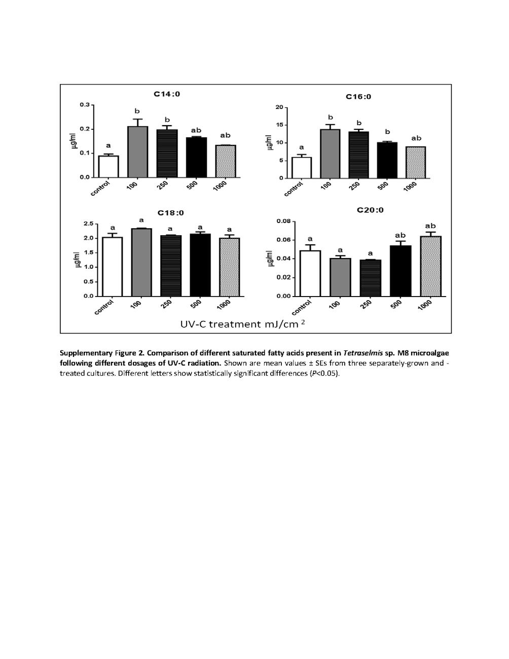
130 107
131 108
132 109
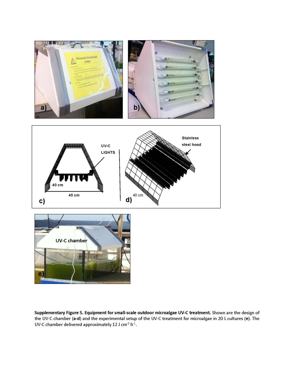
133 110
134 111
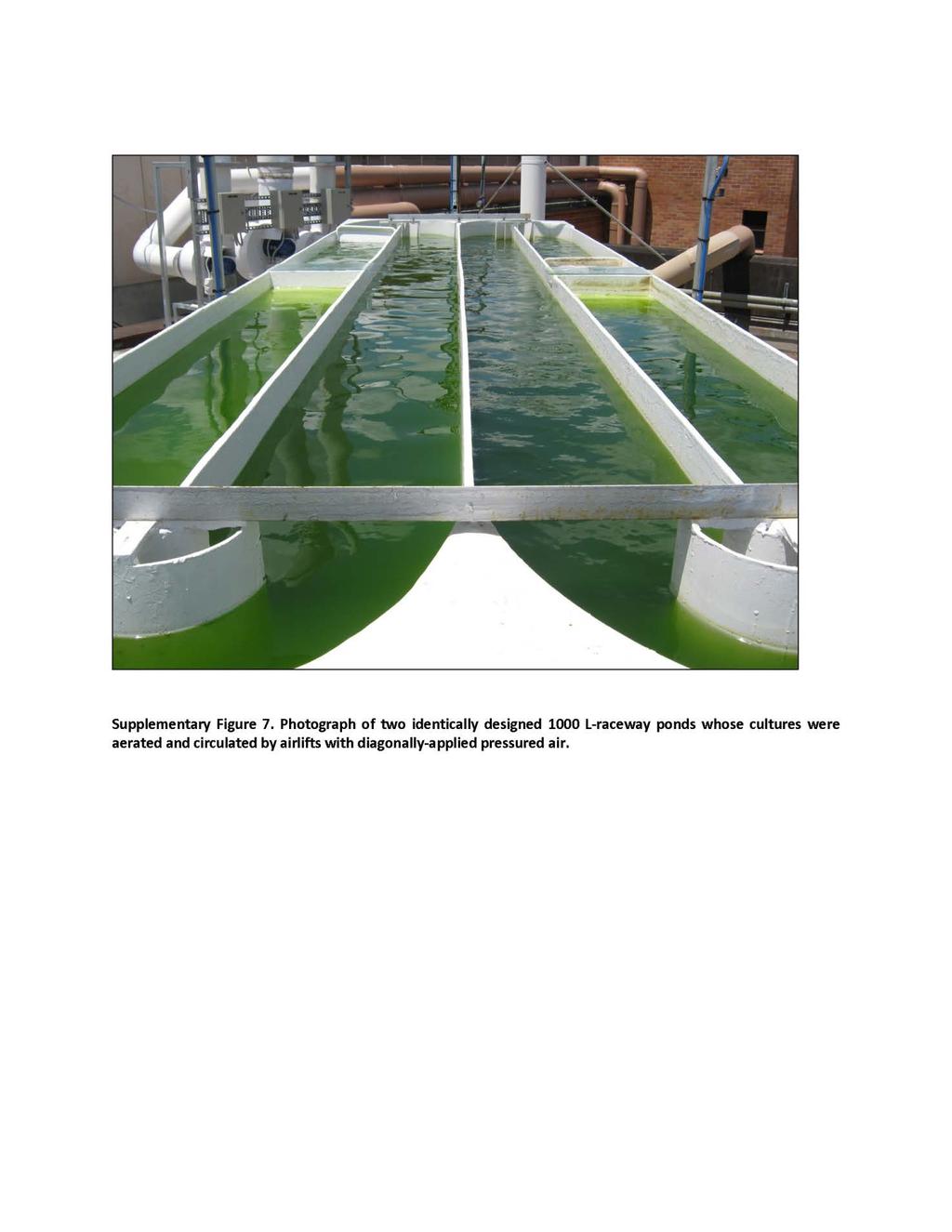
135 112
136 113
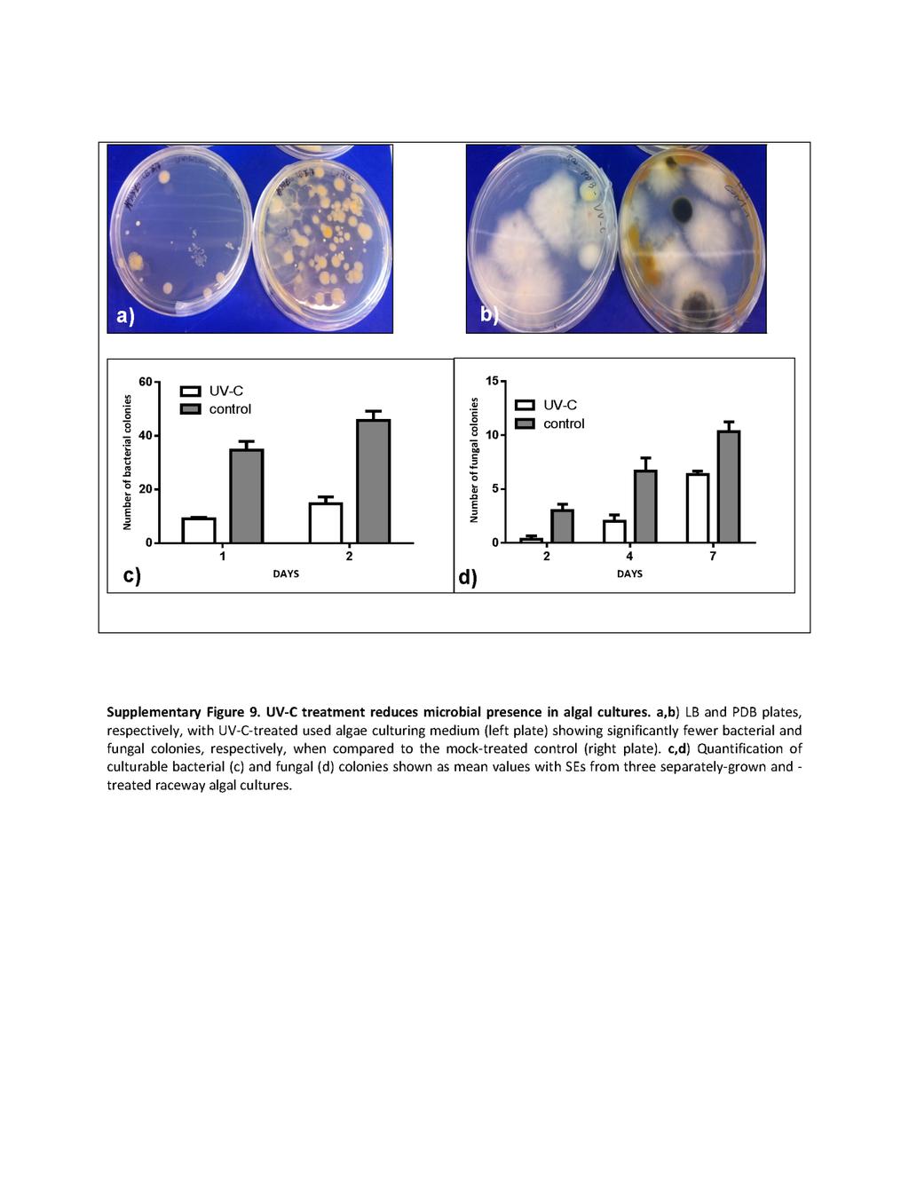
137 114
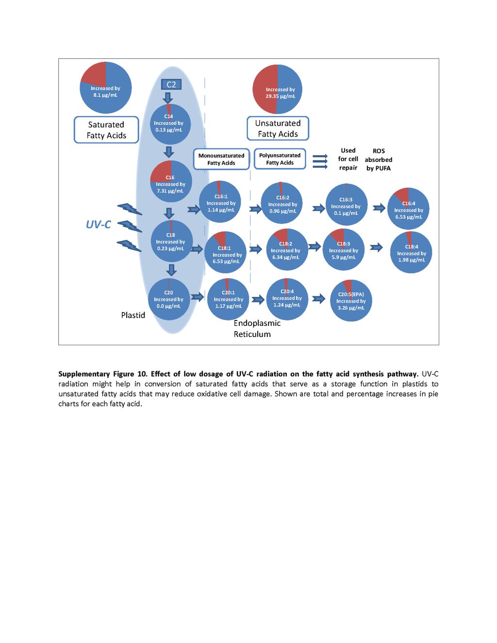
138 115
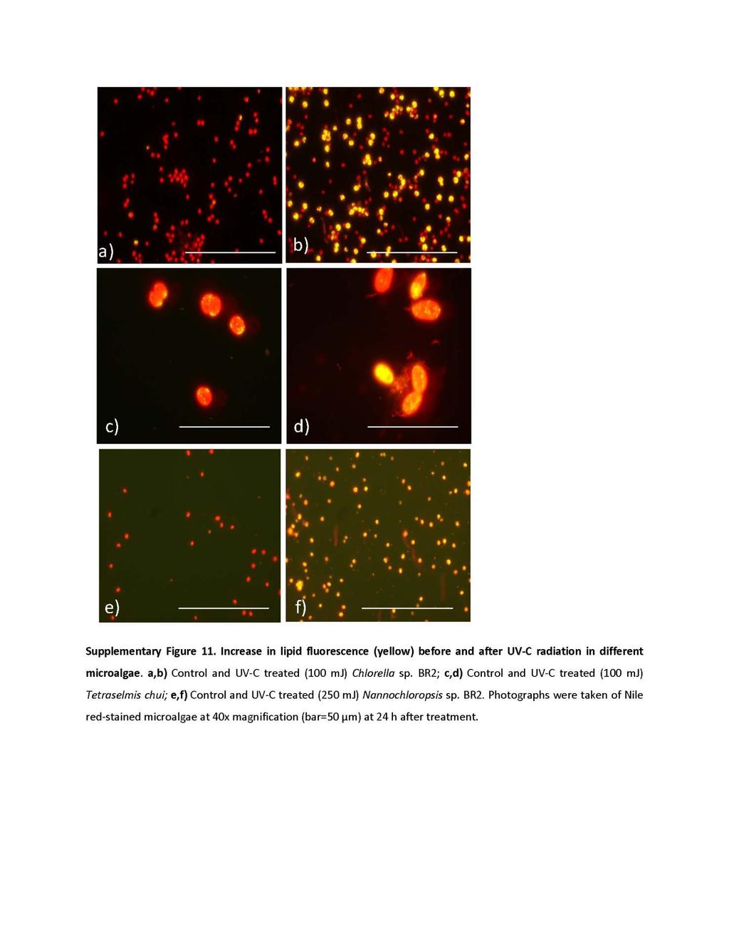
139 116
140 117
141 Chapter 5: Rapid induction of omega-3 fatty acids (EPA) in Nannochloropsis sp. by UV-C radiation (Submitted) Overview Experiments using UV C radiation on Chlorella sp. BR2 and Tetraselmis sp. M8 showed that this external stress can stimulate lipid accumulation, especially PUFAs. Hence, to test weather UV-C radiation could be used to induce high value fatty acids in Nannochloropsis sp. BR2. In particular the production of eicosacenoid pentanoic acid (EPA), an important omega-3 fatty acids with proven health benefits, was targeted. Combined sequential stress treatments were carried out to achieve this aim. Key Findings UV-C treatment of Nannochloropsis sp. resulted in higher total polyunsaturated fatty acids contents, with up to 40% EPA of total fatty acids. The treatment also led to a significant increase of volumetric omega-3 productivity. A combined sequential approach, using UV-C and nutrient starvation may provide a promising approach to produce large amounts of algal omega-3, in an effort to reduce dependency on global fish stocks. 118
142 Chapter 5: Rapid induction of omega-3 fatty acids (EPA) in Nannochloropsis sp. by UV-C radiation (Submitted) Kalpesh Sharma, Peer M Schenk* Algae Biotechnology Laboratory, School of Agriculture and Food Sciences, The University of Queensland, Brisbane, Queensland 4072, Australia *Corresponding author p.schenk@uq.edu.au Abstract Omega-3 fatty acids such as eicosapentaenoic acid (EPA) and docosahexaenoic acid (DHA) provide substantial health benefits. As global fish stocks are declining and in some cases are contaminated with heavy metals, there is a need to find a more sustainable land-based source of these essential fatty acids. The oleaginous microalga Nannochloropsis sp. has been identified as a highly efficient producer of omega-3 fatty acids. In this study, we present a new process to rapidly induce biosynthesis of essential fatty acids, including EPA in Nannochloropsis sp. BR2. Short exposure to UV-C at a dose of 100 or 250 mj/cm2 led to a significant increase in total cellular lipid contents when compared to mock-treated controls. A low dosage of 100 mj/cm 2 also led to a two-fold increase in total EPA content within 24 h that constituted 30% of total fatty acids and up to 12% of total dry weight at higher dosages. UV-C radiation may find uses as an easily applicable external inducer for large-scale production of omega-3 production from microalgae. Introduction Within the last decade microalgae have emerged as a potential source of renewable energy and nutraceutical products. Of particular interest are long chain polyunsaturated fatty acids (LC-PUFAs), including omega 3 and 6 (ω-3,6 ) fatty acids. Omega-3 and 6 fatty acids are essential components for the growth of higher eukaryotes (Ward and Singh, 119
143 2005; Adarme-Vega et al. 2012;). Nutritionally, arachidonic acid (AA, 20:4, ω-6), eicosapentaenoic acid (EPA, 20:5), and docosahexaenoic acid (DHA, 22:6) are the most important fatty acids belonging to this group of bioactive compounds. AA and EPA are components of mammalian cell membranes and are also precursors of the eicosannids, including the prostaglandins, a family of biological effectors involved in inflammatory responses, blood pressure regulation, blood clotting and cell signaling (Kinsella et al. 1990). These compounds have been proven to help in neonatal retinal and brain development, as well as cardiovascular health and disease prevention (Carlson et al. 1993; Crawford 2000; Gill and Valivety 1997; Neuringer et al. 1988). Current sources of ω-3,6 LC-PUFAs, EPA and DHA are marine fish oil (e.g. from mullet and krill), however global fish stocks have been in decline since the late 1980s (Worm et al. 2006). Moreover, traces of heavy metals have been found in marine fish, rendering these fish harmful to consumers (Bourdon et al. 2010). Fish oil is also not suitable for vegetarians and the odor makes it unattractive. Microalgae are the primary producers of these compounds. Their ability to grow under autotrophic, mixotrophic and heterotrophic culture condition and on non-arable land with limited water recourses makes them a preferred choice for commercial production of ω-3,6 PUFAs, EPA and DHA. There is a wide range of lipid induction techniques in microalgae, such as the use of nutrient stress, including nitrogen and/or phosphorus starvation, light irradiation, ph or temperature change, and exposure to heavy metals or other chemicals (Sharma et al. 2012). Of all the microalgae studied, Nannochloropsis sp. are known to produce high amounts of ω-3,6 LC-PUFAs, EPA [10-13]. For example, Nannochloropsis oculata cultivated under nitrogen limitation conditions and an increase in temperature from 20 C to25 C resulted in an increase in total lipids by two fold increase (Converti et al. 2009). Moderated use of UV-A radiation for seven days could lead to an increased production of fatty acids in Nannochloropsis sp. (Forján et al. 2011) and in a study carried out by Srinivas and Ochs (2012) on N. oculata the effect of UV-A at different levels resulted in significantly increased lipid to chlorophyll ratio. Although these protocols highlighted the possibility to increase lipid production based on laboratory research, there are still many concerns for their application, such as difficulties with culture maintenance, elongated cultivation periods/low lipid productivity, high cost for large-scale cultivation, oil extractability and potential environmental impacts. In most 120
144 cases, it is also uncertain whether the induced lipid production during cultivation was derived from the sacrifice of algal growth or not. Moreover, under nutrient stress condition in Nannochloropsis sp. usually higher accumulation of SFA is favored when compared to LC-PUFA s.therefore, there is a desire to develop an effective protocol to stimulate lipid production more importantly LC-PUFA, while sustaining cell growth. Previous published study on Tetraselmis sp (M8) indicated that exposure of M8 cells to small dosage of UV-C mj/cm 2 radiation resulted increase LC-PUFA, especially C18:4, followed by twofold increase of ω-3 fatty acids, However the actual production of ω-3 fatty acids was only 8 µg/ml. The study also highlighted the impact to low dosage of UV-C radiation on fatty acid biosynthesis pathway in converting saturated fatty acids (SFA s) into LC-PUFA s. Based on these results it was hypothesized that the contents of these LC-PUFA can be further increased in Nannochloropsis sp by low dosage of UV-C radiation as naturally these species is known to produce high amounts of LC-PUFA s especially EPA. Materials and methods Laboratory-scale microalgae culturing and UV-C treatment Nannochloropsis sp. BR2, family (Eustigmataceae) was originally collected from the Brisbane River and was cultured in the Algae Biotechnology Laboratory at The University of Queensland (Lim et al. 2012). Cultures were grown in f/2 medium (Guillard and Ryther 1962) in artificial sea water obtained from CSIRO at 23 C on an orbital shaker (Thermoline) at 100 rpm with 12 hours of light and dark rhythm. When the cultures were at late exponential growth phase and the cell densities reached 7.5x10 5 cells/ml, cultures were subjected to UV-C radiation. The method for UV-C treatments has previously been reported (Sharma et al. 2014). Lipid fluorescence analysis and GC-MS analyses Nile red staining was conducted) followed by flow cytometry analysis using a BD LSR II: Analyzing flow cytometer with 573 nm of excitation wavelength as described previously (Sharma et al., 2014). A total of 10,000 cells were counted in each sample. A gate was set to separate lipid producing fluorescence-activated cells and inactivated cells based on the analysis of cells without Nile red staining. The background absorbance from chloroplast auto-fluorescence was subtracted from each sample using mock-treated (control) 121
145 Nannochloropsis sp. BR2 cultures that were not Nile red stained. Qualitative observation was conducted simultaneously by fluorescence microscopy as described below. Gas chromatography-mass spectrometry (GC-MS) analyses were carried out as described previously (Sharma et al., 2014). Dry weight measurements A total of 5 ml of the culture were used for dry weight measurements. The culture was filtered through a 0.27 µm glass fiber filter (Millipore) which was pre-weighed and prewashed with 1 ml distilled water in a vacuum-subjected filter unit (three biological replicates were used from each culture (UV-C treated and mock-treated control)). After filtration, the filters were kept in individual Petri dishes to avoid contamination and dried in a drying oven for 24 h at 80 C with the plate lid half open, prior to weighing. To determine the dry weight the following formula was used: Dry weight mg/l = Filter dry weight - Filter pre-weight Filtered volume ml x 1000 Dry weight in mg/l was determined from the average of three weight measurements for each replicate. Analytical methods Data for growth rates and lipid productivities were statistically analyzed by one-way analysis of variance (ANOVA) with different microalgal cultures as the source of variance and growth rate or lipid productivity as dependent variables. This was followed by Turkey s multiple comparisons test (P>0.05) where appropriate. Results Low UV-C exposure leads to increased lipid accumulation in Nannochloropsis cells Nannochloropsis sp. cells have previously been shown to accumulate significant amounts of lipids after nutrient deprivation and were identified as one of the most promising autotrophic microalgal sources for omega-3 fatty acid production (Huerlimann et al., 2010; Lim et al. 2012). Preliminary experiments using combined stresses of nutrient starvation and UV-C radiation on Tetraselmis sp. have indicated that UV-C exposure can be an 122
146 efficient stimulus to induce LC-PUFA production, including EPA (Sharma et al. 2014). Thus in an effort to optimise EPA production in Nannochloropsis, UV-C treatment was applied to nutrient-starved Nannochloropsis sp. BR2 cells. Following Nile red staining, it became quickly apparent that lipid fluorescence intensities (yellow florescence) became stronger with an increase of UV-C radiation from 100 to 500 mj/cm 2 (Fig. 1a). The reported cell rupturing that was observed in Tetraselmis sp. at 1000 mj/cm 2 (Sharma et al., 2014) was not apparent in Nannochloropsis sp. BR2, suggesting that cell walls are quite rigid (Fig 1a). Based on the presence of green color in live cells and grey or brown color in dead cells, survival rates were determined by cell counting in a hemocytometer. This showed a gradual decline of live cells when UV-C radiation increased (Fig. 1b). The cell survival rates reached half (LD50) at UV-C radiations of mj/cm 2, and at 500 mj/cm 2 and 1000 mj/cm 2 only 5% of the cells had survived. There was no significant difference observed in the cell size at different UV-C radiation intensities as had previously been observed for Tetraselmis sp. (Sharma et al. 2014). 123
 Cells with maximum lipid fluorescence (yellow) can be observed at 250 mj and 500 mj UV-C radiation. Cells are shown at 40x magnification (bar=50 µm) at 24 h after treatment.")
147 Figure 1: Nile red-stained cells of Nannochloropsis sp. BR2 that received different doses of UV-C exposure after nutrient starvation. a) Cells with maximum lipid fluorescence (yellow) can be observed at 250 mj and 500 mj UV-C radiation. Cells are shown at 40x magnification (bar=50 µm) at 24 h after treatment. b) Kill curve of Nannochloropsis sp. BR2, showing the number of cells that survived UV-C treatment at different dosages. Shown are mean values ± SEs from three independent treatments. The arrow indicates the LD50 value. 124
148 With an increase of UV-C radiation, the algal cells were divided into two different populations of P1 and P2, according to the fluorescence intensity (Fig 2 a-f). P1 population presented the auto-fluorescence of chloroplasts in the cells, corresponding to control cultures which were not stained by Nile red, while P2 represents Nile-red fluorescing cells (Fig 2a). In nutrient-starved control cultures, about 75% and 35% of the cells were present in P1 and P2 populations, respectively. Whereas, as the UV-C radiation increased from 100 to 1000 mj/cm 2 a sharp decline was observed in the P1 population which was accompanied by sharp increase in the P2 population (Fig 2 a-f). P1 started to decline sharply with an increase of UV-C radiation. The P2 proportion increased significantly from 100 to 500 mj/cm 2 (P<0.05) and was maintained at 95% between 500 and 1000 mj/cm 2 (P>0.05) (Fig 3a). Overall an increase in Nile red-fluorescing P2 population and a decrease in unstained P1 population with an increase in UV-C intensity was observed that were consistent with results previously obtained for Tetraselmis sp. (Sharma et al., 2014). Based on the average fluorescence value, P1 cells displayed much less fluorescence intensity than P2 (Fig 3b). Different from P1, P2 cells had a big variation of fluorescence intensity that correlated to the total lipid intensity (Fig 3b). Compared to the mock-treated control cells, the lipid fluorescence intensity of algal cells was significantly increased with the increase in UV-C radiation. However, there was no significant difference between the control (0 mj/cm 2 ) and 100 mj/cm 2 treated cells (Fig 3c). As UV-C dosage was increased to 250 mj/cm 2 and 500 mj/cm 2, the total lipid fluorescence in both the cultures was nearly increased by two fold when compared to the control (Fig 3c). Subsequently, the lipid intensity of P2 cells sharply dropped to the control level at 1000 mj/cm 2 (Fig. 3b,c). However, the total lipid fluorescence was still increased significantly at 1000mJ/cm 2 when compared to the mock-treated control (Fig 3c). 125
149 a b c d e f Figure 2: FACS analysis of Nannochloropsis sp. BR2 unstained cells (a), Nile red-stained mock-treated control cells (b), UV-C treated cells (c-f) at 100, 250, 500 and 1000 mj/cm 2, respectively, showing P1 (background fluorescence) and P2 (Nile red fluorescence) populations. The y-axis shows fluorescence intensity at phycoerythrin excitation wavelength of 575 nm and the X- axis shows the forward scatter based on cell size. 126
 and lipid fluorescence (b) of low (P1) and high (P2) fluorescence cell populations and of the total population (c).")
150 a b c Figure 3: Lipid induction in Nannochloropsis sp. BR2 at 24 h after receiving different UV-C dosages. FACS analysis of Nile red-stained cells showing distribution (a) and lipid fluorescence (b) of low (P1) and high (P2) fluorescence cell populations and of the total population (c). Shown are mean values ± SEs from three separately-grown and treated microalgal cultures. Bars with different letters indicate significant differences (P<0.05). 127
151 Fatty acid profiling in Nannochloropsis following UV-C radiation shows lipid induction and a shift towards LC-PUFAs To further quantify the ability of UV-C stress to increase cellular fatty acid contents and to profile their composition, GC-MS analyses were carried out. These confirmed the results obtained from flow cytometry, showing a significant total fatty acid increase (P=0.002, P=0.031; respectively) of UV-C-treated cultures (100 and 250 mj/cm 2 ) compared to mock-treated nutrient-starved control cultures, whereas cultures treated with 500 and 1000 mj/cm 2 showed no significant difference (Fig. 4a). Moreover, in cultures treated with 100 and 250 mj/cm 2, the amount of unsaturated fatty acids (USFA) significantly increased and also the proportion of USFA compared to total fatty acids (Fig. 4a,c). Amongst the saturated fatty acids (SFA), the highest increase was observed for C16 (palmitic acid), followed by C14 (Fig. 4b). On the other hand, C18 and C20 did not show any increase in cultures treated with 100 and 250 mj/cm 2 UV-C, whereas in cultures treated with 500 and 1000 mj/cm 2 there was no significant increase of any fatty acid (Supplementary Fig. 1 and 2). When comparing different USFA, cultures treated with 100 and 250 mj/cm 2 UV-C showed significant increases for all detected USFA, most notably C20:5 (EPA) which accounted to be nearly 30% of the total fatty acid, and was increased highly significantly at 100 mj/cm 2 (P=0.0075) followed by 250 mj/cm 2 (P= ) (Fig. 4c). Other MUFAs namely C16:1, C18:1 cis+trans, and PUFAs C20:4 were also significantly increased, (Supplementary Fig. 2). EPA contents in cellular dry weight was measured to determine suitability of Nannochloropsis for large-scale EPA production. As shown in Fig. 5, EPA constituted up to 12% of total dry weight at higher dosages, however a lower dose of UV-C resulting in a lower EPA content maybe preferred to ensure high overall EPA productivity. 128
 and unsaturated (USFA) fatty acids, respectively,")

152 Figure 4: Fatty acid profiling and quantification by GC-MS showing (a-c) total fatty acids as well as saturated (SFA) and unsaturated (USFA) fatty acids, respectively, produced by different UV-C-treated Nannochloropsis sp. BR2 cultures. Shown are mean values ± SEs from three separately-grown and treated microalgal cultures. Bars with (*) indicate 129


153 significant differences (P<0.05) and bars with (**) indicate highly significant difference (P<0.01). Figure 5: Dry weight and EPA proportion changes in Nannochloropsis sp. BR2, following nutrient starvation and different UV-C exposures. Shown are mean values ± SEs from three separately-grown and treated microalgal cultures. Bars with different letters indicate significant differences (P<0.05). Discussion Consistent with the previous study conducted on Tetraselmis sp. by Sharma et al. (2014), it was reported that the low dosage of UV-C radiation could also stimulate lipid biosynthesis in Nannochloropsis sp. and induce the production of PUFAs. A radiation of 130
154 100 and 250 mj/cm 2 was optimal to induce total lipids content especially USFAs which were increased by two fold followed by SFA s (Fig. 4). A notable difference when comparing flow cytometry and GC-MC data was observed. The total fluorescence shown by flow cytometry analysis was significantly highest at 250 and 500 mj/cm 2 followed by 1000 mj/cm 2. Whereas, GC-MS analysis indicated maximum total fatty acids at UV-C exposure of 100 and 250 mj/cm 2. This difference in the lipids fluorescence can be attributed to the P2 population as the percentage of this population was higher in the culture treated at 250 mj/cm 2 (75%) and mj/cm 2 (95%). Thus it was understood that the high proportion of P2 population and the considerably high number of live cells resulted in high total lipid fluorescence. But when considering the fluorescence of individual P2 cells, the culture treated at 100 mj/cm 2 displayed the highest fluorescence followed by 250 mj/cm 2 and 500 mj/cm 2 which corresponds well to the results obtained by GC-MS. Moreover, when considering the dry weight measurement, as expected there was a linear decline in the total dry weight due to increased cell death at higher dosages (Fig. 5). But on the other hand, there was a significant increase in the percentage of cellular EPA with maximum values at 1000 mj/cm 2 (12% of total dry weight; Fig 5). Cellular lipids may simply provide more energy reserves for stress responses. Alternatively, a UV-induced morphological variation maybe better adapted to adverse condition/lipid production. Since, UV-C causes genetic mutations (Guihéneuf et al. 2010; Rothschild 1999), UV may have acted as a selection pressure during algal evolution (Cockell 2000; Cockell and Raven 2007). In the previous study conducted on Tetraselmis sp. by Sharma et al. (2014) it was reported that at the higher dosage of UV- C (1000 mj/cm 2 ) the release of lipid bodies became apparent as cell structures disintegrated, However cell rupturing was not observed in this study, the possible reason for this would be the relatively small cell size of the Nannochloropsis cells when compared to Tetraselmis sp. cells, also Nannochloropsis sp. cells reportedly possess rigid cell walls when compared to other microalgal species (Iqbal and Theegala 2013). 131
155 When comparing to other lipids induction techniques (Sharma et al. 2012; Takagi et al. 2006; Wang et al. 2008), UV-C radiation resulted in faster lipid biosynthesis stimulation within 24 h (Fig. 1-4). Of all the fatty acids measured, a significant increase was observed in the production of the omega-3 fatty acid EPA which was induced by 2.5 fold and accounted to be approx. 30% of the total fatty acids (Fig 4c) followed by a two fold increase in C18:1, C18:4, C20:4 (Supplementary Fig. 2). The previous study reported that UV-C radiation on Tetraselmis sp. lead to an increase in reactive oxygen species (ROS) production (Sharma et al., 2014). PUFAs have a strong affinity or absorption to ROS (Bouhamidi et al. 1998), therefore, the increment of PUFAs in this study can be interpreted as a defence response against UV-C-generated cellular ROS. The role of PUFAs as an antioxidant and an agent to prevent cell damage has been well documented in many studies on plants and animals (Bartsch et al. 1999; Cheeseman and Slater 1993; Lenzi et al. 2000; Machlin and Bendich 1987). Omega-3 fatty acids (including EPA) have been found to play an important role in prevention of degenerative and cardiovascular diseases (Hooper et al. 2004; Ross et al. 2007). Moreover EPA has been reported to be part of the cell membrane of microalgae and to play an important role in cellular repair and membrane development in microalgae (Huerlimann et al. 2010; Valentine and Valentine 2004). Thus this can also be one of the reasons that Nannochloropsis sp. cells were still intact at higher UV-C dosage of 1000 mj/cm 2 as cells had relatively high concentration of EPA (Fig. 5) when compared to the previous study (Sharma et al., 2014). Whereas, in previous study on Tetraselmis sp, it was reported that UV-C induces antioxidants activity in microalgal cells and as EPA has strong affinity to absorption to ROS (Bouhamidi et al. 1998), an increase in EPA was assumed as a defence mechanism of microalgal cells. In conclusion, this study highlights the use of low exposure of UV-C radiation to induce lipids, particularly EPA, within 24 h of the treatment. This presents a significant improvement to lipid induction by nutrient starvation only. The optimized large-scale 132
156 production of EPA in Nannochloropsis may provide a much-needed alternative, vegetarian, and land-based source of omega-3. Acknowledgements We wish to thank the Australian Research Council for financial support. 133


157 Supplementary Fig. 1: Comparison of different saturated fatty acids present in Nannochloropsis sp. BR2 following different treatments of UV-C radiation. Values are mean ±SE (n = 3); bars with different alphabets indicate significant differences (P<0.05). 134


158 Supplementary Fig. 2: Comparison of different unsaturated fatty acids present in Nannochloropsis sp. BR2 following different treatments of UV-C radiation. Values are mean ±SE (n = 3); bars with different alphabets indicate significant differences (P<0.05). 135
159 References Adarme-Vega TC, Lim DK, Timmins M, Vernen F, Li Y, Schenk PM Microalgal biofactories: a promising approach towards sustainable omega-3 fatty acid production. Microb Cell Fact 11(1):96. Bartsch H, Nair J, Owen RW Dietary polyunsaturated fatty acids and cancers of the breast and colorectum: emerging evidence for their role as risk modifiers. Carcinogenesis 20(12): Bouhamidi R, Prévost V, Nouvelot A High protection by grape seed proanthocyanidins (GSPC) of polyunsaturated fatty acids against UV-C induced peroxidation. Comptes Rendus de l'académie des Sciences-Series III-Sciences de la Vie 321(1): Bourdon J, Bazinet T, Arnason T, Kimpe L, Blais J, White P Polychlorinated biphenyls (PCBs) contamination and aryl hydrocarbon receptor (AhR) agonist activity of omega-3 polyunsaturated fatty acid supplements: implications for daily intake of dioxins and PCBs. Food and Chemical Toxicology 48(11): Carlson SE, Werkman SH, Peeples JM, Cooke RJ, Tolley EA Arachidonic acid status correlates with first year growth in preterm infants. PNAS 90(3): Cheeseman K, Slater T An introduction to free radical biochemistry. British Medical Bulletin 49(3): Cockell CS The ultraviolet history of the terrestrial planets -- implications for biological evolution. Planetary and Space Science 48(2-3): Cockell CS, Raven JA Ozone and life on the Archaean Earth. Philosophical Transactions of the Royal Society A: Mathematical, Physical and Engineering Sciences 365(1856): Converti A, Casazza AA, Ortiz EY, Perego P, Del Borghi M Effect of temperature and nitrogen concentration on the growth and lipid content of Nannochloropsis oculata and Chlorella vulgaris for biodiesel production. Chemical Engineering and Processing: Process Intensification 48(6): Crawford M Placental delivery of arachidonic and docosahexaenoic acids: implications for the lipid nutrition of preterm infants. The American journal of clinical nutrition 71(1):275S-284S. Forján E, Garbayo I, Henriques M, Rocha J, Vega J, Vílchez C UV-A Mediated Modulation of Photosynthetic Efficiency, Xanthophyll Cycle and Fatty Acid Production of Nannochloropsis. Marine Biotechnology 13(3): Gill I, Valivety R Polyunsaturated fatty acids, part 1: occurrence, biological activities and applications. Tren in Biotech 15(10): Guihéneuf F, Fouqueray M, Mimouni V, Ulmann L, Jacquette B, Tremblin G Effect of UV stress on the fatty acid and lipid class composition in two marine microalgae;pavlova lutheri (Pavlovophyceae) and Odontella aurita (Bacillariophyceae). J of App Phycol 22(5): Guillard RR, Ryther JH Studies of Marine Planktonic Diatoms.1. Cyclotella Nana Hustedt, and Detonula Confervacea (Cleve) Gran. Canadian J of Microbiology 8(2):229-&. 136
160 Hooper L, Thompson R, Harrison R, Summerbell C, Moore H, Worthington H, Durrington P, Ness A, Capps N, Davey Smith G Omega 3 fatty acids for prevention and treatment of cardiovascular disease. Cochrane Database Syst Rev 4. Huerlimann R, de Nys R, Heimann K Growth, lipid content, productivity, and fatty acid composition of tropical microalgae for scale-up production. Biotechnology and Bioengineering 107(2): Iqbal J, Theegala C Optimizing a continuous flow lipid extraction system (CFLES) used for extracting microalgal lipids. GCB Bioenergy 5(3): Kinsella JE, Lokesh B, Broughton S, Whelan J Dietary polyunsaturated fatty acids and eicosanoids: potential effects on the modulation of inflammatory and immune cells: an overview. Nutrition (Burbank) 6(1): Lenzi A, Gandini L, Picardo M, Tramer F, Sandri G, Panfili E Lipoperoxidation damage of spermatozoa polyunsaturated fatty acids (PUFA): scavenger mechanisms and possible scavenger therapies. Front Biosci 5(1):1-15. Lim DKY, Garg S, Timmins M, Zhang ESB, Thomas-Hall SR, Schuhmann H, Li Y, Schenk PM Isolation and Evaluation of Oil-Producing Microalgae from Subtropical Coastal and Brackish Waters. PLoS ONE 7(7):e Machlin LJ, Bendich A Free radical tissue damage: protective role of antioxidant nutrients. The FASEB Journal 1(6): Neuringer M, Anderson GJ, Connor WE The essentiality of n-3 fatty acids for the development and function of the retina and brain. Annual review of nutrition 8(1): Ross BM, Seguin J, Sieswerda LE Omega-3 fatty acids as treatments for mental illness: which disorder and which fatty acid. Lipids Health Dis 6(21):21. Rothschild LJ The Influence of UV Radiation on Protistan Evolution. Journal of Eukaryotic Microbiology 46(5): Sharma K, Li Y, Schenk P UV-C-mediated lipid induction and settling, a step change towards economical microalgal biodiesel production. Green Chem 16, Sharma KK, Schuhmann H, Schenk PM High Lipid Induction in Microalgae for Biodiesel Production. Energies 5(5): Srinivas R, Ochs C Effect of UV-A Irradiance on Lipid Accumulation in Nannochloropsis oculata. Photochemistry and Photobiology 88(3): Takagi M, Karseno, Yoshida T Effect of salt concentration on intracellular accumulation of lipids and triacylglyceride in marine microalgae Dunaliella cells. J of Bioscience and Bioengineering 101(3): Valentine RC, Valentine DL Omega-3 fatty acids in cellular membranes: a unified concept. Progress in Lipid Research 43(5): Wang G, Chen K, Chen L, Hu C, Zhang D, Liu Y The involvement of the antioxidant system in protection of desert cyanobacterium Nostoc sp. against UV-B radiation and the effects of exogenous antioxidants. Ecotoxicology and Environmental Safety 69(1): Ward OP, Singh A Omega-3/6 fatty acids: Alternative sources of production. Process Biochemistry 40(12):
161 Worm B, Barbier EB, Beaumont N, Duffy JE, Folke C, Halpern BS, Jackson JB, Lotze HK, Micheli F, Palumbi SR Impacts of biodiversity loss on ocean ecosystem services. Science 314(5800):
162 Chapter 6: UV-C mediated rapid carotenoid induction and enhanced harvesting performance of Dunaliella salina and Haematococcus pluvialis (Submitted) Overview From the above studies conducted on Chlorella sp. BR2, Tetraselmis sp. M8 and Nannochloropsis sp. BR2, it was discovered that short exposure of UV-C radiation could significantly induce lipid biosynthesis, most importantly polyunsaturated fatty acids (PUFAs) and cause oxidative damage. As carotenoids have been proven to have high anti-oxidative activity and scavenge free radicals the hypothesis was to use a small dosage of UV-C radiation to induce carotenoids in Dunaliella salina and Haematococcus pluvialis., two microalgae that were found to contain high levels of beta-carotene and astaxanthin, respectively. Key Findings A significant increase in total carotenoids and beta-carotene was observed after exposure to 50 mj/cm 2 for D. salina and two-fold increase in total astaxanthin production was observed at 30 mj/cm 2 for Haematococcus sp. UV-C radiation of 50 mj/cm 2 was found sufficient to detach flagellate in both species tested and approximately 95% of settling was observed after 15 h and 3 h for D. salina and H pluvialis., respectively. This was the first study to report UV-C radiation not only induces lipids biosynthesis, but also induces carotenoids biosynthesis in D. salina and H pluvialis. 139
163 Chapter 6: UV-C mediated rapid carotenoid induction and enhanced harvesting performance of Dunaliella salina and Haematococcus pluvialis (Submitted) Kalpesh K Sharma 1, Faruq Ahmed 1, Peer M Schenk 1 and Yan Li 1,2* 1 Algae Biotechnology Laboratory, School of Agriculture and Food Sciences, The University of Queensland, Brisbane, Queensland 4072, Australia; 2 College of Marine and Environmental Science, James Cook University, Townsville, Queensland 4811, Australia. *Corresponding author yan.li3@jcu.edu.au PMS and YL have contributed equally. Abstract Microalgae are primary producers of organic matter in aquatic environments and are a promising source of a wide diversity of compounds, such as carotenoids and essential fatty acids. However, commercial-scale microalgae application is rarely economical due to slow-growing high carotenoid-producing microalgal strains and high harvesting costs. Here, we present a novel UV-C radiation technique to not only induce lipid accumulation but also carotenoid biosynthesis and natural settling in Dunaliella salina and Haematococcus pluvialis. Maximum total carotenoid and β-carotenoid inductions were achieved following 50 mj/cm 2 of UV-C radiation for D. salina, whereas a five-fold increase in total astaxanthin production was achieved at 30 mj/cm 2 in H. pluvialis. Meanwhile, a radiation of 50 mj/cm 2 was sufficient to detach algal flagella in both tested species. Approximately 95% of algal cells settled after 15 h and 2 h for D. salina and H. pluvialis, respectively. Introduction Microalgae are considered as a potential feedstock, not only for second generation biofuels, but also for the production of a wide diversity of compounds, ranging from valueadded products, such as carotenoids, pharmaceuticals and nutraceuticals (Li et al. 2007; Olaizola 2003; Pérez-López et al. 2014; Schenk et al. 2008; Sheehan et al. 1998). Microalgae are the primary producers of organic matter in aquatic environments due to their robust photosynthetic activities. They also possess several advantages over 140
164 terrestrial plants: they are relatively easy to cultivate, do not need to compete with food production, can adapt to some extreme environmental conditions by producing a variety of secondary metabolites, and can also be used as efficient bioremediation tools, for instance for sequestering CO 2 from streams derived from industrial processes (Ahmed et al. 2014; Munir et al. 2012; Olaizola 2003; Suh et al. 2006). Therefore, microalgae have become a promising natural feedstock in the new century. As the cost-effectiveness of algal biodiesel has been challenged, there is mounting interest on high-value bio-compounds produced from microalgae. The typical representatives are Dunaliella salina and Haematococcus pluvialis that can produce a high level of β-carotene and astaxanthin, respectively. Carotenoids are generally non-polar, lipid-soluble organic pigments that give microalgae yellow, orange and red colour due to carotenoids absorbing visible light. Carotenoid pigments have high anti-oxidation properties, so they functionally play an active role in protecting the photosynthetic apparatus by dissipating excess energy (Ahmed et al. 2014; Stahl and Sies 2012). They also play a major role in photosynthesis by harvesting light and stabilising protein folding in the photosynthetic apparatus (Choudhury and Behera 2001). Due to their long chain carbon and hydrogen bonds, carotenoids can scavenge singlet oxygen which mainly arises from sunlight absorption by chromophores and thus protect chlorophylls, lipids, proteins and DNA from oxidative damage (Ahmed et al. 2014; Ibañez and Cifuentes 2013). Furthermore, carotenoids have been used as feed for aquaculture and as colorants by the food industry. In recent years, they have also become popular as dietary supplements due to the findings that carotenoids are powerful antioxidants and therefore can provide protection against ailments, such as cardiovascular diseases, certain cancers, age-related macular degeneration, and neurodegenerative diseases e.g. amyotrophic lateral sclerosis (Ibañez and Cifuentes 2013). Haematococcus algal meal serves as a colour additive in salmonid feeds, and also is a health-proven dietarysupplement for human consumption (Guedes et al. 2011). Therefore, microalgae D. salina and H. pluvialis have attracted a number of researchers interests as valuable carotenoid producers and also entrepreneurs attention in the last decade. It is well known that microalgae can modify the lipid biosynthesis pathway and redirect it to produce some secondary metabolites upon environmental stress conditions (e.g. nutrient deprivation, light limitation, UV irradiation, ph, temperature; Aflalo et al. 2007; García Malea et al. 2009; Shahid and Mohammad 2013; Sharma et al. 2012, 2014). Hence, many studies were conducted on optimisation of culturing condition, high temperature and salinity stress on D. salina to optimise β-carotenoids production (Fernández-Sevilla et al. 141
165 2010; Herrero et al. 2006; Kleinegris et al. 2011). Similarly, the production of astaxanthin in H. pluvialis has been extensively studied, typically with the aim to increase yields (Domınguez-Bocanegra et al. 2004; García Malea et al. 2009; Olaizola 2000). The stress conditions used to induce carotenoid production in these microlagal strains include a combination of high salinity (for β-carotene), nutrient deprivation, high light, osmotic shock (for astaxanthin) at a stage when cultures are in late exponential growth phase (Ahmed et al., 2013). These attempts have resulted in the accumulation of up to 4% (w/w) astaxanthin and 14% (w/w) β-carotene in the dry biomass weight in H. pluvialis and D. salina (Borowitzka, 2013). However, due to lengthy processes of these induction methods, the development of new efficient techniques is warranted that can significantly reduce the time for high-level carotenoid accumulation in H. pluvialis and D. salina, thereby improving cost-effectiveness of production. Synthesis and accumulation of large amounts of carotenoids in microalgae can be achieved by either acting individually or by applying a combination of stresses, including nutrient stress, radiation, ph and temperature change, heavy metals and other chemicals, and also by some metabolic engineering approaches (Gao et al. 2013; Renaud et al. 2002; Gao et al., 2014). However, it typically takes 3 5 days until significant amounts of carotenoids are synthesised which is accompanied by slow growth rates and thus finally affects the total algal biomass and carotenoid productivities. More importantly, modification of temperature, ph, salinity and/or heavy metals concentration are difficult to apply at large scale. Additionally, the microalgae dewatering/harvesting process constitutes the most energy intensive and expensive component for carotenoid production from microalgae and becomes a major impediment for industrial scale manufacturing of H. pluvialis and D. salina (Garg et al., 2012; Ahmed et al., 2013; Garg et al. 2012). Therefore, it would be highly beneficial for the carotenoid production process if induction methods could simultaneously assist in the dewatering and harvesting process of carotenoidinduced H. pluvialis and D. salina cultures. UV-A and UV-B have already been reported to increase fatty acid and carotenoid production in several microalgal strains but which typically takes more than 7 days (Srinivas and Ochs 2012; Goes, 1995). UV-C is particularly promising due to the fact that it carries more energy per photon. We have previously shown for Tetraselmis sp., that UV-C radiation acts a highly effective method, not only for inducing high-level fatty acid production but also leads to rapid settling of these flagellate microalgae (Sharma et al. 2014). This process was accompanied by an increase in reactive oxygen species (ROS) 142
166 and up-regulation of redox homeostasis-regulating enzymes, superoxide dismutase and glutathione S reductase (Sharma et al. 2014). As carotenoids have a high antioxidative activity and can scavenge free ROS, the rationale behind the present study was to test whether a low dosage of UV-C radiation can not only induce fatty acid synthesis, but also lead to rapid carotenoid production within hours and whether prolonged exposure causes settling of flagellate D. salina and H. pluvialis cultures. Materials and methods Microalgal cultures and UV-C treatments Microalgae Dunaliella salina (Lim et al., 2012) and Haematococcus pluvialis (Lim et al., 2012) were cultured in the Algae Biotechnology Laboratory at The University of Queensland. The isolate of D. salina was grown in f/2 medium in autoclaved 60 ppt saltwater, whereas H. pluvialis was cultured in BMM medium in autoclaved tap water (Bischoff et al., 1963). Primary stock cultures were maintained aerobically in 2.5 L Erlenmeyer flasks with constant orbital shaking (100 rpm) at 25 C, on an orbital shaker (Thermoline) at 100 rpm under a 12:12 h light/dark photoperiod of fluorescent white light (120 μmol photons m 2 s 1 ). The cell density reached 1.7x10 6 /ml and 5x10 5 /ml for D. salina and H. pluvialis, respectively, prior to the UV-C radiation trials. Nitrate and phosphate levels were determined using the corresponding API Nutrient testing kits, according to the manufacturer s instructions. After gently stirring, 25 ml aliquots of D. salina and H. pluvialis cultures were pipetted into Cole-Parmer sterile tissue culture dishes (150 mm), forming a thin layer inside a total of 18 plates, each for both cultures. Plates were randomly divided into six groups with three plates as replicates for UV-C radiation (253 nm) treatment in a UV chamber, with dimension of 31.7 x 24.1 x 15.2 cm containing five G8T5 format, minibipin bulbs (Biorad, Gs-Genelinker, California, USA).. Based on our preliminary tests, the radiation levels were set at 0, 50, 75, 100, 150 and 200 mj/cm 2 for D. salina and subsequently incubated for 24 h, whereas H. pluvialis plates were exposed to 0, 30, 50, 100, 200 and 400 mj/cm 2 and incubated for 48 h. Algae survival rates were measured by counting the live cells based on visibly intact chloroplasts in every 100 cells on each plate. The cell size was also measured by compound microscopy (Olympus). 143
167 Microscopic analyses After the UV-C carotenoid induction phase, microalgae cells were stained with 2 μg/ml Nile red (dissolved in acetone; Sigma, USA) for 15 min and photographed using a fluorescent Olympus BX61 microscope fitted with a 100 W High Pressure Mercury Burner and an Olympus DP10 digital camera. Differential interference contrast and epifluorescent (excitation: nm, emission: 590 nm) images were obtained at 20 magnification. Lipid fluorescence analysis For each replicate, 1 ml of algae cells was sampled and stained with 3 µl Nile red solution (a 10 mg/ml Nile red stock solution was prepared in acetone and stored in the dark at 4 C). After 20 min incubation in the dark, the lipid fluorescence intensity of cells was detected by plate reader fluorescent spectroscopy. A total of 200 µl of each sample was loaded into 96 well-plates and analysed with a Fluostar optima and Polarstar optima (BMG LAB tech) plate reader at excitation and emission wavelengths of 485/584 nm; the gain was set at Settling of microalgae After gently stirring, 25 ml aliquots of D. salina and H. pluvialis cultures were pipetted into a square Petri dish, forming a thin layer inside in a total of 18 plates for D. salina and H. pluvialis, respectively. Plates were randomly divided into six groups with three plates as replicates, and then exposed to UV-C radiation (253 nm) in a UV chamber as mentioned above. The cultures from the plate were then poured into 50 ml tubes and left undisturbed for settling. Cell count was taken at 2 and 15 h. Analyses of fatty acid and carotenoid profiles As described previously (Sharma et al, 2014), 4 ml of algal culture on each plate were centrifuged at 16,000xg for 3 min. The supernatant was discarded and the lipid in the algal pellet was hydrolysed and methyl-esterified with a 300 µl of 2% H 2 SO 4 methanol solution at 80 C by shaking (480 rpm) for 2 h on a thermalmixer (Eppendorf). Prior to esterification, 50 µg of heneicosanoic acid (C21) were added to the pellet of each sample as an internal standard. After esterification, 300 µl of 0.9% (w/v) NaCl and 300 µl of hexane (analytical grade) were added and vortexed for 20 s. Phase separation was achieved by centrifugation at 16,000xg for 3 min and the hexane layer was used for carotenoid profile 144
168 analysis. Gas chromatography-mass spectrometry (GC-MS) analyses were carried out on an Agilent 6890 GC coupled to a 5975 MSD. A DB-Wax column (Agilent, ) was used with running conditions as described in Agilent s RTL DBWax method (Application note: EN). Identification of fatty acid methyl esters (FAME) was based on mass spectral profiles and retention times in the Agilent s RTL DBWax method. Each FAME was quantified using the formula: Fatty acid (µg/ml) = (Total ionic current of fatty acid/ Total ionic current of standard) x (molecular mass of fatty acid/molecular mass of internal standard) x 50/4 For carotenoid extraction, the centrifuged algal samples were freeze-dried for 24 h. The carotenoid extraction was based on a method of Fanning et al. (2010). The concentrated carotenoid extract was dissolved in 2.5 ml of methanol/dichloromethane (50/50, v/v) for HPLC analysis. The gradient of mobile phases in high performance liquid chromatography (HPLC) analysis was set as: 0 min, 80% phase A and 20% phase B; 48 min, 20% A and 80% B; 49 min, 80% A and 20% B; 54 min, 20% A and 80% B (phase A - 92% methanol/8% 10 mm ammonium acetate; phase B - 100% methyl tert-butyl ether). A mass spetrometry scan was undertaken between mass units in the APCI+ mode (Fu et al. 2012) using an Acquity UPLC H-Class system connected to a Quattro Premier triple quad (Micromass MS Technologies, Waters Corporation, Milford, MA, USA). Source temperature and probe temperature were 150 C and 600 C, respectively, while desolvation and cone gas flow were at 450 and 50 L h -1. The corona, cone and extractor voltages were 5.0 µa, 30 V and 3 V, respectively. Carotenoids were identified by their specific retention times, UV/Vis spectra and mass spectra against authentic standards (Lu et al. 2009). The concentrations of the identified carotenoids were determined using individual calibration curves. Malondialdehyde assay in Haematococcus pluvialis To better understand the underlying mechanisms for the observed shift of fatty acids towards unsaturated fatty acids following UV-C treatment, Thiobarbituric Acid Reactive Substances (TBARS) assays were carried out on H. pluvialis. Decomposition of unstable peroxides derived from PUFAs resulting in the formation of malondialdehyde, were quantified colorimetrically following their controlled reaction with TBARS. 145
169 Data analysis Data for lipid and carotenoid productivity was statistically analysed by one-way analysis of variance (ANOVA) with separately-grown microalgal cultures as the source of variance and growth rate or lipid or carotenoid productivity as dependent variables. This was followed by Turkey s multiple comparisons test where appropriate (P<0.05). Student s t- test was used for pair-wise comparisons. Results UV-C exposure induces settling of D. salina and H. pluvialis cultures To test whether nutrient starvation and UV-C treatment can lead to enhanced increases in carotenoids biosynthesis, combined sequential stress treatments were carried out on D. salina and H. pluvialis. For D. salina, the cell survival rate sharply declined with an increase of UV-C radiation (Fig 1a). The survival rate of D. salina was about 50% (LD50) at 50 mj/cm 2, whereas there was only 1-3% of survival rate at 200 mj/cm 2. Radiation of 50 mj/cm 2 was sufficient to detach the flagella and induce settling for almost 100% of the cells within 15 h, but reached 50% in the first 2 h post treatments (Fig 1b). Similarly, H. pluvialis cultures were radiated at different dosages of UV-C radiation. There was a linear decline of cell survival rates in H. pluvialis, reaching 50% (LD50)at 100 mj/cm 2, and dropped to 30% at 200 mj/cm 2 (Fig 1a). However, the cell survival rate did not further decrease between 200 and 1000 mj/cm 2 (Fig 2a). Similar to D. salina, 50 mj/cm 2 could effectively detach the flagella of H. pluvialis, and approximately 75% settling was achieved at this dose, but almost 99% of the Haematococcus cells settled at 150 mj/cm 2. UV-C irradiation induces microalgal lipid biosynthesis in D. salina and H. pluvialis Nitrogen-deprived cells of D. salina and H. pluvialis cultures showed a clear increased amount of lipid droplets within 24 h of UV-C radiation (Fig. 2 and Fig. 3a-e). This is consistent with a recent study on a different UV-C-treated green alga, Tetraselmis sp. (Sharma et al. 2014). With increase of UV-C radiation dosage from 50 to 200 mj/cm 2, the lipid fluorescence intensity inside the cells became stronger for both microalgae. Upon dosages higher than 200 mj/cm 2, algal cell bleaching and rupturing became apparent in 146
170 D. salina. cells also displayed more clumping (arrows on Fig 2d) upon UV-C exposure of 50 mj/cm 2 or higher in D. salina (Fig 2c-d). In H. pluvialis, both, 30 and 50 mj/cm 2, were sufficient to induce lipid droplets and the value became stabilised when UV-C radiation was higher than 50 mj/cm 2. In D. salina, 50 and 75 mj/cm 2 of radiation were more effective for lipid induction, demonstrated by a significant increase of lipid florescence inside the cells (P<0.05, Fig 2e). However, both microalgae showed no significant difference on cell mortality between the control and treated cultures when exposed to higher than 100 mj/cm 2 of UV-C radiation. 147


171 Figure1. a) Kill curve of D. salina and H. pluvialis cells, showing the number of survival cells after different UV-C radiation dosages; b) Settling curve of D. salina and H. pluvialis. Shown are mean values ± SEs from three independent treatments of separately-grown algal cultures, each. 148
, 30 and 100 mj/cm 2, respectively. g,h) Lipid fluorescence analysis of different UV-C treated samples of D. salina and H. pluvialis.")
172 Figure 2. a-c) Nile red-stained cells of D. salina cultures exposed to UV-C radiation at 0 (control), 75 and 100 mj/cm 2, respectively. d-f) Nile red-stained cells of H. pluvialis cultures exposed to UV-C radiation at 0 (control), 30 and 100 mj/cm 2, respectively. g,h) Lipid fluorescence analysis of different UV-C treated samples of D. salina and H. pluvialis. UV-C treatment leads to increased β-carotene and astaxanthin biosynthesis Carotenoid contents were measured in microalgal cells to test whether UV-C exposure can stimulate biosynthesis of these anti-oxidative compounds. At 24 h after exposure to 50 mj/cm 2, there was a clear increase of total carotenoids content in D. salina (P<0.05, Fig. 3a). As a major component, β-carotene content nearly doubled compared to the mocktreated control cultures (P<0.05). However, when UV-C radiation was higher than 50 mj/cm 2, both total carotenoids and β-carotene content declined sharply and dropped back to the control levels. Apart from β-carotene, the mean value of lutein content was also higher than the control at 50 mj/cm 2 but was not statistically significant (P>0.05, Fig. 3c). 149
173 In H. pluvialis, a significant increase of astaxanthin production was obtained at 30 mj/cm 2 (P<0.05,Fig. 4a-g), but gradually declined with an increase of UV-C radiation ( 50 mj/cm 2 ) and became similar to the control level (Fig. 4d). Three isomers of astaxanthin were detected in this study, showing about three times less of cis-astaxanthin content than either di-astaxanthin or mono-astaxanthin in tested H. pluvailis cells (Fig. 4d-g). With increased UV-C radiation, these astaxanthins displayed a similar fluctuation pattern as total astaxanthins, but only mono-astaxanthin was still significantly higher than in the control cultures at 50 mj/cm 2 (P<0.05). Microscopy analyses revealed that some cells had burst open and cell debris was observed (Fig. 2f). UV-C treatment induces polyunsaturated fatty acid production and malondialdehyde activity Although lipid florescence analysis showed a large of amount of lipid droplets induced in UV-C treated D. salina cells, GC-MS data did not show any significant increase in total fatty acid determination. Only a slight increase was detected in the cells radiated at 75 mj/cm 2 of UV-C treatment (Fig. 5a). However, the total unsaturated fatty acid content significantly increased almost two-fold at 75 mj/cm 2 (P<0.05, Fig. 5b). By comparison, the content of total saturated fatty acids was unaltered in the tests (Fig. 5c). It appears plausible that UV-C stress leads to cellular lipid biosynthesis to improve survival of microalgal cells. Figure 4a shows that as the UV-C radiation increased from 0 to 1000 mj/cm 2 the formation of malondialdehyde increased in H. pluvialis cells and reached the highest level at 1000 mj/cm 2 (fig 6), indicating that UV-C-treated cells underwent oxidative stress and likely caused damage to DNA and cellular membranes. Hence the increased amount and proportion of astaxanthin in UV-C-stressed cells may be required for providing sufficient antioxidant capacity and repairing cellular damage to restore redox homeostasis in the cells. 150
 Total carotenoids, β-carotenoids and lutein")

174 Figure 3. a-c) Total carotenoids, β-carotenoids and lutein measured in D. salina cells following different UV-C treatments. Shown are mean values ± SEs from three independent treatments of separately-grown algae cultures. 151


175 a b c Figure4: a-c) Visible carotenoid induction in UV-C-treated H. pluvialis cultures at 0 (control), 30 and 100 mj/cm 2, respectively. d-g) Total astaxanthin, cis-astaxanthin, monoastaxanthin and di-astaxanthin contents in H. pluvialis cells upon different UV-C radiations. Shown are mean values ± SEs from three independent treatments of separately-grown algae cultures. Different small letters represent significant differences between treatments (P<0.05). 152


176 Figure 5 :a-c) Total fatty acids, total unsaturated fatty acids and total saturated fatty acids induced in D. salina cultures at different dosages of UV-C radiation. Shown are mean values ± SEs from three independent treatments of separately-grown D. salina cultures. Different small letters represent statistically significant differences between treatments (P<0.05). 153
.")

177 Figure 6: Decomposition of unstable peroxides (TBRAS) assay of UV-C-stressed and mock-treated H. pluvialis cells at different dosages. Values are mean ± SEs from three separately-grown and -treated cultures. Different small letters represent statistically significant differences between treatments (P<0.05). Discussion To our knowledge, this is the first study to report that UV-C radiation can induce both lipid and carotenoid biosynthesis in D. salina and H. pluvialis. In particular, this study highlights the effectiveness of using low dosage of UV-C radiation to stimulate the production of valuable carotenoids which only took 24 h for D. salina and 48 h for H. pluvialis, and also provides a cost-effective way to pre-concentrate microalgae. Maximum amounts of total carotenoids and β-carotene were obtained at 50 mj/cm 2 in D. salina. About a five-fold increase of total astaxanthin production was measured following UV-C treatment at 30 mj/cm 2 in Haematococcus cells. However, 50 mj/cm 2 of UV-C radiation was found sufficient to detach flagella in both microalgal species and approximately 95% of settling rate was achieved after 15 h and 2 h for D. salina and H. pluvialis, respectively. Probably caused by the lack of rigid cell walls, D. salina showed a high level of cell mortality at 100 mj/cm 2 of UV-C radiation and almost 100% of the cells were dead up to 1000 mj/cm 2 (Fig 1a). In contrast, H. pluvialis is known to have a rigid cell wall and turns into cysts during stress conditions. This is reflected in the reduced cell mortality for H. pluvialis in this study. Interestingly, a significant increase in lipid florescence was observed 154
178 in H. pluvialis at 30 and 50 mj/cm 2, whereas it was at 75 mj/cm 2 on D. salina. It is likely that this was associated with the cell density because D. salina ( /ml) was used at a much higher cell concentration than H. pluvialis ( /ml). However, the report on Tetraselmis sp. showed that UV-C radiation of 100 mj/cm 2 was optimal to induce significant amounts of lipids, based on a similar cell density ( /ml; Sharma et al. 2014) as our tested D. salina cultures. Clearly, the effectiveness of UV-C-mediated lipid induction is dependent on the structure and dimension of target algal cells. In the present study, a significant increase of total carotenoids and two-fold increase of β- carotene in D. salina were found after 50 mj/cm 2 UV-C treatments within 24 hours. This is consistent with a study on Dunaliella bardawil with UV-A radiation ( nm), resulting in a two-fold induction of β-carotene in 24 days (Mogedas et al. 2009). As lutein production did not fluctuate significantly following UV-C exposure, it is conceived that β-carotene could be more active than α-carotene to deal with radiation stress in Dunaliella microalgae. This role seems to be taken by astaxanthin in UV-C-stressed H. pluvialis cells as discussed below. Commercial production of astaxanthin by Haematococcus sp. has been implemented by microalgae companies, like Cyanotech, Algaetech and Aquasearch. These companies use a two-stage cultivation system, consisting of a first step to produce green biomass under optimal growth conditions (green stage), followed by a second stage when the microalgae are exposed to adverse environmental conditions to induce accumulation of astaxanthin (red stage) (Guerin et al. 2003). In the present study, once the culture density had reached an exponential phase (green stage), cultures were irradiated with different dosages of UV- C and a five-fold induction of total astaxanthins was detected following treatment at 30 mj/cm 2 within 48 h, which reached up to 2.7 mg/g (2.7 mg/l) of astaxanthin compared to 0.5 mg/g of control level (Fig. 4d). As the tested Haematococcus cells were not fully transformed into cysts in this study, the increased astaxanthin level was below the astaxanthin contents in large-scale facilities which are typically 2.2 mg L 1 even though maximum astaxanthin productivities of 11.5 mg L -1 d -1 can be attained at bench scale (Olaizola, 2000). Future studies may focus on maximising astaxanthin productivity by combining UV-C stress with other cyst-inducing treatments. In the present study, UV-C radiation induced more unsaturated fatty acids, than saturated fatty acids. This is similar to the results obtained for Tetraselmis sp. (Sharma et al. 2014). In microalgae, the deleterious effect of UV light on thylakoid membrane integrity 155
179 leads to the production of ROS and PUFAs have a strong affinity or absorption to ROS (Gupta et al. 2008). Therefore, the increment of PUFAs can be interpreted as a defence response against UV-C-generated cellular ROS. This inference is also coincident with MDA determination which gradually increased with UV-C radiation. It has been reported that ROS play a crucial role in triggering synthesis of carotenoids, as part of a cellular strategy aiming for cell protection against oxidative damage (Ip and Chen 2005; Salguero et al. 2003). Thus in H. pluvialis, the increased MDA concentration indicates that UV-Ctreated cells were synthesising astaxanthin to prevent oxidative damage. Other studies have reported that flashing light increased the rate of astaxanthin production in H. pluvialis by at least four-fold relative to that under continuous light sources (Fábregas et al. 2001; Kim et al. 2006). However, the effect of light irradiance depends on several factors, such as culture density, cell maturity (flagellates are much more sensitive than palmelloids) (Wang et al. 2003). A low UV-C radiation of 30 mj/cm 2 was sufficient for H. pluvailis to transform quickly into the palmelloid/apalanospore stage, thus providing a short cut for astaxanthin accumulation. The information derived from this study suggests that further studies using UV-C for astaxanthin induction would be best focused on the aplanospore stage of Haematococcus, to investigate the effectiveness and evaluate commercial contribution of rapid UV-C induction to commercial H. pluvialis cultivation. One of the highlight of this study was that, UV-C not only induced total carotenoid contents but that it also bridged the gap between the time taken to harvest the induced cells. The optimum carotenoids induction dosage of 50 mj/cm 2 was found to be sufficient to detach the flagella of both the species tested. Moreover, given that the fragile cell structures of D. salina poses a problem for using high- speed centrifuges, UV-C mediated settling might present a good alternative for primary dewatering. Finally it should be mentioned that all the results obtained in this study were obtained from lab-scale cultivation. Further optimisation of UV-C treatment might lead to the development of rapid and cost-effective protocols of induction and harvesting of microalgae in commercial cultivation systems. Acknowledgements This work was supported by the Australian Research Council and a James Cook University-The University of Queensland Collaborative Research Fund (JCU20732). We would like to thank Drs Skye Thomas Hall and Simon Tannock for useful discussions and Drs Kent Fanning and Michael Netzel for kindly providing access to HPLC equipment and 156
180 analysis software at the Department of Agriculture, Forestry and Fisheries, Coopers Plains, during this study. References Aflalo C, Meshulam Y, Zarka A, Boussiba S On the relative efficiency of two vs. one stage production of astaxanthin by the green alga Haematococcus pluvialis. Biotechnology and bioengineering 98(1): Ahmed F, Fanning K, Netzel M, Turner W, Li Y, Schenk PM Profiling of carotenoids and antioxidant capacity of microalgae from subtropical coastal and brackish waters. Food Chemistry 165(0): Bischoff, H.W. & Bold, H.C Phycological studies. IV. Some soil algae from Enchanted Rock and related algal species. - University of Texas Publications 6318: Borowitzka, M.A High-value products from microalgae their development and commercialisation. Journal of Applied Phycology, 25 (3). pp Choudhury NK, Behera RK Photoinhibition of Photosynthesis: Role of Carotenoids in Photoprotection of Chloroplast Constituents. Photosynthetica 39(4): Domınguez-Bocanegra A, Guerrero Legarreta I, Martinez Jeronimo F, Tomasini Campocosio A Influence of environmental and nutritional factors in the production of astaxanthin from Haematococcus pluvialis. Bioresource technology 92(2): Fábregas J, Otero A, Maseda A, Domínguez A Two-stage cultures for the production of Astaxanthin from Haematococcus pluvialis. Journal of biotechnology 89(1): Fanning KJ, Martin I, Wong L, Keating V, Pun S, O Hare T Screening sweet corn for enhanced zeaxanthin concentration. J Sci Food Agr 90: Fernández-Sevilla JM, Fernández FA, Grima EM Biotechnological production of lutein and its applications. Applied Microbiology and Biotechnology 86(1):
181 Fu W, Magnúsdóttir M, Brynjólfson S, Palsson BO, Paglia G UPLC-UV-MSE analysis for quantification and identification of major carotenoid and chlorophyll species in algae. Analytical and Bioanalytical Chemistry 404: Gao Z, Meng C, Gao H, Li Y, Zhang X, Xu D, Zhou S, Liu B, Su Y, Ye N Carotenoid genes transcriptional regulation for astaxanthin accumulation in fresh water unicellular alga Haematococcus pluvialis by gibberellin A3 (GA3). Indian journal of biochemistry & biophysics 50(6): García Malea MC, Acién FG, Del Río E, Fernández JM, Cerón MC, Guerrero MG, Molina Grima E Production of astaxanthin by Haematococcus pluvialis: Taking the one step system outdoors. Biotechnology and bioengineering 102(2): Garg S, Li Y, Wang L, Schenk PM Flotation of marine microalgae: Effect of algal hydrophobicity. Bioresource Technology 121(0): Guedes AC, Amaro HM, Malcata FX Microalgae as sources of carotenoids. Marine drugs 9(4): Guerin M, Huntley ME, Olaizola M Haematococcus astaxanthin: applications for human health and nutrition. Trends in Biotechnology 21(5): Gupta R, Bhadauriya P, Chauhan VS, Bisen PS Impact of UV-B radiation on thylakoid membrane and fatty acid profile of Spirulina platensis. Current microbiology 56(2): Herrero M, Jaime L, Martín-Álvarez PJ, Cifuentes A, Ibáñez E Optimization of the extraction of antioxidants from Dunaliella salina microalga by pressurized liquids. Journal of agricultural and food chemistry 54(15): Ibañez E, Cifuentes A Benefits of using algae as natural sources of functional ingredients. Journal of the Science of Food and Agriculture 93(4): Ip P-F, Chen F Production of astaxanthin by the green microalga Chlorella zofingiensis in the dark. Process Biochemistry 40(2): Kim Z-H, Kim S-H, Lee H-S, Lee C-G Enhanced production of astaxanthin by flashing light using Haematococcus pluvialis. Enzyme and microbial technology 39(3):
182 Kleinegris DM, Janssen M, Brandenburg WA, Wijffels RH Continuous production of carotenoids from Dunaliella salina. Enzyme and microbial technology 48(3): Li X, Xu H, Wu Q Large scale biodiesel production from microalga Chlorella protothecoides through heterotrophic cultivation in bioreactors. Biotechnology and bioengineering 98(4): Lu QY, Zhang Y, Wang Y, Wang D, Lee RP, Gao K, Byrns R, Heber D California hass avocado: Profiling of carotenoids, tocopherol, fatty acid, and fat content during maturation and from different growing areas. Journal of Agricultural and Food Chemistry 57: Mogedas B, Casal C, Forján E, Vílchez C β-carotene production enhancement by UV-A radiation in Dunaliella bardawil cultivated in laboratory reactors. Journal of Bioscience and Bioengineering 108(1): Munir SM, Abdul Manan Z, Wan Alwi SR Holistic carbon planning for industrial parks: a waste-to-resources process integration approach. Journal of Cleaner Production 33: Olaizola M Commercial production of astaxanthin from Haematococcus pluvialis using 25,000-liter outdoor photobioreactors. Journal of Applied Phycology 12(3-5): Olaizola M Commercial development of microalgal biotechnology: from the test tube to the marketplace. Biomolecular Engineering 20(4): Pérez-López P, González-García S, Jeffryes C, Agathos SN, McHugh E, Walsh D, Murray P, Moane S, Feijoo G, Moreira MT Life cycle assessment of the production of the red antioxidant carotenoid astaxanthin by microalgae: from lab to pilot scale. Journal of Cleaner Production 64(0): Renaud SM, Thinh L-V, Lambrinidis G, Parry DL Effect of temperature on growth, chemical composition and fatty acid composition of tropical Australian microalgae grown in batch cultures. Aquaculture 211(1 4): Salguero A, de la Morena B, Vigara J, Vega JM, Vilchez C, León R Carotenoids as protective response against oxidative damage in Dunaliella bardawil. Biomolecular engineering 20(4):
183 Schenk P, Thomas-Hall S, Stephens E, Marx U, Mussgnug J, Posten C, Kruse O, Hankamer B Second Generation Biofuels: High-Efficiency Microalgae for Biodiesel Production. BioEnergy Research 1(1): Shahid M, Mohammad F Recent advancements in natural dye applications: a review. Journal of Cleaner Production 53: Sharma K, Li Y, Schenk P UV-C-mediated lipid induction and settling, a step change towards economical microalgal biodiesel production. Green Chemistry 16: Sharma KK, Schuhmann H, Schenk PM High Lipid Induction in Microalgae for Biodiesel Production. Energies 5(5): Sheehan J, Dunahay T, Benemann J, Roessler P A look back at the US Department of Energy's aquatic species program: biodiesel from algae: National Renewable Energy Laboratory Golden. Srinivas R, Ochs C Effect of UV A Irradiance on Lipid Accumulation in Nannochloropsis oculata. Photochemistry and photobiology 88(3): Stahl W, Sies H Photoprotection by dietary carotenoids: concept, mechanisms, evidence and future development. Molecular nutrition & food research 56(2): Suh IS, Joo H-N, Lee C-G A novel double-layered photobioreactor for simultaneous Haematococcus pluvialis cell growth and astaxanthin accumulation. Journal of biotechnology 125(4): Wang B, Zarka A, Trebst A, Boussiba S Astaxanthin accumulation in haematococcus pluvialis (chlorophyceae) as an active photoprotective process under high irradiance. Journal of Phycology 39(6):
184 Conclusion and Future direction Microalgae cultivation in a biorefinery concept requires optimal and cost-effective production of multiple product streams, including high-value carotenoids and omega-3 fatty acids (e.g. for nutraceuticals), as well as triacylglycerides as biodiesel feedstock. Methods for the induction of lipid and carotenoid biosynthesis as well as cost effective harvesting technology are vital for successful microalgae cultivation in a biorefinery module. After considering different lipid induction (Chapter 1) and harvesting (Chapter 2) techniques it was concluded that, lipid induction with light irradiation is a potential method to serve this purpose. Hence a novel technique, LIS (Lipid induction and settling) was developed that can not only induce lipids in exponential phase within hours, but also bridges the gap between the time taken in microalgae biomass production to harvesting the biomass. In the first study of UV-C radiation conducted on Chlorella sp. BR2, it was found that along with rapid (within 24 h) lipid induction, the algal cells were also relatively altered. While the cell size gradually increased, the cell survival rate significantly declined by UV-C radiation ranging from mj/cm 2. This study highlighted the efficiency of UV-C radiation on microalgae lipid induction, and also provided a benchmark for rapid lipid analysis by FACS. One of the major finding in this study was a significant increase in total unsaturated fatty acid content in the cells when compared to total saturated fatty acids at low UV-C radiation. To test whether both, nutrient starvation and UV-C treatment can lead to further lipid biosynthesis and facilitate settling in flagellate microalgae, combined sequential stress treatments were carried out on Tetraselmis sp. M8. The procedure essentially separates biomass growth from lipid accumulation and harvesting, and was optimised to be completed within 48 h for Tetraselmis sp. M8, using pilot-scale outdoor cultivation. Interestingly, the decrease of saturated fatty corresponded to the increase of unsaturated fatty acids like which coincided with the results obtained for Chlorella sp. To investigation whether, UV-C radiation could be used to induce high value fatty acids in Nannochloropsis sp. BR2. in particular the production of eicosacenoid pentanoic acid (EPA), an important fatty acid with proven health benefits. Combined sequential stress treatments were carried out to achieve this aim. The UV-C treated culture showed significant increase for all detected USFA, most notably C20:5 (EPA) which accounted to be nearly 50 % of the total fatty acid. 161
185 In studies conducted on different microalgae it was discovered that short exposure of UV- C radiation could not only significantly induce lipid biosynthesis but also leads to increase in ROS production followed by up-regulation of enzymatic activity of superoxide dismutase (SOD), glutathione reductase (GR) and MDA, indicating high oxidative damage. As carotenoids have been proven to have high antioxidation activity and scavenge free ROS the later experiments were conducted on D. salina and Haematococcus sp to induce carotenoids. Significant increase in total carotenoids and beta-carotenoids induction was observed in D. salina and a twofold increase in total astaxanthin production was observed in Haematococcus sp. Whereas, radiation of 50 mj/cm 2 was found sufficient to detach flagella in both the species tested and approximately 95% of settling was observed after 15 h and 3 h for D. salina and Haematococcus sp., respectively. This was the first study to report UV-C radiation not only induces TAG biosynthesis, but also carotenoids biosynthesis in D. salina and Haematococcus sp. It highlights the potential use of low dosage UV-C radiation to induce carotenoids within 48 h of treatment and also provides a cost-effective way to settle microalgae for easy dewatering and harvesting. The below summary shows the results for the different microalgae tested. 162
186 Table 1- Summary of different microalgae tested by UV-C lipid induction and settling (LIS) method. Red colour text indicates that the method has been optimised for pilot scale. Microalgae species tested Optimum UV-C Dose Targeted induced compounds Settling induced (BR2) Chlorella sp 100 mj C18:3 n/a Nanochloropsis sp mj C20:5 (EPA) n/a (M8) Tetraselmis sp D. Salina 100 mj C16:4 Yes 50 mj Total Caroteniods Yes (3-15 hr) Haematococcus sp mj Astaxanthin Yes (2 hr) D. Salina (lab) P. Lutheri mj - Yes (2-15 hr) mj - Yes Future directions Large-scale optimisation of LIS treatment at demonstration facility, Pinjarra Hills UQ. Future studies are well-positioned to take the findings of this thesis to commercial levels of large-scale production of microalgae, where the use of UV-C could assist in lipid biosynthesis, in particular high-value fatty acids, such as EPA, carotenoid biosynthesis and settling of flagellate microalgae. During the present study, a 250,000-L algae demonstration farm was constructed in Pinjarra Hills adjacent to the Brisbane River to develop large-scale production. Preliminary tests showed that UV-C exposure in pipes maybe the best option for cost-effective induction of lipids and/or carotenoids and settling for microalgal biomass harvesting. 163
187 Gene expression analysis and transformation. In studies conducted on Chlorella sp. BR2, Tetraselmis sp. M8, Nannochloropsis, sp,. BR2, D. salina and Haematococcocus sp., it was discovered that short exposure of UV-C radiation could significantly induce lipid biosynthesis, more importantly PUFAs and carotenoids. Moreover, an increase in ROS was also reported followed by up-regulation of ROS-detoxifying enzymes, indicating high oxidative damage. Thus future studies are wellpositioned to take the findings of this thesis deeper into identifying the molecular mechanisms that control the underlying physiological pathways in microalgal cells, including the identification of different key genes and proteins that are differentially regulated in response to UV-C induction. This may lead to the development of different strategies to manipulate microalgal cells based on the desired compounds. Optimisation of UV-C settling technique While conducting experiments on flagellate Tetraselmis sp. it was discovered that UV-C treatment not only induced lipids in these microalgae but also induced settling. But as UV- C settling experiments were conducted alongside with lipid induction studies, only apparent experiments were carried out; hence there was a need to conduct more detailed settling experiments on different flagellate microalgae. Moreover, there was a need to compare UV-C settling experiments other harvesting techniques like sedimentation, flotation and centrifuge. UV-C for extraction of oil from microalgae In the UV-C studies conducted on different microalgal species, along with the increase of cell size, the cell survival rate declined dramatically. Moreover, as the cell walls became fragile after higher UV radiation, some of the cells had burst open and lipid bodies were released in the medium, especially for Chlorella sp. BR2 and Tetraselmis sp. M8. Given the difficulty of breaking cells for lipid extraction, this adverse impact can be praised, as both lipid stimulation and extraction could be achieved by proper use of UV-C radiation on microalgae. Thus future research will be conducted into optimisation of the UV-C technique, not only to induce high valve compounds and settling but also as a pretreatment to more efficiently extract the desired compounds. 164
188 Optimisation of UV-C method for other microalgal species Considering that microalgae are one of most varied and diverse groups of organisms that are present on the planet, moreover Australia being blessed with one of the longest coastline and various climate zones, there are tremendous opportunities to discover new strains of microalgae and to optimise the UV-C method for these strains to induce lipids, promote settling and/or improve compound extraction efficiency. 165
189 Appendices 166
190 Energies 2012, 5, ; doi: /en Article OPEN ACCESS energies ISSN High Lipid Induction in Microalgae for Biodiesel Production Kalpesh K. Sharma, Holger Schuhmann and Peer M. Schenk * Algae Biotechnology Laboratory, School of Agriculture and Food Sciences, The University of Queensland, Brisbane, Queensland 4072, Australia; s: kalpesh.sharma@uqconnect.edu.au (K.K.S.); h.schuhmann@uq.edu.au (H.S.) * Author to whom correspondence should be addressed; p.schenk@uq.edu.au; Tel.: ; Fax: Received: 30 March 2012; in revised form: 3 May 2012 / Accepted: 8 May 2012 / Published: 18 May 2012 Abstract: Oil-accumulating microalgae have the potential to enable large-scale biodiesel production without competing for arable land or biodiverse natural landscapes. High lipid productivity of dominant, fast-growing algae is a major prerequisite for commercial production of microalgal oil-derived biodiesel. However, under optimal growth conditions, large amounts of algal biomass are produced, but with relatively low lipid contents, while species with high lipid contents are typically slow growing. Major advances in this area can be made through the induction of lipid biosynthesis, e.g., by environmental stresses. Lipids, in the form of triacylglycerides typically provide a storage function in the cell that enables microalgae to endure adverse environmental conditions. Essentially algal biomass and triacylglycerides compete for photosynthetic assimilate and a reprogramming of physiological pathways is required to stimulate lipid biosynthesis. There has been a wide range of studies carried out to identify and develop efficient lipid induction techniques in microalgae such as nutrients stress (e.g., nitrogen and/or phosphorus starvation), osmotic stress, radiation, ph, temperature, heavy metals and other chemicals. In addition, several genetic strategies for increased triacylglycerides production and inducibility are currently being developed. In this review, we discuss the potential of lipid induction techniques in microalgae and also their application at commercial scale for the production of biodiesel. Keywords: algaculture; biofuels; biodiesel; induction; lipids; microalgae; oil production; triacylglycerides
191 Energies 2012, Introduction Sustainable production of renewable energy is being debated globally since it is increasingly understood that first generation biofuels, primarily produced from food crops and mostly oil seeds, compete for arable land, freshwater or biodiverse natural landscapes and are limited in their ability to achieve targets for biofuel production. These concerns have increased the interest in developing second and third generation biofuels such as lignocellulosics and microalgae, respectively, which potentially offer great opportunities in the longer term and do not need to compete for arable land and precious freshwater [1,2]. Due to continuous and increasing combustion of fossil carbon, the amount of greenhouse gas CO 2 has increased. As a result global warming and climate change are threatening ecological stability, food security and social welfare [3,4]. The transportation and energy sector are the two major sources, responsible for the generation of 20% and 60% of greenhouse gases (GHG) emissions, respectively, and it is expected that with the development of emerging economies the global consumption of energy will rise considerably and this will lead to more environmental damage [5]. Photosynthesis is the only process that can convert CO 2 into organic compounds with high energy content, and thus can provide a source for sustainable transport fuel production. There is an urgent need to develop technologies which are able to produce an additional five to six billion tons of organic carbon apart from the current harvest from agricultural crops [3]. Large-scale cultivation of microalgae may be times more productive on a per hectare basis than other biofuel crops, are able to use a wide variety of water sources, and have a strong potential to produce biofuels without the competition for food production [2]. Algae can be produced either as macrophytes via marine aquaculture [6] or in large-scale microalgae cultivation systems in open ponds or in photobioreactors [1]. Microalgae are currently considered the most promising types of algae for biofuel production, based on their high lipid contents. Recent progress in the production of microalgae has been intensively reviewed [7], and future perspectives have been presented by Stephens et al. [5]. Microalgae can also be cultivated as an integrated concept with wastewater treatment to optimize the energetic and financial input for the production process [8]. Triacylglycerides (TAGs) generally serve as energy storage in microalgae that, once extracted, can be easily converted into biodiesel through transesterification reactions [3,9]. These neutral lipids bear a common structure of triple esters where usually three long-chain fatty acids (FAs) are coupled to a glycerol molecule. Transesterification displaces glycerol with small alcohols (e.g., methanol). Recently, the rise in petroleum prices and the need to reduce greenhouse gas emission has seen a renewed interest in large-scale biodiesel production [10]. Within the last few decades the concept of lipid induction in microalgae has been intensively studied to increase TAG production in microalgae, but at present different lipid induction techniques have not been compared to each other. Here we provide a review of different lipid inducing techniques in microalgae and their potential to be used for biodiesel production. 2. Lipids in Microalgae Lipids produced by microalgae generally include neutral lipids, polar lipids, wax esters, sterols and hydrocarbons, as well as prenyl derivatives such as tocopherols, carotenoids, terpenes, quinines and pyrrole derivatives such as the chlorophylls. Lipids produced by microalgae can be grouped into two
192 Energies 2012, categories, storage lipids (non-polar lipids) and structural lipids (polar lipids). Storage lipids are mainly in the form of TAG made of predominately saturated FAs and some unsaturated FAs which can be transesterified to produce biodiesel. Structural lipids typically have a high content of polyunsaturated fatty acids (PUFAs), which are also essential nutrients for aquatic animals and humans. Polar lipids (phospholipids) and sterols are important structural components of cell membranes which act as a selective permeable barrier for cells and organelles. These lipids maintain specific membrane functions, providing the matrix for a wide variety of metabolic processes and participate directly in membrane fusion events. In addition to a structural function, some polar lipids may act as key intermediates (or precursors of intermediates) in cell signaling pathways (e.g., inositol lipids, sphingolipids, oxidative products) and play a role in responding to changes in the environment. Of the non-polar lipids, TAGs are abundant storage products, which can be easily catabolized to provide metabolic energy [11]. In general, TAGs are mostly synthesized in the light, stored in cytosolic lipid bodies, and then reutilized for polar lipid synthesis in the dark [12]. Microalgal TAGs are generally characterized by both, saturated and monounsaturated FAs. However, some oil-rich species have demonstrated a capacity to accumulate high levels of long-chain polyunsaturated fatty acids (PUFA) as TAG [13,14]. A detailed study on both accumulation of TAG in the green microalga Parietochloris incisa and storage into chloroplastic lipids (following recovery from nitrogen starvation) led to the conclusion that TAGs may play an additional role beyond being an energy storage product in this alga [13,15]. Hence, PUFA-rich TAGs are metabolically active and are suggested to act as a reservoir for specific fatty acids. In response to a sudden change in the environmental condition, when the de novo synthesis of PUFA may be slower, PUFA-rich TAG may donate specific acyl groups to monogalactosyldiacylglycerol (MGDG) and other polar lipids to enable rapid adaptive membrane reorganization [15,16]. 3. Methods of Lipid Induction The ability of microalgae to survive in diverse and extreme conditions is reflected in the tremendous diversity and sometimes unusual pattern of cellular lipids obtained from these microalgae [17]. Moreover, some of these microalgae can also modify lipid metabolism efficiently in response to changes in environmental conditions [12,18]. A review of microalgal lipid metabolism has recently been published [19]. Under optimal growth conditions, large amounts of algal biomass are produced but with relatively low lipid contents (Figure 1), which constitute about 5 20% of their dry cell weight (DCW), including glycerol-based membrane lipids. Essentially, microalgae biomass and TAGs compete for photosynthetic assimilate and a reprogramming of physiological pathways is required to stimulate lipid biosynthesis. Under unfavorable environmental or stress conditions many microalgae alter their lipid biosynthetic pathways towards the formation and accumulation of neutral lipids (20 50% DCW), mainly in the form of TAG, enabling microalgae to endure these adverse conditions (Figure 1).
193 Energies 2012, Figure 1. Lipid induction in microalgae under stress condition. Lipid inducing stress conditions: Nutrients starvation Temperature (low, high) shift Change in salinity and ph Light or UV irradiation Microalgae cell division phase Accumulation of lipid bodies (yellow droplets) as a stress response High capital costs due to low lipid productivity of FA-synthesizing microalgae are a major bottleneck, hindering the commercial production of microalgal oil-derived biodiesel. One who has grown microalgae under laboratory or outdoor condition is well aware of the fact that to obtain high lipid content, external stress or lipid induction techniques need to be applied. Many microalgae produce saturated and unsaturated FAs naturally under ideal growth conditions, which have high nutritional value, but are less ideal for biofuels. However, the synthesis of neutral lipids in the form of TAG can be induced in many species under stress conditions, and these lipids are suitable precursors for biodiesel production [20,21]. The occurrence and the extent to which TAGs are produced is species/strain-specific, and are ultimately controlled by the genetic make-up of individual organisms. Synthesis and accumulation of large amounts of TAGs accompanied by considerable alterations in lipid and FA composition can occur in microalgae when placed under stress conditions imposed by chemical or physical environmental stimuli, either acting individually or in combination [21]. There has been a wide range of studies carried out on lipid induction techniques in microalgae such as the use of nutrients stress, including nitrogen and/or phosphorus starvation, light irradiation, ph, temperature, heavy metals and other chemicals. The following paragraphs review the different TAG induction techniques and discuss their potential in different microalgae species Nutrient Starvation Nutrient availability has a significant impact on growth and propagation of microalgae and broad effects on their lipid and FA composition. Environmental stress condition when nutrients are limited, invariably cause a steadily declining cell division rate. Surprisingly, active biosynthesis of fatty acids is maintained in some algae species under such conditions, provided there is enough light and CO 2 available for photosynthesis [12]. When algal growth (as measured by cell divisions) slows down and there is no requirement for the synthesis of new membrane compounds, the cells instead divert and deposit fatty acids into TAG. Under these conditions, TAG production might serve as a protective
194 Energies 2012, mechanism. Under normal growth conditions, ATP and NADPH produced by photosynthesis are consumed by generating biomass, with ADP and NADP + eventually being available again as acceptor molecules in photosynthesis. When cell growth and proliferation is impaired due to the lack of nutrients, the pool of the major electron acceptor for photosynthesis, NADP +, can become depleted. Since photosynthesis is mainly controlled by the abundance of light, and cannot be shut down completely, this can lead to a potentially dangerous situation for the cell, damaging cell components. NADPH is consumed in FA biosynthesis, therefore, increased FAs production (which in turn are stored in TAGs) replenishes the pool of NADP + under growth-limiting conditions [12,21]. Nutrient starvation is one of the most widely used and applied lipid induction techniques in microalgal TAG production and has been reported for many species (Table 1). For example, when the diatom Stephanodiscus minutulus was grown under silicon, nitrogen or phosphorus limitation, an increase in TAG accumulation and a decrease of polar lipids (as percentage of total lipids) was noticed in all of the nutrient-limited cultures [22]. In the green alga Chlamydomonas moewusii, nutrient limitation resulted in decreased PUFA C16:3, C16:4, and C18:3 contents whereas overall levels of C16:1 and C18:1 FA were increased [23]. Nitrogen is the single most critical nutrient affecting lipid metabolism in algae. A general trend towards accumulation of lipids, particularly TAG, in response to nitrogen deficiency has been observed in numerous species or strains of various microalgae [24 26]. Hu et al. [27] conducted a study on nitrogen stress responses of several green microalgae, diatoms and cyanobacteria and all tested species showed a significant rise in lipid production. A detailed and large-scale model of lipid induction by nutrient starvation (nitrogen, phosphorus) on several diatoms, green algae, red algae, prymnesiophytes and eustimatophytes is presented in a study carried out by Rodolfi et al. [28]. In the diatom Cyclotella cryptica, higher levels of neutral lipids (primarily TAG) and higher proportions of saturated and mono-unsaturated FAs were produced due to silicon deficiency [20]. However, only a small increase in TAG levels (from 69 to 75% from total lipids) together with phospholipids (from 6 to 8%) was reported for the microalga Phaeodactylum tricornutum as a result of reduced nitrogen concentrations [29]. Scenedesmus sp. subjected to nitrogen or phosphorus limitation showed an increase in lipids as high as 30% and 53%, respectively [30]. Lipid content of freshwater green alga Chlorella vulgaris could be significantly increased by 40% in low nitrogen-containing medium [31]. With manipulated culture conditions of 1 mm KNO 3, 1.0% CO 2, 60 µmol photon m 2 s 1 and 25 C, lipid production of C. vulgaris was increased by 2.5-fold [32]. In addition, lipid stimulation in Chlorella was also achieved via silicon deficiency [33] and iron supplementation [34]. Moreover, it was found for C. vulgaris that changing from normal nutrient to nitrogen depletion media gradually changed the lipid composition from free FA-rich lipids to lipid mostly contained as TAG [35]. Nitrogen starvation in microalgae not only affects the fatty acid metabolism, but also affects pigment composition. For Parietochloris incise grown in nitrogen-replete medium a considerable increase in the ratio of carotenoid and chlorophyll contents was recorded [36]. Phosphorus limitation resulted in increased lipid content, mainly as TAG, in P. tricornutum, Chaetoceros sp., Isochrysis galbana and Pavlova lutheri, but decreased lipid content in Nannochloris atomus and Tetraselmis sp. [37]. Due to phosphorus deprivation, production of C16:0 and C18:1 was increased and production of C18:4ω3, C20:5ω3 and C22:6ω3 was decreased [37]. In contrast, for phosphorus-starved cells of the green alga Chlorella kessleri, an elevated level of unsaturated fatty
195 Energies 2012, acids in all identified individual lipids, namely phosphatidylcholine (PC), phosphatidylglycerol (PG), digalactosyldiacylglycerol (DGDG), monogalactosyldiacylglycerol (MGDG), and sulfoquinovosyl diacylglycerols (SQDG) were found [38]. Phosphorus limitation was also found to increase the overall TAG production from 6.5% up to 39.3% with a gradual decrease in eicosapentaenoic acid (EPA) concentration. The cellular total lipid content increased, mainly due to TAG accumulation in Monodus subterraneus [15]. In studies carried out on other organisms, including higher plants, the authors have also acknowledged replacement of membrane phospholipids by non-phosphorus containing glycolipids and betain lipids under phosphate limitation [39,40]. A study by Sato et al. [17] on sulphur and phosphorus depletion in green alga C. reinhardtii showed that sulphur depletion leads to decrease in SQDG but on the other hand PG was increased by 2-fold, representing a compensatory mechanism where lipids containing sulphur are substituted by lipids containing phosphate. When C. reinhardtii was grown in media with limited phosphorus it showed a 40% decrease in PG and also stimulated increase in the SQDG content. Thus, mechanisms that keep the total sum of SQDG and PG concentrations constant under both phosphorus and sulphur-limiting conditions appear to occur [17]. Other studies have also shown that sulfur deprivation led to increased total lipid content in the green algae Chlorella sp. and C. reinhardtii [41]. Based on the literature reviewed, it is clear that amongst all nutrient starvation approaches, nitrogen starvation technique is most widely applied and studied in almost all the microalgae species that can be considered for the production of biofuel (Table 1). Nitrogen is the most growth-limiting factor for eukaryotic microalgae and would be one of the first nutrients to be depleted during algae cultivation. It is relatively easy to apply controlled nitrogen stress on microalgae by subtracting the nitrogen source in the growth media. Moreover all the microalgae species studied so far (Table 1), seem to increase TAG production under nitrogen stress. Hence, nitrogen starvation is the most successful lipid inducing technique at present. However, high lipid production due to nitrogen stress may take 2 5 days and is complemented with slow growth rates and low cell counts and thus finally effecting the total biomass and lipid productivity as detailed by Widjaja et al. [35]. Table 1. Examples of different types of nutrient starvation stress which have been studied to induce lipids in microalgae. Microalgae species or strain Chlamydomonas reinhardtii, Scenedesmus subspicatus Nannochloropsis oculata Nutrient stress Changes in lipid profile after induction Reference Nitrogen limitation Increase in total lipids (lipid: amide ratio) [42] Nitrogen limitation Total lipid increased by 15.31% [43] Chlorella vulgaris Nitrogen limitation Total lipid increased by 16.41% [43] Chlorella vulgaris Nitrogen limitation Lipid productivity of 78 mg/l d [24] Chlorella sp. Nitrogen limitation Lipid productivity of ± 0.63 mg/l d [25] Phaeodactylum tricornutum Nitrogen limitation TAG levels increased from 69 to 75% [29]
196 Energies 2012, Table 1. Cont. Dunaliella tertiolecta Nitrogen limitation Five times increase in lipid fluorescence [44] Chlorella vulgaris Nitrogen medium Lipids increased by 40% [31] Chlorella vulgaris Nitrogen limitation Increase in TAG [35] Chlorella sp. Nutrient-deprived conditions (nitrogen, phosphate-potassium, Total lipid production of ± 1.36 mg/l d [25] iron, and all three combined) Chlorella sp. Urea limitation Total lipid productivity of g/ L d [26] Neochloris oleoabundans Ammonium nitrate Lipid productivity of g /L d [45] Scenedesmus sp., Combined effect of Coelastrella sp. Ph and N-limitation Increase in TAG [46] Phaeodactylum tricornutum, Chaetoceros sp., Isochrysis galbana Phosphorus limitation Increase in total lipids with higher relative content of 16:0 and 18:1 Monodus subterraneus Phosphorus limitation Increase in TAG [15] Scenedesmus sp Nitrogen and phosphorus starvation Lipids increased 30% and 53%, respectively [30] Chlorella sp. Silicon deficiency - [33] Chlorella kessleri Phosphorus limitation Increase in unsaturated FAs [38] Chlamydomonas reinhardtii Sulphur limitation PG was increased by 2-fold [17] Chlamydomonas reinhardtii Sulphur limition Increase in TAG [41] Cyclotella cryptica Silicon starvation Increased in total lipids from 27.6% to 54.1% [47] 3.2. Temperature Stress Temperature has been found to have a major effect on the fatty acid composition of microalgae (Table 2) [18,48]. A general trend towards increasing FA unsaturation with decreasing temperature and increasing saturated FA with increasing temperature has been observed in many microalgae and cyanobacteria (Table 2) [49 52]. It is generally accepted that many of the lipid profile changes alter the physical properties of membranes so that normal functions (e.g., ion permeability, photosynthetic and respiratory processes) can continue unimpaired [53]. The most commonly observed change in membrane lipids following a temperature shift is an alteration in FA unsaturation [54]. Due to their geometry, FAs with carbon-carbon double bonds cannot be as densely packed as saturated FA, therefore the fluidity of membranes containing unsaturated FA is increased. As membrane fluidity is decreased at lower temperatures, increased FA unsaturation provides an adaptation to the changing environment. Dunaliella salina has been extensively analyzed for low temperature modification of lipid composition [12]. A temperature shift from 30 C to 12 C increased the level of unsaturated lipids significantly by 20% [12]. In Ochromonas danica, as the incubation temperature rose from 15 to [37]
197 Energies 2012, C, the cell number per unit volume of medium was increased thus increasing total lipid content [55]. In Chlorella vulgaris and Botryococcus braunii, increased temperature resulted in a decrease of the relative content of unsaturated intracellular fatty FAs [56]. Increases in growth rate and total lipid production were obtained in Nannochloropsis salina with an increase in temperature [57]. Whereas, a decrease in culture temperature from 25 to 10 C led to an elevation in the relative proportion of oleate but a decrease in linoleate and stearidonic acid (C18:4n-3) in the green alga Selenastrum capricornutum [58]. In a culture of I. galbana grown at 30 C, total lipids accumulated at a higher rate with a slight decrease in the proportion of nonpolar lipids [59]. On the other hand, higher levels of omega-3 PUFA α-linolenic acid (ALA) and docosahexaenoic acid (DHA) with a corresponding decrease in saturated, monounsaturated, and linoleic fatty acids were found in the cells grown at 15 C [59]. Moreover, in the diatom P. tricornutum the highest yields of PUFA and EPA per unit dry mass were 4.9 and 2.6%, respectively, when temperature was shifted from 25 C to 10 C for 12 h, with both being raised by 120% compared with the control [60]. Study on the effects of low temperatures in some higher plants have also been shown to increase the amount of unsaturated FAs [61]. Similar results were also obtained in Chlorella ellipsoidea where the content of unsaturated FA was increased by 2-fold. Moreover, a low temperature-adapted strain of this species also showed increased ALA and, therefore, more unsaturation in its PG [62]. In the marine microalga Pavlova lutheri, significant changes in acidic lipid and fatty acid composition have been reported for cultures grown at 15 C compared with 25 C [63]. The culture grown at 15 C resulted in an increased relative amount of EPA and DHA [63]. Variations of FA composition with growth temperature were also studied by Fork et al. on the thermophilic cyanobacterium Synechococcus lividus [64]. When the growth temperature was lowered from 55 C to 38 C, the amount of saturated FA C18:0 decreased while the unsaturated FAs C18:1 and C16:1 increased [64]. In general, there was an increase in the more fluid lipids in all of the lipid classes when the cells were grown at the lower temperature [64]. The cyanobacterium Spirulina platensis and eukaryotic microalgae Chlorella vulgaris and Botryococcus braunii were studied for the effect of ambient temperature on the composition of intracellular FAs and the release of free fatty acids (FFA) into the medium [56]. It was found that all of the above species studied, regardless of their taxonomic status, responded to the temperature regime by similar changes in their intracellular FA composition: the relative content of more unsaturated FAs decreased and saturated FAs increased with the elevation of temperature [56]. In contrast, no significant change in the lipid content was observed in Chlorella sorokiniana grown at various temperatures [65]. There was no effect of temperature shift on the content of the acidic thylakoid lipids, SQDG and PG, in C. reinhardtii [17]. It should be noted that only a limited amount of information is available on this subject and that all studies were carried at laboratory scale where it is very easy to maintain the desired temperature. Thus maintaining, decreasing or increasing temperature is feasible only in closed system photobioreactors which are costly when compared to open systems. At present, we are not aware of any study that has highlighted the effect of temperature to induce lipids on large-scale cultivation; but as lipid profiles clearly change at different temperatures, properties of algal-derived biodiesel would also change for different climates and seasons. Different strains or species may be used for different seasons (e.g., summer or winter strains) and efforts are underway to use flue gases and other heat sources to increase algae growth in colder climates.
198 Energies 2012, Table 2. Lipid induction in microalgae with different temperatures. Microalgae species or strain Stressing agent Lipid profile change after induction Reference Chaetoceros sp. Grown at 25 C Total lipid increased by 16.8% [49] Rhodomonas sp., Cryptomonas sp., Isochrysis sp. Range of 27 C to 30 C Nannochloropsis oculata Increase from 20 C to 25 C Lipid production increased by 15.5, 12.7, and 21.7% respectively Lipid production increased by 14.92% Isochrysis galbana Increase from 15 C to 30 C Increase in neutral lipids [59] Chlorella ellipsoidea Lowering temperature Unsaturated FA was increased by 2-fold [62] Nannochloropsis salina Increase in temperature Increase in total lipids [57] Dunaliella salina Shift from 30 C to 12 C Increase in unsaturated lipids [12] Ochromonas danica Increase from 15 C to 30 C Increase in total lipids [55] Selenastrum capricornutum Temperature from 25 C to 10 C Increase in oleate fatty acid [58] Isochrysis galbana Grown at 30 C Increase in total lipids [59] Phaeodactylum tricornutum Shifted from 25 C to 10 C for 12 h Highest yields of PUFA and EPA [60] Pavlova lutheri Grown at 15 C Increased relative amount of EPA [63] Spirulina platensis, Chlorella vulgaris, Botryococcus braunii Increase in temperature Saturated FAs increased [56] 3.3. Salinity-Induced Lipid Production Dunaliella species provide the best examples of microalgae that can tolerate high salt concentrations. The ability of Dunaliella species to proliferate over practically the saturation range of salinities makes them one of the favorite candidates to study salinity effects on microalgae [66 68]. In a study carried out by Azachi et al. [66] cells of D. salina were transferred from 0.5 to 3.5 M (29 to 205 g/l) NaCl, and there was a significantly higher ratio of C18 (mostly unsaturated) to C16 (mostly saturated) FAs in the cells grown in 3.5 M (205 g/l) NaCl compared with those grown at 0.5 M (29 g/l) NaCl [66]. An increase of the initial NaCl concentration from 0.5 M (29 g/l) to 1.0 M (58 g/l) followed by further addition of NaCl to 2.0 M (117 g/l) during cultivation of Dunaliella tertiolecta resulted in an increase in intracellular lipid content and a higher percentage of TAG [67]. An even stronger increase in salinity from 0.4 M to 4 M (23 to 234 g/l) in Dunaliella sp. increased the proportion of total saturated fatty and monounsaturated fatty acids, whereas the proportion of PUFA was decreased [68]. The diatom Nitzschia laevis is known to produce high amounts of EPA [69]. When these cells were subjected to high salt concentrations, the degree of FA unsaturation of both neutral and polar lipid fractions increased sharply when salt concentrations increased from 10 to 20 g/l, but decreased at salt concentrations of 30 g/l [69]. Highest contents of total fatty acids, EPA and polar lipids were all obtained at NaCl concentration of 20 g/l, under which 71.3% of total EPA existed in polar lipid [49] [43]
199 Energies 2012, fractions [69]. The amount of total free sterols was also increased with an increase in salt concentration. In three marine heterotrophic microalgae strains, Crythecodinium cohnii ATCC 30556, C. cohnii ATCC and C. cohnii RJH grown at different salinities, the FA composition was also affected [70]. At 9 g/l NaCl, C. cohnii ATCC had the highest total FA content and DHA (C22:6) proportion. In contrast, C. cohnii ATCC and C. cohnii RJH had the highest DHA content at 5 g/l NaCl. C. cohnii ATCC and ATCC had the highest DHA yield (132 and 68 mg/l respectively) at 9 g/l NaCl while C. cohnii RJH had the highest DHA yield (129 mg/l) at 5 g/l NaCl [70]. Growth, lipid content and FA composition of heterotrophic microalga Schizochytrium limacinum OUC88 at different temperatures (16 C, 23 C, 30 C and 37 C) and salinities (0, 9, 18, 27 and 36 g/l) were analyzed [71]. Highest lipid content was obtained at salinities of 9 36 g/l at a temperature range of C and the content of saturated fatty acids C15:0 and C17:0 was increased greatly [71]. In addition, the ratio of DHA to DPA changed at different temperatures and salinities [71] The Effect of ph and Heavy Metals Stress Fluctuations of the ph in the medium also have been found to alter the lipid composition of microalgae (Table 3). For example, alkaline ph stress led to TAG accumulation in Chlorella CHLOR1 and was not dependent on nitrogen or carbon limitation levels, and led to a decrease in membrane lipids [72]. Based on morphological observations, alkaline ph inhibited the growth of microalgae, thus diverting the energy to form TAG [72]. The effects of ph on the lipid and FA composition of a Chlamydomonas sp. isolated from a volcanic acidic lake, and C. reinhardtii have been studied and compared [73]. In the unidentified Chlamydomonas sp., FAs of polar lipids were more saturated than those in C. reinhardtii. The relative proportion of TAG (as percentage of total lipids) was higher in Chlamydomonas sp. grown at ph 1 than that in the cells cultivated at higher ph. The increase in saturation of fatty acids in membrane lipids of Chlamydomonas has been suggested to represent an adaptive reaction at low ph to decrease membrane lipid fluidity [73]. Heavy metals like cadmium, iron, copper and zinc have also been found to increase the lipid content in some microalgae [74]. The effect of high levels of cadmium was studied in Euglena gracilis grown as autotrophic, heterotrophic (in the dark) and mixotrophic (in the light with an organic carbon source) cultures [74]. Cadmium caused an increase in the total lipid content per cell in all three culture systems [74]. Among the membrane lipids, sterol content was lower in cadmium-treated cells cultivated under illumination. There were no changes in the total phospholipid content, although there was an increase in PG. E. gracilis has also been shown to display somewhat different sensitivities to copper and zinc [74]. The effect of iron on growth and lipid accumulation in Chlorella vulgaris was investigated by Liu et al. [34]. The culture in the late exponential growth phase when supplemented with Fe 3+ at different concentrations, showed increased total lipid content of up to 56.6% biomass by dry weight [34].
200 Energies 2012, Table 3. Examples of lipid induction in microalgae due to salinity and ph stress. Microalgae sp Salinity change Lipid profile change after induction Reference Dunaliella salina Transferred from 29 to 205 g/l NaCl Increased concentration of C18 FA [66] Dunaliella tertiolecta Transferred from 29 g/l to 58 g/l NaCl Increase in lipid content and TAG [67] Dunaliella sp. Increased salinity from Increase in total FA and 23 to 234 g/l NaCl monounsaturated FA [68] Nitzschia laevis NaCl concentration increased from 10 to 20 g/l Increase in unsaturated FA [69] Crythecodinium. cohnii ATCC At 9 g/l NaCl Increase in total FA content and DHA [70] Salinity at 9 36 g/l at Schizochytrium Saturated FA C15:0 and C17:0 was temperature range of limacinum greatly increased C [71] Unidentified Chlamydomonas sp. Low ph Increase in saturated FAs [73] Chlorella sp. alkaline ph Increase in TAG [72] Euglenia gracilis Cadmium, copper, zinc Increase in total lipids [74] Chlorella vulgaris Fe 3+ Increase in total lipids to 56.6% of biomass [34] 3.5. Light Irradiation Stress Light is the most important element for photosynthesis, without which no autotrophic life can sustain or flourish. Microalgae have been reported to grow on various light intensities exhibiting remarkable changes in their gross chemical composition, pigment content and photosynthetic activity [75] (Table 4). Moreover, different light intensities and wavelengths have been reported to change the lipid metabolism in microalgae altering the lipid profile [76] (Table 4). High light intensity leads to oxidative damage of PUFA [76], and is also required for the synthesis of C16:1 (3 trans) and alters the level of this fatty acid in microalgae. Typically, low light intensity induces the formation of polar lipids, particularly the membrane polar lipids associated with the chloroplast, whereas high light intensity decreases total polar lipid content with a simultaneous increase in the amount of neutral storage lipids, mainly TAGs [77 80]. High light exposure decreased the total phospholipid content and increased the level of nonpolar lipid (namely TAG) in the filamentous green alga Cladophora sp. [79]. In the red microalga Tichocarpus crinitus exposure to low light intensity resulted in increased levels of some cell membrane lipids, especially SQDG, PG and PC, whereas higher light intensities increased the level of TAG [78]. In the haptophyte Pavlova lutheri higher light intensities act as a catalyst to increase the lipid content and were associated with lower dilution rate-promoted increases in both cell population and weight per cell [81]. TAG production under high light conditions might serve as a protective mechanism for the cell. As outlined above, electron acceptors needed by the photosynthetic machinery might be depleted under high light conditions as well. Increased FA synthesis which in turn are stored as TAG, potentially helps the cell to re-generate its electron acceptor pool.
201 Energies 2012, Light intensity not only affects the fatty acid composition in microalgae, but also the pigment composition. In the green microalga Parietochloris incise under low irradiance photosynthetically active radiation, cultures displayed slow growth and a relatively low carotenoid-to-chlorophyll ratio [36]. At higher irradiances on complete medium, the alga displayed a higher growth rate and an increase in the carotenoid content, especially that of β-carotene and lutein [36]. Light/dark cycles at different growth phases also have a significant effect on algal lipid composition, as was successfully demonstrated in a detailed study on various light regimes on lipids of the diatom Thalassiosira pseudonana [77]. A culture grown to stationary phase under strong continuous light or under 12:12 h strong light/dark conditions had a higher amount of TAG with saturated and monounsaturated fatty acids compared to cultures grown with less light. At the exponential growth phase, however, the proportion of PUFA was highest under high light conditions [77]. This demonstrates the important role of growth phase in the accumulation of certain fatty acids. With the onset of stationary phase, algae typically show increased proportions of saturated and monounsaturated fatty acids and decreased amounts of PUFA [77]. The lipid and fatty acid compositions of three species of sea ice diatoms grown in chemostats have been analyzed and compared when cultivated at light-limiting conditions of 2 and 15 µmol photons m 2 s 1 [82]. Growing cultures at 2 µmol photons m 2 s 1 resulted in 50% more MGDG containing EPA than those grown at 15 µmol photons m 2 s 1. Growing cultures at 2 µmol photons m 2 s 1 resulted in higher amounts of non-polar lipid bilayer-forming MGDG in relation to total bilayer-forming lipids, especially DGDG (the ratio of MGDG:DGDG increased from 3.4 to 5.7) than in cultures grown at 15 µmol photons m 2 s 1. A shorter light period seemed to increase the production of PUFA in Isochrysis galbana [83]. Sitosterol and stigmasterol were the two main sterols detected at and mg/100 g, respectively. A continuous increase in the level of total sterols was recorded during the life cycle at 24 h lighting [83]. The reduction of the photoperiod led to a decrease in the total sterols produced in the decay phase. A gradual increase in α-tocopherol production during the life cycle was also recorded [83]. Dark treatment caused a decrease in the relative proportion of oleate fatty acid and an increase in linoleate fatty acid in the green alga Selenastrum capricornutum [58]. In the dinoflagellate Prorocentrum minimum dark exposure led to a reduced content of TAG and galactosylglycerides, while the total content of phospholipids changed little [58]. Light irradiation can only be controlled in a closed system bioreactors or in laboratory-scale cultures, as shown by the examples above. Moreover, operational costs for controlled light add up to the production cost of biofuels from microalgae, although there are several commercial approaches of using LEDs or diverted sunlight in large-scale photobioreactors. Light is essential for TAG production, but if high light irradiation is used as a stimulant for increased TAG production, based on the examples above and in Table 4, it can be expected that this will differ for different species. In addition, TAG FA composition is different for different species in response to different light exposures. For example, in Nannochloropsis sp. the degree of unsaturation of FAs was lower with increasing irradiance with a significant decrease in omega-3 fatty acids (29% to 8% of total FA), caused mainly by a decrease of EPA (20:5n-3) [84]. In conclusion, light will normally stimulate fatty acid synthesis, growth and the formation of (particularly chloroplast) membranes. Therefore, the overall lipid content of algae will reflect such morphological changes.
202 Energies 2012, UV Irradiance for Lipid Induction Current research on the effect of UV irradiance in microalgae is mainly focused on the impact of UV-A and UV-B radiation on algal growth, morphology, physiology and oxidative stress [85 89], with a special emphasis on pigments and photosynthesis [90]. Examples of studies on UV radiation on lipid profiles in microalgae are shown in Table 4. In a study carried out by Srinivas and Ochs [91] on Nannochloropsis oculata the effect of UV-A at different levels of exposure on total lipid accumulation was investigated. UV-A treatments significantly increased the PUFA (chlorophyll-specific lipid concentration) of N. oculata cells, and UV-A and decreased nutrients had a synergistic effect on lipid accumulation. The effects of UV-B radiation on the total lipid, FA and sterol composition and content of three Antarctic marine phytoplankton species Odontella weissflogii, Chaetoceros simplex and the haptophyte Phaeocystis antarctica were examined in a preliminary culture experiment [92]. The cultures were exposed to constant UV-A and low or high UV-B radiation. The sterol, fatty acid and total lipid content for Odontella weissflogii changed little under low UV-B when compared with control conditions. In contrast, when P. antarctica was exposed to low UV-B irradiance, storage lipids were reduced and structural lipids increased [92]. P. antarctica also contained a higher proportion of polyunsaturated fatty acids under low UV-B exposure. Exposure of P. antarctica to high UV-B irradiance increased total lipid, TAG and FFA concentrations. Lipid concentrations per cell also increased when C. simplex was exposed to high UV-B irradiance [92]. This resulted from increases in FFA concentration principally saturated FA and may indicate degradation of complex lipids during high UV-B treatment [92]. Effect of UV-B radiation on lipid productivity was studied in detail in Tetraselmis sp. [93]. A 4 hour-exposure to UV-B radiation resulted in an overall increase in saturated FA and monounsaturated FA, whereas the PUFA content was decreased by 50% [93]. In addition, UV irradiance caused a decline in the overall rate of carbon incorporated into amino acids and a reduction in the pool size of total cellular amino acids [93]. In contrast, intracellular dissolved free amino acid increased [93]. The effect of UV radiation on growth and fatty acid composition of two diatoms, P. tricornutum and Chaetoceros muelleri, were examined in batch cultures [94]. UV radiation induced significant differences in all the major fatty acids of P. tricornutum. The percentages of EPA and PUFA increased while monounsaturated FA decreased in the UV-A treatment in comparison with no UV irradiance or combined UV-A + UV-B treatments [94]. On the other hand, all the major fatty acids of C. muelleri varied with harvest stage and UV irradiance. The percentage of monounsaturated FA in C. muelleri increased, while EPA and PUFA decreased under combined UV-A + UV-B treatment [94]. The study indicated that UV-A exposure may promote EPA and PUFA formation in P. tricornutum, whereas combined UV-A + UV-B exposure enhanced short FA and monounsaturated FA content, but suppressed PUFA formation in C. muelleri [94]. PUFAs, especially EPA and DHA, are abundantly synthesized by some phytoplankton species and play a key role in the marine food chain. However, they are generally considered to be sensitive to oxidation by UV radiation. P. lutheri and Odontella aurita were exposed to a combination of UV-A and UV-B radiation with a total daily dose of 110 kj/m 2 and lipid composition was then determined on days 3, 5, and 8 of UV exposure [95]. In P. lutheri exposure to UV led to a decrease in the proportion
203 Energies 2012, of PUFAs, especially those in structural lipids (glycolipids and phospholipids) and a reduction of 20% in EPA levels and 16% in DHA levels, after 8 days; whereas for O. aurita, exposure to UV did not change the fatty acid composition of the total lipids and lipid fractions of the cells [95]. Interestingly, UV radiation has been suggested for microalgal lipid induction in large-scale cultivation systems. As UV radiation has genetically and physiologically deleterious effects on many life forms including microalgae [95], the impact is conceivably related to the radiation intensity. A recent study showed that the modulated use of UV-A radiation for seven days could lead to an increased production of fatty acids in Nannocloropsis sp. [96]. However, there is concern that constant use of UV-A light may not be viable for industrial-scale cultivation, while shorter, but stronger UV radiation could also affect microalgal lipid composition and production as a result of inhibiting nutrient uptake, carbon assimilation mechanism and damaging DNA [97]. Table 4. Lipid induction due to different light irradiation stress in microalgae. Microalgae sp Irradiation type Lipid profile change after induction Reference Tichocarpus crinitus Low light intensity Increased TAG [78] Pavlova lutheri High light intensities Increased total lipid content [81] Thalassiosira pseudonana Continuous or light/dark cycled strong light at Increased PUFA [77] exponential growth Thalassiosira pseudonana Continuous or light/dark cycled strong light at Increased TAG [77] stationary phase Unidentified diatoms Low light (2 µmol photons m 2 s 1 ) 50% more MGDG [82] Selenastrum capricornutum Dark treatment Increase in linoleate FA [58] Prorocentrum minimum Dark treatment Marginal increase in phospholipids [58] Isochrysis galbana Shorter light period Increase of PUFA [83] Nannochloropsis oculata UV-A Increase of PUFA, structural lipids [91] P. antarctica Low UV-B Increase in PUFA, structural lipids [70] C. simplex High UV-B Increase in total lipids [70] Tetraselmis sp. UV-B radiation Increase in saturated and monounsaturated FA [93] Phaeodactylum tricornutum UV radiation Increased EPA and PUFA [98] Chaetoceros muelleri UV-A Increased monounsaturated FA [98] Nannochloropsis sp. UV-A Increase in saturated FA to PUFA ratio [96] 4. Genetic Engineering of Microalgae to Increase Lipid Production Apart from inducing lipid biosynthesis in microalgae by external cues, some progress is emerging towards metabolic engineering towards higher TAG or omega-3 accumulation capacities. The model species C. reinhardtii has been the focus of most molecular, genetic and physiological
204 Energies 2012, research [99 106]. Significant advances in microalgae genomics have been achieved during the last decade [103,104,106]. Expressed sequence tag (EST) databases have been established; transcriptomes as well as nuclear, mitochondrial and chloroplastidial genomes from several microalgae have been sequenced; and several more are in progress of being sequenced [102]. Of particular relevance in relation to fatty acid biosynthesis, is acetyl-coa carboxylase (ACC) which was first isolated from the microalga Cyclotella cryptica in 1990 by Roessler [107] and was later successfully transformed into the diatoms C. cryptica and Navicula saprophila [108]. ACC1 was over-expressed leading to 2-3-fold enhanced enzyme activity. These experiments demonstrated that ACC could be transformed efficiently into microalgae although no significant increase of lipid accumulation was observed in the transgenic diatoms [108]. It also suggests that over-expression of ACC enzyme alone might not be sufficient to significantly enhance the lipid biosynthesis pathway. For a recent review on the potential of metabolic engineering and other genetic targets to enhance lipid accumulation in microalgae, see Schuhmann et al. [19]. However, it should be pointed out that at present, GM strains of microalgae can only be used in small-scale closed bioreactors and are very strictly regulated. This may increase the total cost of production when compared to non-gm algae in open pond systems. 5. Conclusions and Future Directions We have discussed the different lipid induction techniques that can be used to stimulate lipid biosynthesis, in microalgae, in particular TAG. It is clear that different microalgae species react to different stresses by producing different fatty acids or by altering their composition of fatty acids. Thus which techniques to apply for the lipid induction in particular microalgae might depend on the environmental conditions and cultivation systems. Perhaps the most obvious way to advance our understanding of how the environment can alter lipid metabolism in microalgae is to study one species under controlled laboratory conditions. Based on the literature reviewed, it is clear that amongst different lipid induction techniques, nitrogen starvation is most widely applied and studied in almost all the microalgae species that can be considered for the commercial production of biodiesel (Table 1). Change in temperature, ph, salinity and heavy metals can also induce lipids effectively, but may be difficult to regulate on a large-scale cultivation system (Tables 2 4). Rapid induction of lipid biosynthesis maybe achieved through hybrid systems where a nutrient-rich flow culture in exponential phase continuously produces algal biomass of which batches (e.g., 50%) can be transferred to low nutrient conditions but with sufficient light irradiance for photosynthesis. The lipid accumulation phase maybe further shortened by applying a combination of different induction types. For example, this may include a sudden induction, as possible in a hybrid system, combined with nutrient depletion, a salinity/ph change as well as exposure to UV irradiance. The exact combination of induction stresses that provides optimum lipid productivity in a large-scale commercial cultivation system for biodiesel production, will differ for every microalgae strain and depends on nutrient supply, environmental and climatic conditions.
205 Energies 2012, References 1. Schenk, P.M.; Thomas-Hall, S.R.; Stephens, E.; Marx, U.; Mussgnug, J.; Posten, C.; Kruse, O.; Hankamer, B. Second generation biofuels: High-efficiency microalgae for biodiesel production. BioEnergy Res. 2008, 1, Chisti, Y. Biodiesel from microalgae. Biotechnol. Adv. 2007, 25, Chisti, Y. Biodiesel from microalgae beats bioethanol. Trends Biotech. 2008, 26, Christenson, L.; Sims, R. Production and harvesting of microalgae for wastewater treatment, biofuels, and bioproducts. Biotechnol. Adv. 2011, 29, Stephens, E.; Ross, I.L.; Mussgnug, J.H.; Wagner, L.D.; Borowitzka, M.A.; Posten, C.; Kruse, O.; Hankamer, B. Future prospects of microalgal biofuel production systems. Trends Plant Sci. 2010, 15, Bruton, T.; Lyons, H.; Lerat, Y.; Stanley, M.; Rasmussen, M.B. A Review of the Potential of Marine Algae as a Source of Biofuel in Ireland; Technical Report; Sustainable Energy Ireland: Dublin, Ireland, Brennan, L.; Owende, P. Biofuels from microalgae A review of technologies for production, processing, and extractions of biofuels and co-products. Renew. Sustain. Energy Rev. 2010, 14, McGinn, P.; Dickinson, K.; Bhatti, S.; Frigon, J.-C.; Guiot, S.; O Leary, S. Integration of microalgae cultivation with industrial waste remediation for biofuel and bioenergy production: Opportunities and limitations. Photosynth. Res. 2011, 109, Fukuda, H.; Kondo, A.; Noda, H. Biodiesel fuel production by transesterification of oils. J. Biosci. Bioeng. 2001, 92, Chen, Y.F. Production of Biodiesel from Algal Biomass: Current Perspectives and Future; Academic Press: Waltham, MA, USA, 2011; p Gurr, M.I.; Harwood, J.L.; Frayn, K.N. Lipid Biochemistry: An Introduction, 5th ed.; Blackwell: Oxford, UK, 2002; p Thompson, G.A. Lipids and membrane function in green algae. Biochim. Biophys. Acta 1996, 1302, Bigogno, C.; Khozin-Goldberg, I.; Cohen, Z. Accumulation of arachidonic acid-rich triacylglycerols in the microalga Parietochloris incisa (trebuxiophyceae, chlorophyta). Phytochemistry 2002, 60, Alonso, D.L.; Belarbi, E.-H.; Rodríguez-Ruiz, J.; Segura, C.I.; Giménez, A. Acyl lipids of three microalgae. Phytochemistry 1998, 47, Khozin-Goldberg, I.; Cohen, Z. The effect of phosphate starvation on the lipid and fatty acid composition of the fresh water eustigmatophyte Monodus subterraneus. Phytochemistry 2006, 67, Makewicz, A.; Gribi, C.; Eichenberger, W. Lipids of Ectocarpus fasciculatus (phaeophyceae). Incorporation of [l- 14 C]oleate and the role of TAG and MGDG in lipid metabolism. Plant Cell Physiol. 1997, 38, Sato, N.; Hagio, M.; Wada, H.; Tsuzuki, A.M. Environmental effects on acidic lipids of thylakoid membranes. Biochem. Soc. Trans. 2000, 28,
206 Energies 2012, Guschina, I.A.; Harwood, J.L. Lipids and lipid metabolism in eukaryotic algae. Prog. Lipid Res. 2006, 45, Schuhmann, H.; Lim, D.K.Y.; Schenk, P.M. Perspectives on metabolic engineering for increased lipid contents in microalgae. Biofuels 2011, 3, Miao, X.; Wu, Q. Biodiesel production from heterotrophic microalgal oil. Bioresour. Technol. 2006, 97, Hu, Q.; Sommerfeld, M.; Jarvis, E.; Ghirardi, M.; Posewitz, M.; Seibert, M.; Darzins, A. Microalgal triacylglycerols as feedstocks for biofuel production: Perspectives and advances. Plant J. 2008, 54, Lynn, S.G.; Kilham, S.S.; Kreeger, D.A.; Interlandi, S.J. Effect of nutrient availability on the biochemical and elemental stoichiometry in the freshwater diatom Stephanodiscus minutulus (bacillariophyceae). J. Phycol. 2000, 36, Arisz, S.A.; van Himbergen, J.A.J.; Musgrave, A.; van den Ende, H.; Munnik, T. Polar glycerolipids of Chlamydomonas moewusii. Phytochemistry 2000, 53, Yeh, K.L.; Chang, J.S. Nitrogen starvation strategies and photobioreactor design for enhancing lipid production of a newly isolated microalga Chlorella vulgaris esp-31: Implications for biofuels. Biotechnol. J. 2011, 6, Praveenkumar, R.; Shameera, K.; Mahalakshmi, G.; Akbarsha, M.A.; Thajuddin, N. Influence of nutrient deprivations on lipid accumulation in a dominant indigenous microalga Chlorella sp., bum11008: Evaluation for biodiesel production. Biomass Bioenerg. 2012, 37, Hsieh, C.-H.; Wu, W.-T. Cultivation of microalgae for oil production with a cultivation strategy of urea limitation. Bioresour. Technol. 2009, 100, Hu, Q. PSA abstracts. J. Phycol. 2006, 42, Rodolfi, L.; Chini Zittelli, G.; Bassi, N.; Padovani, G.; Biondi, N.; Bonini, G.; Tredici, M.R. Microalgae for oil: Strain selection, induction of lipid synthesis and outdoor mass cultivation in a low-cost photobioreactor. Biotechnol. Bioeng. 2009, 102, Alonso, D.L.; Belarbi, E.-H.; Fernández-Sevilla, J.M.; Rodríguez-Ruiz, J.; Grima, E.M. Acyl lipid composition variation related to culture age and nitrogen concentration in continuous culture of the microalga Phaeodactylum tricornutum. Phytochemistry 2000, 54, Xin, L.; Hong-ying, H.; Ke, G.; Ying-xue, S. Effects of different nitrogen and phosphorus concentrations on the growth, nutrient uptake, and lipid accumulation of a freshwater microalga Scenedesmus sp. Bioresour. Technol. 2010, 101, Illman, A.M.; Scragg, A.H.; Shales, S.W. Increase in Chlorella strains calorific values when grown in low nitrogen medium. Enzyme Microb. Technol. 2000, 27, Lv, J.-M.; Cheng, L.-H.; Xu, X.-H.; Zhang, L.; Chen, H.-L. Enhanced lipid production of Chlorella vulgaris by adjustment of cultivation conditions. Bioresour. Technol. 2010, 101, Griffiths, M.; Harrison, S. Lipid productivity as a key characteristic for choosing algal species for biodiesel production. J. Appl. Phycol. 2009, 21, Liu, Z.-Y.; Wang, G.-C.; Zhou, B.-C. Effect of iron on growth and lipid accumulation in Chlorella vulgaris. Bioresour. Technol. 2008, 99,
207 Energies 2012, Widjaja, A.; Chien, C.-C.; Ju, Y.-H. Study of increasing lipid production from fresh water microalgae Chlorella vulgaris. J. Taiwan Inst. Chem. Eng. 2009, 40, Solovchenko, A.; Khozin-Goldberg, I.; Didi-Cohen, S.; Cohen, Z.; Merzlyak, M. Effects of light and nitrogen starvation on the content and composition of carotenoids of the green microalga Parietochloris incisa. Russ. J. Plant Physiol. 2008, 55, Reitan, K.I.; Rainuzzo, J.R.; Olsen, Y. Effect of nutrient limitation on fatty acid and lipid content of marine microalgae. J. Phycol. 1994, 30, El-Sheek, M.M.; Rady, A.A. Effect of phosphorus starvation on growth, photosynthesis and some metabolic processes in the unicellular green alga Chlorella kessleri. Phyton 1995, 35, Härtel, H.; Dörmann, P.; Benning, C. DGD1-independent biosynthesis of extraplastidic galactolipids after phosphate deprivation in Arabidopsis. Proc. Nat. Acad. Sci. USA 2000, 97, Andersson, M.X.; Stridh, M.H.; Larsson, K.E.; Liljenberg, C.; Sandelius, A.S. Phosphate-deficient oat replaces a major portion of the plasma membrane phospholipids with the galactolipid digalactosyldiacylglycerol. FEBS Lett. 2003, 537, Matthew, T.; Zhou, W.; Rupprecht, J.; Lim, L.; Thomas-Hall, S.R.; Doebbe, A.; Kruse, O.; Hankamer, B.; Marx, U.C.; Smith, S.M.; et al. The metabolome of Chlamydomonas reinhardtii following induction of anaerobic H 2 production by sulfur depletion. J. Biol. Chem. 2009, 284, Dean, A.P.; Sigee, D.C.; Estrada, B.; Pittman, J.K. Using FTIR spectroscopy for rapid determination of lipid accumulation in response to nitrogen limitation in freshwater microalgae. Bioresour. Technol. 2010, 101, Converti, A.; Casazza, A.A.; Ortiz, E.Y.; Perego, P.; Del Borghi, M. Effect of temperature and nitrogen concentration on the growth and lipid content of Nannochloropsis oculata and Chlorella vulgaris for biodiesel production. Chem. Eng. Process. 2009, 48, Chen, M.; Tang, H.; Ma, H.; Holland, T.C.; Ng, K.Y.S.; Salley, S.O. Effect of nutrients on growth and lipid accumulation in the green algae Dunaliella tertiolecta. Bioresour. Technol. 2011, 102, Li, Y.; Horsman, M.; Wang, B.; Wu, N.; Lan, C. Effects of nitrogen sources on cell growth and lipid accumulation of green alga Neochloris oleoabundans. Appl. Microbiol. Biotechnol. 2008, 81, Gardner, R.; Peters, P.; Peyton, B.; Cooksey, K.E. Medium ph and nitrate concentration effects on accumulation of triacylglycerol in two members of the chlorophyta. J. Appl. Phycol. 2011, 23, Roessler, P.G. Effects of silicon deficiency on lipid composition and metabolism in the diatom Cyclotella cryptica. J. Phycol. 1988, 24, Morgan-Kiss, R.M.; Priscu, J.C.; Pocock, T.; Gudynaite-Savitch, L.; Huner, N.P.A. Adaptation and acclimation of photosynthetic microorganisms to permanently cold environments. Microbiol. Mol. Biol. Rev. 2006, 70,
208 Energies 2012, Renaud, S.M.; Thinh, L.-V.; Lambrinidis, G.; Parry, D.L. Effect of temperature on growth, chemical composition and fatty acid composition of tropical Australian microalgae grown in batch cultures. Aquaculture 2002, 211, Lynch, D.V.; Thompson, T.G., Jr. Low temperature-induced alterations in the chloroplast and microsomal membranes of Dunaliella salina. Plant Physiol. 1982, 69, Murata, N.; Troughton, J.H.; Fork, D.C. Relationships between the transition of the physical phase of membrane lipids and photosynthetic parameters in Anacystis nidulans and lettuce and spinach chloroplasts. Plant Physiol. 1975, 56, Sato, N.; Murata, N. Temperature shift-induced responses in lipids in the blue-green alga, Anabaena variabilis: The central role of diacylmonogalactosylglycerol in thermo-adaptation. BBA-Lipid Lipid Metab. 1980, 619, Somerville, C. Direct tests of the role of membrane lipid composition in low-temperatureinduced photoinhibition and chilling sensitivity in plants and cyanobacteria. Proc. Nat. Acad. Sci. USA 1995, 92, Harwood, J.L.; Jones, A.L. Lipid Metabolism in Algae. In Advances in Botanical Research; Callow, J.A., Ed.; Academic Press: Waltham, MA, USA, 1989; Volume 16, pp Aaronson, S. Effect of incubation temperature on the macromolecular and lipid content of the phytoflagellate Ochromonas danica. J. Phycol. 1973, 9, Sushchik, N.N.; Kalacheva, G.S.; Zhila, N.O.; Gladyshev, M.I.; Volova, T.G. A temperature dependence of the intra- and extracellular fatty-acid composition of green algae and cyanobacterium. Russ. J. Plant Physiol. 2003, 50, Boussiba, S.; Vonshak, A.; Cohen, Z.; Avissar, Y.; Richmond, A. Lipid and biomass production by the halotolerant microalga Nannochloropsis salina. Biomass 1987, 12, McLarnon-Riches, C.J.; Rolph, C.E.; Greenway, D.L.A.; Robinson, P.K. Effects of environmental factors and metals on Selenastrum capricornutum lipids. Phytochemistry 1998, 49, Zhu, C.; Lee, Y.; Chao, T. Effects of temperature and growth phase on lipid and biochemical composition of Isochrysis galbana tk1. J. Appl. Phycol. 1997, 9, Jiang, H.; Gao, K. Effects of lowering temperature during culture on the production of polyunsaturated fatty acids in the marine diatom Phaeodactylum tricornutum (bacillariophyceae). J. Phycol. 2004, 40, Murata, N. Molecular species composition of phosphatidylglycerols from chilling-sensitive and chilling-resistant plants. Plant Cell Physiol. 1983, 24, Joh, T.; Yoshida, T.; Yoshimoto, M.; Miyamoto, T.; Hatano, S. Composition and positional distribution of fatty acids in polar lipids from Chlorella ellipsoidea differing in chilling susceptibility and frost hardiness. Physiol. Plant. 1993, 89, Tatsuzawa, H.; Takizawa, E. Changes in lipid and fatty acid composition of Pavlova lutheri. Phytochemistry 1995, 40, Fork, D.C.; Murata, N.; Sato, N. Effect of growth temperature on the lipid and fatty acid composition, and the dependence on temperature of light-induced redox reactions of cytochrome f and of light energy redistribution in the thermophilic blue-green alga Synechococcus lividus. Plant Physiol. 1979, 63,
209 Energies 2012, Patterson, G. Effect of culture temperature on fatty acid composition of Chlorella sorokiniana. Lipids 1970, 5, Azachi, M.; Sadka, A.; Fisher, M.; Goldshlag, P.; Gokhman, I.; Zamir, A. Salt induction of fatty acid elongase and membrane lipid modifications in the extreme halotolerant alga Dunaliella salina. Plant Physiol. 2002, 129, Takagi, M.; Yoshida, T. Effect of salt concentration on intracellular accumulation of lipids and triacylglyceride in marine microalgae Dunaliella cells. J. Biosci. Bioeng. 2006, 101, Xu, X.-Q.; Beardall, J. Effect of salinity on fatty acid composition of a green microalga from an antarctic hypersaline lake. Phytochemistry 1997, 45, Chen, G.-Q.; Jiang, Y.; Chen, F. Salt-induced alterations in lipid composition of diatom Nitzschia laevis (bacillariophyceae) under heterotrophic culture condition1. J. Phycol. 2008, 44, Jiang, Y.; Chen, F. Effects of salinity on cell growth and docosahexaenoic acid content of the heterotrophic marine microalga Crypthecodinium cohnii. J. Ind. Microbiol. Biotechnol. 1999, 23, Zhu, L.Y.; Zhang, X.C.; Ji, L.; Song, X.J.; Kuang, C.H. Changes of lipid content and fatty acid composition of Schizochytrium limacinum in response to different temperatures and salinities. Process Biochem. 2007, 42, Guckert, J.B.; Cooksey, K.E. Triglyceride accumulation and fatty acid profile changes in Chlorella (chlorophyta) during high ph-induced cell cycle inhibition. J. Phycol. 1990, 26, Tatsuzawa, H.; Takizawa, E.; Wada, M.; Yamamoto, Y. Fatty acid and lipid composition of the acidophilic green alga Chlamydomonas sp. J. Phycol. 1996, 32, Einicker-Lamas, M.; Mezian, G.A.; Fernandes, T.B.; Silva, F.L.S.; Guerra, F.; Miranda, K.; Attias, M.; Oliveira, M.M. Euglena gracilis as a model for the study of Cu 2+ and Zn 2+ toxicity and accumulation in eukaryotic cells. Environ. Pollut. 2002, 120, Richardson, K.; Beardall, J.; Raven, J.A. Adaptation of unicellular algae to irradiance: An analysis of strategies. New Phytol. 1983, 93, Harwood, J.L. Membrane Lipids in Algae. In Lipids in Photosynthesis: Structure, Fuction and Genetics; Siegenthaler, P.-A., Murata, N., Eds.; Kluwer Academic Publishers: Kluwer, The Netherlands, 1998; pp Brown, M.R.; Dunstan, G.A.; Norwood, S.J.; Miller, K.A. Effects of harvest stage and light on the biochemical composition of the diatom Thalassiosira pseudonana. J. Phycol. 1996, 32, Khotimchenko, S.V.; Yakovleva, I.M. Lipid composition of the red alga Tichocarpus crinitus exposed to different levels of photon irradiance. Phytochemistry 2005, 66, Napolitano, G.E. The relationship of lipids with light and chlorophyll measurements in freshwater algae and periphyton. J. Phycol. 1994, 30, Orcutt, D.; Patterson, G. Effect of light intensity upon lipid composition Nitzschia closterium (cylindrotheca fusiformis). Lipids 1974, 9, Carvalho, A.P.; Malcata, F.X. Optimization of ω-3 fatty acid production by microalgae: Crossover effects of CO 2 and light intensity under batch and continuous cultivation modes. Mar. Biotech. 2005, 7,
210 Energies 2012, Mock, T.; Kroon, B.M.A. Photosynthetic energy conversion under extreme conditions ii: The significance of lipids under light limited growth in antarctic sea ice diatoms. Phytochemistry 2002, 61, Bandarra, N.M.; Pereira, P.A.; Batista, I.; Vilela, M.H. Fatty acids, sterols and α-tocopherol in isochrysis galbana. J. Food Lipids 2003, 10, Fábregas, J.; Maseda, A.; Domínguez, A.; Otero, A. The cell composition of Nannochloropsis sp. changes under different irradiances in semicontinuous culture. World J. Microbiol. Biotechnol. 2004, 20, Holzinger, A.; Lütz, C. Algae and UV irradiation: Effects on ultrastructure and related metabolic functions. Micron 2006, 37, Xue, L.; Zhang, Y.; Zhang, T.; An, L.; Wang, X. Effects of enhanced ultraviolet-b radiation on algae and cyanobacteria. Crit. Rev. Microbiol. 2005, 31, He, Y.-Y.; Häder, D.-P. UV-B-induced formation of reactive oxygen species and oxidative damage of the cyanobacterium Anabaena sp.: Protective effects of ascorbic acid and n-acetyl-cysteine. J. Photochem. Photobiol. B-Biol. 2002, 66, Bhandari, R.; Sharma, P. Photosynthetic and biochemical characterization of pigments and UV-absorbing compounds in Phormidium tenue due to UV-B radiation. J. Appl. Phycol. 2011, 23, Wiley, P.S. Photosynthetic and Oxidative Stress in the Green Alga Dunaliella tertiolecta: The Effects of UV-B and UV-A Radiation. Ph.D. Thesis, University of New Hampshire, Durham, NH, USA, Fouqueray, M.; Mouget, J.-L.; Morant-Manceau, A.; Tremblin, G. Dynamics of short-term acclimation to UV radiation in marine diatoms. J. Photochem. Photobiol. B-Biol. 2007, 89, Srinivas, R.; Ochs, C. Effect of UV-A irradiance on lipid accumulation in Nannochloropsis oculata. Photochem. Photobiol. 2012, 88, Skerratt, J.H.; Davidson, A.D.; Nichols, P.D.; McMeekin, T.A. Effect of UV-B on lipid content of three antarctic marine phytoplankton. Phytochemistry 1998, 49, Goes, J.I.; Handa, N.; Taguchi, S.; Hama, T.; Saito, H. Impact of uv radiation on the production patterns and composition of dissolved free and combined amino acids in marine phytoplankton. J. Plankton Res. 1995, 17, Liang, Y.; Beardall, J.; Heraud, P. Effect of uv radiation on growth, chlorophyll fluorescence and fatty acid composition of Phaeodactylum tricornutum and Chaetoceros muelleri (bacillariophyceae). Phycologia 2006, 45, Guihéneuf, F.; Fouqueray, M.; Mimouni, V.; Ulmann, L.; Jacquette, B.; Tremblin, G. Effect of UV stress on the fatty acid and lipid class composition in two marine microalgae Pavlova lutheri (pavlovophyceae) and Odontella aurita (bacillariophyceae). J. Appl. Phycol. 2010, 22, Forján, E.; Garbayo, I.; Henriques, M.; Rocha, J.; Vega, J.; Vílchez, C. UV-A mediated modulation of photosynthetic efficiency, xanthophyll cycle and fatty acid production of Nannochloropsis. Mar. Biotechnol. 2011, 13, Guihéneuf, F.; Mimouni, V.; Ulmann, L.; Tremblin, G. Combined effects of irradiance level and carbon source on fatty acid and lipid class composition in the microalga Pavlova lutheri commonly used in mariculture. J. Exp. Mar. Biol. Ecol. 2009, 369,
211 Energies 2012, Liang, Y.; Beardall, J.; Heraud, P. Effects of nitrogen source and uv radiation on the growth, chlorophyll fluorescence and fatty acid composition of Phaeodactylum tricornutum and Chaetoceros muelleri (bacillariophyceae). J. Photochem. Photobiol. B-Biol. 2006, 82, Shrager, J.; Hauser, C.; Chang, C.-W.; Harris, E.H.; Davies, J.; McDermott, J.; Tamse, R.; Zhang, Z.; Grossman, A.R. Chlamydomonas reinhardtii genome project. A guide to the generation and use of the cdna information. Plant Physiol. 2003, 131, Jain, M.; Shrager, J.; Harris, E.H.; Halbrook, R.; Grossman, A.R.; Hauser, C.; Vallon, O. Est assembly supported by a draft genome sequence: An analysis of the Chlamydomonas reinhardtii transcriptome. Nucl. Acid. Res. 2007, 35, Molnar, A.; Schwach, F.; Studholme, D.J.; Thuenemann, E.C.; Baulcombe, D.C. Mirnas control gene expression in the single-cell alga Chlamydomonas reinhardtii. Nature 2007, 447, Courchesne, N.M.D.; Parisien, A.; Wang, B.; Lan, C.Q. Enhancement of lipid production using biochemical, genetic and transcription factor engineering approaches. J. Biotechnol. 2009, 141, Merchant, S.S.; Kropat, J.; Liu, B.; Shaw, J.; Warakanont, J. TAG, You re it! Chlamydomonas as a reference organism for understanding algal triacylglycerol accumulation. Curr. Opin. Biotechnol. 2011, 23, Boyle, N.R.; Page, M.D.; Liu, B.; Blaby, I.K.; Casero, D.; Kropat, J.; Cokus, S.; Hong-Hermesdorf, A.; Shaw, J.; Karpowicz, S.J.; et al. Three acyltransferases and a nitrogen responsive regulator are implicated in nitrogen starvation-induced triacylglycerol accumulation in Chlamydomonas. J. Biol. Chem. 2012, 287, ; doi: /jbc.m Yohn, C.; Mendez, M.; Behnke, C.; Brand, A. Stress-Induced Lipid Trigger. Patent No. WO/2011/097261, 11 August Siaut, M.; Cuine, S.; Cagnon, C.; Fessler, B.; Nguyen, M.; Carrier, P.; Beyly, A.; Beisson, F.; Triantaphylides, C.; Li-Beisson, Y.; et al. Oil accumulation in the model green alga Chlamydomonas reinhardtii: Characterization, variability between common laboratory strains and relationship with starch reserves. BMC Biotechnol. 2011, 11, Roessler, P.G. Purification and characterization of acetyl-coa carboxylase from the diatom Cyclotella cryptica. Plant Physiol. 1990, 92, Dunahay, T.G.; Jarvis, E.E.; Roessler, P.G. Genetic transformation of the diatoms Cyclotella cryptica and Navicula saprophila. J. Phycol. 1995, 31, by the authors; licensee MDPI, Basel, Switzerland. This article is an open access article distributed under the terms and conditions of the Creative Commons Attribution license (
212 This article was downloaded by: [UQ Library] On: 08 February 2015, At: 16:49 Publisher: Taylor & Francis Informa Ltd Registered in England and Wales Registered Number: Registered office: Mortimer House, Mortimer Street, London W1T 3JH, UK Biofuels Publication details, including instructions for authors and subscription information: Critical analysis of current Microalgae dewatering techniques Kalpesh K Sharma a, Sourabh Garg a, Yan Li ab, Ali Malekizadeh a & Peer M Schenk c a Algae Biotechnology Laboratory, School of Agriculture and Food Sciences, The University of Queensland, Brisbane, QLD 4072, Australia b School of Tropical and Marine Sciences, James Cook University, Douglas, QLD 4811, Australia c Algae Biotechnology Laboratory, School of Agriculture and Food Sciences, The University of Queensland, Brisbane, QLD 4072, Australia. Published online: 09 Apr To cite this article: Kalpesh K Sharma, Sourabh Garg, Yan Li, Ali Malekizadeh & Peer M Schenk (2013) Critical analysis of current Microalgae dewatering techniques, Biofuels, 4:4, To link to this article: PLEASE SCROLL DOWN FOR ARTICLE Taylor & Francis makes every effort to ensure the accuracy of all the information (the Content ) contained in the publications on our platform. However, Taylor & Francis, our agents, and our licensors make no representations or warranties whatsoever as to the accuracy, completeness, or suitability for any purpose of the Content. Any opinions and views expressed in this publication are the opinions and views of the authors, and are not the views of or endorsed by Taylor & Francis. The accuracy of the Content should not be relied upon and should be independently verified with primary sources of information. Taylor and Francis shall not be liable for any losses, actions, claims, proceedings, demands, costs, expenses, damages, and other liabilities whatsoever or howsoever caused arising directly or indirectly in connection with, in relation to or arising out of the use of the Content. This article may be used for research, teaching, and private study purposes. Any substantial or systematic reproduction, redistribution, reselling, loan, sub-licensing, systematic supply, or distribution in any form to anyone is expressly forbidden. Terms & Conditions of access and use can be found at
 4(4), 397 407 Downloaded by [UQ Library] at 16:49 08 February 2015 Kalpesh K Sharma 1, Sourabh Garg 1,")


213 For reprint orders, please contact Perspective Critical analysis of current microalgae dewatering techniques Biofuels (2013) 4(4), Downloaded by [UQ Library] at 16:49 08 February 2015 Kalpesh K Sharma 1, Sourabh Garg 1, Yan Li 1,2, Ali Malekizadeh 1 & Peer M Schenk* 1 Oil-accumulating microalgae have the potential to enable large-scale biodiesel production without competing for arable land or biodiverse natural landscapes. However, microalgae harvesting/dewatering is a major obstruction to industrial-scale processing for biofuel production. The dilute nature of microalgae in cultivation creates high operational costs for harvesting, thus making microalgal fuel less economical. Within the last decade, significant advances have been made to develop new technologies for dewatering or harvesting of microalgae. The choice of which harvesting technique to apply depends on the microalgae cell size and the desired product. Microalgae dewatering processes can broadly be classified as primary and secondary dewatering. This article provides an overview of current dewatering techniques along with a critical analysis of costs and efficiencies, and provides recommendations towards cost-effective dewatering. Microalgae have a robust photosynthetic capability for fixing CO 2 and converting solar energy into chemical energy. Moreover, they do not need to compete with arable land and freshwater, and have been considered as one of the most promising feedstocks for biofuels [1,2]. Microalgae are typically 2 50 µm in size with a negative charge on the cell surface [3 5], but some microalgae, under certain conditions, have a larger cell size. In most cases they are motile (i.e., swimming or gliding), such as dinoflagellates or raphid diatoms, and form stable suspensions. Unfortunately, microalgal biomass is fairly dilute in cultures (up to g dry biomass/l), resulting in difficulties in harvesting and dewatering algae cost effectively [6]. Microalgae harvesting can typically make up to 20 30% of the total biomass production cost [7 9]. This makes the harvesting process a major bottleneck, hindering the development of the microalgae industry. To date, there are a multitude of techniques being used for microalgae dewatering, but with low economical feasibility. Based on their large biodiversity, microalgae harvesting processes are to a large extent species specific [10,11]. They are also closely linked to cell density and cultivation conditions [12]. The production of biofuel, such as biodiesel, from microalgae is a multistep process involving cultivation, biomass harvest, lipid extraction and oil conversion. Compared with the other processes, harvesting is arguably still the most critical and challenging stage in microalgae biomass production [4,8, 12 15]. When considering commercial-scale processes for dewatering and recovering algal biomass for further downstream processes, a traditional harvesting method may involve up to two steps; the first is known as primary harvesting/bulk harvesting, and the second is known as secondary dewatering/thickening (Figure 1) [8 10,16]. During the primary harvesting process, the microalgae mass ratio to water volume is increased [17]. This step aims to achieve a concentration containing 2 7% total solid matter, from the initial biomass concentration [16]. Secondary dewatering concentrates the biomass up to 15 25%, which when followed by drying, aims to further concentrate the slurry, increasing the total solid matter up to 90 95%. This step is generally a more energy-intensive step than 1 Algae Biotechnology Laboratory, School of Agriculture and Food Sciences, The University of Queensland, Brisbane, QLD 4072, Australia 2 School of Tropical and Marine Sciences, James Cook University, Douglas, QLD 4811, Australia *Author for correspondence: p.schenk@uq.edu.au These authors contributed equally future science group /BFS Future Science Ltd ISSN
214 Perspective Sharma, Garg, Li, Malekizadeh & Schenk Downloaded by [UQ Library] at 16:49 08 February 2015 Key terms Microalgae: Generally unicellular photosynthetic microorganisms, some of which can also form a chain or colony ranging from a few micrometers to a few hundred micrometers. Secondary dewatering: includes drying. Aims to further concentrate the slurry, increasing the total solid matter up to 90 95%. Flocculation: Process of forming algae flocs that is often performed as a pretreatment to destabilize algae cells from water and to increase the particle size before using another method such as settling or flotation. Flotation: Microalgae cells are trapped on micro-air bubbles and float at the surface of water. Primary dewatering: Aims to concentrate an algae culture from the initial biomass concentration. primary harvesting. Several techniques for dewatering of microalgae cultures have been developed [16]. This article attempts to provide an overview of these techniques to estimate their efficiencies, and then classify these techniques based on their properties. It also highlights the need for developing hybrid technology. It is desired to optimize microalgae dewatering processes by combining the strengths of several different harvesting techniques. Primary harvesting Primary harvesting methods reviewed here include flocculation, flotation, sedimentation and electroflocculation (Figure 1) [1,6,18]. Flocculation Flocculation is often performed as a pretreatment to increase the particle size before using another method (Table 1). Hence, flocculation is commonly used before secondary dewatering processes to facilitate further steps such as centrifugation or filtration [4,10,19]. In some cases negative charges of microalgae cells inhibit aggregation; therefore, cationic flocculants, cationic polymers and metal salts (e.g., ferric chloride, alum, aluminum sulfate and ferric sulfate) are used to neutralize charges and facilitate aggregation [4,13,16,19 22]. The efficiency of electrolytes to induce coagulation is measured by the critical coagulation concentration, or the concentration required to cause rapid coagulation. Coagulation efficiency of metal ions increases with increasing ionic charge. Multivalent metal salts such as alum have been widely used to flocculate algal biomass in wastewater treatment processes [23,24]. Alum is an effective flocculant for freshwater species such as Scenedesmus and Chlorella [25]; however, for maximizing the economic value derived from the feedstock, there is a need to produce various co-products such as pigments, protein, omega-3 fatty acids and animal feed along with biofuel production [26]. Hence, flocculation by metal salts may be unacceptable if biomass is to be used in certain aquaculture applications or to be used as food or feed. Polyferric sulphates are reported to be a better flocculant compared with the more traditional nonpolymerized metal salt flocculants, as shown by Jiang et al. [27]. Prepolymerized metal salts are effective over a wider ph range than nonpolymerized salts [27]. Moreover, flocculation was carried out by adjustment of ph using sodium hydroxide and addition of the non anionic polymer Magnafloc LT-25 to a final concentration of 0.5 mgl -1 by Knuckey et al. [28]. Ultrasound has also been used to induce aggregation in microalgae [29]. Microbial flocculation under nutrient-depletion stress has been investigated by Lee et al. [30]. Flocculation occurs naturally in some microalgae; for example, by high light, nitrogen stress, and changes in ph, salinity or the level of dissolved oxygen [22]. This typically leads to flocculation and settling, and probably presents a protective survival mechanism for algae in their natural environment. Electrolytes and synthetic polymers are typically added to coagulate (neutralize charge) and flocculate the cells, respectively [31]. Smith and Davis recently investigated autoflocculation using magnesium-based flocculants naturally available in brackish water [32]. Moreover, magnesium-based flocculants can be obtained from wastewater treatment plants. A recent study carried out by Taylor et al. on Nannochloropsis oculata observed that artificially treating the algae with algal extracts does not only effectively flocculate microalgae, but also increase overall lipid content [33]. Interrupting CO 2 to algae culture may also cause autoflocculation [34,35]. However, autoflocculation may not be as reliable as chemical flocculation [22]. Electrocoagulation flocculation (ECF) has been evaluated as a method for harvesting a freshwater (Chlorella vulgaris) and a marine (Phaeodactylum tricornutum) microalgal species by Vandamme et al. [36]. In this study, ECF was shown to be more efficient using an aluminium anode than using an iron anode. Moreover, the efficiency of the ECF process could be substantially improved by reducing the initial ph and by increasing the turbulence in the microalgal suspension. In another study conducted by Xu et al., a rapid and efficient electroflocculation method integrated with dispersed air flotation was developed for harvesting Botryococcus braunii with a recovery of 98.6% within 14 min [37]. When considering downstream processes to produce bioproducts from algae, the use of metal salts for coagulation and flocculation poses many challenges. In wastewater sludge treatment, aluminum and sulfate have been shown to affect the specific methanogenic activity of methanogenic and acetogenic bacteria, and reduce their anaerobic digestion ability [38]. A similar problem may be faced when using algal biomass for anaerobic digestion. Land application of aluminum-treated sludge can increase heavy metal uptake and cause phosphorus deficiency in plants [39]. Natural polymers that do not raise environmental concerns may also be used as flocculants, although these are less studied. One of the most widely used and studied natural polymers for flocculation is chitosan (at a ph of ~7), which is typically derived from crab shell. Divakaran et al. reported successful flocculation and settling of algae by adding chitosan [40], which is considered an environmentally friendly option that has 398 Biofuels (2013) 4(4) future science group
215 Critical analysis of current microalgae dewatering techniques Perspective Table 1. Examples of various flocculation studies that have been used to harvest microalgae. Algae Flocculant Source Ph Dosage Result Ref. Tetraselmis suecica Praestol Acrylamide (nonbiological) mgl -1 70% in 30 min [47] Spirulina platensis Praestol Acrylamide (nonbiological) mgl -1 70% in 30 min [47] Rhodopseudomonas Praestol Acrylamide (nonbiological) mgl -1 86% in 30 min [47] palustris Chaetoceros calcitrans Magnafloc LT25 Polyacrylamide (nonbiological) 10.2 >1 mgl -1 93% in 4 h [48] C. calcitrans Chitosan Inorganic polymer (biological) mgl -1 83% in 4 h [48] Cholrella minutissima Aluminium chloride Inorganic salt (nonbiological) 0.5 mgl -1 90% in 5 h [49] Scenedesmus Greenfloc 120 Cationic starch >10 mgl -1 <90% in min [45] Phaeodactylum tricornutum Sodium hydroxide Alkaline agent % in 1 h [50] P. tricornutum Chitosan Inorganic polymer (biological) mgl -1 90% in 30 min [50] Chlorella vulgaris Magnesium + sodium hydroxide mm <90% in min [51] Downloaded by [UQ Library] at 16:49 08 February 2015 also been used in various other studies [41 43]. Other nonconventional flocculants such as Moringa oleifera seed flour has been used by Teixeira et al. as another nontoxic microalgae flocculant [44]. Cationic starch is also mentioned as another potential effective flocculant for freshwater microalgae by Vandamme et al. [45]. Gravity-assisted sedimentation This process is commonly used in wastewater treatment. However, this process is also appropriate for microalgae larger than 70 µm in size [16,46], but is typically fairly slow due to the low specific gravity of algal cells [4]. Flotation In this process, microalgal cells are trapped on microair bubbles and float to the surface [16]. Efficient flotation relies on successful collision and attachment of bubbles and particles, and works best when algal cells are hydrophobic [3,47]. Dissolved air flotation (DAF) has been successfully used in water treatment plants and is also widely used for microalgae harvesting (Table 2) [48,49]. It involves the release of pressurized water (saturated with air) into the tank containing microalgae. Due to the difference of pressure, many fine bubbles form, carrying algal cells as a froth, which can be skimmed off. The effectiveness of this process depends on air bubble size, solubility and the pressure difference of air, the hydraulic retention time, and the floated particle size [50]. Before algae can be removed using DAF they need to be flocculated. The flocculation increases the efficiency of removal. A study carried out by Edzwald found DAF to be more effective than sedimentation [50]. Suspended air flotation is an alternative method that could potentially harvest microalgae with a lower air:solids ratio, lower energy requirements and higher loading rates compared with DAF [51]. In dispersed air flotation (or foam flotation), algal cells are floated in a mechanical cell with a high-speed agitator through which a constant stream of air is passed [3]. Fine bubbles of approximately 1 mm diameter are generated by either agitation combined with air injection or bubbling air through porous media [52]. Hydrophobic interaction plays an important role for attachment particles, such as microalgae, to the bubbles [3]. Bubbles then rise to the surface and constantly accumulate as foam as a result of solid liquid separation [3]. Foam fractionation is considered as an alternative to the use of expensive centrifugation for microalgae harvesting [53]. Secondary dewatering In secondary dewatering or thickening, the algae slurry is concentrated approximately times, and consequently the water content of the produced algae paste can be as low as 20 25% (Figure 1) [13]. Energy-intensive processes such as centrifugation and ultrasonic aggregation are commonly used at this stage [16]. This step requires more energy input than primary dewatering, and therefore needs more capital and operating costs [46]. Centrifugation Centrifugation is the ideal method for rapid harvesting of algae containing high-value products. Generally centrifuges can be of various types and sizes depending on the uses. A disc stack centrifuge consists of a relatively shallow cylindrical bowl containing a number (stack) of closely spaced metal cones (discs) that rotate, and it is mostly used in commercial plants for highvalue algal products and in algal biofuel pilot plants. Decanter centrifuges have been found to be as effective as solid-bowl centrifuges for separating microalgae, but the energy consumption of decanter centrifuges is higher than that of disc-bowl centrifuges at 8 kwh m -3 [8]. A hydrocyclone is a relatively low-energy (0.3 kwh m -3 ) particle-sorting device compared with other centrifuge methods, but on the other hand it was reported future science group 399
216 Perspective Sharma, Garg, Li, Malekizadeh & Schenk Primary dewatering Secondary harvesting Flotation Flocculation Gravity-assisted settling Centrifugation Filtration Direct drying DAF Chemical Belt filtration Freeze drying DiAF Biochemical Microfiltration Spray drying Electroflotation Electroflocculation Ultrafiltration Downloaded by [UQ Library] at 16:49 08 February 2015 Figure 1. Overview of microalgae harvesting techniques. to be an unreliable means of concentrating microalgae as only a maximum concentration factor of 4 could be achieved [8]. Spiral plate centrifuges are considered a relatively new generation of centrifuges, manufactured by Evodus. The suspension flows outwards in thin films over vertical plates with the solid sediment or microalgae being forced by centrifugal force to collect on the outer bottom edge of the vanes. Table 3 provides more analyses and details about the harvesting of 10,000 l of Chlorella Rotary filtration Cross-flow filtration Vacuum drum filtration sp. with an Evodos centrifuge. More detailed studies on centrifuge harvesting have been carried out by Molina Grima et al. [8]. However, centrifugation is energy intensive, not easily scalable and requires high maintenance due to fast-moving mechanical parts [8,16,22]. Therefore, centrifugation has high capital and operating costs, and is considered too expensive for low-value products such as biofuel [8,54]. Furthermore, high speed spinning can disrupt algae cells [19,55]. Table 2. Examples of various flotation studies that have been used to harvest microalgae. Algae Surfactant Surfactant type Ph Dosage (mgl -1 ) Result Ref. Scenedesmus SDS + chitosan Anionic surfactant % in 20 min [82] quadricauda Chlorella sp. CTAB Cationic surfactant % in 20 min [83] Chlorella sp. SDS + chitosan Anionic surfactant % in 20 min [83] Scenedesmus CTAB Cationic surfactant >90% in 20 min [84] quadricauda Chlorella sp. CTAB Cationic surfactant % in 11 min [3] Chlorella sp. CTAB Cationic surfactant min [85] Dunaliella salina Aluminium sulphate Ferric sulphate Ferric chloride Inorganic metallic coagulants CTAB: Cetyl trimethyl ammonium bromide; SDS: Sodium dodecyl sulfate % in 30 min 98% in 30 min 98.7% in 30 min [86] 400 Biofuels (2013) 4(4) future science group
217 Critical analysis of current microalgae dewatering techniques Perspective Table 3. Cost of harvesting 10,000 l of Chlorella sp. with different harvesting techniques. Single step Primary Secondary Centrifugation Sedimentation Flotation (1) Flotation (2) Filtration Centrifugation Total energy [4] 5.5 consumed (kwh/10 m 3) Energy cost (AU$) # # # 1.21 # Dosage required (g) 10 mgl -1 [1] 3 mgl -1 [3] 10 mgl -1 Chemical cost (AU$) 2.50 $25/kg) 0.24 $8/kg) 0.8 $8/kg) Downloaded by [UQ Library] at 16:49 08 February 2015 ph adjustment l acetic acid dosage ph adjustment cost 1.20 $800/ton (AU$) Total cost (AU$) An Evodos centrifuge was used for this study. Flotation cell considered is Jameson cell and energy consumption was determined using various published studies as well as our own published [3] and unpublished data. Flotation cell considered is column flotation cell and energy consumption was determined using work done by Coward et al. [85]. 1 AU$ = ~US$1.04. # Electricity prices were calculated based on AU$0.22/kWh. The volume was estimated by doing an experiment with 1 l of algae culture. CTAB: Cetyl trimethyl ammonium bromide. Filtration Filtration methods such as microstrainers, vibrating screen filters, and micro- and ultra-filtration have been widely studied, and have proven to be efficient (Table 4) [56 58]. One of the major disadvantages of these techniques is the high capital and operating costs to avoid filter blinding and disruptive pressure changes (high pressure or vacuum). Membrane filtration and ultrafiltration are costly for large-scale operations due to high operating costs for membrane replacement, clogging and pumping [4,8,10,13,16,22,46,57]. Although the filtration process may be considered slower than centrifugation for some applications [10], it is still a simpler and lower cost alternative when compared with centrifugation, if implemented properly. Fast formation of thick filter cake, which dramatically decreases flow rate, is another disadvantage of conventional filtration processes [22]. Cross-flow filtration (tangential flow filtration) has been shown to solve these problems as the filter cake is washed away during the cross-flow filtration process, which increases the operation time of the filtration system [59]. However, this technology is still very expensive for low-value products and is not easily scalable. In addition, most studies consider the conventional filtration process as unsuitable for harvesting of small microalgae (smaller than 30 µm) [4,10,13,16,22,60]. Drying The water content of algal paste after secondary de watering should not exceed 50% before oil extraction [12]. Because the cost of thermal drying is high (even higher than mechanical drying), a harvesting method with a high solid content is preferable before drying [10]. Common methods for drying microalgae after secondary dewatering are: spray drying, drum drying, freeze drying and sun drying [10]. Spray drying is considered too expensive for low-value products such as biofuel [10]. The influence of short-term storage and spray and freeze drying of fresh microalgal paste on the stability of lipids and carotenoids of P. tricornutum was investigated by Ryckebosch et al. [61]. Solar drying is considered the most economical drying process; however, it requires large land areas for large-scale operations [12,62]. Technoeconomic assessment Using the information from previously completed studies and specifications provided by companies that supply the equipment and chemicals, a theoretical calculation Table 4. Examples of various filtration studies that have been used to harvest microalgae. Species Type of filtration Effective (% TSS) Coelastrum sp. Coelastrum sp. and Scenedesmus sp. Non-precoated vacuum drum filter Potato starch vacuum drum filter Ref. 18 [6] 37 [6] Coelastrum sp. Belt filter 9.5 [6] Chlorella and Cyclotella Microfiltration [50] Scenedesmus quadricauda Ultrafiltration membranes [87] Spirulina sp. Ultrafiltration membranes 20 [88] Spirulina micractinium Rotary vacuum filter 1 2 [89] Spirulina Belt filters 18 [8,90] Haslea ostrearia, Skeletonema costatum TSS: Total suspended solids. Crossflow microfiltration and ultrafiltration [91] future science group 401
![Perspective Sharma, Garg, Li, Malekizadeh & Schenk Downloaded by [UQ Library] at 16:49 08 February 2015 Key term was carried out to determine the Costing: For the cost analysis, technoeconomic](/docs-images/74/70558664/images/218-0.jpg "feasibility of overall biomass recovery in a one-step harvesting of 10,000 l of Chlorella sp. culture was considered and was as well as a two-step method.")
. Assessments equipment to Brisbane (Australia).")


218 Perspective Sharma, Garg, Li, Malekizadeh & Schenk Downloaded by [UQ Library] at 16:49 08 February 2015 Key term was carried out to determine the Costing: For the cost analysis, technoeconomic feasibility of overall biomass recovery in a one-step harvesting of 10,000 l of Chlorella sp. culture was considered and was as well as a two-step method. For calculated by using information from the costing purpose, harvesting of previously carried out studies and information provided by companies 10,000 l Chlorella sp. culture was that supply the required chemicals and considered (Table 3). Assessments equipment to Brisbane (Australia). were independently developed in accordance with Australian conditions and, where possible, were compared to equivalent costing from previous economic analyses of microalgal biofuel systems. Table 3 compares some of the traditionally used harvesting methods in microalgal bioprocessing. From the table it can be summarized that due to its high energy consumption, single-step centrifugation is the most expensive method when compared with other techniques. Flotation appears to be the most cost-effective method for primary dewatering; however, if used with centrifugation, the overall setup costs will increase and would result in higher capital costs. On the other hand, for flotation, if used in conjunction with filtration, the overall process may become more feasible but there is still room for improvement. Moreover, the cetyl trimethyl ammonium bromide chemical used for flotation is not only toxic to the environment but also makes the biomass unfit for Mechanical Centrifugation Chem-mechanical Flotation Filtration Electroflocculation Chemical Flocculation Biomechanical Figure 2. Interaction between different harvesting techniques. Biochemical Bioflocculation human and animal consumption. Flocculation coupled with filtration may be more cost effective, but chitosan used for flocculation is biodegradable, as it is derived from a biological source (crustacean). However, large-scale use of chitosan may not be possible as it is expensive, as well as this putting pressure on crustacean populations. Commercial chitosan is derived from the shells of shrimp and other sea crustaceans, including Pandalus borealis. Hence, some harvesting techniques are more feasible than others when considering costs only, but some of these may not be environmentally friendly. Thus, there is a need to optimize current methods or to develop improved methods that are not only cost effective, but also environmentally friendly. Classification of current harvesting processes Current harvesting methods mentioned above can be divided into chemically based, mechanically based and biologically based (Figure 2). Various combinations or sequences of these methods can be used for cost-effective harvesting. Currently, biologically based methods are being investigated as a cost-reducing and environmentally friendly means of harvesting [63]. In any case, it needs to be checked if any desirable valuable compounds are lost during the process. To develop a cost-effective harvesting technique, apart from the costs, one has to consider the following three main aspects: species-specific requirements of microalgae that need to be harvested; recovery/ yield of desired product; and environmental impact. Chemically based methods can be termed as a harvesting method that involves the addition of chemicals to the microalgae culture to induce flocculation, which is used in various solid liquid separation processes as a pretreatment stage [64]. The chemical reactions are highly sensitive to ph, and high doses of flocculants are required to produce large Biological Zooplankton amounts of sludge, which may leave a residue in the treated effluent. Although cost effective, a major disadvantage could be the presence of harmful salts and chemicals in the extracted biomass, which can possibly pose health and environmental risks. For example, use of aluminium oxide to flocculate microalgae can lead 402 Biofuels (2013) 4(4) future science group
219 Critical analysis of current microalgae dewatering techniques Perspective Downloaded by [UQ Library] at 16:49 08 February 2015 to accumulation of aluminium salt precipitates in the biomass. Mechanical harvesting, as the name suggests, is the method that involves the use of a mechanical machine to harvest microalgae, which generally includes centrifugation, filtration and flotation. Molina Grima et al. concluded that centrifugation is a preferred method for harvesting of microalgal biomass, especially for producing extended shelf-life concentrates for aquaculture, pharmaceuticals and other high-value products such as omega-3 [8]. However, Knuckey et al. state that exposure of microalgae cells to high gravitational and shear forces can damage the cell structure [28]. In addition, processing a large amount of culture using centrifugation is time consuming and increases the overall costs of microalgae biomass production (Table 3). Filtration and gravitational sedimentation are widely applied in wastewater treatment facilities to harvest relatively large (>70 µm) microalgae such as Coelastrum and Spirulina. However, they cannot be used to harvest algae species approaching bacterial dimensions (<30 µm) such as Scenedesmus, Dunaliella and Chlorella, which can rapidly and easily blind the filter [16]. This may result in higher operating costs and frequent replacement of filters. In summary, most technologies including chemical and mechanical methods greatly increase operational costs for algal production and are only economically feasible for production of high-value products [65]. Biological harvesting is the method in which bioproducts or other microorganisms are used for the harvesting of microalgae. When cultivating microalgae, some cultures tend to aggregate and grow as fluffy pellets, or tightly packed, compact or dense granules. These fluffy pellets are caused by filamentous microorganisms, including some species of molds and bacteria [66 68], and may assist in trapping additional microalgal cells, one of the major advantages of cell pelletization [7,63,68]. Fungal cell growth can be induced by changing operational conditions during cell cultivation, rather than using CaCO 3 powder or other nuclei to induce the fungal pelletization [66], which are costly and cause solid waste disposal issues. A preliminary study was recently conducted by Zhou et al. to inoculate filamentous fungal spores when culturing mixotrophic green algae, C. vulgaris, with the result that pellets clearly formed within 2 days of culture [68]. Microalgal cells, aggregated together with fungal cells, were immobilized in the pellets [67]. Bioflocculation using flocculating microalgae has also been investigated by others [63,68]. The advantage of this method is that neither addition of chemical flocculants is required nor the cultivation conditions have to be changed. This method is as simple and effective as chemical flocculation; however, it is potentially more sustainable and cost effective. No additional costs are involved for pretreatment of the biomass before oil extraction and for the medium before it can be reused [27]. An interesting method is the use of zooplankton to harvest microalgae [69]. Biological harvesting could be a cost-effective method to harvest microalgae, but it is time consuming and has limitations in large-scale cultivation, as enough bioproduct must be co-produced. In addition, chances of cross-contamination are very high. Based on our analyses conducted on microalgae harvesting technologies (Table 3), it is evident that harvesting techniques should not only be cost effective and rapid, but also have to be environmentally safe and easily scalable for a microalgae-based biorefinery industry. Thus, there is a need to think outside the box and develop new hybrid methods that may combine the best aspects of several techniques (Figure 2). Biochemically based methods As described above, flocculation assisted by chitosan (biologically derived) has been used in many studies on different microalgae and has proven to be very promising [41,70,71]. Another example of biologically derived flocculation is the use of M. oleifera seeds, which have also been used for water treatment due to their high flocculation potential, low cost and low toxicity. Recently, Teixeira et al. demonstrated M. oleifera as a successful flocculating agent for C. vulgaris [44]. In addition, a range of new bioflocculants are proposed to address the cost and environmental concerns for current flocculation methods [14]. Microalgae flocculation was also achieved by using naturally available ions in brackish water, and a variety of precipitating ions, including Mg 2+, Ca 2+ and CO 3 2-, can lead to autoflocculation of microalgae [32]. A combination of bioflocculants together with a low dose of chemicals may lead to the best flocculation outcome. Emerging technologies When considering chemical, mechanical and biological harvesting methods, each method has its advantages and disadvantages. Biomechanical and chemical mechanical methods for flocculation are less explored when compared with other methods. Developing hybrid techniques, which make use of all three harvesting categories, may be a viable option that is worth exploring. The conceptual photobioreactor shown by Chen et al. has the potential to be developed into a commercially viable microalgae cultivation system with zero electricity consumption [56]. This was made possible by combining sunlight and multi-led light sources with solar panels and a wind power generator. Similarly, when considering harvesting, electricity cost is the key factor that makes the process costly, but renewable energy sources such as solar and wind can be used to generate green future science group 403
220 Perspective Sharma, Garg, Li, Malekizadeh & Schenk Downloaded by [UQ Library] at 16:49 08 February 2015 electricity [72]. The main disadvantage of these systems is the high construction costs. Another option to reduce the cost of harvesting could be by combining two or more stages of microalgal biodiesel production with a harvesting method into one step; for example, as done in the study carried out by Taylor et al. [33]. By doing so, not only can the cost be reduced, but also the overall time required for a full production cycle. For example, developing a process that can help in rapid induction of lipids as well as flocculation could accelerate the harvesting process. Similarly, a method was developed by Hejazi et al. for milking β-carotene from Dunaliella salina in a two-phase bioreactor [73]. In this technique, cells were first grown under normal growth conditions and then stressed by excess light to produce larger amounts of β-carotene, and later a biocompatible organic phase was added and the β-carotene was extracted selectively via continuous recirculation of a biocompatible organic solvent through the aqueous phase containing the cells. Because the cells continue to produce β-carotene, the extracted product was continuously replaced by newly produced molecules. Therefore, the cells are continuously reused and do not need to be grown again. Thus, in contrast to existing commercial processes, this method does not require harvesting, concentrating and disruption of cells for extraction of the desired product [73,74]. Matrix-attached algae culture systems have been developed for growing microalgae on the surface of polystyrene foam to simplify the cell harvest [7,75]. These methods are innovative and will decrease the harvesting costs to some extent if developed successfully, but require heavy investments on equipment and chemical supplies with various combinations or sequences of these methods. Xu et al. developed a simple and rapid in situ magnetic harvesting method by using Fe 3 O 4 nanoparticles on B. braunii and Chlorella ellipsoidea. Magnetic particles were added to the microalgal culture broth and then separated by an external magnetic field [76]. Recently a genetically modified approach has also been used for harvesting microalgae of genera Chlamydomonas, Dunaliella, Scenedesmus and Hematococcus sp. [76]. Conclusion When considering the research carried out in the field of harvesting microalgae over the past few decades, much progress has been made. Researchers have optimized various techniques; machines have become more energy efficient. There is a need to optimize current methods or to develop improved techniques that are not only cost effective, but also environmentally friendly. Moreover, there is a need to develop hybrid harvesting technology that can use the best of all current harvesting methods. The costing calculation in this review suggests that flotation for primary dewatering coupled with filtration maybe the most cost-effective method for microalgal harvesting, but this may be different for different microalgal strains. The comparison also highlights the fact that none of the harvesting methods are cost effective when considering cultivation of microalgae solely for biodiesel production. Hence, it is a necessity to derive a secondary product that has a higher market value when compared with biodiesel. In the past, the majority of studies have focused on freshwater microalgae species and not much work has been done on marine species. With limited availability of freshwater, further research should be focused more on the processing of marine microalgae. Future perspective Rapid depletion of fossil fuels and rising GHG emissions have made the case of microalgae as a biofuel source even more compelling. Moreover, microalgae grown on nonarable land have great potential for provision of animal feed, and microalgae can also be used for wastewater purification. At present, harvesting technologies are costly and labor intensive, but recent studies indicate that major efforts are underway to develop new, more efficient and cheaper harvesting technologies, many of which will be microalgae strain specific. Microalgae are being grown in outdoor ponds, greenhouses, photobioreactors, fermenters and hybrid systems combining bioreactors and ponds. As more and larger microalgae pilot plants will be in operation within 5 10 years, more accurate economic assessments of different harvesting methods will be possible that will feed into the life cycle analyses of future algal biorefineries. With the availability of new and more efficient harvesting systems, microalgae harvesting will be less costly, easier to manage and more accessible for farmers, rural communities and industry around the world. Microalgal biorefineries are expected to be first established on a large scale in countries with high irradiation, flat, nonarable, desert, saline or low-biodiversity land, and access to water unsuitable for human consumption or irrigation (brackish, marine or polluted). Financial & competing interests disclosure The authors wish to thank the Australian Research Council and Meat & Livestock Australia for financial support. The authors have no other relevant affiliations or financial involvement with any organization or entity with a financial interest in or financial conflict with the subject matter or materials discussed in the manuscript apart from those disclosed. No writing assistance was utilized in the production of this manuscript. 404 Biofuels (2013) 4(4) future science group
221 of of In-depth Comprehensive Critical analysis of current microalgae dewatering techniques Perspective Downloaded by [UQ Library] at 16:49 08 February 2015 Executive summary Background Microalgae harvesting/dewatering is a major obstruction to industrial-scale processing of microalgae for biofuel production. When considering commercial-scale processes in order to dewater and prepare algal biomass for further downstream processing, a traditional harvesting method may involve up to two steps; the first is known as primary harvesting/bulk harvesting and second is known as secondary dewatering/thickening. Classification of harvesting techniques Primary harvesting includes flocculation, flotation and sedimentation. Secondary harvesting includes centrifugation and filtration. Current harvesting methods can be classified into chemically, mechanically and biologically based categories. Costing & analyses of current harvesting techniques Single-step centrifugation is the most expensive method when compared with other techniques. Flocculation coupled with filtration may be cost effective, but chemicals used in flocculation of microalgae may lead to environmental damage. Flotation could be the most cost-effective method for primary dewatering; however, if used with centrifugation the overall setup costs increase and result in higher capital costs. Future perspective There is a need to think outside the box and develop new hybrid methods that use best-of-all techniques and are not only cost effective, but also have a low environmental impact. References Papers of special note have been highlighted as: interest considerable interest 1 Li Y, Moheimani NR, Schenk PM. Current research and perspectives of microalgal biofuels in Australia. Biofuels 3(4), (2012). 2 Sharma KK, Schuhmann H, Schenk PM. High lipid induction in microalgae for biodiesel production. Energies 5(5), (2012). 3 Garg S, Li Y, Wang L, Schenk PM. Flotation of marine microalgae: effect of algal hydrophobicity. Bioresour. Technol. 121, (2012). 4 Pittman JK, Dean AP, Osundeko O. The potential of sustainable algal biofuel production using wastewater resources. Bioresour. Technol. 102(1), (2011). 5 Wang B, Li Y, Wu N, Lan CQ. CO 2 biomitigation using microalgae. Appl. Microbiol. Biotechnol. 79(5), (2008). 6 Shelef G, Sukenik A, Green M. Microalgae Harvesting and Processing: A Literature Review. A Subcontract Report. Prepared for the US Department of Energy. Technion Research and Development Foundation Ltd., Haifa, Israel (1984). review about nearly all harvesting technologies that are still relevant today. Based on the US Department of Energy s Aquatic Species Program and focuses on filtration and centrifugation. 7 Johnson M, Wen Z. Development of an attached microalgal growth system for biofuel production. Appl. Microbiol. Biotechnol. 85(3), (2010). 8 Molina Grima E, Belarbi EH, Acién Fernández FG, Robles Medina A, Chisti Y. Recovery of microalgal biomass and metabolites: process options and economics. Biotechnol. Adv. 20, (2003). review on microalgae cultivation and harvesting methods. Includes economic analyses that also considers the downstream recovery of metabolites and EPA. 9 Gudin C, Thepenier C. Bioconversion of solar energy into organic chemicals by microalgae. Adv. Biotechnol. Process. 6, (1986). 10 Mata TM, Martins AA, Caetano NS. Microalgae for biodiesel production and other applications: a review. Renew. Sustain. Energy Rev. 14(1), (2010). 11 Singh A, Nigam PS, Murphy JD. Mechanism and challenges in commercialisation of algal biofuels. Bioresour. Technol. 102(1), (2011). 12 Kumar A, Ergas S, Yuan X et al. Enhanced CO 2 fixation and biofuel production via microalgae: recent developments and future directions. Trends Biotechnol. 28(7), (2010). 13 Wang B, Li Y, Wu N, Lan CQ. CO 2 biomitigation using microalgae. Appl. Microbiol. Biotechnol. 79(5), (2008). 14 Khan SA, Rashmi, Hussain MZ, Prasad S, Banerjee UC. Prospects of biodiesel production from microalgae in India. Renew. Sustain. Energy Rev. 13(9), (2009). 15 Zeng X, Danquah MK, Chen XD, Lu Y. Microalgae bioengineering: from CO 2 fixation to biofuel production. Renew. Sustain. Energy Rev. 15(6), (2011). 16 Brennan L, Owende P. Biofuels from microalgae a review of technologies for production, processing, and extractions of biofuels and co-products. Renew. Sustain. Energy Rev. 14(2), (2010). 17 Yang J, Xu M, Zhang X, Hu Q, Sommerfeld M, Chen Y. Life-cycle analysis on biodiesel production from microalgae: water footprint and nutrients balance. Bioresour. Technol. 102(1), (2011). 18 Danquah MK, Gladman B, Moheimani N, Forde GM. Microalgal growth characteristics and subsequent influence on dewatering efficiency. Chem. Eng. J. 151(1 3), (2009). 19 Harun R, Singh M, Forde GM, Danquah MK. Bioprocess engineering of microalgae to produce a variety of consumer products. Renew. Sustain. Energy Rev. 14(3), (2010). 20 Granados MR, Acien FG, Gomez C et al. Evaluation of flocculants for the recovery of freshwater microalgae. Bioresour. Technol. 118, (2012). 21 Şirin S, Trobajo R, Ibanez C, Salvadó J. Harvesting the microalgae Phaeodactylum tricornutum with polyaluminum chloride, aluminium sulphate, chitosan and alkalinityinduced flocculation. J. Appl. Phycol. 24(5), (2011). future science group 405
222 Clearly Perspective Sharma, Garg, Li, Malekizadeh & Schenk Downloaded by [UQ Library] at 16:49 08 February Schenk PM, Thomas-Hall SR, Stephens E et al. Second generation biofuels: highefficiency microalgae for biodiesel production. BioEnergy Res. 1(1), (2008). 23 McGarry MG. Algal flocculation with aluminum sulfate and polyelectrolytes. Journal (Water Pollution Control Federation) 42(5), R191 R201 (1970). 24 Dodd JC. Algae production and harvesting from animal wastewaters. Agricult. Wastes 1(1), (1979). 25 Golueke CG, Oswald WJ. Harvesting and processing sewage-grown planktonic algae. Journal (Water Pollution Control Federation) 37(4), (1965). 26 Subhadra B, Edwards M. An integrated renewable energy park approach for algal biofuel production in United States. Energy Policy 38(9), (2010). 27 Jiang JQ, Graham NJ, Harward C. Comparison of polyferric sulphate with other coagulants for the removal of algae and algae-derived organic matter. Water Sci. Technol. 27(11), (1993). 28 Knuckey RM, Brown MR, Robert R, Frampton DMF. Production of microalgal concentrates by flocculation and their assessment as aquaculture feeds. Aquacult. Eng. 35(3), (2006). 29 Bosma R, van Spronsen WA, Tramper J, Wijffels RH. Ultrasound, a new separation technique to harvest microalgae. J. Appl. Phycol. 15, (2003). 30 Lee AK, Lewis DM, Ashman PJ. Microbial flocculation, a potentially low-cost harvesting technique for marine microalgae for the production of biodiesel. J. Appl. Phycol. 21(5), (2008). 31 Bernhardt H, Clasen J. Flocculation of micro-organisms. J. Water Supply Res. T. 40(2), (1991). 32 Smith BT, Davis RH. Sedimentation of algae flocculated using naturally-available, magnesium-based flocculants. Algal Res. 1(1), (2012). 33 Taylor R, Rand J, Caldwell GS. Treatment with algae extracts promotes flocculation, and enhances growth and neutral lipid content in Nannochloropsis oculata a candidate for biofuel production. Marine Biotechnol. 14(6), (2012). 34 Amin S. Review on biofuel oil and gas production processes from microalgae. Energy Convers. Manag. 50(7), (2009). 35 Demirbas A. Use of algae as biofuel sources. Energy Convers. Manag. 51(12), (2010). 36 Vandamme D, Pontes SC, Goiris K, Foubert I, Pinoy LJ, Muylaert K. Evaluation of electro-coagulation-flocculation for harvesting marine and freshwater microalgae. Biotechnol. Bioeng. 108(10), (2011). 37 Xu L, Wang F, Li H-Z et al. Development of an efficient electroflocculation technology integrated with dispersed-air flotation for harvesting microalgae. J. Chem. Technol. Biotechnol. 85(11), (2010). 38 Cabiro N, Barragán EJ, Durán A, Noyola A. Effect of aluminium and sulphate on anaerobic digestion of sludge from wastewater enhanced primary treatment. Water Sci. Technol. 48(6), 235 (2003). 39 Bugbee GJ, Frink CR. Alum sludge as a soil amendment: effects on soil properties and plant growth Connecticut Agricultural Experiment Station, CT, USA (1985). 40 Divakaran R, Pillai VS. Flocculation of algae using chitosan. J. Appl. Phycol. 14(5), (2002). 41 Morales J, De La Noüe J, Picard G. Harvesting marine microalgae species by chitosan flocculation. Aquacult. Eng. 4(4), (1985). 42 Ahmad AL, Mat Yasin NH, Derek CJC, Lim JK. Optimization of microalgae coagulation process using chitosan. Chem. Eng. J. 173(3), (2011). 43 Riaño B, Molinuevo B, García-González MC. Optimization of chitosan flocculation for microalgal-bacterial biomass harvesting via response surface methodology. Ecolog. Eng. 38(1), (2012). 44 Teixeira CMLL, Kirsten FV, Teixeira PCN. Evaluation of Moringa oleifera seed flour as a flocculating agent for potential biodiesel producer microalgae. J. Appl. Phycol. 24(3), (2012). 45 Vandamme D, Foubert I, Meesschaert B, Muylaert K. Flocculation of microalgae using cationic starch. J. Appl. Phycol. 22(4), (2010). 46 Amaro HM, Guedes AC, Malcata FX. Advances and perspectives in using microalgae to produce biodiesel. Appl. Energy 88(10), (2011). 47 Henderson RK, Parsons SA, Jefferson B. The potential for using bubble modification chemicals in dissolved air flotation for algae removal. Separ. Sci. Technol. 44(9), (2009). 48 Matis KA, Gallios GP, Kydros KA. Separation of fines by flotation techniques. Separ. Technol. 3(2), (1993). 49 Rubio J, Souza ML, Smith RW. Overview of flotation as a wastewater treatment technique. Miner. Eng. 15(3), (2002). 50 Edzwald JK. Algae, bubbles, coagulants, and dissolved air flotation. Water Sci. Technol. 27(10), (1993). explains how bubbles and coagulants affect the overall dynamics of flotation. Demonstrates that by varying the bubble size and coagulant concentration, the overall algal biomass recovery can be significantly improved. 51 Wiley PE, Brenneman KJ, Jacobson AE. Improved algal harvesting using suspended air flotation. Water Environ. Res. 81(7), (2009). 52 Hill AM, Feinberg DA. Fuel From Microalgae Lipid Products (No. SERI/TP ; CONF ). Solar Energy Research Institute, CO, USA (1984). 53 Csordas A, Wang J-K. An integrated photobioreactor and foam fractionation unit for the growth and harvest of Chaetoceros spp. in open systems. Aquacult. Eng. 30(1 2), (2004). 54 Packer M. Algal capture of carbon dioxide; biomass generation as a tool for greenhouse gas mitigation with reference to New Zealand energy strategy and policy. Energy Policy 37(9), (2009). 55 Mohn FH. Experiences and strategies in the recovery of biomass from mass cultures of microalgae. Algae Biomass (1980). 56 Chen CY, Yeh KL, Aisyah R, Lee DJ, Chang JS. Cultivation, photobioreactor design and harvesting of microalgae for biodiesel production: a critical review. Bioresour. Technol. 102(1), (2011). 57 Rawat I, Ranjith Kumar R, Mutanda T, Bux F. Dual role of microalgae: phycoremediation of domestic wastewater and biomass production for sustainable biofuels production. Appl. Energy 88(10), (2011). 58 Gonzalez Lopez CV, Acien Fernandez FG, Fernandez Sevilla JM et al. Utilization of the cyanobacteria Anabaena sp. ATCC in CO 2 removal processes. Bioresour. Technol. 100(23), (2009). 59 Danquah MK, Gladman B, Moheimani N, Forde GM. Microalgal growth characteristics and subsequent influence on dewatering efficiency. Chem. Eng. J. 151(1 3), (2009). 60 Gong Y, Jiang M. Biodiesel production with microalgae as feedstock: from strains to biodiesel. Biotechnol. Lett. 33(7), (2011). 406 Biofuels (2013) 4(4) future science group
223 Reports Critical analysis of current microalgae dewatering techniques Perspective Downloaded by [UQ Library] at 16:49 08 February Ryckebosch E, Muylaert K, Eeckhout M et al. Influence of drying and storage on lipid and carotenoid stability of the microalga Phaeodactylum tricornutum. J. Agric. Food Chem. 59(20), (2011). 62 Li Y. Biofuels from microalgae. Biotechnol. Prog. 24, (2008). 63 Salim S, Bosma R, Vermuë M, Wijffels R. Harvesting of microalgae by bio-flocculation. J. Appl. Phycol. 23(5), (2011). 64 Lee SJ, Kim SB, Kim JE et al. Effects of harvesting method and growth stage on the flocculation of the green alga Botryococcus braunii. Lett. Appl. Microbiol. 27(1), (1998). 65 Park JBK, Craggs RJ, Shilton AN. Wastewater treatment high rate algal ponds for biofuel production. Bioresour. Technol. 102(1), (2011). 66 Liao W, Liu Y, Chen S. Studying pellet formation of a filamentous fungus Rhizopus oryzae to enhance organic acid production. Appl. Biochem. Biotechnol (1 12), (2007). 67 Zhou W, Cheng Y, Li Y et al. Novel fungal pelletization-assisted technology for algae harvesting and wastewater treatment. Appl. Biochem. Biotechnol. 167(2), (2012). 68 Salim S, Vermue MH, Wijffels RH. Ratio between autoflocculating and target microalgae affects the energy-efficient harvesting by bio-flocculation. Bioresour. Technol. 118, (2012). 69 Li Y, Mangott A, Grierson S, Schenk PM. Thinking outside of the box: potential of zooplankton for microalgae harvesting. Biofuels 4(3), (2013). 70 Lavoie A, De La Noüe J. Harvesting microalgae with chitosan. J. World Mariculture Soc. 14(1 4), (1983). 71 Lubián LM. Concentrating cultured marine microalgae with chitosan. Aquacult. Eng. 8(4), (1989). 72 Ramachandra TV, Mahapatra DM, Gordon R. Milking diatoms for sustainable energy: biochemical engineering versus gasolinesecreting diatom solar panels. Indust. Eng. Chem. Res. 48(19), (2009). 73 Hejazi MA, Holwerda E, Wijffels RH. Milking microalga Dunaliella salina for β-carotene production in two-phase bioreactors. Biotechnol. Bioeng. 85(5), (2004). 74 Hejazi MA, Wijffels RH. Milking of microalgae. Trends Biotechnol. 22(4), (2004). 75 Wilkie AC, Mulbry WW. Recovery of dairy manure nutrients by benthic freshwater algae. Bioresour. Technol. 84(1), (2002). 76 Xu L, Guo C, Wang F et al. A simple and rapid harvesting method for microalgae by in situ magnetic separation. Bioresour. Technol. 102(21), (2011). 77 Pushparaj B, Pelosi E, Torzillo G, Materassi R. Microbial biomass recovery using a synthetic cationic polymer. Bioresour. Technol. 43(1), (1993). 78 Harith ZT. Effect of different flocculants on the flocculation performance of microalgae, Chaetoceros calcitrans, cells. African J. Biotechnol. 8(21), 5971 (2009). 79 Papazi A, Makridis P, Divanach P. Harvesting Chlorella minutissima using cell coagulants. J. Appl. Phycol. 22(3), (2010). 80 Şirin S, Trobajo R, Ibanez C, Salvadó J. Harvesting the microalgae Phaeodactylum tricornutum with polyaluminum chloride, aluminium sulphate, chitosan and alkalinityinduced flocculation. J. Appl. Phycol. 24(5), (2012). 81 Vandamme D, Foubert I, Fraeye I et al. Flocculation of Chlorella vulgaris induced by high ph: role of magnesium and calcium and practical implications. Bioresour. Technol. 105, (2012). 82 Chen YM, Liu JC, Ju Y-H. Flotation removal of algae from water. Colloid. Surface. B. 12(1), (1998). 83 Liu JC, Chen YM, Ju Y-H. Separation of algal cells from water by column flotation. Separ. Sci. Technol. 34(11), (1999). 84 Phoochinda W, White DA. Removal of algae using froth flotation. Environ. Technol. 24(1), (2003). 85 Coward T, Lee JGM, Caldwell GS. Development of a foam flotation system for harvesting microalgae biomass. Algal Res. 2(2), (2013). the use of dispersed air column flotation for harvesting Chlorella sp., achieving a very high (230-fold) concentration. The authors used a very small amount (0.015 kwh/m 3 algae culture) of electricity and 10 mg/l of collector chemical (CTAB). 86 Hanotu J, Bandulasena HCH, Zimmerman WB. Microflotation performance for algal separation. Biotechnol. Bioeng. 109(7), (2012). 87 Zhang X, Hu Q, Sommerfeld M, Puruhito E, Chen Y. Harvesting algal biomass for biofuels using ultrafiltration membranes. Bioresour. Technol. 101(14), (2010). 88 Rossi N, Petit I, Jaouen P et al. Harvesting of cyanobacterium Arthrospira platensis using inorganic filtration membranes. Separ. Sci. Technol. 40(15), (2005). 89 Goh A. Production of Microalgae Using Pig Waste as a Substrate. J Cramer, CO, USA (1984). 90 Mohn FH. Harvesting of micro-algal biomass. In: Microalgal Biotechnology. Cambridge University Press, Cambridge, UK, (1988). 91 Rossignol N, Vandanjon L, Jaouen P, Quéméneur F. Membrane technology for the continuous separation microalgae/ culture medium: compared performances of cross-flow microfiltration and ultrafiltration. Aquacult. Eng. 20(3), (1999). future science group 407

224 Green Chemistry PAPER View Article Online View Journal View Issue Published on 09 May Downloaded by University of Queensland on 09/02/ :50:01. Cite this: Green Chem., 2014, 16, 3539 Received 28th March 2014, Accepted 9th May 2014 DOI: /c4gc00552j Introduction UV-C-mediated lipid induction and settling, a step change towards economical microalgal biodiesel production Kalpesh Sharma, Yan Li and Peer M. Schenk* With growing tension regarding arable land and irrigation water resources, the argument for algae as a future food source, CO 2 sequestration medium, and biofuel supply becomes more compelling. 1 4 The new technologies need to produce an additional 5 6 billion tons of organic carbon apart from agricultural crops. 5 Large-scale microalgae cultivation is considered one of the most promising feedstocks to produce biofuels without competing for food production or biodiverse natural landscapes; recent progress and future perspectives have been intensively reviewed. 3 7 Microalgae can also be cultivated in brackish or seawater and as an integrated concept with wastewater treatment to optimize the energetic, nutrient and financial input for feedstock production. 8 However, high capital costs, low lipid productivity of fast-growing microalgae and high harvesting and processing costs are major bottlenecks hindering commercial production of microalgal oilderived biodiesel. To obtain high lipid contents in fastgrowing microalgae, typically lipid induction techniques need to be applied, but these may slow down growth and add extra costs. 9 Harvesting is another critical and challenging stage in Algae Biotechnology Laboratory, School of Agriculture and Food Sciences, The University of Queensland, Brisbane, Queensland 4072, Australia. p.schenk@uq.edu.au Electronic supplementary information (ESI) available. See DOI: /c4gc00552j Microalgae are highly efficient primary producers that can be grown in most types of water on non-arable land as a promising source of biofuel. However, large-scale microalgal biofuel production is currently uneconomical due to slow growth of high-percentage oleaginous algae and large harvesting costs. Here we present a new strategy, using a small dose of externally applied UV-C radiation, that significantly increases lipid contents of fast growing microalgae and that at higher doses also results in rapid settling. The procedure essentially separates biomass growth from lipid accumulation and harvesting which was tested in several microalgal strains and optimized to be completed within 48 h for Tetraselmis sp. using pilot-scale outdoor cultivation. This process resulted in a significant increase of both volumetric and areal lipid productivity with higher polyunsaturated fatty acid contents, while considerably reducing harvesting costs. Other benefits include control of co-cultured microbes and sanitized water for recirculation. UV-C-mediated lipid induction and settling (LIS) may contribute to commercial microalgal biofuel production. mass microalgae cultivation due to low biomass concentrations (typically g dry weight (DW) L 1 ), 10,11 large volumes of water, and small cell sizes Harvesting consumes 20 30% of biomass production costs, 13,17,18 is algaspecific and may comprise different physical, chemical and biological processes. 11,12,18,19 Synthesis and accumulation of large amounts of triacylglycerides (TAG) in microalgae is required for biodiesel production by transesterification. Cellular lipid and TAG induction techniques (either acting individually or in combination) include nutrient stress, osmotic stress, radiation, ph and temperature change, heavy metals and other chemicals, and some metabolic engineering approaches. 9,20 26 Nitrate starvation is most widely studied in almost all candidate biofuel microalgae 9 and is easily-applied by omitting nitrate from the growth medium or letting the culture use up nutrients. However, it typically takes 3 5 days until significant amounts of lipids are synthesized, which is accompanied by slow growth rates and thus finally affects the total biomass and lipid productivity. 27 Changes in temperature, ph, salinity and heavy metals are difficult to regulate on a large scale. Genetically-modified microalgae potentially produce more lipids, but regulatory issues increase costs, while microalgae naturally high in lipids or hydrocarbons are typically slowgrowing. 28 Thus there is a demand for a process that can not only rapidly induce lipids in exponentially growing algae, but also bridge the gap between the time taken in microalgal biomass This journal is The Royal Society of Chemistry 2014 Green Chem., 2014, 16,
225 View Article Online Paper Green Chemistry Published on 09 May Downloaded by University of Queensland on 09/02/ :50:01. production and harvesting the biomass. After considering different lipid induction techniques, we concluded that external lipid induction with light irradiation is a promising approach that does not involve any alternation in growth media, or leave traces in the biomass and that, moreover, can be easily dosed. Solar ultraviolet light comprises UV-A ( nm; ev per photon), UV-B ( nm; ev per photon), and UV-C ( nm; ev per photon), but the latter does not reach the Earth s surface. UV-A and UV-B have already been tested for microalgae, but lipid induction took more than 2 3 days to show an effect. 22,24,29 31 As UV-C light carries more energy per photon, the hypothesis was to use UV-C (253 nm) radiation as a stress induction technique after reaching a certain cell density, in order to minimize biomass loss and obtain high lipid productivity. Several microalgae were tested and the oleaginous marine microalga Tetrasemis sp. M8 has been chosen as the main strain for this study, based on its previous identification as a candidate strain for biofuel feedstock, including comparatively rapid dominant growth under mid-scale outdoor conditions (growth rate µ = 0.47) and a polyunsaturated fatty acid (PUFA) profile suitable for biodiesel production. 32 Our results demonstrate that UV-C stress not only led to doubling of cellular lipid contents but also led to the loss of flagella and subsequent settling, a convenient, time-saving and cost-effective method for microalgae harvesting. Methods Laboratory-scale microalgae culturing and UV-C treatment Strain Tetraselmis sp. M8 was collected in an intertidal rock pool at Maroochydore, Australia ( S, E; 12 pm on 6 August ). Pure cultures of the isolate M8 were grown in f/2 medium 48 in autoclaved artificial sea water (35 ppt NaCl). Tetraselmis chui, Chlorella sp. BR, Nannochloropsis sp. BR2, and Dunaniella salina strains have been previously described. 32 Laboratory culturing conditions were set at 23 C on an orbital shaker (Thermoline) at 100 rpm with 12 hours of light and dark rhythm. When the cell density reached 10 6 ml 1, the culture was used for UV-C radiation trials. Primary stock cultures were maintained aerobically in 100 ml Erlenmeyer flasks with constant orbital shaking (100 rpm) at 25 C, under a 12 : 12 h light dark photoperiod of fluorescent white light (120 μmol photons m 2 s 1 ). Nitrate and phosphate levels were determined using the corresponding API Nutrient testing kit according to the manufacturer s instructions. After gently stirring, 5 ml aliquots of M8 culture were pipetted into a Petri dish, forming a thin layer inside (a total of 20 plates). Plates subsequently incubated for 24 h. Then the algae survival rate was measured by counting the live cells based on visibly intact chloroplasts in each replicate. The cell size was also measured by compound microscopy (Olympus). Lipid fluorescence analysis For each replicate, 1 ml of algae cells was sampled and stained with 3 µl Nile Red solution (a 10 mg ml 1 Nile Red (Sigma, USA) stock solution was prepared in acetone and stored in the dark at 4 C). After 20 min incubation in the dark, the lipid fluorescence intensity of cells was detected by fluorescence-activated cell analysis (BD LSR II: analyzing flow cytometer) with 573 nm excitation wavelength. A total of cells were counted in each sample. A gate was set up to separate the fluorescence-activated cells and inactivated cells based on the analysis of cells without Nile Red staining. The background absorbance from chloroplast auto-fluorescence was subtracted from each sample using mock-treated (control) Tetraselmis sp. M8. Qualitative observation was conducted simultaneously by fluorescence microscopy as described below. For plate reader analyses, samples were stained with Nile Red stock solution as mentioned above and 200 µl of each sample was loaded into 96 well plates and analyzed with a Fluostar optima and Polarstar optima (BMG LAB tech) plate reader at excitation and emission wavelengths of 485/584 nm; the gain was set at GC-MS analyses GC-MS analysis was carried out as described previously. 32 Briefly, 4 ml of algal culture was centrifuged at g for 3 min. The supernatant was discarded and the lipid in the algal pellet was hydrolyzed and methyl-esterified with 300 µl of 2% H 2 SO 4 methanol solution at 80 C by shaking (480 rpm) for 2 h on a thermal mixture (Eppendorf). Prior to esterification, 50 µg of heneicosanoic acid (C21) was added to the pellet in each sample as an internal standard (IS). After esterification, 300 µl of 0.9% (w/v) NaCl and 300 µl of hexane (analytical grade) were added and vortexed for 20 s. Phase separation was performed by centrifugation at g for 3 min and the hexane layer was used for lipid profile analysis. GC/MS analyses were carried out on an Agilent 6890 GC coupled to a 5975 MSD. A DB-Wax column (Agilent, ) was used with running conditions as described in Agilent s RTL DBWax method (application note: EN). Identification of fatty acid methyl esters was based on mass spectral profiles and retention times in Agilent s RTL DBWax method. Each fatty acid methyl ester (FAME) was quantified using the formula: Fatty acid ðμg ml 1 Þ¼ðintegral of fatty acid=integral internal standardþ ðmolecular mass of fatty acid=molecular mass of internal standardþ50=4 were randomly divided into five groups with three plates used for each UV-C radiation (253 nm) treatment in a UV chamber (BioRAD, Gs-Genelinker). They were separately irradiated at 0, 100, 250, 500 and 1000 mj cm 2. All Petri dish cultures were Enzymatic assays Lipid peroxidation, superoxide dismutase (SOD) and glutathione reductase (GR) activities were determined using the 3540 Green Chem.,2014,16, This journal is The Royal Society of Chemistry 2014
226 View Article Online Green Chemistry Paper Published on 09 May Downloaded by University of Queensland on 09/02/ :50:01. TBARS, Superoxide Dismutase or Glutathione Reductase (GR) Assay Kits, respectively, (Cayman) according to the manufacturer s instructions. A total of 5 ml culture with 10 6 cells ml 1 was used as UV-C treated cells ( mj cm 2 ) and a mocktreated control culture were centrifuged at 2000g and resuspended in 1 ml diluted assay buffer provided by the manufacturer. The cultures were then sonicated three times for 5 min at 40 V setting on ice, centrifuged at 1500g for five min at 4 C and the supernatant was used to perform the assay. UV-C-mediated microalgal lipid biosynthesis induction and settling (LIS) A total of 20 L of exponentially-grown culture of Tetraselmis sp. M8 were taken from a 1000 L raceway pond and poured in two 50 L aquarium fish tanks, each containing 10 L of culture (ESI Fig. 5 ). One of the tanks was UV-C-radiated for 15 min (3 J cm 2 ) with a custom-built UV-C chamber with continuous bubbling to ensure that all cells were evenly radiated, while the other tank was a mock-treated control culture without UV-C radiation that was bubbled for the same time. A total of 5 ml of the culture from both the tanks was taken into 20 ml Falcon tubes for Nile red-staining and cell counting. The culture was left overnight for settling and lipid induction. Cell counting was carried out at 3 h and 15 h after treatment. Outdoor raceway cultivation In order to evaluate the UV-C lipid induction and settling technique of microalgae in a mid-scale outdoor setting, 20 L of laboratory-grown Tetraselmis sp. M8 culture was used to inoculate two identical 1000 L outdoor raceway ponds built by the University of Queensland s Algae Biotechnology Laboratory ( ESI Fig. 7 ). Mid-scale outdoor raceway experiments were conducted between January and September 2012, with most experiments carried out during winter time under sunny conditions when average day temperatures ranged from 22 C to 26.5 C. Cultures were continuously grown in 1000 L outdoor raceway ponds and climatically adapted before experiments commenced. Cultures were continuously grown in seawater containing f/2 medium at uncontrolled ph values of and cell culture densities of ml 1. Cell counts were conducted daily followed by Nile red fluorescence and GC-MS analyses during controlled experiments. Cultures were also checked daily under the microscope to ensure that no contamination with other microalgae occurred. After initial optimization experiments in the outdoor raceway ponds, 500 L of microalgae culture with 12 cm water depth was used for 6 h (72 J cm 2 ) of UV-C treatment. This dosage was found suitable to induce lipid biosynthesis with minimum cell death. Subsequent experiments were set up for 8 days out of which typically by days 3 and 4 the culture was under N starvation and after which an increase in fluorescence of Nile red-stained cells could be measured. To further minimize the initial cell mortality, the UV-C dosage was divided into two phases. An initial dose of UV-C was applied on day 5 for 4 h followed by 36 h continued cultivation for rapid lipid induction. A final dose of UV-C was applied for 2 h at the end of day 7, culture-mixing was stopped and the culture was harvested the next morning after overnight settling. The experiment was repeated three times and included a raceway swap, but included a reduced frequency of measurements. Dry weight measurement At the end of outdoor cultivation, just before harvesting, 25 ml of the culture was used for dry weight measurements. The 25 ml culture was filtered through a 0.27 µm glass fiber filter (Millipore) which was pre-weighed and pre-washed with 1 ml distilled water in a vacuumed filter unit (three biological replicates were used from each culture (UV-C treated and mock-treated control)). After filtration the filters were kept in individual Petri dishes to avoid contamination and dried in a drying oven for 24 h at 80 C with the plate lid half open. After 24 h the filters were immediately weighed. To determine the dry weight the following formula was used: Dry weight mg L 1 Filter dry weight Filter pre-weight ¼ Filtered volume ml 1000 Dry weight in mg L 1 was determined from the average of three weight measurements for each replicate. Microscopic analyses After a lipid induction phase, microalgae cells were stained with 2 μg ml 1 Nile red (dissolved in acetone; Sigma, USA) for 15 min and photographed using a fluorescent Olympus BX61 microscope fitted with a 100 W High Pressure Mercury Burner and an Olympus DP10 digital camera. Differential interference contrast (DIC) and epifluorescent (excitation: nm, emission: 590 nm) images were obtained at 1000 magnification with oil immersion. UV-C disinfectant effect In the morning on day eight of the experiment, just before harvesting, the top layer of both the raceways was collected and 100 µl of the culture was spread on LB plates and PDB plates to screen for bacteria and fungal colonies present after the experiment. The bacterial plates were incubated in a 37 C growth chamber for 48 h, whereas fungal plates were incubated for 7 days at room temperature. Microbial growth was monitored every 24 h. Analytical methods Measurement of nitrate and phosphate levels in the photobioreactor was performed using colorimetric assays (API, Aquarium Pharmaceuticals and Nutrafin, respectively). Data for growth rates and lipid productivity were statistically analyzed by one-way analysis of variance (ANOVA) with different microalgal cultures as the source of variance and the growth rate or lipid productivity as dependent variables. This was followed by Tukey s multiple comparisons test (P > 0.05) where appropriate. Student s t-test was used for pairwise comparisons. This journal is The Royal Society of Chemistry 2014 Green Chem.,2014,16,
227 View Article Online Paper Green Chemistry Table 1 Costs of harvesting L of Tetraselmis sp. culture with different harvesting techniques Single step dewatering Primary dewatering Secondary dewatering Centrifugation Sedimentation Flotation LIS UV-C Filtration Centrifugation Published on 09 May Downloaded by University of Queensland on 09/02/ :50: kwh Total energy 55 kwh per 10 m 3c kwh per 1 3 kwh m kwh m 3c consumed 10 m 3d per 10 m 3 Time required (hours) Energy cost (AUD) a $12.10 b $1.62 $1.84 b $1.26 b $0.22 $0.66 b $1.21 b Dosage required mg L mg L 147 Chemicals (AUD) $10 $6.4 $25 kg 1 ) $8 kg 1 ) ph Adjustment 1.5 to 2 L acetic acid e dosage ph Adjustment cost $1.20 $800 t 1 Total costs (AUD) $12.10 $ $8.02 $8.24 $1.26 $0.22 $0.66 $1.21 a Australian dollar (= approx. US$1.04) b Electricity prices were calculated based on $0.22 kwh 1. c An Evodos (E-25) centrifuge was used for this study (at the flow rate of 1000 L h 1 ). d The flotation cell considered is a Jameson cell and energy consumption was determined using various published studies. 47 e The volume was estimated by conducting an experiment with 1 L of algae culture and mathematical calculation. Values may differ for different algal strains as these may have different preferential harvesting conditions. Construction of a UV-C chamber For scaling up the UV-C lipid induction and settling technique, a UV-C chamber was custom-built using a stainless steel hood of a plant growth chamber light lamp in which the original fluorescent tubes were replaced with six G8T5 (TUV 8W) GE Philips Bi-Pin 288, UV-C (253 nm) lamps connected in series (ESI Fig. 5A ). The height, length and width of the chamber were 40 cm, 45 cm and 40 cm, respectively. Based on a comparison with the specifications of the laboratory UV chamber (BioRAD, Gs-Genelinker), the heat losses of the lamps and the set-up of the outdoor chamber, 12.5% of the energy was estimated to reach the culture surface as UV-C radiation (approx. 12 J cm 2 h 1 ; the actual UV-C surface radiation was not measured). Techno-economic analysis Using the time taken to harvest 1000 L of Tetraselmis sp. M8 culture in mid-scale outdoor cultivation and information from previously carried out studies and specifications provided by companies that supply the equipment and chemicals, a theoretical calculation was carried out to determine the techno-economic feasibility of overall biomass recovery in a one-step as well as a two-step method. For the costing purpose, harvesting of L of Tetraselmis culture was considered (Table 1). Assessments were independently developed in accordance with Australian conditions and, where possible, were compared to equivalent costing from previous economic analyses of microalgae biofuel systems. 11 Results UV-C irradiation induces microalgal lipid biosynthesis Microalgae have been suggested to be a promising source of triacylglycerides for biodiesel production. Tetraselmis sp. M8 microalgal cells had previously been shown to accumulate significant amounts of lipids after nutrient deprivation. 32 Preliminary experiments using UV-C radiation on various microalgal cells, including Tetraselmis sp. M8, indicated that this external stress can also stimulate lipid accumulation (ESI Fig. 11 ). To test whether both, nutrient starvation and UV-C treatment, can lead to further lipid biosynthesis, combined sequential stress treatments were carried out. Nile red-staining of nitrogen-deprived Tetraselmis sp. M8 cells indicated that an additional external stress treatment by UV-C exposure led to an increase in cell sizes and additional lipid accumulation in lipid bodies within 24 h (Fig. 1). As UV-C doses were increased from 100 mj cm 2 to 250 mj cm 2, fluorescence intensities became stronger, but at higher doses cell rupturing was observed. Interestingly, detached flagella were found at UV-C radiation doses at and above 100 mj cm 2 (arrow in Fig. 1a) and this could also occasionally be observed under a fluorescence microscope with blue light excitation within a few seconds. As expected, a sharp decline in cell survival rates was observed with increased UV-C radiation (Fig. 1b). The cell survival rate was approximately half (LD 50 ) at 250 mj cm 2 (Fig. 2). To profile and quantify lipid accumulation of millions of individual nutrient-starved Tetraselmis cells simultaneously following different UV-C exposure, a flow cytometer was used (Fig. 2). With an increase of UV-C radiation, algal cells were divided into two different populations, termed P1 and P2, according to a background fluorescence intensity cutoff in the unstained control population. The majority of the P1 population in this control can be attributed to auto-fluorescence of chloroplasts (ESI Fig. 1a ). In the untreated, but Nile redstained control, 60% of the cells were allocated to the P1 population, whereas 40% were in the P2 population (Fig. 2a; ESI Fig. 1b ); the latter can be attributed to lipid accumulation by nutrient deprivation in these cells. When UV-C radiation of 100 or 250 mj cm 2 was used for treatment of nutrient-starved cells, P1 was reduced to 40%, while the lipid-accumulating P2 increased to 60%, indicating a marked increase in lipid pro Green Chem.,2014,16, This journal is The Royal Society of Chemistry 2014
 Cells with maximum lipid fluorescence (yellow) can be observed at 250 mj and 500 mj UV-C radiation.")
228 View Article Online Paper Published on 09 May Downloaded by University of Queensland on 09/02/ :50:01. Green Chemistry Fig. 1 Nile red-stained cells of Tetraselmis sp. M8 that received different doses of UV-C exposure. (a) Cells with maximum lipid fluorescence (yellow) can be observed at 250 mj and 500 mj UV-C radiation. Starting from UV-C of 100 mj cm 2 detached flagella can be observed (white arrow); as the radiation is increased to 1000 mj cm 2 cell rupturing occurs (white arrow) with lipid bodies released in the medium. Cells are shown at 40 magnification (bar = 50 µm) at 24 h after treatment. (b) Kill curve of Tetraselmis sp. M8, showing the number of cells that survived UV-C treatment at different dosages. Shown are mean values ± SEs from three independent treatments of M8 culture. The arrow indicates the LD50 value. duction after UV-C treatment (Fig. 2a; ESI Fig. 1c and d ). However, at 500 mj cm 2 P1 and P2 were equal and at 1000 mj cm 2 P1 further increased to 65% while the high lipid-containing P2 decreased to 35% (Fig. 2A; ESI Fig. 1e and f ). This is consistent with microscopic observations suggesting that high UV-C doses damaged cells to a point where most cells were unable to produce Nile red fluorescence-detectable lipids. Quantification based on average fluorescence intensities from three separately-grown and -treated microalgal populations showed that P1 cells always had much less fluorescence intensities than the lipid-accumulating P2 populations (Fig. 2b). Overall, fluorescence intensities of both populations combined This journal is The Royal Society of Chemistry 2014 confirmed that nutrient-starved UV-C-treated cells (100 mj cm 2) had significantly increased lipid contents (P = 0.022, Fig. 2c). However, there was no significant fluorescence increase between the untreated control and cells treated with 250 mj cm 2 or higher doses which can probably be attributed to cell damage leading to cell rupturing and release of lipid bodies that were not quantified by flow cytometry. Fatty acid profiles of UV-C-treated microalgae shift towards unsaturated fatty acids To further quantify the ability of UV-C stress to increase cellular TAG contents and to profile the fatty acid composition, Green Chem., 2014, 16,

229 View Article Online Paper Green Chemistry Published on 09 May Downloaded by University of Queensland on 09/02/ :50:01. Fig. 2 Lipid induction in Tetraselmis sp. M8 cultures at 24 h after receiving different UV-C dosages. (a c) FACS analysis of Nile red stained cells showing distribution (a) and lipid fluorescence (b) of low (P1) and high (P2) fluorescence cell populations and of the total population (c). (d) Triacylglyceride quantification by gas chromatography-mass spectroscopy (GC-MS) showing total fatty acids as well as saturated (SFA) and unsaturated (USFA) fatty acids produced by different UV-C-treated cultures. Shown are mean values ± SEs from three separately-grown and treated microalgae cultures. Bars with different letters indicate significant differences (P < 0.05). See ESI Fig. 1 for the corresponding scatter plots of P1 and P2 populations and ESI Fig. 2 and 3 for graphs of individual fatty acids of microalgae treated with different UV-C dosages. GC-MS analyses were performed. These confirmed the results obtained from flow cytometry showing a significant total fatty acid increase (P = 0.032, P = 0.014, respectively) of UV-C-treated cultures (100 and 250 mj cm 2 ) compared to untreated controls, whereas cultures treated with 500 and 1000 mj cm 2 showed no significant difference (Fig. 2d). Moreover, in cultures treated with 100 and 250 mj cm 2, the amount of unsaturated fatty acids (USFA) significantly increased and also the proportion of USFA compared to total fatty acids (Fig. 2d). Amongst the saturated fatty acids (SFA), the highest increase was observed for C16 ( palmitic acid), followed by C14 (ESI Fig. 2 ). On the other hand, C18 and C20 did not show any increase in cultures treated with 100 and 250 mj cm 2 UV-C, whereas in cultures treated with 500 and 1000 mj cm 2 the amount of C20 was significantly higher (P = , P = , respectively). When comparing different USFA, cultures treated with 100 and 250 mj cm 2 UV-C showed significant increases for all detected USFA, most notably C16:4, C18:1 cis + trans, C18:3n3 and C20:5 (ESI Fig. 3 ). It appears plausible that UV-C stress leads to cellular lipid biosynthesis to improve survival of microalgal cells, as has been observed for many other stresses, most of which led to a higher proportion of SFAs. 9 To better understand the underlying mechanisms for the observed shift of fatty acids towards unsaturated fatty acids following UV-C treatment, several enzymatic assays were carried out. Decomposition of unstable peroxides derived from PUFAs resulting in the formation of malondialdehyde (MDA) was quantified colorimetrically following its controlled reaction with thiobarbituric acids (TBARS). ESI Fig. 4a shows that as the UV-C radiation increased from 0 to 1000 mj cm 2 the formation of MDA increased in samples and was highest at 1000 mj cm 2. Similarly, superoxide dismutase (SOD) and glutathione reductase (GR) activities increased with higher UV-C doses (ESI Fig. 4b and c ), suggesting that UV-C-treated cells underwent oxidative stress causing damage to DNA and cellular membranes. Hence an increased amount and proportion of USFA in UV-C-stressed cells may be required for repair of cellular damage to membranes and to provide sufficient antioxidant capacity to restore redox homeostasis. UV-C treatment leads to settling of flagellate algal cultures While conducting UV-C lipid induction experiments with cultures of the flagellate microalgae Tetraselmis sp. M8, it was observed that cells exposed to UV-C radiation also detached their flagella and showed other signs of cell damage (Fig. 1). The effect could also be observed under blue light using fluorescence microscopy and for other flagellate microalgae (data not shown). When applied to entire laboratory-grown cultures, this resulted in settling of the algal cells if left undisturbed. Hence the notion of combining lipid induction with settling for microalgae harvesting was developed. The process of exposing flagellate algal cultures to UV-C irradiation is therefore hereafter referred to as Lipid Induction and Settling (LIS). To test whether this is applicable to outdoor conditions, algal culture settling experiments were performed on saturated 20 L cultures grown in open-lid aquariums using a specially-designed UV-C chamber (ESI Fig. 5 ). Within 3 h of LIS treatment, about 90% of the cells settled when compared to untreated cultures and within 15 h, >99% of the cells settled (ESI Fig. 6 ). Upscaling of lipid induction and settling (LIS) Following initial optimization of UV-C-mediated LIS to determine a suitable dose of UV-C, experiments were conducted on 3544 Green Chem.,2014,16, This journal is The Royal Society of Chemistry 2014

230 View Article Online Published on 09 May Downloaded by University of Queensland on 09/02/ :50:01. Green Chemistry Paper two pilot-scale 1000 L raceways ponds (ESI Fig. 7 ). Both ponds were built with exactly the same specifications for optimal comparisons. Cultures were first pre-adapted to growth in these ponds for 2 months where they were maintained in late exponential growth phase. For the experiment, the same culture was split into two identically-built raceways and grown simultaneously where one of the raceways was UV-C-treated and the other was mock-treated. The trials commenced at a cell density of ml 1 (day 1; Fig. 3). By day 3 cells had reached the nutrient depletion point (nitrate and phosphate not measurable) and days 4 and 5 were intended for nutrient stress induction. In the early morning on day 5, an initial dose of UV-C was applied for 4 h (48 J cm 2). As a result, the cell count dropped slightly from to in the UV-C-treated raceway (Fig. 3a). After 36 h of lipid induction phase, a final dose of UV-C was Fig. 4 Lipid fluorescence of Nile red-stained cells in control and UV-Ctreated raceway ponds. Maximum lipid fluorescence was observed on day 6 which was an indication of UV-C lipid induction. Shown are total culture lipid fluorescence (a) and lipid fluorescence per cell (b) as mean values ± SEs (n = 3). Asterisks indicate the time points when UV-C treatment was applied. Fig. 3 Settling of control and UV-C-treated algal cultures in 1000 L airlift-mixed raceway ponds. (a) Cell counts indicating different phases of the experiment on days 3, 5 and 7. (b) Settling curve of control and UV-C-treated culture on day 8. (c) Settling of UV-C-treated and control culture. (d, e) Settling of UV-C-treated and control cultures, respectively, in 1000 L raceway ponds against the background of a white plate. Shown for a c are mean values ± SEs from three measurements. The experiment was repeated three times with similar results. This journal is The Royal Society of Chemistry 2014 applied in the evening of day 7 for 2 h (24 J cm 2) to induce loss of flagella and settling and cells were left for overnight sedimentation. In the morning, more than 93% of UV-Ctreated cells had settled and the remaining cell count of the culture dropped to 105 ml 1, whereas the mock-treated raceway contained cells ml 1 (Fig. 3b). Along with cell density, lipid fluorescence from Nile red-stained cells and fatty acid profiles were determined at different stages of the experiment (Fig. 4). From day 5, a clear increase in lipid fluorescence was observed, reaching a maximum on day 6 that was twice as high in the UV-C treated raceway (Fig. 4a). As the cell density in UV-C-treated cultures was slightly lower than that in the control, values for total lipids per cell were significantly higher in UV-C-treated culture on day 6 compared to the control (Fig. 4b). As expected, the total DW of harvested biomass obtained from UV-C-treated raceways was marginally reduced compared to the control (Fig. 5a). However, the total fatty acid yield determined by GC-MS was significantly higher (300 µg ml 1) when compared to the control (200 µg ml 1; Fig. 5b), whereas the total lipid productivity was also significantly (P < 0.05) higher (35 mg L 1 per day) when compared to the control (23 mg L 1 per day) (ESI Fig. 8 ). Consistent with the lab-scale studies, the highest increase in fatty acids could be attributed to USFA contents (P = 0.028), whereas the total SFA increase was not significant (P = 0.058; Fig. 5c and d). Specifically, consistent with the Green Chem., 2014, 16,
231 View Article Online Paper Green Chemistry Published on 09 May Downloaded by University of Queensland on 09/02/ :50:01. Fig. 5 Biomass dry weights and fatty acid profiles in harvested culture of control and UV-C-treated raceway-grown algal cultures. Shown are total dry weights (a), total fatty acid contents (b), as well as individual and total saturated (c) and unsaturated (d) fatty acid profiles. Bars with (*) show significant differences (P < 0.05). Shown are mean values ± SEs from three separately-grown and separately-treated raceway cultures. lab-scale studies, the highest increases were observed for C16, C16:3, C16:4, and C18:4 (ESI Fig. 2 and 3 ). UV-C treatment aids in water sanitization for reculturing To investigate whether UV-C treatment had an effect on bacterial and fungal growth in cultures, both groups of organisms were quantified in UV-C-treated vs. untreated used growth medium from outdoor raceway ponds. As expected, the UV-Ctreated raceway pond harbored significantly-reduced culturable bacteria and fungi (ESI Fig. 9 ). biomass, LIS stood further out as a cost-effective method as it would further reduce costs on a per lipid basis. Alternatively, cost savings may be achieved by shortened lipid induction periods of nutrient- and UV-C-stressed cultures, compared to cultures stressed by nutrient depletion alone. Another advantage of LIS compared to flocculant-assisted methods is that no chemical residues remain in the harvested biomass. Full life cycle analyses should be conducted on optimized large-scale cultivation systems to determine how actual costs for algal biomass and lipid production compare to other feedstocks. Techno-economic analysis shows reduced costs for primary dewatering A techno-economic analysis was carried out to determine whether the implementation of LIS would result in significant cost savings compared to other currently-used methods for algal biomass harvesting. As shown in Table 1, an estimated 89% and 45% of the costs for primary dewatering of Tetraselmis sp. M8 can be saved for flocculant-assisted settling or dispersed-air-flotation, respectively, when using LIS instead. When considering lipid productivity rather than algal Discussion This study first introduced UV-C radiation to stimulate lipid biosynthesis in microalgae. It highlights the efficiency of UV-C radiation at lipid induction and also provides a benchmark for UV-C-induced settling of flagellate microalgae. Maximum lipid induction was depicted by cultures radiated at 100 mj cm 2 and 250 mj cm 2 on Petri dishes (Fig. 2c and d), and 48 J cm 2 was found suitable for 12 cm deep Tetraselmis outdoor raceway 3546 Green Chem., 2014, 16, This journal is The Royal Society of Chemistry 2014
232 View Article Online Green Chemistry Paper Published on 09 May Downloaded by University of Queensland on 09/02/ :50:01. cultures containing cells ml 1 (Fig. 4 and 5). UV-Cinduced settling occurred overnight. It should be considered that UV-C may also cause an increase of mutations in the culture. To avoid adaptation to repeated UV-C exposure all the culture was harvested after the experiment. Coincident with studies conducted with UV-B radiation, 33,34 an increase of cell size was found (Fig. 1a; ESI Fig. 1e and f ). A study on nitrogen deprivation in Dunaliella tertiolecta also noted increased cell size following lipid accumulation. 35 So it was understood that the cell size increased by UV-C as a result of lipid induction. However, larger cells may display higher UV stress tolerance as suggested by UV-B research. 33,34 Cellular lipids may simply provide more energy reserves for stress responses. Alternatively, a UV-induced morphological variation may be better adapted to adverse conditions/lipid production. Since UV causes genetic mutations, 36,37 UV may have acted as a selection pressure during algal evolution. 38,39 At high UV-C doses, the release of lipid bodies became apparent as cell structures disintegrated (Fig. 1). Considering the difficulties often experienced for lipid extraction due to rigid cellular structures, UV-C-implemented cell damage may also contribute to higher lipid extraction efficiencies. Compared to previous lipid induction techniques, 9,40,41 UV-C radiation resulted in faster lipid biosynthesis stimulation within 24 h (Fig. 2 4). Depending on the quantification method used, the total lipid or TAG production approximately doubled compared to lipid induction by nutrient depletion only. Under outdoor conditions, TAGs were induced to a lesser extent (Fig. 5b), perhaps because controls also received UVcontaining solar irradiation or because biomass was harvested after the peak in lipid fluorescence of Nile red-stained cells (Fig. 4a). This emphasizes that further fine-tuning of UV-C exposure and harvesting times may be required to maximize TAG induction while minimizing cell mortality, and this is likely to vary for different microalgae, cultivation systems, and climatic conditions. Although the total lipid content increased by UV-C radiation to different extents, an alteration of fatty acid profiles only occurred at low doses (100 and 250 mj cm 2 for laboratory-grown and 48 J cm 2 for outdoor-grown cultures). Interestingly, the decrease of C18 and C20 SFAs corresponded to the increase of C16:2, C16:4, C18:1, C18:2, C18:3 and C20:4 USFAs (Fig. 5; ESI Fig. 2, 3 and 9 ). A similar result was obtained in UV-B-treated Spirulina, concomitant with deleterious effects on thylakoid membrane integrity and protein profiles. 42 Amongst USFAs, the increment of PUFAs was the main change in the present study. As PUFAs are involved in cell repair and growth, 43 it is conceivable that Tetraselmis cells attempted to repair the photo-damage caused by UV-C. However, photodamage appeared irreversible at high UV-C doses since the lipid profile did not change in 24 h after 500 or 1000 mj cm 2 exposure. The fatty acid synthesis pathway of Tetraselmis sp. is considered to be similar to that of Chlamydomonas reinhardtii 44 where fatty acid desaturation results in insertion of double bonds into pre-formed fatty acid chains. Therefore, we speculate that the increases of C16:2, C16:4, C18:1, C18:2, C18:3 and C20:5 unsaturated fatty acids were actually the products of the desaturation of C16, C18 and C20 SFAs in Tetraselmis sp. M8 (ESI Fig. 10 ). Low UV-C dosage may help to convert SFAs to unsaturated fatty acids with a significant increase in PUFAs. In microalgae, the deleterious effect of UV light on thylakoid membrane integrity leads to the production of reactive oxygen species (ROS) 42 (ESI Fig. 4 ). PUFAs have a strong affinity or absorption to ROS; 45 therefore, the increment of PUFAs can be interpreted as a defence response against UV-C-generated cellular ROS. The increase in USFA when compared to SFA in open raceway ponds confirmed the results obtained for lab-scale studies. This may have implications on biodiesel fuel properties that may possess better cold flow properties but may be more prone to oxidation. Further studies should be carried out to determine whether biosynthesis of nutraceuticals such as eicosapentaenoic acid (EPA) can be manipulated by UV-C radiation. A significant two-fold increase of EPA was detected on the laboratory scale (ESI Fig. 3 ). UV-C treatment also significantly reduced co-cultured microorganisms (ESI Fig. 9 ), which may be advantageous for water recycling and for minimizing contamination during continuous cultivation, an important factor in commercial production. Fig. 4a suggests that some lipids are consumed by microalgae after peak accumulation and during harvesting (with or without UV-C), but final UV-C treatment for settling may reduce lipid degradation as cell mortality increases. The protocol of UV-C lipid induction has been successfully applied to non-flagellate Chlorella sp. and Nannochloropsis sp., resulting in an increase of total lipid production. This was visualized by Nile red staining. ESI Fig. 11 shows the effect of UV-C treatment on three other microalgae (one flagellate: Tetraselmis chui, and two nonflagellate strains: Chlorella sp. BR2 and Nannochloropsis sp. BR2). To test whether the LIS protocol also resulted in enhanced settling for other flagellate microalgae, Dunaniella salina and Tetraselmis chui were subjected to UV-C treatment at different dosages. As shown in ESI Fig. 12, UV-C treatment also resulted in effective settling for both flagellate microalgae. In summary, this study used low exposure of UV-C radiation to induce lipids and harvest microalgae at the same time. Outdoor cultivation of Tetraselmis sp. M8 in open raceway modules showed that UV-C radiation could be a rapid and effective tool for LIS of flagellate microalgae. UV-C radiation could also control co-cultured microorganisms, facilitate water recycling and may assist in subsequent lipid extraction by pretreating cells, all of which could potentially be well integrated in a microalgae biorefinery concept. UV-C-mediated LIS may provide an important step towards commercial microalgal biofuel production. Acknowledgements We wish to thank the Australian Research Council for financial support and Dr Skye Thomas-Hall and Simon Tannock for useful discussions. This journal is The Royal Society of Chemistry 2014 Green Chem.,2014,16,
233 View Article Online Published on 09 May Downloaded by University of Queensland on 09/02/ :50:01. Paper References 1 R. H. Wijffels and M. J. Barbosa, Science, 2010, 329, F. X. Malcata, Trends Biotechnol., 2011, 29, C. Wilhelm and T. Jakob, Appl. Microbiol. Biotechnol., 2011, 92, P. Schenk, S. Thomas-Hall, E. Stephens, U. Marx, J. Mussgnug, C. Posten, O. Kruse and B. Hankamer, BioEnerg. Res., 2008, 1, Y. Chisti, J. Biotechnol., 2013, 167, L. Brennan and P. Owende, Renew. Sustain. Energy Rev., 2010, 14, E. Stephens, I. L. Ross, J. H. Mussgnug, L. D. Wagner, M. A. Borowitzka, C. Posten, O. Kruse and B. Hankamer, Trends Plant Sci., 2010, 15, P. McGinn, K. Dickinson, S. Bhatti, J.-C. Frigon, S. Guiot and S. O Leary, Photosynth. Res., 2011, 109, K. K. Sharma, H. Schuhmann and P. M. Schenk, Energies, 2012, 5, B. Wang, Y. Li, N. Wu and C. Q. Lan, Appl. Microbiol. Biotechnol., 2008, 79, K. K. Sharma, S. Garg, Y. Li, A. Malekizadeh and P. M. Schenk, Biofuels, 2013, 4, A. Kumar, S. Ergas, X. Yuan, A. Sahu, Q. Zhang, J. Dewulf, F. X. Malcata and H. Van Langenhove, Trends Biotechnol., 2010, 28, J. K. Pittman, A. P. Dean and O. Osundeko, Bioresour. Technol., 2011, 102, E. Molina Grima, E. H. Belarbi, F. G. Acién Fernández, A. Robles Medina and Y. Chisti, Biotechnol. Adv., 2003, 20, S. A. Khan, R. Chandra, M. Z. Hussain, S. Prasad and U. C. Banerjee, Renew. Sustain. Energy Rev., 2009, 13, X. Zeng, M. K. Danquah, X. D. Chen and Y. Lu, Renew. Sustain. Energy Rev., 2011, 15, T. C. Gudin C, Adv. Biotechnol. Process., 1986, 6, T. M. Mata, A. A. Martins and N. S. Caetano, Renew. Sustain. Energy Rev., 2010, 14, A. Singh, P. S. Nigam and J. D. Murphy, Bioresour. Technol., 2011, 102, J. Yang, M. Xu, X. Zhang, Q. Hu, M. Sommerfeld and Y. Chen, Bioresour. Technol., 2011, 102, S. M. Renaud, L.-V. Thinh, G. Lambrinidis and D. L. Parry, Aquaculture, 2002, 211, R. Srinivas and C. Ochs, Photochem. Photobiol., 2012, 88, B. Riaño, B. Molinuevo and M. C. García-González, Ecol. Eng., 2012, 38, J. I. Goes, N. Handa, S. Taguchi, T. Hama and H. Saito, J. Plankton Res., 1995, 17, Green Chemistry 25 L. Y. Zhu, X. C. Zhang, L. Ji, X. J. Song and C. H. Kuang, Process Biochem., 2007, 42, Z. Xue, P. Sharpe, S.-P. Hong, N. Yadav, D. Xie, D. Short, H. Damude, R. Rupert, J. Seip, J. Wang, D. Pollak, M. Bostick, M. Bosak, D. Macool, D. Hollerbach, H. Zhang, D. Arcilla, S. Bledsoe, K. Croker, E. McCord, B. Tyreus, E. Jackson and Q. Zhu, Nat. Biotechnol., 2013, 31, A. Widjaja, C.-C. Chien and Y.-H. Ju, J. Taiwan Inst. Chem. Engineers, 2009, 40, W. L. Yu, W. Ansari, N. G. Schoepp, M. J. Hannon, S. P. Mayfield and M. D. Burkart, Microb. Cell. Fact., 2011, 10, Y. Jiang and F. Chen, J. Ind. Microbiol. Biotechnol., 1999, 23, Y. Liang, J. Beardall and P. Heraud, J. Photochem. Photobiol., B, 2006, 82, E. Forján, I. Garbayo, M. Henriques, J. Rocha, J. Vega and C. Vílchez, Mar. Biotechnol., 2011, 13, D. K. Y. Lim, S. Garg, M. Timmins, E. S. B. Zhang, S. R. Thomas-Hall, H. Schuhmann, Y. Li and P. M. Schenk, PLoS One, 2012, 7, e M. J. Dring, V. Makarov, E. Schoschina, M. Lorenz and K. Lüning, Mar. Biol., 1996, 126, D. Karentz, J. E. Cleaver and D. L. Mitchell, J. Phycol., 1991, 27, M. Chen, H. Tang, H. Ma, T. C. Holland, K. Y. S. Ng and S. O. Salley, Bioresour. Technol., 2011, 102, F. Guihéneuf, M. Fouqueray, V. Mimouni, L. Ulmann, B. Jacquette and G. Tremblin, J. Appl. Phycol., 2010, 22, L. J. Rothschild, J. Eukaryot. Microbiol., 1999, 46, C. S. Cockell, Planet. Space Sci., 2000, 48, C. S. Cockell and J. A. Raven, Philos. Transact. Roy. Soc. A: Math. Phys. Eng. Sci., 2007, 365, Z.-Y. Liu, G.-C. Wang and B.-C. Zhou, Bioresour. Technol., 2008, 99, M. Takagi, K. Harada and T. Yoshida, J. Biosci. Bioeng., 2006, 101, R. Gupta, P. Bhadauriya, V. Chauhan and P. Bisen, Curr. Microbiol., 2008, 56, M. Brett and D. Müller-Navarra, Freshwater Biol., 1997, 38, X. Chi, X. Zhang, X. Guan, L. Ding, Y. Li, M. Wang, H. Lin and S. Qin, J. Microbiol., 2008, 46, R. Bouhamidi, V. Prévost and A. Nouvelot, C. R. Acad. Sci., Ser. III, 1998, 321, M. Heasman, J. Diemar, W. O Connor, T. Sushames and L. Foulkes, Aquaculture Res., 2000, 31, S. Garg, Y. Li, L. Wang and P. M. Schenk, Bioresour. Technol., 2012, 121, R. R. Guillard and J. H. Ryther, J. Microbiol., 1962, 8, Green Chem.,2014,16, This journal is The Royal Society of Chemistry 2014


234 Biotechnology & Bioengineering Rapid induction of omega-3 fatty acids (EPA) in Nannochloropsis sp. by UV-C radiation Journal: Biotechnology and Bioengineering Manuscript ID: Draft For Peer Review Wiley - Manuscript type: Article Date Submitted by the Author: n/a Complete List of Authors: Sharma, Kalpesh; The University of Queensland, School of Agriculture and Food Sciences Schenk, Peer; University of Queensland, School of Biological Sciences Key Words: Nannochloropsis, Microalga, Omega-3, UV-C John Wiley & Sons
235 Page 1 of 24 Biotechnology & Bioengineering Rapid induction of omega-3 fatty acids (EPA) in Nannochloropsis sp. by UV-C radiation Kalpesh Sharma, Peer M Schenk* Algae Biotechnology Laboratory, School of Agriculture and Food Sciences, The University of Queensland, Brisbane, Queensland 4072, Australia For Peer Review *Corresponding author p.schenk@uq.edu.au 1 John Wiley & Sons
236 Biotechnology & Bioengineering Page 2 of Abstract Omega-3 fatty acids, such as eicosapentaenoic acid (EPA), provide substantial health benefits. As global fish stocks are declining and in some cases are contaminated with heavy metals, there is a need to find more sustainable land-based sources of these essential fatty acids. The oleaginous microalga Nannochloropsis sp. has been identified as a highly efficient producer of omega-3 fatty acids. In this study, we present a new process to rapidly induce biosynthesis of essential fatty acids, including EPA in For Peer Review Nannochloropsis sp. BR2. Short exposure to UV-C at a dose of 100 or 250 mj/cm 2 led to a significant increase in total cellular lipid contents when compared to mock-treated controls. A low dosage of 100 mj/cm 2 also led to a two-fold increase in total EPA content within 24 h that constituted 30% of total fatty acids and up to 12% of total dry weight at higher dosages. UV-C radiation may find uses as an easily applicable external inducer for large-scale production of omega-3 production from microalgae. Keywords: EPA; microalgae; Nannochloropsis; omega-3 fatty acids; PUFA; UV-C 2 John Wiley & Sons
237 Page 3 of 24 Biotechnology & Bioengineering Introduction Within the last decade microalgae have emerged as a potential source of renewable energy and nutraceutical products. Of particular interest are long chain polyunsaturated fatty acids (LC-PUFAs), including omega 3 and 6 (ω-3,6) fatty acids. Omega-3 and 6 fatty acids are essential components for the growth of higher eukaryotes (Ward and Singh, 2005; Adarme-Vega et al. 2012). Nutritionally, For Peer Review arachidonic acid (AA, 20:4, ω-6), eicosapentaenoic acid (EPA, 20:5), and docosahexaenoic acid (DHA, 22:6) are the most important fatty acids belonging to this group of bioactive compounds. AA and EPA are components of mammalian cell membranes and are also precursors of the eicosannids, including the prostaglandins, a family of biological effectors involved in inflammatory responses, blood pressure regulation, blood clotting and cell signaling (Kinsella et al. 1990). These compounds have been proven to help in neonatal retinal and brain development, as well as cardiovascular health and disease prevention (Carlson et al. 1993; Crawford 2000; Gill and Valivety 1997; Neuringer et al. 1988). Current sources of ω-3,6 LC-PUFAs, EPA and DHA are marine fish oil (e.g. from mullet and krill), however global fish stocks have been in decline since the late 1980s (Worm et al. 2006). Moreover, traces of heavy metals have been found in marine fish, rendering these fish harmful to consumers (Bourdon et al. 2010). Fish oil is also not suitable for vegetarians and the odor makes it unattractive. Microalgae are the primary producers of these compounds. Their ability to grow under autotrophic, mixotrophic and heterotrophic culture conditions and on non-arable land with limited water recourses makes them a preferred choice for commercial production of ω-3,6 PUFAs, EPA and DHA. There is a wide range of lipid induction techniques in microalgae, such as the use of nutrient stress, including nitrogen and/or phosphorus starvation, light irradiation, ph or temperature change, and exposure to heavy metals or other chemicals (Sharma et al. 2012). Of all the microalgae studied, Nannochloropsis sp. are known to 3 John Wiley & Sons
238 Biotechnology & Bioengineering Page 4 of produce high amounts of ω-3,6 LC-PUFAs, EPA (Huerlimann et al., 2010; Lim et al., 2012). For example, Nannochloropsis oculata cultivated under nitrogen limitation conditions and an increase in temperature from 20 C to 25 C resulted in an increase in total lipids by two fold increase (Converti et al. 2009). Moderated use of UV-A radiation for seven days could lead to an increased production of fatty acids in Nannochloropsis sp. (Forján et al. 2011) and in a study carried out by Srinivas and Ochs (2012) on N. oculata the effect of UV-A at different levels resulted in significantly increased lipid to chlorophyll ratio. For Peer Review Although these protocols highlight the possibility to increase lipid production based on laboratory research, there are still many concerns for their application, such as difficulties with culture maintenance, elongated cultivation periods/low lipid productivity, high cost for large-scale cultivation, oil extractability and potential environmental impacts. In most cases, it is also uncertain whether the induced lipid production during cultivation was derived from the scarification of algal growth or not. Therefore, there is a desire to develop an effective protocol to stimulate lipid production, more importantly LC-PUFA, while sustaining cell growth. A recent study on Tetraselmis sp. indicates that exposure to small dosage of UV-C mj/cm 2 radiation can result in increased LC-PUFA accumulation, especially C18:4. However, the actual production of ω-3 fatty acids was only 8 µg/ml culture. The study also highlighted the impact to low dosage of UV-C radiation on fatty acid biosynthesis pathway in converting saturated fatty acids (SFAs) into LC-PUFAs. Based on these results it was hypothesized that the biosynthesis of these LC-PUFAs can be further increased in Nannochloropsis sp. by low dosage of UV-C radiation, as naturally these species are known to produce high amounts of LC- PUFAs, especially EPA (Huerlimann et al., 2010). Materials and methods Laboratory-scale microalgae culturing and UV-C treatment 4 John Wiley & Sons
239 Page 5 of 24 Biotechnology & Bioengineering Nannochloropsis sp. BR2, family (Eustigmataceae) was originally collected from the Brisbane River and was cultured in the Algae Biotechnology Laboratory at The University of Queensland (Lim et al. 2012). Cultures were grown in f/2 medium (Guillard and Ryther 1962) in artificial sea water obtained from CSIRO at 23 C on an orbital shaker (Thermoline) at 100 rpm with 12 hours of light and dark rhythm. When the cultures were at late exponential growth phase and the cell densities reached 7.5x10 5 cells/ml, cultures were subjected to UV-C radiation. The method for UV-C treatments has previously been reported (Sharma et al. 2014). For Peer Review Lipid fluorescence analysis and GC-MS analyses Nile red staining was conducted followed by flow cytometry analysis using a BD LSR II: Analyzing flow cytometer with 573 nm of excitation wavelength as described previously (Sharma et al., 2014). A total of 10,000 cells were counted in each sample. A gate was set to separate lipid producing fluorescenceactivated cells and inactivated cells based on the analysis of cells without Nile red staining. The background absorbance from chloroplast auto-fluorescence was subtracted from each sample using mock-treated (control) Nannochloropsis sp. BR2 cultures that were not Nile red stained. Qualitative observation was conducted simultaneously by fluorescence microscopy as described below. Gas chromatography-mass spectrometry (GC-MS) analyses were carried out as described previously (Sharma et al., 2014). Dry weight measurements A total of 5 ml of the culture were used for dry weight measurements. The culture was filtered through a 0.27 µm glass fiber filter (Millipore) which was pre-weighed and pre-washed with 1 ml distilled water 5 John Wiley & Sons
240 Biotechnology & Bioengineering Page 6 of in a vacuum-subjected filter unit (three biological replicates were used from each culture (UV-C treated and mock-treated control)). After filtration, the filters were kept in individual Petri dishes to avoid contamination and dried in a drying oven for 24 h at 80 C with the plate lid half open, prior to weighing. To determine the dry weight the following formula was used: Dry weight mg/l = Filter dry weight - Filter pre-weight For Peer Review Filtered volume ml x 1000 Dry weight in mg/l was determined from the average of three weight measurements for each replicate. Analytical methods Data for growth rates and lipid productivities were statistically analyzed by one-way analysis of variance (ANOVA) with different microalgal cultures as the source of variance and growth rate or lipid productivity as dependent variables. This was followed by Turkey s multiple comparisons test (P>0.05) where appropriate. Results Low UV-C exposure leads to increased lipid accumulation in Nannochloropsis cells Nannochloropsis sp. cells have previously been shown to accumulate significant amounts of lipids after nutrient deprivation and were identified as one of the most promising autotrophic microalgal sources for omega-3 fatty acid production (Huerlimann et al., 2010; Lim et al. 2012). Preliminary experiments 6 John Wiley & Sons
241 Page 7 of 24 Biotechnology & Bioengineering using combined stresses of nutrient starvation and UV-C radiation on Tetraselmis sp. have indicated that UV-C exposure can be an efficient stimulus to induce LC-PUFA production in microalgae, including EPA (Sharma et al. 2014). Thus in an effort to optimise EPA production in Nannochloropsis, UV-C treatment was applied to nutrient-starved Nannochloropsis sp. BR2 cells. Following Nile red staining, it became quickly apparent that lipid fluorescence intensities (yellow fluorescence) became stronger with an increase of UV-C radiation from 100 to 500 mj/cm 2 (Fig. 1a). The reported cell rupturing that was For Peer Review observed in Tetraselmis sp. at 1000 mj/cm 2 (Sharma et al., 2014) was not apparent in Nannochloropsis sp. BR2, suggesting that cell walls are quite rigid (Fig 1a). Based on the presence of green color in live cells and grey or brown color in dead cells, survival rates were determined by cell counting in a hemocytometer. This showed a gradual decline of live cells when UV-C radiation increased (Fig. 1b). The cell survival rates reached half (LD50) at UV-C radiations of mj/cm 2 and at 500 mj/cm 2 and 1000 mj/cm 2 only 5% of the cells had survived. There was no significant difference observed in the cell size at different UV-C radiation intensities, in contrary to reports for Tetraselmis sp. (Sharma et al. 2014). With an increase of UV-C radiation, the algal cells were divided into two different populations of P1 and P2, according to the fluorescence intensity (Fig 2 a-f). P1 population presented the auto-fluorescence of chloroplasts in the cells, corresponding to control cultures which were not stained by Nile red, while P2 represents Nile-red fluorescing cells (Fig 2a). In nutrient-starved but UV-C untreated control cultures, about 75% and 35% of the cells were present in P1 and P2 populations, respectively. Whereas, as the UV-C radiation increased from 100 to 1000 mj/cm 2 a sharp decline was observed in the P1 population which was accompanied by sharp increase in the P2 population (Fig 2 a-f). P1 started to decline sharply with an increase of UV-C radiation.the P2 proportion increased significantly from 100 to 500 mj/cm 2 (P<0.05) and was maintained at 95% between 500 and 1000 mj/cm 2 (P>0.05) (Fig 3a). Overall an increase in Nile red-fluorescing P2 population and a decrease in unstained P1 population with an 7 John Wiley & Sons
242 Biotechnology & Bioengineering Page 8 of increase in UV-C intensity was observed that were consistent with results previously obtained for Tetraselmis sp. (Sharma et al., 2014). Based on the average fluorescence value, P1 cells displayed much less fluorescence intensity than P2 (Fig 3b). Different from P1, P2 cells had a big variation of fluorescence intensity that correlated to the total lipid intensity (Fig 3b). Compared to the mock-treated control cells, the lipid fluorescence intensity For Peer Review of algal cells was significantly increased with the increase in UV-C radiation. However, there was no significant difference between the mock-treated control (0 mj/cm 2 ) and 100 mj/cm 2 treated cells (Fig 3c). As UV-C was increased to 250 mj/cm 2 and 500 mj/cm 2, the total lipid fluorescence nearly doubled when compared to the control (Fig 3c). Subsequently, the lipid intensity of P2 cells sharply dropped to the control level at 1000 mj/cm 2 (Fig. 3b,c). However, the total lipid fluorescence was still increased significantly at 1000 mj/cm 2 when compared to the mock-treated control (Fig 3c). Fatty acid profiling in Nannochloropsis following UV-C radiation shows lipid induction and a shift towards LC-PUFAs To further quantify the ability of UV-C stress to increase cellular fatty acid contents and to profile their composition, GC-MS analyses were carried out. These confirmed the results obtained from flow cytometry, showing a significant total fatty acid increase (P=0.002, P=0.031; respectively) of UV-Ctreated cultures (100 and 250 mj/cm 2 ) compared to mock-treated, nutrient-starved control cultures, whereas cultures treated with 500 and 1000 mj/cm 2 showed no significant difference (Fig. 4a). Moreover, in cultures treated with 100 and 250 mj/cm 2, the amount of unsaturated fatty acids (USFA) significantly increased and also the proportion of USFA compared to total fatty acids (Fig. 4a,c). Amongst the saturated fatty acids (SFA), the highest increase was observed for C16 (palmitic acid), followed by 8 John Wiley & Sons
243 Page 9 of 24 Biotechnology & Bioengineering C14 (Fig. 4b). On the other hand, C18 and C20 did not show any increase in cultures treated with 100 and 250 mj/cm 2 UV-C, whereas in cultures treated with 500 and 1000 mj/cm 2 there was no significant increase of any fatty acid (Supplementary Fig. 1 and 2). When comparing different USFA, cultures treated with 100 and 250 mj/cm 2 UV-Cshowed significant increases for all detected USFA, most notably C20:5 (EPA) which accounted to be nearly 30% of the total fatty acid, and was increased highly significantly at 100 mj/cm 2 (P=0.0075) followed by 250 mj/cm 2 (P= ) (Fig. 4c). Other PUFAs were For Peer Review also significantly increased, namely C16:1, C18:1 cis+trans, and C20:4 (Supplementary Fig. 2). EPA contents in cellular dry weight was measured to determine suitability of Nannochloropsis for EPA production. As shown in Fig. 5, EPA constituted up to 12% of total dry weight at higher dosages, however a lower dose of UV-C resulting in a lower EPA content, maybe preferred to ensure high overall EPA productivity. Discussion Consistent with the previous study conducted on Tetraselmis sp. by Sharma et al. (2014), it was reported that the low dosage of UV-C radiation could also stimulate lipid biosynthesis in Nannochloropsis sp. and induce the production of PUFAs. A radiation of 100 and 250 mj/cm 2 was optimal to induce total lipids, especially USFAs which were increased by two-fold compared to cells subjected to nutrient stress only, followed by SFAs (Fig. 4). A notable difference when comparing flow cytometry and GC-MC data was observed. The total fluorescence shown by flow cytometry analysis was significantly highest at 250 and 500 mj/cm 2 followed by 1000 mj/cm 2. Whereas, as per GC-MS analysis, maximum total fatty acid contents were measured following UV-C exposure at 100 and 250 mj/cm 2. This difference in the lipid fluorescence can be attributed to the P2 population as the percentage of this population was higher in the culture treated at 250 mj/cm 2 (75%) and mj/cm 2 (95%). Thus it was understood that the 9 John Wiley & Sons
244 Biotechnology & Bioengineering Page 10 of high proportion of P2 population and the considerably high number of live cells resulted in high total lipid fluorescence. But when considering the fluorescence of individual P2 cells, the culture treated at 100 mj/cm 2 displayed the highest fluorescence, followed by 250 mj/cm 2 and 500 mj/cm 2 which corresponds well to the results obtained by GC-MS. Moreover, when considering the dry weight measurements, as expected there was a linear decline in the total dry weight due to increased cell death at higher dosages (Fig. 5). But on the other hand, there was a significant increase in the percentage of For Peer Review cellular EPA with maximum values at 1000 mj/cm 2 (up to 12% of total dry weight; Fig 5). Cellular lipids may simply provide more energy reserves for stress responses. Alternatively, a UVinduced morphological variation maybe better adapted to adverse condition/lipid production. Since, UV- C causes genetic mutations (Guihéneuf et al. 2010; Rothschild 1999), UV may have acted as a selection pressure during algal evolution (Cockell 2000; Cockell and Raven 2007). In the previous study conducted on Tetraselmis sp. (Sharma et al. 2014) it was reported that at the higher dosage of UV-C (1000 mj/cm 2 ) the release of lipid bodies became apparent as cell structures disintegrated, However cell rupturing was not observed in this study, the possible reason for this could be the relatively small cell size of the Nannochloropsis cells when compared to Tetraselmis sp. cells, also Nannochloropsis sp. cells reportedly possess rigid cell walls when compared to other microalgal species (Iqbal and Theegala 2013). When comparing to other lipid induction techniques (Takagi et al. 2006; Wang et al. 2008; Sharma et al. 2012), UV-C radiation resulted in faster lipid biosynthesis stimulation within 24 h (Fig. 1-4). Of all the fatty acids measured, a significant increase was observed in the production of the omega-3 fatty acid EPA which was induced by 2.5-fold compared to control cultures that were subjected to nutrient stress only, and accounted to be approx. 30% of the total fatty acids (Fig 4c), followed by a two fold increase for C18:1, C18:4, C20:4 (Supplementary Fig. 2). The previous study reported that UV-C radiation on 10 John Wiley & Sons
245 Page 11 of 24 Biotechnology & Bioengineering Tetraselmis sp. lead to an increase in reactive oxygen species (ROS) production (Sharma et al., 2014). PUFAs have a strong affinity or absorption to ROS (Bouhamidi et al. 1998), therefore, the increment of PUFAs in this study can be interpreted as a defense response against UV-C-generated cellular ROS. The role of PUFAs as an antioxidant and an agent to prevent cell damage has been well documented in many studies on plants and animals (Bartsch et al. 1999; Cheeseman and Slater 1993; Lenzi et al. 2000; Machlin and Bendich 1987). Omega-3 fatty acids (including EPA) have been found to play an important For Peer Review role in prevention of degenerative and cardiovascular diseases (Hooper et al. 2004; Ross et al. 2007). Moreover EPA has been reported to be part of the cell membrane of microalgae and to play an important role in cellular repair and membrane development in microalgae (Huerlimann et al. 2010; Valentine and Valentine 2004). Thus this can also be one of the reasons that Nannochloropsis sp. cells were still intact at higher UV-C dosage of 1000 mj/cm 2 as cells had relatively high concentrations of EPA (Fig. 5) when compared to the previous study (Sharma et al., 2014). In conclusion, this study highlights the use of low exposure of UV-C radiation to induce lipids in Nannochloropsis sp., particularly EPA, within 24 h of the treatment. This presents a significant improvement to lipid induction by nutrient starvation only. The optimized large-scale production of EPA in Nannochloropsis may provide a much-needed alternative, vegetarian and land-based source of omega-3. Acknowledgements We wish to thank the Australian Research Council for financial support. 11 John Wiley & Sons
246 Biotechnology & Bioengineering Page 12 of References Adarme-Vega TC, Lim DK, Timmins M, Vernen F, Li Y, Schenk PM Microalgal biofactories: a promising approach towards sustainable omega-3 fatty acid production. Microb Cell Fact 11(1):96. Bartsch H, Nair J, Owen RW Dietary polyunsaturated fatty acids and cancers of the breast and For Peer Review colorectum: emerging evidence for their role as risk modifiers. Carcinogenesis 20(12): Bouhamidi R, Prévost V, Nouvelot A High protection by grape seed proanthocyanidins (GSPC) of polyunsaturated fatty acids against UV-C induced peroxidation. Comptes Rendus de l'académie des Sciences-Series III-Sciences de la Vie 321(1): Bourdon J, Bazinet T, Arnason T, Kimpe L, Blais J, White P Polychlorinated biphenyls (PCBs) contamination and aryl hydrocarbon receptor (AhR) agonist activity of omega-3 polyunsaturated fatty acid supplements: implications for daily intake of dioxins and PCBs. Food and Chemical Toxicology 48(11): Carlson SE, Werkman SH, Peeples JM, Cooke RJ, Tolley EA Arachidonic acid status correlates with first year growth in preterm infants. PNAS 90(3): Cheeseman K, Slater T An introduction to free radical biochemistry. British Medical Bulletin 49(3): Cockell CS The ultraviolet history of the terrestrial planets -- implications for biological evolution. Planetary and Space Science 48(2-3): Cockell CS, Raven JA Ozone and life on the Archaean Earth. Philosophical Transactions of the Royal Society A: Mathematical, Physical and Engineering Sciences 365(1856): John Wiley & Sons
247 Page 13 of 24 Biotechnology & Bioengineering Converti A, Casazza AA, Ortiz EY, Perego P, Del Borghi M Effect of temperature and nitrogen concentration on the growth and lipid content of Nannochloropsis oculata and Chlorella vulgaris for biodiesel production. Chemical Engineering and Processing: Process Intensification 48(6): Crawford M Placental delivery of arachidonic and docosahexaenoic acids: implications for the lipid nutrition of preterm infants. The American journal of clinical nutrition 71(1):275S-284S. For Peer Review Forján E, Garbayo I, Henriques M, Rocha J, Vega J, Vílchez C UV-A Mediated Modulation of Photosynthetic Efficiency, Xanthophyll Cycle and Fatty Acid Production of Nannochloropsis. Marine Biotechnology 13(3): Gill I, Valivety R Polyunsaturated fatty acids, part 1: occurrence, biological activities and applications. Tren in Biotech 15(10): Guihéneuf F, Fouqueray M, Mimouni V, Ulmann L, Jacquette B, Tremblin G Effect of UV stress on the fatty acid and lipid class composition in two marine microalgae;pavlova lutheri (Pavlovophyceae) and Odontella aurita (Bacillariophyceae). J of App Phycol 22(5): Guillard RR, Ryther JH Studies of Marine Planktonic Diatoms.1. Cyclotella Nana Hustedt, and Detonula Confervacea (Cleve) Gran. Canadian J of Microbiology 8(2):229-&. Hooper L, Thompson R, Harrison R, Summerbell C, Moore H, Worthington H, Durrington P, Ness A, Capps N, Davey Smith G Omega 3 fatty acids for prevention and treatment of cardiovascular disease. Cochrane Database Syst Rev 4. Huerlimann R, de Nys R, Heimann K Growth, lipid content, productivity, and fatty acid composition of tropical microalgae for scale-up production. Biotechnology and Bioengineering 107(2): Iqbal J, Theegala C Optimizing a continuous flow lipid extraction system (CFLES) used for extracting microalgal lipids. GCB Bioenergy 5(3): John Wiley & Sons
248 Biotechnology & Bioengineering Page 14 of Kinsella JE, Lokesh B, Broughton S, Whelan J Dietary polyunsaturated fatty acids and eicosanoids: potential effects on the modulation of inflammatory and immune cells: an overview. Nutrition (Burbank) 6(1): Lenzi A, Gandini L, Picardo M, Tramer F, Sandri G, Panfili E Lipoperoxidation damage of spermatozoa polyunsaturated fatty acids (PUFA): scavenger mechanisms and possible scavenger therapies. Front Biosci 5(1):1-15. For Peer Review Lim DKY, Garg S, Timmins M, Zhang ESB, Thomas-Hall SR, Schuhmann H, Li Y, Schenk PM Isolation and Evaluation of Oil-Producing Microalgae from Subtropical Coastal and Brackish Waters. PLoS ONE 7(7):e Machlin LJ, Bendich A Free radical tissue damage: protective role of antioxidant nutrients. The FASEB Journal 1(6): Neuringer M, Anderson GJ, Connor WE The essentiality of n-3 fatty acids for the development and function of the retina and brain. Annual review of nutrition 8(1): Ross BM, Seguin J, Sieswerda LE Omega-3 fatty acids as treatments for mental illness: which disorder and which fatty acid. Lipids Health Dis 6(21):21. Rothschild LJ The Influence of UV Radiation on Protistan Evolution. Journal of Eukaryotic Microbiology 46(5): Sharma K, Li Y, Schenk P UV-C-mediated lipid induction and settling, a step change towards economical microalgal biodiesel production. Green Chem 16, Sharma KK, Schuhmann H, Schenk PM High Lipid Induction in Microalgae for Biodiesel Production. Energies 5(5): Srinivas R, Ochs C Effect of UV-A Irradiance on Lipid Accumulation in Nannochloropsis oculata. Photochemistry and Photobiology 88(3): John Wiley & Sons
249 Page 15 of 24 Biotechnology & Bioengineering Takagi M, Karseno, Yoshida T Effect of salt concentration on intracellular accumulation of lipids and triacylglyceride in marine microalgae Dunaliella cells. J of Bioscience and Bioengineering 101(3): Valentine RC, Valentine DL Omega-3 fatty acids in cellular membranes: a unified concept. Progress in Lipid Research 43(5): Wang G, Chen K, Chen L, Hu C, Zhang D, Liu Y The involvement of the antioxidant system in For Peer Review protection of desert cyanobacterium Nostoc sp. against UV-B radiation and the effects of exogenous antioxidants. Ecotoxicology and Environmental Safety 69(1): Ward OP, Singh A Omega-3/6 fatty acids: Alternative sources of production. Process Biochemistry 40(12): Worm B, Barbier EB, Beaumont N, Duffy JE, Folke C, Halpern BS, Jackson JB, Lotze HK, Micheli F, Palumbi SR Impacts of biodiversity loss on ocean ecosystem services. Science 314(5800): John Wiley & Sons
250 Biotechnology & Bioengineering Page 16 of Figure legends Figure 1: Nile red-stained cells of Nannochloropsis sp. BR2 that received different doses of UV-C exposure after nutrient starvation. a) Cells with maximum lipid fluorescence (yellow) can be observed at 250 mj and 500 mj UV-C radiation. Cells are shown at 40x magnification (bar=50 µm) at 24 h after treatment. b) Kill curve of Nannochloropsis sp. BR2, showing the number of cells that survived UV-C For Peer Review treatment at different dosages. Shown are mean values ± SEs from three independent treatments. The arrow indicates the LD50 value. Figure 2: FACS analysis of Nannochloropsis sp. BR2 unstained cells (a), Nile red-stained mock-treated control cells (b), UV-C treated cells (c-f) at 100, 250, 500 and 1000 mj/cm 2, respectively, showing P1 (background fluorescence) and P2 (Nile red fluorescence) populations. The Y-axis shows fluorescence intensity at phycoerythrin excitation wavelength of 575 nm and the X-axis shows the forward scatter based on cell size. Figure 2: Lipid induction in Nannochloropsis sp. BR2 at 24 h after receiving different UV-C dosages. FACS analysis of Nile red-stained cells showing distribution (a) and lipid fluorescence (b) of low (P1) and high (P2) fluorescence cell populations and of the total population (c). Shown are mean values ± SEs from three separately-grown and treated microalgal cultures. Bars with different letters indicate significant differences (P<0.05). Figure 3: Fatty acid profiling and quantification by GC-MS showing (a-c) total fatty acids as well as saturated (SFA) and unsaturated (USFA) fatty acids, respectively, produced by different UV-C-treated Nannochloropsis sp. BR2 cultures. Shown are mean values ± SEs from three separately-grown and 16 John Wiley & Sons
251 Page 17 of 24 Biotechnology & Bioengineering treated microalgal cultures. Bars with (*) indicate significant differences (P<0.05) and bars with (**) indicate highly significant difference (P<0.01). Figure 5: Dry weight and EPA proportion changes in Nannochloropsis sp. BR2, following nutrient starvation and different UV-C exposures. Shown are mean values ± SEs from three separately-grown and treated microalgal cultures. Bars with different letters indicate significant differences (P<0.05). Supplementary Figure 1: Comparison of different saturated fatty acids present in Nannochloropsis sp. For Peer Review BR2 following different treatments of UV-C radiation. Values are mean ±SE (n = 3); bars with different alphabets indicate significant differences (P<0.05). Supplementary Figure 2: Comparison of different unsaturated fatty acids present in Nannochloropsis sp. BR2 following different treatments of UV-C radiation. Values are mean ±SE (n = 3); bars with different alphabets indicate significant differences (P<0.05). 17 John Wiley & Sons


 at 24 h after treatment.")

252 Biotechnology & Bioengineering Page 18 of For Peer Review Figure 1: Nile red-stained cells of Nannochloropsis sp. BR2 that received different doses of UV-C exposure after nutrient starvation. a) Cells with maximum lipid fluorescence (yellow) can be observed at 250 mj and 500 mj UV-C radiation. Cells are shown at 40x magnification (bar=50 µm) at 24 h after treatment. b) Kill curve of Nannochloropsis sp. BR2, showing the number of cells that survived UV-C treatment at different dosages. Shown are mean values ± SEs from three independent treatments. The arrow indicates the LD50 value. 190x275mm (96 x 96 DPI) John Wiley & Sons

 and P2 (Nile red fluorescence) populations.")
253 Page 19 of 24 Biotechnology & Bioengineering For Peer Review Figure 2: FACS analysis of Nannochloropsis sp. BR2 unstained cells (a), Nile red-stained mock-treated control cells (b), UV-C treated cells (c-f) at 100, 250, 500 and 1000 mj/cm2, respectively, showing P1 (background fluorescence) and P2 (Nile red fluorescence) populations. The Y-axis shows fluorescence intensity at phycoerythrin excitation wavelength of 575 nm and the X-axis shows the forward scatter based on cell size. 190x275mm (96 x 96 DPI) John Wiley & Sons

. Shown are mean values ± SEs from three separately-grown and treated microalgal cultures. Bars with different letters indicate significant differences (P<0.05).")
254 Biotechnology & Bioengineering Page 20 of For Peer Review Figure 3: Lipid induction in Nannochloropsis sp. BR2 at 24 h after receiving different UV-C dosages. FACS analysis of Nile red-stained cells showing distribution (a) and lipid fluorescence (b) of low (P1) and high (P2) fluorescence cell populations and of the total population (c). Shown are mean values ± SEs from three separately-grown and treated microalgal cultures. Bars with different letters indicate significant differences (P<0.05). 190x275mm (96 x 96 DPI) John Wiley & Sons
 total fatty acids as well as saturated (SFA) and")
 indicate significant differences (P<0.")
255 Page 21 of 24 Biotechnology & Bioengineering For Peer Review Figure 4: Fatty acid profiling and quantification by GC-MS showing (a-c) total fatty acids as well as saturated (SFA) and unsaturated (USFA) fatty acids, respectively, produced by different UV-C-treated Nannochloropsis sp. BR2 cultures. Shown are mean values ± SEs from three separately-grown and treated microalgal cultures. Bars with (*) indicate significant differences (P<0.05) and bars with (**) indicate highly significant difference (P<0.01). 190x275mm (96 x 96 DPI) John Wiley & Sons


256 Biotechnology & Bioengineering Page 22 of For Peer Review Figure 5: Dry weight and EPA proportion changes in Nannochloropsis sp. BR2, following nutrient starvation and different UV-C exposures. Shown are mean values ± SEs from three separately-grown and treated microalgal cultures. Bars with different letters indicate significant differences (P<0.05). 190x275mm (96 x 96 DPI) John Wiley & Sons

; bars with different alphabets indicate significant differences (P<0.05).")
257 Page 23 of 24 Biotechnology & Bioengineering For Peer Review Supplementary Figure 1: Comparison of different saturated fatty acids present in Nannochloropsis sp. BR2 following different treatments of UV-C radiation. Values are mean ±SE (n = 3); bars with different alphabets indicate significant differences (P<0.05). 190x275mm (96 x 96 DPI) John Wiley & Sons
258 Biotechnology & Bioengineering Page 24 of For Peer Review Supplementary Figure 2: Comparison of different unsaturated fatty acids present in Nannochloropsis sp. BR2 following different treatments of UV-C radiation. Values are mean ±SE (n = 3); bars with different alphabets indicate significant differences (P<0.05). 190x275mm (96 x 96 DPI) John Wiley & Sons
259 BioEnergy Research Rapid lipid induction in Chlorella sp. by UV-C radiation --Manuscript Draft-- Manuscript Number: Full Title: Article Type: Keywords: Corresponding Author: Rapid lipid induction in Chlorella sp. by UV-C radiation Original Research Chlorella; Lipids; Microalgae; PUFA; UV-C Peer M Schenk, PhD University of Queensland St Lucia, Queensland AUSTRALIA Corresponding Author Secondary Information: Corresponding Author's Institution: University of Queensland Corresponding Author's Secondary Institution: First Author: Kalpesh Sharma First Author Secondary Information: Order of Authors: Kalpesh Sharma Yan Li, PhD Peer M Schenk, PhD Order of Authors Secondary Information: Abstract: Rapid induction of lipid accumulation in microalgae is an important prerequisite towards the use of microalgae as a feedstock for biodiesel production. In this study we present a novel approach to induce lipids in Chlorella sp. within 24 h by short term UV- C radiation (UVR) stress at different energy intensities ranging from mj/cm2. Increase in the lipid fluorescence was measured by Nile red staining and fluorescenceactivated cell sorting analysis followed by gas chromatography-mass spectrometry. Lipid fluorescence was significantly increased in cultures radiated at or above 250 mj/cm2 compared to the mock-treated control cultures. Lower dosages at 100 mj/cm2 and 250 mj/cm2 led to a near doubling of total fatty acids, with a significant increase in total unsaturated fatty acids. This study provides a protocol for rapid lipid induction of microalgal cells by UV-C and the possible impact of UV-C radiation on fatty acid metabolism. Powered by Editorial Manager and ProduXion Manager from Aries Systems Corporation
260 Manuscript Click here to download Manuscript: Sharma et al.-manuscript.doc Rapid lipid induction in Chlorella sp. by UV-C radiation Kalpesh K. Sharma, Yan Li, and Peer M. Schenk* Algae Biotechnology Laboratory, School of Agriculture and Food Sciences, The University of Queensland, Brisbane, Queensland 4072, Australia *Corresponding author p.schenk@uq.edu.au Abstract Rapid induction of lipid accumulation in microalgae is an important prerequisite towards the use of microalgae as a feedstock for biodiesel production. In this study we present a novel approach to induce lipids in Chlorella sp. within 24 h by short term UV-C radiation (UVR) stress at different energy intensities ranging from mj/cm 2. Increase in the lipid fluorescence was measured by Nile red staining and fluorescence-activated cell sorting analysis followed by gas chromatography-mass spectrometry. Lipid fluorescence was significantly increased in cultures radiated at or above 250 mj/cm 2 compared to the mock-treated control cultures. Lower dosages at 100 mj/cm 2 and 250 mj/cm 2 led to a near doubling of total fatty acids, with a significant increase in total unsaturated fatty acids. This study provides a protocol for rapid lipid induction of microalgal cells by UV-C and the possible impact of UV- C radiation on fatty acid metabolism. Keywords Chlorella, Lipids, Microalgae, PUFA, UV-C 1
261 Introduction Microalgae have become increasingly important feedstocks for different industrial processing applications like biofuel, high valve compounds and carbon sequestration [1,2]. The interest in microalgae for these potentials predominantly focuses on their high growth rates [3] and high lipid content, especially nonpolar triacylglycerides (TAG) suitable for biodiesel production [4]. Being secondary metabolites, however, the production of TAGs is normally not compatible with the high growth rates in microalgae. Therefore, finding suitable methods to increase the lipid production without challenging algal growth rates is one of the major obstacles hindering the development of microalgae industrial application. As microalgae have immense diversity, screening for optimal strains is the most important criterium for their potential utilization, particularly for uses in valuable oil or biofuel applications strains should display relatively high lipid contents, fast growth and easy cultivation and harvesting properties. Chlorella sp. is successfully cultivated at large-scale for commercial purposes and is considered a good feedstock for algal biodiesel production [5]. Hence, there are many attempts to increase lipid production/productivity in this species. For example, lipid content of C. vulgaris could be significantly increased by 40% in low nitrogen medium [6]. With manipulated culture conditions of 1.0 mm KNO 3, 1.0% CO 2, 60 µmol photon m -2 s -1 and 25 o C, the lipid production of C. vulgaris was further increased 2.5-fold [7]. In addition, the lipid stimulation in Chlorella sp. was also achieved via silicon deficiency [8] and iron supplementation [9]. Similar to these autotrophic culture conditions, the yield of bio-oil from heterotrophically-grown C. protothecoides increased 3.4 times and made up to 58% of dry weight [10-12]. When co-immobilized in alginate beads with the microalgae growth-promoting bacterium Azospirillum brasilense, about 4-fold lipid production was obtained in C. vulgaris [13]. Although these protocols highlight the possibility to increase lipid production based on laboratory research, there are still many concerns for their promotion and application, such as the difficulty of maintenance, elongated cultivation periods leading to low lipid productivity, high cost for large-scale cultivation and potential environmental impacts. In most cases, it is also uncertain whether the induced lipid production during cultivation was derived from sacrificing of algal biomass growth or not. Therefore, there is a need to develop an effective technique to rapidly stimulate lipid production, while sustaining high cell growth. Different from the conventional cultivation protocols that aim at biomass, accumulation of lipids in microalgae has been achieved mainly under unfavorable and stressful conditions [14-16]. Due to the disturbance on normal metabolism of membrane phospholipids during rapid cell division, stress can induce cessation of cell division and diversion of photosynthetic energy into TAG production [15]. In addition to nutrient starvations, stresses also include temperature and ph changes as well as high 2
262 irradiance (e.g. UV-light) [17,15]. In comparison, UV radiation (UVR) is a sound protocol for microalgae lipid induction in large-scale cultivation. As UV radiation has genetically and physiologically deleterious effects on many life forms including microalgae [18], the impact is conceivably related to the radiation intensity. A study conducted by Forján et al. showed that the modulated use of UV-A radiation for seven days could lead to increased in production of fatty acids in Nannochloropsis sp. [19]. Previous UVR research in microalgae was mainly focused on the impact of UV-A and UV-B radiations on algal growth, morphological and physiological changes and triggered oxidative stress [19-25]. Compared to UV-A and UV-B, UV-C has more energetic radiation which is believed to have more adverse impacts on microalgae. UV-C has recently been applied to co-stimulate microalgal lipid production in Tetraselmis sp. together with nutrient stress at the end of its growth phase, resulting in an increase in long chain-polyunsaturated fatty acids (LC-PUFA), especially C18:4, including a two-fold increase of ω-3 fatty acids [26]. Therefore, to find a rapid lipid induction system for microalgae, we investigated application of UV-C stress to microalgae whenever the biomass/growth is optimal during nutrient replete conditions, rather than at the end of the growth phase. In the current study, we used Chlorella sp. exposed to instant UV-C radiation as a module to study the efficiency of UV-C radiation on algal lipid stimulation. The variation of lipid production was quantified by fluorescence intensity changes in the cells by Nile red staining. Changes in individual fatty acid composition were further quantified by GC-MS analysis, demonstrating a clear and rapid significant induction at a dose of 250 mj/cm 2 UV-C. In the meantime, the deleterious impact on algal cells was also measured by cell survival rate and chlorophyll pigments quantification. Materials and methods Microalgae cultivation and UV-C treatment Chlorella sp. BR2 culture was originally collected from the Brisbane River (GPS coordinates South ' 21.36" East ' 32.87") and was cultured in the Algae Biotechnology Laboratory at The University of Queensland as previously described [27]. Primary stock cultures were maintained aerobically in 100 ml Erlenmeyer flasks with constant orbital shaking (100 rpm) at 25 C, under a 12:12 h light/dark photoperiod of fluorescent white light (120 μmol photons m 2s 1). For experiments, Chlorella sp. BR2 was grown in f/2 medium [28] in autoclaved tap water. The culturing conditions were set at 23 C on an orbital shaker (Thermoline) at 100 rpm with 12 hours of light and dark rhythm. When the cell density reached 1.6x10 7 /ml, which is still in the late exponential growth phase for this strain [26], the culture was used for UV-C radiation trials. After gently stirring, 5 ml aliquots of Chlorella sp. BR2 3
263 culture were pipetted into a Petri dish, forming a thin layer inside (total of 20 plates). Plates were randomly divided into five groups with three plates used for each for UV-C radiation (253 nm) treatment in a UV chamber (Biorad, Gs-Genelinker). They were separately irradiated at 0, 100, 250, 500 and 1000 mj/cm 2 as described previously [26]. All Petri dish cultures were subsequently incubated for 24 h and algae survival rates were measured by counting the live cells based on visibly intact chloroplasts in each replicate. The cell size was also measured by compound microscopy (Olympus). One of four Petri dish cultures was randomly selected and used for thin layer chromatography (TLC) analysis as suggested by Timmins et al, [29]. Briefly, all cells were centrifuged at 4000xg for 5 min and dried at 70 C overnight. The dried algal cells were grinded and dissolved in chloroform, methanol and water (v/v/v; 1:2:0.8). The extraction was filtered (0.22 um) and chloroform and water (v/v; 2:1.8) were added for phase separation. Then, the lipid-containing supernatant layer was transferred into an Eppendorff tube and the chloroform was evaporated in a desiccator. While 20 µl chloroform were added to resuspend the extracted lipid, 3 µl were loaded on a 10 x 10 cm HPLC-HL silica gel plate (Anatech) (n=3). The mobile phase consisted of n-hexane, diethyl ether and acetic acid (v/v/v; 80:20:2). Lipid fluorescence analysis and GC-MS analysis For each replicate, 1 ml of algae cells was sampled and stained with Nile Red solution and used for fluorescence-activated cell sorting (FACS) analysis as described previously [26]. For each sample, the fluorescence of 10,000 cells was quantified and a gate was set up to distinguish fluorescence-activated cells and inactivated cells based on the analysis of cells without Nile red staining [26]. The background absorbance from chloroplast auto-fluorescence was subtracted from each sample using mock-treated (control) Chlorella sp. BR-2 culture. Qualitative surveillance of stained cells was carried out simultaneously by fluorescence microscopy as described previously [26]. GC-MS analysis was carried out as described previously [26] using 4 ml of algal culture per sample. Analytical methods Data for lipid productivity was statistically analysed by one-way analysis of variance (ANOVA) using lipid productivity as dependent variables and different microalgal cultures as the source of variance.. This was succeeded by Turkey s multiple comparisons test (P<0.05) where appropriate. 4
264 Results UV-C irradiation stimulates microalgal lipid accumulation but increases cell mortality To test whether rapid lipid induction could be achieved on actively dividing cells, Chlorella sp. BR2 cultures during exponential growth phase were treated with various doses of UV-C radiation. As expected for rapidly dividing cells, lipid yellow fluorescence after Nile red staining was rarely observed among the cells in the control cultures while lipid fluorescence intensities were increasingly stronger with higher of UV-C radiation doses (Fig. 1a). At higher doses, cell rupture was also observed starting from 500 mj/cm 2 and became more prominent at 1000 mj/cm 2, as shown by the presence of free small yellow lipid bodies released into the medium (Fig. 1a). Results obtained from TLC also confirmed the Nile red observations, where a bright band of TAGs was visible for cultures treated at 100 and 250 mj/cm 2 (Online Resource Fig. 1). As expected the b algal survival rate decreased gradually with increasing UV-C radiation (Fig. 1b). Cell survival rates reached half at a UV-C radiation of 100 mj/cm 2 (LD50), and at 500 mj/cm 2 to 1000 mj/cm 2 only 5% of the cells had survived, respectively. By contrast, the cell size gradually became larger with an increase of UV-C radiation (P<0.05, Fig. 1c). UV-C induces lipid fluorescence in Chlorella populations To profile and quantify lipid accumulation in Chlorella cells following different UV-C exposure, a flow cytometer was a used (Online Resource Fig. 2). With an increase of UV-C radiation, algal cells were divided into two different populations, named P1 and P2, according to a background fluorescence intensity cutoff in the unstained control population. The majority of the P1 population in this control was attributed to auto-fluorescence of chloroplasts (Fig. 2a). In the untreated, but Nile red-stained control and c in the culture treated with 100 mj/cm 2 UV-C, 99% of the cells were attributed to the P1 population, whereas only 1% were present in the P2 population (Fig. 2a,b). When UV-C radiation of 250 mj/cm 2 was used for treatment, P1 was reduced to 25%, while the lipid-accumulating P2 increased to 74%, indicating a marked increase in lipid production after UV-C treatment (Fig. 2a). However, at 500 and 1000 mj/cm 2 only 3% of the cells were present in the P1 population and nearly all (97%) cells were present in the P2 population. The P2 population started to split into another two clusters at UV-C radiations of 250 mj/cm 2 and 500 mj/cm 2 (Online Resource Fig. 2). However, this tendency was not apparent at 1000 mj/cm 2 where only one cell cluster was observed. Quantification based on average fluorescence intensities from three separately-grown and -treated microalgal populations, showed that P1 cells always had much lower fluorescence intensities than the lipid-accumulating P2 populations (Fig. 2b). Compared to the mock-treated control, the lipid fluorescence 5
265 intensity of algal cell was significantly increased with an increase in UV-C radiation. However, there was no significant difference between the control and the 100 mj/cm 2 treatment (Fig. 2b,c). When UV-C radiation was applied at 250 and 500 mj/cm 2, the total lipid fluorescence nearly increased by 30 and 70 times respectively. Subsequently, the lipid intensity of P2 cell sharply dropped to the control level at 1000 mj/cm 2 (Fig. 2c). However, the total fluorescence was increased nearly 20 times at 1000 mj/cm 2 when compared to control. It should be mentioned that this strong increase in lipid fluorescence was probably influenced by the presence of free lipid bodies at higher UV-C dosages (Fig. 1a) and not a direct measure of cellular lipid accumulation. UV-C treatment induces production of polyunsaturated fatty acids (PUFA) To further quantify fatty acid contents and profile individual fatty acids, GC-MS was carried out on algal cultures. The results obtained from GC-MS analyses confirmed the results obtained by flow cytometry, showing a significant increase in total fatty acid in UV-C-treated cultures (100 and 250 mj/cm 2 ) compared to untreated controls, whereas cultures treated with 500 and 1000 mj/cm 2 showed no significant difference (Fig. 3). Amongst the saturated fatty acids (SFA), the highest increase was observed for C16 (palmitic acid), followed by C20 and C14 (Online Resource Fig. 3). On the other hand, C12 (lauric acid) did not show any increase in cultures treated with 100 and 250 mj/cm 2 UV-C, whereas in cultures treated with 500 mj/cm 2 the amount of C18 was significantly higher when compared to the mock-treated control cultures. When comparing different unsaturated fatty acids (USFA), cultures treated with 100 and 250 mj/cm 2 UV-C showed significant increases for all detected USFA, most notably C16:1, C16:2, C18:1, C18:3 and C20:4 (Online Resource Fig. 4). Discussion This study firstly introduced UV-C radiation to induce lipids production in Chlorella sp. concomitant with lipid induction, the algal cells were also relatively altered. While the cell size gradually increased with higher UV-C dosages, the cell survival rate significantly declined. Out of the different UV-C intensities tested, the maximum lipid fluorescence was displayed by cultures radiated at 500 mj/cm 2 (Fig. 2c). However, the presence of free lipid bodies from ruptured cells at higher UV-C dosages (Fig. 1a) rather than intracellular lipids probably contributed to this. This is also suggested by the TAG data obtained from TLC (Online Resource Fig. 1) and GC-MS data on total fatty acids (Fig. 3) where UV-C dosages of 100 and 250 mj/cm 2 led to the highest induction compared to the untreated control cells. GC- MS data further revealed that the UV-C-based lipid induction led to both, an increase in saturated and unsaturated fatty acids, especially at 100 and 250 mj/cm 2. (Fig. 3), although to a different extend for 6
266 individual fatty acids (Online Resource Fig. 3 and 4). This study highlights the efficiency of UV-C radiation on rapid microalgae lipid induction on actively-dividing Chlorella cells, and also provides a benchmark for rapid lipid analysis by FACS of microalgae cells by UV-C. Compared to lipid induction by conventional cultivation and stress stimulations, e.g. by gradual nutrient depletion [9,37], UV-C radiation only take a few seconds or minutes and lipid stimulation could be obtained within 24 hours. It should be investigated whether UV-C radiation may serve as a viable alternative to achieve higher lipid productivities in large-scale cultivation systems where cells can remain in exponential growth phase, in comparison to cells that have to undergo lengthy nutrient depletion phases. Coincident with many studies conducted with UV-B radiation [e.g. 30,31], an increase in cell size was also found in this study. Similarly, a study on nitrogen deprivation in the marine microalga Dunaliella tertiolecta also noted an increase in cell size with more lipid accumulation [32]. So it was implied that the cell size may have increased by UV-C as a result of lipid induction. However, larger cells could be better adapted to tolerate UV radiation as suggested by previous UV-B research [30,31]. Since UV can cause genetic mutations on most life forms [18,33], UV may have acted as a selection pressure during algal evolution [34,35]. Along with the increase of cell size, the cell survival rate declined dramatically in this study and some of the cells had burst open and lipid bodies were released in medium (Fig. 1a). This emphasizes the need to carefully optimize UV-C dosages to achieve maximum lipid induction (and lipid productivity) for the culture. However, a pre-treatment with UV-C that leads to breaking of the cells may have a positive effect on lipid extraction efficiency. In this study, the P1 population in FACS analyses presented the auto-fluorescence of algal cells, while P2 was attributed to lipid fluorescence (Online Resource Fig. 2). The conversion of cells from P1 to P2 was a clear sign of lipid stimulation by UV-C radiation. The lipid stimulation in Chlorella sp. BR2 was closely correlated to the amount of P2 population between 100 and 500 mj/cm 2, where the average lipid content in P2 cells also increased significantly (Fig. 2). Although the total lipid content increased by UV-C radiation at different extents, an alteration of fatty acid profiles was significant at 100 and 250 mj/cm 2 (Fig. 3; Online Resource Fig. 3 and 4). Interestingly, the decrease of C16, C18 and C20 SFAs corresponded to the increase of C16:2, C16:3, C18:1, C18:2, C18:3 and C20:4 USFAs (Fig. 4). A similar result was also obtained for Spirulina platensis with UV-B radiation, concomitant with deleterious effect on thylakoid membrane integrity and protein profile [36]. 7
267 Amongst these USFAs, the increment of PUFAs was the main change in this study. As PUFAs are involved in cell repair and growth [38,39], it is conceivable that Chlorella sp. BR2 cells were repairing the photo-damage caused by UV-C. However, it seems that the damage was irreversible when algal cells were exposed to high UV-C radiation since the lipid profile did not change in 24 hours at 500 and 1000 mj/cm 2 of UV-C radiation (Online Resource Fig. 3 and 4) Fatty acid desaturation inserts double bond(s) into pre-formed fatty acid chains [40]. Therefore, it is tempting to speculate that the increase of C16:2, C16:3, C18:1, C18:2, C18:3 and C20:4 USFAs were actually the products of the desaturation of C16, C18 and C20 SFAs in Chlorella sp. BR2 (Fig 4). Thus it can be concluded that lower UV-C dosage (i.e. 100 and 250 mj/cm 2 ) may help to convert SFAs to USFAs with a significant increase in PUFAs within 24 h after treatment. In microalgae, the deleterious effect of UV light on thylakoid membrane integrity and protein profile is accompanied by the generation of reactive oxygen species (ROS) [36,41]. It has been proven that PUFAs have a strong affinity or absorption to ROS [41]. Therefore, the increment of PUFAs may present a protection mechanism of algal cells to ROS, while membrane damage due to lipid peroxidation would also create the need to repair membrane damage with polar lipids containing PUFAs. References 1. Schenk PM, Thomas-Hall S, Stephens E, Marx U, Mussgnug J, Posten C, Kruse O, Hankamer B (2008) Second generation biofuels: high-efficiency microalgae for biodiesel production. Bioenergy Research 1 (1): Li Y, Qin JG, Moore RB, Ball AS (2009) Perspectives of marine phytoplankton as a source of nutrition and bioenergy. In: Marine Phytoplankton. Nova Science Publishers, Inc., 3. Yongmanitchai W, Ward OP (1991) Screening of algae for potential alternative sources of eicosapentaenoic acid. Phytochemistry 30 (9): Rodolfi L, Zittelli GC, Bassi N, Padovani G, Biondi N, Bonini G, Tredici MR (2009) Microalgae for oil: strain selection, induction of lipid synthesis and outdoor mass cultivation in a low-cost phtobioreactor. Biotechnology and Bioengineering 102: Huntley M, Redalje D (2007) CO 2 mitigation and renewable oil from photosynthetic microbes: a new appraisal. Mitigation and Adaptation Strategies for Global Change 12 (4): Illman AM, Scragg AH, Shales SW (2000) Increase in Chlorella strains calorific values when grown in low nitrogen medium. Enzyme and Microbial Technology 27 (8):
268 Lv J-M, Cheng L-H, Xu X-H, Zhang L, Chen H-L (2010) Enhanced lipid production of Chlorella vulgaris by adjustment of cultivation conditions. Bioresource Technology 101 (17): Griffiths M, Harrison S (2009) Lipid productivity as a key characteristic for choosing algal species for biodiesel production. Journal of Applied Phycology 21 (5): Liu Z-Y, Wang G-C, Zhou B-C (2008) Effect of iron on growth and lipid accumulation in Chlorella vulgaris. Bioresource Technology 99 (11): Miao X, Wu Q (2006) Biodiesel production from heterotrophic microalgal oil. Bioresource Technology 97 (6): Miao X, Wu Q (2004) High yield bio-oil production from fast pyrolysis by metabolic controlling of Chlorella protothecoides. Journal of Biotechnology 110 (1): Xiong W, Li X, Xiang J, Wu Q (2008) High-density fermentation of microalga Chlorella protothecoides in bioreactor for microbio-diesel production. Applied Microbiology and Biotechnology 78 (1): de-bashan LE, Bashan Y, Moreno M, Lebsky VK, Bustillos JJ (2002) Increased pigment and lipid content, lipid variety, and cell and population size of the microalgae Chlorella spp. when coimmobilized in alginate beads with the microalgae-growth-promoting bacterium Azospirillum brasilense. Canadian Journal of Microbiology 48: Greenwell HC, Laurens LML, Shields RJ, Lovitt RW, Flynn KJ (2009) Placing microalgae on the biofuels priority list: a review of the technological challenges. Journal of The Royal Society Interface rsif Hu Q, Sommerfeld M, Jarvis E, Ghirardi M, Posewitz M, Seibert M, Darzins A (2008) Microalgal triacylglycerols as feedstocks for biofuel production: perspectives and advances. The Plant Journal 54 (4): Sharma KK, Schuhmann H, Schenk PM (2012) High lipid induction in microalgae for biodiesel production. Energies 5 (5): Thompson GA (1996) Lipids and membrane function in green algae. Biochimica et biophysica acta 1302 (1): Guihéneuf F, Fouqueray M, Mimouni V, Ulmann L, Jacquette B, Tremblin G (2010) Effect of UV stress on the fatty acid and lipid class composition in two marine microalgae Pavlova lutheri (Pavlovophyceae) and Odontella aurita (Bacillariophyceae). Journal of Applied Phycology 22 (5): Forján E, Garbayo I, Henriques M, Rocha J, Vega J, Vílchez C (2011) UV-A mediated modulation of photosynthetic efficiency, xanthophyll cycle and fatty acid production of Nannochloropsis. Marine Biotechnology 13 (3):
269 Guihéneuf F, Mimouni V, Ulmann L, Tremblin G (2009) Combined effects of irradiance level and carbon source on fatty acid and lipid class composition in the microalga Pavlova lutheri commonly used in mariculture. Journal of Experimental Marine Biology and Ecology 369 (2): Holzinger A, Lütz C (2006) Algae and UV irradiation: Effects on ultrastructure and related metabolic functions. Micron 37 (3): Xue L, Zhang Y, Zhang T, An L, Wang X (2005) Effects of enhanced ultraviolet-b radiation on algae and cyanobacteria. Critical Reviews in Microbiology 31 (2): He Y-Y, Häder D-P (2002) UV-B-induced formation of reactive oxygen species and oxidative damage of the cyanobacterium Anabaena sp.: protective effects of ascorbic acid and N-acetylcysteine. Journal of Photochemistry and Photobiology B: Biology 66 (2): Bhandari R, Sharma P (2011) Photosynthetic and biochemical characterization of pigments and UV-absorbing compounds in Phormidium tenue due to UV-B radiation. Journal of Applied Phycology 23 (2): Wiley PS (2009) Photosynthetic and oxidative stress in the green alga Dunaliella tertiolecta: The effects of UV-B and UV-A radiation. Ph.D., University of New Hampshire, United States - New Hampshire 26. Sharma K, Li Y, Schenk P. (2014) UV-C-mediated lipid induction and settling, a step change towards economical microalgal biodiesel production. Green Chem 16, Lim DK, Garg S, Timmins M, Zhang ES, Thomas-Hall SR, Schuhmann H, Li Y, Schenk PM (2012) Isolation and evaluation of oil-producing microalgae from subtropical coastal and brackish waters. PLoS ONE 7 (7):e Guillard RR, Ryther JH (1962) Studies of marine planktonic diatoms.1. Cyclotella nana Hustedt, and Detonula confervacea (Cleve) Gran. Canadian Journal of Microbiology 8 (2):229-& 29. Timmins M, Zhou W, Rupprecht J, Lim L, Thomas-Hall SR, Doebbe A, Kruse O, Hankamer B, Marx UC, Smith SM, Schenk PM (2009) The metabolome of Chlamydomonas reinhardtii following induction of anaerobic H 2 production by sulfur depletion. Journal of Biological Chemistry 284 (35): Dring MJ, Makarov V, Schoschina E, Lorenz M, Lüning K (1996) Influence of ultravioletradiation on chlorophyll fluorescence and growth in different life-history stages of three species of Laminaria (phaeophyta). Marine Biology 126 (2): Karentz D, Cleaver JE, Mitchell DL (1991) Cell survival characteristics and molecular responses of antarctic phytoplankton to ultraviolet-b radiation. Journal of Phycology 27 (3):
270 Chen M, Tang H, Ma H, Holland TC, Ng KYS, Salley SO (2011) Effect of nutrients on growth and lipid accumulation in the green algae Dunaliella tertiolecta. Bioresource Technology 102 (2): Rothschild LJ (1999) The influence of UV radiation on protistan evolution. Journal of Eukaryotic Microbiology 46 (5): Cockell CS (2000) The ultraviolet history of the terrestrial planets - implications for biological evolution. Planetary and Space Science 48 (2-3): Cockell CS, Raven JA (2007) Ozone and life on the Archaean Earth. Philosophical Transactions of the Royal Society A: Mathematical, Physical and Engineering Sciences 365 (1856): Gupta R, Bhadauriya P, Chauhan V, Bisen P (2008) Impact of UV-B radiation on thylakoid membrane and fatty acid profile of Spirulina platensis. Current Microbiology 56 (2): Takagi M, Karseno, Yoshida T (2006) Effect of salt concentration on intracellular accumulation of lipids and triacylglyceride in marine microalgae Dunaliella cells. Journal of Bioscience and Bioengineering 101 (3): Lim DK, Sharma K, Garg S, Schenk PM (2010) The race for highly productive microalgae strains. Biofuels 1 (6): Brett M, Müller-Navarra D (1997) The role of highly unsaturated fatty acids in aquatic foodweb processes. Freshwater Biology 38 (3): Chi X, Zhang X, Guan X, Ding L, Li Y, Wang M, Lin H, Qin S (2008) Fatty acid biosynthesis in eukaryotic photosynthetic microalgae: Identification of a microsomal delta 12 desaturase in Chlamydomonas reinhardtii. The Journal of Microbiology 46 (2): Bouhamidi R, Prévost V, Nouvelot A (1998) High protection by grape seed proanthocyanidins (GSPC) of polyunsaturated fatty acids against UV-C induced peroxidation. Comptes Rendus de l'académie des Sciences - Series III - Sciences de la Vie 321 (1):
271 Figure legends Figure 1 a) Nile red-stained Chlorella sp. BR2 cells observed at 40x magnification with a scale size of 50 µm. Cells with maximum lipid fluorescence (shown as yellow) were observed at 250 and 500 mj/cm 2 UV-C radiation. As the radiation was increased to 1000 mj/cm 2 cell rupturing was apparent (white arrow) with lipid bodies released into the medium. b) Killing curve of Chlorella sp. BR2, showing the number of cells that survived treatment with UV-C at certain UV-C radiation doses. Values are mean ± standard error (n = 3). c) Cell size of Chlorella sp. BR2 after different UV-C treatments. Values are mean ± SE from three separately grown cultures (n = 3). Different letters indicate significance differences (P<0.05). Figure 2: FACS analysis of Chlorella sp. BR2 at 24 h after receiving different UV-C dosages. (a) cells present in P1 and P2 population (b) Lipid fluorescence of P1 and P2 population (c) Total fluorescence of 10,000 cells at different UV-C dosage. Values are mean ± SE from three separately-grown cultures (n = 3). Bars with different letters indicate significant differences (P<0.05). Figure 3: Total fatty acids produced by different UV-C-treated Chlorella sp. BR2 cultures. Values are mean ± SE from three separately grown cultures (n = 3); bars with different letters indicate significant differences (P<0.05). Figure 4: Effect of low dosage of UV-C radiation on the fatty acid synthesis pathway. Energy obtained from UV-C radiation might help in conversion of saturated fatty acids that serve as a storage function in plastids to unsaturated fatty acids that serve as antioxidants and repair damage to membrane lipids. 12
272 Online Resource Figure 1: Analysis of the non-polar fraction of TAGs in Chlorella sp. BR2 treated with different UV-C radiation doses ( mj/cm 2 ). Online Resource Figure 2: FACS analysis of Chlorella sp. BR2. Shown are cells without Nile red staining (Unstained Cells) and Nile red-stained with different UV-C dosages ranging from 0 mj/cm 2 (Control) to 1000 mj/cm 2 showing P1 and P2 populations. The Y-axis shows fluorescence intensity at the phycoerythrin excitation wavelength of 575 nm and the X-axis shows the forward scatter based on cell size. Online Resource Figure 3: Comparison of different saturated fatty acids present in Chlorella sp. BR2 cultures treated with different doses of UV-C radiation. Values are mean ± SE from three separatelygrown cultures (n = 3); bars with different letters indicate significant differences (P<0.05). Online Resource Figure 4: Comparison of different unsaturated fatty acids present in Chlorella sp. BR2 cultures treated with different doses of UV-C radiation. Values are mean ± SE from three separatelygrown cultures (n = 3); bars with different letters indicate significant differences (P<0.05). 13

 Figure 1 a) Nile red-stained Chlorella sp.")
 were observed at 250 and 500 mj/cm 2 UV-C radiation.")
 Killing curve of Chlorella sp.")
. c) Cell size of Chlorella sp. BR2 after different UV-C treatments.")
273 c e ll s iz e ( m ) a b 50µm 50µm 50µm Control 100 mj/cm mj/cm 2 50µm 500 mj /cm 2 50µm 1000 mj/cm 2 c 8 b 6 a c b c a c a c o n t r o ll U V - C t r e a tm e n t (m J /c m 2 ) Figure 1 a) Nile red-stained Chlorella sp. BR2 cells observed at 40x magnification with a scale size of 50 µm. Cells with maximum lipid fluorescence (shown as yellow) were observed at 250 and 500 mj/cm 2 UV-C radiation. As the radiation was increased to 1000 mj/cm 2 cell rupturing was apparent (white arrow) with lipid bodies released into the medium. b) Killing curve of Chlorella sp. BR2, showing the number of cells that survived treatment with UV-C at certain UV-C radiation doses. Values are mean ± standard error (n = 3). c) Cell size of Chlorella sp. BR2 after different UV-C treatments. Values are mean ± SE from three separately grown cultures (n = 3). Different letters indicate significance differences (P<0.05). 14
274 T o ta l flu o r e s e n c e /1 0 4 c e lls In c r e a s e in lip id flu o r e s c e n c e p e r c e n t o f to ta l p o p u la tio n a b P 1 P co n tro l co n tro l c b d a a c 0 c o n tr o l U V -C tre a tm e n t (m J /c m 2 ) Figure 2: FACS analysis of Chlorella sp. BR2 at 24 h after receiving different UV-C dosages. (a) cells present in P1 and P2 population (b) Lipid fluorescence of P1 and P2 population (c) Total fluorescence of 10,000 cells at different UV-C dosage. Values are mean ± SE from three separately-grown cultures (n = 3). Bars with different letters indicate significant differences (P<0.05). 15
275 T o ta l F a tty a c id s ( g / m L ) U n s a tu ra te d F a tty a c id s S a tu ra te d F a tty a c id s b b a a a C o n t r o l U V -C tre a tm e n t (m J /c m 2 ) Figure 3: Total fatty acids produced by different UV-C-treated Chlorella sp. BR2 cultures. Values are mean ± SE from three separately grown cultures (n = 3); bars with different letters indicate significant differences (P<0.05). 16

276 Figure 4: Effect of low dosage of UV-C radiation on the fatty acid synthesis pathway. Energy obtained from UV-C radiation might help in conversion of saturated fatty acids that serve as a storage function in plastids to unsaturated fatty acids that serve as antioxidants and repair damage to membrane lipids. 17
277 Supplementary Material Click here to download Supplementary Material: Online Resource Figure 1.docx
278 Supplementary Material Click here to download Supplementary Material: Online Resource Figure 2.docx
279 Supplementary Material Click here to download Supplementary Material: Online Resource Figure 3.docx
280 Supplementary Material Click here to download Supplementary Material: Online Resource Figure 4.docx
281 BioEnergy Research Isolation of high-lipid Tetraselmis suecica strains following repeated UV-C mutagenesis, FACS and high-throughput growth selection --Manuscript Draft-- Manuscript Number: Full Title: Article Type: Keywords: Corresponding Author: BERE-D R2 Isolation of high-lipid Tetraselmis suecica strains following repeated UV-C mutagenesis, FACS and high-throughput growth selection Original Research biodiesel; breeding; cell sorting; lipid production; microalgae; mutation; selection Peer M Schenk, PhD University of Queensland St Lucia, Queensland AUSTRALIA Corresponding Author Secondary Information: Corresponding Author's Institution: University of Queensland Corresponding Author's Secondary Institution: First Author: David KY Lim First Author Secondary Information: Order of Authors: David KY Lim Holger Schuhmann, PhD Kalpesh Sharma Peer M Schenk, PhD Order of Authors Secondary Information: Abstract: Mutagenesis and selection of microalgae can be used for accelerated breeding of elite strains, providing a significant advantage over genetic engineering as prior biochemical and genetic information is not required. UV-C-induced mutagenesis combined with fluorescence-activated cell sorting (FACS) and microtiter plate reader cell density screening was used to produce Tetraselmis suecica strains with increased lipid contents without compromising on cell growth. After five rounds of mutation-selection, two dosages of UV-C (50% and >98% lethality) yielded two improved strains (M5 and M24) that produced significantly more neutral lipids (increases of 114% and 123%, respectively). This study highlights that repeated UV-C mutagenesis and highthroughput selection for cell growth can be a viable combined approach to improve lipid productivity in microalgae. These maybe used as elite strains for future breeding programs and as potential feedstock for biodiesel production. Powered by Editorial Manager and ProduXion Manager from Aries Systems Corporation
282 Response to Reviewer Comments Editor s comments: Reviewer 1 is happy with the revisions and has no further comments. Reviewer 2 still has some remaining requests for revisions, and I am concerned that these are somewhat unfair, especially the comment about the starch, which you indicated would be part of a follow-up study. My suggestion is that you make a number of minor edits to the text, and include a statement about the need to investigate the relationship between starch and TAG, and plans to investigate this further, but that you do not conduct any further experiments. Please let me know if that sounds like an acceptable solution. Authors response: We agree that altered carbon partitioning in the selected strains could explain the increase in lipids and we have therefore added the following sentence to the Discussion (Line 477): Future studies should also investigate whether carbon partitioning is altered in the selected strains and whether the increased lipid content may result from a decrease of starch reserves as was found for other strains with elevated lipid contents [42]. Reviewer 1: I think Manuscript Number BERE-D should be accepted to be published in BioEnergy research now since the authors have answered the questions decently. Reviewer 2: The authors provide answers to my comments. However, I am not convinced about the fact that the mutants do not present higher lipid content under N replete conditions by looking at Figure 5, which shows at t0 a more elevated fluorescence for M5, with a very
283 large standard deviation, from only 2 independent replicates (by the way usually 3 biological replicates are used). So in my mind, this point needs to be clarified by experimental data (change deplete by replete in lines of p 13). Authors comments: We agree that it is possible that lipid content in the selected strains could also slightly be elevated during nutrient replete conditions. However, no statistically significant differences could be found (even in earlier studies that were not part of this time course experiment). The mistake was corrected (Line 416). Reviewer 2: In addition, in my mind, it would be relevant to measure at least starch content, to obtain a clear view of the changes occurring in the mutants. Indeed, many papers (for example de Jaeger et al 2014, Yu et al 2013, Work et al 2010) report about decreased starch content when increased TAG levels and it seems for me a rather elementary point to be known at that stage of the research. Authors response: Please see Response to Editor above. Reviewer 2: Additional comment If I understand well, figure 6 only shows relative values of the different fatty acids. I imagine that it would also be possible to give total FA content per cell (in pg) from the information got by GC-MS. This value would be very useful because none of the other figures give absolute lipid quantification. Authors response: The GC-MS data was analysed to determine absolute values and the
284 following sentence was added to the manuscript (Line 445): GC-MS data only showed a slight increase of total fatty acid contents in the selected strains (30.7 and 29.7 µg/ml for M5 and M24, respectively) compared to the wt (26.8 µg/ml), raising the question whether lipids other than fatty acids may have contributed to the higher lipid fluorescence in the selected strains. Other changes include the addition of another author and a new citation [42].
285 Manuscript Click here to download Manuscript: David Lim Mutant Paper-revised -2-new.docx Isolation of high-lipid Tetraselmis suecica strains following repeated UV-C mutagenesis, FACS and high-throughput growth selection David K. Y. Lim, Holger Schuhmann, Kalpesh Sharma, Peer M. Schenk* Algae Biotechnology Laboratory, School of Agriculture and Food Sciences, The University of Queensland, Brisbane, Queensland 4072, Australia. *Corresponding author p.schenk@uq.edu.au Highlights UV-C mutagenesis and high throughput selection technology has been combined to improve lipid productivity in Tetraselmis suecica. Both 50% and >98% lethal dosage successfully produced improved strains with approximately 100% increase in lipid accumulation. Growth rates of improved strains remained unchanged. Abstract Mutagenesis and selection of microalgae can be used for accelerated breeding of elite strains, providing a significant advantage over genetic engineering as prior biochemical and genetic information is not required. UV-C-induced mutagenesis combined with fluorescence-activated cell sorting (FACS) and microtiter plate reader cell density screening was used to produce Tetraselmis suecica strains with increased lipid contents without compromising on cell growth. After five rounds of mutation-selection, two dosages of UV-C (50% and >98% lethality) yielded two improved strains (M5 and M24) that produced significantly more neutral lipids (increases of 114% and 123%, respectively). This study highlights that repeated UV-C mutagenesis and high-throughput selection for cell growth can be a viable combined approach to improve lipid productivity in microalgae. These maybe used as elite strains for future breeding programs and as potential feedstock for biodiesel production. 1
286 Introduction As fossil fuel resources diminish and cause environmental damage, there is a rapidly-growing global demand for lipids, particularly triacylglycerides (TAGs) for the biofuel, aquaculture and pharmaceutical industry. The need to develop sustainable lipid sources is now widely apparent. Traditional crop-based plants are increasingly being used for oil and biofuel production, but these cannot reasonably meet the growing demand [1]. Photosynthetic microalgae have repeatedly been proposed as a more viable lipid source due to their high productivity, environmental benefits and ability to produce different kinds of oils [2-4]. Theoretically, microalgae can produce 10 to 20 times more lipids than oil palms [5], corn and soybean [6-8] while achieving CO2 capture efficiencies of up to 99% [9]. Furthermore, the production of microalgal biomass can be carried out without competing for valuable resources, such as arable land, biodiverse landscapes (e.g. rainforests) and freshwater [5]. However, the industry is still in its infancy and the cost of microalgal lipid production is still too high to achieve full commercialization of microalgal lipid feedstocks. Having the ideal algal strain with elevated lipid content, high growth rate and robust environmental tolerance remains one of the most important factors to improve algae economics [10,11]. While many studies have focused on species selection and characterization to identify strains that contain two seemingly antagonistic traits: fast growth and high lipid content, genetic and metabolic engineering provide opportunities to create potential elite strains that meet these requirements. At present, induced mutagenesis provides a significant advantage over genetic engineering, as little biochemical or genetic information regarding the chosen organisms is needed [12,13]. This approach requires relatively little technical manipulation. Improved non-transgenic microorganisms can be bred by incorporating mutagenesis and high-throughput selection, including microalgal strains with enhanced lipid performance. Mutation studies involving ultraviolet (UV), ethyl methane sulfonate (EMS) and nitrosomethylguanidine (NTG) have been performed on a range of microalgal species (Phaeodactylum tricornutum, Pavlova lutheri, Nannochloropsis oculata, 2
287 Haematococcus pluvialis, Schizochytrium sp., Chlorella sorokiana, Scenedesmus obliquus, Isochrysis galbana, Dunaliella salina) to produce mutants that exhibited increased/modified lipid content and growth rate [14-23]. Many of these studies, however, rely upon time-consuming techniques like metabolite antibiotic response to select for mutants and lipid extraction techniques to analyze their lipid content. Lately, the combination of lipophilic dyes (e.g. Nile red) and highthroughput technologies such as microplate readers and flow cytometry provided a powerful tool to isolate potential mutants from a complex population based on specific fluorescence cell properties. A study using the Nile red fluorochrome in conjunction with microplate reader technology has been shown to be able to select C. sorokiniana and S. obliquus mutants with elevated lipid contents [21]. Automated fluorescence-assisted cell sorting (FACS) can be more efficient than a microplate reader-based selection, as much larger populations of single cells can be handled, resulting in the recovery of a high number of candidate cells with the desired lipid content. The use of FACS in combination with mutagenesis has been described in mutation-selection studies that produced high-lipid content strains of I. galbana [23] and carotenoidhyperproducing D. salina strains [22]. Even without mutagenesis, the use of FACS has been successful in generating higher lipid content strains of Nannochloropsis sp. and T. suecica through selection alone [1,24]. While most studies involve only one screening step, the present study combines a mutation-selection approach using UV-C-induced mutagenesis [19-21,23] with FACS [1,24] and microplate reader screening [21] to mutate, identify and isolate T. suecica cells with a higher lipid content without reduced growth rate. Tetraselmis suecica is a flagellate green microalga commonly used as aquaculture feedstock, and that is also considered a good candidate for biofuel production. This species is known to have a high lipid content as well as being robust enough to tolerate a range of environmental conditions [25,26]. Moreover, Tetraselmis cells have recently been shown to lose their flagella during stressful conditions, quickly settling and thus reducing harvesting/dewatering cost [27]. The growth characteristics of the T. suecica strain, used in the present study, have been previously described, displaying one of the highest comparative microalgal 3
288 growth rates, although with a slightly lower TAG content compared to other strains tested [28]. Therefore, this microalga was chosen as a suitable candidate to generate an improved strain with fast growth and high-lipid content properties. Material and Methods Microalgae culturing conditions The parent culture of wild-type Tetraselmis suecica (wt) was originally collected by Queensland Sea Scallops (Bundaberg, Australia; [28]) and cultured in autoclaved artificial seawater with Guillard F medium [29] and maintained aerobically in 100 ml Erlenmeyer flasks with constant orbital shaking (100 rpm) at 25 C, under a 12:12 h light/dark photoperiod of fluorescent white light (120 μmol photons m -2 s -1 ). Mutagenesis The mutation program used UV-C ( nm) as the mutagenic agent. First, the appropriate dosage of UV-C treatment was determined in order to obtain an equivalent of the rate of non-lethal mutations. To achieve this, kill curves were produced by two methods, to establish 50% (LD50) and >98% lethal dosage. Mutagenesis was achieved by placing a 2 cm-deep culture of T. suecica (1 x 10 6 cells/ml; early starvation phase) in a 140 mm-petri dish under the lamps of a Bio-Rad GS Gene Linker UV Chamber and exposure to 15, 25, 50 and 100 mj of UV-C. Cultures were then left in the dark for 24 h to prevent DNA repair by photo-reactivation. The first kill curve was obtained by plating 200 µl aliquots of 1/1000-diluted mutagenized cells onto a 1% agar plate containing F medium in artificial seawater, which were then allowed to grow for 3 weeks before algal colonies from control and UV-exposed cells were counted. The second kill curve was carried out in 96 well-plates inoculated with mutagenized cells after serial 4
289 dilution down to 1 cell per well [22]. Wells with surviving microalgae that multiplied were then counted after 3 weeks of growth. Both, agar plates and 96 well-plates were grown under fluorescent white lights (50 μmol photons m -2 s -1 ; 16:8 h light:dark photoperiod) and maintained at 24 C. Further stages of this study used UV-C dosages of 25 mj and 100 mj to induce mutagenesis, as these provided a survival rate of 50% and <2%, respectively. FACS and lipid quantification by Nile red fluorescence Nile red (Sigma Inc.) was used to stain lipids for (i) FACS and (ii) quantification of lipid contents via a 96 well-microplate reader. First, the appropriate amount of Nile red working solution required to produce the best lipid staining, while maintaining a high FACS survival rate, had to be determined. Cell samples in starvation phase (1 ml at x 10 6 cells/ml) were treated with 1, 2 and 3 µl of a working solution of Nile red in acetone or dimethyl sulfoxide (both 1 mg/ml). Samples were then gently mixed and incubated in the dark for 10 min. Single cells were sorted using a BD FACSVantage SE (Becton Dickinson) cell sorter with a 485 nm argon laser and 100 μm nozzle into 96 well-plates using F in seawater medium. Cell fluorescence was measured at 585 nm for yellow-gold fluorescence, indicative of neutral lipid content. Approximately 10,000 cells were analyzed, with dot plots of yellow-gold fluorescence (PE-A) vs. forward light scatter (FSC-A, cell size). Cell sorting regions were positioned to include cells presenting increased fluorescence and size compared to the general population of cells. FACS survival rates were then determined after 2 weeks of growth. To enable quantification of neutral lipid contents of T. suecica cells in a microtiter plate reader, the ability of Nile red (in acetone working solution) to stain neutral lipids without killing the cells was first established. Two populations of microalgal cells (10 6 cells/ml): (i) cells in late starvation phase and (ii) cells in exponential growth phase were mixed to produce a population of 0, 25, 50, 75 and 100% of starved cells, as a proxy for increasing lipid content within a given volume. A total of 1 ml of these samples was then stained with 2 5
290 μl of a working solution of Nile red in acetone (1 mg/ml). Samples were then gently mixed and incubated in the dark for 10 min. A total of 100 µl from each sample was then loaded into a 96 well-microtiter plate (Sarsted) in triplicates. Yellow-gold fluorescence was measured on a POLARstar OPTIMA (BMG Labtech) plate reader using fluorescence intensity mode. Gain was set at 3000, with excitation and emission wavelengths of 485 nm and 590 nm, respectively. These settings were used for further fluorescence intensity measurements in this study. Mutation-selection cycles Cultures were grown until late log phase and then mutagenized according to the optimized method at 25 mj and 100 mj. Mutagenized cultures were then left in the dark for 24 h and then cultured for 2 weeks to allow for culture recovery (addition of fresh F medium every 7 days). Two days prior to FACS, nutrient deprivation to stimulate lipid production was achieved by removal of previous medium by centrifugation (1,200 x g, 5 min) and replacement with only seawater (without F medium). FACS was then carried out according to the optimized methodology, with mutagenized single cells sorted into 96 well-plates. Plates were incubated at 24 C under a 16:8 h light:dark photoperiod of fluorescent white lights (50 μmol photons m -2 s -1 ). For generations (cycles of mutagenesis and selection rounds) 1, 2 and 3, the plates after FACS were monitored daily, and the first 6 wells that showed visual signs of algal growth were selected and scaled up for subsequent rounds of mutagenesis and cell sorting. For generations 4 and 5, the top 24 wells from the 96 well-plates after FACS that showed visual signs of growth were scaled up (1:10 dilution) in a 24 well-plate. After 7 days of growth at 24 C under a 16:8 h light:dark photoperiod of fluorescent white light (50 μmol photons m -2 s -1 ), the absorbance value (450 nm) and fluorescence intensity of the wells were measured. Cell counts were performed on the top 12 wells that showed the highest fluorescence/absorbance ratio using a haemocytometer. The top 3 clones that showed the highest fluorescence/cell count ratio were selected and scaled up for subsequent rounds of mutagenesis. In all cycles of the program, selected strains were allowed to 6
291 grow for 3 to 4 weeks to ensure a genetically stable population before mutagenesis. Standard protocol for culture growth analysis, lipid induction phase, sampling for fluorescence and lipid analysis A standard protocol was designed to allow direct comparison of growth rates, fluorescence intensity and fatty acid (FA) profile between selected strains and wt based on a modified method by Lim et al. [28]. Briefly, a total of 5 ml of selected strain or wt culture in late log phase was used as inoculum (8 h after start of the light cycle) for 50 ml artificial seawater complemented with F medium in 100 ml Erlenmeyer flasks, and grown under constant orbital shaking (100 rpm) at 25 C, under a 12:12 h light:dark photoperiod of fluorescent white light (120 μmol photons m -2 s -1 ). After day 7, nutrient deprivation to stimulate lipid production was achieved by centrifugation (1200 x g, 5 min) and replacement with only seawater (without F medium). Cultures were then grown for another 5 days post starvation. Cell counts were performed on days 0, 2, 3, 4, 5, 6, 7, 9, 10, 11, 12 & 13, while fluorescence intensities were measured on days 0, 2, 3, 4, 5, 6 post starvation. The fatty acid methyl ester (FAME) contents of the cultures were analyzed on day 4 post starvation. Fatty Acid Methyl Ester (FAME) analysis For FAME analyses, 4 ml of algal culture was collected from each replicate and centrifuged at 16,000 x g for 3 min. After the supernatant was discarded, lipids in the algal pellet were hydrolyzed and methyl-esterified by shaking (1,200 rpm) with 300 µl of a 2% H2SO4/methanol solution for 2 h at 80 C. Prior to the reaction, 50 mg of heneicosanoic acid (Sigma, USA) was added as internal standard to the pellet. A total of 300 µl of 0.9% (w/v) NaCl and 300 µl of hexane was then added followed by mixing for 20 s. Subsequently, phase separation was performed by centrifugation at 16,000 x g for 3 min. A total of 1 ml of the hexane layer was then injected splitless into an Agilent 6890 Gas Chromatograph coupled to a 5975 MSD Mass Spectrometer. A DB-Wax column 7
292 (Agilent, ) was used with running conditions as described for Agilent s RTL DBWax method (Application note: EN). Quantification of FAMEs was carried out by taking the ratio of the integral of each FAME s total ion current peak to that of the internal standard (50 mg), with the molecular mass of each FAME also factored into the equation. FAMEs were then identified based on mass spectral profiles, in comparison to standards and expected retention times from Agilent s RTL DBWax method (Application note: EN). Analytical methods Growth rates and doubling times were calculated from day 0 to day 7 to measure the growth rate during growth phase, day 0 to day 10 to measure the overall growth rate, and from day 7 to day 10 to measure the growth rate during starvation phase. Calculations for growth rate and doubling time are based on the following equations: Growth rate μ = Ln(Ny/Nx)/(ty-tx) Mean doubling time TAve = (ty-tx)/ log2 (Ny/Nx) with Ny and Nx being the number of cells from the selected days of analysis. Results Mutagenesis survival rate The appropriate dosage of UV-C exposure to obtain a 50% and <2% survival rate was determined by treating microalgal cells with a range of UV-C dosages (0-100 mj), followed by growth on agar- (solid media) and in 96 wellplates (liquid media). Survival rate (Figure 2a) was found to be dosagedependent, with the survival rate decreasing as UV-C dosage increased. The 8
293 growth method was also had an effect of post-uv-c exposure survival rates. When grown on agar plates, the LD50 was found to be at 16 mj, while <2% survival rate was found to be at 63 mj. In liquid medium the 50% survival rate and <2% survival rate was at 26 mj and 92 mj, respectively (Figure 2a) and this dosage was used during the subsequent experiments. Optimization of Nile red staining for Fluorescence-Activated Cell Sorting (FACS) The use of appropriate Nile red staining solution was required to ensure maximum fluorescence while maintaining a high recovery of viable clones. While the increase in staining solution did not affect fluorescence intensities or clone recovery, it was found that cells stained with Nile red dissolved in acetone produced fluorescence intensities markedly higher than cells stained with Nile red in DMSO (Figure 3). Unstained populations achieved a recovery rate of 78% viable cells, while both DMSO and acetone solvents achieved 40-50% recovery. Therefore for this study, the addition of 1 μl Nile red in acetone working solution was chosen for FACS. The ability of Nile red staining in acetone to determine varying levels of neutral lipids in T. suecica was also established (Figure 4). This study also found that 2 mg/ml of Nile red was suitable to detect populations of T. suecica with >12.5% of cells containing neutral lipids, demonstrating a strong linear correlation (r 2 = 0.98, n=6) between the percentage of starved cells and fluorescence intensity. Growth rates and cell density After the 5th cycle of mutation-selection, two strains with one of the best fluorescence/cell count ratio (one from each UV dosage), were chosen for further analyses : (i) M5 (originating from 25 mj UV-C (50% survival rate) mutagenesis) and (ii) M24 (100 mj UV-C (<2% survival rate)) were compared with wt T. suecica to determine their growth and lipid production performance. During the first 7 days of growth, all cultures exhibited similar growth rates and cell densities. After starvation was induced, both M5 (0.2 day -1 ; P=0.06) and M24 (0.2 9
294 day -1 ; P<0.05) exhibited higher growth rates than wt. The strains also achieved significantly higher (P<0.05) cell densities than wt (1.25 x 10 6 cells/ml) on day 10, and reached 1.82 x 10 6 cells/ml and 1.71 x 10 6 cells/ml, respectively. Overall growth rates (μ10 days) of the selected strains were also found to be slightly higher than wt, although not at a significant rate (Figure 5). Neutral lipid content After 7 days of growth, nutrient deplete conditions were used to induce lipid production, and the neutral lipid content of selected T. suecica strains and wt were determined. The total neutral lipid content of wt increased until day 3 and then plateaued, while the total neutral lipid content of M5 and M24 continued to increase. From day 4 onwards, total neutral lipid production of the selected strains was significantly higher than wt (P<0.05), with M5 and M24 achieving maximum total neutral lipids on day 5 (114% increase from wt) and day 6 (123% increase from wt), respectively (Figure 5a). On a per cell basis, wt cells accumulated lipids from day 2 to day 3 and then stopped, while selected strains cells continued to accumulate lipids until day 6. M5 cells had 80-90% more neutral lipids than wt on day 4 and 5 (P<0.05), while M24 cells contained 96%-100% more neutral lipids than wt on day 5 and 6 (P<0.05; Figure 5b). GC/MS analyses revealed an overall reduction in monounsaturated FAs (MUFAs; P<0.05) in the selected strains on day 5 (Figure 6). This trend was observed for C16, C16:1, C18, C18:1, C20 and C20:1 FAs, where significant reductions were accompanied by significant increases in polyunsaturated FAs (PUFAs), such as C16:4, C18:4 and C20:5. The selected strains also exhibited an increase in C14 and a decrease in C18:2, although this was only significant (P<0.05) in the M24 strain. Discussion This study describes a combined and repeated mutation-selection method designed to increase the neutral lipid content of T. suecica without compromising 10
295 its growth rate. It also includes the optimization of key steps within the mutation-selection cycle, such as the appropriate UV-C dosage and Nile red concentration. UV-C has been successfully used to generate microalgae mutants [19, 21], and was selected as a mutagenic agent because it was practical and safer than chemical mutagens such as EMS and MTG. As chemical mutagens are more effective than UV radiation, UV-C mutagenesis requires higher dosages (with lower survival rates) [30]. However, although the frequency of mutants increases among survivors at high dosages, so does damage to the genetic background which reduces clone recovery and fitness. Therefore, two different UV-C dosages were chosen to either increase mutation probability (>98% lethal dosage) or increase recovery of a high number of clones (50% lethal dosage) with less genetic background damage, a survival rating similarly applied in other microalgae mutation studies [19,22]. The recovery of UV-C-exposed cells in liquid media was more effective, as survival rates in 96 well-plates were higher compared to solid media agar plates. The poor growth of other flagellate microalgae on solid media was also demonstrated for Pavlova lutheri as well as other filamentous and flagellate organisms, primarily due to the dehydration of the medium [31,32]. The use of Nile red as a fluorescence probe for neutral lipid detection and quantification in microalgae has been well documented [1,21,22,24,23,33,34]; a strong correlation between lipid content and fluorescence intensity has already been established [34]. The addition of solvents such as acetone and DMSO has been shown to improve the transition of the dye into lipids, although their efficacy varies between species, depending on the characteristics of the individual algae species [34]. The optimization stage of this study revealed acetone as a better stain carrier than DMSO for staining T. suecica cells that displayed higher fluorescence intensities during FACS. Other flow cytometry studies involving D. salina [33], Nannochloropsis sp. [1] and I. galbana [23] also utilized acetone as a carrier. The percentage of viable cells post-sorting achieved in this study (40-50%) was lower than the 80% reported by Montero et al. [24], which was achieved by seawater as sheath fluid, but still higher than the 20-30% reported by other studies involving the sorting of other phytoplankton 11
296 flagellates [35]. The staining efficacy of DMSO has been shown to be better than acetone at a higher volume/volume [34], but would prove toxic and reduce FACS clone recovery. Therefore when at similar volumes, acetone produced a higher fluorescence intensity than DMSO and was the preferred solvent in this study. This study has also established a correlation between Nile red fluorescence intensity between percentage of starved cell (a proxy of total lipid content within a volume). In T. suecica, correlations between Nile red fluorescence signal and TAG content [24], as well as neutral lipids estimated by gravimetry, have been established [36]. Other studies have also found a strong correlation between fluorescence signal and total lipid content [34,37], thus confirming the use of Nile red fluorescence to quantify lipid content in this study to allow for sorting of hyperlipidic T. suecica strains. The overall aim of the mutation-selection program was to develop a strain that had a high lipid content while maintaining its high growth rate. For generations 1 to 3, lipid selection was achieved during flow cytometry, while growth rate selection was carried out post-sorting during the grow-up phase of the sorted cells. While other studies pooled their sorted cells [1,24], individual cells were sorted into individual 96 well-plate wells to facilitate the selection of the top six fastest growing individuals that would be carried into the next cycle of selection. The growth screening step was introduced to maintain the growth rate of the selected strains, as there have been studies that indicated reduced growth rates in strains isolated for high lipid content [22,8,38,21]. To confirm that the selected individuals still maintained their high lipid content, an additional screening step similar to that of Vigeolas et al. [21] was introduced in generations 4 and 5. A total of 24 instead of six of the fastest growing strains were selected and scaled up in 24 well-plates before their Nile red fluorescence intensity and absorbance values were measured to obtain fluorescence/absorbance. As absorbance values do not account for cell viability and can be misleading, cell numbers were then used to confirm the top six performers with the highest fluorescence/absorbance. The final mutation-selection program therefore now incorporates FACS with a fast growth selection step, followed by a high lipid per cell step. 12
297 After five cycles of mutation-selection that yielded two improved strains: M5 and M24, a standard protocol to compare the growth rates, lipid content and FA content was performed. Growth rate comparisons found the overall growth rates of improved strains to be slightly higher than wt, with significant increases occurring during the starvation period. Improved strains also achieved a significantly higher maximum cell density compared to wt. While the growth rates achieved in this study were expected to be lower than reported by Montero et al. [24] due to the lack of CO2 aeration, FACS-isolated T. suecica cells in that study exhibited lower growth rates than the original wt. Reduced growth and cell density were also reported in a mutant study involving another flagellate, D. salina, that did not incorporate a growth selection step [22], although no reduction in growth rates was reported for I. galbana after two rounds of mutation-selection [23]. Other mutation studies that reported maintained or increased growth rates in mutants were found to have incorporated a growth selection step as well [15-17,21]. When comparing neutral lipid productivities, selected T. suecica cultures in the current study exhibited a % increase in total fluorescence compared to wt, and a 90%-100% increase on a fluorescence per cell basis (but the cultures did not show any significant differences during nutrient rdeplete conditions on day 0; Figure 5). These results were much higher than other UV mutation studies without FACS or Nile red fluorescence screening, which reported only a 8-35% increase in FA content [14,18,19]. Nevertheless, the yield improvement of this study was more similar to studies involving microplate reader screening and FACS. Vigeolas et al. [21] screened UV-mutated cells based on Nile red fluorescence using a 96 well-plate reader to develop C. sorokiana and S. obliquus strains with % increase in fluorescence units per cell, corresponding to similar increases in TAG content per cell, while Bougaran et al. [23] combined UV mutagenesis and FACS in an I. galbana mutation-selection procedure that increased lipid productivity by 80%. It is interesting to note that in FACS studies without mutagenesis, a T. suecica strain with up to 4-fold increase in fluorescence signal was obtained after two rounds of sorting [24], while a 13
298 Nannochloropsis sp. strain with a 3-fold increase in total lipid content was selected after three rounds of sorting [17]. While significantly higher than wt, the neutral lipid content of both improved T. suecica strains in the present study was not significantly different from each other (Figure 5). This indicates that both 50% and >98% lethal dosage can be used to produce viable mutants with selectable traits. Nevertheless, a 50% lethal dosage was preferred as the recovery of clones was easier and less time-consuming. As cell sizes between selected cells and wt cells were not significantly different during FACS analyses, it is expected that, similar to a per cell basis (Figure 5b), lipid contents per dry weight would also be increased (although this was not directly measured here). Although selected cells were not found to be significantly smaller, it appears that both, a more rapid growth after N depletion (Figure 4) as well as an increased cellular lipid fluorescence (Figure 5b), contributed to an increased overall lipid fluorescence in the selected cells (Figure 5a). This is in alignment with the selection protocol (Figure 1) that selected for both of these traits. GC-MS data only showed a slight increase of total fatty acid contents in the selected strains (30.7 and 29.7 µg/ml for M5 and M24, respectively) compared to the wt (26.8 µg/ml), raising the question whether lipids other than fatty acids may have contributed to the higher lipid fluorescence in the selected strains. The comparison of FA profiles between improved strains and wt T. suecica revealed a decrease in MUFAs such C16:1 and C18:1, accompanied by increases in PUFAs such as C16:4, C18:4 and C20:5 (Figure 6). While certain studies report unaltered FA composition in their mutants [1,17,21], there have been studies that have also reported elevated PUFAs. The increase of PUFA in mutants has been documented in P. lutheri mutants irradiated by UV [19] and Nannochloropsis sp. irradiated by EMS and N- methyl-n-nitrosourea [15,16]. Furthermore, Chaturvedi & Fujita (2006) and Chaturvedi et al. (2004) also reported increases in C14 and reductions in C18:1. This decrease in the proportion of saturated FAs and MUFAs in comparison to an increase in PUFAs suggests that the mutants preferentially store lipids in the form of PUFAs. This, along with an overall increase in neutral lipids detected by elevated Nile red fluorescence, point towards mutations occurring in the gene 14
299 coding for ACCase enzyme and the coding sequences of key desaturases(s) genes. Changes in the ACCase enzyme, considered to be the rate-limiting step in FA biosynthesis [39], would increase the substrate pool of TAG production, leading to more short-chain FAs (e.g. C12, C14, C16) that become precursors for MUFAs and PUFAs, as well as an increase in overall TAG accumulation [16]. Variations in PUFA contents between wt and selected strains also point towards mutations in key desaturase genes, which could explain the shift towards PUFA production [16]. Another explanation towards increased PUFA production in selected strains is the antioxidant effect of PUFAs against reactive oxygen species (ROS) generated during mutagenesis [40]. The antioxidant function of PUFAs in marine microorganisms, particularly eicosapentaenoic acid (EPA), has been reviewed and points towards their stabilizing function against oxidation by ROS. This is achieved by increased PUFA presence as membrane phospholipids to function as shield molecules [41]. Therefore, during the course of multiple rounds of mutation and selection, selected strains with increased PUFA production would have increased survivability towards UV-C radiation. Future studies should also investigate whether carbon partitioning is altered in the selected strains and whether the increased lipid content may result from a decrease of starch reserves as was found for other strains with elevated lipid contents [42]. Considering the average doubling time of 2.55 days of the selected strains, the fluorescence values obtained during the comparison to wt were obtained more than 24 cell divisions after the final selection cycle step, and 36 divisions after UV radiation in cycle 5. This supports the idea of a stable genetic makeup of the obtained putative mutants that produce increased neutral lipid content. Although the nature of these putative mutations are beyond the scope of this study, the observed improvements could be a result of adaptation (e.g. by epigenetics), and not DNA mutations. It cannot be ruled out that strains adapted to, instead of mutated to UV-C exposure. Therefore, the lipid content of these strains should be evaluated again in the future, particularly after long-term storage without selection pressure for high lipid content. At that point, unchanged lipid content values in comparison to wt would indicate a stable genetic mutation, while epigenetic change would cause these strains to adapt 15
300 back a more wt phenotype. Unlike plant crops that undergo very few reproductive cycles between harvests (typically just one), bred elite microorganisms are constantly at high risk to revert back to faster growing wtlike strains. At present, the risks of this occurring for the microalgae in this study appear reduced, as the selected strains did not compromise on their ability to grow. Acknowledgements We wish to thank the Australian Research Council for financial support. 16
301 References 1. Doan YTT, Obbard JP (2011) Enhanced intracellular lipid in Nannochloropsis sp. via random mutagenesis and flow cytometric cell sorting. Global Change Biology Bioenery 3: Chisti Y (2007) Biodiesel from microalgae. Biotechnol Adv 25: Malcata FX (2011) Microalgae and biofuels: A promising partnership? Trends Biotechnol 29: Schenk PM, Thomas-Hall SR, Stephens E, Marx UC, Mussgnug JH, Posten C, Kruse O, Hankamer B (2008) Second generation biofuels: high-efficiency microalgae for biodiesel production. Bioenerg Res 1: Ahmad AL, Mat Yasin NH, Derek CJC, Lim JK (2011) Microalgae as a sustainable energy source for biodiesel production: A review. Renew Sustain Energy Rev15: Chisti Y (2008) Biodiesel from microalgae beats bioethanol. Trends Biotechnol 26: Gouveia L, Oliveria A (2009) Microalgae as a raw material for biofuels production. J Ind Microbiol Biotechnol 36: Hu Q, Sommerfeld M, Jarvis E, Ghirardi M, Posewitz M, Seibert M, Darzins A (2008) Microalgal triacylglycerols as feedstocks for biofuel production: perspectives and advances. Plant J 54: Zeiler KG, Heacox DA, Toon ST, Kadam AL, Brown LM (1995) The use of microalgae for assimilation and utlization of carbon dioxide from fossil fuel-fired power plant flue gas. Energy Convers Manag 36: Van Thang D, Li Y, Nowak E, Schenk PM (2012) Microalgae isolation and selection for prospective biodiesel production. Energies 5: Gallagher BJ (2011) The economics of producing biodiesel from algae. Renewable Energy 36: Queener SW, Lively DH (1986) Screening and selection for strain improvement. In: Demain AL, Solomon NA (eds) Manual of industrial microbiology and biotechnology. American Society for Microbiology, Washington DC, pp
302 Rowlands RT (1984) Industrial strain improvement: Mutagenesis and random screening procedures. Enzyme Microb Technol 6: Alonso DL, Castillo CIS, Grima EM, Cohen Z (1996) First insights into improvement of eicosapentaenoic acid content in Phaeodactylum tricornutum (Bacillariophyceae) by induced mutagenesis. J Phycol 32: Chaturvedi R, Fujita Y (2006) Isolation of enhanced eicosapentanoic acid producing mutant of Nannochloropsis oculata ST-6 using ethyl methane sulfonate induced mutagenesis techniques and their characterization at mrna transcript level. Phycol Res 54: Chaturvedi R, Uppalapati SR, Alamsjah MA, Fujita Y (2004) isolation of quizalofop-resistant mutants of Nannochloropsis oculata (Eustigmatophyceae) with high eicosapentanoic acid following N-methyl-N-nitrosourea-induced mutagenesis. J Appl Phycol 16: Anandarajah K, Mahendraperumal G, Sommerfeld M, Hu Q (2012) Characterization of microalga Nannochloropsis sp. mutants for improved production of biofuels. Appl Energy 96: Lian M, Huang H, Ren L, Ji X, Zhu J, Jin L (2010) Increase of docosahexaenoic acid production by Schizochytrium sp. through mutagenesis and enzyme assay. Appl Microbiol Biotechnol 162: Meireles LA, Catarina Guesdes A, Xavier Malcata F (2003) Increase of the yields of eicosapentanoic and docosahexanoic acids by the microalga Pavlova lutheri following random mutagenesis. Biotechnol Bioeng 81: Kamath BE, Vidhyavathi R, Sarada R, Ravishakar GA (2008) Enhancement of carotenoids by mutation and stress induced carotenogenic genes in Haematococcus pluvialis mutants. Bioresour Technol 99: Vigeolas H, Duby F, Kaymak E, Niessen G, Motte P, Franck F, Remacle C (2012) Isolation and partial characterization of mutants with elecated lipid content in Chlorella sorokiana and Scenedesmus obliquus. J Biotechnol 162: Mendoza H, de la Jara A, Presmanes KF, Duarte LC, Ramos AA, Duarte VS, Varela JCS (2008) Characterization of Dunaliella salina strains by flow cytometry: a new approach to select carotenoid hyperproducing strains. Electronic J Biotechnol 11:
303 Bougaran G, Rouxel C, Dubois N, Kaas R, Grouas S, Lukomska E, Le Coz J, Cadoret J (2012) Enhancement of neutral lipid productivity in the microalga Isochrysis affinis galbana (T-Iso) by a mutation-selection procedure. Biotechnol Bioeng 11: Montero MF, Aristizabal M, Reina GG (2011) Isolation of high-lipid content strains of the marine microala Tetraselmis suecica for biodiesel prodction by flow cytometry and single-cell sorting. J Appl Phycol 23: Rodolfi L, Zittelli GC, Bassi N, Padovani G, Biondi N, Bonini G, Tredici MR (2009) Microalgae for oil: strain selection, induction of lipid synthesis and outdoor mass cultivation in a low-cost photobioreactor. Biotechnol Bioeng 102: Chini Zitelli G, Rodolfi L, Biondi N, Tredici MR (2006) Productivitty and photosynthetic efficiency of outdoor cultures of Tetraselmis suecica in annular collumns. Aquaculture 261: Sharma KS, Li Y, Schenk PM (2014) UV-C-mediated lipid induction and settling, a step change towards economical microalgal biodiesel production. Green Chem doi: /c4gc00552j 16: Lim DKY, Garg S, Timmins M, Zhang ESB, Thomas-Hall SR, Schumann H, Li Y, Schenk PM (2012) Isolation and evaluation of oil-producing microalgae from subtropical coastal and brackish waters. PLoS One 7:e Guillard RR, Ryther JH (1962) Studies of marine planktonic diatoms.1. Cyclotella nana Hustedt, and Detonula confervacea (Cleve) Gran. Can J Microbiol 8: Carlton BC, Brown BJ (1981) Gene mutation. In: Gerhardt P (ed) Manuals of methods for general bacteriology. American Society for Microbiology, Washington DC, pp Droop MR (1954) A note on the isolation of small marine algae and flagellates for pure cultures. J Marine Biol Assoc 33: Williams E, Lambert J, O'Brien P, Houghton JA (1979) Evidenec for dark repair of far ultraviolet light damage in the blue-green algae Gleocapsa aplicola. Photochem Photobiol 29:
304 Mendoza H, de la Jara A, Presmanes KF, Duarte LC (2012) Quick estimation of intraspeciific variation of fatty acid composition in Dunaliella salina using flow cytometry and Nile Red. J Appl Phycol 24: Chen W, Zhang C, Song L, Sommerfeld M, Hu Q (2009) A high throughput Nile red method for quantitative measurement of neutral lipids in microalgae. J Microbiol Meth 77: Sensen CW, Heimann K, Melkonian M (1993) The production of clonal and axenic cultures of microalgae using fluorescence-activated cell sorting. European Journal of Phycology 28: Mendoza H, de la Jara A, Carmona L, Presmanes KF (2009) Estimate by means of flow cytometry of variation in composition of fatty acids from Tetraselmis suecica in response to cluture conditions. Aquaculture Intern 18: de la Jara A, Mendoza H, Martel A, Molina C, Nordstro L, de la Rosa V, Diaz R (2003) Flow cytometric determination of lipid content in a marine dinoflagellate Cryptecodinium cohnii. J Appl Phycol 15: Sheehan J, Dunahay T, Benemann J, Roessler PG (1998) A look back at the US Department of Energy's Aquatic Species Program - biodiesel from algae, close out report. National Renewable Energy Laboratory. 39. Post-Beittenmiller D, Roughan G, Ohlrogge J (1992) Regulation of plant fatty acid biosynthesis: Analysis of acyl-coa and acyl-acp substrate pools in spinach and pea chloroplasts. Plant Physiol 100: Kocavik J, Klejdus B, Backor M (2010) Physiological responses of Scenedesmus quadricauda (Chlorophyceae) to UV-A and UV-C light. Photochem Photobiol 86: Okuyama H, Orikasa Y, Nishida T (2008) Significant of antioxidative functions of eicosapentaenoic and docosahexaenoic acids in marine microorganisms. Appl Environm Microbiol 74: Breuer G, de Jaeger L, Artus VP, Martens DE, Springer J, Draaisma RB, Eggink G, Wijffels RH, Lamers PP (2014) Superior triacylglycerol (TAG) accumulation in starchless mutants of Scenedesmus obliquus: (II) evaluation of TAG yield and productivity in controlled photobioreactors. Biotech for Biofuels, 7:
305 Figure Legends Figure 1. Mutation-selection cycle for the production of T. suecica cells for improved lipid production. Cycles 1-3: the top 6 clones with the fastest growth after each sorting run were selected for subsequent mutagenesis. Cycle 4 & 5: the top 24 fastest growers were scaled up and their lipid production performance evaluated. The top 3 lipid producing cultures were selected for subsequent cycles. Figure 2. Optimization of UV-C lethal dosage and Nile red staining. (a) Survival rate of T. suecica in agar plates and 96 well-plates after exposure to varying UV-C dosages. The 10 mj survival rates in 96 well-plates were not measured. (b) Fluorescence units of mixed starved & unstarved T. suecica population demonstrating the ability of Nile red staining in acetone to determine varying levels of neutral lipids in T. suecica. Data represent mean ± SEM from three independent replicates. Figure 3. Two-dimensional dot plots of T. suecica stained with varying volumes of Nile red in acetone or DMSO working solution: (a) 1 μl Nile red-acetone, (b) 2 μl Nile red-acetone, (c) 1 μl Nile red-dmso, (d) 2 μl Nile red-dmso. Selected region of population P1 is an example of what was gated for cell sorting. Figure 4. Cell density of T. suecica selected strains and wild-type over the span of 13 days with induced starvation on day 7. Boxed data points indicate significant differences from wild-type (P<0.05). Inserted table: Growth rates and doubling time for the first 7 days, first 10 days and from day 7 to day 10. Asterisks indicate significant differences from wild-type (P<0.05). Data represent mean ± SEM from two independent replicates. Figure 5. Lipid accumulation in T. suecica wild-type and selected strains (M5 & M24) during nutrient starvation phase measured by Nile red-stained fluorescence units at 485/590. (a) Total fluorescence units measured represent total lipid accumulated per ml of culture (b) Total fluorescence/cell represent 21
306 total lipid accumulated per cell. Boxed data points indicate significant differences from wild-type (P<0.05), data represent mean ± SEM from two independent replicates. Figure 6. Fatty acid composition in percentage of total FAME of wild-type and selected T. suecica strains. Inserted table: Percentage of saturated, monounsaturated and polyunsaturated FA. Arrows and asterisk indicate significant differences from wild-type (P<0.05). Data represent mean ± SEM from two independent replicates. 22
307 Wild-type culture (+) Mutation by UV Figure 1. Mutation-selection cycle for the production of T. suecica cells for improved lipid production. Cycles 1-3: the top 6 clones with the fastest growth after each sorting run were selected for subsequent mutagenesis. Cycle 4 & 5: the top 24 fastest growers were scaled up and their lipid production performance evaluated. The top 3 lipid producing cultures were selected for subsequent cycles. 23 Flow cytometry sorting Generation 1-3 Top 6 growers Top 3 Lipid selection Screening in plate reader 1. Total fluorescence 2. Fluorescence/Absorbance 3. Fluorescence/Cell count Growth in 24 well plate Top 24 growers Generation 4 & 5 Growth in 6 x 96 well plate
308 Survival % Fluorescence units (gain 3000) a Figure 2. Optimization of UV-C lethal dosage and Nile red staining. (a) Survival rate of T. suecica in agar plates and 96 well-plates after exposure to varying UV-C dosages. The 10 mj survival rates in 96 well-plates were not measured. (b) Fluorescence units of mixed starved & unstarved T. suecica population demonstrating the ability of Nile red staining in acetone to determine varying levels of neutral lipids in T. suecica. Data represent mean ± SEM from three independent replicates well plate Agar Plate mj of UV-C b % 75% 50% 25% 12.5% 0% % of starved cells

309 a" b" c" d" Figure 3. Two-dimensional dot plots of T. suecica stained with varying volumes of Nile red in acetone or DMSO working solution: (a) 1 μl Nile red-acetone, (b) 2 μl Nile red-acetone, (c) 1 μl Nile red-dmso, (d) 2 μl Nile red-dmso. Selected region of population P1 is an example of what was gated for cell sorting. 25
310 Figure 4. Cell density of T. suecica selected strains and wild-type over the span of 13 days with induced starvation on day 7. Boxed data points indicate significant differences from wild-type (P<0.05). Inserted table: Growth rates and doubling time for the first 7 days, first 10 days and from day 7 to day 10. Asterisks indicate significant differences from wild-type (P<0.05). Data represent mean ± SEM from two independent replicates. 26
311 Fluorescence units Fluorescence units/cell a b Starvation Time Starvation Time Figure 5. Lipid accumulation in T. suecica wild-type and selected strains (M5 & M24) during nutrient starvation phase measured by Nile red-stained fluorescence units at 485/590. (a) Total fluorescence units measured represent total lipid accumulated per ml of culture (b) Total fluorescence/cell represent total lipid accumulated per cell. Boxed data points indicate significant differences from wild-type (P<0.05), data represent mean ± SEM from two independent replicates Wild Type M5 M Wild Type M5 M24

312 Figure 6. Fatty acid composition in percentage of total FAME of wild-type and selected T. suecica strains. Inserted table: Percentage of saturated, monounsaturated and polyunsaturated FA. Arrows and asterisk indicate significant differences from wild-type (P<0.05). Data represent mean ± SEM from two independent replicates. 28
Van Thang Duong Master of Environmental Sciences
 Isolation and evaluation of microalgae strains from The Northern Territory and Queensland - Australia that have adapted to accumulate triacylglycerides and protein as storage Van Thang Duong Master of
Isolation, Characterization of algal Chlorophyll and Hydrocarbon content in algae found in National Capital Region
 Isolation, Characterization of algal Chlorophyll and Hydrocarbon content in algae found in National Capital Region Bhatnagar Tripti, Awasthi Shashank, Kumar Sanjeev Codon Biotech Pvt. Ltd., C-23, Sector
Effect of nitrogen source on the growth and lipid production of microalgae
 Effect of nitrogen source on the growth and lipid production of microalgae H. Varsha rani 1*, K. T.Vijaya Kumar and V. Eswarappa 1 Department of Agricultural Microbiology, University of Agricultural Sciences,
Isolation of Microalgae Strains from Pond Water and their Medium Standardization for Lipid Production
 Research Article Isolation of Microalgae Strains from Pond Water and their Medium Standardization for Lipid Production Vidyadharani Gopalakrishnan, Dhandapani Ramamurthy * Department of Microbiology, Periyar
ALTERNATIVE SOURCES OF OMEGA-3 OILS FOR BARRAMUNDI, Lates calcarifer, AQUACULTURE
 ALTERNATIVE SOURCES OF OMEGA-3 OILS FOR BARRAMUNDI, Lates calcarifer, AQUACULTURE By Ramez Alhazzaa B.Sc., Grad. Dip. Animal Husbandry A thesis submitted in fulfilment of the requirements for the degree
Biodiesel Production from Algae
 University of Southern California, Undergraduate Symposium for Scholarly and Creative Work, April 9-11, 2012 Biodiesel Production from Algae Undergraduate Student Researchers: Avril Pitter, Kirsten Rice,
Heterotrophic Growth of Chlorella sp. KKU-S2 for Lipid Production using Molasses as a Carbon Substrate
 2011 International Conference on Food Engineering and Biotechnology IPCBEE vol.9 (2011) (2011)IACSIT Press, Singapoore Heterotrophic Growth of Chlorella sp. KKU-S2 for Lipid Production using Molasses as
APPLICATION OF CAROTENOIDS WITH SPECIAL REFERENCE TO MICROALGAE
 APPLICATION OF CAROTENOIDS WITH SPECIAL REFERENCE TO MICROALGAE Dr. Ranga Rao Ambati Assistant Research Professor Department of Science and Technology Beijing Normal University-Hong Kong Baptist University
Simultaneous quantification of cellular lipids and carotenoids inside Chlorella vulgaris using Raman spectrometry
 Available online at www.sciencedirect.com ScienceDirect Energy Procedia 61 (2014 ) 829 833 The 6 th International Conference on Applied Energy ICAE2014 Simultaneous quantification of cellular lipids and
SOMATIC HYBRIDIZATION OF SELECTED MICRO ALGAL SPECIES BY PROTOPLAST FUSION-AN ATTEMPT TO GET DESTINED ALGAL CROP FOR COMMERCIAL BIOFUEL PRODUCTION
 SOMATIC HYBRIDIZATION OF SELECTED MICRO ALGAL SPECIES BY PROTOPLAST FUSION-AN ATTEMPT TO GET DESTINED ALGAL CROP FOR COMMERCIAL BIOFUEL PRODUCTION PROJECT REFERENCE NO.: 38S _B_MSC_002 COLLEGE : CMR INSTITUTE
Identifying and Characterising MicroalgalStrains as Potential Sources of Biodiesel
 Identifying and Characterising MicroalgalStrains as Potential Sources of Biodiesel Krys Bangert dtp09kb@sheffield.ac.uk Dr D.J. Gilmour d.j.gilmour@sheffield.ac.uk -8th June 2010 - Mini project overview
PRODUCTION OF VALUE ADDED SUBSTANCES BY TROPICAL MICROALGAE. Nurul Ashyikin Yahya*, Marshila Kaha, Noraiza Suhaimi, Hirofumi Hara, Koji Iwamoto**
 PRODUCTION OF VALUE ADDED SUBSTANCES BY TROPICAL MICROALGAE Nurul Ashyikin Yahya*, Marshila Kaha, Noraiza Suhaimi, Hirofumi Hara, Koji Iwamoto** Department of Environmental Engineering and Green Technology,
Algal Biofuels Research: Using basic science to maximize fuel output. Jacob Dums, PhD candidate, Heike Sederoff Lab March 9, 2015
 Algal Biofuels Research: Using basic science to maximize fuel output Jacob Dums, PhD candidate, jtdums@ncsu.edu Heike Sederoff Lab March 9, 2015 Outline Research Approach Dunaliella Increase Oil Content
Foundations in Microbiology Seventh Edition
 Lecture PowerPoint to accompany Foundations in Microbiology Seventh Edition Talaro Chapter 8 An Introduction to Microbial Metabolism Copyright The McGraw-Hill Companies, Inc. Permission required for reproduction
INFLUENCE OF ILLUMINATION ON THE GROWTH AND LIPID PRODUCTION BY Chaetoceros calcitrans
 INFLUENCE OF ILLUMINATION ON THE GROWTH AND LIPID PRODUCTION BY Chaetoceros calcitrans Natalia T. Ribeiro 1, Daniela A. Nogueira 1, Natalia T. Ribeiro 1, Juliane Machado da Silveira 1, Évelin Vidal 1 e
PUFAChain - a value chain from algal biomass to lipid-based products
 PUFAChain - a value chain from algal biomass to lipid-based products Thomas Friedl, Stefan Durm, Anastasiia Kryvenda Congress Innovations from biomass, Papenburg, Germany, June 18 2015 Project Objectives
Genetic modification of microalgae for the sustainable production of high value products
 Rothamsted Research where knowledge grows Genetic modification of microalgae for the sustainable production of high value products Olga Sayanova 9 May 2016 Cambridge Metabolic Engineering of Microalgae
Extraction of lipids from microalgae: optimization of an analytical method
 Extraction of lipids from microalgae: optimization of an analytical method Eline Ryckebosch K.U.Leuven Campus Kortrijk Research Unit Food & Lipids Belgium Outline Introduction Materials & Methods Results
Post Graduate template
 Post Graduate template Please fill in the information for the headings below. Only once you have all the information, please send to Ngwenyaa2@ukzn.ac.za Please fill in details below: Brief Description
Safety Assessment of eicosapentaenoic acid (EPA)-rich oil from Phaeodactylum tricornutum
-rich oil from Phaeodactylum tricornutum") Safety Assessment of eicosapentaenoic acid (EPA)-rich oil from Phaeodactylum tricornutum Name of Applicant: Simris Alg AB, Hammenhög, Sweden Contact person(s): Rhodri Evans, Exponent International Ltd.
Safety Assessment of eicosapentaenoic acid (EPA)-rich oil from Phaeodactylum tricornutum Name of Applicant: Simris Alg AB, Hammenhög, Sweden Contact person(s): Rhodri Evans, Exponent International Ltd.
Boosting lipid productivity of the oleaginous microalga N. gaditana via genome engineering
 Boosting lipid productivity of the oleaginous microalga N. gaditana via genome engineering Imad Ajjawi, John Verruto, Moena Aqui, Eric Moellering and Robert Brown October 25th 216 Outline n Brief intro
As Easy as ABC -- Always Be Commercializing: Cellana s Multiproduct, Biorefinery- Based Business Model: Today, Tomorrow and in the Future
 As Easy as ABC -- Always Be Commercializing: Cellana s Multiproduct, Biorefinery- Based Business Model: Today, Tomorrow and in the Future Valerie Harmon, Senior Director of R&D Cellana, LLC Page 1 Cellana
Spirulin BOTANY V 03-10/ ,
 Spirulin BOTANY Spirulina spp. Spirulina is a blue green micro-alga. These tiny green spiral coils harvest the energy of the sun, growing a treasure of bioavailable nutrients. This first photosynthetic
Microalgae Production and Their Use in Animal Feeds Cyanotech
 Microalgae Production and Their Use in Animal Feeds Cyanotech Gerald R. Cysewski, Ph.D. Chief Science Officer Cyanotech Corporation Cyanotech Specializing in Microalgae Technology Operating since 1984
Isolation and Evaluation of Oil-Producing Microalgae from Subtropical Coastal and Brackish Waters
 Isolation and Evaluation of Oil-Producing Microalgae from Subtropical Coastal and Brackish Waters David K. Y. Lim 1, Sourabh Garg 1, Matthew Timmins 1,2, Eugene S. B. Zhang 1, Skye R. Thomas-Hall 1, Holger
Photosynthetic Performance in Relation to Nitrogen Limitation-Induced Starch Production in a Marine Green Microalga Tetraselmis subcordiformis
 Photosynthetic Performance in Relation to Nitrogen Limitation-Induced Starch Production in a Marine Green Microalga Tetraselmis subcordiformis Changhong Yao 1*, Yadong Chu 2, Yinghui Liu 2 and Xupeng Cao
Chapter 5. Microbial Metabolism
 Chapter 5 Microbial Metabolism Metabolism Collection of controlled biochemical reactions that take place within a microbe Ultimate function of metabolism is to reproduce the organism Metabolic Processes
Recovery of nutrients from biomass for nutrient recycling
 Recovery of nutrients from biomass for nutrient recycling John Hewson, Ryan Davis, Todd Lane, Nicholas Wyatt Sandia National Laboratories Anthony Siccardi, Zachary Fuqua Texas A&M University 2013 Algae
-Absolute safety of food. - Unconditional spatial and temporal availability. - Lowest possible price
 -Absolute safety of food - Unconditional spatial and temporal availability - Lowest possible price - clean label - Inherent technological functionalities of the ingredient - If additives are used, they
An Analysis of Diatom Growth Rate and the Implications for the Biodiesel Industry
 Occam's Razor Volume 5 (2015) Article 6 2015 An Analysis of Diatom Growth Rate and the Implications for the Biodiesel Industry Sara Spitzer Western Washington University, sara.spitzer@wwu.edu Follow this
Non-fuel Products from Algae An Overview
 List of Contents Non-fuel Products from Algae An Overview Introduction Non-fuel Products from Algae o Pharmaceuticals & Nutraceuticals o Food & Feed o Specialty Chemicals o Personal Care Products o Natural
Bioprospecting for high PUFA content in Northern marine microalgae. Pia Steinrücken University of Bergen Norway
 Bioprospecting for high PUFA content in Northern marine microalgae Pia Steinrücken University of Bergen Norway 1 Focus new microalgae with industrial potential University of Bergen, Norway University of
International Journal of Environment and Bioenergy, 2012, 3(2): International Journal of Environment and Bioenergy
: International Journal of Environment and Bioenergy") International Journal of Environment and Bioenergy, 2012, 3(2): 67-74 International Journal of Environment and Bioenergy Journal homepage: www.modernscientificpress.com/journals/ijee.aspx ISSN: 2165-8951
International Journal of Environment and Bioenergy, 2012, 3(2): 67-74 International Journal of Environment and Bioenergy Journal homepage: www.modernscientificpress.com/journals/ijee.aspx ISSN: 2165-8951
A mutant in Arabidopsis Lacking a Chloroplast Specific Lipid. Lewis Kurschner and Karen Thulasi Masters in Botany
 A mutant in Arabidopsis Lacking a Chloroplast Specific Lipid Lewis Kurschner and Karen Thulasi Masters in Botany Fatty acid nomenclature Fatty acyl composition Chain length Degree of unsaturation and position
Optimization of Lipid Accumulation by Starchless Mutant Chlorella sorokiniana for Biodiesel Production
 Kasetsart J. (Nat. Sci.) 49 : 54-66 (2015) Optimization of Lipid Accumulation by Starchless Mutant Chlorella sorokiniana for Biodiesel Production Watchara Jantasee, Wichien Yongmanitchai and Duenrut Chonudomkul*
Introduction to Lipid Chemistry
 Introduction to Lipid Chemistry Benjamin Schwartz Ontario SCC Education Day September 18, 2018 Lipid knowledge for the personal care industry What is a Lipid? Lipids are fatty acids and their derivatives,
ALGAE ENERGY A Renewable, Sustainable and Emerging Profitable Business
 ALGAE ENERGY A Renewable, Sustainable and Emerging Profitable Business Disclaimer This presentation has been prepared by Algae.Tec Limited. This presentation contains a number of forward-looking statements
Corresponding author: İsmail Ayhan Şengil,
 16 th International Conference on Clean Energy (ICCE-2018) 9-11 May 2018, Famagusta, N. Cyprus MICROALGAE CULTURE DEVELOPMENT IN BIOLOGICALLY TREATED WATER OF POULTRY SLAUGHTERHOUS Gamze Katırcıoğlu Sınmaz
GUTS Lecture Syllabus for Lipid Structure and Nomenclature
 GUTS Lecture Syllabus for Lipid Structure and Nomenclature For Questions or Assistance contact: Dr. Gwen Sancar, gsancar@ad.unc.edu Learning bjectives After completing the GUTS lecture and associated self-
MANNOSYLERYTHRITOL LIPIDS (MEL) AS ADDITIVES IN COSMETIC FORMULATIONS
 AS ADDITIVES IN COSMETIC FORMULATIONS") MANNOSYLERYTHRITOL LIPIDS (MEL) AS ADDITIVES IN COSMETIC FORMULATIONS Fraunhofer Institute for Interfacial Engineering and Biotechnology IGB Alexander Beck SuperBIO Workshop Biosurfactants, Gent, Belgium
MANNOSYLERYTHRITOL LIPIDS (MEL) AS ADDITIVES IN COSMETIC FORMULATIONS Fraunhofer Institute for Interfacial Engineering and Biotechnology IGB Alexander Beck SuperBIO Workshop Biosurfactants, Gent, Belgium
Bioenergetics. Finding adequate sources of energy is a constant challenge for all living organisms, including this bear.
 33 Bioenergetics Finding adequate sources of energy is a constant challenge for all living organisms, including this bear. Introduction to General, Organic, and Biochemistry, 10e John Wiley & Sons, Inc
THS General Biology S1 Interim Assessment
 1. What is biodiversity? 3. What is an abiotic factor? A) All of the variety of living organisms within a given ecosystem. B) The types of topics and genres covered within biology. C) The maximum quantity
3.1.3 Lipids. Source: AQA Spec
 alevelbiology.co.uk SPECIFICATION Triglycerides and phospholipids are two groups of lipid. Triglycerides are formed by the condensation of one molecule of glycerol and three molecules of fatty acid. A
Omega 3 oil sources for use in aquaculture Alternatives to the unstainable harvest of wildfish
 Omega 3 oil sources for use in aquaculture Alternatives to the unstainable harvest of wildfish Prepared by: Matt Miller, Peter Nichols & Chris Carter Problem Aquaculture is growing Requires wild caught
Biochemical shifts in Chlorella lipid metabolism for two-stage bioprocessing
 Biochemical shifts in Chlorella lipid metabolism for two-stage bioprocessing Julian Rosenberg, PhD Candidate Department of Chemical & Biomolecular Engineering Johns Hopkins University, Baltimore, MD September
Heterotrophic cultivation of Chlorella sp. using different waste extracts
 International Journal of Biochemistry and Biotechnology ISSN: 2169-3048 Vol. 2 (3), pp. 289-297, March, 2013. Available online at http://internationalscholarsjournals.org International Scholars Journals
Help-seeking behaviour for emotional or behavioural problems. among Australian adolescents: the role of socio-demographic
 Help-seeking behaviour for emotional or behavioural problems among Australian adolescents: the role of socio-demographic characteristics and mental health problems Kerry A. Ettridge Discipline of Paediatrics
Lipids. Lipids: a Diverse group of chemicals. Storage Lipids: derivatives of fatty acids. 11/21/10
 1 Lipids Lehninger 3 rd ed. Chapter 11 (For biosynthesis see Chapter 21) 2 Lipids: a Diverse group of chemicals Insolubility in water. Fats and oils: energy stores. Phospholipids and sterols: structural
Chapter 5 Microbial Metabolism: The Chemical Crossroads of Life
 Chapter 5 Microbial Metabolism: The Chemical Crossroads of Life Copyright The McGraw-Hill Companies, Inc. Permission required for reproduction or display. The Metabolism of Microbes metabolism all chemical
A farm that producing a consistent supply of pure Astaxanthin from Haematococcus Pluvialis microalgae. Astaxanthin. Microalga Technologies
 A farm that producing a consistent supply of pure Astaxanthin from Haematococcus Pluvialis microalgae. Astaxanthin Microalga Technologies Product description Astaxanthin - the substance is considered to
EFFECTS OF PHOSPHORUS AND IRON LIMITATION ON THE ACCUMULATION OF LIPIDS BY CHLORELLA KESSLERI GROWN IN THE DARK ON LIGNOCELLULOSIC HYDROLYSATE
 EFFECTS OF PHOSPHORUS AND IRON LIMITATION ON THE ACCUMULATION OF LIPIDS BY CHLORELLA KESSLERI GROWN IN THE DARK ON LIGNOCELLULOSIC HYDROLYSATE A Thesis by Nayan Shrestha Bachelor of Science, Kathmandu
Advantageous characteristics of the diatom Chaetoceros gracilis as a sustainable biofuel producer
 DOI 10.1186/s13068-016-0649-0 Biotechnology for Biofuels RESEARCH Open Access Advantageous characteristics of the diatom Chaetoceros gracilis as a sustainable biofuel producer Hiromi Tokushima 1, Natsuko
Impact of UV-B Radiation on Haematococcus pluvialis Flotow Isolated from Himachal Pradesh under Laboratory Conditions
 Volume 3, Issue 11 April 2015 581 RESEARCH ARTICLE ISSN: 2278-5213 Impact of UV-B Radiation on Haematococcus pluvialis Flotow Isolated from Himachal Pradesh under Laboratory Conditions G. Kavitha, C. Kurinjimalar,
Enhanced microbial lipid production with genetically modified yeast and fungus
 Enhanced microbial lipid production with genetically modified yeast and fungus Pacific Rim Summit on Industrial Biotechnology and Bioenergy 2014 Kari Koivuranta, Marilyn Wiebe, Laura Ruohonen and Merja
Lecture-3. Water and Phospholipid
 Lecture-3 Water and Phospholipid Life on earth began in water and evolved there for three billion years before spreading onto land. Although most of the water in liquid form, it is also in solid form and
POTENTIAL USE OF MOUGEOTIA SP. ALGAE IN FOOD PRODUCTION, BASED ON ITS CAROTENOID CONTENT. Abstract
 E. Muntean, et all. Journal of Agroalimentary Processes and Technologies, Volume XIII, No.1 (2007), 143-148 Full Paper Natural Food Extract and Additives POTENTIAL USE OF MOUGEOTIA SP. ALGAE IN FOOD PRODUCTION,
PURE ASTAXANTHIN. Characteristic. Origin. Concept. Function. Product
 ASTAXANTHIN 1 2 3 4 5 Characteristic Origin Concept Function Product Characteristic Astaxanthin is a carotenoid. It belongs to a larger class of phytochemicals known as terpenes, which are built from five
ABSTRACT OF Ph.D. THESIS IN VIVO DETECTION OF LIGHT STRESS INDUCED REACTIVE OXYGEN SPECIES IN PLANTS. Ph.D. Thesis. Csengele Barta
 ABSTRACT OF Ph.D. THESIS IN VIVO DETECTION OF LIGHT STRESS INDUCED REACTIVE OXYGEN SPECIES IN PLANTS Ph.D. Thesis Csengele Barta Supervisor: Dr. Éva Hideg Biological Research Centre of the Hungarian Academy
In glycolysis, glucose is converted to pyruvate. If the pyruvate is reduced to lactate, the pathway does not require O 2 and is called anaerobic
 Glycolysis 1 In glycolysis, glucose is converted to pyruvate. If the pyruvate is reduced to lactate, the pathway does not require O 2 and is called anaerobic glycolysis. If this pyruvate is converted instead
Role of Microorganisms in Wastewater Treatment
 Role of Microorganisms in Wastewater Treatment The stabilization of organic matter is accomplished biologically using a variety of microorganisms Carbonaceous organic matter Gases + cell tissue Colloidal
Cultivation of freshwater microalga Scenedesmus sp. using a low-cost inorganic fertilizer for enhanced biomass and lipid yield
 J. Gen. Appl. Microbiol., 62, 7 13 (2016) doi 10.2323/jgam.62.7 2016 Applied Microbiology, Molecular and Cellular Biosciences Research Foundation Full Paper Cultivation of freshwater microalga Scenedesmus
Microbial Metabolism (Chapter 5) Lecture Materials for Amy Warenda Czura, Ph.D. Suffolk County Community College Eastern Campus
 Lecture Materials for Amy Warenda Czura, Ph.D. Suffolk County Community College Eastern Campus") Microbial Metabolism (Chapter 5) Lecture Materials for Amy Warenda Czura, Ph.D. Suffolk County Community College Eastern Campus Primary Source for figures and content: Tortora, G.J. Microbiology An Introduction
Microbial Metabolism (Chapter 5) Lecture Materials for Amy Warenda Czura, Ph.D. Suffolk County Community College Eastern Campus Primary Source for figures and content: Tortora, G.J. Microbiology An Introduction
By Daniel C. Perrinez Morse Hall 103. Faculty Contact: Ihab H. Farag, Sc.D., P.E.
 By Daniel C. Perrinez Morse Hall 103 Faculty Contact: Ihab H. Farag, Sc.D., P.E. Overview Background Project Process Description Approach Chemicals, Equipment, and Wastes Status & Future Steps http://www.rbgsyd.nsw.gov.au/
Chemistry 5.07 Problem Set
 Chemistry 5.07 Problem Set 8 2013 Problem 1. All oxidation steps in the pathway from glucose to CO 2 result in the production of NADH, except the succinate dehydrogenase (SDH) step in the TCA cycle, which
JJEES Jordan Journal of Earth and Environmental Sciences
 JJEES Jordan Journal of Earth and Environmental Sciences Volume 4 (Special Publication, Number 2), Jan. 2012 ISSN 1995-6681 Pages 23-27 Production of Natural β-carotene from Dunaliella Living in the Dead
2013 W. H. Freeman and Company. 10 Lipids
 2013 W. H. Freeman and Company 10 Lipids CHAPTER 10 Lipids Key topics: Biological roles of lipids Structure and properties of storage lipids Structure and properties of membrane lipids Structure and properties
Chapter 8. An Introduction to Microbial Metabolism
 Chapter 8 An Introduction to Microbial Metabolism The metabolism of microbes Metabolism sum of all chemical reactions that help cells function Two types of chemical reactions: Catabolism -degradative;
Limitations to yield in saline-sodic soils: Quantification of the osmotic and ionic regulations that affect the growth of crops under salinity stress
 Limitations to yield in saline-sodic soils: Quantification of the osmotic and ionic regulations that affect the growth of crops under salinity stress Ehsan Tavakkoli BSc. Hon (Agricultural Engineering)
An evolving model for algae. Joe Miller
 An evolving model for algae Joe Miller inqubator@me.com How are algae grown? Open Ponds Contamination - rotifers wild strains heavy metals Low yield (~1/2) Evaporation PBRs Costly (CapX, energy) Higher
A New Method for the Early Detection of Edible Oil Oxidation
 WHITE PAPER Early Detection of Edible Oil Oxidation A New Method for the Early Detection of Edible Oil Oxidation Edible oils are used in a wide range of culinary applications. Oils containing unsaturated
CELLULAR METABOLISM. Metabolic pathways can be linear, branched, cyclic or spiral
 CHM333 LECTURE 24 & 25: 3/27 29/13 SPRING 2013 Professor Christine Hrycyna CELLULAR METABOLISM What is metabolism? - How cells acquire, transform, store and use energy - Study reactions in a cell and how
Effect of Various Nitrogen Sources on Microalgal Growth and Lipid Content in Chlorella pyrenoidosa NCIM 2738 and ANK-1
 International Journal of Current Microbiology and Applied Sciences ISSN: 2319-7706 Volume 6 Number 8 (2017) pp. 3099-3108 Journal homepage: http://www.ijcmas.com Original Research Article https://doi.org/10.20546/ijcmas.2017.608.371
COMPARATIVE STUDY OF BIOCHEMICAL COMPOSITION OF FIVE MICROALGAE FOR BIODIESEL/BIOPRODUCTS APPLICATION
 COMPARATIVE STUDY OF BIOCHEMICAL COMPOSITION OF FIVE MICROALGAE FOR BIODIESEL/BIOPRODUCTS APPLICATION R. FELLER 1, A.P. MATOS 2, E.H.S. MOECKE 2,3, R.M. CARVALHO Jr 4, R.G. LOPES 5, C.P.A. CAMARGO 1, E.S.
Optimizing biodiesel production in marine Chlamydomonas sp. JSC4 through metabolic profiling and an innovative salinity-gradient strategy
 Ho et al. Biotechnology for Biofuels 214, 7:97 RESEARCH Open Access Optimizing biodiesel production in marine Chlamydomonas sp. JSC4 through metabolic profiling and an innovative salinity-gradient strategy
Lipids: Fats, Oils & Waxes: AP Biology
 Lipids: Fats, Oils & Waxes: Lipids long term energy storage concentrated energy *9 Cal/gram Lipids: Triglycerides Lipids are composed of C, H, O u long hydrocarbon chains (H-C) Family groups u fats u phospholipids
Lipid accumulation and growth characteristics of Chlorella zofingiensis under different nitrate and phosphate concentrations
 Journal of Bioscience and Bioengineering VOL. 114 No. 4, 405e410, 2012 www.elsevier.com/locate/jbiosc Lipid accumulation and growth characteristics of Chlorella zofingiensis under different nitrate and
Metabolism III. Aim: understand gluconeogenesis, pentose phosphate pathway, photosynthesis and amino acid synthesis
 Metabolism III Aim: understand gluconeogenesis, pentose phosphate pathway, photosynthesis and amino acid synthesis Anabolism From a carbon source and inorganic molecules, microbes synthesize new organelles
Cellular Respiration
 Cellular Respiration 1. To perform cell work, cells require energy. a. A cell does three main kinds of work: i. Mechanical work, such as the beating of cilia, contraction of muscle cells, and movement
Int.J.Curr.Microbiol.App.Sci (2015) 4(3):
 4(3):") ISSN: 2319-7706 Volume 4 Number 3 (2015) pp. 207-215 http://www.ijcmas.com Original Research Article Effect of Various Carbon Sources on Biochemical Production in Marine Microalgae Nannochloropsis salina
ISSN: 2319-7706 Volume 4 Number 3 (2015) pp. 207-215 http://www.ijcmas.com Original Research Article Effect of Various Carbon Sources on Biochemical Production in Marine Microalgae Nannochloropsis salina
NEW GREEN TECHNOLOGIES TO EXTRACT BIOACTIVES FROM Isochrysis galbana MICROALGA
 NEW GREEN TECHNOLOGIES TO EXTRACT BIOACTIVES FROM Isochrysis galbana MICROALGA M. Herrero, J.A. Mendiola, A.L. Cediel, L. Montero, I. López- Expósito, E. Ibáñez Foodomics Laboratory, Institute of Food
Dynamic metabolic profiling of the marine microalga Chlamydomonas sp. JSC4 and enhancing its oil production by optimizing light intensity
 Ho et al. Biotechnology for Biofuels (2015) 8:48 DOI 10.1186/s13068-015-0226-y RESEARCH ARTICLE Open Access Dynamic metabolic profiling of the marine microalga Chlamydomonas sp. JSC4 and enhancing its
Changes of the Total Lipid and Omega-3 Fatty Acid Contents in two Microalgae Dunaliella Salina and Chlorella Vulgaris Under Salt Stress
 1 Biological and Applied Sciences Vol.60: e17160555, January-December 2017 http://dx.doi.org/10.190/1678-4324-2017160555 ISSN 1678-4324 Online Edition BRAZILIAN ARCHIVES OF BIOLOGY AND TECHNOLOGY A N I
Important reactions of lipids
 Taif University College of Medicine Preparatory Year Students Medical chemistry (2) Part II (Lipids) week 4 lectures 1435-36 Important reactions of lipids Lectures outlines Definition and importance of
Lipids and Classification:
 Lipids and Classification: Lipids: Biological lipids are a chemically diverse group of organic compounds which are insoluble or only poorly soluble in water. They are readily soluble in non-polar solvents
Lipid Metabolism. Catabolism Overview
 Lipid Metabolism Pratt & Cornely, Chapter 17 Catabolism Overview Lipids as a fuel source from diet Beta oxidation Mechanism ATP production Ketone bodies as fuel 1 High energy More reduced Little water
Nile Red, an Alternative Fluorescence Method for Quantification of Neutral Lipids in Microalgae
 Vol:7, No:9, 213 Nile Red, an Alternative Fluorescence Method for Quantification of Neutral Lipids in Microalgae P. Rattanapoltee and P. Kaewkannetra Abstract According to biodiesel from microalgae is
Selection of potent microalgae for biofuel production
 2017; 3(6): 722-726 ISSN Print: 2394-7500 ISSN Online: 2394-5869 Impact Factor: 5.2 IJAR 2017; 3(6): 722-726 www.allresearchjournal.com Received: 05-04-2017 Accepted: 06-05-2017 Dept of Biotechnology,
The Contribution of Enzymes to Bioprocessing and Industrial Sustainability
 The Contribution of Enzymes to Bioprocessing and Industrial Sustainability Ghent, Belgium September 21st 2005 Kirsten Birkegaard Stær, Director External Affairs Novozymes in brief Danish biotech-based
ROTIFER CULTURE MANUAL. Prime microalgae
 ROTIFER CULTURE MANUAL Prime microalgae Obtain the highest growth rates for your rotifers Proviron Prime microalgae contain freeze dried microalgae ensuring the stable production of rotifers (high fecundity)
Cellular Respiration: Obtaining Energy from Food
 Chapter 6 Cellular Respiration: Obtaining Energy from Food Lectures by Chris C. Romero, updated by Edward J. Zalisko PowerPoint Lectures for Campbell Essential Biology, Fourth Edition Eric Simon, Jane
Influence of different concentrations of sodium bicarbonate on growth rate and chlorophyll content of Chlorella salina
 7 J. Mar. Biol. Ass. India, 5 (1) : 7-78, January - June Reeta 8 Jayasankar and K.K. Valsala Influence of different concentrations of sodium bicarbonate on growth rate and chlorophyll content of Chlorella
NanoVit oa Bioactive phyto-omegas 3, 6, 9 + 7
 NanoVit oa Bioactive phyto-omegas 3, 6, 9 + 7 NanoVit oa Bioactive phyto-omegas 3, 6, 9 + 7 Regeneration and Antioxidant Activity NanoVit oa is a nano-emulsion loaded with sea buckthorn pulp oil. Sea
AP Bio Photosynthesis & Respiration
 AP Bio Photosynthesis & Respiration Multiple Choice Identify the letter of the choice that best completes the statement or answers the question. 1. What is the term used for the metabolic pathway in which
Transesterification of Intracellular Lipids Using a Single Step Reactive-Extraction
 Transesterification of Intracellular Lipids Using a Single Step Reactive-Extraction Daniel Nelson 1, Ron Sims 1, Sridhar Viamajala 2 1, University of Toledo 2 Microorganism derived biodiesel Accumulated
Diluted acid pretreatment for an integrated microalgae bio-refinery to produce lipid- and carbohydrate-based biofuels
 Diluted acid pretreatment for an integrated microalgae bio-refinery to produce lipid- and carbohydrate-based biofuels Tao Dong, Lieve Laurens, Nick Nagle, Stefanie Van Wychen, Nicholas Sweeney, Philip
New Zealand's Marine Resources
 Extracting Health & Wealth from New Zealand's Marine Resources Bioactives Research at Cawthron Jonathan Puddick, Mike Packer, Donato Romanazzi, Matt Miller, Serean Adams THE THOMAS CAWTHRON STORY Philanthropist
Hawaiian Spirulina Pacifica
 Hawaiian Spirulina Pacifica World s Healthiest Food More nutrition gram-per-gram than any other product in the world Spirulina Nutrient Comparison Spirulina has 300% more calcium than whole milk Good for
Comparison of the accumulation of astaxanthin in Haematococcus pluvialis and other green microalgae under N- starvation and high light conditions
 Comparison of the accumulation of astaxanthin in Haematococcus pluvialis and other green microalgae under N- starvation and high light conditions M. Orosa, J.F. Valero, C. Herrero & J. Abalde 1 Biotechnology
Opportunities of algae as ingedrient for animal feed. Rommie van der Weide en Marinus van Krimpen
 Opportunities of algae as ingedrient for animal feed Rommie van der Weide en Marinus van Krimpen Wageningen UR ACRRES ACRRES: Application Centre for Renewable RESources Part of Wageningen Unversity were
AACL BIOFLUX Aquaculture, Aquarium, Conservation & Legislation International Journal of the Bioflux Society
 AACL BIOFLUX Aquaculture, Aquarium, Conservation & Legislation International Journal of the Bioflux Society Investigating the impact of NaCl salinity on growth, β-carotene, and chlorophyll a in the content