Biliary Tree Ultrasound - In a nutshell. Pamela Parker Lead Sonographer
|
|
|
- Suzanna McCarthy
- 5 years ago
- Views:
Transcription
1 Biliary Tree Ultrasound - In a nutshell Pamela Parker Lead Sonographer
2 Aims Review what we know about the biliary system Common pathologies Pitfalls Reporting tips

3 The Nutshell
4 Background Biliary examinations most appropriate and efficacious uses of US Inherently high contrast due to cystic nature of GB and bile ducts, particularly when dilated High quality examination in the majority of pts

5 Modality of choice
6 The things we are good at

7 The things we are good at

8 The things we are good at
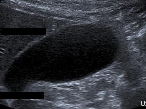
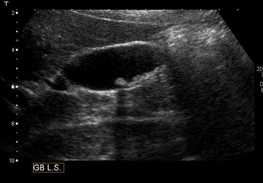
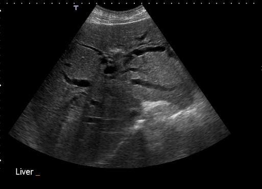
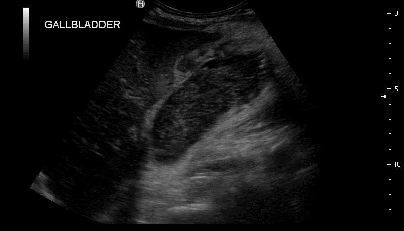
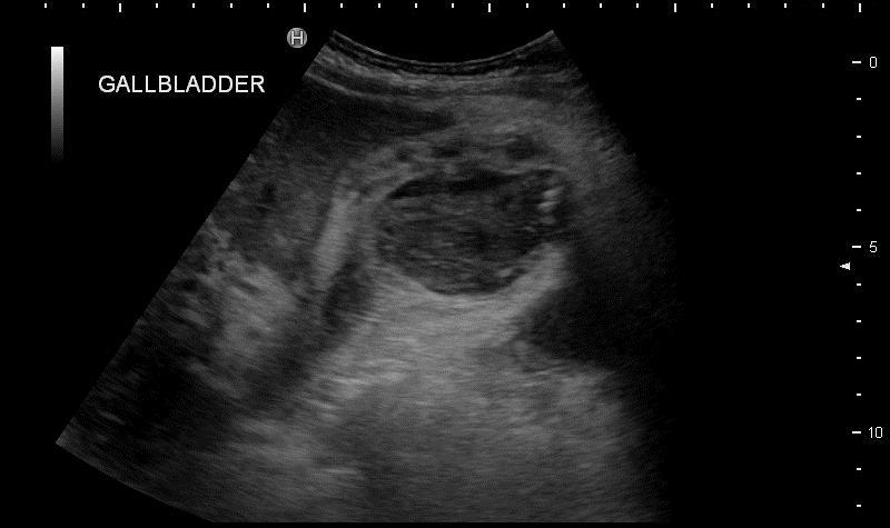
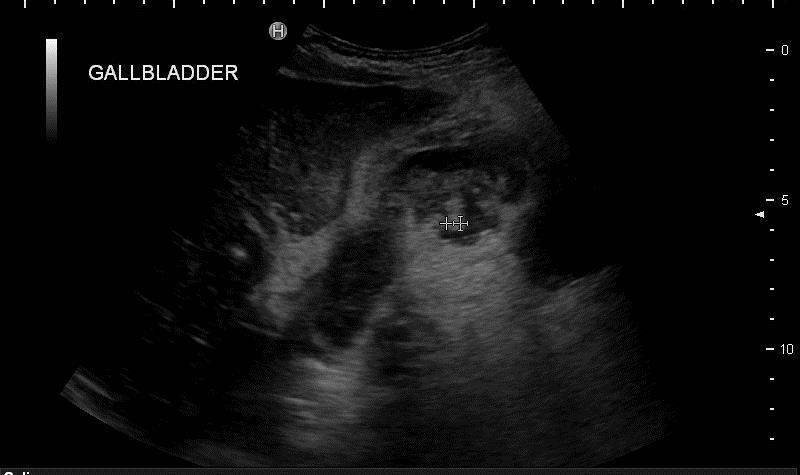
9 Gallbladder The ultrasound appearance of the GB are of a elongated pear-shaped cystic structure. The gallbladder is well delineated and has smooth thin walls

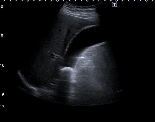
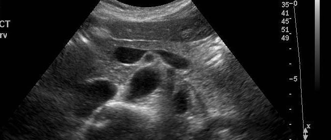
10 Gallbladder Spiral valves are small mucosal folds within the cystic duct **Pitfall alert** Can mimic stones within GB neck

11 Gallbladder The walls are uniform and thin measuring less than 3mm in diameter. 3mm is the upper limit of the normal range. There is no lower limit of normal.

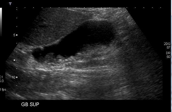
12 Gallbladder Do what we do well, well! Evaluate fully the whole of the gallbladder during every examination. May need to evaluate the neck separately to the fundus.
13 Gallbladder


14 Gallbladder GS can migrate to the fundus, particularly if fold present The fundus has a separate blood supply and this can be reduced, particularly in the elderly Fundus is prone to pathology developing related to chronic cholecystitis leading to adenomyomatosis
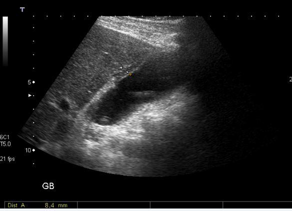
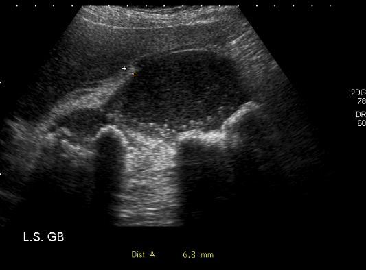
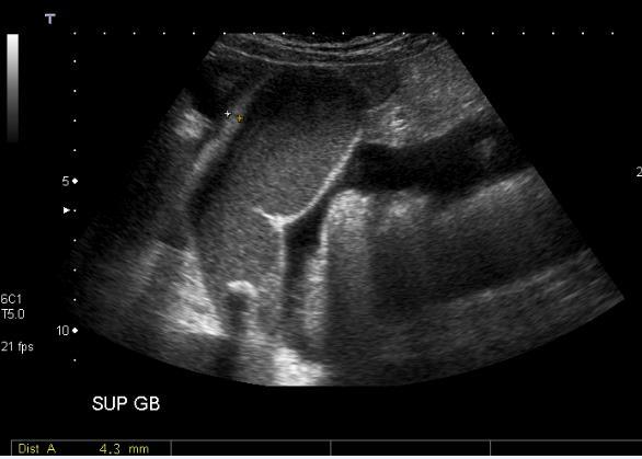
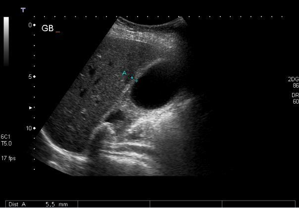
15 Gallbladder Polyps Gallstones Acute Cholecystitis Chronic Cholecystitis Adenomyomatosis Cancer

16 Gallbladder Polyps A gallbladder polyp is defined as any elevated lesion of the mucosal surface of the gallbladder, and as such includes a variety of both benign and malignant entities. Solitary Multiple > 10mm at higher risk of malignant change


17 Gallbladder Polyps Relatively frequent, seen in up to 5% of the population. Frequently identified in patients between 40 and 50 years of age, and are more common in women

18 Gallbladder Polyps 95% of all polyps are benign cholesterol polyps : > 50% of all polyps adenoma : ~ 30%, possibly premalignant inflammatory polyps adenomyomatosis

19 Gallbladder Polyps 5% of all polyps are malignant adenocarcinoma : ~ 90% of malignant polyps other rare entities including metastases to gallbladder squamous cell carcinoma angiosarcoma
20 Gallbladder Polyps Typically polyps are incidentally found on upper abdominal imaging, in patients with upper abdominal discomfort. In most instances polyps are thought to be asymptomatic Surgical management considered if symptomatic and no other cause found The differential for a gallbladder polyp is limited, and includes: gallstone usually mobile, but may be adherent usually casts and acoustic shadow biliary sludge

21 Gallbladder Polyps It is not possible to distinguish an adenoma from an adenocarcinoma The risk of GB malignancy resulting from incidentally detected polyps is extremely low.
22 Gallbladder Polyp Management Any Polyp >9mm should be referred for consideration of Cholecystectomy. Incidental finding of a polyp <9mm in an asymptomatic patient should have a follow up scan in 1 year with 3 caveats. If patient becomes symptomatic within the year they should be referred for consideration of Cholecystectomy, regardless of size of the polyp If Polyp has stayed the same after 1 year can be discharged with advice see GP if becomes symptomatic. If the patient does develop RUQ symptoms they should be referred for consideration of Cholecystectomy, regardless of size of the polyp. If small increase in size, annual follow up until either >9mm, symptomatic or stops growing. Ref Management and follow-up of gallbladder polyps : Joint guidelines between the European Society of Gastrointestinal and Abdominal Radiology (ESGAR), European Association for Endoscopic Surgery and other Interventional Techniques (EAES), International Society of Digestive Surgery - European Federation (EFISDS) and European Society of Gastrointestinal Endoscopy (ESGE). Eur Radiol Sep;27(9):

23 The things we are good at

24 Gallstones 10-15% of people in the adult Western world develop gallstones. 1-4% of asymptomatic cases go on to develop symptoms annually.
25 Gallstones - Presentation Up to 70% of patients with gallstones are asymptomatic at the time of diagnosis. Gallstones may cause acute or chronic cholecystitis, biliary colic, pancreatitis or obstructive jaundice. Biliary colic is the most common presentation, The second most common presentation is acute cholecystitis.
26 Gallstones The adage 'fair, fat, fertile, female and forty' is only part of the story. Other risk factors include: Increasing age. Positive family history. Sudden weight loss - eg, after obesity surgery. Loss of bile salts - eg, ileal resection, terminal ileitis. Diabetes - as part of the metabolic syndrome. Oral contraception - particularly in young women
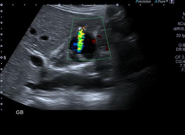
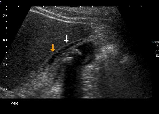
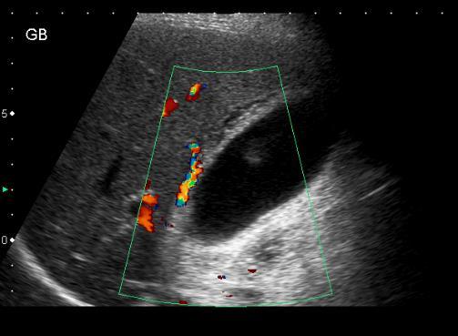
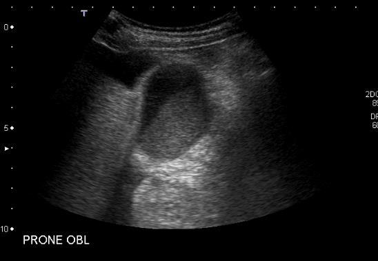
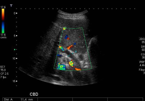
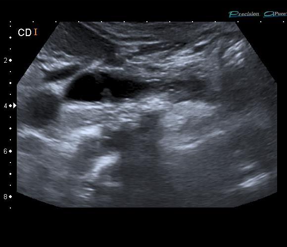
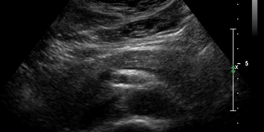
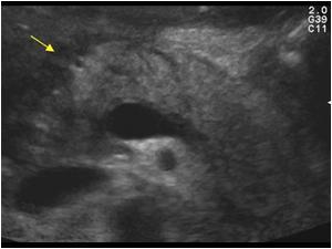
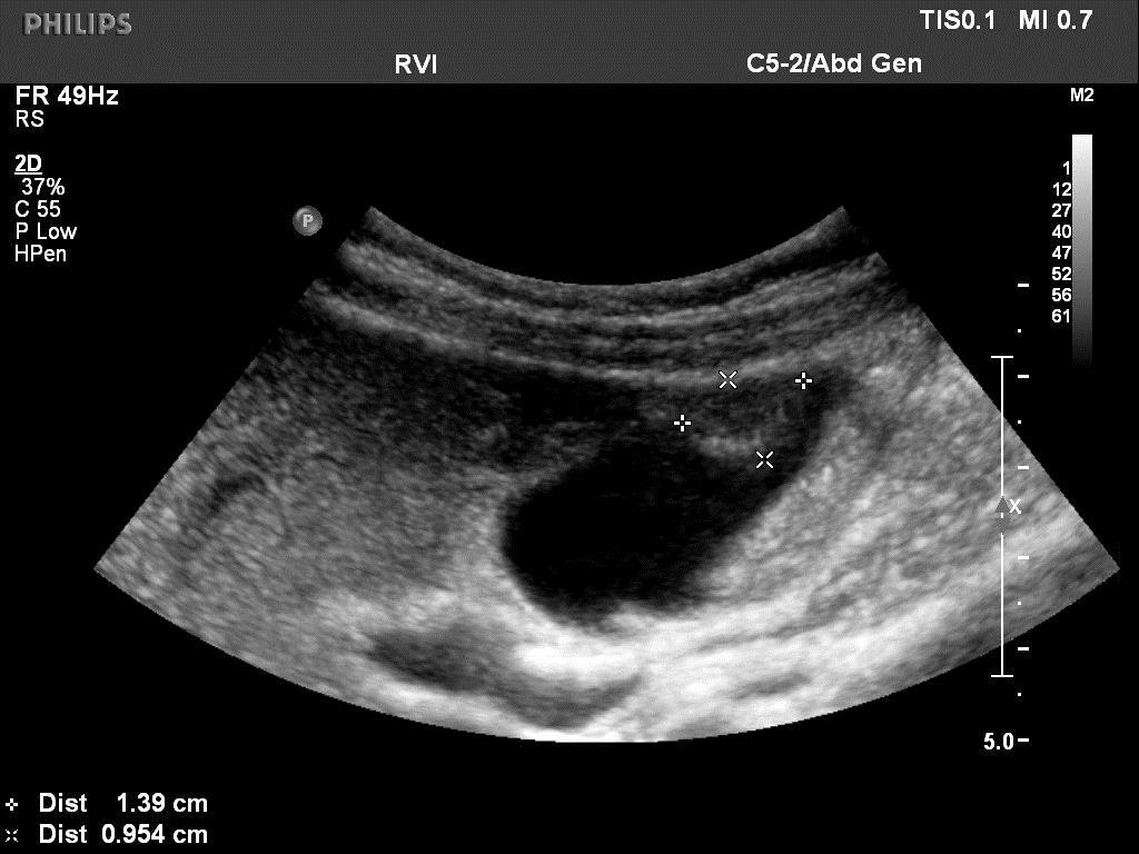
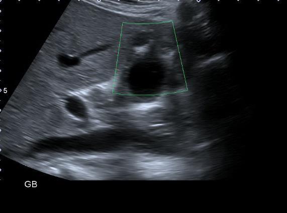
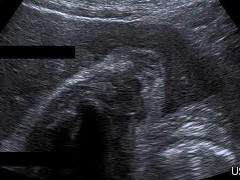
27 Gallstones US features Ultrasound appearances are the same regardless of the composition of the stone. The large acoustic impendence between the stone and the surrounding bile makes them highly reflective. Strong acoustic shadow. * Pitfall Alert* Stones of less than 5mm may not shadow, particularly with the use of compound imaging Moving the patient and observe movement. Colour Doppler can occasionally aid diagnosis as it may produce a twinkle artefact in the stone.
28 Gallstones US features
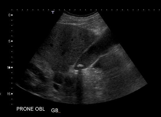
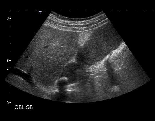
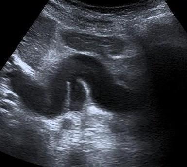
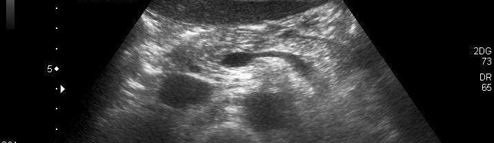
29 Gallstones US features Gallstones? Gallstones?
30 Gallstones? *Pitfall Alert* YES No x Gas filled duodenum
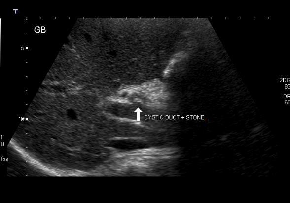
31 Biliary colic Caused by a gallstone impacting in the cystic duct or the ampulla of Vater. The pain starts suddenly in the epigastrium (RUQ) and may radiate around to the back in the interscapular region. Pain persists from 15 minutes up to 24 hours, subsiding spontaneously or with analgesics. Nausea or vomiting often accompanies the pain, Occurs as a result of distension of the gallbladder due to an obstruction or to the passage of a stone through the cystic duct
32 Gallstone Facts Female gallstone subjects had a higher BMI than controls but males did not. Gallstones are twice as common in those with diabetes. A third of elderly patients of both sexes have gallstones but most had not had surgery Gallstones are seldom a cause of death
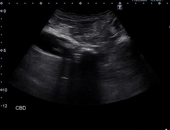
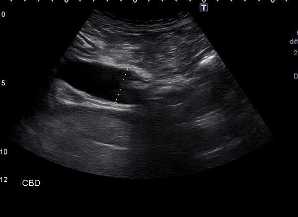
33 Gallstones Investigations US is 90-95% sensitive. Immobile stones misinterpreted as polyps; very small ones may be missed or fail to throw a helpful acoustic shadow. If US negative but there is a high level of suspicion, repeat after an interval; May pick up stones which were previously missed. US can also asses CBD liver and hepatic bile ducts; but it can only identify with certainty about half of any stones in the CBD.
34 Gallstones - Colic

35 Gallstones - Colic CBD stones may occur in % of all patients for whom cholecystectomy is performed
36 Causes of Cholecystitis Gallstones or biliary sludge (95% of patients). Trauma or acute biliary illness (5% acalculus disease). Female gender. Increasing age. Obesity. Rapid weight loss. Pregnancy. Crohn's disease. Hyperlipidaemia
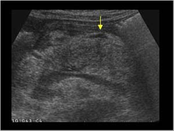
37 Cholecystitis Investigations FBC - the WCC is likely to be raised. Liver enzymes are often mildly abnormal. Ultrasound findings for cholecystitis: Include a thickened GB wall (greater than 3 mm) and may also include pericholecystic fluid or air in the GB or the GB wall. If the GB wall is thickened but there are no gallstones present then the diagnosis could still be acalculous cholecystitis
38 Cholecystitis
39 Additional Findings

40 *Pitfall Alert* Mural oedema, but not cholecystitis
41 Causes of GB wall oedema Physiological o post prandial Inflammatory Adjacent disease Non-inflammatory o adenomyomatosis, cancer, leukaemia, mets Generalised oedema o Ascites, organ failure, portal hypertension Varices
42 Complications of Gallstones Obstructive jaundice Cholangitis Severe Perforated Cholecystitis Pancreatitis Gallstone ileus Chronic Cholecystitis
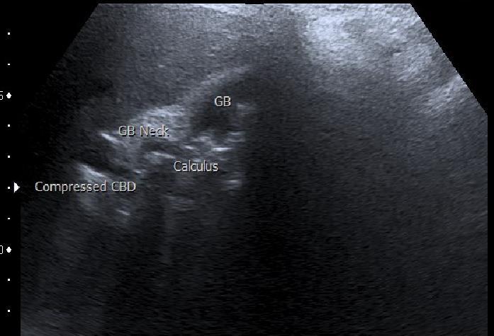
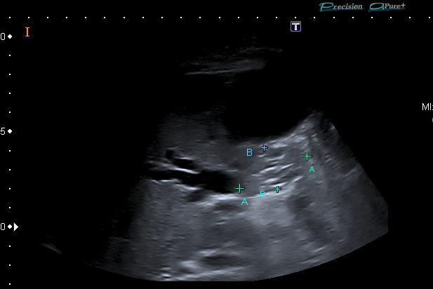
43 Obstructive Jaundice Mirizzi Syndrome

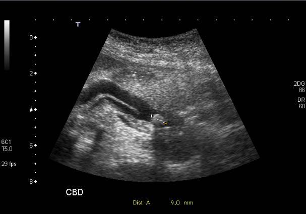
 Should taper to pancreatic")
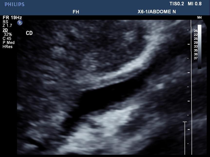
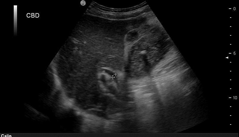
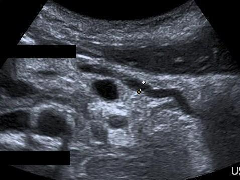
44 *Pitfall Alert* Measuring the duct What is normal? Does size matter? Enlarges with age, bile duct disease (?cholecystectomy) Should taper to pancreatic head Symptomatic vs Incidental
45 Why do we care about a big duct? Because it might represent obstruction Because of the possible causes Cancer Stones Inflammation/Stricture Because of the possible consequences Cholangitis Obstructive jaundice

46 So if you see a big bile duct

47 So if you see a big bile duct
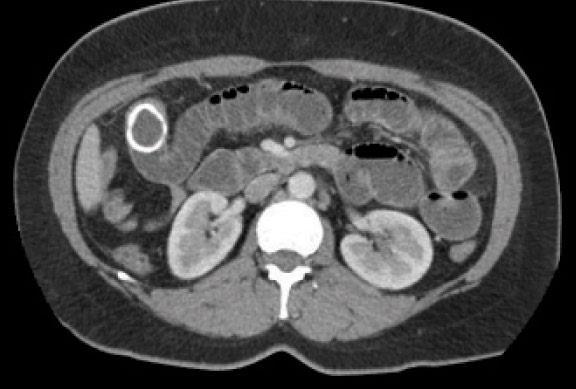
48 What about the incidental big duct? Overall clinical picture Overall radiological picture Intrahepatic ductal dilatation? Good views of pancreas? Old imaging any cross-sectional modality
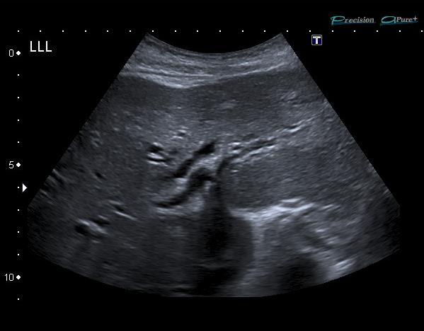
49 Intrahepatic Ducts Complex 3D shape Subjective Confounders Arterial hypertrophy in cirrhotics Saccular dilatation
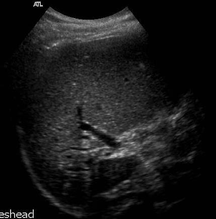
50 Segmental Intrahepatic Biliary Dilatation


51 Liver Cysts v Duct dilatation Central dot sign in Caroli s Disease (dilatation or ectasia of the bile ducts)

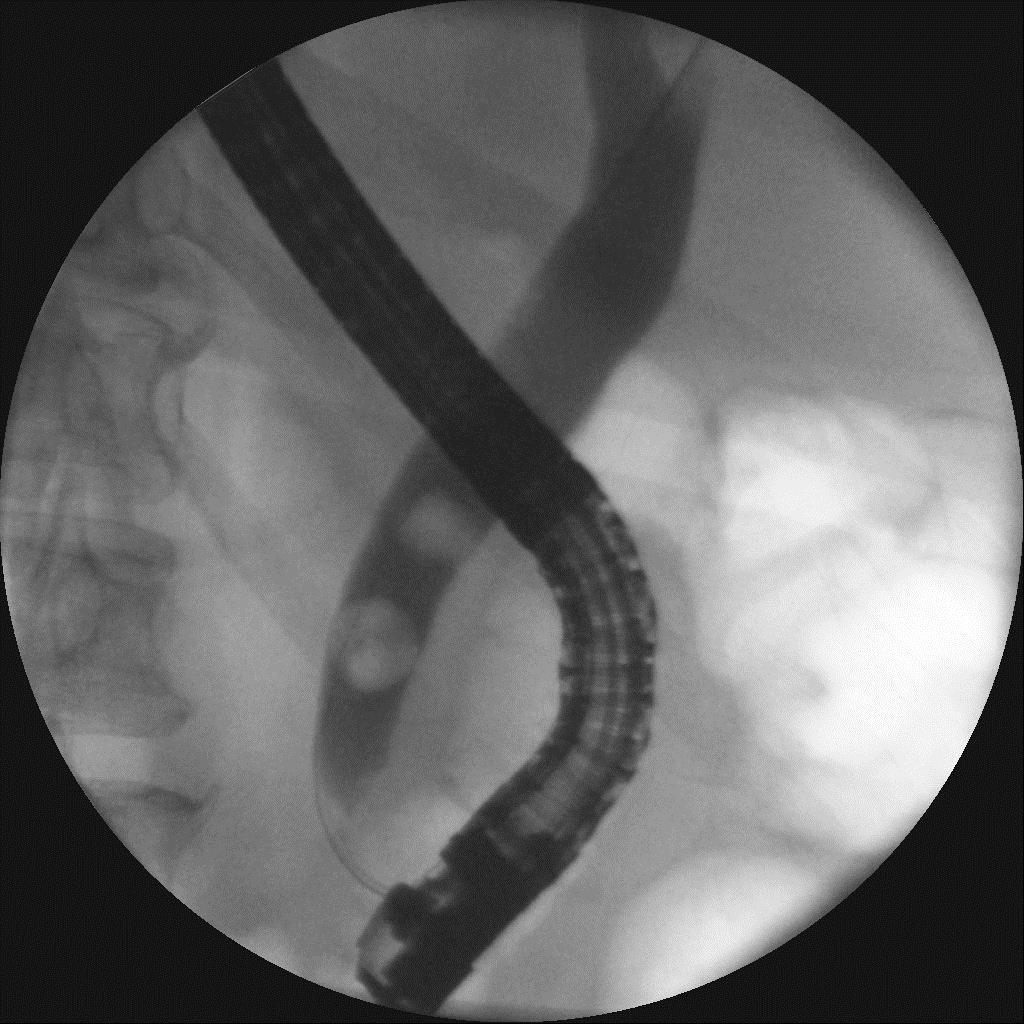
52 Ascending Cholangitis Charcot's triad: Infected CBD leading to jaundice and high swinging fevers with rigors and chills Retrograde infection up the CBD as a result of acute cholecystitis or ERCP
53 Severe, Perforated Cholecystitis
54 Pancreatitis Gallbladder disease and excess alcohol consumption account for most cases and typically cause periductal necrosis. Gallstones cause pancreatitis by blocking the bile duct, causing back pressure in the main pancreatic duct


55 Normal Pancreas 50 Shades of Grey

56 Pancreatitis

57 Gallstone ileus

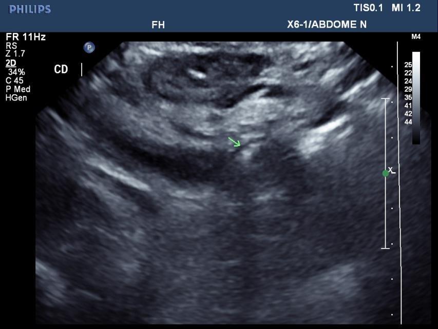
58 Chronic Cholecystitis GB Walls irregularly thickened Hyperechoic Fibrosed Complicated by episodes of acute on chronic cholecystitis


59 Adenomyomatosis Benign condition associated with stone disease and strictures Wall thickening, solid nodules often at the fundus, shadowing from Rokitanski-Achoff sinuses Surgeons tend to take out symptomatic gallbladders
60 Focal Adenomyomatosis
61 Learning Points Clinical History Acute, spasmodic, WCC Previous history Previous cholecystectomy? Gallstones present now what? Wall thickness, oedema, pain Big ducts Why? Intraductal Stones Pancreas Secondary Signs
62 What are you going to report? Clinical history: Admitted RUQ pain, jaundice and puritis. Mr ****** would like an US to look at CBD. Bil 148, ALP 521 and ALT 201. US ABDO The liver appears normal in size and shape with a homogenous echo pattern. No focal lesions identified. Normal hepatopetal flow shown in the portal vein. Pneumobilia again noted. Cholecystectomy noted. The common duct is dilated measuring 17mm and intrahepatic duct dilation is present around the porta. There appear to be some filling defects in the CBD. The right kidney appears normal. Left parapelvic cyst again seen. The pancreas was obscured by overlying bowel. No free fluid. Patient went on to have CT then MRCP
63 Biliary Tree Ultrasound in a Nutshell Fantastic Tricky Common duct Gallbladder and stone disease Pancreatic Cancers (!) Intrahepatic ductal pathology Differentiating cancer from inflammation Handling the incidentally dilated common duct Think beyond the gallstone
Biliary Tree Ultrasound - In a nutshell. Pamela Parker Lead Sonographer
 Biliary Tree Ultrasound - In a nutshell Pamela Parker Lead Sonographer Aims Review what we know about the biliary system Common pathologies Pitfalls Reporting tips The Nutshell Background Biliary examinations
4/9/2018 OBJECTIVES PANCREAOTO BILIARY ULTRASOUND: BEYOND CHOLECYSTITIS
 PANCREAOTO BILIARY ULTRASOUND: BEYOND CHOLECYSTITIS Jean Yves Sewah Kaiser Permanente West Los Angeles 1 OBJECTIVES Discuss the role of ultrasound in the evaluation of the gallbladder, biliary tree and
Radiology of hepatobiliary diseases
 GI cycle - Lecture 14 436 Teams Radiology of hepatobiliary diseases Objectives 1. To Interpret plan x-ray radiograph of abdomen with common pathologies. 2. To know the common pathologies presentation.
Abdominal ultrasound:
 Abdominal ultrasound: Non-traumatic acute abdomen Wittanee Na-ChiangMai, MD Department of Radiology ChiangMai University 26/04/2017 Contents Technique of examination Normal anatomy Emergency conditions
Biliary Ultrasonography Kathleen O Brien MD MPH RDMS Kaiser Permanente South Sacramento
 Biliary Ultrasonography Kathleen O Brien MD MPH RDMS Kaiser Permanente South Sacramento https://www.google.com/search?sa=g&hl=en&q=public+disclosure&tbm=isch&tbs=simg:caqsigeahwelekju2aqaaawlelcmpwgaygpgcamskpib_1qnza7ai
Sonography of Gall Bladder
 Sonography of Gall Bladder Vikram Dogra,MD Professor of Radiology, Urology and BME Director of Ultrasound Associate Chair of Education and Research University of Rochester, NY Objectives Describe the Congenital
Case Study: #3: Gallbladder Carcinoma?
 Case Study: #3: Gallbladder Carcinoma? By: Megan Wyatt K. SON Wyatt 225 2B1 RDMS, RVT Patient: Male 85 YOA Caucasian Indication: Elevated Alkaline Phosphatase History Annual physical showed elevated alkaline
Cholelithiasis & cholecystitis
 1 Cholelithiasis & cholecystitis Dr. Muhammad Shamim FCPS (Pak), FACS (USA), FICS (USA) Assistant Professor, Dept. of Surgery College of Medicine, Prince Sattam bin Abdulaziz University Email: surgeon.shamim@gmail.com
Hepatobiliary Ultrasound Rimon Bengiamin, MD, RDMS Assistant Clinical Professor Director of Emergency Ultrasound UCSF Fresno. Objectives. Why?
 Hepatobiliary Ultrasound Rimon Bengiamin, MD, RDMS Assistant Clinical Professor Director of Emergency Ultrasound UCSF Fresno Objectives Discuss the goals of point-of-care biliary ultrasound Review the
Management of Gallbladder Disease
 Management of Gallbladder Disease Steven B. Johnson, MD, FACS, FCCM Professor and Chairman, Department of Surgery Program Director, Phoenix Integrated Surgical Residency University of Arizona College of
My Patient Has Abdominal Pain PoCUS of the Biliary Tract and the Urinary Tract
 My Patient Has Abdominal Pain PoCUS of the Biliary Tract and the Urinary Tract Objectives PoCUS for Biliary Disease PoCUS for Renal Colic PoCUS for Urinary Retention Biliary Disease A patient presents
Navigating the Biliary Tract with CT & MR: An Imaging Approach to Bile Duct Obstruction
 Navigating the Biliary Tract with CT & MR: An Imaging Approach to Bile Duct Obstruction Ann S. Fulcher, MD Medical College of Virginia Virginia Commonwealth University Richmond, Virginia Objectives To
REFERRAL GUIDELINES: GALLSTONES
 REFERRAL GUIDELINES: GALLSTONES Document Purpose To ensure patients with gallstones disease are managed appropriately in primary/ secondary care Oxford Radcliffe Hospital Surgical Department Surgical Registrar
Management of Gallbladder Disease. Cory Buschmann, MD PGY-5 11/28/2017
 Management of Gallbladder Disease Cory Buschmann, MD PGY-5 11/28/2017 Financial disclosures None Content Scope of gallbladder diseases Evaluation H&P Labs Imaging Cholecystectomy vs cholecystostomy Ancillary
Biliary tree dilation - and now what?
 Biliary tree dilation - and now what? Poster No.: C-1767 Congress: ECR 2012 Type: Educational Exhibit Authors: I. Ferreira, A. B. Ramos, S. Magalhães, M. Certo; Porto/PT Keywords: Pathology, Diagnostic
Vesalius SCALpel : Biliary (see also: biliary/pancreatic folios) Physiology
 Physiology") Vesalius SCALpel : Biliary (see also: biliary/pancreatic folios) Physiology 95% of bile acids reabsorbed; colic and chenodeoxycolic primary bile acids cholecystokinin (CCK) major stimulus of gallbladder
Vesalius SCALpel : Biliary (see also: biliary/pancreatic folios) Physiology 95% of bile acids reabsorbed; colic and chenodeoxycolic primary bile acids cholecystokinin (CCK) major stimulus of gallbladder
Imaging of Biliary Tract Emergencies in Jorge A. Soto, MD Professor of Radiology Boston University Medical Center.
 Imaging of Biliary Tract Emergencies in 2011 Jorge A. Soto, MD Professor of Radiology Boston University Medical Center Introduction Biliary emergencies are: Common Come in many flavors Deceiving: frequent
Hilar cholangiocarcinoma. Frank Wessels, Maarten van Leeuwen, UMCU utrecht
 Hilar cholangiocarcinoma Frank Wessels, Maarten van Leeuwen, UMCU utrecht Content Anatomy Biliary strictures (Hilar) Cholangiocarcinoom Staging Biliary tract 1 st order Ductus hepatica dextra Ductus hepaticus
DISCLAIMER. No Conflict of Interest
 DISCLAIMER No Conflict of Interest EXCLAIMER No Interest in Conflict GALLSTONES FAQs and FACTS John Dunn, FRACS Laparoscopy Auckland YOU GOTTA KNOW THIS STUFF HOW DO THEY FORM? Gallbladder Lithogenic
Imaging of common diseases of hepatobiliary and GI system
 Imaging of common diseases of hepatobiliary and GI system Natthaporn Tanpowpong, M.D. Diagnostic radiology Faculty of Medicine, Chulalongkorn University Normal plain radiograph A = Common bile duct
Certificate in Clinician Performed Ultrasound (CCPU) Syllabus. Biliary
 Syllabus. Biliary") Certificate in Clinician Performed Ultrasound (CCPU) Syllabus Biliary Page 1 of 6 12/18 Biliary Syllabus Purpose: This unit is designed to cover the theoretical and practical curriculum for basic ultrasound
Certificate in Clinician Performed Ultrasound (CCPU) Syllabus Biliary Page 1 of 6 12/18 Biliary Syllabus Purpose: This unit is designed to cover the theoretical and practical curriculum for basic ultrasound
Objectives. Hepatobiliary Ultrasound: Anatomy, Technique, Pathology. RUQ: Normal Anatomy. Emergency Ultrasound: Gallbladder Location
 Hepatobiliary Ultrasound: Anatomy, Technique, Pathology Laleh Gharahbaghian, MD FAAEM Associate Director, EM Ultrasound Co-Director, EM Ultrasound Fellowship Stanford University Medical Center Seric Cusick,
Imaging of liver and pancreas
 Imaging of liver and pancreas.. Disease of the liver Focal liver disease Diffusion liver disease Focal liver disease Benign Cyst Abscess Hemangioma FNH Hepatic adenoma HCC Malignant Fibrolamellar carcinoma
BILIARY TRACT & PANCREAS, PART II
 CME Pretest BILIARY TRACT & PANCREAS, PART II VOLUME 41 1 2015 A pretest is mandatory to earn CME credit on the posttest. The pretest should be completed BEFORE reading the overview. Both tests must be
A patient with an unusual congenital anomaly of the pancreaticobiliary tree
 A patient with an unusual congenital anomaly of the pancreaticobiliary tree Thomas Hocker, HMS IV BIDMC Core Radiology Case Presentation September 17, 2007 Review of Normal Pancreaticobiliary Tract Anatomy
Biliary Tract Disease. Emmet Andrews Cork University Hospital 6 th September 2010
 Biliary Tract Disease Emmet Andrews Cork University Hospital 6 th September 2010 Overview Gallstones Biliary tract tumours Other conditions Acute acalculous cholecystitis Mirizzi s syndrome Primary Biliary
Personal Profile. Name: 劉 XX Gender: Female Age: 53-y/o Past history. Hepatitis B carrier
 Personal Profile Name: 劉 XX Gender: Female Age: 53-y/o Past history Hepatitis B carrier Chief complaint Fever on and off for 2 days Present illness 94.10.14 Sudden onset of epigastric pain 94.10.15 Fever
Case 1. Intro to Gallbladder & Pancreas Pathology. Case 1 DIAGNOSIS??? Acute Cholecystitis. Acute Cholecystitis. Helen Remotti M.D.
 Cholecystitis acute chronic Gallbladder tumors Adenomyoma (benign) Adenocarcinoma Pancreatitis acute chronic Pancreatic tumors Intro to Gallbladder & Pancreas Pathology Helen Remotti M.D. Case 1 70 year
Dr Claire Smith, Consultant Radiologist St James University Hospital Leeds
 Dr Claire Smith, Consultant Radiologist St James University Hospital Leeds Imaging in jaundice and 2ww pathway Image protocol Staging Limitations Pancreatic cancer 1.2.4 Refer people using a suspected
Pictorial review of Benign Biliary tract abnormality on MRCP/MRI Liver with Endoscopic (including splyglass) and Endoscopic Ultrasound correlation
 and Endoscopic Ultrasound correlation") Pictorial review of Benign Biliary tract abnormality on MRCP/MRI Liver with Endoscopic (including splyglass) and Endoscopic Ultrasound correlation Poster No.: C-2617 Congress: ECR 2015 Type: Educational
Pictorial review of Benign Biliary tract abnormality on MRCP/MRI Liver with Endoscopic (including splyglass) and Endoscopic Ultrasound correlation Poster No.: C-2617 Congress: ECR 2015 Type: Educational
Abdominal Ultrasound. Diane Hallinen, MD. Bloodroot
 Abdominal Ultrasound Diane Hallinen, MD Bloodroot Abdominal Ultrasound Vasculature Hepatobiliary Spleen Kidney Bladder Bowel Where to put the probe? Vasculature We are going to talk about Celiac Trunk
Gallbladder & Pancreas Ultrasonography
 복부초음파 : 담낭과췌장 Gallbladder & Pancreas Ultrasonography 김정훈 Department of Radiology 1 Interaction of sound with matter (1) 반사 (Reflection) (2) 굴절 (Refraction) (3) 흡수 (Absorption) (4) 산란 (Scattering) 음향저항
Commissioning Policy Individual Funding Request
 Commissioning Policy Individual Funding Request Laparoscopic Cholecystectomy for Gallstones in Adults Criteria Based Access Policy Date Adopted: 22 December 2017 Version: 1718.3.01 Individual Funding Request
Intraductal papillary mucinous neoplasm of the bile ducts: a rare form of premalignant lesion of invasive cholangiocarcinoma
 Intraductal papillary mucinous neoplasm of the bile ducts: a rare form of premalignant lesion of invasive cholangiocarcinoma Authors: R. Revert Espí, Y. Fernandez Nuñez, I. Carbonell, D. P. Gómez valencia,
Anatomy of the biliary tract
 Harvard-MIT Division of Health Sciences and Technology HST.121: Gastroenterology, Fall 2005 Instructors: Dr. Jonathan Glickman Anatomy of the biliary tract Figure removed due to copyright reasons. Biliary
The Bile Duct (and Pancreas) and the Physician
 and the Physician") The Bile Duct (and Pancreas) and the Physician Javaid Iqbal Consultant in Gastroenterology and Pancreato-biliary Medicine University Hospital South Manchester Not so common?! Two weeks 38 ERCP s 20 15
The Bile Duct (and Pancreas) and the Physician Javaid Iqbal Consultant in Gastroenterology and Pancreato-biliary Medicine University Hospital South Manchester Not so common?! Two weeks 38 ERCP s 20 15
US in non-traumatic acute abdomen. Lalita, M.D. Radiologist Department of radiology Faculty of Medicine ChiangMai university
 US in non-traumatic acute abdomen Lalita, M.D. Radiologist Department of radiology Faculty of Medicine ChiangMai university Sagittal Orientation Transverse (Axial) Orientation Coronal Orientation Intercostal
IT 의료융합 1 차임상세미나 복부질환초음파 이재영
 IT 의료융합 1 차임상세미나 2013-4-3 복부질환초음파 이재영 나는오늘누구를위하여 종을울리나? 전통적의료 의사 공학설계자 의사 최첨단진단장비들 USG, CT, MRI 환자 환자 현대의료 사용자중심의사고 US in the Abdomen Detection DDx Look Behavior Response by external stimuli Guiding Tool
NATIONAL INSTITUTE FOR HEALTH AND CLINICAL EXCELLENCE SCOPE
 Appendix B: Scope NATIONAL INSTITUTE FOR HEALTH AND CLINICAL EXCELLENCE SCOPE Post publication note: The title of this guideline changed during development. This scope was published before the guideline
Appendix 5. EFSUMB Newsletter. Gastroenterological Ultrasound
 EFSUMB Newsletter 87 Examinations should encompass the full range of pathological conditions listed below A log book listing the types of examinations undertaken should be kept Training should usually
Information for Consent Cholecystectomy (Laparoscopic/Open) 膽囊切除術 ( 腹腔鏡 / 開放性 )
 膽囊切除術 ( 腹腔鏡 / 開放性 )") Version 1.0 Page 1 of 3 Information for Consent Cholecystectomy (Laparoscopic/Open) 膽囊切除術 ( 腹腔鏡 / 開放性 ) Introduction Gallbladder is a sac connected to the biliary tree. It serves the function of concentration
Version 1.0 Page 1 of 3 Information for Consent Cholecystectomy (Laparoscopic/Open) 膽囊切除術 ( 腹腔鏡 / 開放性 ) Introduction Gallbladder is a sac connected to the biliary tree. It serves the function of concentration
Elastography in the. technically difficult patient. EPIQ ultrasound system. Ultrasound
 Ultrasound Elastography in the technically difficult patient EPIQ ultrasound system Chairman Department of Diagnostic Radiology Allegheny General Hospital Pittsburgh, PA, USA You can offer more information
Biliary Tract Disease NIKI TADAYON GENERAL & VASCULAR SURGEON SHOHADA TAJRISH HOSPITAL
 Biliary Tract Disease NIKI TADAYON GENERAL & VASCULAR SURGEON SHOHADA TAJRISH HOSPITAL Differential Diagnosis of RUQ pain Gallstone disease (and its related complications) Gastritis/duodenitis Peptic ulcer
The Radiologic Features of Xanthogranulomatous Cholecystitis: An Important Mimic of Gallbladder Carcinoma
 The Radiologic Features of Xanthogranulomatous Cholecystitis: An Important Mimic of Gallbladder Carcinoma Poster No.: C-0691 Congress: ECR 2014 Type: Authors: Keywords: DOI: Educational Exhibit H. L. khosa
Jaundice. Agnieszka Dobrowolska- Zachwieja, MD, PhD
 Jaundice Agnieszka Dobrowolska- Zachwieja, MD, PhD Jaundice definition Jaundice, as in the French jaune, refers to the yellow discoloration of the skin. It arises from the abnormal accumulation of bilirubin
GASTROINTESTINAL IMAGING STUDY GUIDE
 GASTROINTESTINAL IMAGING STUDY GUIDE Pharynx Diverticula Foreign bodies Trauma o Motility Disorders Esophagus Diverticula Trauma Esophagitis Barrett esophagus Rings, webs, and strictures Varices Benign
Congenital dilatation of the common bile duct and pancreaticobiliary maljunction clinical implications
 Langenbecks Arch Surg (2009) 394:209 213 DOI 10.1007/s00423-008-0330-6 CURRENT CONCEPT IN CLINICAL SURGERY Congenital dilatation of the common bile duct and pancreaticobiliary maljunction clinical implications
Cholelithiasis (Gallstones)
") GALL BLADDER Cholelithiasis (Gallstones) Gallstones afflict 10-20% of adult populations in northern hemisphere Western countries. Adult prevalence rates are higher in Latin American countries (20-40%)
GALL BLADDER Cholelithiasis (Gallstones) Gallstones afflict 10-20% of adult populations in northern hemisphere Western countries. Adult prevalence rates are higher in Latin American countries (20-40%)
Bedside RUQ Ultrasound. Replace Formal ULS? Why Bedside ULS RUQ? RUQ Ultrasound. Bedside ULS is Limited, Goal-Directed
 Bedside RUQ Ultrasound RUQ Ultrasound Why do it How to do it Elizabeth Kwan UCSF Emergency Ultrasound Fellow Why Bedside ULS RUQ? Dx or Rule Out Acute Cholecystitis Cholelithiasis, Choledocolithiasis Earlier
Newcastle HPB MDM updated radiology imaging protocol recommendations. Author Dr John Scott. Consultant Radiologist Freeman Hospital
 Newcastle HPB MDM updated radiology imaging protocol recommendations Author Dr John Scott. Consultant Radiologist Freeman Hospital This document is intended as a guide to aid radiologists and clinicians
Pediatric Hepatobiliary, Pancreatic & Splenic US
 Pediatric Hepatobiliary, Pancreatic & Splenic US Susan J. Back, MD Department of Radiology, The Children s Hospital of Philadelphia No Disclosures Objectives Normal Abnormal: cases and US advances Objectives
Hepatobiliary Disease
 Hepatobiliary Disease 大林慈濟綜合醫院 魏昌國 一般外科 Cholelithiasis 膽道結石 Incidence (USA) 10% in adult 85% cholesterol stones 15% pigment stone Risk factors Cholesterol stones Age Female(2x, 25% versus 12% at age 60),
Case Cholecystoduodenal fistula with migrated gallstone leading to gastric outlet obstruction: Bouveret's syndrome
 Case 14613 Cholecystoduodenal fistula with migrated gallstone leading to gastric outlet obstruction: Bouveret's syndrome Eva De Backer 1, Filip Vanhoenacker 2, 3, 4, Adelard De Backer5 1: Ghent University,
Tools of the Gastroenterologist: Introduction to GI Endoscopy
 Tools of the Gastroenterologist: Introduction to GI Endoscopy Objectives Endoscopy Upper endoscopy Colonoscopy Endoscopic retrograde cholangiopancreatography (ERCP) Endoscopic ultrasound (EUS) Endoscopic
Primary Sclerosing Cholangitis and Cholestatic liver diseases. Ahsan M Bhatti MD, FACP Bhatti Gastroenterology Consultants
 Primary Sclerosing Cholangitis and Cholestatic liver diseases Ahsan M Bhatti MD, FACP Bhatti Gastroenterology Consultants I have nothing to disclose Educational Objectives What is PSC? Understand the cholestatic
Disclosures. Overview. Case 1. Common Bile Duct Sizes 10/14/2016. General GI + Advanced Endoscopy: NAFLD/Stones/Pancreatitis
 Disclosures General GI + Advanced Endoscopy: NAFLD/Stones/Pancreatitis 123 Blank Blank, LLC Aldo Maspons, MD Assistant Professor Director of Endoscopy Department of Pediatrics Texas Tech University Health
Biliary Tract Disease HPB Division, Surgery Department of Ramathibodi. Paramin Muangkaew MD.
 Biliary Tract Disease HPB Division, Surgery Department of Ramathibodi Paramin Muangkaew MD. Anatomy Proximal Intrahepatic bile duct IHD Primary Secondary Extrahepatic bile duct Rt & Lt HD CHD Cystic duct
Multiple Primary Quiz
 Multiple Primary Quiz Case 1 A 72 year old man was found to have a 12 mm solid lesion in the pancreatic tail by computed tomography carried out during a routine follow up study of this patient with adult
Case Scenario 1. Discharge Summary
 Case Scenario 1 Discharge Summary A 69-year-old woman was on vacation and noted that she was becoming jaundiced. Two months prior to leaving on that trip, she had had a workup that included an abdominal
Hepatobiliary and Pancreatic Malignancies
 Hepatobiliary and Pancreatic Malignancies Gareth Eeson MD MSc FRCSC Surgical Oncologist and General Surgeon Kelowna General Hospital Interior Health Consultant, Surgical Oncology BC Cancer Agency Centre
What Are Gallstones? GALLSTONES. Gallstones are pieces of hard, solid matter that form over time in. the gallbladder of some people.
 What Are Gallstones? Gallstones are pieces of hard, solid matter that form over time in the gallbladder of some people. The gallbladder sits under the liver and stores bile (a key digestive juice ). Gallstones
Alice Fung, MD Oregon Health and Science University
 Alice Fung, MD Oregon Health and Science University Disclosure Comments The speaker Alice Fung, MD Has relevant financial relationships to disclose. Received honorarium from (Guerbet). This individual
Abdomen and Retroperitoneum Ultrasound Protocols
 Abdomen and Retroperitoneum Ultrasound Protocols Reviewed By: Anna Ellermeier, MD Last Reviewed: March 2018 Contact: (866) 761-4200, Option 1 **NOTE for all examinations: 1. If documenting possible flow
Cholangiocellular carcinoma. Dr. med. Henrik Csaba Horváth PhD
 Cholangiocellular carcinoma Dr. med. Henrik Csaba Horváth PhD Acalculous biliary diseases April 12, 2017 2 Cholangiocarcinoma A slow growing malignancy of the biliary tract which tend - to infiltrate locally
Rokitansky-Aschoff sinuses are epithelial invaginations in the gallbladder wall that from as a result of increased gallbladder pressures.
 Anatomy The complexity of the biliary tree can be broken down into much simpler segments. The intrahepatic ducts converge to form the right and left hepatic ducts which exit the liver and join to become
What Is Pancreatitis?
 What Is Pancreatitis? Pancreatitis is inflammation (swelling) of the pancreas that is most often caused by gallstones or alcohol abuse. There are other causes that your gastroenterologist will look for,
A LEADER IN ADVANCED ENDOSCOPY AND HEPATOBILIARY SURGERY
 A LEADER IN ADVANCED ENDOSCOPY AND HEPATOBILIARY SURGERY St. Peter s Hospital Advanced Endoscopy & Hepatobiliary Center Welcome The St. Peter s Hospital Advanced Endoscopy & Hepatobiliary Center is a leader
The campaign on laboratory: focus on Gallstone Disease and ERCP
 The campaign on laboratory: focus on Gallstone Disease and ERCP Mauro Giuliani, MD, Specialist in Visceral Surgery, Vice Head Physician, Surgical Ward, Ospedale Regionale di Locarno Alberto Fasoli, MD,
CLINICAL PRESENTATION AND RADIOLOGY QUIZ QUESTION
 Donald L. Renfrew, MD Radiology Associates of the Fox Valley, 333 N. Commercial Street, Suite 100, Neenah, WI 54956 8/27/2011 Radiology Quiz of the Week # 35 Page 1 CLINICAL PRESENTATION AND RADIOLOGY
Expected and unexpected gallstones in primary care
 Expected and unexpected gallstones in primary care 7 Speets AM, Van der Graaf Y, Hoes AW, Kalmijn S, De Wit NJ, Mali WPThM. Expected and unexpected gallstones in primary care. Submitted. CHAPTER 7 Abstract
The role for contrast-enhanced ultrasonography outside of focal liver lesions
 The role for contrast-enhanced ultrasonography outside of focal liver lesions Paul S. Sidhu King s College Hospital, London, UK Introduction Contrast-enhanced ultrasonography (US) of focal liver lesions
In The Name of God. Advanced Concept of Nursing- II UNIT- V Advance Nursing Management of GIT diseases. Cholecystitis.
 In The Name of God (A PROJECT OF NEW LIFE HEALTH CARE SOCIETY, KARACHI) Advanced Concept of Nursing- II UNIT- V Advance Nursing Management of GIT diseases. Cholecystitis. Shahzad Bashir RN, BScN, DCHN,MScN
ERCP / PTC Surgical Laparoscopic vs open Timing and order of approach
 Choledocholithiasis Which Approach and When? Lygia Stewart, MD University of California, San Francisco 2010 Naffziger Post-Graduate Course Clinical Manifestations of Choledocholithiasis Asymptomatic (no
Anatomy Jessica Ferguson Ashley Dobos May 31, 2006 LIVER
 Anatomy Jessica Ferguson Ashley Dobos May 31, 2006 LIVER 1) Other Names: Reidel s Lobe normal anatomic variant; projection of the right lobe that can extend as far as the iliac crest (Tempkin, p.54, Anatomy).
The role of endoscopy in the diagnosis and treatment of cystic pancreatic neoplasms
 The role of endoscopy in the diagnosis and treatment of cystic pancreatic neoplasms CYSTIC LESIONS AND FLUID COLLECTIONS OF THE PANCREAS Their pathology ranges from pseudocysts and pancreatic necrosis
Liver Cancer (Hepatocellular Carcinoma or HCC) Overview
 Overview") Liver Cancer (Hepatocellular Carcinoma or HCC) Overview Recent advances in liver cancer care seek to address the rising incidence of liver cancer, which has steadily increased over the past three decades.
Liver Cancer (Hepatocellular Carcinoma or HCC) Overview Recent advances in liver cancer care seek to address the rising incidence of liver cancer, which has steadily increased over the past three decades.
6 th August 2018 Day 1 - Gallbladder & Bile duct Topic
 Venue: Sterling Hospital Auditorium, Sterling Hospitals, Gurukul Road Ahmedabad, Gujarat 6 th August 2018 Day 1 - Gallbladder & Bile duct Registration(8:00am-8:15am) Inauguration(8:15am-8:30am) Welcome
Guidelines, Policies and Statements D5 Statement on Abdominal Scanning
 Guidelines, Policies and Statements D5 Statement on Abdominal Scanning Disclaimer and Copyright The ASUM Standards of Practice Board have made every effort to ensure that this Guideline/Policy/Statement
Abdominal Imaging. Gallbladder perforation: color Doppler findings
 Abdom Imaging 27:47 50 (2002) DOI: 10.1007/s00261-001-0048-1 Abdominal Imaging Springer-Verlag New York Inc. 2002 Gallbladder perforation: color Doppler findings K. Konno, 1 H. Ishida, 1 M. Sato, 1 H.
What can you expect after your ERCP?
 ERCP Explained and respond to bed rest, pain relief and fasting to rest the gut with the patient needing to stay in hospital for only a few days. Some patients develop severe pancreatitis and may require
Evolving Gallstone Ileus. SUNY Downstate Case Conference January 12, 2012
 Evolving Gallstone Ileus SUNY Downstate Case Conference January 12, 2012 Initial Presentation HPI: 90 yo F c 1wk h/o abdominal pain and N/V. Denied F/C. Passing flatus/bm. PMH: DM, HTN, CAD. PSH: C-sections
Intraductal papillary neoplasms in the bile ducts
 Intraductal papillary neoplasms in the bile ducts Seok Hwa Youn Myunghee Yoon Dong Hoon Shin Kosin University Gospel Hospital Department of general surgery Hepato-biliary-pancreatic division Introduction
Abdominal Imaging - 9 Topics in 90 min
 Abdominal Imaging 9 topics in 90 min Antonio C. Westphalen, MD PhD Departments of Radiology and Biomedical Imaging, and Urology Liver Biliary tree Gallbladder Pancreas Kidneys Small bowel Colon Abscess?
Clinics in Diagnostic Imaging (79)
") Singapore Med J 2002 Vol 43(11) : 591-596 M e d i c a l E d u c a t i o n Clinics in Diagnostic Imaging (79) P Lerttumnongtum, M Muttarak, K Wasanavijit 1a Fig. 1a Subcostal US scan taken at the porta
Singapore Med J 2002 Vol 43(11) : 591-596 M e d i c a l E d u c a t i o n Clinics in Diagnostic Imaging (79) P Lerttumnongtum, M Muttarak, K Wasanavijit 1a Fig. 1a Subcostal US scan taken at the porta
Classification of choledochal cyst with MR cholangiopancreatography in children and infants: special reference to type Ic and type IVa cyst
 Classification of choledochal cyst with MR cholangiopancreatography in children and infants: special reference to type Ic and type IVa cyst Poster No.: C-1333 Congress: ECR 2011 Type: Educational Exhibit
Case 1- B.N. 66 yr old F with PMHx of breast cancer s/ p mastectomy, HTN, DM presented with dysphagia to solids and liquids.
 Case 1- B.N 66 yr old F with PMHx of breast cancer s/ p mastectomy, HTN, DM presented with dysphagia to solids and liquids. Reports retching to clear esophagus. Case 1- B.N EGD: Stricture in the distal
Contrast enhanced ultrasound (CEUS) in gallbladder and bile duct pathology: technique, interpretation and clinical applications
 in gallbladder and bile duct pathology: technique, interpretation and clinical applications") Contrast enhanced ultrasound (CEUS) in gallbladder and bile duct pathology: technique, interpretation and clinical applications Poster No.: C-2099 Congress: ECR 2011 Type: Scientific Exhibit Authors: E.
Contrast enhanced ultrasound (CEUS) in gallbladder and bile duct pathology: technique, interpretation and clinical applications Poster No.: C-2099 Congress: ECR 2011 Type: Scientific Exhibit Authors: E.
GALLBLADDER CANCER. Lidie M. Lajoie MD Downstate Surgery M&M July 21, 2011
 GALLBLADDER CANCER Lidie M. Lajoie MD Downstate Surgery M&M July 21, 2011 Agenda Case Presentation Epidemiology Pathogenesis & Pathology Staging Presentation & Diagnosis Stage-wise Management Outcomes/Prognosis
Clinical Spectrum of Presentation of Obstructive Jaundice in Inflammation, Stone Disease, and Malignancy
 Original Article Print ISSN: 2321-6379 Online ISSN: 2321-595X DOI: 10.17354/ijss/2017/326 Clinical Spectrum of Presentation of Obstructive Jaundice in Inflammation, Stone Disease, and Malignancy R Selvasekaran
Appendix 9: Endoscopic Ultrasound in Gastroenterology
 Appendix 9: Endoscopic Ultrasound in Gastroenterology This curriculum is intended for clinicians who perform endoscopic ultrasonography (EUS) in gastroenterology. It includes standards for theoretical
 P R E S E N T S Dr. Mufa T. Ghadiali is skilled in all aspects of General Surgery. His General Surgery Services include: General Surgery Advanced Laparoscopic Surgery Surgical Oncology Gastrointestinal
Colangitis Esclerosante Primaria: Manejo Clínico y Endoscópico
 Colangitis Esclerosante Primaria: Manejo Clínico y Endoscópico Andrés Cárdenas, MD, MMSc, PhD, AGAF, FAASLD GI / Liver Unit, Hospital Clinic Institut de Malalties Digestives i Metaboliques Associate Professor
Northumbria Healthcare NHS Foundation Trust. Laparoscopic Cholecystectomy. Issued by the Department of Upper Gastrointestinal Surgery
 Northumbria Healthcare NHS Foundation Trust Laparoscopic Cholecystectomy Issued by the Department of Upper Gastrointestinal Surgery Laparoscopic Cholecystectomy This leaflet explains why you have been
A CASE REPORT OF SPONTANEOUS BILOMA - AN ENIGMATIC SURGICAL PROBLEM
 A CASE REPORT OF SPONTANEOUS BILOMA - AN ENIGMATIC SURGICAL PROBLEM *Sumanta Kumar Ghosh and Biswajit Mukherjee ESIC Medical College, Joka, Kolkata, India *Author for Correspondence ABSTRACT Occurrence
Background. RUQ Ultrasound Normal, Recommend Clinical Correlation. Sohail R. Shah, MD, MSHA, FACS, FAAP Texas Children s Hosptial
 RUQ Ultrasound Normal, Recommend Clinical Correlation Sohail R. Shah, MD, MSHA, FACS, FAAP Texas Children s Hosptial Background Incidence of pediatric gallbladder disease continues to rise U.S. Pediatric
The Pancreas. Basic Anatomy. Endocrine pancreas. Exocrine pancreas. Pancreas vasculature. Islets of Langerhans. Acinar cells Ductal System
 SGNA: Back to Basics Rogelio G. Silva, MD Assistant Clinical Professor of Medicine University of Illinois at Chicago Department of Medicine Division of Gastroenterology Advocate Christ Medical Center GI
ULTRASOUND AND ABDOMINAL MASSES
 Med. J. Malaysia Vol. 37 No. I March 1982. ULTRASOUND AND ABDOMINAL MASSES AHMAD KAMAL BIN MD ALIF INTRODUCTION It is approximately 30 years since ultrasound was first introduced into the field of medicine,
Gallstones. Classification
 Gallstones Nariman Karanjia Tahir Ali Abstract Gallstones are extremely common in the UK and have a major effect on healthcare resources. Presentation depends on whether the stones occlude the cystic duct
Endoscopic treatment of primary sclerosing cholangitis: Is there something new?
 Endoscopic treatment of primary sclerosing cholangitis: Is there something new? Arnaud Lemmers, MD, PhD Gastroenterology Department, Erasme Hospital, ULB, Brussels BASL December 1st 2017 AGENDA Introduction
Endoscopic Ultrasonography Assessment for Ampullary and Bile Duct Malignancy
 Diagnostic and Therapeutic Endoscopy, Vol. 3, pp. 35-40 Reprints available directly from the publisher Photocopying permitted by license only (C) 1996 OPA (Overseas Publishers Association) Amsterdam B.V.