SPINAL CORD DISEASE IN DOGS PART TWO: MOST LIKELY CAUSES
|
|
|
- Alvin Richardson
- 5 years ago
- Views:
Transcription
1 Vet Times The website for the veterinary profession SPINAL CORD DISEASE IN DOGS PART TWO: MOST LIKELY CAUSES Author : RITA GONÇALVES Categories : Vets Date : April 7, 2014 RITA GONÇALVES in the second part of her article, discusses the reasons why this neurological disorder occurs in canines, the use of imaging to aid diagnosis and treatment options SPINAL cord disease is one of the most common reasons for neurology referral. When assessing these dogs, it is important to perform a thorough physical and neurological examination to localise the origin of the problem, to guide the investigation process as described in part one (VT 44.09). Taking a good history is essential as discussed previously. Most importantly, determining the speed of onset (acute versus chronic) and the presence or absence of pain, as well as establishing if the disease is progressive or not, will be crucial in elaborating this list. To be thorough and consistent, the author likes to follow the vitamin D differential diagnoses list. To make it easier to narrow these down, Table 1 divides them according to whether they cause pain and are progressive. Vascular conditions Ischaemic myelopathy in dogs is most commonly associated with a fibrocartilaginous embolism (FCE). In these cases, a small piece of intervertebral disc penetrates and blocks a spinal blood vessel and results in spinal cord ischaemia. Dogs present with acute onset of neurological deficits (often very lateralised) that are not associated with pain and do not get worse with time. Diagnosis requires MRI and myelography can be used to rule other problems out (Figure 1a and 1b). 1 / 22
2 Treatment mainly involves physiotherapy and hydrotherapy and, occasionally, management of urinary dysfunction in the more affected cases. There is no need for surgery as the injury is due to ischaemic damage to the spinal cord and only requires time for healing to occur. Inflammatory conditions Immune-mediated causes for spinal cord disease are most common, but infectious causes are also possible. For example, discospondylitis, which is an infection of the intervertebral disc space and adjacent vertebrae, typically causes severe spinal pain. Diagnosis is achieved on radiography (remember, it may take two to four weeks for changes to be identifiable with this imaging modality) or MRI if felt this is appropriate in the early stages (Figure 2a). When confirmed, a cause for the infection should be looked for; blood cultures are positive in up to 75 per cent of cases and urine culture in up to 50 per cent. Treatment with antibiotics should be continued for six to eight weeks and initial pain relief is appropriate in most cases. The most common cause of spinal immune-mediated disease is meningomyelitis of unknown origin. In this group of diseases inflammation of the spinal cord occurs. They mostly affect the brain, or both brain and spinal cord, but sometimes only affect the spinal cord. They most commonly present as acute, progressive and usually painful (although not always) myelopathies. It is often confused with intervertebral disc disease as most commonly affect young to middle- aged small breed dogs. Diagnosis is usually made with MRI and, most importantly, analysis of the cerebrospinal fluid (CSF) to document the inflammation. Treatment is made with immunosuppressive drugs (corticosteroids), but supportive care and physiotherapy is essential in the initial stages. Traumatic conditions Traumatic injury may cause different types of lesions that will present in different ways and require very different treatment. The two most common results of spinal trauma are fractures and luxations usually associated with instability, which results in pain and progression of the clinical signs until stabilisation is performed and traumatic disc extrusions, which are mainly associated with spinal cord contusion without compression or significant instability, so is usually not associated with pain and with neurological deficits that improve with time without any treatment. Cases resulting in fractures and luxations typically happen with more significant trauma, such as RTAs or falls from height, and are associated with pain and progression of the clinical signs. It is important to start by performing radiographs of the spine (always orthogonal views) and look for other possible trauma with radiographs of the entire spine, chest and abdomen. 2 / 22
3 It is essential to determine the prognosis at presentation: spinal fractures in the absence of pain perception carry a poor prognosis, with it being usually thought a less than five per cent likelihood of walking again. Treatment can be conservative, such as splinting/cage rest don t forget pain management, or surgical if there is instability. Those cases where the traumatic injury does not result in fracture or luxation, but put the intervertebral disc under significant pressure, result in a traumatic disc extrusion also called high velocity/low volume disc or acute non-compressive nucleus pulposus extrusion (ANNPE). When the disc is healthy (hydrated and not significantly degenerated), there is extrusion of a hydrated disc that hits the spinal cord at high speed and causes mainly spinal cord contusion with minimal associated compression. Often associated with mild trauma (often just exuberant exercise), dogs typically present with acute onset of neurological deficits that are less commonly associated with pain and that do not get worse with time (very similar presentation to FCEs). Diagnosis requires MRI (Figure 1c and 1d), and radiographs and myelography can be used to rule other problems out. Treatment mainly involves physiotherapy and hydrotherapy and, occasionally, management of urinary dysfunction in more affected cases. Surgery is not needed as there is no significant compression of the spinal cord and the injury is mainly contusive damage only requiring time to heal. Anomalous conditions Many anomalous conditions affect the spine and spinal cord, such as block, transitional or hemivertebrae, but they do not always result in neurological deficits only if they cause significant compression of the spinal cord or nerve roots. It is important, therefore, to couple imaging findings, such as anomalous vertebrae on a radiograph, with neurological deficits that localise to that region of the spine. One of the most common malformations associated with significant clinical signs is atlantoaxial instability. This usually affects young dogs that present with acute or waxing and waning episodes of neck pain, with or without ataxia and tetraparesis. It occurs from failure of ligamentous support, usually associated with aplasia/hypoplasia of the dens in toy breeds. Diagnosis is made through radiographs of the cervical spine: the lateral view shows an increased space between the dorsal lamina of the atlas (C1) and the dorsal spinous process of the axis (C2), and the ventrodorsal view will allow assessment of the presence and size of the dens. Treatment can be conservative with splinting for six to 10 weeks or involve surgical stabilisation. Neoplastic conditions 3 / 22
4 Dogs with tumours of the spine and spinal cord characteristically present with slow onset, progressive and usually painful (except in intramedullary lesions) signs of spinal cord dysfunction ( ). Diagnosis can be achieved through spinal radiographs, CT myelography or MRI. Figure 3a Treatment options include surgery, chemotherapy or, rarely, radiotherapy, but most often palliative treatment with analgesia is used. Degenerative conditions There are many degenerative conditions that result in spinal cord dysfunction. They can be associated with degeneration of the intervertebral disc, joints (articular facets of the spine), spinal cord itself and so on. Degenerative intervertebral disc disease type I Degenerative intervertebral disc disease type I (extrusion) occurs when there is herniation of the nucleus pulposus (middle of the disc) through the annular fibres (outer thicker layer of the disc) with extrusion of the nuclear material into the spinal canal. Onset of clinical signs is usually acute and often progressive. Diagnosis can be achieved through myelography (with or without CT) or MRI ( Figure 4a and 4b). Treatment is conservative in many cases, but often involves surgery when the neurological deficits are more severe, in those where conservative treatment has been attempted, but was unsuccessful or in cases associated with severe pain. Degenerative intervertebral disc disease type II Degenerative intervertebral disc disease type II (protrusion) occurs when there is protrusion of the annulus caused by shifting of central nuclear material there is a bulge of the disc that compresses the spinal cord. Protrusions are more common in older, non-chondrodystrophic dogs, which present with chronic, slowly progressive signs, which may or may not include spinal pain. The neurological deficits are often less severe than with type I disease. The diagnosis can be achieved through myelography (with or without CT) or MRI (Figure 4c and 4d). Treatment is conservative in milder cases, but surgery is indicated when the neurological deficits are more severe. Caudal cervical spondylomyelopathy Caudal cervical spondylomyelopathy (wobbler) is typically seen in large breed dogs presenting with progressive ataxia, tetraparesis and sometimes neck pain. The clinical signs are usually worse in the pelvic limbs, with a short, stilted gait in the thoracic limbs. It can result from a combination of 4 / 22
5 problems, but most commonly associated with intervertebral disc protrusion in the caudal cervical spine. Diagnosis can be achieved through myelography (with or without CT) or MRI. Treatment can be conservative in mild cases, but often involves surgery (decompression or distractionstabilisation). Lumbosacral disease In the lumbosacral region the vertebral canal contains only the cauda equina as the spinal cord ends at L6 in most dogs and L7 in cats and small dogs. Therefore, the clinical signs are usually related to pain (often manifested through lameness and reluctance to jump and exercise) and mild weakness of the pelvic limbs. It is most commonly caused by intervertebral disc protrusion at the lumbosacral region, but can also be due to other problems in this region. Diagnosis can be confusing as the clinical signs are often vague and should take into account a combination of the results of the physical, neurological and orthopaedic examinations, as well as those from MRI. It is important to evaluate the nerve roots at this level so myelography is not indicated for these cases. Treatment can be conservative in milder cases, otherwise it involves surgery (decompression or distraction-stabilisation). Degenerative myelopathy Degenerative myelopathy (DM) is a degenerative disease that affects older dogs (usually around eight to 10 years of age), causing chronic onset weakness of the pelvic limbs that slowly progresses over six to 12 months until complete loss of movement. This condition is not associated with pain. Keep in mind many of the breeds that have this condition (such as German shepherd dogs, corgis and boxers) are also commonly affected with chronic intervertebral disc disease, which can present very similarly. Myelography and MRI are normal in these dogs; analysis of the CSF often shows increases in protein levels. There is a genetic test for the mutation (SOD1) associated with DM, but unfortunately a homozygous positive result does not mean the dog has, or will, develop the disease (a negative test though makes it very unlikely). There is no specific treatment available for this condition, but physiotherapy and hydrotherapy help maintain muscle strength and are associated with longer survival. Treatment options Conservative treatment mainly involves rest and appropriate analgesia (NSAIDs, paracetamol and gabapentin are most commonly used). In many cases, physiotherapy and hydrotherapy are indicated, either in cases where there is no compression (FCE, ANNPE), but also post-surgery to improve and speed up recovery. 5 / 22

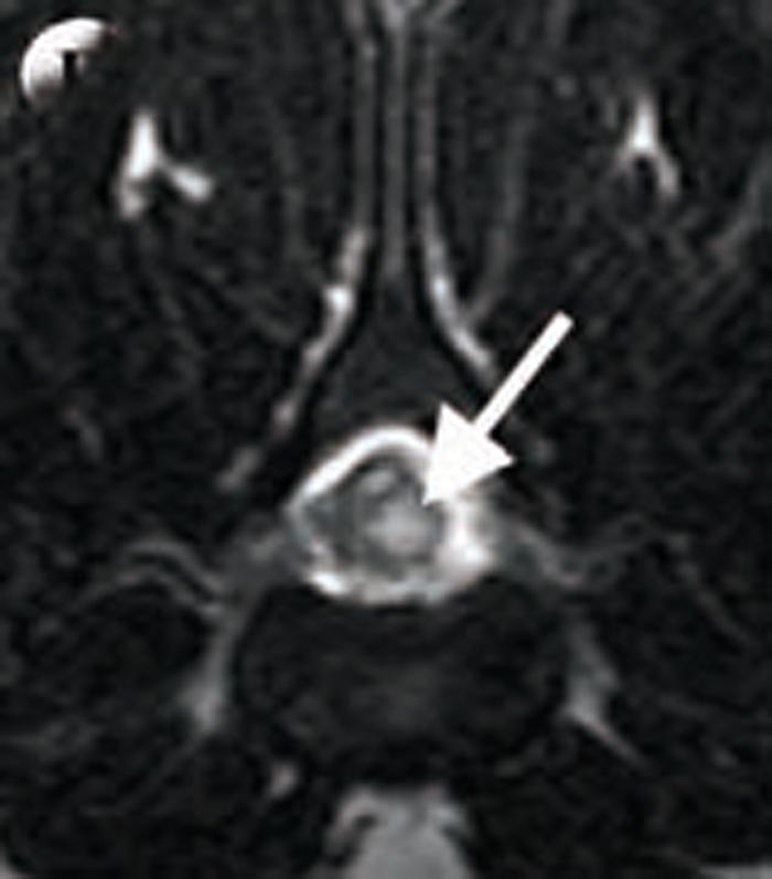
6 In cases where there is compression of the neural tissue, surgery is often indicated (for example, cases with intervertebral disc disease or any other compressive material close to the spinal cord) when these are not improving with conservative treatment or are causing severe deficits. In cases where there is instability, stabilisation of fractures or luxations is often necessary. Note that gabapentin is not licensed for veterinary use. Figure 1. MRIs of non-compressive spinal cord injuries. A (sagittal T2-WI) and B (transverse T2-WI) are MRIs of a dog with a fibrocartilagenous embolism. C (sagittal T2-WI) and D (transverse T2-WI) are MRIs of a dog with a traumatic disc extrusion. It appears similar to the above, but most changes are localised just above the intervertebral disc space. 6 / 22

7 Figure 1. MRIs of non-compressive spinal cord injuries. A (sagittal T2-WI) and B (transverse 7 / 22

8 T2-WI) are MRIs of a dog with a fibrocartilagenous embolism. C (sagittal T2-WI) and D (transverse T2-WI) are MRIs of a dog with a traumatic disc extrusion. It appears similar to the above, but most changes are localised just above the intervertebral disc space. Figure 1. MRIs of non-compressive spinal cord injuries. A (sagittal T2-WI) and B (transverse T2-WI) are MRIs of a dog with a fibrocartilagenous embolism. C (sagittal T2-WI) and D (transverse T2-WI) are MRIs of a dog with a traumatic disc extrusion. It appears similar to the above, but most changes are localised just above the intervertebral disc space. 8 / 22
9 9 / 22
 and D (transverse T2-WI) are MRIs of a dog with a traumatic disc extrusion. It appears similar to the above, but most changes are localised just above the intervertebral disc space.")
10 Figure 1. MRIs of non-compressive spinal cord injuries. A (sagittal T2-WI) and B (transverse T2-WI) are MRIs of a dog with a fibrocartilagenous embolism. C (sagittal T2-WI) and D (transverse T2-WI) are MRIs of a dog with a traumatic disc extrusion. It appears similar to the above, but most changes are localised just above the intervertebral disc space. Figure 2. Lateral radiograph (A) and sagittal T1-WI image (B) of two dogs with discospondylitis. Note the narrowing of the intervertebral disc space, and irregularity and lysis of the endplates, as well as development of spondylosis. 10 / 22

11 Figure 2. Lateral radiograph (A) and sagittal T1-WI image (B) of two dogs with discospondylitis. Note the narrowing of the intervertebral disc space, and irregularity and lysis of the endplates, as well as development of spondylosis. 11 / 22
12 Figure 3. Spinal neoplasia. A: normal spinal cord. B: extradural neoplasia compressing the spinal cord (soft tissue sarcoma). C: intradural extramedullary neoplasia (meningioma). D: intradural intramedullary neoplasia (glioma). 12 / 22
13 Figure 3. Spinal neoplasia. A: normal spinal cord. B: extradural neoplasia compressing the spinal cord (soft tissue sarcoma). C: intradural extramedullary neoplasia (meningioma). D: intradural intramedullary neoplasia (glioma). 13 / 22
.")
14 Figure 3. Spinal neoplasia. A: normal spinal cord. B: extradural neoplasia compressing the spinal cord (soft tissue sarcoma). C: intradural extramedullary neoplasia (meningioma). D: intradural intramedullary neoplasia (glioma). 14 / 22
.")
15 Figure 3. Spinal neoplasia. A: normal spinal cord. B: extradural neoplasia compressing the spinal cord (soft tissue sarcoma). C: intradural extramedullary neoplasia (meningioma). D: intradural intramedullary neoplasia (glioma). 15 / 22
 and D (transverse T2-WI) are MRIs of a dog with IVDD type II.")
16 Figure 4. Degenerative intervertebral disc disease (IVDD). A (sagittal T2-WI) and B (transverse T2-WI) are MRIs of a dog with IVDD type I. Note the disc material has extruded to the spinal canal and is causing significant compression of the spinal cord. C (sagittal T2-WI) and D (transverse T2-WI) are MRIs of a dog with IVDD type II. Note the disc is bulging as a whole and causing milder compression of the spinal cord. 16 / 22
 and B (transverse T2-WI) are MRIs of a dog with IVDD type I.")
17 Figure 4. Degenerative intervertebral disc disease (IVDD). A (sagittal T2-WI) and B (transverse T2-WI) are MRIs of a dog with IVDD type I. Note the disc material has extruded to the spinal canal 17 / 22
.")
18 and is causing significant compression of the spinal cord. C (sagittal T2-WI) and D (transverse T2-WI) are MRIs of a dog with IVDD type II. Note the disc is bulging as a whole and causing milder compression of the spinal cord. Figure 4. Degenerative intervertebral disc disease (IVDD). A (sagittal T2-WI) and B (transverse T2-WI) are MRIs of a dog with IVDD type I. Note the disc material has extruded to the spinal canal and is causing significant compression of the spinal cord. C (sagittal T2-WI) and D (transverse T2-WI) are MRIs of a dog with IVDD type II. Note the disc is bulging as a whole and causing milder compression of the spinal cord. 18 / 22
19 19 / 22

20 20 / 22
 and D (transverse T2-WI) are MRIs of a dog with IVDD type II.")
21 Figure 4. Degenerative intervertebral disc disease (IVDD). A (sagittal T2-WI) and B (transverse T2-WI) are MRIs of a dog with IVDD type I. Note the disc material has extruded to the spinal canal and is causing significant compression of the spinal cord. C (sagittal T2-WI) and D (transverse T2-WI) are MRIs of a dog with IVDD type II. Note the disc is bulging as a whole and causing milder compression of the spinal cord. Table 1. Possible aetiologies of spinal cord disease 21 / 22
22 22 / 22 Powered by TCPDF (
Treating neck pain in dogs neurological five-step approach
 Vet Times The website for the veterinary profession https://www.vettimes.co.uk Treating neck pain in dogs neurological five-step approach Author : Johnny (Ioannis) Plessas Categories : Canine, Vets Date
CANINE LUMBOSACRAL DISEASE
 Vet Times The website for the veterinary profession https://www.vettimes.co.uk CANINE LUMBOSACRAL DISEASE Author : Brent Higgins Categories : Vets Date : April 6, 2009 Brent Higgins discusses differing
This article appeared in a journal published by Elsevier. The attached copy is furnished to the author for internal non-commercial research and
 This article appeared in a journal published by Elsevier. The attached copy is furnished to the author for internal non-commercial research and education use, including for instruction at the authors institution
Radiography of the Spine
 Radiography of the Spine Radiography of the Spine Attila ARANY-TóTH, DVM Complex anatomy Vertebrae: 7 cervical, 13 thoracal, 7 lumbal, 3 sacral, n caudal Thorough neurological examination - localization!!!
Fibrocartilaginous embolic myelopathy and traumatic IVDE
 Fibrocartilaginous embolic myelopathy and traumatic IVDE Luisa De Risio DVM, MRCVS, PhD, Dipl ECVN, RCVS recognised specialist in veterinary neurology Head of Neurology/ Neurosurgery Animal Health Trust
Proceedings of the 33rd World Small Animal Veterinary Congress
 www.ivis.org Proceedings of the 33rd World Small Animal Veterinary Congress Dublin, Ireland - 2008 Next WSAVA Congress : Reprinted in IVIS with the permission of the Congress Organizers 20 Neurology Com
NEUROLOGICAL EXAMINATIONS: LOCALISATION AND GRADING
 Vet Times The website for the veterinary profession https://www.vettimes.co.uk NEUROLOGICAL EXAMINATIONS: LOCALISATION AND GRADING Author : MARK LOWRIE Categories : Vets Date : June 16, 2014 MARK LOWRIE
What s Your Diagnosis? Lindsay Banks, Class of Murphy 9 year old M/C Dachshund. History:
 What s Your Diagnosis? Lindsay Banks, Class of 2011 Murphy 9 year old M/C Dachshund History: Presented to KSU Veterinary Medical Teaching Hospital with cervical neck pain Prior to presentation, Murphy
Clinical approach to the adult Doberman Pinschers with cervical spondylomyelopathy ( wobbler syndrome )
") Clinical approach to the adult Doberman Pinschers with cervical spondylomyelopathy ( wobbler syndrome ) Dr Decker Steven, DVM, PhD, MvetMed, MRCVS Department of Veterinary Clinical Sciences, Royal Veterinary
Clinical approach to the adult Doberman Pinschers with cervical spondylomyelopathy ( wobbler syndrome ) Dr Decker Steven, DVM, PhD, MvetMed, MRCVS Department of Veterinary Clinical Sciences, Royal Veterinary
WHEN IS A SPINAL NOT A DISC PROLAPSE?
 WHEN IS A SPINAL NOT A DISC PROLAPSE? Dr Sara Boyd Johannesburg Specialist Veterinary Centre 63 Kayburne Venue Randpark Ridge Email: sara.boyd@jsvc.co.za ABSTRACT Dogs showing the early signs of spinal
Spinal diseases: non-surgical options
 Vet Times The website for the veterinary profession https://www.vettimes.co.uk Spinal diseases: non-surgical options Author : MARIANNE STABAEK MARTIN Categories : Vets Date : June 2, 2008 MARIANNE STABAEK
Thoracolumbar Intervertebral Disk Disease Basics
 Thoracolumbar Intervertebral Disk Disease Basics OVERVIEW The spine is composed of multiple bones (vertebrae) with disks (intervertebral disks) located in between adjacent bones; the disks act as shock
Spine. Neuroradiology. Spine. Spine Pathology. Distribution of fractures. Radiological algorithm. Role of radiology 18/11/2015
 Spine Neuroradiology Spine Prof.Dr.Nail Bulakbaşı X Ray: AP/L/Oblique Vertebra & disc spaces CT & CTA Vertebra, discs, vessels MRI & MRA Vertebra, disc, vessels, meninges Spinal cord & nerves Myelography
NEURORADIOLOGY. Part III. Angela Csomor University of Szeged Department of Radiology
 NEURORADIOLOGY Part III Angela Csomor University of Szeged Department of Radiology DISEASES OF SPINE AND SPINAL CORD I. Non-tumourous diseases developmental anomalies vascular disorders inflammatory processes
HERNIATED DISCS AN INTRODUCTION TO
 AN INTRODUCTION TO HERNIATED S This booklet provides general information on herniated discs. It is not meant to replace any personal conversations that you might wish to have with your physician or other
Degenerative Disease of the Spine
 Degenerative Disease of the Spine Introduction: I. Anatomy Talk Overview II. Overview of Disease Processes: A. Spondylosis B. Intervertebral Disc Disease III. Diagnosis IV. Therapy Introduction: Myelopathy
Acute Thoracolumbar IVD Extrusion. Tracy Sutton, DVM, DACVIM (Neurology)
") Acute Thoracolumbar IVD Extrusion Tracy Sutton, DVM, DACVIM (Neurology) CONTACT INFORMATION Austin Veterinary Emergency Specialty Center (AVES) 7300 Ranch Road 2222, Austin, TX 78730 (512) 343-2837 DrSutton@AustinVets.com
Acute Thoracolumbar IVD Extrusion Tracy Sutton, DVM, DACVIM (Neurology) CONTACT INFORMATION Austin Veterinary Emergency Specialty Center (AVES) 7300 Ranch Road 2222, Austin, TX 78730 (512) 343-2837 DrSutton@AustinVets.com
8/31/2018 IMPORTANT CONSIDERATIONS. Signalment History Symmetry Progression of signs Painful vs non-painful SURGICAL CONSIDERATIONS
 IMPORTANT CONSIDERATIONS Signalment History Symmetry Progression of signs Painful vs non-painful SURGICAL CONSIDERATIONS Specific region of TL spine Differences in size and shape of articular processes
Wobbler Syndrome: A Review and New Advanced Treatment Options.
 Wobbler Syndrome: A Review and New Advanced Treatment Options. PVMA meeting April 20, 2010 Filippo Adamo, DVM, Dipl. ECVN, President Bay Area VNC (Veterinary Neurology Neurosurgery Consulting) San Mateo,
1. General principles. Goal : 17/07/2017 INDEX: Afecções da medula espinhal de felinos
 Afecções da medula espinhal de felinos Gualtiero Gandini Describe the most common diseases affecting the spinal cord of the cat INDEX: 1. General principles 2. Vitamin D and feline spinal cord disorders
RADICULOPATHY AN INTRODUCTION TO
 AN INTRODUCTION TO RADICULOPATHY This booklet provides general information on radiculopathy. It is not meant to replace any personal conversations that you might wish to have with your physician or other
CT FINDINGS OF THORACOLUMBAR SPINE LESIONS IN DOGS
 CT FINDINGS OF THORACOLUMBAR SPINE LESIONS IN DOGS C. DARABAN 1, V. VULPE 1, FLORENTINA BOCĂNEŢI 1, GIUSEPPINA MENNONNA 2, M. SACCONE 2, G. FATONE 2, L. MEOMARTINO 2 1 University of Agriculture Science
Cervical intervertebral disc disease Degenerative diseases F 04
 Cervical intervertebral disc disease Degenerative diseases F 04 How is a herniated cervical intervertebral disc treated? Conservative treatment is generally sufficient for mild symptoms not complicated
Spinal Cord Disease appearance and differentials. MG Young DVM,MS, DACVIM
 Spinal Cord Disease appearance and differentials MG Young DVM,MS, DACVIM Localization There are four localizations: these are derived from the changes to UMN or LMN signs to either thoracic or pelvic limbs.
Standards of Care (How I treat) INTERVERTEBRAL DISC DISEASE
 INTERVERTEBRAL DISC DISEASE") Standards of Care (How I treat) INTERVERTEBRAL DISC DISEASE Richard A. LeCouteur, BVSc, PhD, Diplomate ACVIM (Neurology), Diplomate ECVN University of California Davis CA 95616 USA ralecouteur@ucdaviss.edu
Standards of Care (How I treat) INTERVERTEBRAL DISC DISEASE Richard A. LeCouteur, BVSc, PhD, Diplomate ACVIM (Neurology), Diplomate ECVN University of California Davis CA 95616 USA ralecouteur@ucdaviss.edu
Surgical Considerations of Thoracolumbar Intervertebral Disk Disease Pathogenesis Clinical Signs Diagnosis Survey spinal radiography Myelography
 Surgical Considerations of Thoracolumbar Intervertebral Disk Disease Joan R. Coates, DVM, MS, Diplomate ACVIM (Neurology) Associate Professor, Department of Veterinary Medicine and Surgery University of
International Journal of Science, Environment and Technology, Vol. 6, No 1, 2017,
 International Journal of Science, Environment and Technology, Vol. 6, No 1, 2017, 191 198 ISSN 2278-3687 (O) 2277-663X (P) EVALUATION OF RADIOLOGICAL FINDINGS OF DOGS WITH THORACOLUMBAR DISORDERS Thanigaivel
Spinal canal stenosis Degenerative diseases F 06
 What is spinal canal stenosis? The condition known as spinal canal stenosis is a narrowing (stenosis) of the spinal canal that in most cases develops due to the degenerative (wear-induced) deformation
Properties of Purdue. Anatomy. Positioning AXIAL SKELETAL RADIOLOGY FOR PRIVATE PRACTITIONERS 11/30/2018
 AXIAL SKELETAL RADIOLOGY FOR PRIVATE PRACTITIONERS Anatomy Complex Text book is needed Species Contrast Positioning Painful/ non cooperative Sedation General anesthesia Species Contrast 1 Slightly oblique
Nursing the spinal patient
 Vet Times The website for the veterinary profession https://www.vettimes.co.uk Nursing the spinal patient Author : Lisa Thompson Categories : RVNs Date : November 1, 2009 Lisa Thompson DipAVN(surg), looks
RETROLISTHESIS. Retrolisthesis. is found mainly in the cervical spine and lumbar region but can also be often seen in the thoracic spine
 RETROLISTHESIS A retrolisthesis is a posterior displacement of one vertebral body with respect to adjacent vertebrae Typically a vertebra is to be in retrolisthesis position when it translates backward
NECK AND BACK PAIN AN INTRODUCTION TO
 AN INTRODUCTION TO NECK AND BACK PAIN This booklet provides general information on neck and back pain. It is not meant to replace any personal conversations that you might wish to have with your physician
Acute Spinal Cord Myelopathy
 Acute Spinal Cord Myelopathy Acute Non-compressive Nucleus Pulposus Extrusion (ANNPE) and Fibrocartilaginous Embolism (FCE) Mary Stallings, DVM Neurology Intern BVNS - Richmond November 5, 2017 Overview
VERTEBRAL COLUMN ANATOMY IN CNS COURSE
 VERTEBRAL COLUMN ANATOMY IN CNS COURSE Vertebral body Sections of the spine Atlas (C1) Axis (C2) What type of joint is formed between atlas and axis? Pivot joint What name is given to a fracture of both
Epidemiology of Low back pain
 Low Back Pain Definition Pain felt in your lower back may come from the spine, muscles, nerves, or other structures in that region. It may also radiate from other areas like the mid or upper back, a inguinal
It consist of two components: the outer, laminar fibrous container (or annulus), and the inner, semifluid mass (the nucleus pulposus).
, and the inner, semifluid mass (the nucleus pulposus).") Lumbar Spine The lumbar vertebrae are the last five vertebrae of the vertebral column. They are particularly large and heavy when compared with the vertebrae of the cervical or thoracicc spine. Their bodies
Lumbar Spine The lumbar vertebrae are the last five vertebrae of the vertebral column. They are particularly large and heavy when compared with the vertebrae of the cervical or thoracicc spine. Their bodies
ACDF. Anterior Cervical Discectomy and Fusion. An introduction to
 An introduction to ACDF Anterior Cervical Discectomy and Fusion This booklet provides general information on ACDF. It is not meant to replace any personal conversations that you might wish to have with
Comprehension of the common spine disorder.
 Objectives Comprehension of the common spine disorder. Disc degeneration/hernia. Spinal stenosis. Common spinal deformity (Spondylolisthesis, Scoliosis). Osteoporotic fracture. Anatomy Anatomy Anatomy
Discospondylitis in dogs: a review
 Vet Times The website for the veterinary profession https://www.vettimes.co.uk Discospondylitis in dogs: a review Author : Luca Motta Categories : Vets Date : August 1, 2009 Luca Motta discusses possible
A Patient s Guide to Artificial Cervical Disc Replacement
 A Patient s Guide to Artificial Cervical Disc Replacement Each year, hundreds of thousands of adults are diagnosed with Cervical Disc Degeneration, an upper spine condition that can cause pain and numbness
1 Normal Anatomy and Variants
 1 Normal Anatomy and Variants 1.1 Normal Anatomy MR Technique. e standard MR protocol for a routine evaluation of the spine always comprises imaging in sagittal and axial planes, while coronal images are
PARADIGM SPINE. Patient Information. Treatment of a Narrow Lumbar Spinal Canal
 PARADIGM SPINE Patient Information Treatment of a Narrow Lumbar Spinal Canal Dear Patient, This brochure is intended to inform you of a possible treatment option for narrowing of the spinal canal, often
Vertebral Column. Backbone consists of 26 vertebrae. Five vertebral regions. Cervical
 Vertebral Column Backbone consists of 26 vertebrae. Five vertebral regions Cervical vertebrae (7) in the neck. Thoracic vertebrae (12) in the thorax. Lumbar vertebrae (5) in the lower back. Sacrum (5,
Common Thoraco- Lumbar Problems in the Mature Athlete
 Common Thoraco- Lumbar Problems in the Mature Athlete Diana Heiman, MD Associate Professor, Family Medicine Residency Director East Tennessee State University Objectives Review the pathophysiology of the
Lumbar Disc Prolapse. Dr. Ahmed Salah Eldin Hassan. Professor of Neurosurgery & Consultant spinal surgeon
 Lumbar Disc Prolapse By Dr. Ahmed Salah Eldin Hassan Professor of Neurosurgery & Consultant spinal surgeon 1-What are the Functions of the Spine Structural support for upright posture Protection of Spinal
2. The vertebral arch is composed of pedicles (projecting from the body) and laminae (uniting arch posteriorly).
 and laminae (uniting arch posteriorly).") VERTEBRAL COLUMN 2018zillmusom I. VERTEBRAL COLUMN - functions to support weight of body and protect spinal cord while permitting movements of trunk and providing for muscle attachments. A. Typical vertebra
VERTEBRAL COLUMN 2018zillmusom I. VERTEBRAL COLUMN - functions to support weight of body and protect spinal cord while permitting movements of trunk and providing for muscle attachments. A. Typical vertebra
8/4/2012. Causes and Cures. Nucleus pulposus. Annulus fibrosis. Vertebral end plate % water. Deforms under pressure
 Causes and Cures Intervertebral discs Facet (zygopophyseal) joints Inter body joints Spinal nerve roots Nerve compression Pathological conditions Video Causes of back pain Nucleus pulposus Annulus fibrosis
Wobbler Syndrome in dogs. Pathogenesis and Diagnosis. Part 1 P. Filippo Adamo, DVM, DECVN, San Mateo, CA, USA
 Wobbler Syndrome in dogs. Pathogenesis and Diagnosis. Part 1 P. Filippo Adamo, DVM, DECVN, San Mateo, CA, USA Abstract Wobbler syndrome in dogs refers to a disorder of the cervical vertebrae and intervertebral
Fecal Incontinence. Inability to retain feces or bowel movements, resulting in involuntary passage of feces or bowel movements
 Fecal Incontinence (Involuntary Passage of Feces or Bowel Movements) Basics OVERVIEW Inability to retain feces or bowel movements, resulting in involuntary passage of feces or bowel movements GENETICS
DEGENERATIVE DISC DISEASE
 DEGENERATIVE DISC DISEASE What is a disc, and what is its purpose? The spinal cord is one of the most important and most sensitive organs in the body. If it is damaged the nerve cells do not regenerate
The surgical treatment of metastatic disease of the spine
 The surgical treatment of metastatic disease of the spine Péter Banczerowski National Institute of Neurosurgery, Budapest Spine tumours 15% of the primary tumours of the CNS affect the spine The spine
Introduction to Neuroimaging spine. John J. McCormick MD
 Introduction to Neuroimaging spine John J. McCormick MD Neuroanatomy Netter drawings Radiographic Anatomy Cervical Spine Cervical Spine Oblique View Cervical Spine Dens View Thoracic Spine Lumbar Spine
Intervertebral Disc Disease and Nursing Care of the Down Dog. Deanna M. Swartzfager, RVT
 Intervertebral Disc Disease and Nursing Care of the Down Dog Deanna M. Swartzfager, RVT Intervertebral Disc Disease (IVDD) Syndrome of pain and neurologic deficits, and sometimes complete paralysis, resulting
Peggers Super Summaries: The Aging Spine
 Aging Spine: AGING PROCESS Osteopenia 10% of 50 year old males and 25% of 50 year females Disc dehydration Facet degeneration Soft tissue hypertrophy 2 0 deformity Leg pain worse than back pain from nerve
Objectives. Comprehension of the common spine disorder
 Objectives Comprehension of the common spine disorder Disc degeneration/hernia Spinal stenosis Common spinal deformity (Spondylolisthesis, Scoliosis) Osteoporotic fracture Destructive spinal lesions Anatomy
22110 vertebral segment; cervical vertebral segment; thoracic vertebral segment; lumbar
 The following codes are authorized by Palladian Health for applicable product lines. Visit palladianhealth.com to request authorization and to access guidelines. Palladian Musculoskeletal Program Codes
CERVICAL SPONDYLOSIS & CERVICAL DISC DISEASE
 CERVICAL SPONDYLOSIS & CERVICAL DISC DISEASE Cervical spondylosis l Cervical osteophytosis l Most common progressive disease in the aging cervical spine l Seen in 95% of the people by 65 years Pathophysiology
Intervertebral Disc Disease A Major Pain in the Neck or Back
 Intervertebral Disc Disease A Major Pain in the Neck or Back Dogs, like people, can be afflicted with problems of the spinal column. One of the most common issues with this part of the body is an abnormality
River North Pain Management Consultants, S.C., Axel Vargas, M.D., Regional Anesthesiology and Interventional Pain Management.
 River North Pain Management Consultants, S.C., Axel Vargas, M.D., Regional Anesthesiology and Interventional Pain Management. Chicago, Illinois, 60611 Phone: (888) 951-6471 Fax: (888) 961-6471 Clinical
AXIAL SKELETON FORM THE VERTICAL AXIS OF THE BODY CONSISTS OF 80 BONES INCLUDES BONES OF HEAD, VERTEBRAL COLUMN, RIBS,STERNUM
 AXIAL SKELETON FORM THE VERTICAL AXIS OF THE BODY CONSISTS OF 80 BONES INCLUDES BONES OF HEAD, VERTEBRAL COLUMN, RIBS,STERNUM APPENDICULAR SKELETON BONES OF THE FREE APPENDAGES & THEIR POINTS OF ATTACHMENTS
VERTEBRAL COLUMN VERTEBRAL COLUMN
 VERTEBRAL COLUMN FUNCTIONS: 1) Support weight - transmits weight to pelvis and lower limbs 2) Houses and protects spinal cord - spinal nerves leave cord between vertebrae 3) Permits movements - *clinical
MRI of the spine: disc disease and spinal trauma
 MRI of the spine: disc disease and spinal trauma Ruth Dennis MA,VetMB,DVR,DipECVDI,MRCVS Veterinary Magnetic Resonance Imaging Advanced Course University of Bologna, 9 th -10 th August 2014 INTRODUCTION
Bony framework of the vertebral column Structure of the vertebral column
 5.1: Vertebral column & back. Overview. Bones o vertebral column. o typical vertebra. o vertebral canal. o spinal nerves. Joints o Intervertebral disc. o Zygapophyseal (facet) joint. Muscles o 2 compartments:
Usefulness of Hemilaminectomy for Cervical Intervertebral Disk Disease in Small Dogs
 FULL PAPER Surgery Usefulness of Hemilaminectomy for Cervical Intervertebral Disk Disease in Small Dogs Hiroshi TANAKA 1), Masanari NAKAYAMA 1) and Katsuaki TAKASE 2) 1) Nakayama Veterinary Hospital, 6
University of Jordan. Professor Freih Abuhassan -
 Freih Odeh Abu Hassan F.R.C.S.(Eng.), F.R.C.S.(Tr.& Orth.). Professor of Orthopedics University of Jordan 1 A. Sacroiliitis History Trauma is very common Repetitive LS motion--lumbar rotation or axial
Three Consecutive Ventral Slots for the Treatment of Cervical Intervertebral Disk Disease in a Dog
 Three Consecutive Ventral Slots for the Treatment of Cervical Intervertebral Disk Disease in a Dog Merbl, Y.,* Shamir, M.H., Chamisha, Y., Peeri, D., Benzioni, H. and Chai, O. Koret School of Veterinary
Cervical Spine: Pearls and Pitfalls
 Cervical Spine: Pearls and Pitfalls Presenters Dr. Rob Donkin Functional Anatomy Current research Cervical Radiculopathy Dr. Gert Ferreira Red flags Case Study Kinesio Taping Chris Neethling Gonstead adjusting
NECK PAIN DISORDERS IN DOGS: CASE WORKUPS Simon Platt, BVM&S, MRCVS DACVIM (Neurology), DECVN
, DECVN") NECK PAIN DISORDERS IN DOGS: CASE WORKUPS Simon Platt, BVM&S, MRCVS DACVIM (Neurology), DECVN NEUROLOGY Chiari-Like Malformation and Syringomyelia (CM/SM) Chiari-like malformation (CM) and syringomyelia
NECK PAIN DISORDERS IN DOGS: CASE WORKUPS Simon Platt, BVM&S, MRCVS DACVIM (Neurology), DECVN NEUROLOGY Chiari-Like Malformation and Syringomyelia (CM/SM) Chiari-like malformation (CM) and syringomyelia
SUBAXIAL CERVICAL SPINE TRAUMA- DIAGNOSIS AND MANAGEMENT
 SUBAXIAL CERVICAL SPINE TRAUMA- DIAGNOSIS AND MANAGEMENT 1 Anatomy 3 columns- Anterior, middle and Posterior Anterior- ALL, Anterior 2/3 rd body & disc. Middle- Posterior 1/3 rd of body & disc, PLL Posterior-
Spinal radiographs are indicated for: CerviCal Spine radiography. Small animal Spinal RadiogRaphy SeRieS. ImagIng EssEnTIals
 ImagIng EssEnTIals Peer reviewed Small animal Spinal RadiogRaphy SeRieS CerviCal Spine radiography Danielle Mauragis, CVT, and Clifford R. erry, DVM, Diplomate CVR Imaging Essentials provides comprehensive
Spinal Cord Diseases Part 1. Casey P. Neary, DVM, DACVIM (Neurology) Neurology/Neurosurgery 7/16/17
 Neurology/Neurosurgery 7/16/17") Spinal Cord Diseases Part 1 Casey P. Neary, DVM, DACVIM (Neurology) Neurology/Neurosurgery 7/16/17 About Me. Hometown Roswell, GA High School Roswell High Vet Assistant WHVH Smyrna, GA About Me. Auburn
Spinal Cord Diseases Part 1 Casey P. Neary, DVM, DACVIM (Neurology) Neurology/Neurosurgery 7/16/17 About Me. Hometown Roswell, GA High School Roswell High Vet Assistant WHVH Smyrna, GA About Me. Auburn
SpineFAQs. Neck Pain Diagnosis and Treatment
 SpineFAQs Neck Pain Diagnosis and Treatment Neck pain is a common reason people visit their doctor. Neck pain typically doesn't start from a single injury. Instead, the problem usually develops over time
Spine Pain Management Program
 Spine Pain Management Program Please complete the following information: Patient Name: Patient ID Number: Patient DOB: The procedure being requested: Facet Injection Please check the indication (reason)
THE VERTEBRAL COLUMN. Average adult length: In male: about 70 cms. In female: about 65 cms.
 THE VERTEBRAL COLUMN Average adult length: In male: about 70 cms. In female: about 65 cms. 1 Vertebral Column (Regions and Curvatures) Curvatures of the vertebral column: A. Primary curvature: C-shaped;
Cox Technic Case Report #124 published at ( sent October 2013 ) 1
 1") Cox Technic Case Report #124 published at www.coxtechnic.com ( sent October 2013 ) 1 5 th Lumbar Disc Herniation with Spondylolisthesis Treated with Cox Technic Flexion Distraction by Travis Cross BS,
Cox Technic Case Report #124 published at www.coxtechnic.com ( sent October 2013 ) 1 5 th Lumbar Disc Herniation with Spondylolisthesis Treated with Cox Technic Flexion Distraction by Travis Cross BS,
DEGENERATIVE SPONDYLOLISTHESIS
 AN INTRODUCTION TO DEGENERATIVE SPONDYLOLISTHESIS This booklet is designed to inform you about lumbar degenerative spondylolisthesis. It is not meant to replace any personal conversations that you might
Anterior Cervical Discectomy and Fusion Surgery
 Disclaimer This movie is an educational resource only and should not be used to manage orthopaedic health. All decisions about the management of orthopaedic conditions must be made in conjunction with
Dr Ajit Singh Moderator Dr P S Chandra Dr Rajender Kumar
 BIOMECHANICS OF SPINE Dr Ajit Singh Moderator Dr P S Chandra Dr Rajender Kumar What is biomechanics? Biomechanics is the study of the consequences of application of external force on the spine Primary
Orthopadic cors. Topic : -Cervical spondylitis. -Development disorders(spondylolysis and Spodylolsithesis)
") Orthopadic cors Topic : -Cervical spondylitis. -Development disorders(spondylolysis and Spodylolsithesis) Cervical spondylitis. Definition : - a painful condition of the cervical spine resulting from the
Orthopadic cors Topic : -Cervical spondylitis. -Development disorders(spondylolysis and Spodylolsithesis) Cervical spondylitis. Definition : - a painful condition of the cervical spine resulting from the
Key Primary CPT Codes: Refer to pages: 7-9 Last Review Date: October 2016 Medical Coverage Guideline Number:
 National Imaging Associates, Inc. Clinical guidelines CERVICAL SPINE SURGERY: ANTERI CERVICAL DECOMPRESSION WITH FUSION CERVICAL POSTERI DECOMPRESSION WITH FUSION CERVICAL ARTIFICIAL DISC CERVICAL POSTERI
Ligaments of the vertebral column:
 In the last lecture we started talking about the joints in the vertebral column, and we said that there are two types of joints between adjacent vertebrae: 1. Between the bodies of the vertebrae; which
Study the Strut: Gait Changes in Dogs: Cased Based Analysis Mike Thoesen, DVM, DACVS th Ave NE, Shoreline, WA (206)
") Study the Strut: Gait Changes in Dogs: Cased Based Analysis Mike Thoesen, DVM, DACVS 14810 15th Ave NE, Shoreline, WA 98155 (206) 545-4322 September 17 th, 2017 Copyright 2015 Animal Surgical Clinic of
Study the Strut: Gait Changes in Dogs: Cased Based Analysis Mike Thoesen, DVM, DACVS 14810 15th Ave NE, Shoreline, WA 98155 (206) 545-4322 September 17 th, 2017 Copyright 2015 Animal Surgical Clinic of
Spinal Cord Injuries: The Basics. Kadre Sneddon POS Rounds October 1, 2003
 Spinal Cord Injuries: The Basics Kadre Sneddon POS Rounds October 1, 2003 Anatomy Dorsal columntouch, vibration Corticospinal tract- UMN Anterior horn-lmn Spinothalamic tractpain, temperature (contralateral)
Caudal Occipital Malformation Syndrome
 Caudal Occipital Malformation Syndrome Robert L Bergman, DVM, MS, Dip ACVIM Carolina Veterinary Specialists With the increasing availability of MRI, as well as improved education of pet owners, caudal
Outline. Epidemiology Indications for C-spine imaging Modalities Interpretation Types of fractures
 C-Spine Plain Films Outline Epidemiology Indications for C-spine imaging Modalities Interpretation Types of fractures Epidemiology 7000-10000 c-spine injuries treated each year Additional 5000 die at the
Spine Pain Management Program
 Spine Pain Management Program Please complete the following information: Patient Name: Patient ID Number: Patient DOB: The procedure being requested: Epidural Injection Please check the indication (reason)
Computed tomographic characteristics of acute thoracolumbar intervertebral disc disease in dogs
 J. Vet. Sci. (), (), 7 79 DOI:./jvs...7 JOURNAL OF Veterinary Science Computed tomographic characteristics of acute thoracolumbar intervertebral disc disease in dogs Changyun Lim, Oh-Kyeong Kweon, Min-Cheol
Skeletal System. Axial Division
 Skeletal System Axial Division The Axial Skeleton You will see that each bone has special features (overviewed in section I below) that provide Sites of Attachment (for muscles, ligaments, tendons, etc.)
A Journey Down The Canal
 A Journey Down The Canal Radiological Assessment of Spinal Cord Masses John Berry-Candelario HMS III Gillian Lieberman, MD BIDMC Objectives Patient review Anatomy of the spine Imaging techniques Classification
Neck Pain: Help! Eric M. Massicotte, MD, MSc, MBA, FRCSC Associate Professor University of Toronto
 Neck Pain: Help! Eric M. Massicotte, MD, MSc, MBA, FRCSC Associate Professor University of Toronto Copyright 2017 by Sea Courses Inc. All rights reserved. No part of this document may be reproduced, copied,
The vault bones Frontal Parietals Occiput Temporals Sphenoid Ethmoid
 The Vertebral Column Head, Neck and Spine Bones of the head Some consider the bones of the head in terms of the vault bones and the facial bones hanging off the front of them The vault bones Frontal Parietals
RVC OPEN ACCESS REPOSITORY COPYRIGHT NOTICE
 RVC OPEN ACCESS REPOSITORY COPYRIGHT NOTICE This is the peer reviewed version of the following article: Fenn, J., Drees, R., Volk, H. A. and Decker, S. D. (2016), INTER- AND INTRAOBSERVER AGREEMENT FOR
POSTERIOR CERVICAL FUSION
 AN INTRODUCTION TO PCF POSTERIOR CERVICAL FUSION This booklet provides general information on the Posterior Cervical Fusion (PCF) surgical procedure for you to discuss with your physician. It is not meant
DIFFERENTIAL DIAGNOSIS
 NEUROGENIC LAMENESS Ronaldo C. da Costa, DMV, MSc, PhD, Dipl. ACVIM Neurology Professor and Service Head, Neurology and Neurosurgery College of Veterinary Medicine, The Ohio State University Key points
Spinal Imaging. ssregypt.com. Mamdouh Mahfouz MD
 Spinal Imaging Degenerative diseases ssregypt.com Mamdouh Mahfouz MD mamdouh.m5@gmail.com MRI Open MRI Closed Extremity MRI Dynamic MRI Dynamic MRI The bed rotates from Upright to Recumbent, stopping at
Herniated Disk in the Lower Back
 Herniated Disk in the Lower Back This article is also available in Spanish: Hernia de disco en la columna lumbar (topic.cfm?topic=a00730). Sometimes called a slipped or ruptured disk, a herniated disk
102 Results RESULTS. Age Mean=S.D Range 42= years -84 years Number % <30 years years >50 years
 102 Results RESULTS A total of 50 cases were studied 39 males and 11females.Their age ranged between 16 years and 84 years (mean 42years). T1 and T2WI were acquired for all cases in sagittal and axial
Module: #15 Lumbar Spine Fusion. Author(s): Jenni Buckley, PhD. Date Created: March 27 th, Last Updated:
: Jenni Buckley, PhD. Date Created: March 27 th, Last Updated:") Module: #15 Lumbar Spine Fusion Author(s): Jenni Buckley, PhD Date Created: March 27 th, 2011 Last Updated: Summary: Students will perform a single level lumbar spine fusion to treat lumbar spinal stenosis.
Module: #15 Lumbar Spine Fusion Author(s): Jenni Buckley, PhD Date Created: March 27 th, 2011 Last Updated: Summary: Students will perform a single level lumbar spine fusion to treat lumbar spinal stenosis.
Degenerative Joint Diseases. Alfonso López Atlantic Veterinary College University of Prince Edward Island Canada
 Degenerative Joint Diseases Alfonso López Atlantic Veterinary College University of Prince Edward Island Canada January 27, 2014 Degenerative Joint Diseases (DJD) Examples of DJDs in Domestic Animals:
Alan H Daniels, MD. Spine Division, Department of Orthopaedics Warren Alpert School of Medicine of Brown University
 Spinal and Orthopaedic Surgery in the Elderly Alan H Daniels, MD Spine Division, Department of Orthopaedics Warren Alpert School of Medicine of Brown University As the population ages, and patients remain