Supplementary Materials
|
|
|
- Blaze Harvey
- 6 years ago
- Views:
Transcription
1 Supplementary Materials Conserved differences in protein sequence determine the human pathogenicity of Ebolaviruses Authors: Morena Pappalardo, Miguel Juliá, Mark J. Howard, Jeremy S. Rossman*, Martin Michaelis*, Mark N. Wass* Affiliation: Centre for Molecular Processing and School of Biosciences, University of Kent, Canterbury, Kent CT 7NJ, UK. *Correspondence to: (Mark N. Wass), (Martin Michaelis), (Jeremy S. Rossman) equal contribution.
2 Supplementary Material Supplementary Methods - Subsampling of sequence data The sensitivity of the SDP analysis to the number of sequences available was considered by subsampling the sequences. Sampling was performed for; only the human pathogenic group; only the Reston group; and for both groups simultaneously. Subsampling was performed using between 0%-90% of sequences in the group, increasing in 0% increments. For each percentage setting the group was sampled 50 times. Where both groups were sampled simultaneously they were done so with the same percentage of sequences i.e. at 0% sampling the SDPs were predicted each time using 0% of the human pathogenic sequences in one group and 0% of the Reston sequences in the other. For each sample s3det was run to predict SDPs using the same settings as for the full dataset. Completely conserved SDPs are also compared to those that are not completely conserved. the The total number of SDPs predicted when sampled is shown in supplementary Figure 6. When the sequences of human pathogenic Ebolaviruses were sampled, while the number of Reston sequences remained constant, we observed that the number of SDPs predicted decreased as the proportion of sequences sampled increased. We further observed that even when a very high proportion of sequences was sampled (70%-90%), that there was still some variation in the number of SDPs, indicating that there was still further information present in the excluded sequences. When the Reston virus sequences were sampled, the pattern observed varied between the proteins (Supplementary Figure 6B). For GP, L and VP30, sampling resulted in more SDPs being predicted than in the full dataset, with the number reducing as the proportion of sequences sampled increased. For NP, sampling the Reston sequences generated some samples where fewer SDPs than the total present in the full dataset were predicted and other samples where a larger number of SDPs were predicted. This is possible for SDPs that are not completely conserved in the two groups, as sampling may generate some sets of sequences where these positions appear variable and others where they are conserved. For VP35, sampling led to fewer SDPs being predicted until 90% of sequences were used. The number of SDPs in VP4 and VP40 was invariant across all samples. When sampling both groups (Supplementary Figure 6C) we found that the number of SDPs predicted very quickly converged to the number of SDPs present in the full dataset. We then considered the number of SDPs predicted that are present in the full dataset and those that are present only in sampling (Supplementary Figure 7). When the human pathogenic sequences were sampled (Supplementary Figure 7A), we found that the vast majority of SDPs in the full data set were predicted at all sampling levels. We also found that when a small proportion of sequences were sampled,
3 that many new SDPs were predicted, which for some proteins (e.g. GP, NP and VP40) may be greater than the total number of SDPs present in the full dataset. This may not be too surprising given that positions that are variable in the full dataset may appear to be conserved when a small sample of sequences was taken. As the proportion of sequences sampled increased, very few new SDPs were predicted. Sampling the Reston sequences (Supplementary Figure 7B) we again found that the vast majority of SDPs present in the full dataset was present in all samples. The number of new SDPs present in samples was much smaller than for sampling of the human pathogenic sequences, which is likely to be due to the smaller number of Reston sequences, resulting in fewer samples where positions are conserved that are not conserved in the full data set. When both groups were sampled, results were very similar to that observed when the human pathogenic group was sampled (Supplementary Figure 7C). Finally, we considered the number of SDPs in the sampling sets that are completely conserved and those that are not (Supplementary Figure 8). In conjunction with the data from Supplementary Figure 7, this shows that sampling generates new SDPs that are completely conserved (i.e. only one amino acid in each group) and also some where there is variation within one or both groups. As the proportion of sequences sampled increased these numbers quickly converged to the numbers observed in the full dataset. Some of these included SDPs which in some samples were completely conserved but as further sequences were added, variation was introduced and they were no longer completely conserved. In such cases there was a change ranking for the SDP, as when completely conserved it was ranked, and this ranking was reduced once the position was not completely conserved.
 genome Bayesian tree.")
4 Supplementary Figures Supplementary Figure. Phylogenetic tree of the Ebolavirus genomes and individual proteins. Bayesian and Maximum Likelihood phylogenetic trees are shown for the Ebolavirus genomes and each of the Ebolavirus proteins. A) genome Bayesian tree. B) Genome maximum likelihood tree, C) Bayesian tree for protein L, D)Maximum likelihood tree for protein L, E)Bayesian tree for protein GP, F)Maximum likelihood tree for protein GP, G)Bayesian tree for protein NP, H)Maximum likelihood tree for protein NP, I)Bayesian tree for protein VP4, J)Maximum likelihood tree for protein VP4, K)Bayesian tree for protein VP30, L)Maximum likelihood tree for protein VP30, M)Bayesian tree for protein VP35, N)Maximum likelihood tree for protein VP35, O)Bayesian tree for protein VP40. P)Maximum likelihood tree for protein VP40. All trees use Ebola virus as root (EBOV, Ebola virus; BDBV, Bundibugyo virus; SUDV, Sudan virus; TAFV, Taϊ Forest virus; RESTV, Reston virus). Fig SA
5 Fig SB.
6 Fig SC.
7 Fig SD.
8 Fig SE.
9 Fig SF.
10 Fig SG.
11 Fig SH
12 Fig SI.
13 Fig SJ.
14 Fig SK.
15 Fig SL
16 Fig SM.
17 Fig SN.
18 Fig SO.
19 Fig SP.
 or specificity determining positions (SDPs) that discriminate Reston viruses from the")
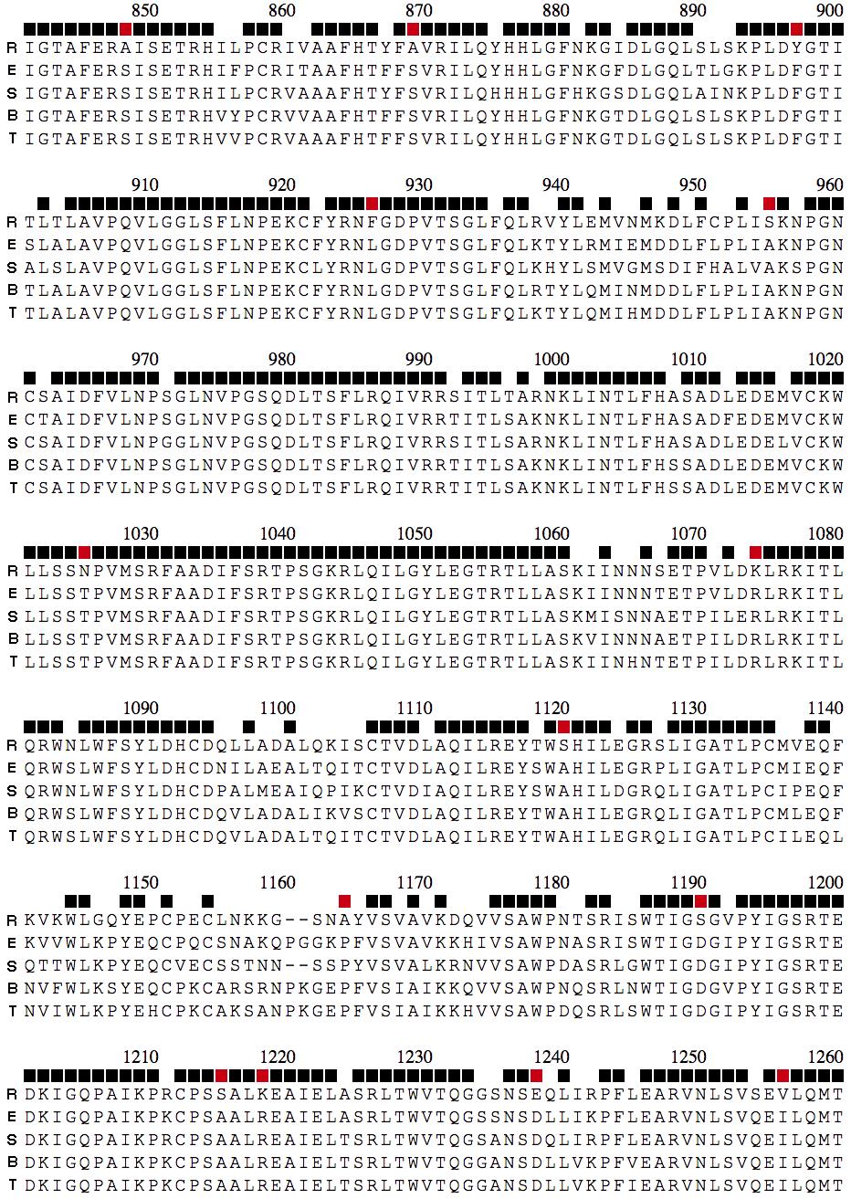
20 Supplementary Figure. Ebolavirus protein consensus sequences and SDPs. The consensus sequence for each Ebolavirus species is shown for each Ebolavirus protein. The row above the alignment indicates positions that are 00% conserved across all Ebolavirus sequences (black) or specificity determining positions (SDPs) that discriminate Reston viruses from the four human pathogenic Ebolavirus species (red); R, Reston virus; E, Ebola virus; S, Sudan virus; B, Bundibugyo virus; T, Taϊ Forest virus. A) for VP4, B) for GP, C) for VP40, D) VP35, E)VP30, F) sgp, G) NP, H)L. A VP4
21 B - GP
22
23 C VP40
24 D VP35
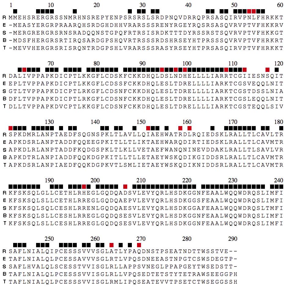
25 E VP30
26 F sgp
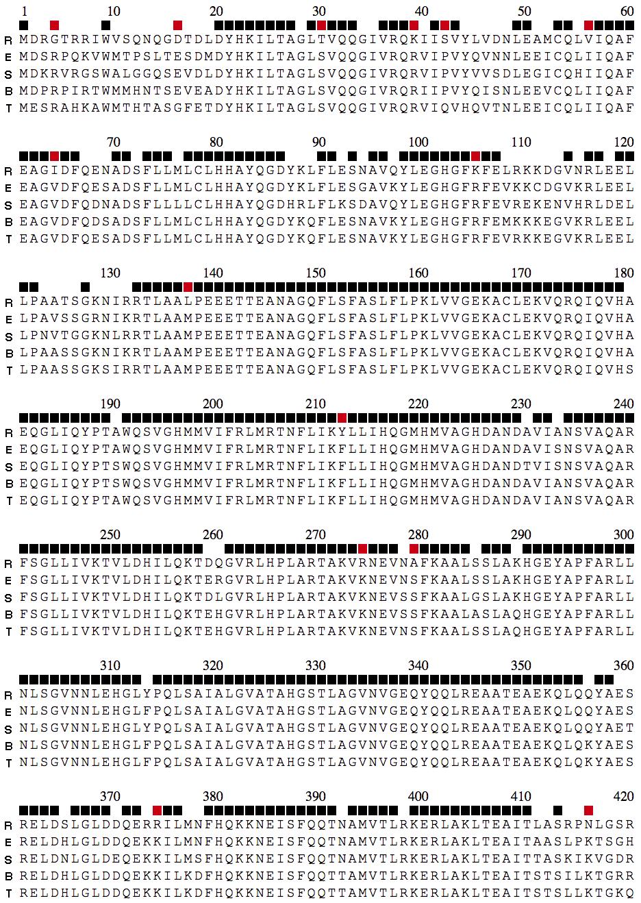
27 G NP
28
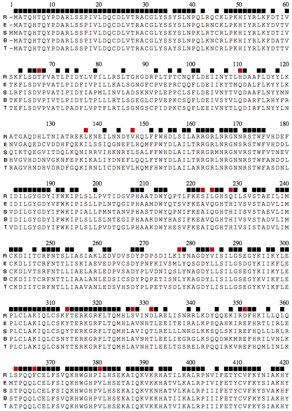
29 H L
30
31
32
33
34
35 Supplementary Figure 3. Solvent Accessible surface area for Ebolavirus SDPs. Histograms showing the Solvent Accessible surface area in square ångstroms of SDPs. Values are calculated for the Ebola virus structure and residues.
 Heatmap of intra- and inter-species GP sequence identity (EBOV, Ebola virus; BDBV, Bundibugyo virus; SUDV, Sudan virus; TAFV, Taϊ Forest virus; RESTV, Reston virus).")
36 Supplementary Figure 4. GP SDPs. A) Heatmap of intra- and inter-species GP sequence identity (EBOV, Ebola virus; BDBV, Bundibugyo virus; SUDV, Sudan virus; TAFV, Taϊ Forest virus; RESTV, Reston virus). B) Monomeric representation of GP with GP (grey) and GP (blue). D) EBOV GP trimer (PDB code: 3CSY) with SDPs colored red. The three GP chains are colored grey. The three GP chains are colored blue, green and yellow. C) Electrostatics surfaces for the EBOV structure (3CSY) and a model of a RESTV GP trimer based on 3CSY.

37 Supplementary Figure 5. GP SDPs are located outside the putative NPC binding site. GP SDPS are shown in red. The putative NPC binding site is shown in cyan.
38 Supplementary Figure 6. SDP prediction with subsampling of Ebolavirus sequences. The two groups of sequences human pathogenic and Reston ( non human pathogenic ) were sampled and SDP predictions made (see materials and methods). The boxplots show the distributions of the number of SDPs predicted in the simulations where A) only human pathogenic sequences were sampled, B) only Reston sequences were sampled and C) both sets were sampled. Sampling was performed for samples consisting of between 0%-90% of sequences (x axis). Red lines indicate the number of SDPs predicted in the full dataset without sampling. Note the scale of the Y-axis varies between each plot. A. Human pathogenic sequence sampled. GP L SDPs predicted SDPs predicted NP VP4 SDPs predicted SDPs predicted VP30 VP35 SDPs predicted SDPs predicted VP40 SDPs predicted
39 B. Reston Sequences Sampled SDPs predicted GP SDPs predicted L NP VP4 SDPs predicted SDPs predicted SDPs predicted VP30 SDPs predicted VP35 VP40 SDPs predicted
40 C. Both groups sampled GP L SDPs predicted SDPs predicted NP VP4 SDPs predicted SDPs predicted VP30 VP35 SDPs predicted SDPs predicted VP40 SDPs predicted
41 Supplementary Figure 7. Change in SDP prediction with subsampling of Ebolavirus sequences. The two groups of sequences human pathogenic and and Reston ( non human pathogenic ) were sampled and SDP predictions made (see materials and methods). The boxplots show the number of SDPs predicted in each sampling that are also in the full dataset (red) and new SDPs that are predicted only in subsamples (blue). The black horizontal line indicates the number of SDPs predicted using the full dataset. Subsampling performed for A) only human pathogenic sequences were sampled, B) only Reston sequences were sampled and c) both sets were sampled. A. Human pathogenic sequence sampled. GP 60 VP Var 40 0 Var L 80 VP Var Var 0 0 NP VP Var Var 0 0 VP Var 0
42 B. Reston Sequences Sampled GP Var VP30 Var 0 0 L VP Var Var NP VP Var Var VP Var 0.0
43 C. Both groups sampled GP VP Var Var L VP Var Var 0 0 NP VP Var Var 0 0 VP Var 0
44 Supplementary Figure 8. Analysis of completely conserved SDP with subsampling of Ebolavirus sequences. The two groups of sequences human pathogenic and and Reston ( non human pathogenic ) were sampled and SDP predictions made (see materials and methods). The boxplots show the number of SDPs predicted in each sampling that are are completely conserved (red) and not completely conserved (blue). The red horizontal line indicates the number of completely conserved SDPs present in the full dataset and the blue line represents the equivalent for SDPs that are not completely conserved. Subsampling performed for A) only human pathogenic sequences were sampled, B) only Reston sequences were sampled and c) both sets were sampled. A. Human pathogenic sequence sampled. GP 00 VP Var Var L 00 VP Var Var 0 0 NP VP Var Var VP Var 0
45 B. Reston Sequences Sampled GP 5 0 VP Var Var 5 0 L VP Var 0 5 Var NP VP Var Var VP Var 0.0
46 C. Both groups sampled 50 GP 80 VP Var Var 0 0 L VP Var Var 0 0 NP VP Var Var 0 0 VP Var 0
47 Supplementary Tables completely conserved positions Number of Positions with variation % of positions with variation All species % Ebola virus % Sudan virus % Bundibugyo % virus Tai forest virus % Reston virus % Supplementary Table. Variation within the Ebolavirus genomes. The number of positions in the Ebolavirus protein multiple sequence alignments that are completely conserved and those that have variation are shown.
48 Alignme nt position RESTV EBO V BDB V SUDV TAFV BLOSU M 6 score SASA (Å ) 7 M7 L7 L7 L7 L7 70 I V V V V I3 V3 V3 V3 V S3 T3 T3 T3 T T3 36 L36 N3 N3 N3 N3 9 M3 6 M36 M36 M36 Q3 9 Q39 Q39 Q39 3 mcsm (Δ Δ G, Kcal/mol) (destabilisin (destabilisin S3det Rank g) g) (destabilisin g) (destabilisin -. (destabilisin g) g) -.7 (destabilisin g) R39 (stabilising) (destabilisin 6 A6 T6 T6 T6 T6 0 g) 48 L48 S48 S48 S48 S Supplementary Table. VP4 SDPs. The position in the multiple sequence alignment, the amino acid position, and amino acid present in each of the species is shown. The BLOSUM6 score represents how frequently such amino acid changes are observed in nature. SASA is the solvent accessible surface area, which is only available for SDPs that could be mapped to protein structure. SASA was calculated using the protein structure with PDB code 4M0Q. RESTV, Reston virus; EBOV, Ebola virus; B, Bundibugyo virus; SUDV, Sudan virus; TAFV, Taϊ Forest virus. The s3det column shows the ranking of the SDPs by s3det.
49 Alignmen t position RESTV EBOV BDBV SUDV TAFV BLOSUM 6 score SASA (Å ) mcsm (Δ Δ G, Kcal/ mol) S3det rank 53 N53 T5 T5 T5 T L54 V53 V53 V53 V53-64 I64 T63 T63 T63 T D94 E93 E93 E93 E93-97 N97 T96 T96 T96 T H99 R98 R98 R98 R R08 K07 K07 K07 K07 - I S S S S S7 K6 K6 K6 K6 0 - S A0 A0 A0 A (stabili 5 I5 T50 T50 T50 T50-7 sing) 58 R58 Q57 Q57 Q57 Q (destab ilising) 60 L60 I59 I59 I59 I (destab ilising) 97 H97 R96 R96 R96 R (destab ilising) 06 D06 E05 E05 E05 E (destab ilising) 63 A63 R6 R6 R6 R (destab ilising) 69 Q69 S68 S68 S68 S Supplementary Table 3. VP30 SDPs. The position in the multiple sequence alignment, the amino acid position, and amino acid present in each of the species is shown. The BLOSUM6 score represents how frequently such amino acid changes are observed in nature. SASA is the solvent accessible surface area, which is only available for SDPs that could be mapped to protein structure. SASA was calculated using the protein structure with PDB code I8B. RESTV, Reston virus; EBOV, Ebola virus; B, Bundibugyo virus; SUDV, Sudan virus; TAFV, Taϊ Forest virus. The s3det column shows the ranking of the SDPs by s3det.
50 Alignment position REST V EBOV BDB V SUDV TAFV BLOSUM 6 SCORE SAS A (Å ) mcs M (Δ Δ G, Kcal/ mol) S3det rank 7 T5 S6 S6 S6 S6-49 D37 E48 E48 E48 E48-77 E65 D76 D76 D76 D76-86 K74 E85 E85 E85 D M8 S9 S9 S9 S T86 V97 V97 V97 I N90 T0 T0 T0 A A95 S06 S06 S06 S06 - I0 V V V M S43 A54 A54 A54 A54-60 V48 T59 T59 T59 T D49 E60 E60 E60 E60-68 K56 G67 G67 G67 G A63 S74 S74 S74 S74-8 L70 I8 I8 I8 I8-70 D58 E69 E69 E69 E (desta bilisin g) 9 V79 A90 A90 A90 A (desta bilisin g) 35 A303 V34 V34 V34 V (desta bilisin g) 330 K38 Q39 Q39 Q39 Q (desta bilisin g) Supplementary Table 4. VP35 SDPs. The position in the multiple sequence alignment, the amino acid position, and amino acid present in each of the species is shown. The BLOSUM6 score represents how frequently such amino acid changes are observed in nature. SASA is the solvent accessible surface area, which is only available for SDPs that could be mapped to protein structure. SASA was calculated using the protein structure with PDB code 4IBB. RESTV, Reston virus; EBOV, Ebola virus; B, Bundibugyo virus; SUDV, Sudan virus; TAFV, Taϊ Forest virus. The s3det rank column shows the ranking of the SDPs by s3det. The s3det column shows the ranking of the SDPs by s3det.
51
52 Alignme nt position REST V EBOV BDBV SUDV TAFV BLOSU M 6 SCORE SASA (Å ) mcsm (Δ Δ G, Kcal/m ol) -0.3 (destab S3det rank 46 V46 T46 T46 T46 T ilising) (destab 85 T85 P85 P85 P85 P85-4 ilising) V I I I I (destab 0 N0 G0 G0 G0 G ilising) 09 L09 F09 F09 F09 F (destab ilising) 45 P45 Q45 Q45 Q45 Q (stabili sing) 69 Q69 H69 H69 H69 H V93 I93 I93 I93 I (destab ilising) 35 D35 E35 E35 E35 E35 - Supplementary Table 5. VP40 SDPs. The position in the multiple sequence alignment, the amino acid position, and amino acid present in each of the species is shown. The BLOSUM6 score represents how frequently such amino acid changes are observed in nature. SASA is the solvent accessible surface area, which is only available for SDPs that could be mapped to protein structure. SASA was calculated using the protein structure with PDB code ES6. RESTV, Reston virus; EBOV, Ebola virus; B, Bundibugyo virus; SUDV, Sudan virus; TAFV, Taϊ Forest virus. The s3det column shows the ranking of the SDPs by s3det.
53 Alignmen t position BLOSU M 6 SCORE mcs M (Δ Δ G, Kcal/ mol) REST V EBOV BDBV SUDV TAF V SASA (Å ) 4 G4 R4 R4 R4 R4-6 D6 E6 E6 E6 G6 30 T30 S30 S30 S30 S30 39 K39 R39 R39 R39 R S4 P4/ Q4 P4 P4 Q V56 I56 I56 I56 I I64 V64 V64 V64 V K05 R05 R05 R05 R05 37 L37 M37 M37 M37 M37 37 Y F F F F R74 K74 K74 K74 K A79 S79 S79 S79 S (desta bilisin g) -.73 (desta bilisin g) (desta bilisin g) (desta bilisin g) (desta bilisin g) (desta bilisin g) (desta bilisin g) (desta bilisin g) -0.8 (desta bilisin g) (desta bilisin g) 374 R374 K374 K374 K374 K N46 K46 K46 K46 K Q4 Y4 Y4 Y4 Y4-46 E46 D46 D46 D46 D N435 D435 D435 D435 D E443 D443 D443 D443 D I453 T453 T453 T453 T453 - S3det rank
54 49 E49 D49 D49 D49 D A497 P497 P497 P497 P (-) P56 P56 P56 P56 57 S563 T563 T563 T563 T V565 I565 I565 I565 I T60 P60 P60 P60 N Q64 N64 N64 N64 K R705 A705 A705 A705 A (desta bilisin g) 76 N76 D76 D76 D76 D (stabil ising) 77 N77 G77 G77 G77 G (desta bilisin g) Supplementary Table 6. NP SDPs. The position in the multiple sequence alignment, the amino acid position, and amino acid present in each of the species is shown. The BLOSUM6 score represents how frequently such amino acid changes are observed in nature. SASA is the solvent accessible surface area, which is only available for SDPs that could be mapped to protein structure. SASA was calculated using the protein structure with PDB code 4QB0 for the C terminal and 4YPI for the N terminal regions. RESTV, Reston virus; EBOV, Ebola virus; B, Bundibugyo virus; SUDV, Sudan virus; TAFV, Taϊ Forest virus. The s3det rank column shows the ranking of the SDPs by s3det. The s3det column shows the ranking of the SDPs by s3det.
55 BLOSU M 6 Score SAS A (Å ) mcs M (Δ Δ G, Kcal/ mol) Alignment position RESTV EBOV BDBV SUDV TAF V G M M M M -3 3 S3 G V E/G G I3 F3 F3 F3 F D55 N54 N54 N54 N (desta bilisin S3det rank 38 I38 V37 V37 V37 V g) -.76 (desta bilisin 46 A46 V45 V45 V45 V g) (desta bilisin 76 I76 V75 V75 V75 V g) 97 A97 S96 S96 S96 S96 08 D08 E07 T07 E07 T07 9 T S0 S0 S0 S (desta bilisin 6 L6 I60 I60 I60 I60 5 g) (desta bilisin 70 S70 T69 T69 T69 T69 99 g) S308/ 308 H308 L307 S308 S308 S G36 R35 V35 R35 V L355 H354 R354 H354 Q P40 Q403 N40 Q397 S E4 S48 A409 S4 T P449 T448 S44 T448 T448-7 Y57/ 497 H57 H56 H56 H56 H K499 R498 R498 R498 R498 5 K50 R500 R500 R500 R (desta bilisin 54 V5 Q5 Q5 Q5 L5 9 g) (stabil ising) 6
56 568 V548 L547 I547 L547 I (desta bilisin g) L585 I584 I584 I584 I S608 D607 D607 D607 D E63 K6 K6 K6 K6 659 H639 Q638 Q638 Q638 Q L643 D64 D64 D64 S L645 W644 W644 W644 W I660 T569 T569 T569 T569 - Supplementary Table 7. GP SDPs. The position in the multiple sequence alignment, the amino acid position, and amino acid present in each of the species is shown. The BLOSUM6 score represents how frequently such amino acid changes are observed in nature. SASA is the solvent accessible surface area, which is only available for SDPs that could be mapped to protein structure. SASA was calculated using the protein structure with PDB code 3CSY. RESTV, Reston virus; EBOV, Ebola virus; B, Bundibugyo virus; SUDV, Sudan virus; TAFV, Taϊ Forest virus. The s3det rank column shows the ranking of the SDPs by s3det. The s3det column shows the ranking of the SDPs by s3det.
57 Alignment position RESTV EBOV BDBV SUDV TAFV BLOSUM 6 SCORE SASA (Å ) S3det rank 47 G M M M M I3 F3 F3 F3 F I38 V37 V37 V37 V A46 V45 V45 V45 V I76 V75 V75 V75 V A97 S96 S96 S96 S96 56 T S0 S0 S0 S0 306 L6 I60 I60 I60 I S70 T69 T69 T69 T69 48 Supplementary Table 8. sgp SDPs. The position in the multiple sequence alignment, the amino acid position, and amino acid present in each of the species is shown. The BLOSUM6 score represents how frequently such amino acid changes are observed in nature. SASA is the solvent accessible surface area, which is only available for SDPs that could be mapped to protein structure. SASA was calculated using the Phyre structural model that used template structure 3s88I. RESTV, Reston virus; EBOV, Ebola virus; B, Bundibugyo virus; SUDV, Sudan virus; TAFV, Taϊ Forest virus. The s3det rank column shows the ranking of the SDPs by s3det. The s3det column shows the ranking of the SDPs by s3det.
58 Alignment position RESTV EBOV BLOSU M6 SCORE mcsm (Δ Δ G, Kcal/m ol) BDB V SUDV TAF V SASA (Å ) S3det rank 67 T66 V66 V66 V66 V H09 Q09 Q09 Q09 Q L36 I36 I36 I36 I36 47 V46 L46 L46 L46 L46 S A A A A 4 L3 Q3 Q3 Q3 Q3-8 Q7 H7 H7 H7 H (destab ilising) 77 I76 L76 L76 L76 L V83 L83 L83 L83 L83 33 F3 Y3 Y3 Y3 Y S36 A36 A36 A36 A36 33 D330 T330 T330 T330 T D350 E350 E350 E350 E S36 T36 T36 T36 T F365 L365 L365 L365 L I379 V379 V379 V379 V H447 Q447 Q447 Q447 Q S450 P450 P450 P450 P N465 D465 D465 D465 D S689 E689 E689 E689 E A847 S847 S847 S847 S A868 S868 S868 S868 S Y896 F896 F896 F896 F F95 L95 L95 L95 L S954 A954 A954 A954 A T995 S995 S995 S995 S N04 T04 T0 4 T04 T K073 R073 R07 3 R073 R S9 A9 A 9 A9 A 9 64 A6 F63 F6 3 F63 F S87 D89 D8 9 D89 D S A4 A 4 A4 A 4 8 K5 R7 R 7 R7 R 7 38 E35 D37 D3 7 D37 D V53 I55 I55 I55 I K53 R534 R53 R534 R53
59 A354 T366 T36 6 T366 T T393 S395 S39 5 S395 S M406 I408 I408 I408 I L4 I44 I44 I44 I N434 S436 S43 6 S436 S Q459 K46 K46 K46 K C47 S473 S47 3 S473 S Y486 L488 L48 8 L488 L L497 I499 I499 I499 I A504 S506 S50 6 S506 S V507 I509 I509 I509 I S536 A535 A53 5 A535 A Y64 L64 L6 4 L64 L S68 C68 C6 8 C68 C I760 V76 V76 V76 V T848 V850 V85 0 V850 V S87 T873 T87 3 T873 T N94 R96 R9 6 R96 R R939 E94 E94 E94 E I006 L008 L00 8 L008 L I04 L044 L04 4 L044 L T075 S077 S07 7 S077 S07 7 E09 E09 D096 E098 E L30 Q05 Q0 5 Q05 Q E06 Q08 Q0 8 Q08 Q F9 Y3 Y3 Y3 Y3 3 8 V55 L57 L5 7 L57 L N7 R68 R6 8 R68 R K73 R75 R7 R75 R7
60 0 F75 L77 M8 L L7 L7 7 L M8 M8 6 M86 6 Supplementary Table 9. L SDPs. The position in the multiple sequence alignment, the amino acid position, and amino acid present in each of the species is shown. The BLOSUM6 score represents how frequently such amino acid changes are observed in nature. SASA is the solvent accessible surface area, which is only available for SDPs that could be mapped to protein structure. SASA was calculated using the Phyre structural model which used template 4n48A ( cap-specific mrna ( cap-specific mrna (nucleoside-'-o-)-methyltransferase protein in complex with capped rna fragment ). RESTV, Reston virus; EBOV, Ebola virus; B, Bundibugyo virus; SUDV, Sudan virus; TAFV, Taϊ Forest virus. The s3det rank column shows the ranking of the SDPs by s3det. The s3det column shows the ranking of the SDPs by s3det.
61 Protein EBOV Res RESTV Res Mutation position Mutation Effect GP Q638 H 638 Q V No effect on release of soluble GP,delta. GP R498 K RTRR ATAA No effect on cleavage between GP and GP. GP D64 L 64 D V No effect on release of soluble GP,delta. VP4 M36 L 34/36 F-A/M-A Near complete loss of KPNA5 binding * VP4 Q39 R RTQ AAA Near complete loss of KPNA5 binding * Supplementary Table 0. SDPs that coincide with known mutagenesis data. Functional data extracted from UniProt unless stated. Res, residue; EBOV, Ebola virus; RESTV, Reston virus *Data from Bornholdt et al., 35
62 PROTEIN SPECIES OLIGOMERIC STATE GP EBOV Trimer of Heterodimers PDB/TEMPLATE REGION IN SEQUENCE 3CSY (structure) sgp EBOV Dimer 3s88I (model) 3-87 sgp RESTV Dimer 3s88I (model) L EBOV Monomer 4n48A (model) 3-38 NP (C-terminal) EBOV Monomer 4QB0 (structure) NP (N-terminal) EBOV Monomer 4YPI (structure) VP4 EBOV Heterodimer 4M0Q (structure) 0-3 VP4 EBOV Heterodimer 4UX (structure) 6-3 VP4 RESTV Dimer 4D9O (structure) 0-3 VP30 EBOV Dimer I8B (structure) VP30 RESTV Dimer 3V70 (structure) 4-7 VP35 EBOV Heterodimer 4IBB (structure) VP35 EBOV Dimer of heterodimers 3L5 (structure) VP35 RESTV Dimer of heterodimers 3KS8 (structure) VP40 EBOV Monomer ES6 (structure) 44-3 VP40 EBOV Dimer 4LDB (structure) VP40 EBOV Hexamer 4LDD (structure) VP40 EBOV Octamer 4LDM (structure) VP40 RESTV Monomer es6a (model) 44-3 Supplementary Table. Protein structures available for Ebolavirus Proteins. EBOV, Ebola virus; RESTV, Reston virus
63 Reston virus residue I3 I38 A46 Pathogenic consensus F3 V37 V45 Comments Note- Ebola virus GP structure has R3 rather than F3. Surface residue close to interface with GP in the trimer. Unclear what functional effect may be if any. Surface residue, appears to be a conservative change of amino acid that could be well tolerated Also a surface residue. Conservative change of hydrophobic amino acid that could be well accommodated. Functional effect unclear unlikely unlikely I76 L6 S70 H308 D55 V5 V548 L585 V75 I60 T69 S308/ L307 N54 Q5 L547 I584 Surface residue, conservative change of amino acid. Change should be well accommodated One of three SDPs located in the glycan cap region of GP. The glycan cap binds the host cell receptor(s) but is highly glycosylated so it is not clear if the amino acids directly contact the host cell. Surface residue in a cavity. It is part packed quite tightly with residue F34, V36, T40 but should be possible to accommodate change to Leu in Reston virus. Could there be a role with the three SDPs combined in this region. Located at the top of the structure, is a surface residue (with side chain pointing to the solvent) representing a conservative amino acid change. Again could it have a role in conjunction with the other SDPs in this region? Also located in the glycan cap and also a surface residue. Present in loop so unlikely to alter structure but could have a functional role, and alters charge on the protein surface. Surface residue, results in loss of negative charge in Reston virus GP. Located at the end of a beta sheet. Seems unlikely to have a structural effect. Possible combined effect with adjacent L547V? Close to trimer interface (GP-GP) but directly within the interface. Not clear what effect this change would have on protein structure Surface residue at end of a beta sheet. Appears to be minor change in amino acid. Possible combined effect with adjacent N54D? Largely buried amino acid. At the interface with GP (in the same GP monomer). EBOV I584 interacts with F57, not clear if this interaction would change in with Leu in Reston virus. unlikely possible* possible* possible* unlikely unclear unlikely unlikely Supplementary Table. Structural analysis of GP SDPs. Details of the structural analysis are included with an assessment of whether the amino acid change is likely to have an effect on the protein. Four categories are used for the effect column unlikely (the change seems unlikely to alter the structure/function), unclear (the change could be functional but there is limited evidence), possible (more confident that there is an effect than the unclear group) and probably (highly confident that the change will have a structural/functional effect).
64 Reston virus residue K39 S4 V56 I64 K05 L37 Y R74 A79 R374 R705 N76 N77 Pathogenic consensus R39 P4/ Q4 I56 V64 R05 M37 F K74 S79 K374 A705 D76 G77 Comments R39 forms a H bond with D7. Change to K is likely to maintain this H bond. Unusual to see Pro in a sheet. The amino acid is on the protein surface and it there is nothing to suggest that a change to Ser would alter the protein I56 is largely buried and packed against other sidechains. While change to Val would reduce the size of the side chain, it seems likely that it would be accommodated within the structure. Also V64I is adjacent to this SDP. In a surface loop facing the helix containing I56V. Possible coevolution with I56 reduce size in one, matched with increased size in the other. The side chain guanidino group of R05 provides a hydrogen bond with the side chain of Q38 as well as with the local backbone NH of G03 to provide a stabilized region of the protein. Although the mutation R05K appears conservative and maintains the side chain positive charge, the ability to form multiple hydrogen bonds is reduced due to resonance stabilization in the guanidino group being lost in the transfer to the lysine side chain amino group. This has the potential to weaken interactions in this region. M37 is located at the end of helix and packs against an adjacent helix. The conservative change to L37 in Reston virus seems unlikely to have a significant effect on structure/function A minor change in side chains. P is located in an alpha helix and the sidechain is largely buried. The change to Y in Reston virus is unlikely to have a significant effect on protein structure/function K74 is located in the VP35 binding site. K74 forms a hydrogen bond with VP35 D46 and a change to Arg should be able to maintain this interaction. S79 is located in an alpha helix on the protein surface. The change to A79 in Reston virus would introduce a hydrophobic amino acid on the protein surface that could have an effect on protein structure. K374 is located in an alpha helix on the protein surface. It is not unlikely that the change to R374 in Reston virus will alter protein structure. It is a conservative change of side chain. A695 is located on the protein surface so the charge introduce by the change to R695 in Reston virus should be tolerated. Proximity of Reston virus R705 to E694 may result in a salt bridge that would reduce flexibility in Reston virus NP. There could different hydrodynamic volumes between the Reston virus and pathogenic NP proteins as well as in the pathogenic ebolaviruses exposing residues that remain buried in the Reston virus NP. The salt bridge could make RESTV more thermostable (and possibly more resistant to proteolysis and denaturants). Present in a surface loop this change will change the charge properties. Should be considered with adjacent amino acid, which is also an SDP. Overall we see the removal of a negatively charged amino acid with two polar side chains. Adjacent to D76N psdp. The loss of Gly would change the turn from type to a type turn. Also See comment above. Functional effect unlikely unclear unlikely unlikely possible unlikely unlikely unlikely unclear unlikely Possible unclear unclear Supplementary Table 3. Structural analysis of NP SDPs. Details of the structural analysis are included
65 with an assessment of whether the amino acid change is likely to have an effect on the protein. Four categories are used for the effect column unlikely (the change seems unlikely to alter the structure/function), unclear (the change could be functional but there is limited evidence), possible (more confident that there is an effect than the unclear group) and probably (highly confident that the change will have a structural/functional effect).
66 Reston virus Residue D58 V79 A303 K38 Pathogenic consensus E69 A90 V34 Q39 Comments Present in dimer interface (only for one of the subunits as the dimer is asymmetric). Forms hydrogen bonds with R30, R3 and W33 (RESTV numbering). Distances between atoms are slightly different between the species. W34 3.A (.8 in Ebola virus), R30 3.A (.9 in Ebola virus) R3.8 and 3.0 (both.8a in Ebola virus). Also close to A303 across interface, they could compensate or presence of both changes could have greater effect on interface in this area. (6.A in RESTV, 7.5 in Ebola virus) Present in a surface loop packs against adjacent helix, conservative change of hydrophobic amino acid. Could be some local conformational changes and is located adjacent to the linker between the two subdomains, which is in RESTV has a short alpha helix that is not present in EBOV. Present in a surface loop near the VP35 dimer interface. Close in space to D58 in the other subunit. Located at the end of a beta sheet. Adjacent to His85 in next strand. His85 is completely conserved in all Ebolavirus species. So Reston virus VP35 has increased positive charge in this position Functional effect probable Unclear unclear unclear Supplementary Table 4. Structural analysis of VP35 SDPs. Details of the structural analysis are included with an assessment of whether the amino acid change is likely to have an effect on the protein. Four categories are used for the effect column unlikely (the change seems unlikely to alter the structure/function), unclear (the change could be functional but there is limited evidence), possible (more confident that there is an effect than the unclear group) and probably (highly confident that the change will have a structural/functional effect).
67 RESTV residue I5 R58 L60 H97 D06 A63 Pathogenic consensus T50 Q57 I59 R96 E05 R6 Comments The side chain is largely buried and it appears that Reston virus I5 would be tolerated although a hydrogen bond with the backbone of the previous turn of the helix will be lost. Located in a surface loop, will increase surface charge. It is possible that Reston virus forms a salt bridge with D59, which would increase stability and reduce flexibility in this area of the protein. This SDP is in a region of SDPs and very close to another SDP (I59L). So possible effects may be compensated by other changes. Located in a surface close to another SDP (see above). Appears to be a conservative change that given the other species specific changes in this area it seems unlikely that it will have a functional effect on the protein. Surface residue so change in size/shape should well accommodated, positive charge maintained in side chain. Exposed surface residue, conservative change of amino acid. Unlikely to alter protein structure. This residue is present in the dimer interface. In Ebola virus VP30 R6 hydrogen bonds with the backbone of A4 and G40. Reston virus A63 will be unable to hydrogen bond. This is likely to reduce the affinity of the dimer (given that it is symmetrical and so the Ebola virus R6 in each subunit forms hydrogen bonds with the other subunit. The Reston virus dimer has been observed to be rotated relative to the Ebola virus. The loss of the hydrogen bonds may explain this. functional effect unlikely unlikely unlikely unlikely unlikely probable Supplementary Table 5. Structural analysis of VP30 SDPs. Details of the structural analysis are included with an assessment of whether the amino acid change is likely to have an effect on the protein. Four categories are used for the effect column unlikely (the change seems unlikely to alter the structure/function), unclear (the change could be functional but there is limited evidence), possible (more confident that there is an effect than the unclear group) and probably (highly confident that the change will have a structural/functional effect).
68 Reston virus residue V46 T85 V N0 L09 P45 Q69 V93 Pathogenic consensus T46 P85 I G0 F09 Q45 H69 I93 Comments Present in a surface loop (although only third amino acid in structure). Reston virus V46 introduces a hydrophobic amino acid on surface, could affect stability but no evidence for this. Ebola virus P85 is in a S-G-P-K beta-turn, proline confers backbone rigidity and change to Thr in Reston virus would introduce backbone flexibility and provide a side chain with H- bond donor. Located in the Ebola virus octamer interface, will result in changes to this interface and likely alter the octamer structure. In an octamer structure (if it were to remain similar to the Ebola virus octamer), T85 could hydrogen bond with the backbone of L7 or the sidechain of R37. This change appears to be conservative substitution of two hydrophobic amino acids. Ebola virus I is packed with other hydrophobic residues and it appears that the region would be able to accommodate the change to Reston virus V with a slightly smaller side chain. Located in a surface loop. Based on the Ebola virus structure, the Reston virus N0 side chain would be likely to point into the protein structure. But not clear what effect this would have on the protein structure, if any given that the structure has gaps in this region so cannot be confident. Packed in a largely hydrophobic region the SDP results in a reduction in side chain size in Reston virus. The smaller Leucine may adopt different side chain conformations to aid stability. Ebola virus F09 does not interact with other aromatic side chains so the structure is unlikely to be adversely affected by the swap to Leucine. Surrounding hydrophobic residues are aliphatic (I6, I85, V98, A38, P37) so the change to Leucine could be well accommodated. Located at the end of an alpha helix, the Reston virus P45 would break the helix and shorten it to either L44 or more likely M4, which is a better C-capping residue. This could have a destabilizing effect on the two helices in this region and the base of the hydrophobic core because secondary structure will most likely change to accommodate the inflexible Proline. A surface residue, loss of charge to polar side chain. This is a highly charged region with E65, R70, K74, K75. So the positive charge would be reduced in Reston virus VP40. Packs with other hydrophobic residues. Appears to be a conservative change Possible Functional effect unclear probably unlikely unclear unlikely probably unclear Unlikely Supplementary Table 6. Structural analysis of VP40 SDPs. Details of the structural analysis are included with an assessment of whether the amino acid change is likely to have an effect on the protein. Four categories are used for the effect column unlikely (the change seems unlikely to alter the structure/function), unclear (the change could be functional but there is limited evidence), possible (more confident that there is an effect than the unclear group) and probably (highly confident that the change will have a structural/functional effect). Analysis is based on the VP40 dimer structure unless otherwise stated.
69 Reston virus residue M7 I I3 S3 T3 L36 R39 A6 Pathogenic consensus L7 V V3 T3 N3 M36 Q39 T6 Comments Located in a helix. Appears to be a conservative change in amino acid. No suggestion from structure that it would alter structure/function. Located in a helix and is fairly tightly packed against the adjacent helix but would expect the pocket to accommodate the change. Located in a sheet facing a loop. Side chain is relatively exposed so structure should be able to accommodate. Adjacent in space to another SDP (3) Ebola virus T3 forms hydrogen bonds with the side chains of T9, W5 and with the backbone of H33. Model of Reston virus VP4 suggests S3 would continue to interact with the same residues. This residue is on the edge of the KPNA5 binding site. Appears to be a conservative change of amino acid. Exposed polar residue exchanges for another polar residue. Unlikely to affect structure. Adjacent in space to an SDP (V3S) and in sequence to 3. Part of the interface site with KPNA5. Mutagenesis of M36 in combination with other residues resulted in loss of KPNA5 binding 34. Although it appears to be a conservative substitution. Interface residue. In Ebola virus Q39 forms an H bond with the backbone of R37. This is likely to be lost in Reston virus VP4 with the longer R39 side chain. Change will also introduce positive charge at interface site. Located in a helix facing a sheet. Ebola virus T6 forms a hydrogen bond with the backbone of D48. Reston virus A6 will not be able to form this hydrogen bond. This is likely to reduce the stability of the protein and increase flexibility. Possible functional effect unlikely unlikely unlikely probable unlikely probable probable Probable Supplementary Table 7. Structural analysis of VP4 SDPs. Details of the structural analysis are included with an assessment of whether the amino acid change is likely to have an effect on the protein. Four categories are used for the effect column unlikely (the change seems unlikely to alter the structure/function), unclear (the change could be functional but there is limited evidence), possible (more confident that there is an effect than the unclear group) and probably (highly confident that the change will have a structural/functional effect).
70 Region Residue Conservation L36 SDP R39 SDP S40 Not an SDP but conserved S in Reston viruses and mainly R in Ebola viruses, not conserved enough to be SDP L07 Vary in species specific manner H09 Vary in species specific manner T6 Vary in species specific manner G0 Not an SDP G in Reston viruses and Ebola viruses (mainly), differs in others 3 S84 3 T85 Not an SDP. T in Reston viruses, mainly N in other species 3 H86 Vary in species specific manner 3 T87 Not an SDP, primarily T in most species (A in Sudan viruses) 3 F97 Vary in species specific manner 4 V0 Vary in species specific manner 5 S50 Not an SDP Supplementary Table 8. Residues in VP4 previously identified to differ between Reston viruses and Ebola viruses and/or Sudan viruses. Zhang et al., identified five regions that differed between Reston viruses and Ebola viruses and/or Sudan viruses 7.The five regions are listed along with conservation information i.e. whether the position is an SDP, varies in a species specific manner (i.e. not an SDP, but a different residue is conserved in each of the different species) or otherwise conserved. Region one is part of the KPNA5 (karyopherin α5) binding site and region two is thought to be part of the STAT binding site 7.
71 Mutation Location/Comments Relationship to SDPs From Volchhkov et al., 43 experiment M7I Surface residue. Not clear what functional effect would be. Not close L47P Part of an alpha helix, the proline would be expected to break the helix Close to SDPs T87I and could lead to conformational changes that would alter function. Adjacent to interface site. T87 forms Hydrogen bonds with the backbone of H86 and E03. Mutation to I would remove these hydrogen bonds and reduce stability/increase flexibility in this area. (Also close to L6F mutation from a separate study) L7M, VI Not close From Volchhkov et al., 43 experiment H86Y Present in interface with KPNA5. Forms a hydrogen bond with the backbone of T434 in KPNA5. Mutation to Tyr would still enable Hydrogen bonding with KPNA as the functional group is maintained. From Ebihara et al., 44 T50I The side chain of Ebola virus T50 can hydrogen bond with the backbones of Q36 and K5. Removal of these interactions with mutation Ile will reduce stability/increase flexibility. From Dowall et al., 45 L6F Largely buried side chain. Increase in size to phenylalanine could require some conformational change. Interesting that is located close to T87I (see above). F9V* Largely buried side chain. Reduction in size would create space and therefore likely to result in some conformational change? A43P* K8R* Close in space to L6F (see above). Present in a turn. Appears to be a conservative change. K8 is present in the KPNA5 interface. Is close to M436 and D489. Possible electrostatic interaction. Possible the mutation to R enables this interaction to continue in the different species. Not close Close to SDP T6A Close to VI Close in space to SDPs T3S, N3T, V3I. Supplementary Table 9. VP4 Mutations occurring in adaption of Ebola virus to rodent species. The location of the mutation and how it may alter structure and function is listed with details of proximity to SDPs. *indicates that after passage one the predominant amino acid at that position was the wild type 44. In the Dowall et al. 45, study L6F is the only mutation where the mutation is predominantly maintained in in all passages. Separate experimental evidence suggests that the L6F mutation along results in pathogenicity in guinea pigs 37.
72 Genome Identifier Ebola virus species Host gb:kj Organism:Zaire ebolavirus H.sapiens-wt/GIN/04/Makona-Kissidougou-C5 Human gb:kj Organism:Zaire ebolavirus H.sapiens-wt/GIN/04/Makona-Gueckedou-C07 Human gb:kj Organism:Zaire ebolavirus H.sapiens-wt/GIN/04/Makona-Gueckedou-C05 Human gb:kp34330 Organism:Zaire ebolavirus H.sapiens-wt/GIN/04/Conacry-9 Human gb:kp0964 Organism:Zaire ebolavirus H.sapiens-tc/GIN/4/WPG-C5 Human gb:kp0964 Organism:Zaire ebolavirus H.sapiens-tc/GIN/4/WPG-C07 Human gb:kp09640 Organism:Zaire ebolavirus H.sapiens-tc/GIN/4/WPG-C05 Human gb:kc4800 Organism:Zaire ebolavirus EBOV/H.sapiens-tc/GAB/00/Ilembe Human gb:kc4794 Organism:Zaire ebolavirus EBOV/H.sapiens-tc/GAB/996/Nza Human gb:kc4797 Organism:Zaire ebolavirus EBOV/H.sapiens-tc/GAB/996/Oba Human gb:kc4795 Organism:Zaire ebolavirus EBOV/H.sapiens-tc/GAB/996/Mbie Human gb:kc4798 Organism:Zaire ebolavirus EBOV/H.sapiens-tc/GAB/996/Ikot Human gb:kc4793 Organism:Zaire ebolavirus EBOV/H.sapiens-tc/GAB/996/Eko Human gb:kc479 Organism:Zaire ebolavirus EBOV/H.sapiens-tc/GAB/994/Gabon Human gb:kc4784 Organism:Zaire ebolavirus EBOV/H.sapiens-tc/COD/007/9 Luebo Human gb:kc4790 Organism:Zaire ebolavirus EBOV/H.sapiens-tc/COD/007/5 Luebo Human gb:kc4788 Organism:Zaire ebolavirus EBOV/H.sapiens-tc/COD/007/43 Luebo Human gb:kc4789 Organism:Zaire ebolavirus EBOV/H.sapiens-tc/COD/007/4 Luebo Human gb:kc4787 Organism:Zaire ebolavirus EBOV/H.sapiens-tc/COD/007/3 Luebo Human gb:kc4786 Organism:Zaire ebolavirus EBOV/H.sapiens-tc/COD/007/ Luebo Human gb:kc4785 Organism:Zaire ebolavirus EBOV/H.sapiens-tc/COD/007/0 Luebo Human gb:kc4799 Organism:Zaire ebolavirus EBOV/H.sapiens-tc/COD/995/3709 Kikwit Human gb:kc4796 Organism:Zaire ebolavirus EBOV/H.sapiens-tc/COD/995/365 Kikwit Human gb:kc479 Organism:Zaire ebolavirus EBOV/H.sapiens-tc/COD/977/Bonduni Human gb:kc480 Organism:Zaire ebolavirus EBOV/H.sapiens-tc/COD/976/deRoover Human gb:km338 Organism:Zaire ebolavirus Ebola virus/h.sapiens-wt/sle/04/makona-nm04.3 Human gb:km337 Organism:Zaire ebolavirus Ebola virus/h.sapiens-wt/sle/04/makona-nm04. Human gb:km336 Organism:Zaire ebolavirus Ebola virus/h.sapiens-wt/sle/04/makona-nm04. Human gb:km335 Organism:Zaire ebolavirus Ebola virus/h.sapiens-wt/sle/04/makona-g3857 Human gb:km334 Organism:Zaire ebolavirus Ebola virus/h.sapiens-wt/sle/04/makona-g Human gb:km333 Organism:Zaire ebolavirus Ebola virus/h.sapiens-wt/sle/04/makona-g3856. Human gb:km33 Organism:Zaire ebolavirus Ebola virus/h.sapiens-wt/sle/04/makona-g385 Human gb:km33 Organism:Zaire ebolavirus Ebola virus/h.sapiens-wt/sle/04/makona-g3850 Human gb:km330 Organism:Zaire ebolavirus Ebola virus/h.sapiens-wt/sle/04/makona-g3848 Human gb:km3309 Organism:Zaire ebolavirus Ebola virus/h.sapiens-wt/sle/04/makona-g3846 Human gb:km3308 Organism:Zaire ebolavirus Ebola virus/h.sapiens-wt/sle/04/makona-g3845 Human gb:km3307 Organism:Zaire ebolavirus Ebola virus/h.sapiens-wt/sle/04/makona-g384 Human gb:km3306 Organism:Zaire ebolavirus Ebola virus/h.sapiens-wt/sle/04/makona-g3840 Human gb:km3305 Organism:Zaire ebolavirus Ebola virus/h.sapiens-wt/sle/04/makona-g3838 Human gb:km3304 Organism:Zaire ebolavirus Ebola virus/h.sapiens-wt/sle/04/makona-g3834 Human gb:km3303 Organism:Zaire ebolavirus Ebola virus/h.sapiens-wt/sle/04/makona-g383 Human gb:km330 Organism:Zaire ebolavirus Ebola virus/h.sapiens-wt/sle/04/makona-g389 Human gb:km330 Organism:Zaire ebolavirus Ebola virus/h.sapiens-wt/sle/04/makona-g387 Human gb:km3300 Organism:Zaire ebolavirus Ebola virus/h.sapiens-wt/sle/04/makona-g386 Human gb:km33099 Organism:Zaire ebolavirus Ebola virus/h.sapiens-wt/sle/04/makona-g385. Human gb:km33098 Organism:Zaire ebolavirus Ebola virus/h.sapiens-wt/sle/04/makona-g385. Human gb:km33097 Organism:Zaire ebolavirus Ebola virus/h.sapiens-wt/sle/04/makona-g383 Human gb:km33096 Organism:Zaire ebolavirus Ebola virus/h.sapiens-wt/sle/04/makona-g38 Human gb:km33095 Organism:Zaire ebolavirus Ebola virus/h.sapiens-wt/sle/04/makona-g38 Human gb:km33094 Organism:Zaire ebolavirus Ebola virus/h.sapiens-wt/sle/04/makona-g380 Human gb:km33093 Organism:Zaire ebolavirus Ebola virus/h.sapiens-wt/sle/04/makona-g389 Human gb:km3309 Organism:Zaire ebolavirus Ebola virus/h.sapiens-wt/sle/04/makona-g388 Human
73 gb:km3309 Organism:Zaire ebolavirus Ebola virus/h.sapiens-wt/sle/04/makona-g387 Human gb:km33090 Organism:Zaire ebolavirus Ebola virus/h.sapiens-wt/sle/04/makona-g386 Human gb:km33089 Organism:Zaire ebolavirus Ebola virus/h.sapiens-wt/sle/04/makona-g384 Human gb:km33088 Organism:Zaire ebolavirus Ebola virus/h.sapiens-wt/sle/04/makona-g380. Human gb:km33087 Organism:Zaire ebolavirus Ebola virus/h.sapiens-wt/sle/04/makona-g380. Human gb:km33086 Organism:Zaire ebolavirus Ebola virus/h.sapiens-wt/sle/04/makona-g3809 Human gb:km33085 Organism:Zaire ebolavirus Ebola virus/h.sapiens-wt/sle/04/makona-g3808 Human gb:km33084 Organism:Zaire ebolavirus Ebola virus/h.sapiens-wt/sle/04/makona-g3807 Human gb:km33083 Organism:Zaire ebolavirus Ebola virus/h.sapiens-wt/sle/04/makona-g3805. Human gb:km3308 Organism:Zaire ebolavirus Ebola virus/h.sapiens-wt/sle/04/makona-g3805. Human gb:km3308 Organism:Zaire ebolavirus Ebola virus/h.sapiens-wt/sle/04/makona-g3800 Human gb:km33080 Organism:Zaire ebolavirus Ebola virus/h.sapiens-wt/sle/04/makona-g3799 Human gb:km33079 Organism:Zaire ebolavirus Ebola virus/h.sapiens-wt/sle/04/makona-g3798 Human gb:km33078 Organism:Zaire ebolavirus Ebola virus/h.sapiens-wt/sle/04/makona-g3796 Human gb:km33077 Organism:Zaire ebolavirus Ebola virus/h.sapiens-wt/sle/04/makona-g3795 Human gb:km33076 Organism:Zaire ebolavirus Ebola virus/h.sapiens-wt/sle/04/makona-g3789. Human gb:km33075 Organism:Zaire ebolavirus Ebola virus/h.sapiens-wt/sle/04/makona-g3788 Human gb:km33074 Organism:Zaire ebolavirus Ebola virus/h.sapiens-wt/sle/04/makona-g3787 Human gb:km33073 Organism:Zaire ebolavirus Ebola virus/h.sapiens-wt/sle/04/makona-g3786 Human gb:km3307 Organism:Zaire ebolavirus Ebola virus/h.sapiens-wt/sle/04/makona-g378 Human gb:km3307 Organism:Zaire ebolavirus Ebola virus/h.sapiens-wt/sle/04/makona-g377 Human gb:km33070 Organism:Zaire ebolavirus Ebola virus/h.sapiens-wt/sle/04/makona-g3770. Human gb:km33069 Organism:Zaire ebolavirus Ebola virus/h.sapiens-wt/sle/04/makona-g3770. Human gb:km33068 Organism:Zaire ebolavirus Ebola virus/h.sapiens-wt/sle/04/makona-g Human gb:km33067 Organism:Zaire ebolavirus Ebola virus/h.sapiens-wt/sle/04/makona-g Human gb:km33066 Organism:Zaire ebolavirus Ebola virus/h.sapiens-wt/sle/04/makona-g3769. Human gb:km33065 Organism:Zaire ebolavirus Ebola virus/h.sapiens-wt/sle/04/makona-g3769. Human gb:km33064 Organism:Zaire ebolavirus Ebola virus/h.sapiens-wt/sle/04/makona-g3765. Human gb:km33063 Organism:Zaire ebolavirus Ebola virus/h.sapiens-wt/sle/04/makona-g3764 Human gb:km3306 Organism:Zaire ebolavirus Ebola virus/h.sapiens-wt/sle/04/makona-g3758 Human gb:km3306 Organism:Zaire ebolavirus Ebola virus/h.sapiens-wt/sle/04/makona-g375 Human gb:km33060 Organism:Zaire ebolavirus Ebola virus/h.sapiens-wt/sle/04/makona-g Human gb:km33059 Organism:Zaire ebolavirus Ebola virus/h.sapiens-wt/sle/04/makona-g3750. Human gb:km33058 Organism:Zaire ebolavirus Ebola virus/h.sapiens-wt/sle/04/makona-g3750. Human gb:km33057 Organism:Zaire ebolavirus Ebola virus/h.sapiens-wt/sle/04/makona-g3735. Human gb:km33056 Organism:Zaire ebolavirus Ebola virus/h.sapiens-wt/sle/04/makona-g3735. Human gb:km33055 Organism:Zaire ebolavirus Ebola virus/h.sapiens-wt/sle/04/makona-g3734. Human gb:km33054 Organism:Zaire ebolavirus Ebola virus/h.sapiens-wt/sle/04/makona-g379 Human gb:km33053 Organism:Zaire ebolavirus Ebola virus/h.sapiens-wt/sle/04/makona-g374 Human gb:km3305 Organism:Zaire ebolavirus Ebola virus/h.sapiens-wt/sle/04/makona-g373.4 Human gb:km3305 Organism:Zaire ebolavirus Ebola virus/h.sapiens-wt/sle/04/makona-g373.3 Human gb:km33050 Organism:Zaire ebolavirus Ebola virus/h.sapiens-wt/sle/04/makona-g373. Human gb:km33049 Organism:Zaire ebolavirus Ebola virus/h.sapiens-wt/sle/04/makona-g3707 Human gb:km Organism:Zaire ebolavirus Ebola virus/h.sapiens-wt/sle/04/makona-g3687. Human gb:km03456 Organism:Zaire ebolavirus Ebola virus/h.sapiens-wt/sle/04/makona-g3686. Human gb:km03456 Organism:Zaire ebolavirus Ebola virus/h.sapiens-wt/sle/04/makona-g3683. Human gb:km Organism:Zaire ebolavirus Ebola virus/h.sapiens-wt/sle/04/makona-g368. Human gb:km Organism:Zaire ebolavirus Ebola virus/h.sapiens-wt/sle/04/makona-g3680. Human gb:km Organism:Zaire ebolavirus Ebola virus/h.sapiens-wt/sle/04/makona-g3679. Human gb:km Organism:Zaire ebolavirus Ebola virus/h.sapiens-wt/sle/04/makona-g3677. Human gb:km Organism:Zaire ebolavirus Ebola virus/h.sapiens-wt/sle/04/makona-g3677. Human gb:km Organism:Zaire ebolavirus Ebola virus/h.sapiens-wt/sle/04/makona-g3676. Human gb:km Organism:Zaire ebolavirus Ebola virus/h.sapiens-wt/sle/04/makona-g3676. Human
Amino Acids. Review I: Protein Structure. Amino Acids: Structures. Amino Acids (contd.) Rajan Munshi
 Rajan Munshi") Review I: Protein Structure Rajan Munshi BBSI @ Pitt 2005 Department of Computational Biology University of Pittsburgh School of Medicine May 24, 2005 Amino Acids Building blocks of proteins 20 amino acids
Review I: Protein Structure Rajan Munshi BBSI @ Pitt 2005 Department of Computational Biology University of Pittsburgh School of Medicine May 24, 2005 Amino Acids Building blocks of proteins 20 amino acids
Introduction to proteins and protein structure
 Introduction to proteins and protein structure The questions and answers below constitute an introduction to the fundamental principles of protein structure. They are all available at [link]. What are
CS612 - Algorithms in Bioinformatics
 Spring 2016 Protein Structure February 7, 2016 Introduction to Protein Structure A protein is a linear chain of organic molecular building blocks called amino acids. Introduction to Protein Structure Amine
Detergent solubilised 5 TMD binds pregnanolone at the Q245 neurosteroid potentiation site.
 Supplementary Figure 1 Detergent solubilised 5 TMD binds pregnanolone at the Q245 neurosteroid potentiation site. (a) Gel filtration profiles of purified 5 TMD samples at 100 nm, heated beforehand for
Introduction to Protein Structure Collection
 Introduction to Protein Structure Collection Teaching Points This collection is designed to introduce students to the concepts of protein structure and biochemistry. Different activities guide students
PHAR3316 Pharmacy biochemistry Exam #2 Fall 2010 KEY
 1. How many protons is(are) lost when the amino acid Asparagine is titrated from its fully protonated state to a fully deprotonated state? A. 0 B. 1 * C. 2 D. 3 E. none Correct Answer: C (this question
Supplementary Figure 1 Preparation, crystallization and structure determination of EpEX. (a), Purified EpEX and EpEX analyzed on homogenous 12.
, Purified EpEX and EpEX analyzed on homogenous 12.") Supplementary Figure 1 Preparation, crystallization and structure determination of EpEX. (a), Purified EpEX and EpEX analyzed on homogenous 12.5 % SDS-PAGE gel under reducing and non-reducing conditions.
Supplementary Figure 1 Preparation, crystallization and structure determination of EpEX. (a), Purified EpEX and EpEX analyzed on homogenous 12.5 % SDS-PAGE gel under reducing and non-reducing conditions.
Bioinformatics for molecular biology
 Bioinformatics for molecular biology Structural bioinformatics tools, predictors, and 3D modeling Structural Biology Review Dr Research Scientist Department of Microbiology, Oslo University Hospital -
(B D) Three views of the final refined 2Fo-Fc electron density map of the Vpr (red)-ung2 (green) interacting region, contoured at 1.4σ.
 Three views of the final refined 2Fo-Fc electron density map of the Vpr (red)-ung2 (green) interacting region, contoured at 1.4σ.") Supplementary Figure 1 Overall structure of the DDB1 DCAF1 Vpr UNG2 complex. (A) The final refined 2Fo-Fc electron density map, contoured at 1.4σ of Vpr, illustrating well-defined side chains. (B D) Three
Supplementary Figure 1 Overall structure of the DDB1 DCAF1 Vpr UNG2 complex. (A) The final refined 2Fo-Fc electron density map, contoured at 1.4σ of Vpr, illustrating well-defined side chains. (B D) Three
Secondary Structure North 72nd Street, Wauwatosa, WI Phone: (414) Fax: (414) dmoleculardesigns.com
 Fax: (414) dmoleculardesigns.com") Secondary Structure In the previous protein folding activity, you created a generic or hypothetical 15-amino acid protein and learned that basic principles of chemistry determine how each protein spontaneously
Secondary Structure In the previous protein folding activity, you created a generic or hypothetical 15-amino acid protein and learned that basic principles of chemistry determine how each protein spontaneously
Arginine side chain interactions and the role of arginine as a mobile charge carrier in voltage sensitive ion channels. Supplementary Information
 Arginine side chain interactions and the role of arginine as a mobile charge carrier in voltage sensitive ion channels Craig T. Armstrong, Philip E. Mason, J. L. Ross Anderson and Christopher E. Dempsey
Phenylketonuria (PKU) Structure of Phenylalanine Hydroxylase. Biol 405 Molecular Medicine
 Structure of Phenylalanine Hydroxylase. Biol 405 Molecular Medicine") Phenylketonuria (PKU) Structure of Phenylalanine Hydroxylase Biol 405 Molecular Medicine 1998 Crystal structure of phenylalanine hydroxylase solved. The polypeptide consists of three regions: Regulatory
Phenylketonuria (PKU) Structure of Phenylalanine Hydroxylase Biol 405 Molecular Medicine 1998 Crystal structure of phenylalanine hydroxylase solved. The polypeptide consists of three regions: Regulatory
BIO 311C Spring Lecture 15 Friday 26 Feb. 1
 BIO 311C Spring 2010 Lecture 15 Friday 26 Feb. 1 Illustration of a Polypeptide amino acids peptide bonds Review Polypeptide (chain) See textbook, Fig 5.21, p. 82 for a more clear illustration Folding and
This exam consists of two parts. Part I is multiple choice. Each of these 25 questions is worth 2 points.
 MBB 407/511 Molecular Biology and Biochemistry First Examination - October 1, 2002 Name Social Security Number This exam consists of two parts. Part I is multiple choice. Each of these 25 questions is
Secondary Structure. by hydrogen bonds
 Secondary Structure In the previous protein folding activity, you created a hypothetical 15-amino acid protein and learned that basic principles of chemistry determine how each protein spontaneously folds
Supplementary Materials for
 advances.sciencemag.org/cgi/content/full/4/3/eaaq0762/dc1 Supplementary Materials for Structures of monomeric and oligomeric forms of the Toxoplasma gondii perforin-like protein 1 Tao Ni, Sophie I. Williams,
Multiple-Choice Questions Answer ALL 20 multiple-choice questions on the Scantron Card in PENCIL
 Multiple-Choice Questions Answer ALL 20 multiple-choice questions on the Scantron Card in PENCIL For Questions 1-10 choose ONE INCORRECT answer. 1. Which ONE of the following statements concerning the
Ionization of amino acids
 Amino Acids 20 common amino acids there are others found naturally but much less frequently Common structure for amino acid COOH, -NH 2, H and R functional groups all attached to the a carbon Ionization
PROTEINS. Amino acids are the building blocks of proteins. Acid L-form * * Lecture 6 Macromolecules #2 O = N -C -C-O.
 Proteins: Linear polymers of amino acids workhorses of the cell tools, machines & scaffolds Lecture 6 Macromolecules #2 PRTEINS 1 Enzymes catalysts that mediate reactions, increase reaction rate Structural
Biomolecules: amino acids
 Biomolecules: amino acids Amino acids Amino acids are the building blocks of proteins They are also part of hormones, neurotransmitters and metabolic intermediates There are 20 different amino acids in
Molecular Biology. general transfer: occurs normally in cells. special transfer: occurs only in the laboratory in specific conditions.
 Chapter 9: Proteins Molecular Biology replication general transfer: occurs normally in cells transcription special transfer: occurs only in the laboratory in specific conditions translation unknown transfer:
Supplementary Figure 1 (previous page). EM analysis of full-length GCGR. (a) Exemplary tilt pair images of the GCGR mab23 complex acquired for Random
. EM analysis of full-length GCGR. (a) Exemplary tilt pair images of the GCGR mab23 complex acquired for Random") S1 Supplementary Figure 1 (previous page). EM analysis of full-length GCGR. (a) Exemplary tilt pair images of the GCGR mab23 complex acquired for Random Conical Tilt (RCT) reconstruction (left: -50,right:
S1 Supplementary Figure 1 (previous page). EM analysis of full-length GCGR. (a) Exemplary tilt pair images of the GCGR mab23 complex acquired for Random Conical Tilt (RCT) reconstruction (left: -50,right:
Objective: You will be able to explain how the subcomponents of
 Objective: You will be able to explain how the subcomponents of nucleic acids determine the properties of that polymer. Do Now: Read the first two paragraphs from enduring understanding 4.A Essential knowledge:
2. Which of the following amino acids is most likely to be found on the outer surface of a properly folded protein?
 Name: WHITE Student Number: Answer the following questions on the computer scoring sheet. 1 mark each 1. Which of the following amino acids would have the highest relative mobility R f in normal thin layer
Review II: The Molecules of Life
 Review II: The Molecules of Life Judy Wieber BBSI @ Pitt 2007 Department of Computational Biology University of Pittsburgh School of Medicine May 24, 2007 Outline Introduction Proteins Carbohydrates Lipids
Biochemistry - I. Prof. S. Dasgupta Department of Chemistry Indian Institute of Technology, Kharagpur Lecture 1 Amino Acids I
 Biochemistry - I Prof. S. Dasgupta Department of Chemistry Indian Institute of Technology, Kharagpur Lecture 1 Amino Acids I Hello, welcome to the course Biochemistry 1 conducted by me Dr. S Dasgupta,
Chemistry 135, First Exam. September 23, Chem 135, Exam 1 SID:
 Chemistry 135, First Exam September 23, 2015 This exam will be worth 15% of your overall grade. Please read all instructions/questions carefully and provide answers in the space provided. There should
Chemical Nature of the Amino Acids. Table of a-amino Acids Found in Proteins
 Chemical Nature of the Amino Acids All peptides and polypeptides are polymers of alpha-amino acids. There are 20 a- amino acids that are relevant to the make-up of mammalian proteins (see below). Several
List of Figures. List of Tables
 Supporting Information for: Signaling Domain of Sonic Hedgehog as Cannibalistic Calcium-Regulated Zinc-Peptidase Rocio Rebollido-Rios 1, Shyam Bandari 3, Christoph Wilms 1, Stanislav Jakuschev 1, Andrea
3.2 Ligand-Binding at Nicotinic Acid Receptor Subtypes GPR109A/B
 3.2 Ligand-Binding at Nicotinic Acid Receptor Subtypes GPR109A/B 3.2.1 Characterization of the Ligand Binding Site at GPR109A Receptor Ligands of GPR109A Receptor are Carboxylic Acids Nicotinic acid (pyridine-3-carboxylic
PAPER No. : 16, Bioorganic and biophysical chemistry MODULE No. : 22, Mechanism of enzyme catalyst reaction (I) Chymotrypsin
 Chymotrypsin") Subject Paper No and Title 16 Bio-organic and Biophysical Module No and Title 22 Mechanism of Enzyme Catalyzed reactions I Module Tag CHE_P16_M22 Chymotrypsin TABLE OF CONTENTS 1. Learning outcomes 2.
Subject Paper No and Title 16 Bio-organic and Biophysical Module No and Title 22 Mechanism of Enzyme Catalyzed reactions I Module Tag CHE_P16_M22 Chymotrypsin TABLE OF CONTENTS 1. Learning outcomes 2.
Transient β-hairpin Formation in α-synuclein Monomer Revealed by Coarse-grained Molecular Dynamics Simulation
 Transient β-hairpin Formation in α-synuclein Monomer Revealed by Coarse-grained Molecular Dynamics Simulation Hang Yu, 1, 2, a) Wei Han, 1, 3, b) Wen Ma, 1, 2 1, 2, 3, c) and Klaus Schulten 1) Beckman
SUPPORTING INFORMATION FOR. A Computational Approach to Enzyme Design: Using Docking and MM- GBSA Scoring
 SUPPRTING INFRMATIN FR A Computational Approach to Enzyme Design: Predicting ω- Aminotransferase Catalytic Activity Using Docking and MM- GBSA Scoring Sarah Sirin, 1 Rajesh Kumar, 2 Carlos Martinez, 2
Practice Problems 3. a. What is the name of the bond formed between two amino acids? Are these bonds free to rotate?
 Life Sciences 1a Practice Problems 3 1. Draw the oligopeptide for Ala-Phe-Gly-Thr-Asp. You do not need to indicate the stereochemistry of the sidechains. Denote with arrows the bonds formed between the
The three important structural features of proteins:
 The three important structural features of proteins: a. Primary (1 o ) The amino acid sequence (coded by genes) b. Secondary (2 o ) The interaction of amino acids that are close together or far apart in
Copyright 2008 Pearson Education, Inc., publishing as Pearson Benjamin Cummings
 Concept 5.4: Proteins have many structures, resulting in a wide range of functions Proteins account for more than 50% of the dry mass of most cells Protein functions include structural support, storage,
Structure of the measles virus hemagglutinin bound to the CD46 receptor. César Santiago, María L. Celma, Thilo Stehle and José M.
 Supporting Figures and Table for Structure of the measles virus hemagglutinin bound to the CD46 receptor César Santiago, María L. Celma, Thilo Stehle and José M. Casasnovas This PDF file includes: Supplementary
Proteins consist of joined amino acids They are joined by a Also called an Amide Bond
 Lecture Two: Peptide Bond & Protein Structure [Chapter 2 Berg, Tymoczko & Stryer] (Figures in Red are for the 7th Edition) (Figures in Blue are for the 8th Edition) Proteins consist of joined amino acids
Biological systems interact, and these systems and their interactions possess complex properties. STOP at enduring understanding 4A
 Biological systems interact, and these systems and their interactions possess complex properties. STOP at enduring understanding 4A Homework Watch the Bozeman video called, Biological Molecules Objective:
P450 CYCLE. All P450s follow the same catalytic cycle of;
 P450 CYCLE All P450s follow the same catalytic cycle of; 1. Initial substrate binding 2. First electron reduction 3. Oxygen binding 4. Second electron transfer 5 and 6. Proton transfer/dioxygen cleavage
Supplementary Figure-1. SDS PAGE analysis of purified designed carbonic anhydrase enzymes. M1-M4 shown in lanes 1-4, respectively, with molecular
 Supplementary Figure-1. SDS PAGE analysis of purified designed carbonic anhydrase enzymes. M1-M4 shown in lanes 1-4, respectively, with molecular weight markers (M). Supplementary Figure-2. Overlay of
BIRKBECK COLLEGE (University of London)
") BIRKBECK COLLEGE (University of London) SCHOOL OF BIOLOGICAL SCIENCES M.Sc. EXAMINATION FOR INTERNAL STUDENTS ON: Postgraduate Certificate in Principles of Protein Structure MSc Structural Molecular Biology
BIRKBECK COLLEGE (University of London) SCHOOL OF BIOLOGICAL SCIENCES M.Sc. EXAMINATION FOR INTERNAL STUDENTS ON: Postgraduate Certificate in Principles of Protein Structure MSc Structural Molecular Biology
Structural biology of viruses
 Structural biology of viruses Biophysical Chemistry 1, Fall 2010 Coat proteins DNA/RNA packaging Reading assignment: Chap. 15 Virus particles self-assemble from coat monomers Virus Structure and Function
Proteins are sometimes only produced in one cell type or cell compartment (brain has 15,000 expressed proteins, gut has 2,000).
.") Lecture 2: Principles of Protein Structure: Amino Acids Why study proteins? Proteins underpin every aspect of biological activity and therefore are targets for drug design and medicinal therapy, and in
Lecture 2: Principles of Protein Structure: Amino Acids Why study proteins? Proteins underpin every aspect of biological activity and therefore are targets for drug design and medicinal therapy, and in
Supplementary Information
 Supplementary Information Two common structural motifs for TCR recognition by staphylococcal enterotoxins Karin Erica Johanna Rödström 1, Paulina Regenthal 1, Christopher Bahl 2, Alex Ford 2, David Baker
!"#$%&' (#%) /&'(2+"( /&3&4,, ! " #$% - &'()!% *-sheet -(!-Helix - &'(&') +,(-. - &'()&+) /&%.(0&+(! - &'(1&2%( Basic amino acids
 /&'(2+( /&3&4,, ! #$% - &'()!% *-sheet -(!-Helix - &'(&') +,(-. - &'()&+) /&%.(0&+(! - &'(1&2%( Basic amino acids") Basic amino acids pk ~ 10.5 pk ~ 12.5 pk ~ 6.0 Polar 25!"#$%&' (#%)! " #$% - &'()!% *-sheet -(!-Helix - &'(&') +,(-. - &'()&+) /&%.(0&+(! - &'(1&2%( /&'(2+"( /&3&4,, :++55 ('&.! 6($.(" 40 > 3&4,, ('&.!
Basic amino acids pk ~ 10.5 pk ~ 12.5 pk ~ 6.0 Polar 25!"#$%&' (#%)! " #$% - &'()!% *-sheet -(!-Helix - &'(&') +,(-. - &'()&+) /&%.(0&+(! - &'(1&2%( /&'(2+"( /&3&4,, :++55 ('&.! 6($.(" 40 > 3&4,, ('&.!
Judy Wieber. Department of Computational Biology. May 27, 2008
 Review II: The Molecules of Life Judy Wieber BBSI @ Pitt 2008 Department of Computational Biology University it of Pittsburgh School of Medicine i May 27, 2008 Outline Introduction Proteins Carbohydrates
Lecture 10 More about proteins
 Lecture 10 More about proteins Today we're going to extend our discussion of protein structure. This may seem far-removed from gene cloning, but it is the path to understanding the genes that we are cloning.
SUPPLEMENTARY INFORMATION. Computational Assay of H7N9 Influenza Neuraminidase Reveals R292K Mutation Reduces Drug Binding Affinity
 SUPPLEMENTARY INFORMATION Computational Assay of H7N9 Influenza Neuraminidase Reveals R292K Mutation Reduces Drug Binding Affinity Christopher Woods 1, Maturos Malaisree 1, Ben Long 2, Simon McIntosh-Smith
Copyright Mark Brandt, Ph.D. 46
 Examples of tein Structures tein types teins fall into three general classes, based on their overall three-dimensional structure and on their functional role: fibrous, membrane, and globular. Fibrous proteins
The Basics: A general review of molecular biology:
 The Basics: A general review of molecular biology: DNA Transcription RNA Translation Proteins DNA (deoxy-ribonucleic acid) is the genetic material It is an informational super polymer -think of it as the
Properties of amino acids in proteins
 Properties of amino acids in proteins one of the primary roles of DNA (but far from the only one!!!) is to code for proteins A typical bacterium builds thousands types of proteins, all from ~20 amino acids
Protein Secondary Structure
 Protein Secondary Structure Reading: Berg, Tymoczko & Stryer, 6th ed., Chapter 2, pp. 37-45 Problems in textbook: chapter 2, pp. 63-64, #1,5,9 Directory of Jmol structures of proteins: http://www.biochem.arizona.edu/classes/bioc462/462a/jmol/routines/routines.html
Draw how two amino acids form the peptide bond. Draw in the space below:
 Name Date Period Modeling Protein Folding Draw how two amino acids form the peptide bond. Draw in the space below: What we are doing today: The core idea in life sciences is that there is a fundamental
SDS-Assisted Protein Transport Through Solid-State Nanopores
 Supplementary Information for: SDS-Assisted Protein Transport Through Solid-State Nanopores Laura Restrepo-Pérez 1, Shalini John 2, Aleksei Aksimentiev 2 *, Chirlmin Joo 1 *, Cees Dekker 1 * 1 Department
SAM Teacher s Guide Four Levels of Protein Structure
 SAM Teacher s Guide Four Levels of Protein Structure Overview Students explore how protein folding creates distinct, functional proteins by examining each of the four different levels of protein structure.
Levels of Protein Structure:
 Levels of Protein Structure: PRIMARY STRUCTURE (1 ) - Defined, non-random sequence of amino acids along the peptide backbone o Described in two ways: Amino acid composition Amino acid sequence M-L-D-G-C-G
The Structure and Function of Large Biological Molecules Part 4: Proteins Chapter 5
 Key Concepts: The Structure and Function of Large Biological Molecules Part 4: Proteins Chapter 5 Proteins include a diversity of structures, resulting in a wide range of functions Proteins Enzymatic s
Short polymer. Dehydration removes a water molecule, forming a new bond. Longer polymer (a) Dehydration reaction in the synthesis of a polymer
 Dehydration reaction in the synthesis of a polymer") HO 1 2 3 H HO H Short polymer Dehydration removes a water molecule, forming a new bond Unlinked monomer H 2 O HO 1 2 3 4 H Longer polymer (a) Dehydration reaction in the synthesis of a polymer HO 1 2 3
HO 1 2 3 H HO H Short polymer Dehydration removes a water molecule, forming a new bond Unlinked monomer H 2 O HO 1 2 3 4 H Longer polymer (a) Dehydration reaction in the synthesis of a polymer HO 1 2 3
Catalysis & specificity: Proteins at work
 Catalysis & specificity: Proteins at work Introduction Having spent some time looking at the elements of structure of proteins and DNA, as well as their ability to form intermolecular interactions, it
Gentilucci, Amino Acids, Peptides, and Proteins. Peptides and proteins are polymers of amino acids linked together by amide bonds CH 3
 Amino Acids Peptides and proteins are polymers of amino acids linked together by amide bonds Aliphatic Side-Chain Amino Acids - - H CH glycine alanine 3 proline valine CH CH 3 - leucine - isoleucine CH
Methionine (Met or M)
") Fig. 5-17 Nonpolar Fig. 5-17a Nonpolar Glycine (Gly or G) Alanine (Ala or A) Valine (Val or V) Leucine (Leu or L) Isoleucine (Ile or I) Methionine (Met or M) Phenylalanine (Phe or F) Polar Trypotphan (Trp
Fig. 5-17 Nonpolar Fig. 5-17a Nonpolar Glycine (Gly or G) Alanine (Ala or A) Valine (Val or V) Leucine (Leu or L) Isoleucine (Ile or I) Methionine (Met or M) Phenylalanine (Phe or F) Polar Trypotphan (Trp
Proteins are linear polymers built of monomer units called amino acids. Proteins contain a wide range of functional groups.
 Chapter 2: Protein Structure and Function Proteins arevery versatile with regards to functions for the cell Uses? Proteins are linear polymers built of monomer units called amino acids. One dimensional
Structural analysis of fungus-derived FAD glucose dehydrogenase
 Structural analysis of fungus-derived FAD glucose dehydrogenase Hiromi Yoshida 1, Genki Sakai 2, Kazushige Mori 3, Katsuhiro Kojima 3, Shigehiro Kamitori 1, and Koji Sode 2,3,* 1 Life Science Research
Amino acids & Protein Structure Chemwiki: Chapter , with most emphasis on 16.3, 16.4 and 16.6
 Amino acids & Protein Structure Chemwiki: Chapter 16. 16.1, 16.3-16.9 with most emphasis on 16.3, 16.4 and 16.6 1 1. Most jobs (except information storage) in cells are performed by proteins. 2. Proteins
Nafith Abu Tarboush DDS, MSc, PhD
 Nafith Abu Tarboush DDS, MSc, PhD natarboush@ju.edu.jo www.facebook.com/natarboush Protein conformation Many conformations are possible for proteins due to flexibility of amino acids linked by peptide
Activities for the α-helix / β-sheet Construction Kit
 Activities for the α-helix / β-sheet Construction Kit The primary sequence of a protein, composed of amino acids, determines the organization of the sequence into the secondary structure. There are two
Proteins. Amino acids, structure and function. The Nobel Prize in Chemistry 2012 Robert J. Lefkowitz Brian K. Kobilka
 Proteins Amino acids, structure and function The Nobel Prize in Chemistry 2012 Robert J. Lefkowitz Brian K. Kobilka O O HO N N HN OH Ser65-Tyr66-Gly67 The Nobel prize in chemistry 2008 Osamu Shimomura,
AP Bio. Protiens Chapter 5 1
 Concept.4: Proteins have many structures, resulting in a wide range of functions Proteins account for more than 0% of the dry mass of most cells Protein functions include structural support, storage, transport,
SUPPLEMENTARY INFORMATION
 doi:10.1038/nature14008 Supplementary Figure 1. Sequence alignment of A/little yellow-shouldered bat/guatemala/060/2010 (H17N10) polymerase with that of human strain A/Victoria/3/75(H3N2). The secondary
Structure of proteins
 Structure of proteins Presented by Dr. Mohammad Saadeh The requirements for the Pharmaceutical Biochemistry I Philadelphia University Faculty of pharmacy Structure of proteins The 20 a.a commonly found
Protein structure. Dr. Mamoun Ahram Summer semester,
 Protein structure Dr. Mamoun Ahram Summer semester, 2017-2018 Overview of proteins Proteins have different structures and some have repeating inner structures, other do not. A protein may have gazillion
Page 8/6: The cell. Where to start: Proteins (control a cell) (start/end products)
 (start/end products)") Page 8/6: The cell Where to start: Proteins (control a cell) (start/end products) Page 11/10: Structural hierarchy Proteins Phenotype of organism 3 Dimensional structure Function by interaction THE PROTEIN
Page 8/6: The cell Where to start: Proteins (control a cell) (start/end products) Page 11/10: Structural hierarchy Proteins Phenotype of organism 3 Dimensional structure Function by interaction THE PROTEIN
Proteins and their structure
 Proteins and their structure Proteins are the most abundant biological macromolecules, occurring in all cells and all parts of cells. Proteins also occur in great variety; thousands of different kinds,
H C. C α. Proteins perform a vast array of biological function including: Side chain
 Topics The topics: basic concepts of molecular biology elements on Python overview of the field biological databases and database searching sequence alignments phylogenetic trees microarray data analysis
Lecture 33 Membrane Proteins
 Lecture 33 Membrane Proteins Reading for today: Chapter 4, section D Required reading for next Wednesday: Chapter 14, sections A and 14.19 to the end Kuriyan, J., and Eisenberg, D. (2007) The origin of
paper and beads don t fall off. Then, place the beads in the following order on the pipe cleaner:
 Beady Pipe Cleaner Proteins Background: Proteins are the molecules that carry out most of the cell s dayto-day functions. While the DNA in the nucleus is "the boss" and controls the activities of the cell,
The Structure and Function of Macromolecules
 The Structure and Function of Macromolecules Macromolecules are polymers Polymer long molecule consisting of many similar building blocks. Monomer the small building block molecules. Carbohydrates, proteins
Raghad Abu Jebbeh. Amani Nofal. Mamoon Ahram
 ... 14 Raghad Abu Jebbeh Amani Nofal Mamoon Ahram This sheet includes part of lec.13 + lec.14. Amino acid peptide protein Terminology: 1- Residue: a subunit that is a part of a large molecule. 2- Dipeptide:
SUPPLEMENTARY INFORMATION
 doi:10.1038/nature10913 Supplementary Figure 1 2F o -F c electron density maps of cognate and near-cognate trna Leu 2 in the A site of the 70S ribosome. The maps are contoured at 1.2 sigma and some of
Protein Structure Monday, March
 Michael Morales ffice: 204/202 ary; knock hard if you visit as my office is some door. Lab: 238 & 205 ary ffice hours Either make an appointment or just drop by Phone: 829-3965 E-Mail: moralesm@buffalo.edu
Human Biochemistry Option B
 Human Biochemistry Option B A look ahead... Your body has many functions to perform every day: Structural support, genetic information, communication, energy supply, metabolism Right now, thousands of
CHAPTER 21: Amino Acids, Proteins, & Enzymes. General, Organic, & Biological Chemistry Janice Gorzynski Smith
 CHAPTER 21: Amino Acids, Proteins, & Enzymes General, Organic, & Biological Chemistry Janice Gorzynski Smith CHAPTER 21: Amino Acids, Proteins, Enzymes Learning Objectives: q The 20 common, naturally occurring
Supplementary Information A Hydrophobic Barrier Deep Within the Inner Pore of the TWIK-1 K2P Potassium Channel Aryal et al.
 Supplementary Information A Hydrophobic Barrier Deep Within the Inner Pore of the TWIK-1 K2P Potassium Channel Aryal et al. Supplementary Figure 1 TWIK-1 stability during MD simulations in a phospholipid
Modeling holo-acp:dh and holo-acp:kr complexes of modular polyketide synthases: a docking and molecular dynamics study
 Anand and Mohanty BMC Structural Biology 2012, 12:10 RESEARCH ARTICLE Open Access Modeling holo-acp:dh and holo-acp:kr complexes of modular polyketide synthases: a docking and molecular dynamics study
Cbl ubiquitin ligase: Lord of the RINGs
 Cbl ubiquitin ligase: Lord of the RINGs Not just quite interesting - really interesting! A cell must be able to degrade proteins when their activity is no longer required. Many eukaryotic proteins are
Reading from the NCBI
 Reading from the NCBI http://www.ncbi.nlm.nih.gov/books/bv.fcgi?highlight=thermodyn amics&rid=stryer.section.156#167 http://www.ncbi.nlm.nih.gov/books/bv.fcgi?highlight=stability,pr otein&rid=stryer.section.365#371
SRTUCTURE OF PROTEINS DR. A. TARAB DEPT. OF BIOCHEMISTRY HKMU
 SRTUCTURE OF PROTEINS DR. A. TARAB DEPT. OF BIOCHEMISTRY HKMU I. OVERVIEW The twenty amino acids commonly found in proteins are joined together by peptide bonds The linear sequence of the linked amino
Supplementary Information for Structural basis for the inhibition of Polo-like kinase 1
 Supplementary Information for Structural basis for inhibition of Polo-like kinase 1 Jun Xu, Chen Shen, Tao Wang & Junmin Quan Correspondence authors: Tao Wang, taowang@pkusz.edu.cn; Junmin Quan, quanjm@szpku.edu.cn
Green Segment Contents
 Green Segment Contents Parts Reference Guide Green Segment 1 8 2 6 3 4 5 7 1. Amino Acid Side Chain Chart shows the properties and atomic structure of side chains. 2. Amino Acid Side Chains affect protein
9/6/2011. Amino Acids. C α. Nonpolar, aliphatic R groups
 Amino Acids Side chains (R groups) vary in: size shape charge hydrogen-bonding capacity hydrophobic character chemical reactivity C α Nonpolar, aliphatic R groups Glycine (Gly, G) Alanine (Ala, A) Valine
Lecture 15. Membrane Proteins I
 Lecture 15 Membrane Proteins I Introduction What are membrane proteins and where do they exist? Proteins consist of three main classes which are classified as globular, fibrous and membrane proteins. A
Nafith Abu Tarboush DDS, MSc, PhD
 Nafith Abu Tarboush DDS, MSc, PhD natarboush@ju.edu.jo www.facebook.com/natarboush Types of proteins Proteins can be divided into two groups according to structure: Fibrous (fiber-like with a uniform secondary-structure
a) The statement is true for X = 400, but false for X = 300; b) The statement is true for X = 300, but false for X = 200;
 The statement is true for X = 400, but false for X = 300; b) The statement is true for X = 300, but false for X = 200;") 1. Consider the following statement. To produce one molecule of each possible kind of polypeptide chain, X amino acids in length, would require more atoms than exist in the universe. Given the size of
1. Consider the following statement. To produce one molecule of each possible kind of polypeptide chain, X amino acids in length, would require more atoms than exist in the universe. Given the size of
Protein Modeling Event
 Protein Modeling Event School Name: School Number: Team Member 1: Team Member 2: : Pre-Build Score: On-Site Build Score: Test Score: Tie Breaker: Total: Final Rank: Part I: Pre-Build (40% of total score)
Biology 2E- Zimmer Protein structure- amino acid kit
 Biology 2E- Zimmer Protein structure- amino acid kit Name: This activity will use a physical model to investigate protein shape and develop key concepts that govern how proteins fold into their final three-dimensional
Moorpark College Chemistry 11 Fall Instructor: Professor Gopal. Examination # 5: Section Five May 7, Name: (print)
") Moorpark College Chemistry 11 Fall 2013 Instructor: Professor Gopal Examination # 5: Section Five May 7, 2013 Name: (print) Directions: Make sure your examination contains TEN total pages (including this
Moorpark College Chemistry 11 Fall 2013 Instructor: Professor Gopal Examination # 5: Section Five May 7, 2013 Name: (print) Directions: Make sure your examination contains TEN total pages (including this
Supplementary Materials for
 www.sciencemag.org/cgi/content/full/science.aal4326/dc1 Supplementary Materials for Structure of a eukaryotic voltage-gated sodium channel at near-atomic resolution Huaizong Shen, Qiang Zhou, Xiaojing
Q1: Circle the best correct answer: (15 marks)
") Q1: Circle the best correct answer: (15 marks) 1. Which one of the following incorrectly pairs an amino acid with a valid chemical characteristic a. Glycine, is chiral b. Tyrosine and tryptophan; at neutral
Q1: Circle the best correct answer: (15 marks) 1. Which one of the following incorrectly pairs an amino acid with a valid chemical characteristic a. Glycine, is chiral b. Tyrosine and tryptophan; at neutral